Fluorescence microscopy for the observation of nematophagous fungi inside soil
5
Volume 12, Part 3, August 1998 Fluorescence Microscopy for the Observation of Nematophagous Fungi inside Soil CHRISTIAN JENSEN, HElKE NEUMEISTER-KEMP &GERNOT LYSEK Institute of Hygiene, Free University Berlin. Hindenburgdamm 27, D-12203 Berlin. Fax: +49 30 8736489; Email: [email protected] Fluorochroming of soil samples makes it possible to study soil fungi in their natural habitat. This is demonstrated with some nematophagous (predacious) fungi. Keywords: Fluorescence techniques, soil fungi, nematophagous fungi, FDA, calcofluor W. tron microscopic view of the typical capture organ of Arthrobotrys oligospora Fresenius, the • H G Fig 2 SEM of a trap (three-dimensional sticky network) of Arthrobotrys oligospora. Bar represents 100 urn. A F Fig 1 Schematics of the main structures of nematophagous fungi which are frequently found by fluorescence microscopy of soil samples: a) Sporophore and spores of Arthrobotrys; b) sporophore with terminal conidium of Monacrosporium; c), d), e) sticky branches and f) connected sticky branches of Monacrosporium (the glue is given by the dots); g) three-dimensional sticky network of Arthrobotrys (glue not given); h) and i) three-celled ring trap of Arthrobotrys,in i) constricted. Bar represents 20 [.tm. Fluorescence techniques have become an effective tool in microscopy. With the help of sophisticated equipment and an increasing number of flu- orochromes, it is now pos- sible to observe and differentiate a range of materials, microorgan- isms, electric, water or redox potentials, and even living and dead cells or tissues. Most of the fluorochromes also allow the unrestricted observa- tion of living cells. Fluorescence microscopy has now been successfully adapted to observe soil- dwelling fungi in situ (Jensen, 1994; Jensen & Lysek, 1991; Saxena & Lysek, 1993). This has been made possible by the use of specific fluo- rochromes such as fluorescein-di-acetate (FDA), which stains actively metabolizing cells or hyphae, and calcofluor white, which marks the hyphal walls. Here, we present the results of the observation of soil-dwelling, predacious (mainly nematophagous) fungi. This ecological group can in part be well characterized by the morphol- ogy of their spores (Monacrosporium- or Arthrobotrys-type) and also by the presence of specific capture organs, which are often associat- ed with the remains of captured nematodes. To give a better overview and to allow a comparison of fluorescence and conventional microscopical images, these structures are given as schemes in Fig L In addition Fig 2 shows a scanning elec-
-
Upload
christian-jensen -
Category
Documents
-
view
214 -
download
1
Transcript of Fluorescence microscopy for the observation of nematophagous fungi inside soil

Volume 12, Part 3, August 1998
Fluorescence Microscopy for the Observationof Nematophagous Fungi inside Soil
CHRISTIAN JENSEN, HElKE NEUMEISTER-KEMP & GERNOT LYSEK
Institute of Hygiene, Free University Berlin. Hindenburgdamm 27, D-12203 Berlin.
Fax: +49 30 8736489; Email: [email protected]
Fluorochroming of soil samples makes it possible to study soil fungi in their natural habitat.This is demonstrated with some nematophagous (predacious) fungi.
Keywords: Fluorescence techniques, soil fungi,nematophagous fungi, FDA, calcofluor W.
tron microscopic view of the typical captureorgan of Arthrobotrys oligospora Fresenius, the
•
HG
Fig 2 SEM of a trap (three-dimensional sticky network)of Arthrobotrys oligospora. Bar represents 100 urn.
A
F
Fig 1 Schematics of the main structures of nematophagous fungi which are frequentlyfound by fluorescence microscopy of soil samples: a) Sporophore and spores ofArthrobotrys; b) sporophore with terminal conidium of Monacrosporium; c), d), e)sticky branches and f) connected sticky branches of Monacrosporium (the glue is givenby the dots); g) three-dimensional sticky network of Arthrobotrys (glue not given); h)and i) three-celled ring trap of Arthrobotrys,in i) constricted. Bar represents 20 [.tm.
Fluorescence techniqueshave become an effectivetool in microscopy. Withthe help of sophisticatedequipment and anincreasing number of flu-orochromes, it is now pos-sible to observe anddifferentiate a range ofmaterials, microorgan-isms, electric, water orredox potentials, andeven living and dead cellsor tissues. Most of thefluorochromes also allowthe unrestricted observa-tion of living cells.Fluorescence microscopyhas now been successfullyadapted to observe soil-dwelling fungi in situ (Jensen, 1994; Jensen &Lysek, 1991; Saxena & Lysek, 1993). This hasbeen made possible by the use of specific fluo-rochromes such as fluorescein-di-acetate (FDA),which stains actively metabolizing cells orhyphae, and calcofluor white, which marks thehyphal walls. Here, we present the results of theobservation of soil-dwelling, predacious (mainlynematophagous) fungi. This ecological groupcan in part be well characterized by the morphol-ogy of their spores (Monacrosporium- orArthrobotrys-type) and also by the presence ofspecific capture organs, which are often associat-ed with the remains of captured nematodes. Togive a better overview and to allow a comparisonof fluorescence and conventional microscopicalimages, these structures are given as schemes inFig L In addition Fig 2 shows a scanning elec-
Volume 12, Part 3, August 1998
Calcofluor White: this stain(correct name CaIcofluor whiteM2R new) is an optical bright-ener with a high affinity to 13-1,4-glucans such cellulose orchitin. It is thus specific forhyphal walls, and does not dif-ferentiate between living anddead tissue. Calcofluor stainsmany types of fungal structures,for example spores or fruitingbody initials (Von Sengbusch,1983; Cohen et al., 1987). Astock solution was preparedfrom 50 mg calcofluor dissolved
Fluorochroming
For this study, we used two stains, as follows.Fluorescein-di-acetate (FDA): this substanceitself does not give any fluorescence; rather it istaken up by actively metabolizing cells orhyphae, where it is hydrolyzed enzymatically to
yield free fluorescein, a true flu-orochrome. Thus, only livingand actively metabolizingmicroorganisms become visiblewhen stained with FDA(Soderstrom & Erland, 1986;Corren et al., 1986; Jensen,1994).
For the experiments a stocksolution was prepared from 500mg FDA dissolved in 100 mlacetone and stored at -18°C. Aworking solution was preparedby diluting 0.1 ml of the stock in5 ml 60 mM phosphate bufferpH 6.88. Since it degraded veryrapidly, the working solution wasfreshly prepared for every obser-vation. It was mixed into thesoil and microscopic observationstarted immediately; imageswere visible one minute afterpreparation. Due to the rapidphotofading of fluorescein, stain-ing lasted not longer than 20minutes.
fungi from pre-cultures on malt extract agarplates. Incubation was done in petri dishes atroom temperature.
Soils
We used sterilized (autoclaved) compost to avoidany effect of soil fungistasis and inoculated the
Materials and methods
nematophagous hyphomycete on which most ofour experiments were based and which is com-mon in many types of soils (Nordbring-Hertz,1988).
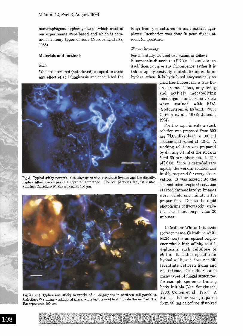
Fig 4 (left) Hyphae and sticky networks of A. oligospora in between soil particles.Calcofluor W staining - additional lateral white light is used to illuminate the soil particles.Bar represents 100 [km.
Fig 3 Typical sticky network of A..oligospora with vegetative hyphae and the digestivehyphae filling the corpse of a captured nematode. The soil particles are just visible.Staining; Calcofluor W. Bar represents 100 um,
•
Volume 12, Part 3, August 1998
•
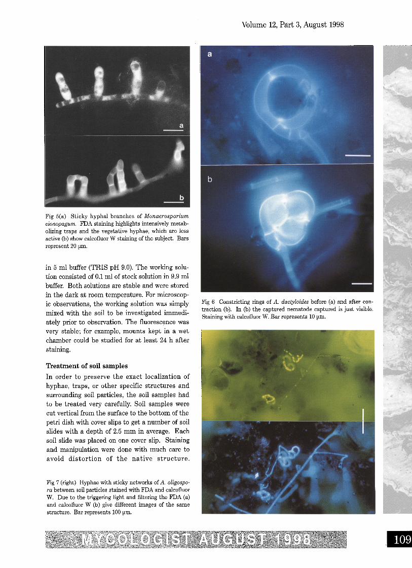
Fig 6 Constricting rings of A. dactyloides before (a) and after con-traction (b). In (b) the captured nematode captured is just visible.Staining with calcofluor W. Bar represents 10 [Lm.
Treatment of soil samples
In order to preserve the exact localization ofhyphae, traps, or other specific structures andsurrounding soil particles, the soil samples hadto be treated very carefully. Soil samples werecut vertical from the surface to the bottom of thepetri dish with cover slips to get a number of soilslides with a depth of 2.5 mm in average. Eachsoil slide was placed on one cover slip. Stainingand manipulation were done with much care toavoid distortion of the native structure.
Fig 7 (right) Hyphae with sticky networks of A. oligospo-ra between soil particles stained with FDA and calcofluorW. Due to the triggering light and filtering the FDA (a)and calcofluor W (b) give different images of the samestructure. Bar represents 100 [Lm.
Fig 5(a) Sticky hyphal branches of Monacrosporiumcionopagum. FDA staining highlights intensively metab-olizing traps and the vegetative hyphae, which are lessactive (b) show calcofluor W staining of the subject. Barsrepresent 20 [Lm.
in 5 ml buffer (TRIS pH 9.0). The working solu-tion consisted of 0.1 ml of stock solution in 9.9 mlbuffer. Both solutions are stable and were storedin the dark at room temperature. For microscop-ic observations, the working solution was simplymixed with the soil to be investigated immedi-ately prior to observation. The fluorescence wasvery stable; for example, mounts kept in a wetchamber could be studied for at least 24 h afterstaining.
Volume 12, Part 3, August 1998
Observation was best through the cover slip inthe hanging drop technique (Jensen, 1994;Jensen & Lysek, 1995).
Microscopic and photographic equipment
Observations were made using a Leitz Dialux 20research microscope equipped with an (epi-) fluo-rescence illuminator PLOEMOPAK 2.4 suppliedwith a mercury lamp HBO 50 W (Osram). Thefilter blocks used were A, E3, 13 and N2(Jensen, 1994). Photographs were taken with anMPS 11 microscope camera connected with anMPS 15 semiphotomat (Wild). Kodak negativefilms of 100, 200, 400 and 1000ASA were used.
SEM-photos were taken using a Cambridgestereoscan 90 B 100/SE scanning electron micro-scope; for details see Neumeister (1994).
Results and discussion
Fig 3 shows calcofluor-stained fluorescence of atypical three-dimensional sticky network of A.oligospora hyphae between soil particles. Thehyphae within the sticky network of the trap aswell as the digestive hyphae filling the emptycorpse of a captured nematode are clearly visible.Due to the Calcofluor W staining, all hyphae givea similar fluorescence - a distinction of active orinactive hyphae is not possible. However, it maybe assumed, at this state of degradation, thatthe digestive hyphae are only present as emptytubes of hyphal walls.
Fig 4 shows hyphae and capture organs of A.oligospora occupying a larger area; the clustersof traps found often in cultures are clearly visi-ble, in addition to the soil particles which havebeen illuminated by additional lateral whitelight. Another type of capture organ, namely theshort sticky branches of Monacrosporiumcionopagum (Drechsler) is shown in Fig 5. Thesecomparatively simple structures are typical forvarious Monacrosporium-species and can fuse togive larger structures (see Fig 5b). Such captureorgans are typically intensively metabolizing andproduce the glue to hold trapped nematodes aswell as nematode-digesting enzymes.
Another group of capture organs, constrictingrings, are shown in Fig 6. These act by veryrapid inflation, triggered when a nematodetouches the inside of the ring. The three cellsenlarge their volume such that the ring is com-pletely filled - the nematode is quickly ensnared
and cannot escape. Fig 6 also shows a recentlycaptured nematode, but due to the selectivenessof calcofluor W for glucans, it is stained poorlyand hence is only visible as a shadow.
Fig 7 shows an example of double staining ofA. oligospora with FDA and calcofluor. If the fil-ters are chosen adequately, the stains highlightdifferent cellular components: FDA (Fig 7a)stains actively metabolizing areas such as cap-ture organs, while calcofluor W makes the entirecomplex visible.
Fluoresence microscopy can be used for thedirect observation of a wide range of soil-dwellingfungi, not just the nematophagous fungi studiedfor this article. Staining of key taxonomic fea-tures (for example the developing conidiophoresand spores of A. oligospora) make it an ideal toolfor the rapid identification of species of fungithat grow actively in soil, and which would beextremely difficult to observe by conventionalmicroscopy. The technique can also be used tostudy fungal growth in soils over time, whichexhibited periodic effects (Jensen et al., 1997).We expect fluorescence microscopy to play anincreasing role in environmental mycology.Recently, for example, it has been used to studythe colonisation of soils contaminated withurban pollutants (Neumeister et al., 1997) and tostudy the occurrence of fungi in the ventilationsystems of buildings (Neumeister et al., 1996).
The authors acknowledge a grant given by theBiological Research and Investment Corporation,Miami/Florida, USA.
ReferencesCohen, Y., Peter, S. Balass, O. & Coffey, M. D. (1987) A
fluorescent technique for studying growth ofPeronospora tabacina on leaf surfaces.Phytopathology 77: 201 - 204.
Corren, B., Purchio, A., Paula, C. R. & Gambale, W.(1986)Evaluation of a fluorescent method (fluoresceindiacetate and ethidium bromide solution) in the studyof the viability of Cryptococcus neoformans strains.Mycopathologia 96: 91 - 96.
Jensen, C. & Lysek, G. (1991)Direct observation of trap-ping activities of nematode-destroying fungi in thesoil using fluorescence microscopy. FEMSMicrobiology Ecology 85: 207 - 210.
Jensen, C. (1994) FluoreszenzmikroskopischeMoglichkeiten zur in situ - Beobachtungnematophager Pilze im Boden. Doctoral Thesis, FUBerlin (1993). Dissertationes Botanicae 217: J.Cramer Verlag Berlin, Stuttgart.
Jensen, C. & Lysek, G. (1995)Fluorescence microscopy offungi in native soil - improvements by additional sub-
III
stances. Microscopy and Analysis 1-9: Sept. 1995.Jensen, C., Neumeister & Lysek, G. (1997)
Nematophagous fungi - study by fluorescencemicroscopy and EDX-technique of the periodicity oftrap formation in soil. Biological Rhythm Research28;,No. 3: 365 - 373.
Neumeister, H. (1994) Entwickung einerPraparationstechnik fiir die energiedispersiveRontgen-Mikroanalyse am Beispiel Arthrobotrysoligospora. Doctoral Thesis, FU Berlin (1993), ShakerVerI. Aachen 1994.
Neumeister, H. Hesse, M. & Quader, S. (1997) Wirkingvon ausgewahlten PAK und PCB auf nematophagePilze. Final Report (Abschlusbericht) for the BMFT;GSF-Forschungsxentrum fur Umwelt u. GesundheitGmbH. Neuherberg.
Neumeister, H., Moritz, M., Schleibinger, H. & Martiny,H. (1996) Investigations on the allergenic potential
Volume 12, Part 3, August 1998
induced by fungi on air filters of HVAC systems.Proceedings of Indoor Air '96; vol. 3, p. 125 - 130;Nagoya/Japan.
Nordbring-Hertz, B. (1988) nematophagous fungi: strate-gies for nematode exploitation and for survival.Microbiological Sciences 5: 108 - 116.
Saxena, G. & Lysek, G. (1993) Observation ofnematophagous fungi in natural soils by fluorescencemicroscopy and their correlation with isolation.Mycological Research 97: 1005 - 1001.
Soderstrom, B. Erland S. (1986) Isolation of fluoresceindiacetate stained hyphae from soil by micromanipula-tion. Transactions of the British Mycological Society86: 465 - 468.
Von Sengbusch, P., Hechler, J. & Muller, U. (1983)Molecular architecture of fungal cell walls - anapproach by use of fluorescent markers. EuropeanJournal of Cell Biology 30: 305 - 312
Cookery Corner
SADDLED CHEESE
Until recently I had not eaten the 'Dryad's sad-dle' - Polyporus squamosus. Most books do notsing its praises but I have discovered that it isdelicious. It has to be fresh and so young it'salmost 'new born'! This recipe serves one and issimple and quick to prepare.
Ingredients1 small round goats cheeseOlive oil for marinadeFresh or dried herbs of your choiceA few rocket leavesSalad dressing of your choiceThin slices of Dryad's saddleOil to deep fry
MethodMarinate the goats cheese in olive oil and the
herbs. I particularly like parsley, thyme andbay. You could also add crushed garlic, lemonzest or anything you fancy. This is better doneovernight but a minimum of four hours shouldbe sufficient.
Dress the rocket, or salad leaves could be sub-stituted, and place onto a plate.
Bake the cheese in a moderate oven for 5-10minutes until soft. Deep fry the slices of Dryad'ssaddle for a few minutes; do not cook for toolong or they may become bitter. Place thecheese on the leaves and sprinkle the Dryad'ssaddle over the top.
Diana Bateman
© Copyright of the author; not to be reproduced withoutpermission.
Volvariella surrecta
Editor's note: Profiles of Fungi will continue inthe November issue with No 97 Volvariella sur-recta, held over from the two other species ofthat genus which appear in this issue. Althoughit is a fungus which seems to be recorded ratherinfrequently, it has been illustrated in two shortitems in recent issues of the Mycologist. It is ofparticular interest as a mycoparasite.
Further correspondence is now in hand,together with a short summary paper by SheilaWells, to be published in November, on theoccurrence and distribution of this unusualspecies. Any other information will be welcomeand could contribute to the debate, if receivedvery soon!
G. Hadley
•


![Basidiomycetes as Potential Biocontrol Agents against ... · entomophagous and nematophagous specialized fungi (YANG& al. [9]). These organisms have the advantage of being effective](https://static.fdocuments.in/doc/165x107/5ed69c0d843ed9152066b50e/basidiomycetes-as-potential-biocontrol-agents-against-entomophagous-and-nematophagous.jpg)