![Videotapes and Movies on Fluid Dynamics and Fluid Machines · VIDEOTAPES AND MOVIES ON FLUID DYNAMICS 1175 Examples of Flow Instability [motion picture], NCFMF, Chicago, IL, Encyclo](https://static.fdocuments.in/doc/165x107/5e88c3b1df80523df73cefe5/videotapes-and-movies-on-fluid-dynamics-and-fluid-machines-videotapes-and-movies.jpg)
Fluid therapy and case examples for specific diseases
31
Close this window to return to IVIS www.ivis.org Proceeding of the LAVECCS Congreso Latinoamericano de Emergencia y Cuidados Intensivos Ju1. 28-30, 2011 – Santiago de Chile, Chile www.laveccs.org Reprinted in IVIS with the permission of the LAVECCS
-
Upload
manuel-martinez -
Category
Science
-
view
279 -
download
0
Transcript of Fluid therapy and case examples for specific diseases
Close this window to return to IVIS www.ivis.org
Proceeding of the LAVECCS
Congreso Latinoamericano de Emergencia y Cuidados Intensivos
Ju1. 28-30, 2011 – Santiago de Chile, Chile
www.laveccs.org
Reprinted in IVIS with the permission of the LAVECCS

LAVECCS.o
rg
CHAPTER 9: FLUID THERAPY AND CASE EXAMPLES FOR SPECIFIC DISEASES CASE EXAMPLE 1 Gunther Gunther, a 10 year old, 86 pound neutered male German shepherd presents to your clinic with a 1 week history of waxing and waning lethargy and inappetance. His owner tells you that today, the dog’s abdomen appears to be more distended. He has not eaten today. There has been no vomiting or diarrhea. The owner does not know of any potential exposure to toxins or chemicals. He is currently on no medications, is up-to-date on vaccinations and has had no prior medical problems. At the time of presentation, his mucous membranes are very pale pink with a prolonged (2.5 seconds) capillary refill time. His heart sounds slightly muffled, but there are no obvious murmurs or dysrhythmias. His heart rate seems high at 170 beats per minute. The femoral pulses are synchronous and slightly decreased in character. The lungs are clear on thoracic auscultation, and there is no evidence of respiratory difficulty. The abdomen is distended, and there is possibly a ballotable fluid wave. The neurologic and orthopedic and dermatologic systems appear normal at this time. What are Gunther’s problems? Waxing and waning lethargy Distended abdomen, Inappetance Possible abdominal effusion Slightly muffled cardiac sounds Tachycardia Decreased pulse quality Prolonged capillary refill time Can you characterize Gunther’s cardiovascular status? Gunther is showing signs of decompensatory shock, with tachycardia, muffled heart sounds, prolonged capillary refill time, and decreased pulse quality. What is your list of differential diagnoses? Differential diagnoses include: neoplasia (hepatic, adrenal, renal, splenic), , perforated bowel and peritonitis, cardiac disease (heart failure, pericardial tamponade), , pancreatitis, and hepatic failure. What would you like to do to start stabilizing Gunther before diagnostic tests? You decide to place a peripheral cephalic intravenous catheter, and start ¼ shock (860 ml, you can add a zero to a dog’s body weight in pounds, and that is the ¼(90 ml/kg) bolus of a crystalloid to administer during hypovolemic shock.) bolus of Normosol-R. After the ¼ shock bolus has been administered, you reassess his heart rate, capillary refill time, mucous membrane color, and blood pressure. His blood pressure is essentially the same, although his heart rate has started to decrease and his mucous
Reprinted in the IVIS website with the permission of LAVECCS
Close window to return to IVIS www.ivis.org
Proceedings of the Congreso Latinoamericano de Emergencia y Cuidados Intensivos LAVECCS - 2011
LAVECCS.o
rg
membrane color and capillary refill time have improved. You decide to administer a second ¼ shock bolus of a crystalloid. What is your diagnostic plan? Initial diagnostic plan includes taking a blood pressure, electrocardiogram (ECG), complete blood count, serum biochemistry panel, urinalysis, thoracic radiographs, and abdominal radiographs, and serum lactate. Results of Diagnostic Tests: Evaluation of thoracic radiographs showed no abnormalities. Complete blood count: 18, 580 WBC/ul with 78% neutrophils, 12% lymphocytes, 8% monocytes, and 2% eosinophils; platelet count is 123,000 platelets/uL; the Hct is 32% Chemistry panel was normal except for hyperglycemia glucose 197 mg/dL, and hypoproteinemia with a total protein 3.8 g/dL. Evaluation of the abdominal Radiographs show a large soft tissue density in the midabdomen, and a decrease in abdominal detail. Reading of the blood pressure showed hypotension with a systolic measurement of 98 mm Hg and diastolic measurement of 48 mm Hg What other problems do you want to add to Gunther’s problem list? Abdominal mass Poor abdominal detail Mild thrombocytopenia Hyperglycemia Hypoproteinemia Mild anemia Hypotension Can you lump together Gunther’s problem list and make the abnormalities fit with a particular situation? Gunther has a mid abdominal mass with poor detail, and is showing clinical signs of hypovolemic or hemorrhagic shock. What other things should you consider? You decide to ask the technician to perform a blood type, as you suspect that Gunther has a hemoabdomen based on the loss of abdominal detail, clinical signs referable to hypovolemic/hemorrhagic shock, and the presence of a large midabdominal mass effect on radiographs. While the technician is performing the blood type, you decide to perform an abdominocentesis, and find nonclotting hemorrhagic fluid. Your suspicions of hemoabdomen are confirmed.
Reprinted in the IVIS website with the permission of LAVECCS
Close window to return to IVIS www.ivis.org
Proceedings of the Congreso Latinoamericano de Emergencia y Cuidados Intensivos LAVECCS - 2011
LAVECCS.o
rg
What should you consider next? Ideally, an abdominal ultrasound and cardiac ultrasound should be performed to rule out the presence of metastasis, and to locate the organ from which the mass arises. However, you do not have an ultrasound machine available, and are concerned that Gunther may continue to hemorrhage through the night, so take him to surgery. At the time of surgery, you find a large cavitated splenic mass, with no obvious metastasis. You perform a routine splenectomy, and repeat a hematocrit intra-operatively. At this time, Gunther’s hematocrit has dropped to 22%, and he is hypotensive under anesthesia (76 mm Hg systolic and 34 mm Hg diastolic). What are your concerns? The degree of hypotension and anemia are concerning. Although some of the apparent anemia might be explained from intraoperative crystalloid fluids and hemodilution, anemia with hypotension is dangerous as both can lead to decreased oxygen delivery to the tissues. While your technician is performing Gunther’s blood type, you can administer a colloid bolus (5 ml/kg IV) in an attempt to raise Gunther’s blood pressure. Next, you decide to give him a blood transfusion. What type of blood product are you going to administer? Initially Gunther’s total protein was low, so fresh frozen or frozen plasma may be beneficial to slightly raise the total protein, but you remember that he also needs red blood cells, so you decide to give him type-specific whole blood. Since his hematocrit has dropped to 22%, you want to ideally raise his hematocrit to 30%. That is an 8% increase from his current Hct. Remember that for every 1 ml per 1 pound of whole blood, you will raise the Hct by 1%. So, if Gunther is 86 pounds, and you want to raise the Hct 8%, (8 x 86) = 688 mls of whole blood. Of course, the specific number is unrealistic, and you likely will administer incremental units of whole blood to be nearly equal, or slightly more than the desired calculated volume. If you did not have blood available, a hemoglobin-based oxygen carrier, if available, could also be administered for oxygen carrying capacity and as a colloid to augment intravascular fluid volume and blood pressure. Post-operatively, Gunther does well on maintenance crystalloid fluids, analgesia, and restricted activity until his abdominal incision heals. Good job!
Reprinted in the IVIS website with the permission of LAVECCS
Close window to return to IVIS www.ivis.org
Proceedings of the Congreso Latinoamericano de Emergencia y Cuidados Intensivos LAVECCS - 2011
LAVECCS.o
rg
CASE EXAMPLE 2: Eli Eli, a 35 kg, 5 year old neutered male Australian Shepherd x Border Collie mixed breed dog, presents to you on emergency referral from another veterinary hospital for possible ethylene glycol intoxication. Eli has been vomiting and having diarrhea for the past 3 days after ingesting a pound of raisins. The owner reports that there have been raisins visible in the dog’s diarrheic feces. Eli has a history of having a sensitive stomach in the past, but no other medical problems. His owner does not know of any chemical or garbage exposure. He is not on any medications except for monthly heartworm prevention. At the referring veterinary hospital, Eli was initially given subcutaneous fluids and metoclopramide. Blood and urine were obtained for evaluation by an outside laboratory. He was discharged to his owner with instructions to administer a bland diet and to contact them if his vomiting did not resolve. This morning, the bloodwork results were returned, and showed renal azotemia with a BUN 99 mg/dL, Creatinine 4.2 mg/dL, his urine specific gravity was 1.018 with glucosuria and amorphous debris, but no white or red blood cells and no bacteria. Urine ketones were negative. At the time of presentation, Eli is lethargic, and has dry mucous membranes and a normal capillary refill time. His heart rate is 68 beats per minute, and there is a normal sinus rhythm with strong synchronous femoral pulses. He has serous nasal discharge, and slightly edematous conjunctiva. He has halitosis, and vomitus staining around his mouth. Thoracic auscultation reveals normal rhythm and no murmurs. His lungs sound mildly moist with slight crackles. He has pain in his perirenal area on abdominal palpation, and the urinary bladder is small. Rectal examination was normal. What are Eli’s problems? Vomiting Diarrhea Overhydration Azotemia with low urine specific gravity Halitosis Serous nasal discharge Conjunctival edema Relative bradycardia Dry mucus membranes Harsh lung sounds Perirenal pain What are your thoughts? Eli clinically appears overhydrated, yet also has fluid losses in the form of vomiting and diarrhea. Normally, in the face of fluid loss, the kidneys should compensate and retain fluid in an attempt to maintain circulating blood volume. He has a relatively slow heart rate, which could be associated with a number of diseases including hypoadrenocorticism, hyperkalemia from other causes including renal failure and uroabdomen, or late decompensatory shock. Although not very sensitive for assessing a
Reprinted in the IVIS website with the permission of LAVECCS
Close window to return to IVIS www.ivis.org
Proceedings of the Congreso Latinoamericano de Emergencia y Cuidados Intensivos LAVECCS - 2011
LAVECCS.o
rg
patient’s actual blood pressure, the presence of very strong pulses makes decompensatory shock less likely. One of the major concerns is primary or secondary renal failure. What is your initial diagnostic plan? Initial diagnostic plans should include an ECG and blood pressure, abdominal radiographs to rule out a foreign body or gastrointestinal obstruction, urethral calculi, or decreased abdominal detail from abdominal effusion. Since the original bloodwork was obtained approximately 24 hours ago, ideally you should consider repeating the complete blood count, serum biochemical analyses, and urinalysis to compare with the bloodwork obtained yesterday. Thoracic radiographs should be considered because of the moist lung sounds. Whenever renal values are elevated, bacterial infections such as pyelonephritis and Leptospirosis should also be considered. You see this rhythm on an ECG. What is it? This ECG strip represents atrial standstill. Although lone atrial standstill can occur in some breeds of dogs such as the English Springer Spaniel, the absence of p waves in combination with widened QRS complexes makes hyperkalemia a real concern. Before performing any additional diagnostics, is there something that you’d like to do? Atrial standstill in a patient with suspected oliguric or anuric renal failure can rapidly become life-threatening if left untreated. Treatment to protect the heart from the toxic effects of hyperkalemia include the administration of calcium gluconate or calcium chloride IV. Alternatively, potassium can be driven intracellularly with the administration of sodium bicarbonate or IV regular insulin with dextrose. You elect to administer a slow bolus of calcium gluconate over 10 minutes. Next, what would you like to do? You perform an oscillometric blood pressure, and find that Eli is hypertensive, with systolic 203 mm Hg and diastolic 120 mm Hg The results of the bloodwork have been completed and reveal that Eli’s azotemia appears to have worsened. The BUN is 128 mg /dL, and the creatinine is 7.6 mg/dL. Eli is also hypercalcemic (total calcium 13 mmol/liter) and hyperphosphatemic (Phosphorus > 16 mmol/liter). The serum potassium is 9.6 mmol/liter. You attempt to obtain a urine sample by cystocentesis, but are unsuccessful as the urinary bladder is very small. You pass a urinary catheter, and obtain just 3 mls of urine. Grossly the urine appears very light yellow and clear. The Specific Gravity is 1.006. The sediment examination shows amorphous debris, but no crystals or casts. Are there any additional problems to add to Eli’s list? Hypertension Atrial standstill Hyposthenuria Worsened azotemia
Reprinted in the IVIS website with the permission of LAVECCS
Close window to return to IVIS www.ivis.org
Proceedings of the Congreso Latinoamericano de Emergencia y Cuidados Intensivos LAVECCS - 2011
LAVECCS.o
rg
Hypercalcemia Hyperphosphatemia Small urinary bladder with little urine What are your thoughts or concerns? In the face of continued vomiting, Eli’s azotemia is worsening, and there is little to no urine being produced. He has clinical signs referable to anuric renal failure, with pulmonary crackles, chemosis, and serous nasal discharge. Additionally, his electrolytes are abnormal, with hyperkalemia, hypercalcemia, and hyperphosphatemia. All of these abnormalities can be attributable to renal failure, however other diseases such as cholecalciferol rodenticide intoxication and hypoadrenocorticism should be considered, as well. Could this be ethylene glycol? Ethylene glycol could cause signs of vomiting, dehydration, azotemia, perirenal pain, hyperkalemia, and hyperphosphatemia. However, with the sedimentation of calcium oxalate crystals in the renal tubules, hypocalcemia, not hypercalcemia would be expected. Additionally, there are no crystals (calcium oxalate monohydrate or calcium oxalate dehydrate, nor hippurate) on the urine sediment examination. Although these findings do not definitively rule out ethylene glycol intoxication, it makes this cause of renal failure less likely. What is your initial treatment plan? You want to attempt to “jump start” Eli’s kidneys, which is a logistical concern, given the presence of overhydration (chemosis, serous nasal discharge, and pulmonary crackles). Drugs such as furosemide (4 mg/kg IV), mannitol (0.5 to 1 g/kg IV), dopamine (3 – 5 mcg/kg/min IV CRI), and diltiazem (DOSE) have been reported to aid in the production of urine. Some, like diltiazem, also have been used to promote diuresis in patients with renal failure, and have the added benefit of decreasing hypertension. How much fluid would you like to administer to Eli? Do you have any concerns? Eli is already showing signs of anuria and volume overload. Additional fluids to a volume overloaded patient can worsen pulmonary edema. However, the kidneys need fluid to make urine, and some fluid therapy along with drugs to promote diuresis is in order. You elect to take into consideration just insensible losses (0.3 ml/kg/hr) and administer (35 kg x 0.3) = 10 ml/hour. In acute renal failure, calcium is thought to promote perpetuation of renal damage. Calcium channel blocking drugs such as diltiazem have been recommended to treat hypertension in oliguric or anuric renal failure patients. You elect to place Eli on a diltiazem constant rate infusion (0.3 – 0.5 mg/kg IV slowly over 10 minutes, then 1 – 5 mcg/kg/min IV CRI), and monitor blood pressure very carefully, as patients can become very hypotensive. Administration of furosemide ( 2 – 4 mg/kg IV, followed by a constant rate infusion 0.7 – 1 mg/kg/hour) can also be considered. The use of mannitol in an anuric patient with signs of intravascular volume overload is contraindicated, as the
Reprinted in the IVIS website with the permission of LAVECCS
Close window to return to IVIS www.ivis.org
Proceedings of the Congreso Latinoamericano de Emergencia y Cuidados Intensivos LAVECCS - 2011
LAVECCS.o
rg
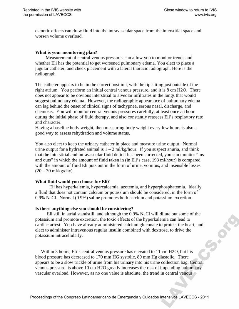
osmotic effects can draw fluid into the intravascular space from the interstitial space and worsen volume overload. What is your monitoring plan? Measurement of central venous pressures can allow you to monitor trends and whether Eli has the potential to get worsened pulmonary edema. You elect to place a jugular catheter, and check placement with a lateral thoracic radiograph. Here is the radiograph. The catheter appears to be in the correct position, with the tip sitting just outside of the right atrium. You perform an initial central venous pressure, and it is 8 cm H2O. There does not appear to be obvious interstitial to alveolar infiltrates in the lungs that would suggest pulmonary edema. However, the radiographic appearance of pulmonary edema can lag behind the onset of clinical signs of tachypnea, serous nasal, discharge, and chemosis. You will monitor central venous pressures carefully, at least once an hour during the initial phase of fluid therapy, and also constantly reassess Eli’s respiratory rate and character. Having a baseline body weight, then measuring body weight every few hours is also a good way to assess rehydration and volume status. You also elect to keep the urinary catheter in place and measure urine output. Normal urine output for a hydrated animal is 1 – 2 ml/kg/hour. If you suspect anuria, and think that the interstitial and intravascular fluid deficit has been corrected, you can monitor “ins and outs” in which the amount of fluid taken in (in Eli’s case, 193 ml/hour) is compared with the amount of fluid Eli puts out in the form of urine, vomitus, and insensible losses (20 – 30 ml/kg/day). What fluid would you choose for Eli? Eli has hyperkalemia, hypercalcemia, azotemia, and hyperphosphatemia. Ideally, a fluid that does not contain calcium or potassium should be considered, in the form of 0.9% NaCl. Normal (0.9%) saline promotes both calcium and potassium excretion. Is there anything else you should be considering? Eli still in atrial standstill, and although the 0.9% NaCl will dilute out some of the potassium and promote excretion, the toxic effects of the hyperkalemia can lead to cardiac arrest. You have already administered calcium gluconate to protect the heart, and elect to administer intravenous regular insulin combined with dextrose, to drive the potassium intracellularly. Within 3 hours, Eli’s central venous pressure has elevated to 11 cm H2O, but his blood pressure has decreased to 170 mm HG systolic, 80 mm Hg diastolic. There appears to be a slow trickle of urine from his urinary into his urine collection bag. Central venous pressure is above 10 cm H2O greatly increases the risk of impending pulmonary vascular overload. However, as no one value is absolute, the trend in central venous
Reprinted in the IVIS website with the permission of LAVECCS
Close window to return to IVIS www.ivis.org
Proceedings of the Congreso Latinoamericano de Emergencia y Cuidados Intensivos LAVECCS - 2011
LAVECCS.o
rg
pressure combined with what this animal is doing clinically is perhaps more important. If the central venous pressure rises by more than 5 cm H2O from baseline in a 24 hour period, or if the value is above 10 cm H2O and the animal is becoming more tachypneic with pulmonary crackles, worsening nasal discharge , peripheral edema and/or chemosis, the clinical picture is more grim. Eli appears to be improving, and you recommend a renal biopsy. You perform a tru-cut percutaneous, ultrasound-guided renal biopsy, and find the following: Histopathologically, the renal tubules are blocked with amorphous debris. There is evidence of mitosis at the glomerular basement membrane, suggesting that the kidneys are improving and attempting to regenerate. Many substances can cause insult to the kidneys. Raisins and grapes have been found to be toxic in some animals. The exact toxic dose, and toxic principle are unknown. However, classic clinical signs associated with renal failure from grape or raisin ingestion include vomiting, diarrhea with grape skins or raisins in the feces, and the development of oliguric or anuric renal failure. Renal tubular obstruction appears to be a component of the oliguria or anuria. As the kidneys recover, and overwhelming post-obstructive diuresis can occur, making calculation of fluid ins and outs an absolute necessity. After 24 hours of intravenous fluids and replenishment of the fluid deficits, you start calculating ins and outs, and find that it is 1 ml/kg/day initially, and has increased to 8 ml/kg/day 12 hours later. What should you do? You must keep up with Eli’s fluid output. Monitor ins and outs, and change the fluid rate to match the fluid coming out. Weigh Eli at least three to four times a day, to make sure that you are keeping up with his fluid output. Rapid changes in body weight are always associated with fluid loss or gain in animals with vomiting, diarrhea, wound exudates, and renal failure.
Reprinted in the IVIS website with the permission of LAVECCS
Close window to return to IVIS www.ivis.org
Proceedings of the Congreso Latinoamericano de Emergencia y Cuidados Intensivos LAVECCS - 2011
LAVECCS.o
rg
CASE EXAMPLE 3: Zeke Zeke, a 70 pound, 4 year old neutered male Bassett Hound, presents to you with an acute onset of collapse. His owners report that he had been normal earlier today, and had access to a fenced in back yard while they were out of the house. He was found approximately 1 hour ago, collapsed and obtunded in the back yard. His owners report that there were several piles of vomit on the back porch, and brown liquid diarrhea near Zeke. To their knowledge, he had not had any exposure to chemicals, toxins, or garbage. There has been no diet change. Recently in the past 2 months, they had placed Zeke on a diet because of obesity. He has lost a small amount of weight. No other abnormalities were noted until his owners found him approximately 1 hour ago. On physical examination, you find that Zeke is morbidly obese, and obtunded. His mucous membranes are brick red and dry, with a severely prolonged capillary refill time of almost 4 seconds. You find that it is difficult to heart his heart on thoracic auscultation. His temperature, pulse, and respiration are: 97.4 degrees F, 120 bpm, and 40 respirations/minute. What are Zeke’s problems? Zeke’s problem list includes: Obesity Obtunded Dry mucous membranes Brick red mucous membranes Prolonged CRT Vomiting Diarrhea Hypothermia Inappropriate bradycardia What are your thoughts? Causes of vomiting and diarrhea are numerous, and include dietary indiscretion, toxin, anaphylactic reaction, inflammatory bowel disease, gastrointestinal obstruction (including foreign body, intussusception or neoplasia), pancreatitis, bacterial or viral gastroenteritis or secondary to some other infection, mesenteric volvulus, renal or hepatic failure, and metabolic insults such as diabetic ketoacidosis or hypoadrenocorticism. Also, Zeke is showing signs of neurologic problems with circulatory compromise. The heart sounds are muffled, which can be due to pericardial effusion, pneumothorax, pleural effusion, or hypovolemia. His mucous membranes are brick red, which often is suggestive of septic shock. In the face of dehydration and/or septic shock, one would expect his heart rate to be elevated, unless in the case of end-stage decompensatory shock, where the heart rate can become bradycardic. Inappropriate bradycardia can be associated with a number of conditions, and is significant in this case because Zeke appears to be very hypovolemic. The body’s normal response to a decrease in intravascular volume is to compensate with an increase in heart rate in an attempt to maintain cardiac output and blood pressure. Inappropriate
Reprinted in the IVIS website with the permission of LAVECCS
Close window to return to IVIS www.ivis.org
Proceedings of the Congreso Latinoamericano de Emergencia y Cuidados Intensivos LAVECCS - 2011
LAVECCS.o
rg
bradycardia can be associated with decompensatory shock, increase vagal tone, or electrolyte abnormalities such as hyperkalemia and hypermagnesemia. His obesity may not be directly associated with his current problems, however, it may impair your abilities to diagnose and treat his illness. What is your initial diagnostic plan? The initial diagnostic plan in any patient with severe vomiting, diarrhea, and collapse should include blood pressure, ECG, complete blood count, biochemistry profile, electrolytes, urinalysis, fecal flotation and cytology, thoracic radiographs, abdominal radiographs, and possibly an abdominal ultrasound. What is your initial treatment plan? Initial treatment plan in any animal with a history of vomiting, diarrhea, and collapse should include administration of intravenous fluids. Blood samples should ideally be obtained at the time of initial presentation, before the administration of intravenous fluids. You attempt to place peripheral cephalic, lateral saphenous, and medial saphenous catheters, and all attempts are unsuccessful because of patient anatomy, peripheral vasoconstriction, and the severity of obesity. What else can you try? If peripheral vascular access is impossible, a central catheter in the jugular vein can be attempted. Alternatively, you can perform a cutdown over the vessel of your choice. You attempt to place a jugular catheter, and find that the degree of obesity prevents catheterization. In an attempt to provide intravenous fluids as soon as possible, are there any other places that you can place a catheter? Intraosseous catheters can be used in patients in whom vascular access is difficult or impossible. Unfortunately, because Zeke is so obese, and because his bones are ossified, it will likely be difficult to place and maintain an intraosseous catheter. Instead, you elect to attempt an auricular catheter in Zeke’s ears. Fortunately you are successful in placing two 20 gauge catheters in the right and left ears, and can start intravenous crystalloid fluid boluses. What fluid would you like to administer? In the case of decompensatory shock, it is important to first establish that there is no cardiac disease (in this case, the absence of cardiac sounds makes pericardial effusion a possibility), then administer a balanced crystalloid fluid such as Lactated Ringers, Normosol-R, Plasmalyte-A, or 0.9% sodium chloride. You start with a ¼ “shock” bolus of fluids (700 mls in Zeke’s case, as he is 70 pounds), and reassess perfusion parameters of capillary refill time, heart rate, blood pressure, and whether he is producing urine.
Reprinted in the IVIS website with the permission of LAVECCS
Close window to return to IVIS www.ivis.org
Proceedings of the Congreso Latinoamericano de Emergencia y Cuidados Intensivos LAVECCS - 2011

LAVECCS.o
rg
The results of your diagnostic blood panel are returned, and are relatively normal except for a hypocholesterolemia (77 mg/dL), hypoglycemia (63 mg/dL), hyponatremia (123 mmol/liter), hypochloremia (78 mmol/liter), and hyperkalemia (7.2 mmol/liter) The ECG rhythm strip shows a normal sinus rhythm despite the presence of hyperkalemia. It is important to note that atrial standstill can be present even with mild hyperkalemia, and the absence of atrial standstill, or the presence of a normal sinus rhythm does not rule out the presence of hyperkalemia. Thoracic radiographs are obtained, and evaluation showed a mild to moderate increase in interstitial to alveolar infiltrates consistent with pneumonia, with a megaesophagus. What are your thoughts? Zeke’s signalment and history, being a young Bassett Hound with an acute onset of collapse, vomiting and diarrhea, combined with radiographic appearance of megaesophagus, and bloodwork abnormalities of azotemia, hypocholesterolemia, hypoglycemia, hyponatremia, hypochloremia, and hyperkalemia are very common in animals with hypoadrenocorticism, or Addison’s disease. None of the aforementioned findings are pathognomonic for Addison’s, however, and definitive diagnostic testing in the form of an ACTH stimulation test must be performed. One of the interesting and often commonly overlooked “abnormalities” on the bloodwork of an Addisonian patient is a normal complete blood count. In an animal with severe dehydration, hypovolemia, and collapse, the stress response would result in the demargination of white blood cells, and cause a classic stress leukogram with a neutrophilic leukocytosis and lymphopenia. A normal leukogram in a critical patient is an abnormal finding until proven otherwise. The absence of a stress leukogram in an ill patient, should increase your index of suspicion for hypoadrenocorticism. In a patient with pneumonia, likely from aspiration of vomitus, white blood cells could potentially infiltrate the diseased lungs, and a leukocytosis may not be present. A neutrophilia should still be present relative to lymphocytes, however. How would you like to treat Zeke? Treatment of the hypovolemic shock in the form of intravenous crystalloid fluid boluses is of paramount importance. First, treat the decompensatory shock with crystalloid fluid boluses (1/4 shock increments) until the blood pressure has normalized. Addition of a colloid such as hetastarch (5 ml/kg increments) can also be administered. If both colloid and crystalloid administration are not successful in raising blood pressure, positive inotropic drugs or vasopressors may be necessary, Ideally, administration of 0.9% sodium chloride should be considered to promote potassium excretion. The hypoglycemia can be treated with supplemental dextrose (2.5%) mixed in with the crystalloid fluids. Antiemetic drugs can be administered to treat the vomiting, and broad spectrum antibiotics and supplemental oxygen can be administered to treat the pneumonia. Careful monitoring of Zeke’s glucose and electrolytes should be performed at least twice to three times a day, to avoid continued hypoglycemia or too rapid correction of serum sodium that can lead to central pontine myelinolysis. Definitive treatment of hypoadrenocorticism includes replacement of
Reprinted in the IVIS website with the permission of LAVECCS
Close window to return to IVIS www.ivis.org
Proceedings of the Congreso Latinoamericano de Emergencia y Cuidados Intensivos LAVECCS - 2011
LAVECCS.o
rg
glucocorticosteroids in the form of prednisone, and replacement of mineralocorticoid activity in the form of fludrocortisone acetate (Florinef) or desoxycorticosterone pivalate (DOCP). You elect to administer Dexamethasone-sodium phosphate (0.5 mg/kg IV), as it will provide glucocorticoids support, and will not interfere with the ACTH stimulation test.
Reprinted in the IVIS website with the permission of LAVECCS
Close window to return to IVIS www.ivis.org
Proceedings of the Congreso Latinoamericano de Emergencia y Cuidados Intensivos LAVECCS - 2011
LAVECCS.o
rg
CASE EXAMPLE 4: Buster Buster, a 13-year old neutered male domestic shorthaired cat, presents to you with a 2 week history of intermittent vomiting, inappetance, and possible weight loss. He is a strictly indoor cat that shares his household with a pug. There is no possibility of toxin, chemical, garbage, plant, or foreign body ingestion to his owner’s knowledge. At the time of presentation, you find that Buster is in poor body condition, with a relatively unkempt haircoat. His mucous membranes are fairly dry and there is moderate dental calculus and gingivitis. He has increased skin tenting. The muscle mass around his head and dorsal spinous processes are more prominent. You auscult a Grade II- III/VI left parasternal murmur with strong pulse quality and no dysrhythmias. His lungs sound clear. Although he still appears overweight, you think that the kidneys may be slightly small but nonpainful on abdominal palpation. His urinary bladder is large, but not tense or painful. What are Buster’s problems? Vomiting Inappetance Muscle wasting Dehydration Dental calculus and gingivitis Murmur Possibly small kidneys What would you like to do? Diagnostic tests would include a complete blood count, biochemistry panel, urinalysis, thoracic radiographs, echocardiogram and possibly an abdominal ultrasound. You perform thoracic radiographs, and find the following: There is a classic valentine shape to the heart on the dorsoventral view. You schedule an ultrasound appointment with a radiologist in 2 days to have an echocardiogram performed. A complete blood count revealed a WBC 14, 280 with 82% segmented neutrophils, 13% lymphocytes, and 5% monocytes. Platelet count was normal at 178,000 platelets/ul. There was some clumping of platelets at the feathered edge of the blood smear. The hematocrit was 32% with a total protein of 8.2 g/dL. The biochemistry panel revealed significant azotemia with BUN 125 mg/dL, creatinine 5.6 mg/dL, hypokalemia 3.2 mmol/liter. The urinalysis showed a urine specific gravity of 1.018 with some rods and white blood cells in the urine. What is Buster’s updated problem list? Valentine shaped heart consistent with hypertrophic cardiomyopathy Anemia relative to dehydration Azotemia
Reprinted in the IVIS website with the permission of LAVECCS
Close window to return to IVIS www.ivis.org
Proceedings of the Congreso Latinoamericano de Emergencia y Cuidados Intensivos LAVECCS - 2011
LAVECCS.o
rg
Hypokalemia Isosthenuria Bactiuria Pyuria The degree of anemia initially does not seem severe when you look at the hematocrit alone (32%). However, when the anemia is considered in the presence of clinical signs of dehydration and hyperproteinemia, the anemia is likely significant. A reticulocyte count can be performed to determine whether the anemia is regenerative or nonregenerative. What would you like to do now? Given that there are bacteria and white blood cells on the urine sediment examination, a urine culture is in order to determine whether a urinary tract infection or pyelonephritis are contributing to Buster’s clinical signs and bloodwork abnormalities. An abdominal ultrasound should also be performed to look at the kidneys and urinary bladder. Ideally, the ultrasound should be performed prior to the administration of intravenous fluids, as dilation of the renal pelvises, or pyelectasia, can occur with pyelonephritis and with intravenous fluid therapy. Some clinicians empirically double or triple the patient’s maintenance fluid requirements when treating an animal with chronic renal failure. In many cats with chronic renal failure, empiric calculation of intravenous fluid volume, rather than taking the time to calculate out a fluid deficit, maintenance needs, and ongoing losses can lead to dehydration in the face of therapy. Intravenous fluid therapy is necessary to replenish interstitial hydration, as well as diurese uremic toxins out of the body that are causing Buster to feel nauseated and vomit. Buster is approximately 7% dehydrated. He is 6 kg. To determine the hydration deficit, use the following formula: mls deficit = (body weight in kg x % dehydration) x 1000 = = (6 x 0.07) x 1000 = 420 ml deficit/24 hours = 17.5 ml/hour To calculate his maintenance fluid requirements: Maintenance fluids = (30 x Body weight in kg) + 70 = mls/day = (30 x 6) + 70 = 250 ml/day or 10.4 ml/hour Adding the deficit + maintenance = 17.5 ml/hour + 10.4 ml/hour = 29 ml/hour Do you have any concerns? Buster has a cardiac murmur and signs of biatrial enlargement on the thoracic radiographs. Overzealous administration of intravenous crystalloid or colloid fluid can evoke congestive heart failure with pulmonary edema, pleural effusion, or both in a previously asymptomatic patient. Placement of a jugular or medial saphenous long catheter such that the tip of the catheter sits just outside of the right atrium or in the caudal vena cava can be used to measure central venous pressures in cats. Monitoring for trends in change from baseline (no more than 5 cm H2O increase from baseline within 24 hours) as well as the actual CVP, changes in patient’s body weight, and clinical signs of impending pulmonary edema such as tachypnea, increased respiratory effort, pulmonary
Reprinted in the IVIS website with the permission of LAVECCS
Close window to return to IVIS www.ivis.org
Proceedings of the Congreso Latinoamericano de Emergencia y Cuidados Intensivos LAVECCS - 2011
LAVECCS.o
rg
crackles, serous nasal discharge, or chemosis should all be used together to aid in preventing pulmonary vascular overload. In Buster’s case, however, it is prudent to be a little more cautious, with frequent patient assessment, daily monitoring of renal values, and careful monitoring of body weight and central venous pressure. Usually, renal values will not decrease significantly within the first 48 hours of aggressive fluid therapy. If the renal values are increasing in the face of such therapy, the prognosis worsens. After 48 hours, ideally the renal values will continue to decrease until a plateau is reached. Once a plateau is reached, gradually decreasing the patient’s intravenous fluids by 25% per day is a cautious method to not decrease fluid support and diuresis too quickly. In addition to Buster’s renal insufficiency and cardiac disease, he has electrolyte imbalances in the form of hypokalemia, which can contribute to muscle weakness. The intravenous fluids should have potassium chloride supplemented, for a dose not to exceed 0.5 mEq/kg/hour. After 48 hours of crystalloid fluid therapy at a rate of 29 ml/hour, Buster clinically appears more comfortable, and the vomiting has ceased. His body weight has increased by 0.5 kg, and his BUN and creatinine have decreased to 56 mg/dL, and 3.2 mg/dL, respectively. Overall, his central venous pressure has not risen above 5 cm H2O. You continue with your treatment of Buster, and are happy that he is responding to your treatment. Good job!
Reprinted in the IVIS website with the permission of LAVECCS
Close window to return to IVIS www.ivis.org
Proceedings of the Congreso Latinoamericano de Emergencia y Cuidados Intensivos LAVECCS - 2011
LAVECCS.o
rg
CASE EXAMPLE 5: Lolita Lolita, a 35 kg 10 month old spayed female Labrador Retriever presents to you from another veterinary hospital, where she has been hospitalized for the past 3 days. She was spayed 6 days ago, and her owners left her Elizabethan collar off for approximately 1 hour when they left the house. Upon their return, they found Lolita had chewed her midline abdominal incision, and had chewed on a portion of her jejunum. The veterinarian at the other hospital performed emergency surgery on Lolita, and needed to remove approximately 9 inches of damaged jejunum. She had appeared to be doing better and was eating for her owner, until this morning, when she was found to be extremely lethargic with brick red mucous membranes and a prolonged capillary refill time. There was vomitus in her cage, and stained around her muzzle. her rectal temperature is 104 degrees Fahrenheit. Her heart rate is 160 bpm, and her respiratory rate is 60 per minute. You palpate her abdomen, and find it to be tense and painful despite intermittent hydromorphone (0.1 mg/kg IV Q6 hours). What are Lolita’s problems? Recent jejunal resection and anastomosis Lethargy Vomiting Fever Tachycardia Tachypnea Brick red mucous membranes Prolonged capillary refill time Tense painful abdomen What is going on? Given Lolita’s recent surgery 3 days ago, tachycardia, tachypnea, abdominal pain, and brick red mucous membranes with a short capillary refill time and vomiting, there is a strong possibility that Lolita’s jejunal incisions have dehisced and she has peritonitis. She has signs of septic shock. What would you like to do? Ideally, an abdominal ultrasound would be ideal to evaluate the abdomen for pockets of fluid. You can also perform a blind abdominocentesis. If there is more than 5 – 7 ml/kg abdominal effusion, there may be a positive tap. You perform a blind abdominocentesis, and obtain this fluid. What should you do next? This fluid is yellow and grossly cloudy, and suspiciously looks like pus. You perform a cytologic examination of the fluid, and find this: The fluid is consistent with septic peritonitis, with degenerative neutrophils and intra- and extracellular bacteria. What should you do next?
Reprinted in the IVIS website with the permission of LAVECCS
Close window to return to IVIS www.ivis.org
Proceedings of the Congreso Latinoamericano de Emergencia y Cuidados Intensivos LAVECCS - 2011
LAVECCS.o
rg
Given all of the clinical signs and cytologic examination of the fluid from the abdomen, you elect to take Lolita to surgery to re-explore her abdomen. Do you have any concerns about what treatments to implement? Lolita requires intravenous fluids. She has lost fluid into her peritoneal cavity, and also from vomiting and lack of appetite for the past 5 days, until she ate a sesame bagel for her owner yesterday. Intravenous crystalloid fluids to replenish her estimated 7% dehydration deficit are ideal. She also has an intravascular fluid deficit, as evidenced by her prolonged CRT and capillary refill time. While you can choose to calculate her interstitial hydration deficit, it is more important, at this time, to treat her intravascular fluid deficit. You elect to administer ¼ of a “shock bolus” of crystalloid fluids (Normosol-R, by adding a “0” to her body weight in pounds) and also a bolus of hydroxyethyl starch (5 ml/hg IV). It is paramount to replenish her intravascular fluid deficit before the administration of any negative inotropic, negative chronotropic, and vasodilatory anesthetic drugs for surgery. Lolita also has a great potential to lose electrolytes and albumin into the peritonitis fluid. She also is at risk for disseminated intravascular coagulation due to the loss of the natural anticoagulant antithrombin into the abdominal fluid. She is also at risk to become hypoglycemic from sepsis; serum glucose should be monitored at least 2 – 3 times per day, and if necessary, additional glucose can be added to the parenteral crystalloid fluids. How can you approach Lolita’s colloid and protein support? Albumin and colloid oncotic pressure (COP) is important in wound healing. COP can be maintained by using natural and synthetic colloids. Fresh frozen plasma only provides a small amount of protein in the form of albumin, some clotting factors, and very small amounts of antithrombin. Fresh frozen plasma is often cost prohibitive to replace albumin. As such, concentrated human or canine specific albumin should be administered to replenish serum albumin up to a level of 2.0 g/dL. In the case of septic shock, with the potential for loss of clotting factors in the peritoneal effusion, administration of fresh frozen plasma to replenish clotting factors can be used in combination with albumin concentrates, crystalloid fluids, and a synthetic colloid to help maintain COP. A synthetic colloid such as hydroxyethyl starch also should be considered to maintain COP by adding 20 – 30 ml/kg/day IV CRI to the fluid therapy regimen. Because hetastarch and albumin are both colloids that will help retain the fluid administered in the intravascular space, you should consider decreasing the fluid dose you calculated by 25 – 50%. That is, instead of administering 153 ml/hour, start by only administering 0.75(153) = 115 ml/hr, the carefully assess body weight at least 2 – 3 times per day. You can also monitor Lolita’s ins and outs by quantitating the amount of fluid in the Jackson-Pratt bulbs (“grenades”) and urine output plus an estimate of insensible losses, then determine how much fluid she is receiving in the form of intravenous fluids. In Lolita’s case, given that she may not eat immediately, nutrition in the form of a jejunostomy tube or parenteral nutrition with a central catheter should also be considered. One of the best remedies to mediate hypoalbuminemia is to provide amino acids as
Reprinted in the IVIS website with the permission of LAVECCS
Close window to return to IVIS www.ivis.org
Proceedings of the Congreso Latinoamericano de Emergencia y Cuidados Intensivos LAVECCS - 2011
LAVECCS.o
rg
building blocks for nutritional support. Finally, broad-spectrum antibiotic coverage will be necessary until the results of abdominal cultures are returned. Is there anything that you should be cautious about? The use of concentrated human albumin is controversial, as all dogs that receive concentrated human albumin can produce anti-albumin antibodies, and have immediate or delayed reactions. Carefully observe Lolita for the development of for clinical signs of vasculitis, urticaria, lames, and joint effusion within days to weeks of receiving concentrated human albumin. Reactions do not occur in all cases, but when they do, they should be treated with anti-inflammatory doses of glucocorticoids, tapering the dose slowly over 3 weeks.
Reprinted in the IVIS website with the permission of LAVECCS
Close window to return to IVIS www.ivis.org
Proceedings of the Congreso Latinoamericano de Emergencia y Cuidados Intensivos LAVECCS - 2011
LAVECCS.o
rg
CASE EXAMPLE 6: Lucky Lucky a 5-year-old 25 pound neutered intact male cocker spaniel, was presented on referral to you for a possible mid-abdominal mass, and anemia. Lucky became lethargic yesterday, and developed a moist cough. His owner took him to another veterinary hospital this morning when she noticed blood-tinged urine. Lucky had abdominal radiographs performed to rule out cystic calculi, and a large soft tissue density was observed in the middle of the abdomen. The other veterinary hospital suspects that there is a splenic mass, and refers to you to perform a splenectomy. At the time of presentation, Lucky is extremely weak with significant respiratory distress. Your receptionist yells out that “He is going!” when he presents at the counter. Your staff rushes to the front, grabs Lucky, and quickly transports him to the back. Upon presentation, you notice that he has scleral hemorrhage, white mucous membranes, and agonal respirations. You intubate, and hook up an ECG and blood pressure cuff. There is no palpable blood pressure, but your astute technician places a peripheral cephalic intravenous catheter while you are performing a perfunctory physical examination. Aside from the scleral hemorrhage, white mucous membranes, agonal respirations, you notice epistaxis, bradycardia, weak femoral pulses, and bruising in the inguinal region. Lucky urinates, and the urine appears bright red in color. What are Lucky’s problems? Weakness Lethargy Cough Anemia Possible mid-abdominal mass Hematuria White mucous membranes Scleral hemorrhage Hypotension Agonal respirations Bruising Epistaxis Weak femoral pulses What is happening with Lucky? The presence of acute onset of clinical signs of a coagulopathy in multiple organ systems (scleral hemorrhage, possible hematuria, pale mucous membranes, and possible mid-abdominal mass effect) is very suspicious for a Vitamin K antagonist rodenticide intoxication or possibly immune-mediated thrombocytopenia. What can you do to confirm a severe coagulopathy? A full coagulation panel usually consists of a platelet count, prothrombin time (PT), activated partial thromboplastin time (APTT), D-dimers, and fibrin degradation
Reprinted in the IVIS website with the permission of LAVECCS
Close window to return to IVIS www.ivis.org
Proceedings of the Congreso Latinoamericano de Emergencia y Cuidados Intensivos LAVECCS - 2011
LAVECCS.o
rg
products (FDPs). However, two tests can be performed quickly and efficiently that can evaluate the patient for the two major causes of clinical bleeding, in this case, Vitamin K antagonist rodenticide intoxication versus immune-mediated thrombocytopenia include an activated clotting time (ACT), and a blood smear to evaluate for the presence of thrombocytopenia. What else needs to be done? Simultaneously as the blood tests are performed, you need to breathe for Lucky, as agonal respirations are non-functional and do not sufficiently aerate the lungs. Additionally, Lucky is showing signs of classic hemorrhagic/hypovolemic shock, with pale mucous membranes, very weak pulses, and clinical signs of bleeding. Although you suspect that he may need red blood cells, you elect to administer 250 mls (1/4 of a “shock” bolus) of intravenous Normosol-R, to refill the vascular space. The initial bolus of fluids only slightly improves mucous membrane color and capillary refill time. He is still hypotensive. What can you do next? Lucky needs both intravascular volume and oxygen carrying capacity. You can administer a bolus of type-specific whole blood, or, if available, can administer 5 ml/kg of a hemoglobin-based oxygen carrier. You elect to administer fresh whole blood, as it will provide both Vitamin K dependent coagulation proteins, as well as red blood cells with oxygen carrying capacity. Whenever there is a patient that its actively hemorrhaging, there is a therapeutic quandary where administration of too little product may not restore blood pressure and perfusion, and too rapid administration of too large a quantity of product can potentially cause dilutional coagulopathies, or can increase the blood pressure and cause clots that have formed to leak off and contribute to active hemorrhage. For this reason, blood pressure monitoring should be used at all times, to resuscitate the patient to a specific blood pressure, ideally 100 mm Hg systolic, greater than 40 mm Hg diastolic, and a mean arterial pressure of 60 mm Hg. In the meantime, your technician tells you that Lucky’s platelet estimate is 8 – 10 per high power field, and the prothrombin time is too high for your analyzer to read. What is going on? A platelet estimate of 8 – 10 per high power field is roughly equivalent to 80,000 – 150,000 platelets/ul. Clinical bleeding does not occur spontaneously until the platelet count decreases to 50,000 platelets/ul or lower. The PT is a test of the extrinsic clotting cascade, namely, Factor VII, the Vitamin K dependent coagulation factor that has the shortest half-life in circulation, and becomes depleted the fastest in cases of Vitamin K antagonist rodenticide toxicity. While you are assessing the results of the bloodwork, your receptionist comes into the back and tells you that Lucky’s owner asked her if Lucky’s problems could possibly be related to Vitamin K antagonist rodenticide intoxication, because the other dog in the household is currently being treated with Vitamin K1 for exposure to the rodenticide. What do you want to do now?
Reprinted in the IVIS website with the permission of LAVECCS
Close window to return to IVIS www.ivis.org
Proceedings of the Congreso Latinoamericano de Emergencia y Cuidados Intensivos LAVECCS - 2011
LAVECCS.o
rg
Treatment of Vitamin K antagonist rodenticide toxicity consists of replacing activated Vitamin K dependent coagulation factors (II, VII, IX, X), Vitamin K, and intravascular fluid volume. Ideally, the use of fresh frozen plasma is necessary to treat Lucky, at a dose of 10 – 15 ml/kg. While the plasma is thawing in a tepid water bath, you notice that Lucky is starting to breathe spontaneously on his own, and his color is improving. His systemic blood pressure has increased to 80 mm Hg systolic, and 45 mm Hg diastolic. You continue the oxygen supplementation, and draw up 5 mg/kg Vitamin K1 to be administered in multiple subcutaneous sites, with a very small gauge needle. What is there to do next? Lucky still needs the plasma, and although it is ideal to start any transfusion slowly to monitor for signs of reaction, Lucky is in dire need of the coagulation factors, as he is bleeding severely internally, and has experienced respiratory arrest once already. Additional fluid boluses may be helpful in replenishing intravascular volume depletion but will do little to combat the hypocoaguability. You elect to administer the plasma as quickly as fast as it can be infused, to replenish clotting factors and help cease active bleeding. When should you recheck the prothrombin time? The prothrombin time usually starts to normalize very quickly after the transfusion of fresh frozen plasma is completed. In some cases, depending on how low the hematocrit falls, you may also have to administer a transfusion of whole blood and/or packed red blood cells. Continued therapy with Vitamin K1 (2.5 mg/kg PO BID) for 4 – 6 weeks is necessary until the Vitamin K antagonist rodenticide is fully metabolized and excreted from the body. Outcome: As you replenish activated Vitamin K dependent coagulation, factors, intravascular fluid volume, Vitamin K, and supplemental oxygen, Lucky continues to improve dramatically and is soon extubated. Within 2 days, his condition normalizes, and he is discharged to his owner with his long-term Vitamin K1 therapy.
Reprinted in the IVIS website with the permission of LAVECCS
Close window to return to IVIS www.ivis.org
Proceedings of the Congreso Latinoamericano de Emergencia y Cuidados Intensivos LAVECCS - 2011
LAVECCS.o
rg
CASE EXAMPLE 7: Mango Mango, a 4-year-old spayed 4 kg domestic shorthaired cat, was presented to you within 30 minutes of being hit by a car. Her owner reports that she initially ran under the wheels, seemed to roll under the car, then ran into the neighbor’s yard across the street. She initially seemed ambulatory and awake, but has since become more lethargic. She has hade no prior health problems, and she is not currently on any medications. At the time of presentation, Mango, has miotic pupils that are sluggishly responsive to light, and is obtunded. You perform a brief physical examination and note that her mucous membrane color is pink with a normal to fast capillary refill time. Her heart and lungs auscult normally with no murmurs or dysrhythmias, and good lung sounds in all lung fields. There is no sign of orthopnea. Her abdomen is soft and nonpainful, and there are strong synchronous femoral pulses. Within 5 minutes of presentation, Mango throws her head back, and has a grand mal seizure. What are Mango’s problems? Trauma/hit by car Miotic pupils with sluggish PLR Seizure What do you think? The history of trauma, miotic pupils and obtundation that progresses to a seizure should make you very concerned about increased intracranial pressure and cerebral swelling. In cats, the degree of hyperglycemia after head trauma may be adversely associated with clinical outcome. As trauma elicits a stress response, catecholamines can result in the release of glucose and hyperglycemia. Hypoglycemia also can cause seizures when the blood glucose falls to less than 60 mg/dL. However, in this case, given the circumstances, hyperglycemia is more commonly expected. To be sure, you perform a blood glucose to rule out hypoglycemia as a cause of the seizure. What would you like to do? First, placing a peripheral medial saphenous venous catheter to gain vascular access is extremely important, so you can administer anticonvulsant drugs. Placing a catheter in the jugular vein is contraindicated, as occluding the vessel to place the catheter can decrease venous outflow from the head, and exacerbate increases in intracranial pressure. Additionally, placing a catheter anywhere in front of the diaphragm can potentially be dangerous to personnel who need to administer anticonvulsant drugs in the event of seizures. You also should obtain a blood pressure and ECG. Cerebral perfusion pressure (CPP) is a combination of mean arterial blood pressure (MAP) minus intracranial pressure (ICP). Perfusion is a result of a pressure differential at different ends of a tube. Cerebral perfusion will decrease as a result of decreased mean arterial pressure, or increased intracranial pressure. As such, to maintain cerebral perfusion, one must implement treatment strategies to increase mean arterial blood pressure, decrease intracranial pressure, or both. Although the brain has a special autoregulatory mechanism to avoid changes in cerebral perfusion in conditions of low mean arterial pressure or increased intracranial pressure, autoregulation does not
Reprinted in the IVIS website with the permission of LAVECCS
Close window to return to IVIS www.ivis.org
Proceedings of the Congreso Latinoamericano de Emergencia y Cuidados Intensivos LAVECCS - 2011
LAVECCS.o
rg
overcome impaired perfusion in all cases. Because the calvarium or skull is a closed vault, essentially, there are very few things that can change to maintain pressure within the skull. The things within the calvarium include the brain parenchyma, blood, and cerebrospinal fluid. As pressure increases, the volume of something within the skull must decrease in order to maintain intracranial pressure. Since blood flow is autoregulated, and cerebrospinal fluid is a constant, rapid changes in intracranial pressure can result in herniation of the brainstem through the back of the foramen magnum. Additionally, the body compensates to avoid increases in cerebral blood flow by a reflex decrease in heart rate as intracranial pressure rises. When the systemic blood pressure is extremely elevated, heart rate will reflexively decrease due to increased vagal tone, in an attempt to decreased cerebral blood flow and intracranial pressure. This is called the Cushing’s Reflex, and is a grave prognostic sign unless emergency measures are implemented. You perform and ECG and blood pressure, and note that the ECG shows a sinus bradycardia with a heart rate of 51 beats per minute, and a systolic blood pressure of 230 mm Hg. This is very characteristic of Cushing’s Reflex. Mango’s blood glucose is 280 mg/dl, consistent with a stress hyperglycemia. What should you do now? You elect to administer 1.25 mg IV diazepam, and place flow by mask supplemental oxygen over Mango’s face. Nasal prongs and nasal tubes are contraindicated due to the risk of sneezing and increasing intracranial pressure. You then consider if there is anything else that you can do to decrease intracranial pressure. Administration of a glucocorticosteroid is contraindicated, as there has been no documented benefit of administration of steroids in head trauma patients, and the effect can worsen hyperglycemia and cerebral acidosis. You elect to administer hypertonic saline. Hypertonic saline works to decrease cerebral edema and draw fluid from the interstitial space into the intravascular space. You administer 3 ml/kg of hypertonic saline as a bolus over 15 minutes. After the hypertonic saline, you administer 5 ml/kg IV Hetastarch, then maintenance (8 ml/hr) Lactated Ringer’s solution. After 10 minutes, Mango’s systemic blood pressure starts to decrease, and her heart rate increases to 120 bpm. Is there anything else that you can do? Mango seems to be responding to the hypertonic saline. However, mannitol (0.5 – 1 g/kg) can also be administered as an osmotic diuretic to decrease cerebral edema. You elect to administer 0.5 g/kg of IV mannitol over 15 minutes. After the administration of the mannitol, Mango’s pupil size increases slightly, and she begins to become more responsive. Although she is far from being “out of the woods”, she seems to be improving. Hypertonic saline is a fluid that should be considered in cases of severe head trauma. Because its effects are short-lived, you need to follow it with intravenous colloids to have a sustained effect, to prevent the fluid that has been pulled into the intravascular space from the interstitial and intracellular space to flow back into its primary location. Because fluid can be pulled from the intracellular space, continued
Reprinted in the IVIS website with the permission of LAVECCS
Close window to return to IVIS www.ivis.org
Proceedings of the Congreso Latinoamericano de Emergencia y Cuidados Intensivos LAVECCS - 2011

LAVECCS.o
rg
therapy with an intravenous crystalloid is necessary to replenish intracellular electrolytes and fluid. You elect to proceed with “Rule of Twenty” monitoring and nursing care, as Mango’s condition is still very critical. You place Mango on a stiff board, and elevate her head 20 degrees by placing a towel under the board. With some care, there is a chance that Mango could make it!!!! CASE EXAMPLE 8: Jake Jake is a 1-year-old, 22 kg intact male Pembroke WelshCorgi that has just been presented to you approximately 20 minutes after being hit by a moving car. The vehicle was moving at approximately 40 miles per hour, and his owner said that Jake was struck on the left side. He was thrown into the air, then ran to the neighbor’s yard, where he lay down. Since that time, his owner noticed some abrasions on his legs, and increased respirations. He did not lose consciousness, and has not urinated or defecated since the accident. To date, he has not had any other medical problems, and is not on any medication. On physical examination you notice that Jake is ambulatory, and tachypneic with a respiratory rate of 60 respirations per minute. His respirations are rapid and shallow. His mucous membrane color is grayish pink, with a capillary refill time of slightly more than 2 seconds. He is tachycardic with a heart rate of 160 beats per minute with strong synchronous pulses. His lungs sound harsh on the left side. His abdomen and limbs palpate normally, with no obvious fractures, and no soft tissue swelling. There are no obvious neurologic deficits. What are Jake’s problems? History of being hit by a cat Abrasions Tachypnea Tachycardia Harsh lung sounds Gray mucous membranes Prolonged capillary refill time What do you want to do? In any patient that has been struck by a moving vehicle, you should be concerned about the possibilities of internal and external injuries, including a pneumothorax, pulmonary contusions, diaphragmatic hernia, internal hemorrhage, ruptured urinary bladder, and avulsed organs. He has the potential to develop myocardial contusions and traumatic myocarditis with cardiac dysrhythmias that can decrease cardiac output and blood pressure. Therefore, ideally you should immediately obtain a blood pressure, and ECG to get baseline readings, then continue to monitor for signs of hypotension and cardiac dysrhythmias. Remember the “ABCs” of trauma and emergency: “Stablize the
Reprinted in the IVIS website with the permission of LAVECCS
Close window to return to IVIS www.ivis.org
Proceedings of the Congreso Latinoamericano de Emergencia y Cuidados Intensivos LAVECCS - 2011
LAVECCS.o
rg
Airway and Breathing”, then address Circulation. You must make certain that Jack’s respiratory and circulatory status are stabilized prior to performing diagnostic tests. In all animals that have been hit by a car, thoracic and abdominal radiographs should be performed after initial stabilization, to rule out a diaphragmatic hernia, pneumothorax, and pulmonary contusions. Baseline bloodwork that consists of a complete blood count and serum biochemical analyses should also be performed at the time of presentation, in the event that the patient’s condition worsens, you have a baseline sample with which to compare. Jake’s ECG shows a sinus tachycardia, and his blood pressure is 80 mm Hg systolic, and 43 mm Hg diastolic. Blood has been obtained, and bloodwork currently is running in the laboratory. His Hct is 48%, and total protein is 6.2 g/dL. What do you want to do ? You also want to treat Jake’s hypotension. You decide to place a peripheral cephalic IV catheter. The placement of a peripheral catheter in any traumatized patient is a good idea, as soon as they present to you. Even the most “stable” animals can decompensate very quickly if there is internal hemorrhage. What are you going to administer to Jake? Ideally, you can start with ¼ shock dose of intravenous crystalloid fluids to improve blood pressure. The full “shock” bolus is 90 ml/kg, and you decide to administer this in ¼ shock increments. Jake weighs approximately 48 pounds, so a ¼ shock bolus is 480 mls of a balanced crystalloid such as Lactated Ringers. One other thing to consider, though, is that Jake is already showing signs of pulmonary contusions. A pulmonary contusion is essentially a large bruise in the lungs, and can worsen both clinically and radiographically over the next 24 – 48 hours. Overzealous fluid administration can worsen the edema in the lungs, and contribute to ventilation perfusion mismatch and hypoxemia. Instead, you elect to administer a bolus of a colloid such as hetastarch, and start with a 5 ml/kg bolus (110 mls), then reassess his perfusion parameters of heart rate, capillary refill time, mucous membrane color, and blood pressure. You also can potentially administer hypertonic saline (3 ml/kg) with your colloid bolus, for a short-lived movement of fluid from the interstitial space into the intravascular space. Is there something else you can do? Jake is hypoxemic, and needs supplemental oxygen. His oxygen saturation is low, and can be remedied by supplemental oxygen. Supplemental oxygen can be administered by an oxygen cage, flow-by oxygen, oxygen hood, or nasal or nasopharyngeal oxygen. You choose to place nasopharyngeal oxygen, and run the humidified oxygen at a rate of 100 ml/kg/minute. Jake’s respiratory rate and effort improve after the onset of supplemental oxygen. In any traumatized patient, and in particular those that display any
Reprinted in the IVIS website with the permission of LAVECCS
Close window to return to IVIS www.ivis.org
Proceedings of the Congreso Latinoamericano de Emergencia y Cuidados Intensivos LAVECCS - 2011
LAVECCS.o
rg
signs of respiratory distress, administration of supplemental oxygen is one of the first things that should be employed, even as you are performing your physical examination. During this time, Jake’s blood pressure has improved to 100 mm Hg systolic, and 54 mm Hg diastolic after his fluid bolus. What would you like to do next? You perform thoracic radiographs, and find the following Abdominal radiographs show no obvious abnormalities. What do you want to do? Ideally, whenever there is a pneumothorax, the air should be removed with thoracocentesis. Both sides of the thorax should be aspirated, until negative pressure is obtained on both sides. If negative pressure cannot be obtained, of if there is frequent reaccumulation of air that is causing increased respiratory effort and respiratory compromise, a thoracic drainage catheter, or thoracostomy tube should be placed. In Jake’s case, pain, anxiety do to increased work of breathing, and the pneumothorax can also potentially be contributing the hypotension, and although he is hypotensive, analgesia should definitely be administered to control discomfort. You administer a dose of hydromorphone (0.1 mg/kg IV) to control discomfort. Next, you perform a thoracocentesis by quickly clipping both sides of Jake’s chest. You insert the needle into the thorax in the 7th intercostal space, and withdraw the air from both sides until you get negative pressure. Although there is improvement in the respiratory rate and effort, Jake still has a restrictive respiratory pattern with pulmonary crackles. His oxygen saturation by pulse oximetry is 86% on room air. What do you want to do next? Jake’s blood pressure is normal at this time. It should be closely monitored. However, overzealous fluid administration can be detrimental and contribute to free lung water. Therefore, administration of maintenance fluids at a rate of (30 x BW in kg) + 70 per day is recommended, with constant reassessment of perfusion parameters in the event of further possible internal hemorrhage. The rest of the treatment consists of supportive care and tincture of time until the bruises in Jake’s lung heal and he can be off of supplemental oxygen.
Reprinted in the IVIS website with the permission of LAVECCS
Close window to return to IVIS www.ivis.org
Proceedings of the Congreso Latinoamericano de Emergencia y Cuidados Intensivos LAVECCS - 2011
LAVECCS.o
rg
CASE EXAMPLE 9: Rocket Rocket, a 5 kg, 10 week old Australian Cattle Dog puppy presents to your clinic with a 2 day history of vomiting white froth and bilious fluid, and diarrhea that has now become hemorrhagic. His owner has attempted to feed him water with an eye dropper, but the puppy promptly vomits. Before now, the puppy was active and healthy, and took daily walks to the dog park. His owner states that he has received 2 vaccinations from the breeder. There is no possibility of ingesting any toxins, chemicals, or garbage, and she has not changed his diet recently. There is no known ingestion of any toys or other foreign objects, and no ingestion of table scraps. Physical examination revealed that the puppy is severely lethargic, and pale pink white dry mucous membranes, and a prolonged capillary refill time. His eyes appear sunken in the orbits, with no apparent ocular discharge. There is no nasal discharge, and the lungs sound normal with no apparent respiratory difficulty. The heart auscults normally with no murmurs or dysrhythmias. When you palpate the abdomen, it feels doughy with no palpable masses. However, the intestines feel like they are fluid-filled. There is blood-tinged diarrhea in the perineal region. The puppy’s skin tents and remains in place for more than 1.5 seconds before slowly returning to its normal place. What is Rocket’s problem list? Vomiting Diarrhea (bloody) Lethargy Pale, dry mucous membranes Dehydration Hypovolemia What would you like to do? You discuss the possibilities of gastrointestinal viruses such as parvovirus and coronavirus, gastrointestinal parasites, toxins, and foreign bodies and GI obstructions with your client. You recommend not only performing a test for parvovirus, but also a complete blood count, serum electrolyte and glucose, and starting the puppy on intravenous fluids. The client agrees to the diagnostic and treatment plan. As the parvovirus fecal antigen ELISA test is running, you ask your technician to set up for an intravenous catheter. What type of catheter would you like to place? Although a peripheral catheter may be easier to place, it is likely to become soiled with vomitus and diarrhea. A jugular catheter is preferable, as it is less likely to become soiled with vomit and excrement, and is relatively easy to place, and is well-tolerated, even by puppies. In addition, you are likely going to want to draw frequent blood samples to check serum glucose and electrolytes, and a central catheter will allow you to do so without the need of repeated venipuncture. It is important to note that if placement of the jugular catheter will delay treatment, placement of a peripheral catheter, or an
Reprinted in the IVIS website with the permission of LAVECCS
Close window to return to IVIS www.ivis.org
Proceedings of the Congreso Latinoamericano de Emergencia y Cuidados Intensivos LAVECCS - 2011
LAVECCS.o
rg
intraosseous catheter, should be considered until a jugular catheter can be placed more easily. There are a variety of single- and multiple lumen catheters available. A multi-lumen catheter will allow you to administer a variety of fluids and blood products such as plasma, if necessary. In addition, its central location will allow you to provide hyperosmolar solutions such as parenteral nutrition in this debilitated puppy. As your technician sets up and places a multi-lumen jugular catheter in the puppy, to want to formulate a fluid therapy plan. How much fluid (ml/hour) do you want to administer? Given the presence of dry mucous membranes, sunken eyes, extreme skin tenting, tachycardia, and mild hypothermia, you estimate that the puppy’s dehydration deficit is approximately 10%. Once an animal is sufficientyly dehydrated that they develop tachycardia, intravasculay hypovolemia has occurred. You also want to consider his maintenance fluid needs, and keep in mind ongoing losses. 5 kg x 0.1 x 1000 = 500 ml deficit 500 ml deficit/24 hours = ml/hour Maintenance fluids per day = (30 x body weight in kg) + 70 = (30 x 5) + 70 = 220 ml/day = 220 ml/24 hours = ml/hour Ongoing losses: Remember that each milliliter of vomitus or diarrheic feces weighs approximately 1 gram. A reasonable method to carefully assess ongoing losses is to weigh bedding before placing it into the cage, then weighing it as it becomes soiled. The best method of assessing whether you are meeting the puppy’s ongoing losses is to weigh him frequently, at least three times a day, to make sure that he is not losing weight in the face of your fluid therapy. What type of fluid do you want to administer? This puppy requires replacement of both intravascular and interstitial fluid deficits. Although he is not currently hypoalbuminemic, he has the potential to lose a great deal of protein through his gastrointestinal tract until the diarrhea ceases. At this time, you elect to start with a balanced replacement crystalloid fluid such as Normosol-R. Do you want to give any additives? The puppy will likely lose electrolytes in his diarrhea and feces. At this time, his serum potassium is normal, although that could change, so much be carefully monitored. You elect to empirically add 20 mEq KCl/liter. In addition, you are concerned about the puppy’s lack of enteral nutrition, and frequent vomiting. Until the puppy’s vomiting decreases in frequency, you elect to not provide all of his nutrition by nasogastric feeding.. However, a nasogastric tube can be used to suction the fluid from the stomach, and decrease vomiting by preventing gastric overdistension. Enteral nutrition is always preferred over microenteral nutrition. You are concerned about the potential to become hypoglycemic. Microenteral nutrition, by trickle feeding small amounts of amino acid
Reprinted in the IVIS website with the permission of LAVECCS
Close window to return to IVIS www.ivis.org
Proceedings of the Congreso Latinoamericano de Emergencia y Cuidados Intensivos LAVECCS - 2011
LAVECCS.o
rg
solution or balanced enteral feeding product such as Clinicare, can be beneficial even if the puppy is still vomiting. Small amounts of microenteral nutrition to the enterocytes has been show to improve survival and decrease length of hospital stay in puppies with parvoviral enteritis.1 Is there anything else you would like to consider?
You just stated that you are concerned about the puppy’s potential to become hypoglycemic, and acknowledge that enteral nutrition is preferred over parenteral nutrition, but the puppy is vomiting profusely. Although it is easier to supply dextrose as an additive in crystalloid fluids, small amounts of dextrose (2.5% - 5%) in intravenous fluids simply supplies enough glucose to maintain serum glucose above 60 mg/dL. The dextrose does not provide sufficient calories to maintain the puppy’s metabolic energy requirements. As such, you decide that the puppy needs parenteral nutrition. What is the puppy’s resting energy requirement? Resting energy requirement (RER)is the same as the metabolic water requirement, as it takes 1 ml of water to metabolize 1 Kcal of energy. RER = (30 x body weight in kg) + 70 = ( 30 x 5) + 70 = 220 Kcal/day Because the puppy is supposed to be growing, it is not improper to consider multiplying this value by 1.2 – 1.4. However, oversupplementation of calories in the form of carbohydrate may be detrimental as he needs to blow off more carbon dioxide. You decide to simply administer his RER on the first day, and will consider making changes tomorrow. How do you want to formulate the parenteral nutrition? The parenteral nutrition should provide 20% of the calories as dextrose, 80% of the calories as lipid, and 3 grams of protein per 100 Kcal of energy. Dextrose: 20% of RER = (0.2 x 220) = 44 Kcal as Dextrose, and 50% Dextrose is 1.7 Kcal/ml = 44 Kcal x 1 ml/1.7 Kcal = 26 ml 50% dextrose Lipid: 80% RER = (0.8 x 220) = 176 Kcal as lipid, and 20% lipid is 2 Kcal/mL = 176 Kcal x 1 ml/2 Kcal = 88 ml 20% lipid Protein: Provide 3 grams per 100 Kcal of energy = 220/100 x 3 = 6.6 grams protein, and 8.5% amino acid solution contains 0.085 g protein/ml = 6.6 g protein x 1 ml/0.085 g protein = 77 ml 8.5% amino acid solution Adding them all together: 26 ml 50% dextrose 88 ml 20% lipid 77 ml 8.5% amino acid
Reprinted in the IVIS website with the permission of LAVECCS
Close window to return to IVIS www.ivis.org
Proceedings of the Congreso Latinoamericano de Emergencia y Cuidados Intensivos LAVECCS - 2011
LAVECCS.o
rg
191 ml Total, dividing by 24 hours = 7.95 or roughly 8 ml/hour parenteral nutrition solution You can administer this in a designated line, or possibly combine it and provide it as a total nutrient admixture, incorporating it into the puppy’s daily fluid requirements, whichever is easier. Remember that it will still be necessary to add in any on-going fluid losses. If you have a multilumen catheter available and multiple pumps, you can provide the parenteral nutrition as a solution by itself. However, if you have a single lumen catheter and limited supply of fluid pumps, then it may be easier to give it as a total nutrient admixture along with the fluids. Is there anything else you want to consider? In any puppy with the potential for electrolyte and fluid deficits, hypoglycemia, compromised immune system, and potential for bacterial translocation and sepsis, a Rule of Twenty monitoring plan is necessary to make sure that you are keeping up with fluid loss, glucose and energy requirements, and electrolyte abnormalities. A broad spectrum antibiotic scheme with a first generation cephalosporin and enrofloxacin, or a cephalosporin plus metronidazole, a 2nd generation cephalosporin, or a beta-lactam such as ampicillin with enrofloxacin are all good choices for provision of broad spectrum antibiotics for this puppy. To medicate the vomiting, potent anti-emetic drugs such as dolasetron (0.6 mg/kg IV once daily) or maripotant (1 mg/kg SQ, 2 mg/kg PO), along with metoclopramide (1 – 2 mg/kg/day IV CRI) should also be administered. Although the puppy’s serum albumin concentration may be normal in the face of dehydration, it may quickly decrease with loss in diarrheic feces. For this reason, some persons would empirically start colloidal support in the form of a hydroxyethyl starch solution (20 ml/kg/day IV CRI) in addition to the parenteral nutrition and crystalloid fluids. Bandage care is of paramount importance, as puppies with parvoviral enteritis can quickly soil catheter bandages, and the fecal material or vomitus can wick through the bandage to the catheter site and cause contamination and infection. Reference: Mohr AJ, Leisewitz AL, Jacobson LS, Steiner JM, Ruaux CG, Williams DA. Effect of early enteral nutrition on intestinal permeability, intestinal protein loss, and outcome in dogs with severe parvoviral enteritis. J Vet Int Med 17(6): 791-798, 2003.
Reprinted in the IVIS website with the permission of LAVECCS
Close window to return to IVIS www.ivis.org
Proceedings of the Congreso Latinoamericano de Emergencia y Cuidados Intensivos LAVECCS - 2011