Flow cytometry: Principles and Applications cytometry: Principles and Applications CME in Hematology...
189
Flow cytometry: Principles and Applications CME in Hematology 2014 Pune Sumeet Gujral, MD Professor, Department of Pathology, TMH, Mumbai [email protected]
Transcript of Flow cytometry: Principles and Applications cytometry: Principles and Applications CME in Hematology...
Flow cytometry: Principles and Applications
CME in Hematology 2014Pune
Sumeet Gujral, MDProfessor,Department of Pathology,TMH, [email protected]

Diagnosis of leukemia / lymphomaFCM: principles and applicationsFCM: Issues and troubleshoots
My talk
Diagnosis of leukemia / lymphomaFCM: principles and applicationsFCM: Issues and troubleshoots
Diagnosis of leukemia / lymphoma

Tumor cells may be mature looking or of blastic morphology..

Some blasts are classical….
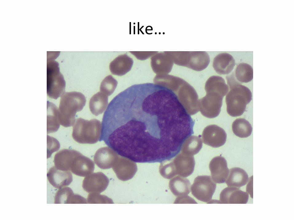
like…
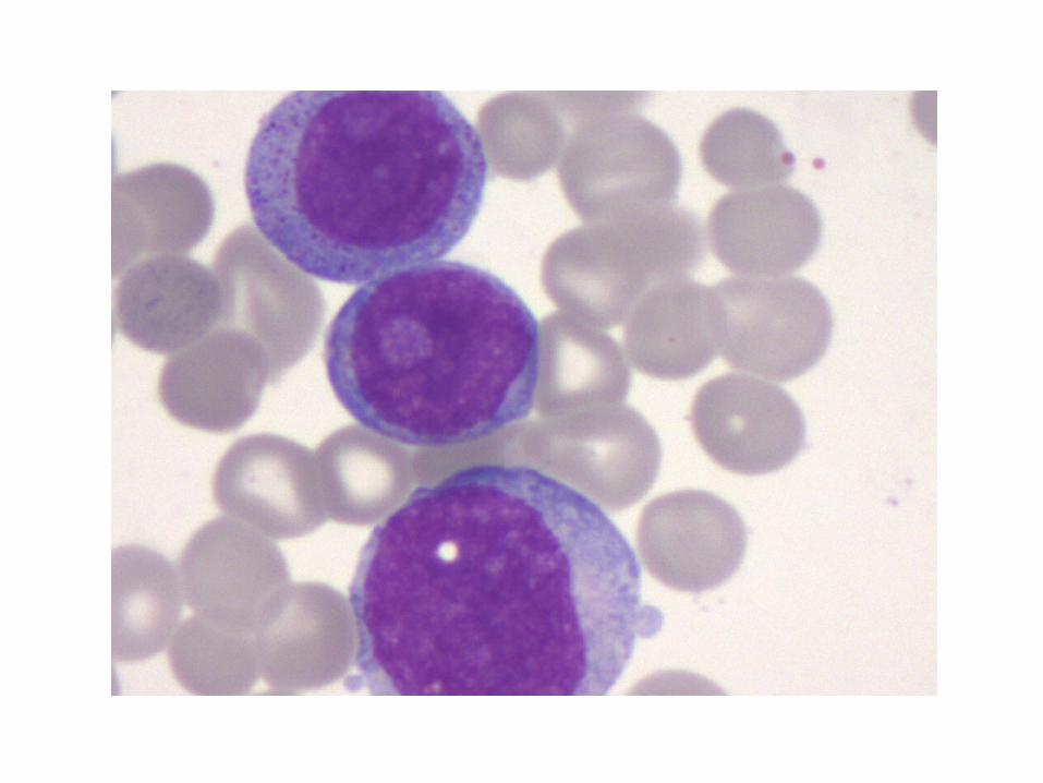
Others are semi classical….

MPO
NSE

And others could be complicated..

Leukemia lymphoma diagnosis

• Morphology
Leukemia lymphoma diagnosis
• Morphology• Ancillary techniques
Leukemia lymphoma diagnosis

• Morphology• Ancillary techniques- Immunophenotyping (IHC and FCI)
Leukemia lymphoma diagnosis
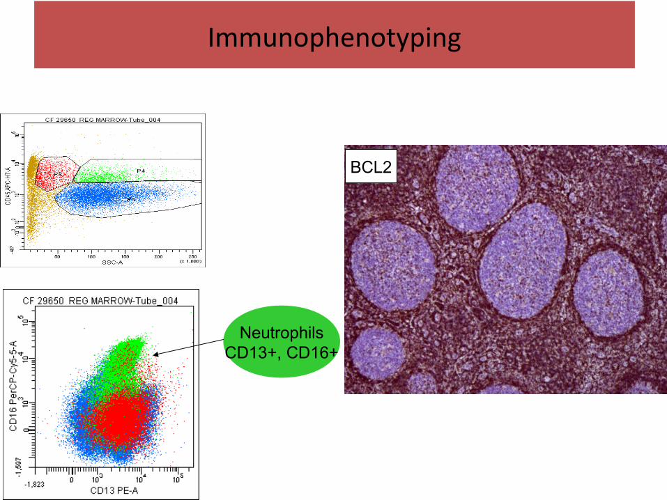
BCL2
NeutrophilsCD13+, CD16+
Immunophenotyping
• Morphology• Ancillary techniques- Immunophenotyping- Cytogenetics
Leukemia lymphoma diagnosis

t(9;22)
Interphase FISH: BCR/ABL fusion
Cytogenetics
PA Amare

• Morphology• Ancillary techniques- Immunophenotyping- Cytogenetics- Molecular diagnostics
Leukemia lymphoma diagnosis

Molecular Diagnostics
NED (Black peak): NPM gene 6 FAM (Blue peak): FLT 3 gene
FLT3Peak 1: 349 bpPeak 2: 412 bpDifference: 63 bp
NPM169 bp

What is the role of ancillary techniques??
Role of a Pathologist
• Diagnostic label

• Diagnostic label• Prognostic marker
Role of a Pathologist
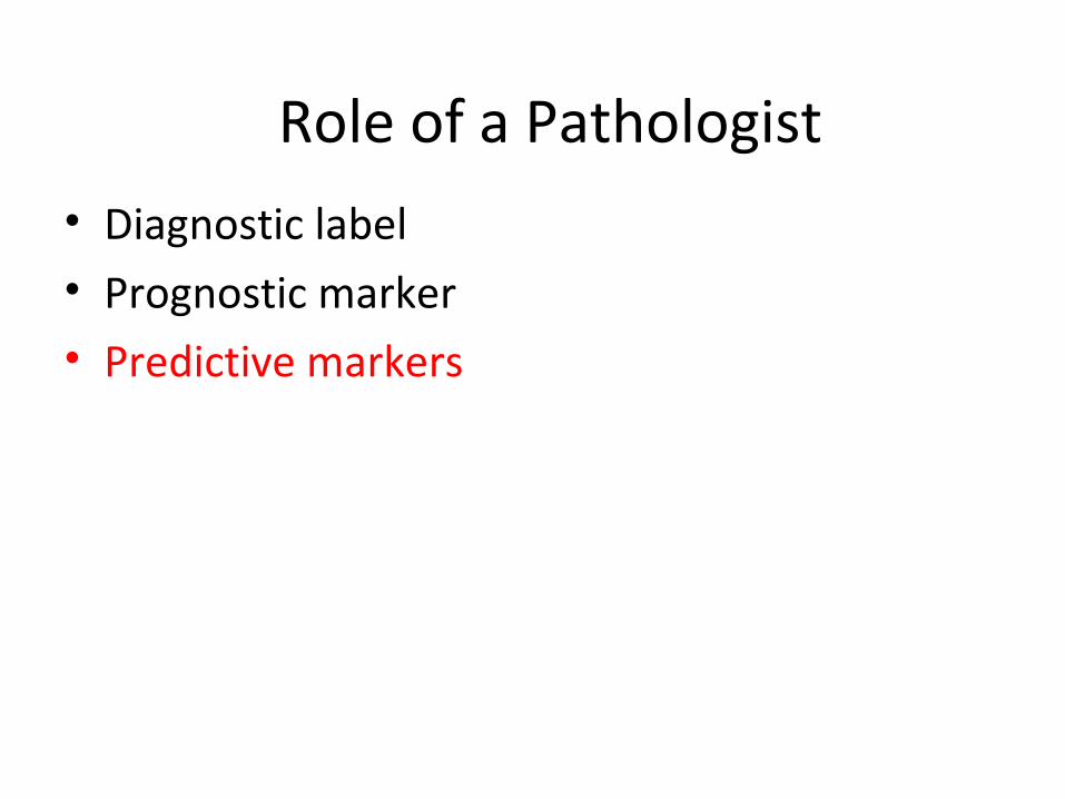
• Diagnostic label• Prognostic marker• Predictive markers
Role of a Pathologist

• Diagnostic label• Prognostic marker• Predictive marker• Minimal residual disease (treatment
effectiveness)
Role of a Pathologist
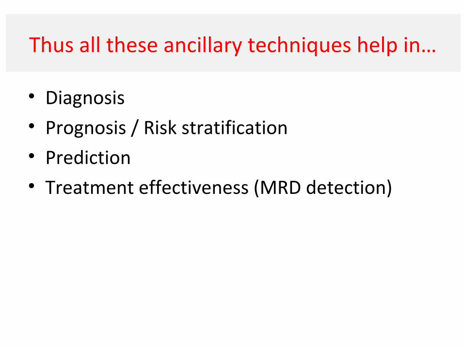
Thus all these ancillary techniques help in…
• Diagnosis• Prognosis / Risk stratification• Prediction• Treatment effectiveness (MRD detection)
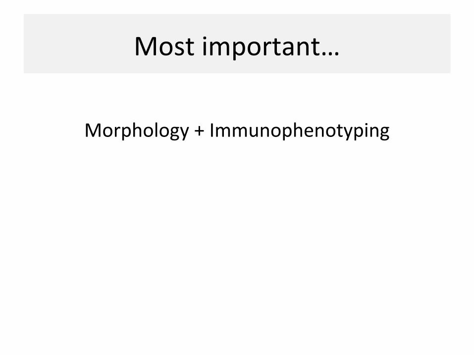
Most important…
Morphology + Immunophenotyping

Diagnosis of leukemia / lymphoma
Flow cytometry principles and applicationsIssues and troubleshoots

IPT helps in subtyping the lineage of the tumors…

What is Immunophenotyping?
• Uses antibodies to identify, locate, and stain specific protein molecules in tissue or in fluids.
• Reaction visualized by a marker (fluorescent dye, enzyme, colloidal gold etc)
• Diagnosis, sub-typing, prognosticating and as a predicting marker of therapeutic response.
Methods for IPT…
ImmunohistochemistryFlow cytometryImmunofluorescence

Immunohistochemistry:Histopathology, paraffin embedded biopsy
Flow cytometry:Peripheral blood,bone marrow aspirate,body fluids
Lymph node biopsy and aspirate
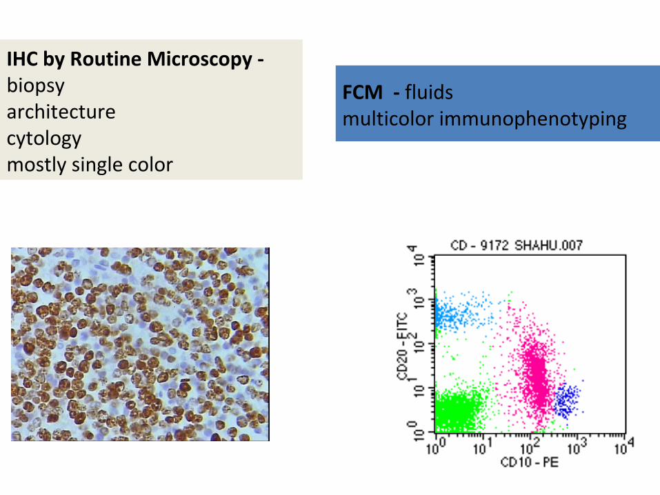
FCM - fluids multicolor immunophenotyping
IHC by Routine Microscopy - biopsy architecturecytologymostly single color

1. IHC
IHC
• Popular in solid tissues
• Subtype tumors
• Architecture + cytology
• But is mostly single color

Some markers best on IHC like epithelial markers, RCT panels
Others on FCM like FMC7, HCL markers
CYTOKERATIN
CD3
CD20
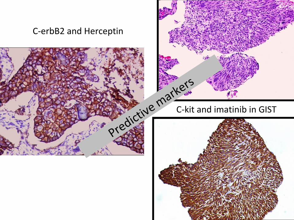
C-kit and imatinib in GIST
C-erbB2 and Herceptin
Predictive markers

IHC is a must in lymphoma diagnosis

Example - DLBCL
• Commonest type of lymphoma
• 50% get cured, rest 50% not…
• Waste basket
• Can we differentiate good ones from bad ones

CD20 and Rituximab
Standard of diagnosis
Predictive markers

Hans Algorithm - DLBCL
BCL6
CD10 Mum1
+
--
GCB
GCB NonGCB
Non GCB
+
+
-

CD 20 CD 10
bcl6
Mib1+ >80%
bcl2
CD 3
Mum1
Diagnosis

Advantages of IHC - architectural relationships and ability to detect scanty tumor cells, as in HL or TCRBCL.
Some antibodies may be better evaluated in paraffin tissue (eg, CyclinD1, CD15, and the presence of Bcl-2, Bcl-6, cyclin D1, ALK-1, and cytoplasmic kappa and lambda).
Likewise, some markers work better on FCI (CD13, CD14, CD19, CD33, etc). Rare markers for BPDCA etc.
FCM is important for fluids…………..

2. FCM

flow + cyto + metry
• 1953 - The first impedance-based flow cytometry device, using the coulter principle (Wallace A Coulter).
• 1968 - The first fluorescence-based flow cytometry device (ICP 11) by Wolfgang Göhde, University of Munster
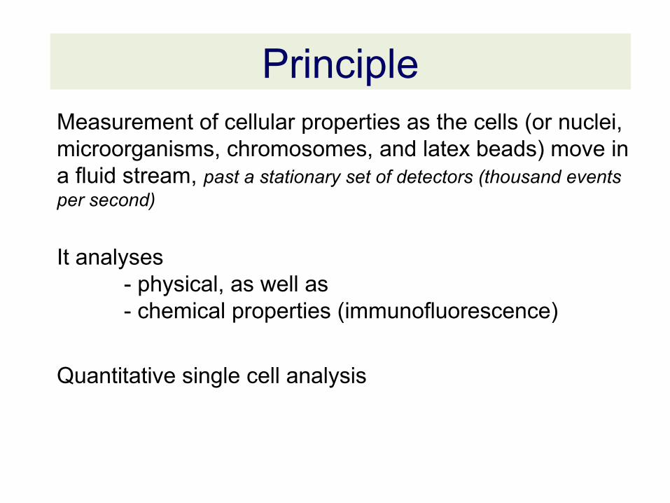
Measurement of cellular properties as the cells (or nuclei, microorganisms, chromosomes, and latex beads) move in a fluid stream, past a stationary set of detectors (thousand events per second)
It analyses - physical, as well as - chemical properties (immunofluorescence)
Quantitative single cell analysis
Principle
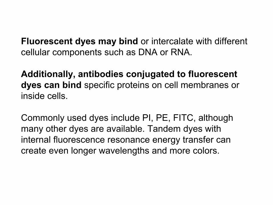
Fluorescent dyes may bind or intercalate with different cellular components such as DNA or RNA.
Additionally, antibodies conjugated to fluorescent dyes can bind specific proteins on cell membranes or inside cells.
Commonly used dyes include PI, PE, FITC, although many other dyes are available. Tandem dyes with internal fluorescence resonance energy transfer can create even longer wavelengths and more colors.
Components of a Flow Cytometer
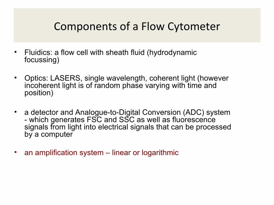
• Fluidics: a flow cell with sheath fluid (hydrodynamic focussing)

Components of a Flow Cytometer
• Fluidics: a flow cell with sheath fluid (hydrodynamic focussing) • Optics: LASERS, single wavelength, coherent light (however
incoherent light is of random phase varying with time and position)
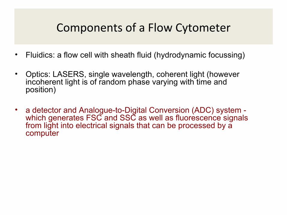
Components of a Flow Cytometer
• Fluidics: a flow cell with sheath fluid (hydrodynamic focussing) • Optics: LASERS, single wavelength, coherent light (however
incoherent light is of random phase varying with time and position)
• a detector and Analogue-to-Digital Conversion (ADC) system - which generates FSC and SSC as well as fluorescence signals from light into electrical signals that can be processed by a computer
Components of a Flow Cytometer
• Fluidics: a flow cell with sheath fluid (hydrodynamic focussing)
• Optics: LASERS, single wavelength, coherent light (however
incoherent light is of random phase varying with time and position)
• a detector and Analogue-to-Digital Conversion (ADC) system - which generates FSC and SSC as well as fluorescence signals from light into electrical signals that can be processed by a computer
• an amplification system – linear or logarithmic

Components of a Flow Cytometer
• Fluidics: a flow cell with sheath fluid (hydrodynamic focussing) • Optics: LASERS, single wavelength, coherent light (however
incoherent light is of random phase varying with time and position)
• a detector and Analogue-to-Digital Conversion (ADC) system - which generates FSC and SSC as well as fluorescence signals from light into electrical signals that can be processed by a computer
• an amplification system – linear or logarithmic
• a computer for analysis of the signals
Maecker, Nature Reviews, 2012
Maecker, Nature Reviews, 2012


Maecker, Nature Reviews, 2012

Maecker, Nature Reviews, 2012

1. Physical propertiesScatter pattern

Forward scatter Side scatter
Size Granularity
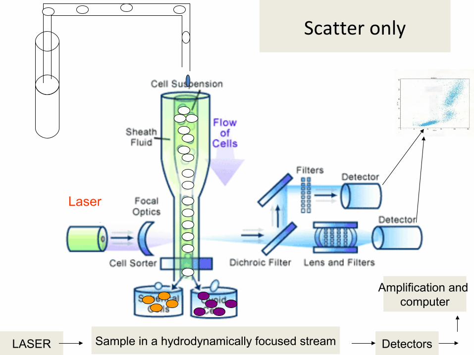
LASER Sample in a hydrodynamically focused stream Detectors
Amplification and computer
Laser
Scatter only

2. Chemical propertiesFCI
PMT
PMT
PMT
LASER Sample in a hydrodynamically focused stream Detectors
Amplification and computer
Laser
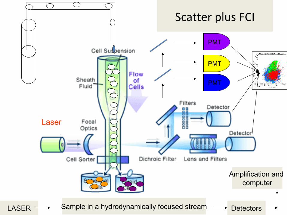
Scatter plus FCI

PMT
PMT
PMT
LASER Sample in a hydrodynamically focused stream Detectors
Amplification and computer
Laser
PMT
PMT
PMT
PMT

Data shown either as a
- single parameter histograms, or - two parameter correlated plots
Data may be shown as
• Linear scale The scale on which the output is directly proportional to the input.
• Logarithmic scale The scale on which the values increase logarithmically.
Data

DNA ploidy
Count
256
384256128
128
384
G0G1 G2M
S phase
Single parameter (Histogram), d
ye (PI),
DNA content, linear scale

Two parameter (dot plot), n
o dye, linear scale
Two parameter (dot plot), d
ye, linear versus log scale

Multiple parameter (d
ot plots), multip
le dyes, FCI, linear/lo
g scale

Types of FCM
- Single Laser or Multiple Lasers(1 laser three color, 4 lasers 18 fluorescence detectors)
- Sorter (so as to purify populations of interest )
- Laser scanning cytometers

Advantages of a FCM• Study of cells, chromosomes and particles (analysis,
counting and sorting)
• Thousand of particles per second
• Multiparametric analysis at a single cell level
• Pattern studies
• Sorting

Research Applications• Autofluorescent Proteins • Antigen or Ligand Density • Apoptosis • Enzyme activity • DNA, RNA content and changes in the cell cycle • Membrane Potential • Cytokine receptors and it's synthesis • Drug uptake and efflux • Phagocytosis • Viability • Changes in Intracellular pH • Changes in Intracellular calcium • Changes in Intracellular glutathione • Changes in Oxidative Burst • Drug discovery and vaccine development

1.Monitoring AIDS patients 2. Immunophenotyping3.Monitoring MRD 4.CD34 counts5.Reticulocyte Counts 6.PNH 7.DNA analysis of S-phase fraction8.Platelet counts
Diagnostic Applications
Start a clinical cytometry facility

Do we really need one? Centralized labs
Hospital / institute based or a stand alone lab
Reagents/maintenance expensive
Stake holders: Management supportCytometrist/Pathologist/ScientistOncologist/Hematologist supportVendor support

Clinical cytometry

RBCsWBCsPlateletsOthers
Cytoplasmic/nuclear characters
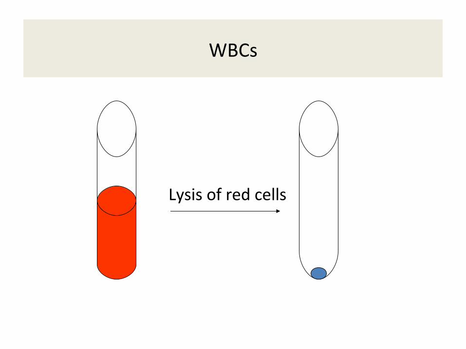
Lysis of red cells
WBCs

Acquire WBCs without any antibodies
FCM - Based on scatter pattern
• Lymphocytes• Monocytes• Neutrophils
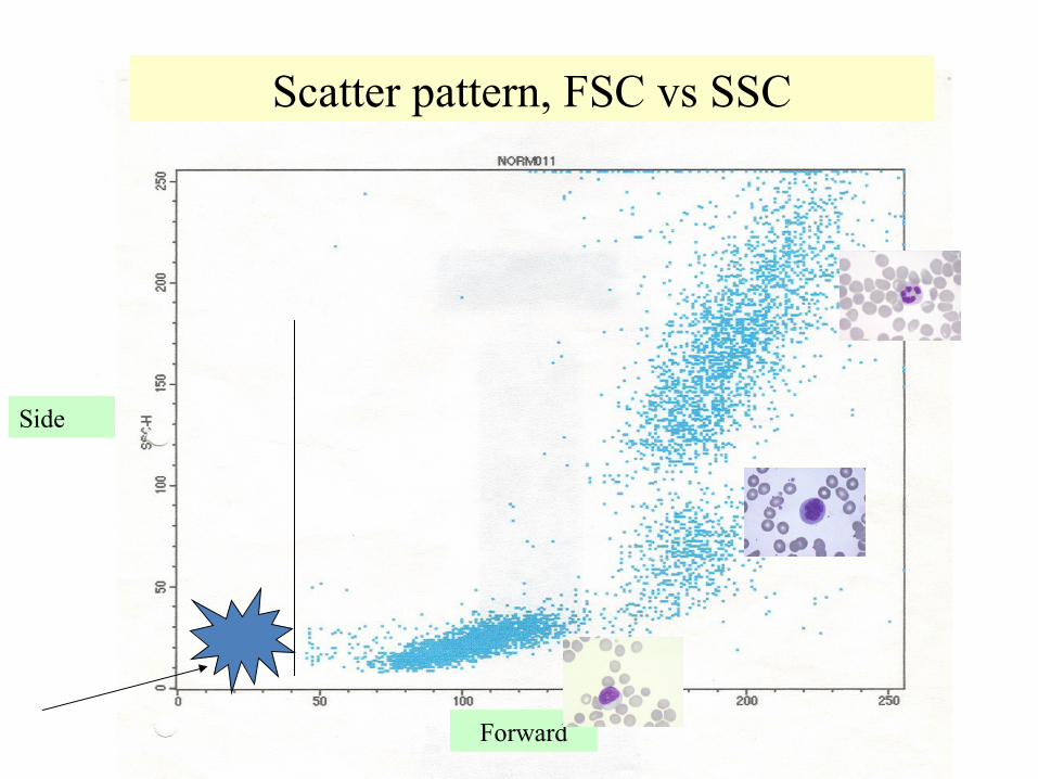
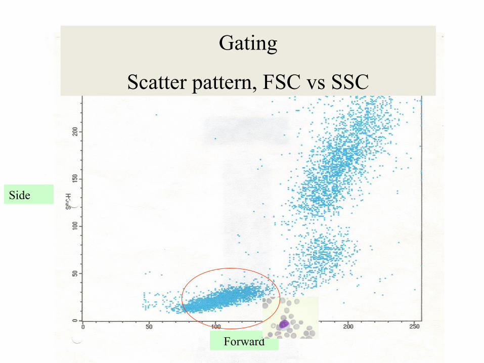
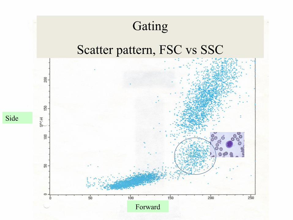
Scatter pattern, FSC vs SSC
Forward
Side
Cells of interest
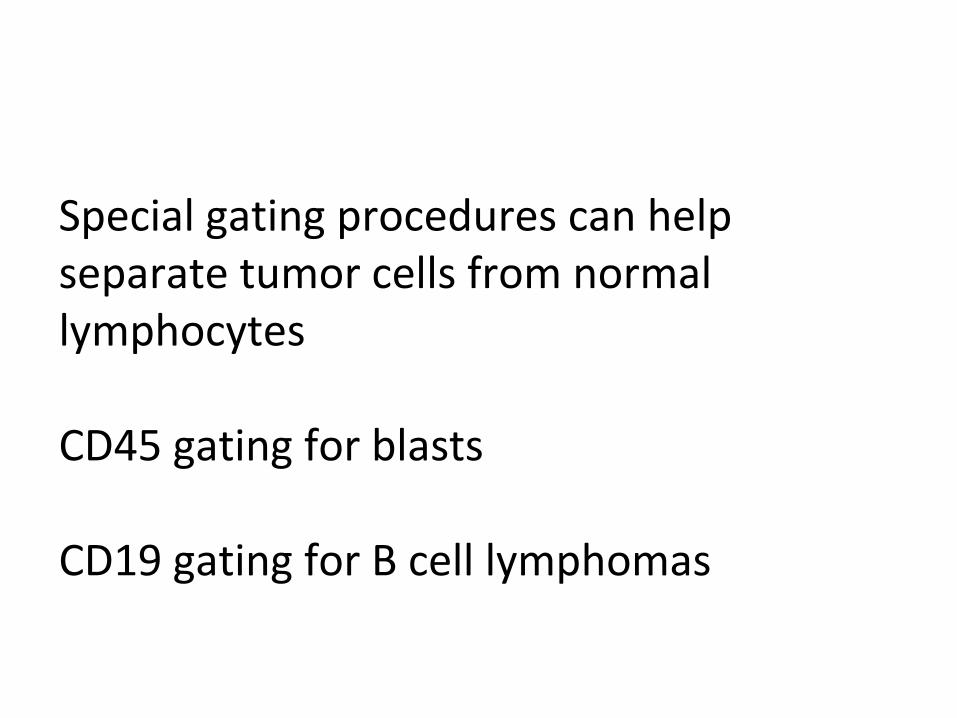
Special gating procedures can help separate tumor cells from normal lymphocytes
CD45 gating for blasts
CD19 gating for B cell lymphomas

Cells of interest and different types of Gating
1. FSC and SSC2. CD45 and SSC3. CD19 and SSC4. CD3 and SSC
5. others

Normal Peripheral bloodForward vs side scatter
Normal Bone marrowForward vs side scatter
FCC-A
FSC-H
SSC-H
FSC-AFSC-A
SSC-ASSC-A
Gating
Scatter pattern, FSC vs SSC
Forward
Side
Gating
Scatter pattern, FSC vs SSC
Forward
Side

Gating
Scatter pattern, FSC vs SSC
Forward
Side

What is an abnormal pattern??

What is an abnormal in this case?

Where do the blasts of acute leukemia appear in scatter pattern plot??

Forward
Side
Where do the blasts of acute leukemia go??
Forward
Side
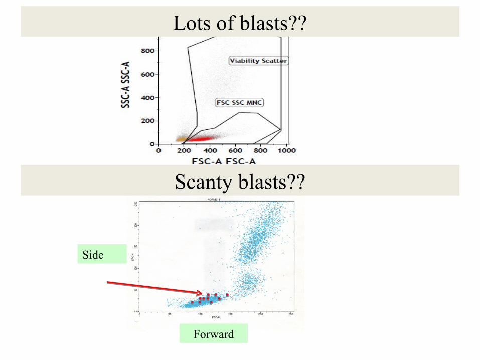
Lots of blasts??
Scanty blasts??

Where do the tumor cells of Hairy cell leukemia go?

Where do the hairy cell leukemia go in flow plots?
Forward
Side

How to separate cells of interest..
Special gating procedures can help separate tumor cells from normal lymphocytes

Lysis of red cells Add Antibodiese.g., CD45
CD 45 gating for blastsSimilarly CD19 gating for HCL tumor cells
Special gating based on CD45..

WBCs
NeutrophilsLymphocytesMonocytesEosinophilsBasophilsVery few stem cells (more in BM)
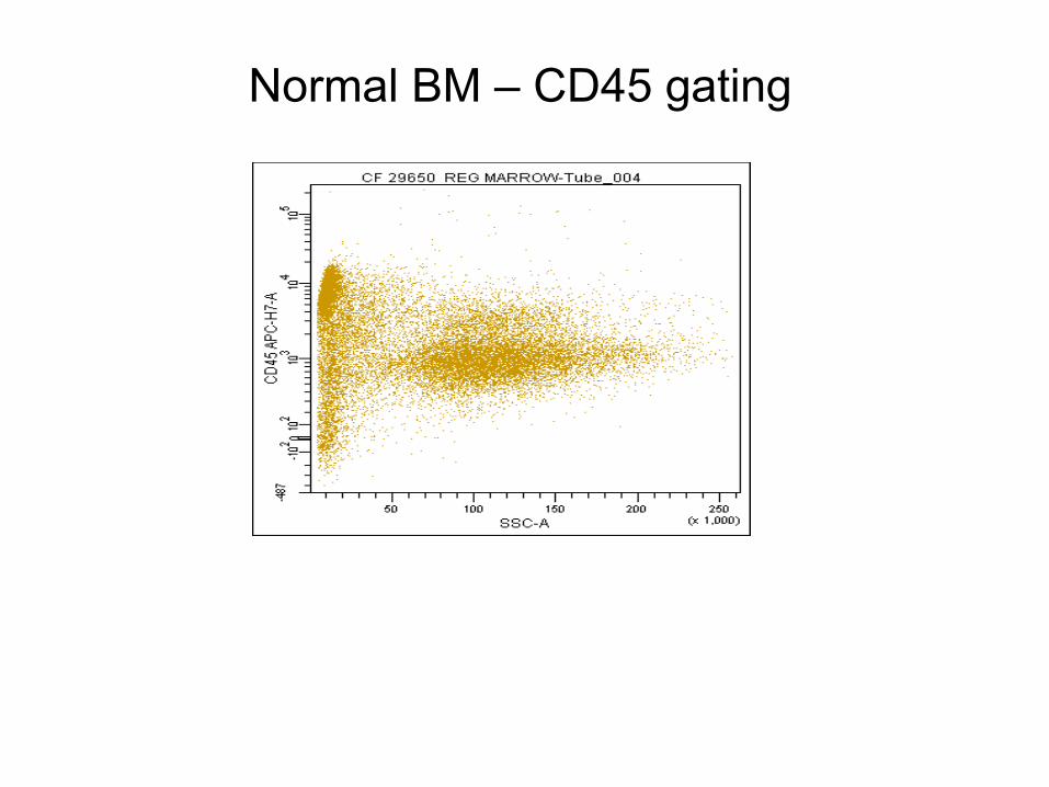
Normal BM – CD45 gating
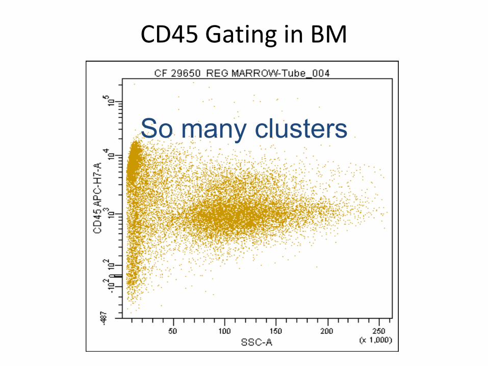
CD45 Gating in BM
So many clusters

CD45 Gating
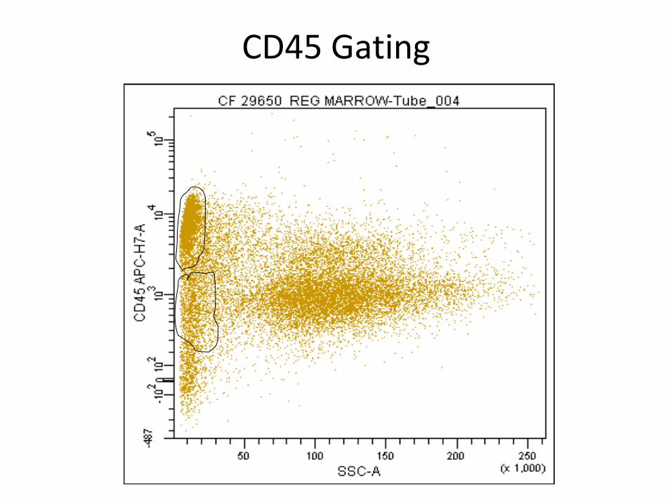
CD45 Gating
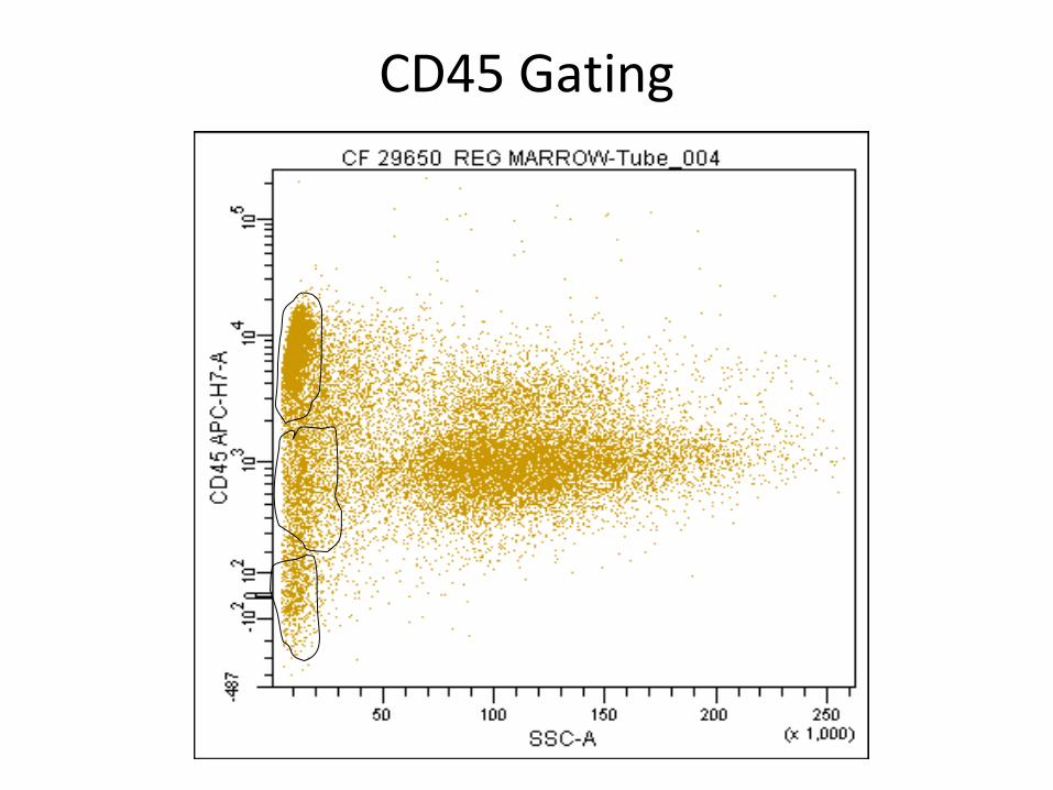
CD45 Gating

CD45 Gating

CD45 Gating

CD45 Gating

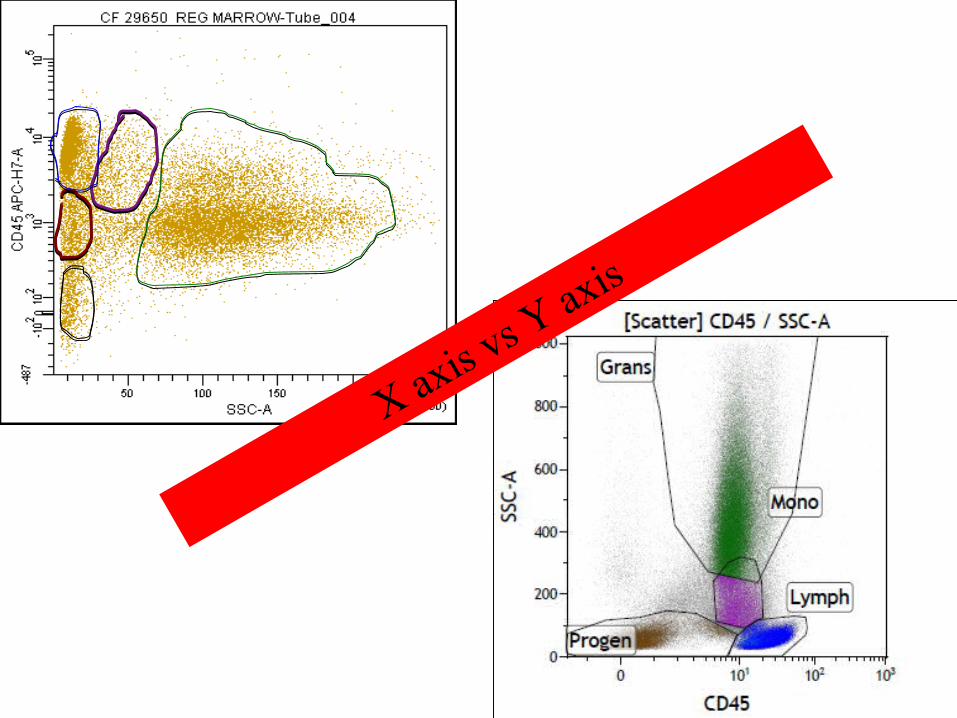
X axis vs Y
axis

CD45 Gating

CD - 23391 INDULKAR (BM).003
CD19 - PE
SS
C -
Hei
ght
100
101
102
103
104
0
256
512
768
1024
CD19 Gating, CLL, post treatment
CD19 Gating, new case of CLL
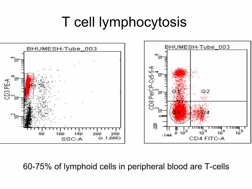
T cell lymphocytosis
60-75% of lymphoid cells in peripheral blood are T-cells
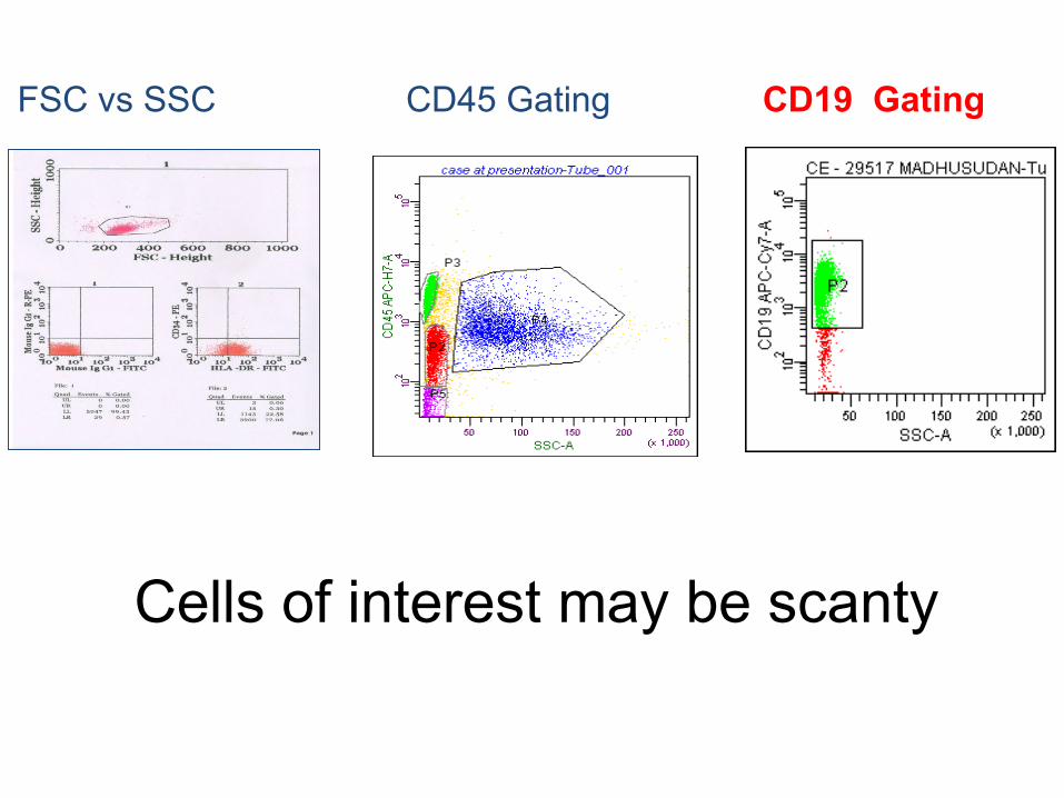
FSC vs SSC CD45 Gating CD19 Gating
Cells of interest may be scanty

CD - 23391 INDULKAR (BM).003
FSC - HeightS
SC
- H
eigh
t0 256 512 768 1024
0
256
512
768
1024
CD - 23391 INDULKAR (BM).003
CD19 - PE
SS
C -
Hei
ght
100
101
102
103
104
0
256
512
768
1024
CD - 23391 INDULKAR (BM).003
CD3 - PerCP
SS
C -
Hei
ght
100
101
102
103
104
0
256
512
768
1024
FSC vs SSC
Study B-cellsStudy T-cells
T and B cells

When to call it positive?

What is positive?
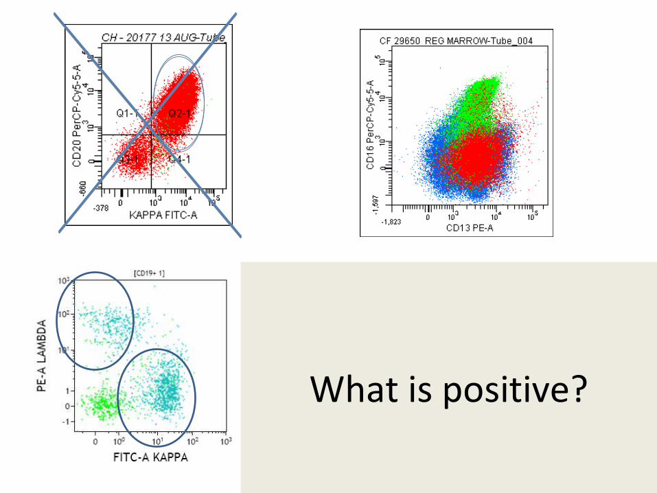
What is positive?
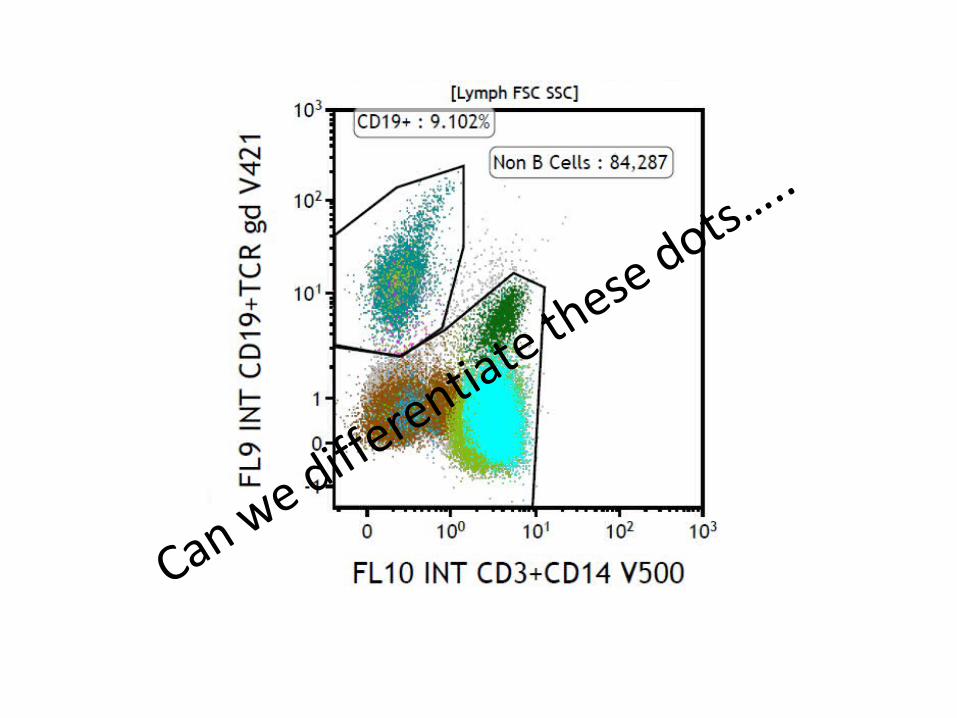
Can we differentiate these dots…
..
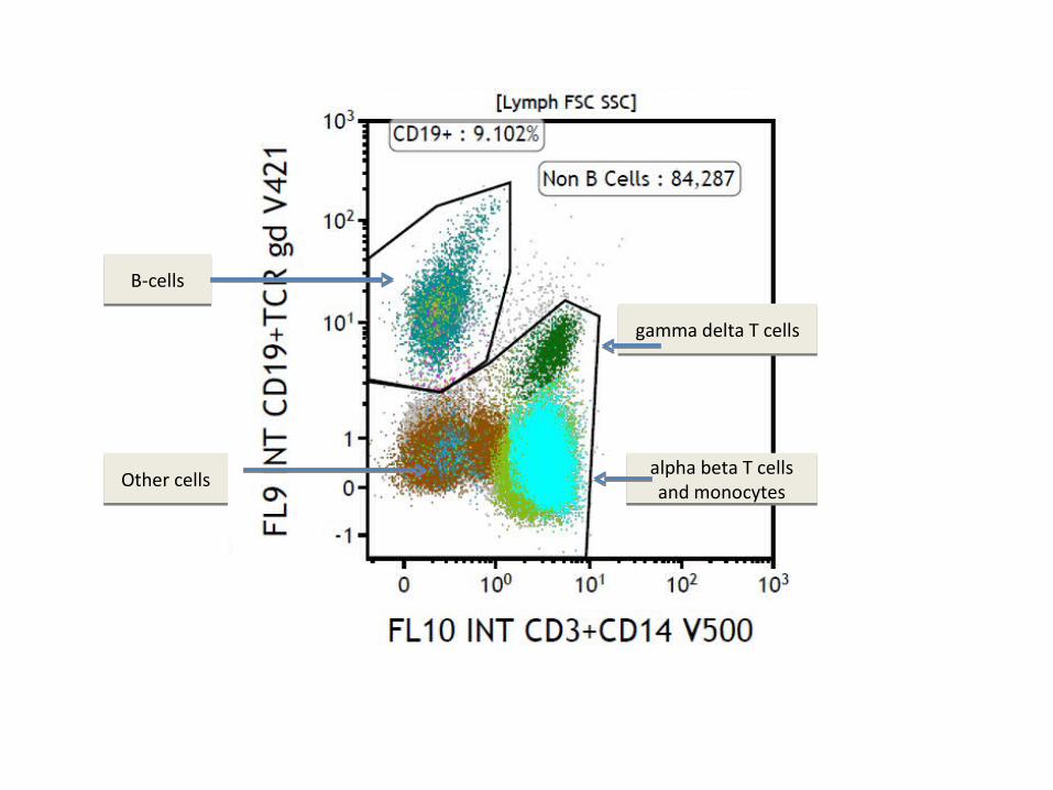
gamma delta T cellsgamma delta T cells
alpha beta T cells and monocytes
alpha beta T cells and monocytes
B-cellsB-cells
Other cellsOther cells

Fluorochromes
Dye Excitation Emission Molecular WeightFITC 488 nm 520 nm 389 DaPE 488 nm 578 nm 240 000 DaECD 488 nm 613 nm 250 000 DaPC 488 nm 668 nm 105 000 DaPerCP 488 nm 688 nm 35 000 DaAPC 613 nm 665 nm 105 000 Da
Each antibody is tagged with a different fluorochrome
Tandem dyes
FCM - ApplicationsFCM - Applications

1. Multicolor Immunophenotyping1. Multicolor Immunophenotyping

What is Multicolor IPT ? 3 or more colors
Single laser: 3-4 colorsTwo lasers: 6 colorsThree lasers: 8 colors plus
More lasers - more colors, more antibodies, more fluorochromes, tandem dyes, better data, more information, flexibility, however, more issues
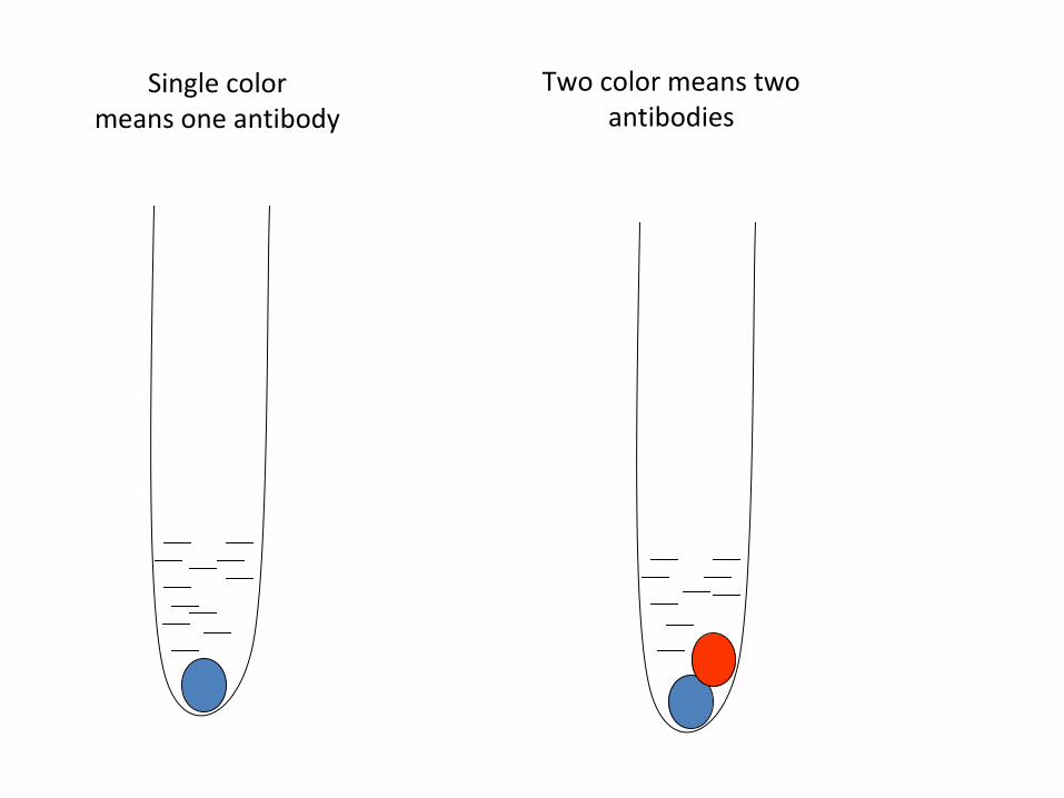
Single colormeans one antibody
Two color means two antibodies
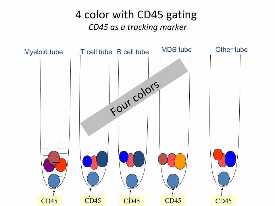
4 color with CD45 gating CD45 as a tracking marker
CD45
Myeloid tube T cell tube B cell tube MDS tube Other tube
CD45 CD45 CD45CD45
Four colors

Immunophenotyping helps in detecting:Immunophenotyping helps in detecting:
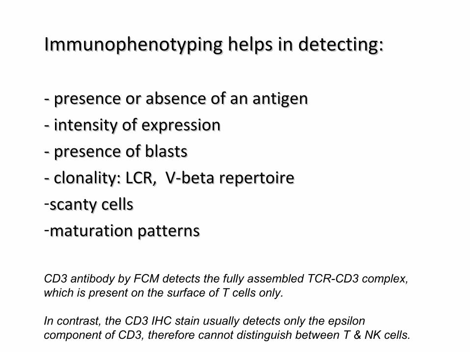
Immunophenotyping helps in detecting:Immunophenotyping helps in detecting:
- - presence or absence of an antigenpresence or absence of an antigen- intensity of expression- intensity of expression- presence of blasts- presence of blasts- clonality: LCR, V-beta repertoire- clonality: LCR, V-beta repertoire-scanty cellsscanty cells-maturation patternsmaturation patterns
CD3 antibody by FCM detects the fully assembled TCR-CD3 complex, which is present on the surface of T cells only.
In contrast, the CD3 IHC stain usually detects only the epsilon component of CD3, therefore cannot distinguish between T & NK cells.
B-cell clonality
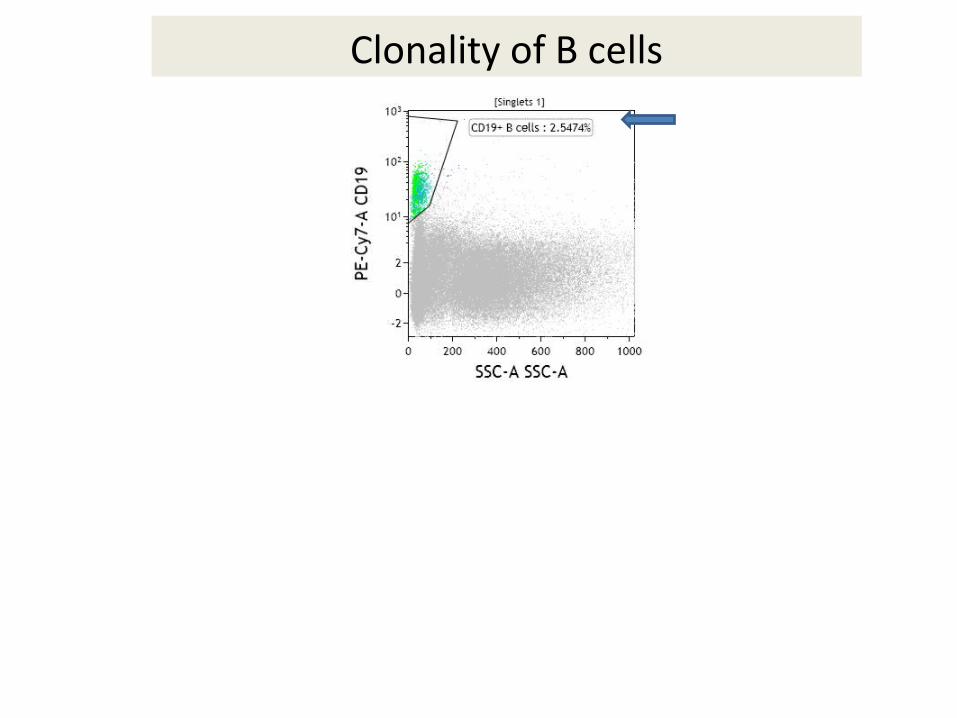
Clonality of B cells

Polyclonal B cells

Monoclonal B cells

T cell normal and abnormal

Normal T cell subsets70% of lymphoid cells in peripheral blood are T cells

Antibodies used for Leuk/Lym diagnosis
- Acute leukemia- Lymphoma/CLPDs

Reagents required in acute leukemia
T-cell tube – CD45, CD2, CD3, CD4, CD5,CD7, CD8B-cell tube – CD45, CD19, CD20, CD10, Myeloid cells – CD45, CD13, CD33, CD117Myeloid tube – CD45, CD14, CD16, CD64Cytopl. tube – CD45, Tdt, cCD3, cCD79a, cCD22, AntiMPOOthers – CD45, CD34, HLADR and so on
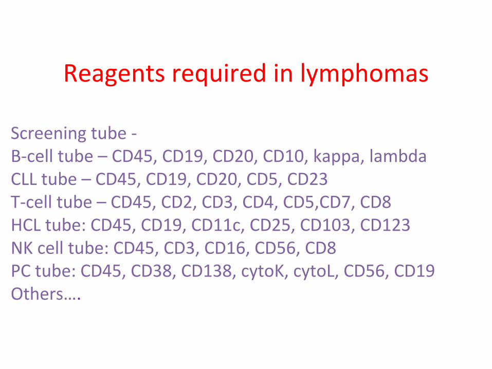
Reagents required in lymphomas
Screening tube - B-cell tube – CD45, CD19, CD20, CD10, kappa, lambdaCLL tube – CD45, CD19, CD20, CD5, CD23T-cell tube – CD45, CD2, CD3, CD4, CD5,CD7, CD8HCL tube: CD45, CD19, CD11c, CD25, CD103, CD123NK cell tube: CD45, CD3, CD16, CD56, CD8PC tube: CD45, CD38, CD138, cytoK, cytoL, CD56, CD19Others….

Indian Guidelines Approach to Acute Leukemia and CLPDs
IJPM, 2008

How to make panels?
- literature based, - training based, - trial or hunch based
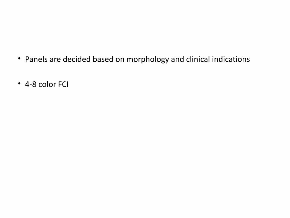
• Panels are decided based on morphology and clinical indications
• 4-8 color FCI
Let us construct a 4 color AL panel

CD3 - FITCCD19 - PE CD13 – PerCPCD45 – TR
a cocktail of antibodies of different lineages..
Four color AL panel
CD3 - FITCCD4 - PE CD8 – PerCPCD45 – TR
a cocktail of antibodies of similar lineage..

CD3 - FITCCD19 - PE CD13 – PerCPCD45 – TR
a cocktail of antibodies of different lineages..
Four color panel
CD3 - FITCCD4 - PE CD8 – PerCPCD45 – TR
T-cell tube

Four color panel - 4/5 tubes
CD3 - FITCCD4 - PE CD8 – PerCPCD45 – TR
T-cell tube
CD19 - FITCCD20- PE CD10- PerCPCD45 – TR
B-cell tube
CD13 - FITCCD33 - PE CD117 – PerCPCD45 – TR
Myeloid- tube
HLADR - FITCCD34 - PE CD7 – PerCPCD45 – TR
Miscellaneous
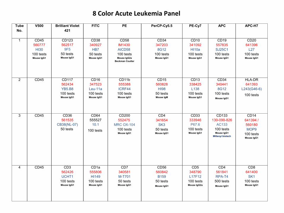
Tube No.
V500 Brilliant Violet 421
FITC PE PerCP-Cy5.5 PE-Cy7 APC APC-H7
1 CD45560777
HI30100 testsMouse IgG1
CD123562517
9F550 tests
Mouse IgG1
CD38340927
HB750 testsMouse IgG1
CD58IM1430 AICD58 100 testsMouse IgG2a
Beckman Coulter
CD343472038G12
100 testsMouse IgG1
CD10341092HI10a
100 testsMouse IgG1
CD19557835SJ25C1100 testsMouse IgG1
CD20641396
L27100 testsMouse IgG1
2 CD45 CD117562434YB5.B8
100 testsMouse IgG1
CD16347523Leu-11a100 testsMouse IgG1
CD11b555388ICRF44
100 testsMouse IgG1
CD15560828
HI9850 testsMouse IgM
CD13338425
L138100 testsMouse IgG1
CD343404418G12
100 testsMouse IgG1
HLA-DR641393
L243(G46-6)
100 tests
3 CD45 CD36561535
CB38(NL-07)50 tests
CD64555527
10.1
100 tests
CD200552475
MRC OX-104100 testsMouse IgG1
CD4341654
SK350 testsMouse IgG1
CD33333946P67.6
100 testsMouse IgG1
CD133130-090-826
AC133100 testsMouse IgG1
Miltenyi biotech
CD14641394 /560180MOP9
100 testsMouse IgG1
4 CD45 CD3562426UCHT1
100 testsMouse IgG1
CD1a555806HI149
100 testsMouse IgG1
CD7340581M-T70150 testsMouse IgG1
CD56560842B159
50 testsMouse IgG1
CD5348790L17F12
100 testsMouse IgG2a
CD4561841RPA-T4500 testsMouse IgG1
CD8641400
SK1100 testsMouse IgG1
8 Color Acute Leukemia Panel
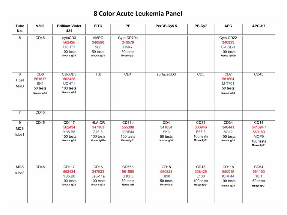
5 CD45 cytoCD3562426UCHT1
100 testsMouse IgG1
AMPO340580
5B850 testsMouse IgG1
Cyto CD79a340579HM47
50 testsMouse IgG1
Cyto CD22340933S-HCL-1100 testsMouse IgG2b
6
T cell
MRD
CD8561617
SK150 testsMouse IgG1
CytoCD3562426UCHT1
100 testsMouse IgG1
Tdt CD4 surfaceCD3 CD5 CD7561604M-T70150 testsMouse IgG1
CD45
7 CD45
9
MDS
tube1
CD45 CD117562434YB5.B8
100 testsMouse IgG1
HLA-DR347363G46-6
100 testsMouse IgG2a
CD11b555388ICRF44
100 testsMouse IgG1
CD4341654
SK350 testsMouse IgG1
CD33333946P67.6
100 testsMouse IgG1
CD343404418G12
100 testsMouse IgG1
CD14641394 /560180MOP9
100 testsMouse IgG1
MDS
tube2
CD45 CD117562434YB5.B8
100 testsMouse IgG1
CD16347523Leu-11a100 testsMouse IgG1
CD66b561650G10F550 testsMouse IgM
CD15560828
HI9850 testsMouse IgM
CD13338425
L138100 testsMouse IgG1
CD11b550019ICRF44
100 testsMouse IgG1
CD64561190
10.150 testsMouse IgG1
8 Color Acute Leukemia Panel
Tube No.
V500 Brilliant Violet 421
FITC PE PerCP-Cy5.5 PE-Cy7 APC APC-H7
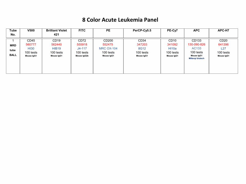
1
MRD
tube
BALL
CD45560777
HI30100 testsMouse IgG1
CD19562440HIB19
100 testsMouse IgG1
CD72555918J4-117
100 testsMouse IgG2b
CD200552475
MRC OX-104100 testsMouse IgG1
CD343472038G12
100 testsMouse IgG1
CD10341092HI10a
100 testsMouse IgG1
CD133130-090-826
AC133100 testsMouse IgG1
Miltenyi biotech
CD20641396
L27100 testsMouse IgG1
8 Color Acute Leukemia Panel
Tube No.
V500 Brilliant Violet 421
FITC PE PerCP-Cy5.5 PE-Cy7 APC APC-H7
Comprehensive panel of antibodies used for L/L diagnosis

Case 1
5 year old boy with fever – 1 month
Classical 3-4 color

Diagnosis
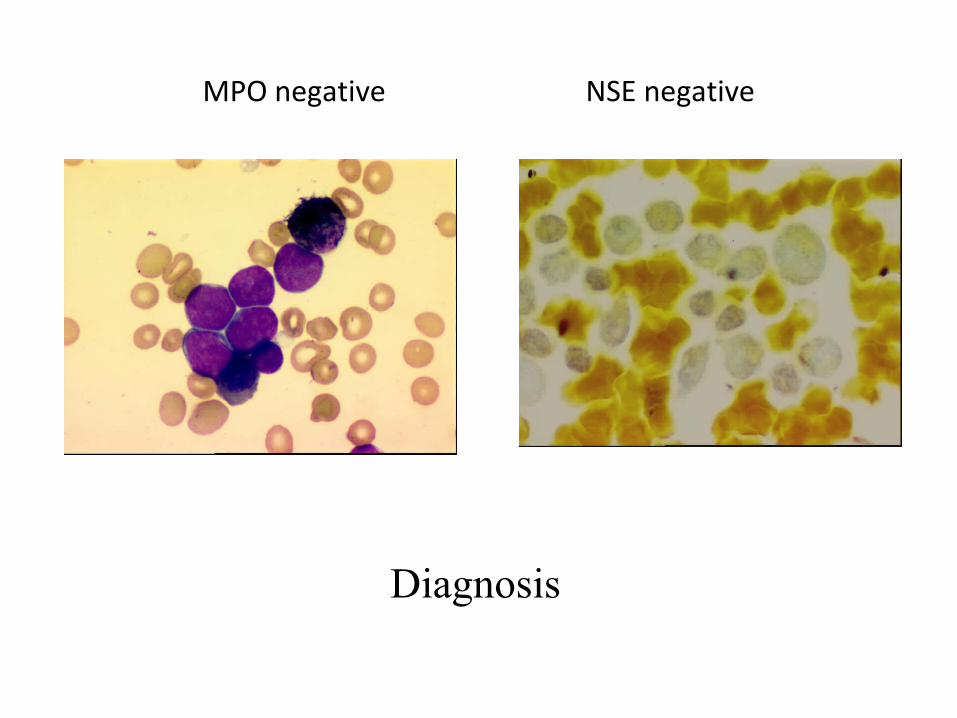
MPO negative NSE negative
Diagnosis

Immunophenotyping
FSC vs SSC
Blasts only
3 color IP
T – Common practice


Guess

CD10+, CD19+, HLADR+
FL, DLBCL
B cell ALL, Burkitt’s Lymphoma
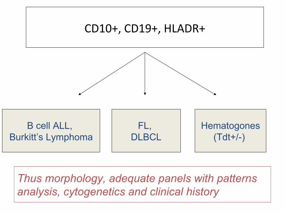
CD10+, CD19+, HLADR+
FL, DLBCL
Hematogones(Tdt+/-)
B cell ALL, Burkitt’s Lymphoma
Thus morphology, adequate panels with patterns analysis, cytogenetics and clinical history

Case 2
12 year old girl, presented with fever for 2 weeks.
Peripheral blood smear reveals high counts with blast like cells

Acute leukemiacytochemical MPO negative
What next?

FCI done
B cell tube – NegativeMyeloid tube – Negative
All T cell markers including CD3 – Negative
Blasts expressed only CD7


T-cell markers- CD3- CD7- CD2- CD5
- CD4/CD8

What do you next?
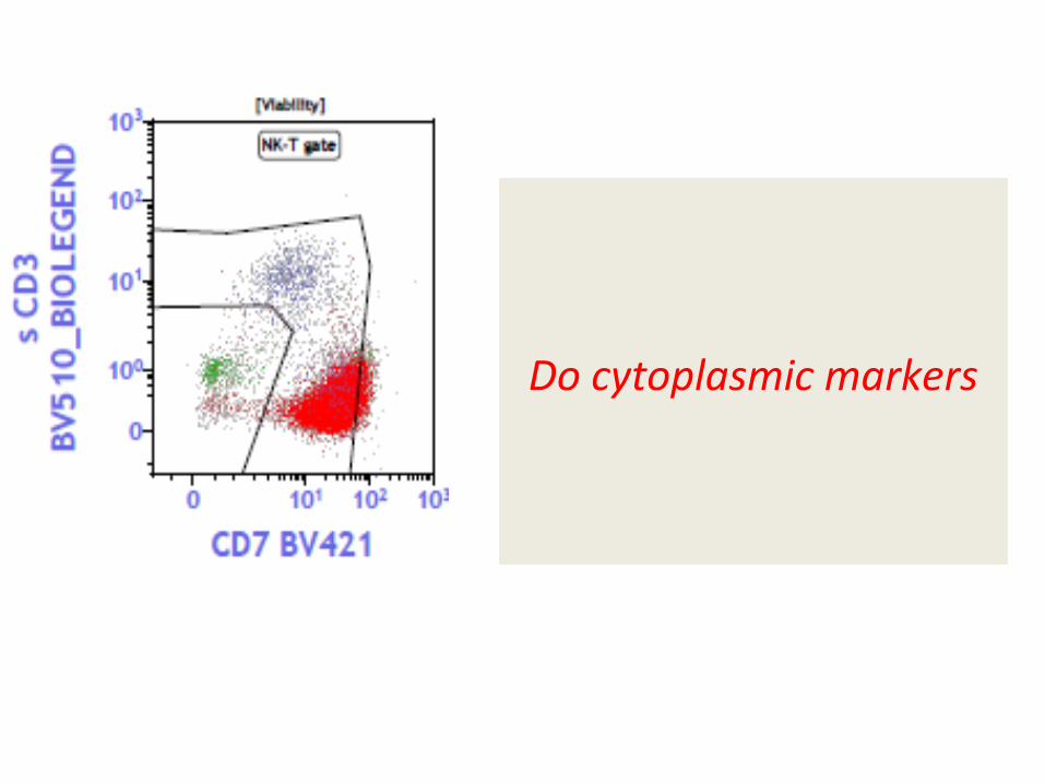
Do cytoplasmic markers
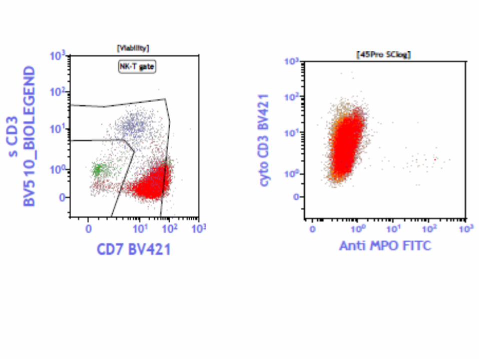

DiagnosisT-cell ALL
Note: Blasts may be surface CD3 negative

Tissue flow
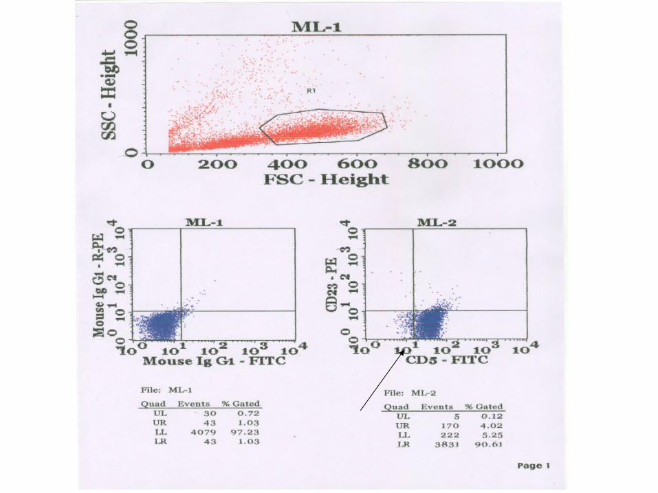
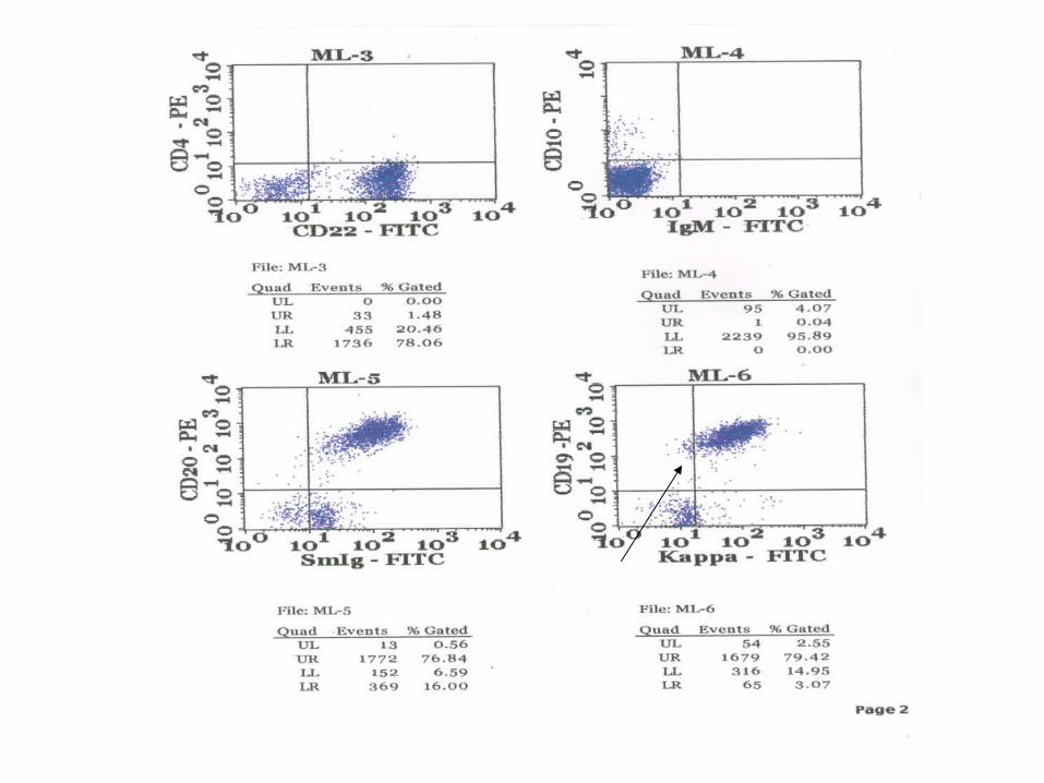
45 year old male, left cervical LN since 1 month
Lymph node FNAC done
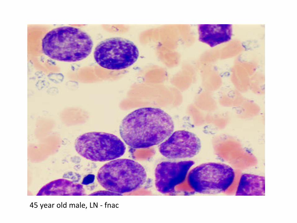
45 year old male, LN - fnac

Issues o
f 3 co
lor IPT

Subtyping of Small B cell lymphomas

Nodular pattern

Common B cell NHLs expressing CD5 (T-cell marker)
1.CLL – express CD232. MCL – express cyclin D1 (not by flow)
Lymphoma Diagnosis
Gold standard is biopsy plus IHC

FCM and lymphoma diagnosis
• Best for CLPDs
• Mostly B cell type and panels are well defined
• Also used for lymph node biopsy, aspirate, fluids and other tissues

T cell lymphomas are rareNeed elaborate panel
• Altered expression of pan T cell markers like CD2, CD3, CD5 and CD7 (also seen in IM, CMV)
• Subset restriction of CD4 or CD8 (also seen in IM, HIV, AID)
• Increased expression of markers like CD25 with/without CD4
• Aberrant expression of CD10, CD30, CD103, Tdt, Alk1 etc.
• Aberrant expression of CD16, CD56
• V beta repertoire
• HTLV-1 serology, PCR for TCR gene rearrangements

Lymphoma Screening Tube
- 15 antibodies in one tube- Mutually exclusive Abs
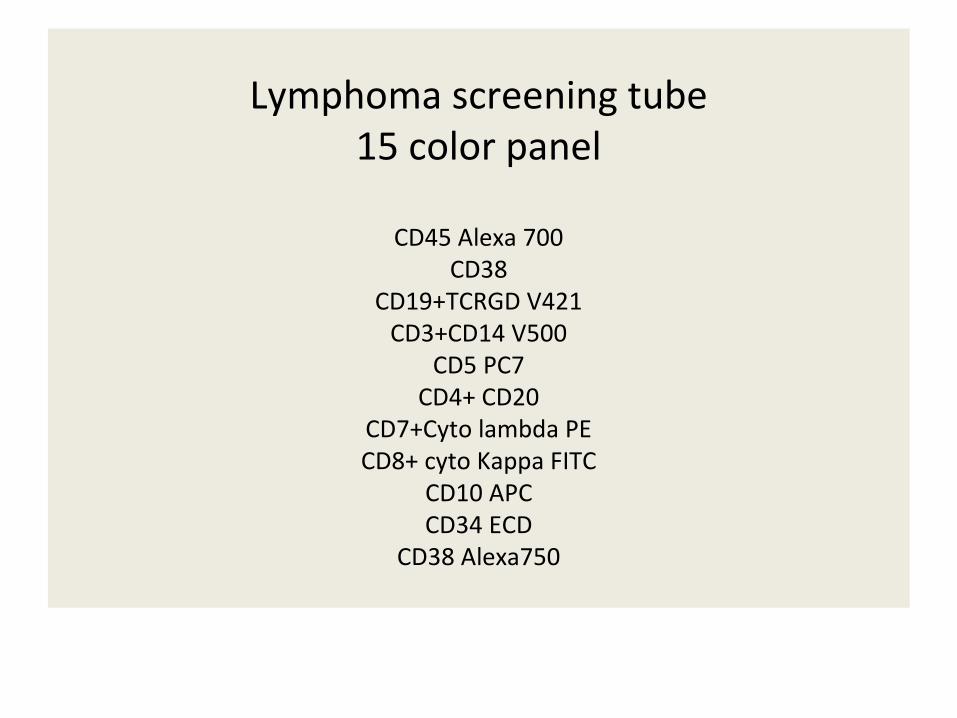
Lymphoma screening tube15 color panel
CD45 Alexa 700CD38
CD19+TCRGD V421CD3+CD14 V500
CD5 PC7CD4+ CD20
CD7+Cyto lambda PECD8+ cyto Kappa FITC
CD10 APCCD34 ECD
CD38 Alexa750


Plasma cell Neoplasms
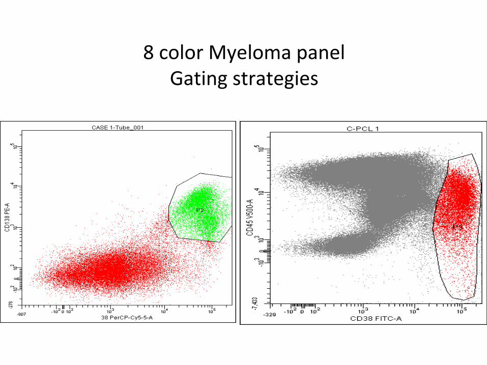
8 color Myeloma panelGating strategies

First tube for Myeloma, TMH • CD38 FITC• CD19 V450• CD20 APCH7• CD27 PE• CD28APC• CD45 V500• CD117PEcy7• CD56 PerCP cy5.5
PG Subramanian

Second myeloma tube, TMH
• Cyto Lambda FITC• Cyto Kappa PE• CD45 APC H7• CD38 PerCP-Cy5.5• CD19 PE-Cy7• CD56 APC• Blank• Blank
PG Subramanian

Case of plasma cell dyscrasia (eight color)

8 color IPT