Flora of drift plastics: a new red algal genus, Tsunamia ... · their origin was the Tohoku coast...
13
Algae 2016, 31(4): 289-301 https://doi.org/10.4490/algae.2016.31.10.20 Open Access Research Article Copyright © 2016 The Korean Society of Phycology 289 http://e-algae.org pISSN: 1226-2617 eISSN: 2093-0860 Flora of drift plastics: a new red algal genus, Tsunamia transpacifica (Stylonematophyceae) from Japanese tsunami debris in the northeast Pacific Ocean John A. West 1, * , Gayle I. Hansen 2 , Takeaki Hanyuda 3 and Giuseppe C. Zuccarello 4 1 School of Biosciences 2, University of Melbourne, Parkville, VIC 3010, Australia 2 Oregon State University, HMSC-EPA, 2111 SE Marine Science Drive, Newport, OR 97365-5260, USA 3 Kobe University Research Center for Inland Seas, Rokkodai, Nadaku, Kobe 657-8501, Japan 4 School of Biological Sciences, Victoria University of Wellington, PO Box 600, Wellington 6140, New Zealand Floating debris provides substrates for dispersal of organisms by ocean currents, including algae that thrive on plastics. The 2011 earthquake and tsunami in Tohuku, Japan resulted in large amounts of debris carried by the North Pacific Cur- rent to North America from 2012 to 2016. In 2015-2016, the plastics in the debris bore a complex biota including pink algal crusts. One sample (JAW4874) was isolated into culture and a three-gene phylogeny (psbA, rbcL, and SSU) indicated it was an unknown member of the red algal class Stylonematophyceae. It is a small pulvinate crust of radiating, branched, uniseriate filaments with cells containing a single centrally suspended nucleus and a single purple to pink, multi-lobed, parietal plastid lacking a pyrenoid. Cells can be released as spores that attach and germinate to form straight filaments by transverse apical cell divisions, and subsequent longitudinal and oblique intercalary divisions produce masses of lateral branches. This alga is named Tsunamia transpacifica gen. nov. et sp. nov. Sequencing of additional samples of red algal crusts on plastics revealed another undescribed Stylonematophycean species, suggesting that these algae may be frequent on drift oceanic plastics. Key Words: biodiversity; dispersal; Japan; Oregon; plastic debris; psbA; rbcL; taxonomy; tsunami; Washington; 18S rDNA INTRODUCTION The increase in plastics in the ocean is a major hazard for oceanic and coastal organisms (Derraik 2002, Barnes et al. 2009, Andrady 2011, Eriksen et al. 2014). Floating de- bris can also provide opportunities for long-distance dis- persal of otherwise poorly dispersing organisms (Fraser et al. 2011, Cumming et al. 2014). Plastics harbor commu- nities that appear to be specific to these substrates (Car- son et al. 2013, Zettler et al. 2013). Diatoms and particular bacterial communities are often known to inhabit small fragments of the marine plastisphere (Bravo et al. 2011, Zettler et al. 2013, Samoray 2016). The North Pacific Ocean is especially rich in plastic debris (Goldstein et al. 2013, Eriksen et al. 2014, Law and Thompson 2014). The highly destructive Tohoku earth- quake and subsequent tsunami of Mar 11, 2011 deposited more than 5 million tons of debris into the ocean (Japa- nese Ministry of the Environment 2012). A part of this de- bris, including many plastic items, was buoyant enough Received August 27, 2016 Accepted October 20, 2016 *Corresponding Author E-mail: [email protected] Tel: +61-3-8344-8080, Fax: +61-3-9347-5460 This is an Open Access article distributed under the terms of the Creative Commons Attribution Non-Com- mercial License (http://creativecommons.org/licenses/by-nc/3.0/) which permits unrestricted non-commercial use, distribution, and reproduction in any medium, provided the original work is properly cited.
Transcript of Flora of drift plastics: a new red algal genus, Tsunamia ... · their origin was the Tohoku coast...

Algae 2016, 31(4): 289-301https://doi.org/10.4490/algae.2016.31.10.20
Open Access
Research Article
Copyright © 2016 The Korean Society of Phycology 289 http://e-algae.org pISSN: 1226-2617 eISSN: 2093-0860
Flora of drift plastics: a new red algal genus, Tsunamia transpacifica (Stylonematophyceae) from Japanese tsunami debris in the northeast Pacific Ocean
John A. West1,*, Gayle I. Hansen2, Takeaki Hanyuda3 and Giuseppe C. Zuccarello4
1School of Biosciences 2, University of Melbourne, Parkville, VIC 3010, Australia2Oregon State University, HMSC-EPA, 2111 SE Marine Science Drive, Newport, OR 97365-5260, USA3Kobe University Research Center for Inland Seas, Rokkodai, Nadaku, Kobe 657-8501, Japan4School of Biological Sciences, Victoria University of Wellington, PO Box 600, Wellington 6140, New Zealand
Floating debris provides substrates for dispersal of organisms by ocean currents, including algae that thrive on plastics.
The 2011 earthquake and tsunami in Tohuku, Japan resulted in large amounts of debris carried by the North Pacific Cur-
rent to North America from 2012 to 2016. In 2015-2016, the plastics in the debris bore a complex biota including pink algal
crusts. One sample (JAW4874) was isolated into culture and a three-gene phylogeny (psbA, rbcL, and SSU) indicated it
was an unknown member of the red algal class Stylonematophyceae. It is a small pulvinate crust of radiating, branched,
uniseriate filaments with cells containing a single centrally suspended nucleus and a single purple to pink, multi-lobed,
parietal plastid lacking a pyrenoid. Cells can be released as spores that attach and germinate to form straight filaments
by transverse apical cell divisions, and subsequent longitudinal and oblique intercalary divisions produce masses of
lateral branches. This alga is named Tsunamia transpacifica gen. nov. et sp. nov. Sequencing of additional samples of red
algal crusts on plastics revealed another undescribed Stylonematophycean species, suggesting that these algae may be
frequent on drift oceanic plastics.
Key Words: biodiversity; dispersal; Japan; Oregon; plastic debris; psbA; rbcL; taxonomy; tsunami; Washington; 18S rDNA
INTRODUCTION
The increase in plastics in the ocean is a major hazard
for oceanic and coastal organisms (Derraik 2002, Barnes
et al. 2009, Andrady 2011, Eriksen et al. 2014). Floating de-
bris can also provide opportunities for long-distance dis-
persal of otherwise poorly dispersing organisms (Fraser
et al. 2011, Cumming et al. 2014). Plastics harbor commu-
nities that appear to be specific to these substrates (Car-
son et al. 2013, Zettler et al. 2013). Diatoms and particular
bacterial communities are often known to inhabit small
fragments of the marine plastisphere (Bravo et al. 2011,
Zettler et al. 2013, Samoray 2016).
The North Pacific Ocean is especially rich in plastic
debris (Goldstein et al. 2013, Eriksen et al. 2014, Law and
Thompson 2014). The highly destructive Tohoku earth-
quake and subsequent tsunami of Mar 11, 2011 deposited
more than 5 million tons of debris into the ocean (Japa-
nese Ministry of the Environment 2012). A part of this de-
bris, including many plastic items, was buoyant enough
Received August 27, 2016 Accepted October 20, 2016
*Corresponding Author
E-mail: [email protected]: +61-3-8344-8080, Fax: +61-3-9347-5460
This is an Open Access article distributed under the terms of the Creative Commons Attribution Non-Com-
mercial License (http://creativecommons.org/licenses/by-nc/3.0/) which permits unrestricted non-commercial use, distribution, and reproduction in any medium, provided the original work is properly cited.

Algae 2016, 31(4): 289-301
https://doi.org/10.4490/algae.2016.31.10.20 290
MATERIALS AND METHODS
Specimens of crusts were collected from plastic debris
items (Table 1) cast ashore on Oregon and Washington
beaches during the winter and spring of 2015 and 2016,
nearly 5 years after the Japanese tsunami. Several of the
debris items contained Japanese writing that indicated
their origin was the Tohoku coast of Japan. Nearly all of
the debris supporting the alga was light in colour and de-
termined to be high-density polyethylene (HDPE), a plas-
tic that typically floats at the waterline. Specimens were
preserved in 5% formalin in seawater for later photogra-
phy and in silica gel for molecular analysis. One collection
of the crust (GIH-416), found to be mostly unialgal, was
kept alive in preliminary culture at 11°C using F/2 me-
dium (Andersen 2005) until further investigations could
be carried out. GIH-416 was taken from a white polyeth-
ylene bottle (Fig. 1A) found on Nov 5, 2015 on the Long
Beach Peninsula in Washington (46°37.385 N, 124°04.246
W). This material was the main reference collection for
our observations of the species, and subsamples of this
collection have been used to prepare the type material.
Living samples of GIH-416 were sent to Australia and a
subculture (JAW4874) was placed in about 80 mL of low
nutrient Modified Provasoli’s ES medium (5 mL per 1 L
sterile seawater at 30‰ salinity) (West and McBride 1999)
with 3 drops of 1% germanium dioxide to control diatoms
and about 1 mg of powdered Na Penicillin G to control
cyanobacteria. The 50 × 70 mm Pyrex dishes were main-
tained at 10 : 14 light-dark daily photoperiod, 19-22°C,
and 4-8 µmol photons m-2 s-1 cool white LED lighting for
several weeks while unialgal status, growth and reproduc-
tion were followed. A higher irradiance (12-34 µmol pho-
tons m-2 s-1) was used to observe morphological changes.
To test for starch, filaments were placed on slides with
drops of I2KI solution (Johansen 1940) and heated for 2 s
at 800 W in a microwave oven.
All photographs except Fig. 1A were taken with a Zeiss
GFL bright field microscope (Zeiss Australia, Sydney, Aus-
tralia) and Canon G3 camera (https://www.canon.com.
au/) and edited with Adobe Photoshop CS4 (http://www.
adobe.com/au/) for publication.
Molecular analyses
Total DNA was isolated from silica gel-dried material
using a modified CTAB procedure (Zuccarello and Lok-
horst 2005). Amplification and sequencing of the plas-
tid-encoded large subunit of the ribulose bisphosphate
carboxylase / oxygenase gene (rbcL) used amplification
to float and be carried by currents across the North Pacific
to the coasts of Oregon and Washington, where they be-
gan arriving in June of 2012. Many debris items were rec-
ognizable as Japanese by their markings, manufacturing
details, and fouling biota. The fouling biota of tsunami
debris is now being investigated by a number of scientists
(Carlton et al. 2016, Hansen et al. 2016), but studies of the
specific organisms that survive on the plastic debris have
just begun.
The red algal class Stylonematophyceae has about 15
genera (West et al. 2007, Zuccarello et al. 2008, Yoon et
al. 2010, Guiry and Guiry 2016). Morphologically they are
simple, varying from unicells and colonies to filamentous
prostrate and upright thalli with various plastid types
(single multilobed or multiple discoid plastids, with or
without a pyrenoid). Most are marine genera but a few
occur in freshwater and terrestrial habitats. Some appear
to be ubiquitous on various other substrates (e.g., algae,
plants, wood, nets, and glass) and do not have clear bio-
geographic patterns (Zuccarello et al. 2008). Seven genera
(Bangiopsis, Chroodactylon, Chroothece, Colacodictyon,
Rhodaphanes, Rhodosorus, and Stylonema) have a single
multi-lobed plastid with a prominent pyrenoid whereas
Empselium, Goniotrichopsis, Kyliniella, Neevea, Phrag-
monema, Purpureofilum, Rhodospora, and Rufusia have
either a single complex multilobed plastid without a pyre-
noid or several small discoid plastids without pyrenoids.
The reproductive modes clearly described for all Stylo-
nematophyceae taxa are (1) cell division in unicells and
colonies to produce spores (e.g., 4-32 spores per cell in
Rhodospora) or (2) by release of vegetative cells directly
as monospores in filamentous forms. Possible sexual re-
production was described once for the freshwater genus
Kyliniella (Flint 1953); otherwise sexual structures have
not been reported.
Within the Rhodophyta, two ultrastructural details are
characteristic of the Stylonematophyceae: (1) plastids
(except Rhodaphanes) have a peripheral encircling thy-
lakoid (also seen in Florideophyceae and Compsopogo-
nophyceae). In other classes, an encircling thylakoid is
absent in plastids; (2) Golgi bodies are associated with
the endoplasmic reticulum in the Stylonematophyceae
and Compsopogonophyceae. In other classes of the Rho-
dophyta, Golgi bodies are associated with mitochondria
(Scott et al. 2010).
As part of a broader study we are investigating the red
algal crusts found on plastic debris in order to determine
their identity and to compare them with other members
of this diverse class of red algae.
West et al. Tsunamia transpacifica gen. et sp. nov.
291 http://e-algae.org
of the class Porphyridiophyceae. Sequences were edit-
ed, assembled and aligned using the Geneious software
package, ver. 9.1 (Biomatters, available from http://www.
geneious.com/). Independent trees were produced for
each gene, and, because there was no hard incongruence
between the data sets, a three-gene (rbcL, psbA, and SSU)
concatenated dataset was produced for the culture isolate
(JAW4874). Two other data sets for both the rbcL and the
psbA genes were produced from PCR amplification and
sequencing of DNA extracted from field samples. These
amplification products gave sequences without apparent
contamination (single peaks in electropherograms) but
in some cases represented two organisms (see “Results”).
The program Modeltest ver. 3.7 (Posada and Crandall
1998) was used to find the model of sequence evolution
that best fit the data set by an Akaike information crite-
rion (AIC) (Posada and Crandall 2001). Maximum likeli-
primers, F8 (Wang et al. 2000) and F753-RrbcS-start
(Freshwater and Rueness 1994). The psbA (photosystem
II reaction center protein D1) gene was amplified using
primers psbA-F and psbA-R1 (Yoon et al. 2002). Ampli-
fication of the nuclear SSU used a nested polymerase
chain reaction (PCR): G01.1 (Müller et al. 2001) and SR12
(Nakayama et al. 1996) for the first round, and G01.1 and
18S-6M (Hanyuda et al. 2002), and LD7 (Kawai et al. 2005)
and SR12 for the second round. Amplified products were
checked for correct length, purity and yield in 1% agarose
gels. PCR products were cleaned and commercially se-
quenced (Macrogen Inc., Seoul, Korea).
DNA sequences of members of the Stylonematophy-
ceae and Compsopogonophyceae from previous pub-
lications were included (Zuccarello et al. 2008, 2011).
Outgroups used in all analyses were Porphyridium aeru-
gineum Geitler and Flintiella sanguinaria Ott, both taxa
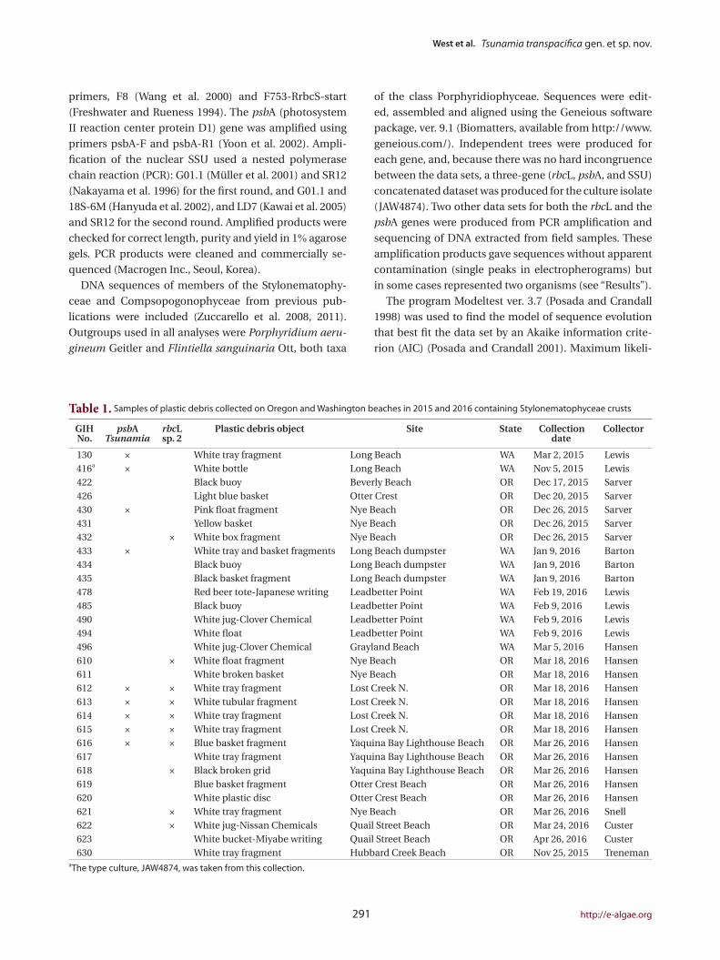
Table 1. Samples of plastic debris collected on Oregon and Washington beaches in 2015 and 2016 containing Stylonematophyceae crusts
GIHNo.
psbATsunamia
rbcLsp. 2
Plastic debris object Site State Collection date
Collector
130 × White tray fragment Long Beach WA Mar 2, 2015 Lewis 416a × White bottle Long Beach WA Nov 5, 2015 Lewis422 Black buoy Beverly Beach OR Dec 17, 2015 Sarver426 Light blue basket Otter Crest OR Dec 20, 2015 Sarver430 × Pink float fragment Nye Beach OR Dec 26, 2015 Sarver431 Yellow basket Nye Beach OR Dec 26, 2015 Sarver432 × White box fragment Nye Beach OR Dec 26, 2015 Sarver433 × White tray and basket fragments Long Beach dumpster WA Jan 9, 2016 Barton434 Black buoy Long Beach dumpster WA Jan 9, 2016 Barton435 Black basket fragment Long Beach dumpster WA Jan 9, 2016 Barton478 Red beer tote-Japanese writing Leadbetter Point WA Feb 19, 2016 Lewis485 Black buoy Leadbetter Point WA Feb 9, 2016 Lewis490 White jug-Clover Chemical Leadbetter Point WA Feb 9, 2016 Lewis494 White float Leadbetter Point WA Feb 9, 2016 Lewis496 White jug-Clover Chemical Grayland Beach WA Mar 5, 2016 Hansen610 × White float fragment Nye Beach OR Mar 18, 2016 Hansen611 White broken basket Nye Beach OR Mar 18, 2016 Hansen612 × × White tray fragment Lost Creek N. OR Mar 18, 2016 Hansen613 × × White tubular fragment Lost Creek N. OR Mar 18, 2016 Hansen614 × × White tray fragment Lost Creek N. OR Mar 18, 2016 Hansen615 × × White tray fragment Lost Creek N. OR Mar 18, 2016 Hansen616 × × Blue basket fragment Yaquina Bay Lighthouse Beach OR Mar 26, 2016 Hansen617 White tray fragment Yaquina Bay Lighthouse Beach OR Mar 26, 2016 Hansen618 × Black broken grid Yaquina Bay Lighthouse Beach OR Mar 26, 2016 Hansen619 Blue basket fragment Otter Crest Beach OR Mar 26, 2016 Hansen620 White plastic disc Otter Crest Beach OR Mar 26, 2016 Hansen621 × White tray fragment Nye Beach OR Mar 26, 2016 Snell622 × White jug-Nissan Chemicals Quail Street Beach OR Mar 24, 2016 Custer623 White bucket-Miyabe writing Quail Street Beach OR Apr 26, 2016 Custer630 White tray fragment Hubbard Creek Beach OR Nov 25, 2015 Treneman
aThe type culture, JAW4874, was taken from this collection.

Algae 2016, 31(4): 289-301
https://doi.org/10.4490/algae.2016.31.10.20 292
Fig. 1. Tsunamia transpacifica (JAW4874). (A) Drift white polyethylene bottle from which the original specimens were scraped for culture. (B) Two-month-old crusts on glass cover slip showing typical radiating horizontally branched filaments with a dark pulvinate central mass of erect branches. Free spores and young sporelings visible around the crusts. (C) Most filaments show transverse divisions and one cell shows longitudinal division (double arrowhead). Enlarged cells (single arrowhead) with nucleus in the cell center. (D) Cell center with nucleus surrounded by particles and supported by thin radiating cytoplasmic strands. (E) Surface view of plastids with multiple lobes interconnected by wide and narrow strands. (F) Black starch grains (arrowheads) around nucleus of most cells treated with I2KI stain. (G) Central cell mass with cell divisions in different planes. Scale bars represent: A, 3 cm; B, 100 µm; C-G, 10 µm.
A
C
D
B
E
G
F

West et al. Tsunamia transpacifica gen. et sp. nov.
293 http://e-algae.org
Culture observations
The live specimen in collection number 416 (culture
number JAW4874) was placed in culture on glass cover
slips. In two months, 0.3-1.0 mm pulvinate crusts were
produced by radiating filaments with many lateral and
vertical branches (Fig 1B). Each vegetative cell had one
spherical central nucleus about 3 µm in diameter, sus-
pended on thin cytoplasmic strands radiating outward
across the central vacuole to the peripheral cytoplasm.
Small granules around the nucleus and in the strands
(Fig. 1C, D & G) stained black with I2KI, a typical starch re-
action (Fig. 1F, arrowheads). Some cells were not stained.
The single parietal plastid had many variously shaped
lobes interconnected by thin to wide strands (Fig. 1E). No
pyrenoid was visible.
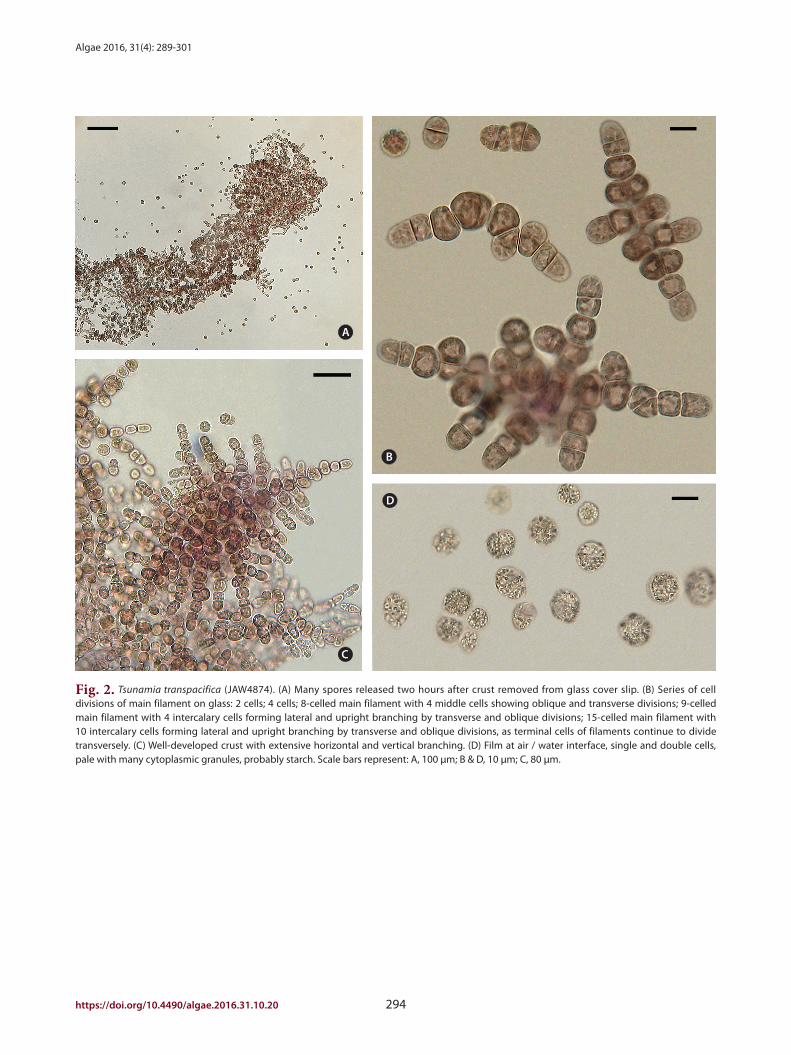
Crusts scraped from the substrate and transferred to
fresh medium released numerous spores within 2 h (Fig.
2A). Vegetative cells in dense pulvinate masses formed
abundant spores via numerous oblique and longitudinal
divisions (Fig. 1B & G). No pit connections were visible
between any cells. Spores were 10 (9-12) µm diam, with a
central nucleus and a single, lobed parietal plastid. Free
spores showed no motility and usually adhered to the sub-
strate less than 12 h after release. Attached spores formed
2-celled (about 10 × 20 µm) and 4-celled (about 10 × 30-
40 µm) stages by apical transverse divisions (Fig. 2B). In
the horizontal young filaments 8-9 cells long (about 10-12
× 65-75 µm), mid-section intercalary cells initiated lon-
gitudinal and oblique divisions to form lateral branches
(Fig. 2B). The apical cells of filaments continued to divide
transversely and filaments radiated horizontally outward.
In the middle section of the main axial filament and in
lateral branches, longitudinal and oblique cell divisions
produced erect branches (Fig. 2C) resulting in compact
three-dimensional crusts up to 1 mm wide and many
cells high.
In stationary cultures with bright light (12-30 µmol
photons m-2 s-1), spores sometimes rose to the air / water
interface, forming floating films up to 2 cm wide and one
or more cells thick. In these films spores divided, produc-
ing separate single cells and 2-celled filaments (Fig. 2D).
The cells were pale, with reduced photosynthetic pig-
ments, and contained many large cytoplasmic granules,
probably starch, a typical response to nutrient deficiency
in red algae.
hood (ML) was performed with RAxML 7.2.8 (Stamatakis
2006). A saturation plot of the rbcL dataset, and not psbA,
indicated that the third codon position for rbcL was satu-
rated for this gene, therefore the third codon position was
removed in the rbcL analysis. Data sets were partitioned
by gene and codon, where appropriate. RAxML was per-
formed with partitioned data and the GTR + gamma
model and 1,000 non-parametric bootstrap replicates
(Felsenstein 1985). Bayesian inference was performed
with MrBayes v3.2 (Ronquist et al. 2012). Analyses con-
sisted of two independent simultaneous runs of one cold
and three incrementally heated chains, and 3 × 10⁶ gen-
erations with sampling every 1,000 generations. The log
files of the runs were checked with Tracer v1.5 (Rambaut
and Drummond 2009) and a burn-in sample of 100 trees
was removed from each run before calculating the major-
ity rule consensus tree.
RESULTS
Field observations
Specimens of crusts were collected from plastic debris
items (Table 1) cast ashore on Oregon and Washington
beaches during the winter and spring of 2015 and 2016,
nearly 5 y after the Japanese tsunami. Several of the de-
bris items bore Japanese writing that indicated their ori-
gin was the Tohoku coast of Japan. Nearly all of the debris
supporting the algae was light in colour and determined
to be HDPE, a plastic that typically floats at the waterline.
The items included small pieces of floats (12-cm in diam-
eter), larger bottles (Fig. 1A), and large totes (160 cm or
more in length). The red algal crusts were typically part
of a small community of species that inhabited the plas-
tic including diatoms, an Ulvella-like green alga, several
hydroids, and Lepas anatifera Linnaeus 1758, the pelagic
gooseneck barnacle.
Microscopic examination of field crusts revealed fea-
tures characteristic of the red algal class Stylonematophy-
ceae. The crusts were palmelloid with numerous single
subglobose cells 8-10 µm in diameter, short filaments and
small packets of cells enclosed in a clear mucilaginous
sheath up to 500 µm thick. The chloroplasts lacked pyre-
noids and appeared to be discoid or highly lobed. When
debris items with crusts were stored in a dark cold room
(5°C) for up to 6 months, the alga remained alive, retain-
ing its colour and structural characteristics.
Algae 2016, 31(4): 289-301
https://doi.org/10.4490/algae.2016.31.10.20 294
Fig. 2. Tsunamia transpacifica (JAW4874). (A) Many spores released two hours after crust removed from glass cover slip. (B) Series of cell divisions of main filament on glass: 2 cells; 4 cells; 8-celled main filament with 4 middle cells showing oblique and transverse divisions; 9-celled main filament with 4 intercalary cells forming lateral and upright branching by transverse and oblique divisions; 15-celled main filament with 10 intercalary cells forming lateral and upright branching by transverse and oblique divisions, as terminal cells of filaments continue to divide transversely. (C) Well-developed crust with extensive horizontal and vertical branching. (D) Film at air / water interface, single and double cells, pale with many cytoplasmic granules, probably starch. Scale bars represent: A, 100 µm; B & D, 10 µm; C, 80 µm.
A
C
D
B
West et al. Tsunamia transpacifica gen. et sp. nov.
295 http://e-algae.org
in the Stylonematophyceae.
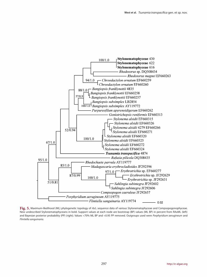
Amplification of rbcL from field samples (10 samples,
dataset of first and second positions, 810 bp) amplified
a different species of Stylonematophyceae (Fig. 5). While
the position of these sequences within the Stylonemato-
phyceae was not well supported, it is clear that they are
different from T. transpacifica and are not similar to any
other sequences available. This new entity was fairly com-
mon on plastics. In some DNA extracts (5 field samples),
both entities were recovered, one using psbA primers (T.
transpacifica) and the other using rbcL primers (Stylon-
ematophyceae). Because we did not notice any morpho-
logically distinct specimens from field-collected samples,
we suspect that this species is probably morphologically
similar to T. transpacifica, but until it is placed in unialgal
culture, this cannot be determined. The alga described in
the field observations above may be either a new species
or something different, in view of the various molecular
results.
Molecular analysis
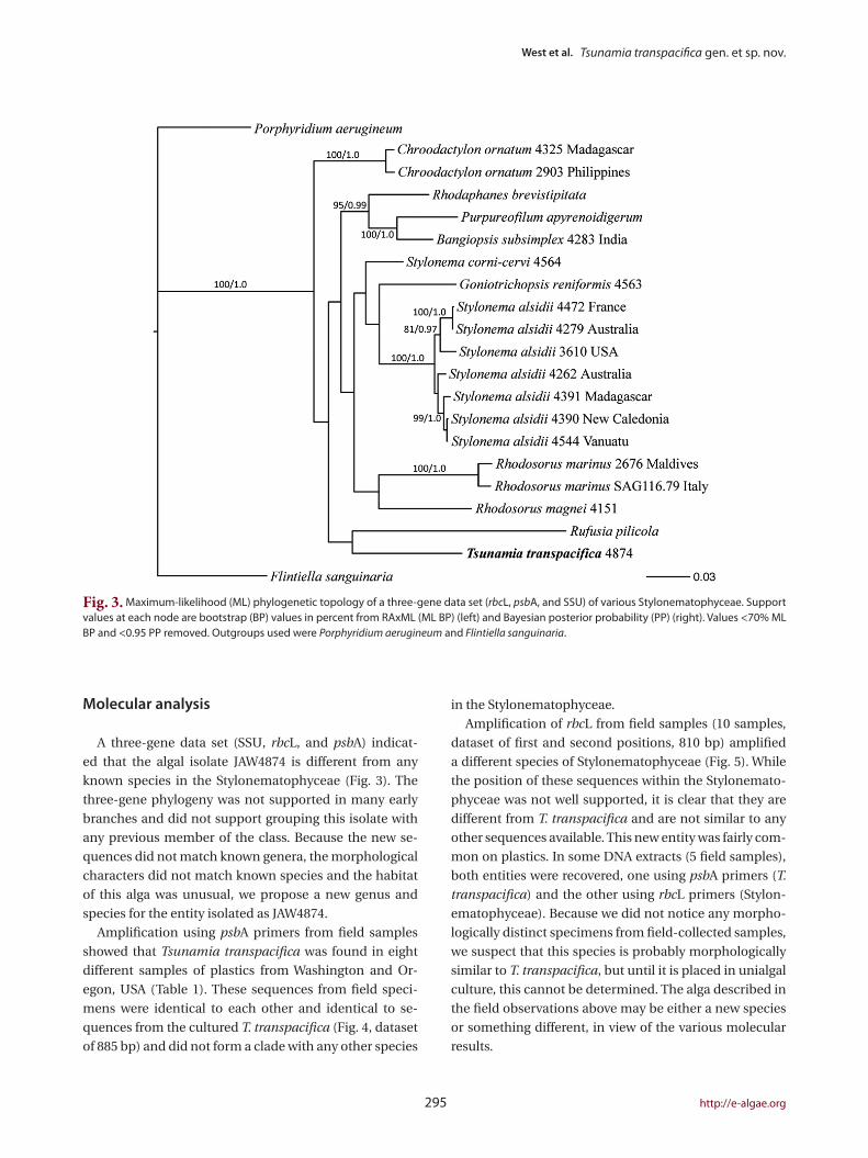
A three-gene data set (SSU, rbcL, and psbA) indicat-
ed that the algal isolate JAW4874 is different from any
known species in the Stylonematophyceae (Fig. 3). The
three-gene phylogeny was not supported in many early
branches and did not support grouping this isolate with
any previous member of the class. Because the new se-
quences did not match known genera, the morphological
characters did not match known species and the habitat
of this alga was unusual, we propose a new genus and
species for the entity isolated as JAW4874.
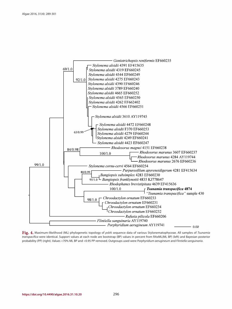
Amplification using psbA primers from field samples
showed that Tsunamia transpacifica was found in eight
different samples of plastics from Washington and Or-
egon, USA (Table 1). These sequences from field speci-
mens were identical to each other and identical to se-
quences from the cultured T. transpacifica (Fig. 4, dataset
of 885 bp) and did not form a clade with any other species
Fig. 3. Maximum-likelihood (ML) phylogenetic topology of a three-gene data set (rbcL, psbA, and SSU) of various Stylonematophyceae. Support values at each node are bootstrap (BP) values in percent from RAxML (ML BP) (left) and Bayesian posterior probability (PP) (right). Values <70% ML BP and <0.95 PP removed. Outgroups used were Porphyridium aerugineum and Flintiella sanguinaria.
Algae 2016, 31(4): 289-301
https://doi.org/10.4490/algae.2016.31.10.20 296
Fig. 4. Maximum-likelihood (ML) phylogenetic topology of psbA sequence data of various Stylonematophyceae. All samples of Tsunamia transpacifica were identical. Support values at each node are bootstrap (BP) values in percent from RAxML(ML BP) (left) and Bayesian posterior probability (PP) (right). Values <70% ML BP and <0.95 PP removed. Outgroups used were Porphyridium aerugineum and Flintiella sanguinaria.
West et al. Tsunamia transpacifica gen. et sp. nov.
297 http://e-algae.org
Fig. 5. Maximum-likelihood (ML) phylogenetic topology of rbcL sequence data of various Stylonematophyceae and Compsopogonophyceae. New undescribed Stylonematophyceans in bold. Support values at each node are bootstrap (BP) values (ML BP) in percent from RAxML (left) and Bayesian posterior probability (PP) (right). Values <70% ML BP and <0.95 PP removed. Outgroups used were Porphyridium aerugineum and Flintiella sanguinaria.

Algae 2016, 31(4): 289-301
https://doi.org/10.4490/algae.2016.31.10.20 298
DISCUSSION
The discovery and description of this species was fa-
cilitated by several events and discoveries of recent years.
The Japanese 2011 tsunami produced large quantities
of marine debris that floated across the North Pacific to
wash ashore in Oregon and Washington where material
could easily be collected. A portion of the debris con-
sisted of HDPE, a plastic widely used in various products.
Since this plastic floats at the waterline, it provided a good
substrate and habitat for light-requiring species in the
Stylonematophyceae. Recent studies on the life history
and molecular biology of the Stylonematophyceae (West
et al. 2005, 2007, 2014) gave us the necessary background
and framework for our recognition of the species as new.
The simple morphology with few diagnostic charac-
ters of the Stylonematophyceae makes specimen identi-
fication difficult without molecular evidence. Definitive
identification also requires isolation into unialgal culture
combined with cytological and chemical observations.
Tsunamia is superficially similar to the endozoic Stylo-
nematophycean Neevea repens Batters which has hori-
zontal palmelloid or pseudo-filamentous thalli and sev-
eral small discoid plastids per cell (Garbary et al. 1980b)
unlike the single multilobed plastid of Tsunamia. Neevea
has never been cultured nor is molecular data available
for this genus.
Without molecular evidence, Tsunamia might have
been identified as a member of the Erythropeltidales
(Compsopogonophyceae) (e.g., Pseudoerythrocladia),
because of its horizontal pulvinate thallus with multi-
branched radiating filaments, although spores in the
Erythropeltidales form by oblique divisions of vegetative
cells (Garbary et al. 1980a, Zuccarello et al. 2010, West et
al. 2012). rather than by oblique and longitudinal divi-
sions, as in Tsunamia. Furthermore, in contrast to spores
of other Stylonematophyceae observed in culture (Pick-
ett-Heaps et al. 2001, Wilson et al. 2002, West et al. 2005,
2007, 2012, 2014), neither gliding nor amoeboid motility
was observed in Tsunamia spores.
Our initial surveys of plastic debris indicate that mem-
bers of the red algal class Stylonematophyceae thrive on
floating plastics (Table 1). With the increasingly wide-
spread use of both polyethylene and polypropylene in a
wide array of items that often end up in debris, the new
“plastisphere” environment in the world’s oceans (Zettler
et al. 2013, Samoray 2016) continues to increase in size,
providing ample habitat for species that thrive on plas-
tic. The Stylonematophyceae asexual life cycle and high
spore production aid in the maintenance of their popu-
Tsunamia gen. nov. J. A. West, G. I. Hansen, G. C. Zuccarello et T. Hanyuda
Description. In culture, vegetative cells are directly
released as single non-motile spores (9-12 µm diam.) at-
taching to a glass substrate and germinating to form radi-
ating, branched uniseriate filaments that become dense
pulvinate crusts up to 1mm wide that are often confluent.
Horizontally radiating filaments are formed by apical cells
undergoing transverse divisions, followed by longitudinal
and oblique divisions of intercalary cells to form numer-
ous horizontal and erect branched filaments. No pit con-
nections form between cells. Each cell has one spherical
central nucleus about 3 µm diameter, suspended on thin
cytoplasmic strands radiating outward across the central
vacuole and a single, peripheral, purple to pink, much-
lobed plastid without a pyrenoid. Identity was also based
on the basis of SSU, rbcL, and psbA sequences.
Tsunamia transpacifica sp. nov. J. A. West, G. I. Hansen, G. C. Zuccarello et T. Hanyuda
Description. Species description as for genus.
Holotype. The holotype culture specimen was depos-
ited in the Royal Botanic Gardens Melbourne, National
Herbarium of Victoria, Australia (MEL 2399992). Strain
JAW4874 (field collection number 416) was from a white
plastic bottle found in debris of the Mar 11, 2011 Tohoku
earthquake and tsunami, obtained on Nov 5, 2015 on
the Long Beach Peninsula, Washington, USA (46°37.385
N, 124°04.246 W) by Russ Lewis (Table 1). Isotype cul-
ture specimens were also deposited in the University of
Michigan Herbarium (MICH 701751) and University Her-
barium, University of California, Berkeley (UC 2050560).
It was not possible to designate a single alga in the field
collection as the type. Consequently, a single thallus was
reisolated from the laboratory culture in Melbourne and
selected as the holotype and isotypes.
Culture deposit. The culture (JAW4874) has been ac-
cessioned (CCMP 3460) by the Provasoli-Guillard Na-
tional Center for Marine Algae and Microbiota (NCMA),
Bigelow Laboratory for Ocean Sciences, P.O. Box 380, 60
Bigelow Drive, East Boothbay, ME 04544, USA.
Etymology. The name reflects the origin of the type
specimen, obtained from a plastic bottle transported in
tsunami drift across the Pacific Ocean to the west coast of
North America.
Genbank accession numbers. SSU, KX787935; psbA,
KX787937; rbcL, KX787936.
West et al. Tsunamia transpacifica gen. et sp. nov.
299 http://e-algae.org
ACKNOWLEDGEMENTS
We thank Russ Lewis and John Chapman who made
the initial collections of the debris objects. The Japanese
Ministry of the Environment through the North Pacific
Marine Science Organization (PICES) provided partial
support for GIH & TH during the current study. The US
Environmental Protection Agency provided laboratory
space for GIH during the study. JW used personal funds
for culturing and light microscopy and is grateful to Geoff
McFadden, University of Melbourne, for use of facilities
and supplies in doing this research. Susan Loiseaux de
Goër provided critical Photoshop advice for arranging
and improving the photographic figures. For accession-
ing Tsunamia herbarium specimens, we thank Nimal
Karunajeewa and Pina Milne, the Royal Botanic Gardens,
Melbourne; Michael Wynne, University of Michigan; and
Kathy Ann Miller, University of California at Berkeley. Ju-
lie Sexton accessioned the culture at CCMP, Bigelow Lab-
oratory for Ocean Sciences, Maine.
REFERENCES
Amsler, C. D. & Searles, R. B. 1980. Vertical distribution of sea-
weed spores in a water column offshore of North Caro-
lina. J. Phycol. 16:617-619.
Andersen, R. A. 2005. Algal culturing techniques. Elsevier,
Amsterdam, 578 pp.
Andrady, A. L. 2011. Microplastics in the marine environ-
ment. Mar. Pollut. Bull. 62:1596-1605.
Barnes, D. K. A., Galgani, F., Thompson, R. C. & Barlaz, M.
2009. Accumulation and fragmentation of plastic debris
in global environments. Philos Trans. R. Soc. Lond. B
Biol. Sci. 364:1985-1998.
Bravo, M., Astudillo, J. C., Lancellotti, D., Luna-Jorquera, G.,
Valdivia, N. & Thiel, M. 2011. Rafting on abiotic substra-
ta: properties of floating items and their influence on
community succession. Mar. Ecol. Prog. Ser. 439:1-17.
Carlton, J., Chapman, J., Geller, J., Miller, J., Ruiz, G., Carlton,
D. & McCuller, M. 2016. The invasion process model and
the long-distance transoceanic dispersal of coastal ma-
rine organisms by Japanese tsunami marine debris. In
9th International Conference on Marine BioInvasions,
Sydney, Australia, p. 15.
Carson, H. S., Nerheim, M. S., Carroll, K. A. & Eriksen, M.
2013. The plastic-associated microorganisms of the
North Pacific Gyre. Mar. Pollut. Bull. 75:126-132.
Concelman, S., Bollens, S. M., Sullivan, B. K., Madin, L. P.,
Horgan, E., Butler, M. & van Keuren, D. 2001. Distribu-
lations, and their floating plastic substrates disperse the
populations widely via the prevailing currents. The pink
crusts on plastic debris included two different members
of the Stylonematophyceae. One of these, Tsunamia
transpacifica, was isolated in culture and characterised as
a new genus, while the other is only known as a DNA se-
quence. It is likely that additional cryptic molecular spe-
cies will be found in this habitat with future studies.
Where did the populations of these two species on
plastic debris originate? Did they exist in Japan on ma-
rine structures or in near-shore areas before the tsunami,
colonizing debris, and drifting across the Pacific to North
America? Did they attach to the plastic in the open ocean?
Or did they attach to the debris in the near-shore regions
of North America? It is difficult to say because the spe-
cies have not been observed previously in either Japan or
North America. On plastic debris, they co-exist with pop-
ulations of species that are known to be pelagic, includ-
ing hydroids and Lepas anatifera. It is possible that the
species either attached to the debris via pelagic spores in
the water column (Amsler and Searles 1980) or that they
hitch-hiked on pelagic hydroids known to drift in the wa-
ter column (Concelman et al. 2001). Once on the plastic
debris, they could survive for long periods of time, propa-
gating repeatedly and jumping between objects that are
in close proximity, such as those that might occur in the
North Pacific garbage patch. Where did Tsunamia occur
before the development of plastic? It may have been a pe-
lagic species that became an opportunistic primary colo-
nizer of the new “plastisphere” in the North Pacific.
It is intriguing that both T. transpacifica and the un-
identified Stylonematophycean were found with differ-
ent sets of primers. The only explanation is variation in
the primer binding sites for these two species, with higher
specificity in T. transpacifica for the psbA primer combi-
nation, while the unidentified Stylonematophyceans
seem to be more readily amplified with the rbcL primer
pairs. Continued study and tests of alternative primers
will help to determine the distribution and relative abun-
dance of these two species on plastic debris.
This investigation highlights our lack of knowledge of
the algal diversity found on this ubiquitous floating ocean
debris, with potential effects on native algal floras. Con-
tinued study of floating algae, possibly on plastics from
the central Pacific gyre or from attached plastics in Japan,
may determine if these algae have a Japanese origin or if
they are native to pelagic substrates.

Algae 2016, 31(4): 289-301
https://doi.org/10.4490/algae.2016.31.10.20 300
Japanese Ministry of the Environment. 2012. Estimated to-
tal amount of debris washed out by the Great East Ja-
pan Earthquake. Available from: https://www.env.go.jp/
en/focus/docs/files/20120901-57.pdf. Accessed Mar 1,
2015.
Johansen, D. A. 1940. Plant microtechnique. McGraw Hill, NY,
523 pp.
Kawai, H., Sasaki, H., Maeba, S. & Henry, E. C. 2005. Mor-
phology and molecular phylogeny of Phaeostrophion
irregulare (Phaeophyceae) with proposal of Phaeostro-
phiaceae fam. nov., and review of Ishigeaceae. Phycolo-
gia 44:169-182.
Law, K. L. & Thompson, R. C. 2014. Oceans: microplastics in
the seas. Science 345:144-145.
Müller, K. M., Oliveira, M. C., Sheath, R. G. & Bhattacharya, D.
2001. Ribosomal DNA phylogeny of the Bangiophycidae
(Rhodophyta) and the origin of secondary plastids. Am.
J. Bot. 88:1390-1400.
Nakayama, T., Watanabe, S., Mitsui, K., Uchida, H. & Inouye,
I. 1996. The phylogenetic relationship between the
Chlamydomonadales and Chlorococcales inferred from
18S rDNA sequence data. Phycol. Res. 44:47-55.
Pickett-Heaps, J. D., West, J. A., Wilson, S. M. & McBride, D. L.
2001. Time-lapse videomicroscopy of cell (spore) move-
ment in red algae. Eur. J. Phycol. 36:9-22.
Posada, D. & Crandall, K. A. 1998. Modeltest: testing the
model of DNA substitution. Bioinformatics 14:817-818.
Posada, D. & Crandall, K. A. 2001. Selecting the best-fit model
of nucleotide substitution. Syst. Biol. 50:580-601.
Rambaut, A. & Drummond, A. J. 2009. Tracer v1.5. Available
from: http://beast.bio.ed.ac.uk/tracer. Accessed Mar 1,
2015.
Ronquist, F., Teslenko, M., van der Mark, P., Ayres, D. L., Dar-
ling, A., Höhna, S., Larget, B., Liu, L., Suchard, M. A. &
Huelsenbeck, J. P. 2012. MrBayes 3.2: efficient Bayesian
phylogenetic inference and model choice across a large
model space. Syst. Biol. 61:539-542.
Samoray, C. 2016. Ocean’s plastics offer a floating fortress
to a mess of microbes: the plastisphere is potentially
causing changes in ocean environments. ScienceNews
189:20-22.
Scott, J. L., Orlova, E. & West, J. A. 2010. Ultrastructural ob-
servations of vegetative cells of two new genera in the
Erythropeltidales (Compsopogonophyceae, Rhodophy-
ta): Pseudoerythrocladia and Madagascaria. Algae
25:11-15.
Stamatakis, A. 2006. RAxML-VI-HPC: maximum likelihood-
based phylogenetic analyses with thousands of taxa and
mixed models. Bioinformatics 22:2688-2690.
Wang, H. W., Kawaguchi, S., Horiguchi, T. & Masuda, M. 2000.
tion, abundance and benthic-pelagic coupling of sus-
pended hydroids on Georges Bank. Deep Sea Res. Part II
Top. Stud. Oceanogr. 48:645-658.
Cumming, R. A., Nikula, R., Spencer, H. G. & Waters, J. M.
2014. Transoceanic genetic similarities of kelp-associat-
ed sea slug populations: long-distance dispersal via raft-
ing? J. Biogeogr. 41:2357-2370.
Derraik, J. G. B. 2002. The pollution of the marine envi-
ronment by plastic debris: a review. Mar. Pollut. Bull.
44:842-852.
Eriksen, M., Lebreton, L. C. M., Carson, H. S., Thiel, M.,
Moore, C. J., Borerro, J. C., Galgani, F., Ryan, P. G. & Reiss-
er, J. 2014. Plastic pollution in the world’s oceans: more
than 5 trillion plastic pieces weighing over 250,000 tons
afloat at sea. PLoS One 9:e111913.
Felsenstein, J. 1985. Confidence limits on phylogenies: an
approach using the bootstrap. Evolution 39:783-791.
Flint, L. H. 1953. Kyliniella in America. Phytomorphology
3:76-80.
Fraser, C. I., Nikula, R. & Waters, J. M. 2011. Oceanic rafting
by a coastal community. Proc. Royal Soc. B Biol. Sci.
278:649-655.
Freshwater, D. W. & Rueness, J. 1994. Phylogenetic relation-
ships of some European Gelidium (Gelidiales, Rho-
dophyta) species, based on rbcL nucleotide sequence
analysis. Phycologia 33:187-194.
Garbary, D. J., Hansen, G. I. & Scagel, R. F. 1980a. A revised
classification of the Bangiophyceae (Rhodophyta). Nova
Hedwigia 33:145-166.
Garbary, D. J., Hansen, G. I. & Scagel, R. F. 1980b. The marine
algae of British Columbia and Northern Washington:
division Rhodophyta (red algae), class Bangiophyceae.
Syesis 13:137-195.
Goldstein, M. C., Titmus, A. J. & Ford, M. 2013. Scales of spa-
tial heterogeneity of plastic marine debris in the North-
east Pacific Ocean. PLoS One 8:e80020.
Guiry, M. D. & Guiry, G. M. 2016. AlgaeBase. World-wide
electronic publication, National University of Ireland,
Galway. Available from: http://www.algaebase.org. Ac-
cessed Mar 1, 2015.
Hansen, G. I., Hanyuda, T. & Kawai, H. 2016. Marine algae
arriving on Japanese tsunami marine debris (JTMD) and
their invasion threat to the coasts of Oregon and Wash-
ington, USA. In 9th International Conference on Marine
BioInvasions, Sydney, Australia, p. 43.
Hanyuda, T., Wakana, I., Arai, S., Miyaji, K., Watano, Y. & Ueda,
K. 2002. Phylogenetic relationships within Cladopho-
rales (Ulvophyceae, Chlorophyta) inferred from 18S
rRNA gene sequences, with special reference to Aegag-
ropila linnaei. J. Phycol. 38:564-571.

West et al. Tsunamia transpacifica gen. et sp. nov.
301 http://e-algae.org
Yoon, H. S., Hackett, J. D., Pinto, G. & Bhattacharya, D. 2002.
The single, ancient origin of chromist plastids. Proc.
Natl. Acad. Sci. U. S. A. 99:15507-15512.
Yoon, H. S., Zuccarello, G. C. & Bhattacharya, D. 2010. Evolu-
tionary history and taxonomy of red algae. In Seckbach,
J. & Chapman, D. J. (Eds.) Red Algae in the Genomic Age.
Springer, NY, pp. 27-42.
Zettler, E. R., Mincer, T. J. & Amaral-Zettler, L. A. 2013. Life
in the “Plastisphere”: microbial communities on plastic
marine debris. Environ. Sci. Technol. 47:7137-7146.
Zuccarello, G. C., Kikuchi, N. & West, J. A. 2010. Molecular
phylogeny of the crustose Erythropeltidales (Compso-
pogonophyceae, Rhodophyta): new genera Pseudo-
erythrocladia and Madagascaria and the evolution of
the upright habit. J. Phycol. 46:363-373.
Zuccarello, G. C. & Lokhorst, G. M. 2005. Molecular phylog-
eny of the genus Tribonema (Xanthophyceae) using rbcL
gene sequence data: monophyly of morphologically
simple algal species. Phycologia 44:384-392.
Zuccarello, G. C., West, J. A. & Kikuchi, N. 2008. Phylogenetic
relationships within the Stylonematales (Stylonemato-
phyceae, Rhodophyta): biogeographic patterns do not
apply to Stylonema alsidii. J. Phycol. 44:384-393.
Zuccarello, G. C., Yoon, H. S., Kim, H., Sun, L., de Goër, S. L.
& West, J. A. 2011. Molecular phylogeny of the upright
Erythropeltidales (Compsopogonophyceae, Rhodophy-
ta): multiple cryptic lineages of Erythrotrichia carnea. J.
Phycol. 47:627-637.
Reinstatement of Grateloupia catenata (Rhodophyta,
Halymeniaceae) on the basis of morphology and rbcL
sequences. Phycologia 39:228-237.
West, J. A., de Goër, S. L. & Zuccarello, G. C. 2014. A new spe-
cies of Bangiopsis: B. franklynottii sp. nov. (Stylonema-
tophyceae, Rhodophyta) from Australia and India and
comments on the genus. Algae 29:101-109.
West, J. A., Loiseaux de Goër, S. & Zuccarello, G. C. 2012. Up-
right Erythropeltidales (Rhodophyta) in Brittany, France
and description of a new species, Erythrotrichia longis-
tipitata. Cah. Biol. Mar. 53:255-270.
West, J. A. & McBride, D. L. 1999. Long-term and diurnal car-
pospore discharge patterns in the Ceramiaceae, Rho-
domelaceae and Delesseriaceae (Rhodophyta). Hydro-
biologia 398/399:101-113.
West, J. A., Zuccarello, G. C., Scott, J., Pickett-Heaps, J. & Kim,
G. H. 2005. Observations on Purpureofilum apyrenoi-
digerum gen. et sp. nov. from Australia and Bangiopsis
subsimplex from India (Stylonematales, Bangiophyceae,
Rhodophyta). Phycol. Res. 53:49-66.
West, J. A., Zuccarello, G. C., Scott, J. L., West, K. A. & Karsten,
U. 2007. Rhodaphanes brevistipitata gen. et sp. nov., a
new member of the Stylonematophyceae (Rhodophyta).
Phycologia 46:440-449.
Wilson, S., West, J., Pickett-Heaps, J., Yokoyama, A. & Hara, Y.
2002. Chloroplast rotation and morphological plasticity
of the unicellular alga Rhodosorus (Rhodophyta, Stylon-
ematales). Phycol. Res. 50:183-191.