Flecainide exerts paradoxical effects on sodium currents and atrial ...
15
Flecainide exerts paradoxical effects on sodium currents and atrial arrhythmia in murine RyR2-P2328S hearts S. C. Salvage, 1, * J. H. King, 1, * K. H. Chandrasekharan, 1 D. I. G. Jafferji, 1 L. Guzadhur, 2 H. R. Matthews, 1 C. L.-H. Huang 1,2 and J. A. Fraser 1 1 Physiological Laboratory, University of Cambridge, Cambridge, UK 2 Department of Biochemistry, University of Cambridge, Cambridge, UK Received 3 October 2014, revision requested 3 November 2014, revision received 27 March 2015, accepted 1 April 2015 Correspondence: J. A. Fraser, Physiological Laboratory, University of Cambridge, Downing Street, Cambridge, CB2 3EG, UK. E-mail: [email protected] *Joint first authors See Editorial Commentary: Curran, J. & Louch, W. E. Linking ryanodine receptor Ca 2+ leak and Na + current in heart: a day in the life of flecainide. Acta Physiol 214, 300–302. Abstract Aims: Cardiac ryanodine receptor mutations are associated with catechol- aminergic polymorphic ventricular tachycardia (CPVT), and some, includ- ing RyR2-P2328S, also predispose to atrial fibrillation. Recent work associates reduced atrial Na v 1.5 currents in homozygous RyR2-P2328S (RyR2 S/S ) mice with slowed conduction and increased arrhythmogenicity. Yet clinically, and in murine models, the Na v 1.5 blocker flecainide reduces ventricular arrhythmogenicity in CPVT. We aimed to determine whether, and how, flecainide influences atrial arrhythmogenicity in RyR2 S/S mice and their wild-type (WT) littermates. Methods: We explored effects of 1 lM flecainide on WT and RyR2 S/S atria. Arrhythmic incidence, action potential (AP) conduction velocity (CV), atrial effective refractory period (AERP) and AP wavelength (k = CV 9 AERP) were measured using multi-electrode array recordings in Langendorff-perfused hearts; Na + currents (I Na ) were recorded using loose patch clamping of superfused atria. Results: RyR2 S/S showed more frequent atrial arrhythmias, slower CV, reduced I Na and unchanged AERP compared to WT. Flecainide was anti- arrhythmic in RyR2 S/S but pro-arrhythmic in WT. It increased I Na in RyR2 S/S atria, whereas it reduced I Na as expected in WT. It increased AERP while sparing CV in RyR2 S/S , but reduced CV while sparing AERP in WT. Thus, RyR2 S/S hearts have low k relative to WT; flecainide then increases k in RyR2 S/S but decreases k in WT. Conclusions: Flecainide (1 lM) rescues the RyR2-P2328S atrial arrhythmo- genic phenotype by restoring compromised I Na and k, changes recently attrib- uted to increased sarcoplasmic reticular Ca 2+ release. This contrasts with the increased arrhythmic incidence and reduced I Na and k with flecainide in WT. Keywords atrial arrhythmia, conduction velocity, CPVT, flecainide, Na + currents, ryanodine receptor. Atrial fibrillation (AF) is the most common sustained arrhythmia, predisposing to significant clinical mor- bidity and mortality (Benjamin et al. 1998, Stewart et al. 2002, Davis et al. 2012), yet its physiological mechanisms are incompletely understood. Neverthe- less, acute atrial arrhythmogenesis may be related not only to cellular Ca 2+ homeostasis but also to altered action potential (AP) conduction and recovery (Zhang et al. 2011, King et al. 2013c). Abnormal Ca 2+ release can arise from cardiac ryanodine receptor-2 (RyR2) mutations or a loss of calsequestrin-2 (CSQ2) (Priori & Chen 2011), poten- tially providing arrhythmic triggers (Mackenzie et al. 2001, 2004, Bootman et al. 2006, Zhang et al. 2010), © 2015 The Authors. Acta Physiologica published by John Wiley & Sons Ltd on behalf of Scandinavian Physiological Society, doi: 10.1111/apha.12505 361 This is an open access article under the terms of the Creative Commons Attribution License, which permits use, distribution and reproduction in any medium, provided the original work is properly cited. Acta Physiol 2015, 214, 361–375
Transcript of Flecainide exerts paradoxical effects on sodium currents and atrial ...

Flecainide exerts paradoxical effects on sodium currents
and atrial arrhythmia in murine RyR2-P2328S hearts
S. C. Salvage,1,* J. H. King,1,* K. H. Chandrasekharan,1 D. I. G. Jafferji,1 L. Guzadhur,2
H. R. Matthews,1 C. L.-H. Huang1,2 and J. A. Fraser1
1 Physiological Laboratory, University of Cambridge, Cambridge, UK
2 Department of Biochemistry, University of Cambridge, Cambridge, UK
Received 3 October 2014,
revision requested 3 November
2014,
revision received 27 March 2015,
accepted 1 April 2015
Correspondence: J. A. Fraser,
Physiological Laboratory,
University of Cambridge,
Downing Street, Cambridge, CB2
3EG, UK.
E-mail: [email protected]
*Joint first authors
See Editorial Commentary:
Curran, J. & Louch, W. E. Linking
ryanodine receptor Ca2+ leak and
Na+ current in heart: a day in the
life of flecainide. Acta Physiol 214,
300–302.
Abstract
Aims: Cardiac ryanodine receptor mutations are associated with catechol-
aminergic polymorphic ventricular tachycardia (CPVT), and some, includ-
ing RyR2-P2328S, also predispose to atrial fibrillation. Recent work
associates reduced atrial Nav1.5 currents in homozygous RyR2-P2328S
(RyR2S/S) mice with slowed conduction and increased arrhythmogenicity.
Yet clinically, and in murine models, the Nav1.5 blocker flecainide reduces
ventricular arrhythmogenicity in CPVT. We aimed to determine whether,
and how, flecainide influences atrial arrhythmogenicity in RyR2S/S mice
and their wild-type (WT) littermates.Methods: We explored effects of 1 lM flecainide on WT and RyR2S/S
atria. Arrhythmic incidence, action potential (AP) conduction velocity
(CV), atrial effective refractory period (AERP) and AP wavelength
(k = CV 9 AERP) were measured using multi-electrode array recordings
in Langendorff-perfused hearts; Na+ currents (INa) were recorded using
loose patch clamping of superfused atria.Results: RyR2S/S showed more frequent atrial arrhythmias, slower CV,
reduced INa and unchanged AERP compared to WT. Flecainide was anti-
arrhythmic in RyR2S/S but pro-arrhythmic in WT. It increased INa in
RyR2S/S atria, whereas it reduced INa as expected in WT. It increased
AERP while sparing CV in RyR2S/S, but reduced CV while sparing AERP
in WT. Thus, RyR2S/S hearts have low k relative to WT; flecainide then
increases k in RyR2S/S but decreases k in WT.Conclusions: Flecainide (1 lM) rescues the RyR2-P2328S atrial arrhythmo-
genic phenotype by restoring compromised INa and k, changes recently attrib-
uted to increased sarcoplasmic reticular Ca2+ release. This contrasts with the
increased arrhythmic incidence and reduced INa and k with flecainide in WT.
Keywords atrial arrhythmia, conduction velocity, CPVT, flecainide, Na+
currents, ryanodine receptor.
Atrial fibrillation (AF) is the most common sustained
arrhythmia, predisposing to significant clinical mor-
bidity and mortality (Benjamin et al. 1998, Stewart
et al. 2002, Davis et al. 2012), yet its physiological
mechanisms are incompletely understood. Neverthe-
less, acute atrial arrhythmogenesis may be related not
only to cellular Ca2+ homeostasis but also to altered
action potential (AP) conduction and recovery (Zhang
et al. 2011, King et al. 2013c).
Abnormal Ca2+ release can arise from cardiac
ryanodine receptor-2 (RyR2) mutations or a loss of
calsequestrin-2 (CSQ2) (Priori & Chen 2011), poten-
tially providing arrhythmic triggers (Mackenzie et al.
2001, 2004, Bootman et al. 2006, Zhang et al. 2010),
© 2015 The Authors. Acta Physiologica published by John Wiley & Sons Ltd on behalf ofScandinavian Physiological Society, doi: 10.1111/apha.12505 361This is an open access article under the terms of the Creative Commons Attribution License,which permits use, distribution and reproduction in any medium, provided the original work is properly cited.
Acta Physiol 2015, 214, 361–375
thereby leading to catecholaminergic polymorphic
ventricular tachycardia (CPVT) (Priori & Chen 2011,
Zhang et al. 2013a). Certain RyR2 mutations are also
associated with AF (Bhuiyan et al. 2007, Sumitomo
et al. 2007). The RyR2-P2328S mutation is associated
with high incidences of both CPVT and atrial tachy-
cardia (AT), despite normal cardiac structure (Swan
et al. 1999, Laitinen et al. 2001). Murine hearts with
a homozygotic RyR2-P2328S (RyR2S/S) mutation
demonstrate both atrial and ventricular arrhythmic
tendencies (Goddard et al. 2008, Zhang et al. 2011,
2013b, King et al. 2013b,c) providing a useful experi-
mental model. Atrial RyR2S/S myocytes show diastolic
elevations in intracellular [Ca2+] attributed to
increased SR Ca2+ release (Zhang et al. 2011). This
would be expected to increase Na+/Ca2+ exchange
(NCX) activity, accounting for delayed afterdepolar-
izations (DADs) causing triggered activity, implicated
in the arrhythmic phenotype (King et al. 2013c).
It has recently been reported that flecainide exerts
anti-arrhythmic effects in human CPVT (Watanabe
et al. 2009, van der Werf et al. 2011). Flecainide
reduced bigeminy and biventricular tachycardia, ECG
features associated with human CPVT, in murine
CSQ2�/� hearts. However, there is debate over the
anti-arrhythmic mechanism of flecainide in CPVT. It
has been suggested that flecainide directly reduces
both RyR2-mediated Ca2+ release and the consequent
triggering events (Watanabe et al. 2009, Hilliard et al.
2010, Hwang et al. 2011). Alternatively, anti-arrhyth-
mic actions of flecainide may be attributed to inhibi-
tion of Nav1.5 function, thereby decreasing membrane
excitability and the likelihood of triggered activity
(Liu et al. 2011).
Further questions concerning the anti-arrhythmic
mechanism of flecainide arise from reports implicating
reduced conduction velocity (CV) in RyR2S/S atria rel-
ative to WT. These reports show that the impaired
CV is secondary to reduced INa rather than abnormal
fibrosis or structural remodelling (King et al. 2013b,
c). Reduced CV has also been shown with other muta-
tions associated with diastolic Ca2+ release and murine
atrial arrhythmias including CREM-IbDC-X (Li et al.
2014) and CSQ2�/� (Glukhov et al. 2013). In each
case, the resultant reduced AP wavelength (k) would
increase the likelihood of re-entrant arrhythmias (King
et al. 2013a).
Nav1.5 inhibition by flecainide might be expected
to further reduce CV and k in RyR2S/S atria. Yet,
Nav1.5 inhibition and consequent reduced Na+ entry
might also increase forward-mode NCX activity (Liu
et al. 2011, Sikkel et al. 2013), thus reducing diastolic
Ca2+. Flecainide has also been shown to reduce RyR2-
mediated Ca2+ release (Watanabe et al. 2009, Hilliard
et al. 2010, Hwang et al. 2011). This study sought to
assess whether, at the tissue level, there was a reduced
arrhythmic tendency in the presence of flecainide in a
system showing a RyR2 abnormality accompanied by
compromised Na+ channel function and AP conduc-
tion velocity. We then investigated the alterations in
arrhythmic tendency brought about by flecainide
through an assessment of Na+ channel function, con-
duction velocity and recovery characteristics that
might together rescue k, otherwise compromised by
the RyR2S/S mutation. This would establish a tissue-
level significance of the previous cellular level results
suggesting that altered Ca2+ homeostasis could affect
Na+ channel function.
The experiments therefore test the influence of fle-
cainide on arrhythmogenicity in RyR2S/S and WT atria
and correlate this with its influence on INa, CV, AERP
and k. We thus complement a recent study reporting
similar anti-arrhythmic inhibitory actions of another
class Ic anti-arrhythmic agent, propafenone, on Ca2+
release events during atrial fibrillation in a CSQ2�/�
model of CPVT (Faggioni et al. 2014), although INa
and CV were not measured in that latter study.
Materials and methods
Experimental animals
All procedures were performed in licensed institutional
premises under a UK Home Office project licence
approved by a university ethics review board, under
the UK Animals (Scientific Procedures) Act (1986),
and conforming to European Parliament Directive
2010/63/EU. 3.5- to 11.5-month-old wild-type (WT,
n = 22) and RyR2-P2328S (RyR2S/S, n = 23) inbred
129/Sv mice (Harlan, UK) were kept in plastic cages
at room temperature in 12-h light/dark cycles. Mice
had free access to sterile rodent chow and water. All
chemical agents were purchased from Sigma-Aldrich
(Poole, UK) except where otherwise indicated, with
effects of flecainide studied at concentrations of 1 and
5 lM and dantrolene Na at 10 lM.
Experimental set-up in isolated Langendorff-perfused
hearts
Mice were killed by cervical dislocation (Schedule 1:
UK Animals (Scientific Procedures) Act 1986). Hearts
were excised and placed in ice-cold bicarbonate-buf-
fered Krebs-Henseleit solution (KH) containing (mM)
NaCl 119, NaHCO3 25, KCl 4, KH2PO4 1.2, MgCl21, CaCl2 1.8, glucose 10 and Na-pyruvate 2; pH 7.4,
95% O2/5% CO2 (British Oxygen Company, Man-
chester, UK), then cannulated and perfused with KH
as previously described (Zhang et al. 2010, 2011).
After a 10- to 15-min stabilization period, hearts were
© 2015 The Authors. Acta Physiologica published by John Wiley & Sons Ltd on behalf ofScandinavian Physiological Society, doi: 10.1111/apha.12505362
Flecainide and arrhythmia in RyR2P2328S atria · S C Salvage et al. Acta Physiol 2015, 214, 361–375

paced using an Ag/AgCl electrode at the epicardial
surface of the right atrium. First, a regular pacing pro-
tocol imposed successive trains of 100 stimuli at fre-
quencies of 5, 6.67, 8 and 10 Hz respectively. This
was followed by a programmed electrical stimulation
(PES) protocol which first paced at 10 Hz for 20 s. It
then applied drive trains consisting of cycles of eight
paced stimuli (S1), each followed by a single extra
stimulus (S2). The S1–S2 interval was initially equal
to the pacing interval, then reduced by 1 ms with each
subsequent cycle until S1–S2 = 6 ms. Both the WT
and RyR2S/S hearts were stimulated using square-wave
stimuli of 2 ms duration and amplitudes of twice dia-
stolic excitation threshold (Sabir et al. 2007) (DS2A
isolated constant voltage stimulator; Digitimer, Wel-
wyn Garden City, Herts., UK). There was no signifi-
cant difference in mean excitation threshold between
WT and RyR2S/S hearts [thresholds: WT,
1.68 � 0.37 V (n = 9); RyR2S/S, 1.75 � 0.39 V
(n = 17); P = 0.69]. This protocol provided both
arrhythmic incidences, defined as an occurrence of
two or more non-stimulated atrial electrograms, and
AERPs, defined as the period when the cell is refrac-
tory to the initiation of new APs, such that no atrial
electrogram results from the S2 stimuli.
Multi-electrode array recordings and conduction velocity
vector analysis
Multi-electrode array (MEA) recordings were made
from the epicardial LA surface of both WT and
RyR2S/S hearts during stimulation protocols. Each
MEA (ME32-FAI-System; Scientifica, Uckfield, UK)
contained 32 recording electrodes of diameter 50 lmthat were arranged in an array of successive rows of
4, 6, 6, 6, 6 and 4 electrodes within a 1.5 9 1.5 mm
configuration with a 300-lm interelectrode distance as
shown in Figure 1. Data were sampled at 10 kHz per
channel. The positions of the stimulating electrode
and the MEA were consistent throughout each experi-
ment.
Local activation times (LATs) were calculated as
the time from stimulation to the maximum negative
rate of voltage change, (dV/dt)max of the extracellular
atrial electrogram recording of the AP. The maximum
negative deflection is a consistently identifiable feature
of the waveform, corresponding to the intracellular
AP peak, which has previously been employed to
assess relative arrival times in extracellular multi-elec-
trode recordings (Lambiase et al. 2009, Zhang et al.
2014). It was then possible to determine the median
LAT for each atrial electrogram (Fig. 1A). Relative
LATs were then determined by subtracting the median
LAT from the individual LATs for each atrial electro-
gram. Finally, the median relative LAT was found for
each recording electrode over all the atrial electro-
grams at each recording frequency.
A velocity vector was calculated and attributed to
the centre of these sites (Fig. 1B). Column (y) and row
(x) time vector components were calculated from the
median atrial electrogram LATs at four neighbouring
recording sites (L1 to L4, ms) as y = ((L1 + L2) �(L3 + L4))/2 and x = ((L1 + L4) � (L2 + L3))/2
respectively. Velocity vector direction was calculated
as h = atan2(x/0.3, y/0.3), and its magnitude
(mm ms�1) was calculated as
ffiffiffiffiffiffiffiffiffiffiffiffiffiffiffiffiffiffiffiffiffiffiffiffiffiffiffiffiffiffiffiffiffiffiffiffiffiffiffiffiffiffiffiffiffiffiffiffiffiffiffiffið0:3 cos hÞ2 þ ð0:3 sin hÞ2
qffiffiffiffiffiffiffiffiffiffiffiffiffiffiffiffix2 þ y2
p ;
where 0.3 is the electrode spacing in mm.
This was done for every interleaved combination
of neighbouring recording sites, producing a 5 9 5
grid of CV vectors each spaced 0.3 mm apart
(Fig. 1C). The vectors were then plotted and
inspected for wave collisions and wave breaks that
would break the necessary assumption of uniform
conduction direction between adjacent pins (Fig. 1D);
any such vectors were manually removed. Median
velocity and standard deviation were calculated from
the remaining vectors, yielding a single value of CV
under each intervention and pacing rate for each of
the hearts studied in each experimental group. k was
subsequently calculated in hearts for which both CV
and AERP values were available (0 and 1 lM flecai-
nide).
Loose patch-clamp recording and assessment of Na+
current
Loose patch-clamp experiments were performed as
previously described (King et al. 2013b). This tech-
nique was chosen to permit measurement of Na+ cur-
rents in whole, perfused, freshly dissected atria,
without the potential disruption of intracellular Ca2+
homeostasis that might occur during cell isolation and
preparation for a tight patch approach. The mainte-
nance of intercellular connectivity allowed recording
of Na+ currents under similar experimental conditions
to those employed in the CV experiments. Na+ cur-
rents recorded from such experiments have been
shown to be in agreement with those obtained from
tight patch techniques (Eickhorn et al. 1990). Micro-
pipettes were pulled from plain thick-walled borosili-
cate glass capillary (GC 150-10; Harvard Apparatus,
Kent, UK) using a micropipette puller (Brown-Flaming
Model P-97, Sutter Instrument Company, Novato,
CA, USA). The pipette was held in a micromanipula-
tor mounted on the stage of a compound microscope
and scribed transversely at a point along its shaft
© 2015 The Authors. Acta Physiologica published by John Wiley & Sons Ltd on behalf ofScandinavian Physiological Society, doi: 10.1111/apha.12505 363
Acta Physiol 2015, 214, 361–375 S C Salvage et al. · Flecainide and arrhythmia in RyR2P2328S atria

where it was a little over 40 lm in diameter using a
diamond knife under visual control at 250 9 magnifi-
cation. Transverse force applied to the distal tip
caused the pipette to fracture at this point orthogonal
to its axis. The pipette was then fire-polished using an
electrically heated nichrome filament at 400 9 mag-
nification to produce a tip with an internal diameter
of approx. 40 lm, previously shown to yield the most
consistent Na+ currents in atrial patches (King et al.
2013b). Internal tip diameters were measured at
1000 9 magnification using a calibrated eyepiece
graticule. Pipettes were bent through an angle of
about 45° approx. 1 mm from the tip, so that it
approached the membrane vertically when mounted
on the headstage of the recording amplifier.
The left atrium was mounted upon a Sylgard (Dow
Chemical Company, Chicago, IL, USA) gel platform
and placed in an actively grounded bath filled with
KH buffer maintained at just above room temperature
(25 � 3 °C) using a heat exchanger and fluid circula-
tor. The pipette was filled to two-thirds along its shaft
with KH buffer; an air-filled line connected to the pip-
ette holder allowed suction to be applied during loose
patch formation. Electrical connections to bath and
pipette were made with Ag/AgCl electrodes. Loose
patch-clamp recordings were carried out using a cus-
tom-built amplifier designed to compensate for leak-
age current, series resistance errors and pipette
capacitance (St€uhmer & Almers 1982). The pipette
was lowered until a resistance increase was observed,
indicating contact with the atrial surface. Gentle suc-
tion was then applied to draw a patch of membrane
into the pipette tip. Voltage-clamp steps were deliv-
ered under computer control; a negative-going clamp
step represents a corresponding depolarization relative
to the resting membrane potential.
Activation properties were investigated with a ser-
ies of depolarizing test pulses of 75 ms duration,
delivered 5 ms following the beginning of the sam-
pling period using a P/4 pulse protocol (Bezanilla &
Armstrong 1977). Although the P/4 protocol corrects
for relative errors during the clamp step itself, it also
adds baseline offsets during the correction procedure.
The underlying drift in clamp voltage was <1 mV in
all cases. The test voltage steps delivered single depo-
larizing voltage excursions from rest ranging from
20 mV to 120 mV, incremented by 10 mV between
trials. The complete series of trials was bracketed by
80 mV depolarizing pulses to check patch stability.
The depolarizing voltage steps elicited distinct inward
currents which activated rapidly and then inactivated,
thus closely resembling previous loose patch-clamp
measurements (Almers et al. 1983, Roberts et al.
1986). These were often followed by increasing out-
ward currents; nevertheless, the time course of peak
INa remained clearly separable permitting an assess-
ment of Na+ channel expression and activation. In
addition, some activation experiments were per-
formed in the presence of 10 lM dantrolene Na, as a
specific RyR blocker, at the 80 mV voltage step used
for testing patch stability. Inactivation properties
were investigated by incorporating a depolarizing
pre-pulse of 5 ms duration and variable amplitude
immediately prior to the test pulse, which had a fixed
magnitude of 100 mV from rest and a duration of
70 ms.
Clamp currents were filtered over the bandwidth
DC-10 kHz (8-pole Bessel filter) and digitized at
50 kHz using custom written software. The resulting
traces were not zeroed to the initial baseline to best
display currents at each voltage step. Resting potential
measurements were performed in a similar superfused
atrial preparation using KCl-filled 10–20 MΩ glass
microelectrodes in the isolated right atrial portions of
the same hearts from which the left atria were
obtained.
(a)
(b)
(c)
(d)
Figure 1 Conduction velocity analysis.
A representative MEA recording is dis-
played as a set of individual traces
obtained at each electrode site in the
centre panel. Panels (a–d) illustrate the
data analysis in which (a) the local acti-
vation time (LAT) is determined from
the maximum negative dV/dt (arrow-
head) of atrial electrograms at each
recording site. (b) LATs from four neigh-
bouring recording sites are used to derive
(c) the conduction velocity vector for
each 2 9 2 square of electrodes and then
visually inspected (d) to ensure the
absence of wavefront collision/splitting.
© 2015 The Authors. Acta Physiologica published by John Wiley & Sons Ltd on behalf ofScandinavian Physiological Society, doi: 10.1111/apha.12505364
Flecainide and arrhythmia in RyR2P2328S atria · S C Salvage et al. Acta Physiol 2015, 214, 361–375

Variations in INa were measured with the loose
patch-clamp method before and following alterations
in the solution bathing the external face of the mem-
brane. These involved raising the patch pipette,
exchanging the solution in the bath and then re-
establishing the patch at the same membrane loca-
tion. INa records obtained in response to progres-
sively increasing depolarizing steps from the resting
potential gave virtually superimposable and thus
reproducible current–voltage relationships before and
following withdrawal and restoration of the patch
pipette in a WT left atrium. Similar electrode with-
drawals and reapplications during which external
[Na+] was first reduced from 146 to 39 mM and
then returned to 146 mM produced fully reversible
reductions in the observed currents. The kinetics of
these inward currents were similar to those described
previously (Lemoine et al. 2011, King et al. 2013b)
consistent with their representing INa. The resting
membrane potential measurements obtained indepen-
dently using sharp intracellular microelectrodes from
n = 15 or 16 cells of 3 or 4 hearts in the presence
and absence of flecainide ranged from
�70.43 � 0.51 mV to �72.49 � 0.56 mV (Table 1),
a variation which was not significantly different and
is within the error of tip potential recordings (Adrian
1956). This represented a consistent baseline voltage
from which voltage excursions (V) could be
imposed.
Statistical analysis
Data are expressed as means � SEM. Different
experimental genotype groups of unpaired data were
Table 1 Effects of flecainide in WT and RyR2S/S atria
Genotype,
flecainide (lM) WT, 0 WT, 1 WT, 5 RyRS/S, 0 RyRS/S
, 1 RyRS/S, 5
Arrhythmic
incidence
(mean events
per heart)
1.1 � 0.22, 10 2.56 � 0.453, 9* N/A 2.65 � 0.377, 17† 1.59 � 0.385, 17* N/A
INa activation properties
INa,max (nA) �14.56 � 0.30, 7 �9.62 � 0.21, 7*** 7.98‡ � 1.94, 7 �9.29 � 0.51, 6††† �12.14 � 0.31, 6**††† �9.81 � 0.90, 6
k (mV) 19.15 � 1.32, 7 14.69 � 1.49, 7 ‡ 13.15 � 5.07, 6 11.97 � 1.98, 6 26.3 � 6.7, 6
V* (mV) 65.86 � 1.06, 7 59.12 � 1.50, 7* ‡ 43.48 � 7.51, 6† 48.42 � 2.50, 6†† 61.44 � 4.98, 6
INa inactivation properties
INa,max (nA) �11.51 � 0.15, 7 �8.48 � 0.06, 7*** �6.53 � 0.37, 7*** �9.58 � 0.15, 6††† �12.01 � 0.20, 6***††† �7.7 � 0.1, 6***†
k (mV) 16.51 � 0.83, 7 14.76 � 0.44, 7 24.24 � 3.75, 7 12.96 � 0.81, 6† 14.58 � 0.97, 6 16.4 � 0.82, 6*
V* (mV) 56.69 � 0.75, 7 56.69 � 0.41, 7 53.1 � 2.51, 7 49.98 � 0.76, 6††† 53.64 � 0.88, 6*† 53.88 � 0.71, 6**
Resting
potential
(mV)
�70.92 � 0.65, 16 �72.49 � 0.56, 15 �70.43 � 0.51, 16 �71.89 � 0.51, 15 �70.67 � 0.58, 15 �71.44 � 0.28, 15
Conduction Velocity
At 6 Hz
(m s�1)
0.326 � 0.018, 9 0.216 � 0.019, 9*** 0.168 � 0.012, 8*** 0.313 � 0.026, 9 0.306 � 0.018, 10††† 0.22 � 0.22, 9**
At 8 Hz
(m s�1)
0.381 � 0.024, 15 0.216 � 0.018, 10*** 0.143 � 0.016, 7*** 0.275 � 0.021, 14††† 0.279 � 0.019, 10 0.188 � 0.02, 9*
At 10 Hz
(m s�1)
0.332 � 0.020, 10 0.215 � 0.021, 9** 0.164 � 0.008, 4*** 0.263 � 0.026, 11 0.275 � 0.020, 9 0.17 � 0.21, 7*
AERP (ms) 24.56 � 1.47, 9 27.67 � 2.957, 9 N/A 23 � 1.77, 17 34 � 3.9, 17** N/A
Wavelength
(cm)
0.869 � 0.072, 8 0.672 � 0.086, 8* N/A 0.627 � 0.078, 10 0.738 � 0.083, 10* N/A
All results shown are means �SEMs, sample size (n). The sample size indicates the number of hearts used for arrhythmic inci-
dence, conduction velocity, AERP, wavelength, INa activation properties and INa inactivation properties. For resting potential,
the sample size indicates the number of patches.
N/A, unavailable data due to loss of excitability. AERP, atrial effective refractory period; k, steepness factor describing the cur-
rents’ dependence upon voltage; V*, half-maximal voltage describing the voltage excursion corresponding to half-maximal cur-
rent.
*Significant effects of 1 or 5 lM flecainide compared to 0 lM.†Differences between RyR2S/S and WT genotypes at the same flecainide concentration. Single, double and triple symbols denote
P < 0.05, P < 0.01 and P < 0.001 respectively.‡A maximal recorded value, or unavailable data, where a Boltzmann function did not fit.
© 2015 The Authors. Acta Physiologica published by John Wiley & Sons Ltd on behalf ofScandinavian Physiological Society, doi: 10.1111/apha.12505 365
Acta Physiol 2015, 214, 361–375 S C Salvage et al. · Flecainide and arrhythmia in RyR2P2328S atria

compared using two-way ANOVA followed by Bonfer-
roni-corrected t-tests if significant differences were
found (GRAPHPAD PRISM v.6; GraphPad Software, Inc.,
La Jolla, CA, USA). Twenty-two WT and 26 RyR2S/S
hearts were used in the experiments – 7 WT and 6
RyRS/S for the loose patch experiments, with the
remainder paced at 6.67, 8 and 10 Hz and then taken
through the PES protocol, allowing the determination
of CV, arrhythmogenicity, AERP and wavelength in
the same hearts when all protocols were successfully
completed; ‘n’ denotes the number of hearts success-
fully studied in each case. Statistical significance was
defined at P < 0.05.
Results
The experiments determined the arrhythmic incidence
during electrical pacing in both RyR2S/S and WT
hearts in the presence and absence of 1 and 5 lM fle-
cainide. These were then correlated with INa, CV,
AERP and k, reductions which have been previously
associated with arrhythmic substrate.
Flecainide increases arrhythmic incidence in WT atria
yet is anti-arrhythmic in RyR2S/S
As summarized in Table 1, incidences of atrial arrhyth-
mia, classified as an occurrence of two or more non-
stimulated atrial electrograms, were greater in
untreated RyR2S/S than WT as previously reported
(King et al. 2013c). Flecainide (1 lM) exerted anti-
arrhythmic effects in RyR2S/S in contrast to pro-
arrhythmic effects in WT (Fig. 2). Two-way ANOVA
demonstrated strong interactions (P = 0.0032;
F = 9.644) between the effects of flecainide and geno-
type upon arrhythmic incidence. Post hoc testing dem-
onstrated that RyR2S/S hearts showed significantly
higher incidences of arrhythmia than WT before flecai-
nide challenge (t = 3.018; P < 0.05). Application of
1 lM flecainide significantly increased arrhythmic inci-
dence in WT (t = 2.584; P < 0.05) but significantly
decreased it in RyR2S/S (t = 2.520; P < 0.05). Follow-
ing application of 1 lM flecainide to both genotypes,
there was no significant difference in arrhythmic inci-
dence between WT and RyR2S/S (t = 1.826; P > 0.05).
Five micromolar flecainide was also tested; however,
over a third of hearts (10 of 27) then became unrespon-
sive to stimulation during either or both regular pacing
(particularly at the higher frequencies) and PES.
Flecainide increases Na+ currents in RyR2S/S in contrast
to decreasing Na+ currents in WT
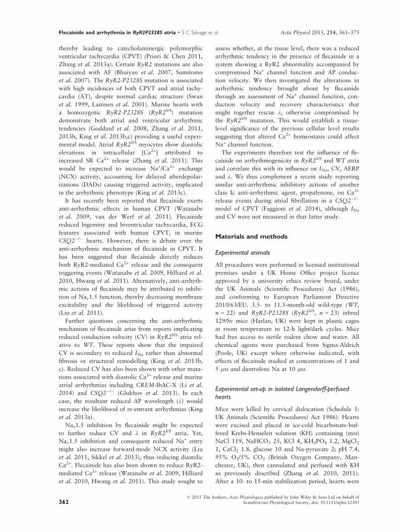
Activation and inactivation curves were obtained by
plotting peak inward currents, INa,max, against V
(Figs 3 and 4 respectively). These could be fitted to
Boltzmann functions to provide empirical indications
of maximum peak currents (INa,max), steepness fac-
tors (k) describing their dependence upon voltage
and the voltage excursions corresponding to half-
maximal current (V*). Such optimizations were pos-
sible for all activation and inactivation data apart
from WT atria studied in 5 lM flecainide. Both pro-
tocols demonstrated that RyR2S/S had a significantly
reduced maximal inward Na+ current (INa(max)) com-
pared to WT (Fig. 3, activation: t = 8.48; P < 0.001;
Fig. 4, inactivation: t = 8.42; P < 0.001), but,
whereas 1 lM flecainide reduced INa(max) in WT
(activation: t = 12.53, P < 0.001, inactivation;
t = 17.88, P < 0.001), it paradoxically increased
such inward currents in RyR2S/S (activation:
t = 4.38, P < 0.01, inactivation; t = 8.84,
P < 0.001). Five micromolar flecainide decreased INa
(max) in both RyR2S/S and WT (inactivation:
t = 9.42, P < 0.001 and t = 11.47, P < 0.001 respec-
tively).
Similarly contrasting effects on INa(max) were
obtained with the specific RyR blocker dantrolene
(10 lM) (Fig. 3, inset). Thus, in response to 80 mV
voltage steps, dantrolene produced a net decrease in
INa(max) in WT atria (�15.08 � 1.68 vs.
�10.89 � 1.42, n = 7, t = 3.05; P < 0.05), but
increased INa(max) in RyR2 atria (�8.04 � 1.19 vs.
�11.54 � 1.00, n = 7, t = 4.26; P < 0.01).
The k of the activation curves were indistinguish-
able between both genotypes and through all flecai-
nide concentrations (Fig. 3, P > 0.05). The k of the
inactivation curves were similar, though smaller in the
RyR2S/S than the WT atria in the absence of flecainide
(Fig. 4, t = 2.79, P < 0.05). This difference was abol-
ished by 1 lM flecainide (t = 0.16, P > 0.05). Addi-
tional increases in flecainide concentration to 5 lMfurther increased k in both RyR2S/S and WT (t = 2.73,
P < 0.05 and t = 1.86, P > 0.05) compared with 0 lMflecainide, and compared with 1 lM flecainide in WT
(t = 2.32, P < 0.05).
Finally, V* of activation was consistently smaller in
RyR2S/S than in WT atria whether in 0 (t = 2.92;
P < 0.05) or 1 lM flecainide (t = 3.48; P < 0.01,
Fig. 3). Flecainide (1 lM) decreased V* in WT
(t = 3.39; P < 0.01), but not RyR2S/S (t = 0.57,
P > 0.05). The V* of inactivation was similarly
reduced in untreated RyR2S/S compared to WT
(t = 5.75; P < 0.001, Fig. 4). However, 1 lM flecai-
nide increased V* in RyR2S/S (t = 2.87, P < 0.05) but
not WT (t = 0, P > 0.05), with RyR2S/S showing a
smaller V* than WT (t = 3.02, P < 0.05). Increases in
flecainide concentration to 5 lM similarly increased V*in RyR2S/S (t = 3.43, P < 0.01) relative to findings
with 0 lM flecainide. It left V* in WT close to that
© 2015 The Authors. Acta Physiologica published by John Wiley & Sons Ltd on behalf ofScandinavian Physiological Society, doi: 10.1111/apha.12505366
Flecainide and arrhythmia in RyR2P2328S atria · S C Salvage et al. Acta Physiol 2015, 214, 361–375

(a)
(b) (i)
(b) (ii)
(c) (i)
(c) (ii)
(d)
Figure 2 Contrasting actions of flecainide on arrhythmic incidence in RyR2S/S and WT. (a) Illustration of the S1S2 stimulation
protocol, consisting of repeated cycles of 8 S1 stimuli, each followed by a single extrasystolic S2 stimulus imposed at succes-
sively shorter S1S2 intervals. The first and last few cycles of the protocol are shown, with the intervening cycles omitted (dashed
lines). The protocol was terminated when an S2 either failed to elicit an AP, as observed by a missing atrial electrogram, or pro-
duced an arrhythmia. Thus, panel (a) depicts the penultimate stimulus cycle, whose S2 stimulus successfully elicited conducting
electrical activity (a), followed by the final cycle that induced either arrhythmia or refractoriness. Typical traces obtained from
(b) WT and (c) RyR2S/S before (i) and following (ii) introduction of 1 lM flecainide were obtained from the last stimulus cycle
whose S2 stimulus successfully elicited electrical activity (left panels) and the final cycle which induced either arrhythmia or
refractoriness (right panels) as described above. The filled arrowheads indicate timings of regular (S1) stimulation, and the filled
arrows indicate the resulting S1 atrial electrogram. The open arrowheads indicate the timing of the extrasystolic (S2) stimuli,
and the open arrows indicate the resulting S2 atrial electrogram. The arrowheads are directly below the stimulus artefact, and
the arrows are directly above the resulting atrial electrogram. Note that atrial electrogram conduction from the point of stimula-
tion to the point of recording is slow relative to conduction of the stimulus artefact, such that the S2 stimulus artefacts can
appear within the preceding S1 waveform at the recording site despite occurring after the atrial electrogram at the stimulus site.
Panel (d) depicts the results of applying the PES protocol to 10 WT and 17 RyR2S/S hearts to assess the incidence of arrhythmic
events normalized to the number of hearts studied in each group. * denotes a difference (P < 0.05) at 0 and 1 lM flecainide
within a genotype. † denotes a difference (P < 0.05) between RyR2S/S and WT genotypes at the same flecainide concentration.
© 2015 The Authors. Acta Physiologica published by John Wiley & Sons Ltd on behalf ofScandinavian Physiological Society, doi: 10.1111/apha.12505 367
Acta Physiol 2015, 214, 361–375 S C Salvage et al. · Flecainide and arrhythmia in RyR2P2328S atria

obtained at 0 lM flecainide (t = 1.26, P > 0.05) as well
as the corresponding result in the RyR2S/S (t = 0.20;
P > 0.05). Thus, both k and V* values in the activation
and inactivation characteristics in the atria of RyR2S/S
and WT mice showed consistent patterns with the addi-
tion of 0, 1 and 5 lM flecainide.
(a)
(b)
(c)
(d)
Figure 3 Paradoxical actions of flecainide on INa activation in RyR2S/S and WTatria . Currents in response to depolarizing steps
increased in 10 mV increments from 20 to 120 mV in voltage-clamped WT (a, n = 7) and RyR2S/S (b, n = 6) left atria in the
presence of 0, 1 and 5 lM flecainide. Currents in response to an 80 mV depolarizing step under control conditions and in the
presence of the specific RyR blocker dantrolene (10 lM) are shown in the inset. The current–voltage relationships were fitted to
Boltzmann functions for WT (c, left panel) and RyR2S/S (d, left panel) in the presence of 0, 1 and 5 lM flecainide. The right pan-
els in (c) and (d) compare the maximum peak currents before and following withdrawal of flecainide. *denotes significant effects
of flecainide or dantrolene. †denotes significant differences between RyR2S/S and WT genotypes at the same flecainide concentra-
tion.
© 2015 The Authors. Acta Physiologica published by John Wiley & Sons Ltd on behalf ofScandinavian Physiological Society, doi: 10.1111/apha.12505368
Flecainide and arrhythmia in RyR2P2328S atria · S C Salvage et al. Acta Physiol 2015, 214, 361–375
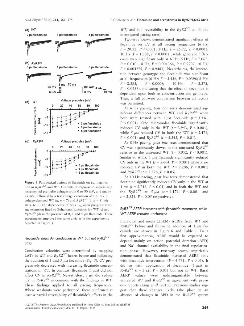
Flecainide slows AP conduction in WT but not RyR2S/S
atria
Conduction velocities were determined by mapping
LATs in WT and RyR2S/S hearts before and following
the addition of 1 and 5 lM flecainide (Fig. 5). CV pro-
gressively decreased with increasing flecainide concen-
trations in WT. In contrast, flecainide (1 lM) did not
affect CV in RyR2S/S. Nevertheless, 5 lM did reduce
CV in RyR2S/S in common with the findings in WT.
These findings applied to all pacing frequencies.
Where washouts were performed, these confirmed at
least a partial reversibility of flecainide’s effects in the
WT, and full reversibility in the RyR2S/S, at all the
investigated pacing rates.
Two-way ANOVA demonstrated significant effects of
flecainide on CV at all pacing frequencies (6 Hz:
F = 20.11, P < 0.001; 8 Hz: F = 25.72, P < 0.0001;
10 Hz: F = 13.88, P < 0.0001), while genotype differ-
ences were significant only at 6 Hz (6 Hz; F = 7.067,
P = 0.0106, 8 Hz; F = 0.001364, P = 0.9707, 10 Hz;
F = 0.004279, P = 0.9481). Nevertheless, the interac-
tion between genotype and flecainide was significant
at all frequencies (6 Hz: F = 3.456, P = 0.0396; 8 Hz:
F = 8.383, P = 0.0006; 10 Hz: F = 3.375,
P = 0.0433), indicating that the effect of flecainide is
dependent upon both its concentration and genotype.
Thus, a full pairwise comparison between all factors
was permitted.
At 6 Hz pacing, post hoc tests demonstrated sig-
nificant differences between WT and RyR2S/S when
both were treated with 1 lM flecainide (t = 3.316,
P < 0.001). One micromolar flecainide significantly
reduced CV only in the WT (t = 3.943, P < 0.001),
while 5 lM reduced CV in both the WT (t = 5.471,
P < 0.001) and RyR2S/S (t = 3.343, P < 0.01).
At 8 Hz pacing, post hoc tests demonstrated that
CV was significantly slower in the untreated RyR2S/S
relative to the untreated WT (t = 3.952, P < 0.001).
Similar to 6 Hz, 1 lM flecainide significantly reduced
CV only in the WT (t = 5.604, P < 0.001) while 5 lMreduced CV in both the WT (t = 7.206, P < 0.001)
and RyR2S/S (t = 2.826, P < 0.05).
At 10 Hz pacing, post hoc tests demonstrated that
flecainide significantly reduced CV only in the WT at
1 lM (t = 3.748, P < 0.01) and in both the WT and
the RyR2S/S at 5 lM (t = 4.179, P < 0.001 and
t = 2.824, P < 0.05 respectively).
RyR2S/S AERP increases with flecainide treatment, while
WT AERP remains unchanged
Individual and mean (�SEM) AERPs from WT and
RyR2S/S before and following addition of 1 lM fle-
cainide are shown in Figure 6 and Table 1. To a
first approximation, AERP would be expected to
depend mainly on action potential duration (APD)
and Na+ channel availability in the final repolariza-
tion phase. However, two-way ANOVA empirically
demonstrated that flecainide increased AERP only
with flecainide intervention (F = 4.761, P < 0.05). It
did so with application of flecainide (1 lM) in
RyR2S/S (t = 3.82; P < 0.01) but not in WT. Basal
AERP values were indistinguishable between
untreated WT and RyR2S/S in agreement with previ-
ous reports (King et al. 2013c). Previous studies sug-
gest that these changes likely take place in an
absence of changes in APD in the RyR2S/S system
(a)
(b)
(c)
(d)
Figure 4 Paradoxical actions of flecainide on INa inactiva-
tion in RyR2S/S and WT. Currents in response to successively
incremented pre-pulse voltages from 0 to 90 mV, and finally
95 mV, followed by a test voltage excursion of 100 mV in
voltage-clamped WT (a, n = 7) and RyR2S/S (b, n = 6) left
atria. (c, d) The dependence of peak INa upon pre-pulse volt-
age excursion fitted to Boltzmann functions for WT (c) and
RyR2S/S (d) in the presence of 0, 1 and 5 lM flecainide. These
experiments employed the same atria as in the experiments
depicted in Figure 3.
© 2015 The Authors. Acta Physiologica published by John Wiley & Sons Ltd on behalf ofScandinavian Physiological Society, doi: 10.1111/apha.12505 369
Acta Physiol 2015, 214, 361–375 S C Salvage et al. · Flecainide and arrhythmia in RyR2P2328S atria

(King et al. 2013c). Furthermore, it has been shown
in the Scn5a+/� system, in which there was a loss of
Na+ channel function, that flecainide produced a
shortening of the APD in the face of a lengthening
VERP (Martin et al. 2011).
Action potential wavelengths correlate with arrhythmic
incidence in both RyR2S/S and WT
Two-way ANOVA demonstrated strong interactions
(P = 0.0021; F = 13.72) between the effects of flecai-
nide and genotype upon k, indicating that the effect of
flecainide is different in WT compared to RyR2S/S.
Flecainide decreased k in WT (t = 2.39, P < 0.05)
while increasing it in RyR2S/S atria (t = 2.42,
P < 0.05).
We then correlated CV, AERP and k with arrhyth-
mic incidences before and following application of
1 lM flecainide (Fig. 7). As indicated above, flecainide
significantly reduced CV in WT but not RyR2S/S,
directly correlating with the increased arrhythmic inci-
dence in WT but not the decreased incidence of
arrhythmia in RyR2S/S (Fig. 7A). In contrast, flecai-
nide significantly increased AERP in RyR2S/S but not
WT atria directly correlating with the decreased
arrhythmic incidences in RyR2S/S but not the increased
arrhythmic incidences in WT (Fig. 7B). However, fle-
cainide decreased k in WT but increased k in RyR2S/S
(Fig. 7C). In contrast to CV and AERP, changes in ktherefore correlated with alterations in arrhythmia in
both RyR2S/S and WT. This implicates k as the
(a)
(b)
(c)
Figure 5 Paradoxical actions of flecainide on conduction velocities in RyR2S/S and WT. Three-dimensional representations of
local activation times (LATs) each accompanied by matrices representing the calculated velocity vectors in WT (a, n = 15) and
RyR2S/S hearts (b, n = 14) in 0, 1 and 5 lM flecainide. Mean (�SEM) epicardial conduction velocities for WT (clear bars) and
RyR2S/S (black bars) in 0, 1, 5 and following subsequent return to 0 lM flecainide during regular 6.67, 8 and 10 Hz pacing (c).
*denotes a difference arising from use of 1 lM flecainide within a genotype compared to the respective control (0 lM flecainide).†denotes a difference between RyR2S/S and WT genotypes with the same concentrations of flecainide. In each case, single, double
and triple symbols denote P < 0.05, P < 0.01 and P < 0.001 respectively.
Figure 6 Paradoxical actions of flecainide on AERP in
RyRS/S and WT. Individual paired and mean (�SEM) AERPs
in 0 and 1 lM flecainide for WT (n = 9) and RyR2S/S
(n = 17) hearts. ** denotes a difference (P < 0.01) arising
from use of 1 lM flecainide within a genotype compared to
the respective control (0 lM flecainide).
© 2015 The Authors. Acta Physiologica published by John Wiley & Sons Ltd on behalf ofScandinavian Physiological Society, doi: 10.1111/apha.12505370
Flecainide and arrhythmia in RyR2P2328S atria · S C Salvage et al. Acta Physiol 2015, 214, 361–375

primary predictor for arrhythmic incidences rather
than either CV or AERP alone, in both RyR2S/S and
WT.
Discussion
The present study demonstrates a novel paradoxical
effect of the INa blocker flecainide on arrhythmic inci-
dence and INa in RyR2-P2328S and WT atria. It fol-
lows directly from evidence for its anti-arrhythmic
effects in human CPVT (Pott et al. 2011, van der
Werf et al. 2011, Watanabe et al. 2013). Its findings
complement a recent report that the alternative class
Ic anti-arrhythmic agent, propafenone, similarly
exerted anti-arrhythmic actions during atrial fibrilla-
tion in a CSQ2�/� model of CPVT (Faggioni et al.
2014). Although INa and CV were not measured in
that latter study, the two reports converge upon com-
mon arrhythmic mechanisms through differing mea-
surements and experimental systems. It also reconciles
several previous studies at the cellular as opposed to
tissue level. These attributed the anti-arrhythmic
effects of flecainide to a range of factors. First, flecai-
nide was suggested to reduce triggered activity arising
from DADs (Liu et al. 2011). This could result from
direct actions inhibiting spontaneous RyR2-mediated
SR Ca2+ release implicated in such DADs: flecainide
blocks RyR2-Ca2+ release channel open states, thereby
reducing Ca2+ wave frequency in CSQ2�/� mice and
rat myocytes (Watanabe et al. 2009, Hilliard et al.
2010, Galimberti & Knollmann 2011). However, a
subsequent study reported that whereas flecainide pre-
vented isoproterenol-induced CPVT, it did not exert
major effects on Ca2+ homeostasis in RyR2-R4496C
hearts (Liu et al. 2011). This suggested that flecainide
increases the threshold for triggered activity by
directly inhibiting Nav1.5 function (Liu et al. 2011).
Second, reductions in Na+ entry could reduce intracel-
lular [Na+], thereby increasing forward-mode NCX
activity, in turn reducing intracellular [Ca2+] (Sikkel
et al. 2013). The alternative INa blockers, tetrodo-
toxin, propafenone or lidocaine similarly reduced
Ca2+ spark and wave frequency, and wave velocity in
WT rat myocytes, doing so only before INa inactiva-
tion brought about by alterations in holding voltage.
Flecainide also increased NCX-mediated Ca2+ efflux,
an effect reversed by reducing extracellular [Na+] (Sik-
kel et al. 2013).
(a)
(b)
(c)
Figure 7 Paradoxical actions of flecai-
nide on CV, AERP and k and their cor-
relations with arrhythmic incidence. Left
panels: comparison of CV (a), AERP (b)
and k (c) in WT (open bars, n = 8) and
RyR2S/S (filled bars, n = 10) hearts in 0
and 1 lM flecainide. These are correlated
with incidences of atrial tachyarrhyth-
mias (AT) (a–c, right panels). * denotes
a difference arising from use of 1 lM fle-
cainide within a genotype compared to
the respective control (0 lM flecainide).† denotes a difference between RyR2S/S
and WT genotypes under the same con-
centration of flecainide. In each case, sin-
gle, double and triple symbols denote
P < 0.05, P < 0.01 and P < 0.001
respectively.
© 2015 The Authors. Acta Physiologica published by John Wiley & Sons Ltd on behalf ofScandinavian Physiological Society, doi: 10.1111/apha.12505 371
Acta Physiol 2015, 214, 361–375 S C Salvage et al. · Flecainide and arrhythmia in RyR2P2328S atria

However, recent findings also associated both cate-
cholamine-induced ventricular arrhythmia (Zhang
et al. 2013b) and atrial arrhythmogenesis with reduc-
tions in CV also associated with RyR2S/S (King et al.
2013a,b,c). RyR2S/S atria showed reduced INa
compared to WT. Increased AF susceptibility in asso-
ciation with conduction abnormalities has also been
observed in other models of altered Ca2+ homeostasis,
including murine CREM-IbDC-X AF (Li et al. 2014)
and CSQ2�/� hearts (Glukhov et al. 2013). In WT,
elevating extracellular Ca2+ and manipulating cellular
Ca2+ homeostasis using caffeine or cyclopiazonic acid
acutely replicated these effects (Zhang et al. 2011).
These findings suggest that RyR2-mediated Ca2+
release in RyR2S/S results in inhibition of INa reducing
CV, thus producing a re-entrant, arrhythmic substrate.
Inhibition of RyR2-mediated Ca2+ release by flecainide
should then paradoxically restore INa and rescue both
the compromised CV and arrhythmic phenotype. Our
findings confirm this prediction: untreated murine
RyR2S/S atria were more arrhythmic than WT, con-
firming recent findings (King et al. 2013b,c), and at
the cellular level showed reduced INa compared to the
corresponding WT. Flecainide (1 lM) was anti-
arrhythmic in RyR2S/S despite being pro-arrhythmic in
WT. These findings were concordant with findings at
the cellular level in which untreated RyR2S/S showed
reduced INa compared to the corresponding WT. Fle-
cainide then reduced INa in WT while increasing it in
RyR2S/S. The use of an alternative more specific RyR
blocker, dantrolene (10 lM), similarly reduced INa in
WT atria while increasing it in RyR2S/S atria. Dantro-
lene has previously been shown to reduce Ca2+ spark
frequency and arrhythmogenicity in induced pluripo-
tent stem cells derived from a CPVT patient carrying
a RYR2 S406L mutation (Jung et al. 2012). The pro-
arrhythmic action of flecainide in the WT may appear
surprising due to its clinical utility for atrial tachycar-
dia without structural abnormality. However, flecai-
nide has proved pro-arrhythmic in various models
(Brugada et al. 1991, Stokoe et al. 2007) and most
notoriously in the cardiac arrhythmia suppression trial
(CAST 1989). It has been proposed that this may
result from effects on cardiac repolarization, and
indeed, there is evidence for reduced IKr in cardiac
cells (Follmer & Colatsky 1990, Wang et al. 1996),
prolonged QT interval in human patients (Katritsis
et al. 1995, Sarubbi et al. 1998), and repolarization
abnormalities and increased arrhythmic incidences in
perfused guinea-pig hearts (Osadchii 2012). The pres-
ent results additionally suggest that reduction in INa,
CV and k may contribute to the pro-arrhythmic
effects of flecainide. Thus, at the tissue level, untreated
RyR2S/S showed reduced CVs compared to WT,
despite similar AERPs. Flecainide decreased CV but
conserved AERP in WT, whereas it spared CV and
increased AERP in RyR2S/S. Nevertheless, k derived
from the product CV x AERP correlated directly with
arrhythmic tendency in both the RyR2S/S and WT
under conditions of either 0 or 1 lM flecainide.
These electrophysiological findings in intact atria
are compatible with previous evidence for interactions
between Ca2+ homeostasis and Nav1.5 expression and
function in WT myocytes at the cellular level.
Increases in pipette Ca2+ concentration reduced INa
density and (dV/dt)max in patch-clamped WT myo-
cytes (Casini et al. 2009). The Ca2+ channel blocker
verapamil and the Ca2+ ionophore calcimycin, respec-
tively, increased and decreased Nav1.5 mRNA and
Nav1.5 protein expression in rat cardiomyocytes (Of-
ford & Catterall 1989, Taouis et al. 1991, Duff et al.
1992). Increased extracellular [Ca2+] and BAPTA-AM,
respectively, expected to increase and decrease intra-
cellular [Ca2+] and correspondingly increased and
decreased INa density in cultured neonatal rat myo-
cytes (Chiamvimonvat et al. 1995).
The findings also agree with previous evidence for
mechanisms linking Ca2+ homeostasis to Nav1.5 at
the molecular level. Nav1.5 is a major calmodulin
kinase II (CaMKII) target. Such phosphorylation shifts
the voltage dependence of inactivation to negative
potentials without affecting channel activation. This
slows recovery from inactivation, enhances Nav1.5
transitions into slower forms of inactivation and
increases late INa (Wagner et al. 2011, Grandi & Her-
ren 2014). However, in the present study, although
RyR2S/S was associated with a negative shift in inacti-
vation, activation properties were similarly affected.
RyR2S/S showed a similar AERP as WT in the absence
of flecainide.
The findings together demonstrate contrasting anti-
and pro-arrhythmic actions of the Nav1.5 channel
blocker flecainide in murine RyR2S/S and WT atria
respectively. They attribute these to corresponding
changes in INa, k and therefore arrhythmic substrate
while not excluding involvement of triggered activity
in initiating arrhythmia with either genotype. This
could involve a mechanism consistent with previously
reported suggestions at the cellular level of interac-
tions between cellular Ca2+ homeostasis and Nav1.5
function.
Funding
This work was supported by the Biotechnology and
Biological Sciences Research Council (BBSRC, UK)
under a David Phillips Fellowship held by JAF (BB/
FO23863/1) and by the Isaac Newton Trust/Wellcome
Trust ISSF/University of Cambridge Joint Research
Grants Scheme.
© 2015 The Authors. Acta Physiologica published by John Wiley & Sons Ltd on behalf ofScandinavian Physiological Society, doi: 10.1111/apha.12505372
Flecainide and arrhythmia in RyR2P2328S atria · S C Salvage et al. Acta Physiol 2015, 214, 361–375

Conflict of interest
None.
We thank Paul Frost and Vicky Johnson for technical sup-
port.
References
Adrian, R.H. 1956. The effect of internal and external potas-
sium concentration on the membrane potential of frog
muscle. J Physiol 133, 631–658.
Almers, W., Stanfield, P.R. & St€uhmer, W. 1983. Lateral dis-
tribution of sodium and potassium channels in frog skeletal
muscle: measurements with a patch-clamp technique. J
Physiol 336, 261–284.
Benjamin, E.J., Wolf, P.A., D’Agostino, R.B., Silbershatz, H.,
Kannel, W.B. & Levy, D. 1998. Impact of atrial fibrillation
on the risk of death: the Framingham Heart Study. Circu-
lation 98, 946–952.
Bezanilla, F. & Armstrong, C.M. 1977. Inactivation of the
sodium channel. I. Sodium current experiments. J Gen
Physiol 70, 549–566.
Bhuiyan, Z.A., van den Berg, M.P., van Tintelen, J.P., Bink-
Boelkens, M.T.E., Wiesfeld, A.C.P., Alders, M., Postma,
A.V., van Langen, I., Mannens, M.M.A.M. & Wilde,
A.A.M. 2007. Expanding spectrum of human RYR2-
related disease: new electrocardiographic, structural, and
genetic features. Circulation 116, 1569–1576.
Bootman, M.D., Higazi, D.R., Coombes, S. & Roderick,
H.L. 2006. Calcium signalling during excitation-contrac-
tion coupling in mammalian atrial myocytes. J Cell Sci 119
(Pt 19), 3915–3925.
Brugada, J., Boersma, L., Kirchhof, C. & Allessie, M. 1991.
Proarrhythmic effects of flecainide. Experimental evidence
for increased susceptibility to reentrant arrhythmias. Circu-
lation 84, 1808–1818.
Casini, S., Verkerk, A.O., van Borren, M.M.G.J., van Ginne-
ken, A.C.G., Veldkamp, M.W., de Bakker, J.M.T. & Tan,
H.L. 2009. Intracellular calcium modulation of voltage-
gated sodium channels in ventricular myocytes. Cardiovasc
Res 81, 72–81.
CAST 1989. Preliminary report: effect of encainide and fle-
cainide on mortality in a randomized trial of arrhythmia
suppression after myocardial infarction. The Cardiac
Arrhythmia Suppression Trial (CAST) Investigators. N
Engl J Med 321, 406–412.
Chiamvimonvat, N., Kargacin, M.E., Clark, R.B. & Duff,
H.J. 1995. Effects of intracellular calcium on sodium cur-
rent density in cultured neonatal rat cardiac myocytes. J
Physiol 483, 307–318.
Davis, R.C., Hobbs, F.D.R., Kenkre, J.E., Roalfe, A.K., Iles,
R., Lip, G.Y.H. & Davies, M.K. 2012. Prevalence of atrial
fibrillation in the general population and in high-risk
groups: the ECHOES study. Europace 14, 1553–1559.
Duff, H.J., Offord, J., West, J. & Catterall, W.A. 1992. Class
I and IV antiarrhythmic drugs and cytosolic calcium regu-
late mRNA encoding the sodium channel alpha subunit in
rat cardiac muscle. Mol Pharmacol 42, 570–574.
Eickhorn, R., Weirich, J., Hornung, D. & Antoni, H. 1990.
Use dependence of sodium current inhibition by tetrodo-
toxin in rat cardiac muscle: influence of channel state. Pflu-
gers Arch 416, 398–405.
Faggioni, M., Savio-Galimberti, E., Venkataraman, R.,
Hwang, H.S., Kannankeril, P.J., Darbar, D. & Knollmann,
B.C. 2014. Suppression of spontaneous ca elevations pre-
vents atrial fibrillation in calsequestrin 2-null hearts. Circ
Arrhythm Electrophysiol 7, 313–320.
Follmer, C.H. & Colatsky, T.J. 1990. Block of delayed recti-
fier potassium current, IK, by flecainide and E-4031 in cat
ventricular myocytes. Circulation 82, 289–293.
Galimberti, E.S. & Knollmann, B.C. 2011. Efficacy and
potency of class I antiarrhythmic drugs for suppression of
Ca2+ waves in permeabilized myocytes lacking calseques-
trin. J Mol Cell Cardiol 51, 760–768.
Glukhov, A.V., Kalyanasundaram, A., Lou, Q., Hage, L.T.,
Hansen, B.J., Belevych, A.E., Mohler, P.J., Knollmann, B.C.,
Periasamy, M., Gy€orke, S. & Fedorov, V.V. 2013. Calseques-
trin 2 deletion causes sinoatrial node dysfunction and atrial
arrhythmias associated with altered sarcoplasmic reticulum
calcium cycling and degenerative fibrosis within the mouse
atrial pacemaker complex. Eur Heart J 36, 686–697.
Goddard, C.A., Ghais, N.S., Zhang, Y., Williams, A.J., Coll-
edge, W.H., Grace, A.A. & Huang, C.L.-H. 2008. Physio-
logical consequences of the P2328S mutation in the
ryanodine receptor (RyR2) gene in genetically modified
murine hearts. Acta Physiol (Oxf), 194, 123–140.
Grandi, E. & Herren, A.W. 2014. CaMKII-dependent regula-
tion of cardiac Na(+) homeostasis. Front Pharmacol 5, 41.
Hilliard, F.A., Steele, D.S., Laver, D., Yang, Z., Le Marc-
hand, S.J., Chopra, N., Piston, D.W., Huke, S. & Knoll-
mann, B.C. 2010. Flecainide inhibits arrhythmogenic Ca2+
waves by open state block of ryanodine receptor Ca2+
release channels and reduction of Ca2+ spark mass. J Mol
Cell Cardiol 48, 293–301.
Hwang, H.S., Hasdemir, C., Laver, D., Mehra, D., Turhan,
K., Faggioni, M., Yin, H. & Knollmann, B.C. 2011. Inhi-
bition of cardiac Ca2+ release channels (RyR2) determines
efficacy of class I antiarrhythmic drugs in catecholaminer-
gic polymorphic ventricular tachycardia. Circ Arrhythm
Electrophysiol 4, 128–135.
Jung, C.B., Moretti, A., Mederos y Schnitzler, M., Iop, L.,
Storch, U., Bellin, M., Dorn, T., Ruppenthal, S., Pfeiffer,
S., Goedel, A. et al. 2012. Dantrolene rescues arrhythmo-
genic RYR2 defect in a patient-specific stem cell model of
catecholaminergic polymorphic ventricular tachycardia.
EMBO Mol Med, 4, 180–191.
Katritsis, D., Rowland, E., O’Nunain, S., Shakespeare, C.F.,
Poloniecki, J. & Camm, A.J. 1995. Effect of flecainide on
atrial and ventricular refractoriness and conduction in
patients with normal left ventricle. Implications for possi-
ble antiarrhythmic and proarrhythmic mechanisms. Eur
Heart J 16, 1930–1935.
King, J.H., Huang, C.L.-H. & Fraser, J.A. 2013a. Determi-
nants of myocardial conduction velocity: implications for
arrhythmogenesis. Front Physiol 4, 154.
King, J.H., Wickramarachchi, C., Kua, K., Du, Y., Jeevarat-
nam, K., Matthews, H.R., Grace, A.A., Huang, C.L.H. &
© 2015 The Authors. Acta Physiologica published by John Wiley & Sons Ltd on behalf ofScandinavian Physiological Society, doi: 10.1111/apha.12505 373
Acta Physiol 2015, 214, 361–375 S C Salvage et al. · Flecainide and arrhythmia in RyR2P2328S atria

Fraser, J.A. 2013b. Loss of Nav1.5 expression and func-
tion in murine atria containing the RyR2-P2328S gain-of-
function mutation. Cardiovasc Res 99, 751–759.
King, J.H., Zhang, Y., Lei, M., Grace, A.A., Huang, C.L.H.
& Fraser, J.A. 2013c. Atrial arrhythmia, triggering events
and conduction abnormalities in isolated murine RyR2-
P2328S hearts. Acta Physiol, 207, 308–323.
Laitinen, P.J., Brown, K.M., Piippo, K., Swan, H., Devaney,
J.M., Brahmbhatt, B., Donarum, E.A., Marino, M., Tiso,
N., Viitasalo, M., Toivonen, L., Stephan, D.A. & Kontula,
K. 2001. Mutations of the cardiac ryanodine receptor
(RyR2) gene in familial polymorphic ventricular tachycar-
dia. Circulation 103, 485–490.
Lambiase, P.D., Ahmed, A.K., Ciaccio, E.J., Brugada, R.,
Lizotte, E., Chaubey, S., Ben-Simon, R., Chow, A.W.,
Lowe, M.D. & McKenna, W.J. 2009. High-density sub-
strate mapping in Brugada syndrome: combined role of
conduction and repolarization heterogeneities in ar-
rhythmogenesis. Circulation 120, 106–117, 1–4.
Lemoine, M.D., Duverger, J.E., Naud, P., Chartier, D., Qi,
X.Y., Comtois, P., Fabritz, L., Kirchhof, P. & Nattel, S.
2011. Arrhythmogenic left atrial cellular electrophysiology
in a murine genetic long QT syndrome model. Cardiovasc
Res 92, 67–74.
Li, N., Chiang, D.Y., Wang, S., Wang, Q., Sun, L., Voigt,
N., Respress, J.L., Ather, S., Skapura, D.G., Jordan, V.K.
et al. 2014. Ryanodine receptor-mediated calcium leak
drives progressive development of an atrial fibrillation sub-
strate in a transgenic mouse model. Circulation 129,
1276–1285.
Liu, N., Denegri, M., Ruan, Y., Avelino-Cruz, J.E., Perissi,
A., Negri, S., Napolitano, C., Coetzee, W.A., Boyden, P.A.
& Priori, S.G. 2011. Short communication: flecainide
exerts an antiarrhythmic effect in a mouse model of cate-
cholaminergic polymorphic ventricular tachycardia by
increasing the threshold for triggered activity. Circ Res
109, 291–295.
Mackenzie, L., Bootman, M.D., Berridge, M.J. & Lipp, P.
2001. Predetermined recruitment of calcium release sites
underlies excitation-contraction coupling in rat atrial myo-
cytes. J Physiol 530(Pt 3), 417–429.
Mackenzie, L., Roderick, H.L., Berridge, M.J., Conway, S.J.
& Bootman, M.D. 2004. The spatial pattern of atrial car-
diomyocyte calcium signalling modulates contraction.
J Cell Sci 117(Pt 26), 6327–6337.
Martin, C.A., Grace, A.A. & Huang, C.L.-H. 2011. Refrac-
tory dispersion promotes conduction disturbance and ar-
rhythmias in a Scn5a (+/-) mouse model. Pflugers Arch
462, 495–504.
Offord, J. & Catterall, W.A. 1989. Electrical activity, cAMP,
and cytosolic calcium regulate mRNA encoding sodium
channel alpha subunits in rat muscle cells. Neuron, 2,
1447–1452.
Osadchii, O.E. 2012. Flecainide-induced proarrhythmia is
attributed to abnormal changes in repolarization and
refractoriness in perfused guinea-pig heart. J Cardiovasc
Pharmacol 60, 456–466.
Pott, C., Dechering, D.G., Reinke, F., Muszynski, A., Zeller-
hoff, S., Bittner, A., K€obe, J., Wasmer, K., Schulze-Bahr,
E., M€onnig, G., Kotthoff, S. & Eckardt, L. 2011. Success-
ful treatment of catecholaminergic polymorphic ventricular
tachycardia with flecainide: a case report and review of the
current literature. Europace 13, 897–901.
Priori, S.G. & Chen, S.R.W. 2011. Inherited dysfunction of
sarcoplasmic reticulum Ca2+ handling and arrhythmogene-
sis. Circ Res 108, 871–883.
Roberts, W.M., St€uhmer, W., Weiss, R.E., Stanfield, P.R. &
Almers, W. 1986. Distribution and mobility of voltage-
gated ion channels in skeletal muscle. Ann N Y Acad Sci
479, 377–384.
Sabir, I.N., Fraser, J.A., Killeen, M.J., Grace, A.A. & Huang,
C.L.-H. 2007. The contribution of refractoriness to
arrhythmic substrate in hypokalemic Langendorff-perfused
murine hearts. Pflugers Arch 454, 209–222.
Sarubbi, B., Ducceschi, V., Briglia, N., Mayer, M.S., Santan-
gelo, L. & Iacono, A. 1998. Compared effects of sotalol,
flecainide and propafenone on ventricular repolarization in
patients free of underlying structural heart disease. Int J
Cardiol 66, 157–164.
Sikkel, M.B., Collins, T.P., Rowlands, C., Shah, M., O’Gara,
P., Williams, A.J., Harding, S.E., Lyon, A.R. & MacLeod,
K.T. 2013. Flecainide reduces Ca(2+) spark and wave fre-
quency via inhibition of the sarcolemmal sodium current.
Cardiovasc Res 98, 286–296.
Stewart, S., Hart, C.L., Hole, D.J. & McMurray, J.J.V.
2002. A population-based study of the long-term risks
associated with atrial fibrillation: 20-year follow-up of the
Renfrew/Paisley study. Am J Med 113, 359–364.
Stokoe, K.S., Balasubramaniam, R., Goddard, C.A., Coll-
edge, W.H., Grace, A.A. & Huang, C.L.-H. 2007. Effects
of flecainide and quinidine on arrhythmogenic properties
of Scn5a+/- murine hearts modelling the Brugada syn-
drome. J Physiol 581(Pt 1), 255–275.
St€uhmer, W. & Almers, W. 1982. Photobleaching through
glass micropipettes: sodium channels without lateral mobil-
ity in the sarcolemma of frog skeletal muscle. Proc Natl
Acad Sci USA 79, 946–950.
Sumitomo, N., Sakurada, H., Taniguchi, K., Matsumura, M.,
Abe, O., Miyashita, M., Kanamaru, H., Karasawa, K., Ay-
usawa, M., Fukamizu, S., Nagaoka, I., Horie, M., Harada,
K. & Hiraoka, M. 2007. Association of atrial arrhythmia
and sinus node dysfunction in patients with catecholamin-
ergic polymorphic ventricular tachycardia. Circ J 71,
1606–1609.
Swan, H., Piippo, K., Viitasalo, M., Heikkil€a, P., Paavonen,
T., Kainulainen, K., Kere, J., Keto, P., Kontula, K. & To-
ivonen, L. 1999. Arrhythmic disorder mapped to chromo-
some 1q42–q43 causes malignant polymorphic ventricular
tachycardia in structurally normal hearts. J Am Coll Car-
diol 34, 2035–2042.
Taouis, M., Sheldon, R.S. & Duff, H.J. 1991. Upregulation
of the rat cardiac sodium channel by in vivo treatment
with a class I antiarrhythmic drug. J Clin Investig 88, 375–
378.
Wagner, S., Ruff, H.M., Weber, S.L., Bellmann, S., Sowa, T.,
Schulte, T., Anderson, M.E., Grandi, E., Bers, D.M.,
Backs, J., Belardinelli, L. & Maier, L.S. 2011. Reactive
oxygen species-activated Ca/calmodulin kinase IId is
© 2015 The Authors. Acta Physiologica published by John Wiley & Sons Ltd on behalf ofScandinavian Physiological Society, doi: 10.1111/apha.12505374
Flecainide and arrhythmia in RyR2P2328S atria · S C Salvage et al. Acta Physiol 2015, 214, 361–375

required for late I(Na) augmentation leading to cellular Na
and Ca overload. Circ Res 108, 555–565.
Wang, D.W., Kiyosue, T., Sato, T. & Arita, M. 1996. Com-
parison of the effects of class I anti-arrhythmic drugs, ci-
benzoline, mexiletine and flecainide, on the delayed
rectifier K+ current of guinea-pig ventricular myocytes.
J Mol Cell Cardiol 28, 893–903.
Watanabe, H., Chopra, N., Laver, D., Hwang, H.S., Davies,
S.S., Roach, D.E., Duff, H.J., Roden, D.M., Wilde,
A.A.M. & Knollmann, B.C. 2009. Flecainide prevents cat-
echolaminergic polymorphic ventricular tachycardia in
mice and humans. Nat Med 15, 380–383.
Watanabe, H., van der Werf, C., Roses-Noguer, F., Adler,
A., Sumitomo, N., Veltmann, C., Rosso, R., Bhuiyan,
Z.A., Bikker, H., Kannankeril, P.J. et al. 2013. Effects of
flecainide on exercise-induced ventricular arrhythmias and
recurrences in genotype-negative patients with catecholam-
inergic polymorphic ventricular tachycardia. Heart Rhythm
10, 542–547.
Van der Werf, C., Kannankeril, P.J., Sacher, F., Krahn, A.D.,
Viskin, S., Leenhardt, A., Shimizu, W., Sumitomo, N.,
Fish, F.A., Bhuiyan, Z.A. et al. 2011. Flecainide therapy
reduces exercise-induced ventricular arrhythmias in
patients with catecholaminergic polymorphic ventricular
tachycardia. J Am Coll Cardiol 57, 2244–2254.
Zhang, Y., Fraser, J.A., Schwiening, C., Killeen, M.J., Grace,
A.A. & Huang, C.L.-H. 2010. Acute atrial arrhythmogene-
sis in murine hearts following enhanced extracellular Ca
(2+) entry depends on intracellular Ca(2+) stores. Acta
Physiol (Oxf), 198, 143–158.
Zhang, Y., Fraser, J.A., Jeevaratnam, K., Hao, X., Hothi,
S.S., Grace, A.A., Lei, M. & Huang, C.L.-H. 2011. Acute
atrial arrhythmogenicity and altered Ca(2+) homeostasis in
murine RyR2-P2328S hearts. Cardiovasc Res 89, 794–804.
Zhang, Y., Matthews, G.D.K., Lei, M. & Huang, C.L.-H.
2013a. Abnormal Ca(2+) homeostasis, atrial arrhythmo-
genesis, and sinus node dysfunction in murine hearts mod-
eling RyR2 modification. Front Physiol 4, 150.
Zhang, Y., Wu, J., Jeevaratnam, K., King, J.H., Guzadhur,
L., Ren, X., Grace, A.A., Lei, M., Huang, C.L.-H. & Fra-
ser, J.A. 2013b. Conduction slowing contributes to sponta-
neous ventricular arrhythmias in intrinsically active murine
RyR2-P2328S hearts. J Cardiovasc Electrophysiol 24, 210–
218.
Zhang, Y., Guzadhur, L., Jeevaratnam, K., Salvage, S.C.,
Matthews, G.D.K., Lammers, W.J., Lei, M., Huang, C.L.-
H. & Fraser, J.A. 2014. Arrhythmic substrate, slowed
propagation and increased dispersion in conduction direc-
tion in the right ventricular outflow tract of murine
Scn5a+/- hearts. Acta Physiol (Oxf), 211, 559–573.
© 2015 The Authors. Acta Physiologica published by John Wiley & Sons Ltd on behalf ofScandinavian Physiological Society, doi: 10.1111/apha.12505 375
Acta Physiol 2015, 214, 361–375 S C Salvage et al. · Flecainide and arrhythmia in RyR2P2328S atria