Fistula occlusion and ligation for a giant right coronary ...
5
CASE REPORT Open Access Fistula occlusion and ligation for a giant right coronary artery aneurysm concurrent with right atrial fistula: a case report Yan Ren 1 , Lin Xie 1 , Weiqiang Ruan 1 , Yajiao Li 2 , Peng Ji 3 , Changping Gan 1 and Ke Lin 1* Abstract Background: Coronary artery aneurysms in most cases require surgical treatment once diagnosed. Lifelong anticoagulation is often needed after surgery. We herein describe a 55-year-old man who was asymptomatic and diagnosed with right giant coronary artery aneurysm combined with right atrial fistula. Case presentation: This is a case of asymptomatic giant right coronary artery aneurysm concurrent with coronary artery fistula. Because the aneurysm was in the distal right posterior descending coronary artery, right coronary artery ligation and fistula occlusion through the right atrium were performed in the absence of cardiopulmonary bypass. The aneurysm was excluded without impacting the myocardial blood supply, and the patient was exempted from lifelong anticoagulation regimen. The follow-up revealed favorable outcomes and the patient’s life expectancy was improved. Conclusion: Decompression and exclusion without cardiopulmonary bypass can be adopted for distal coronary artery aneurysms that do not involve or only have a limited impact on distal blood supply. This procedure can exempt the patient from the lifelong anticoagulation regimen. In addition, the risk for myocardial ischemia caused by the thrombus in the aneurysm can also be avoided. The whole procedure is comparatively easy to perform. Keywords: Coronary artery aneurysm, Coronary artery fistula, Cardiopulmonary bypass Background Coronary artery fistula (CAF) is a rare congenital ana- tomical anomaly with an incidence of about 0.2% [1, 2]. CAF is associated with aneurysms in 19% of the cases. Moreover, these aneurysms may fistulate to the right atrium with an incidence of 26% [3]. An aneurysm > 20 mm in diameter is termed as a giant coronary aneurysm [4]. Though the underlying mechanism remains un- known, it is believed that the increased blood flow and pressure destroy the media structure of the vascular wall and decrease the elasticity of the coronary artery, there- after arousing the concurrent aneurysm. In adults, ath- erosclerosis is the most common cause for coronary artery aneurysm (CAA), which accounts for 50% of all the cases [5, 6]. CAA of all sizes may rupture and the risk of rupture increases in a size-dependent manner [7]. Prompt surgery is necessary for patients with evident symptoms of CAA. Yet for asymptomatic patients, whether surgery is a necessity is controversial. Surgical treatment is recommended to asymptomatic patients due to the low natural closure rate of fistula and the potential complications of aneurysm like myocardial ischemia, bacterial endocarditis and coronary artery aneurysm. This paper reports a case of asymptomatic a giant right coronary artery aneurysm concurrent with coronary artery fistula. Surgery was performed in the ab- sence of cardiopulmonary bypass (CPB) and the patient was exempted from a lifelong postoperative anticoagula- tion regimen. Case presentation A 55-year-old man was admitted to the Cardiovascular Surgery Department of our hospital for CAA detected by transthoracic echocardiography (TTE) during a rou- tine physical examination. He had neither symptoms of © The Author(s). 2019 Open Access This article is distributed under the terms of the Creative Commons Attribution 4.0 International License (http://creativecommons.org/licenses/by/4.0/), which permits unrestricted use, distribution, and reproduction in any medium, provided you give appropriate credit to the original author(s) and the source, provide a link to the Creative Commons license, and indicate if changes were made. The Creative Commons Public Domain Dedication waiver (http://creativecommons.org/publicdomain/zero/1.0/) applies to the data made available in this article, unless otherwise stated. * Correspondence: [email protected]; [email protected] 1 Department of Cardiovascular Surgery, West China Hospital, Sichuan University, No. 37 Guoxue Lane, Wuhou District, Chengdu 610041, Sichuan, China Full list of author information is available at the end of the article Ren et al. BMC Surgery (2019) 19:166 https://doi.org/10.1186/s12893-019-0624-3
Transcript of Fistula occlusion and ligation for a giant right coronary ...
CASE REPORT Open Access
Fistula occlusion and ligation for a giantright coronary artery aneurysm concurrentwith right atrial fistula: a case reportYan Ren1, Lin Xie1, Weiqiang Ruan1, Yajiao Li2, Peng Ji3, Changping Gan1 and Ke Lin1*
Abstract
Background: Coronary artery aneurysms in most cases require surgical treatment once diagnosed. Lifelonganticoagulation is often needed after surgery. We herein describe a 55-year-old man who was asymptomatic anddiagnosed with right giant coronary artery aneurysm combined with right atrial fistula.
Case presentation: This is a case of asymptomatic giant right coronary artery aneurysm concurrent with coronaryartery fistula. Because the aneurysm was in the distal right posterior descending coronary artery, right coronaryartery ligation and fistula occlusion through the right atrium were performed in the absence of cardiopulmonarybypass. The aneurysm was excluded without impacting the myocardial blood supply, and the patient wasexempted from lifelong anticoagulation regimen. The follow-up revealed favorable outcomes and the patient’s lifeexpectancy was improved.
Conclusion: Decompression and exclusion without cardiopulmonary bypass can be adopted for distal coronaryartery aneurysms that do not involve or only have a limited impact on distal blood supply. This procedure canexempt the patient from the lifelong anticoagulation regimen. In addition, the risk for myocardial ischemia causedby the thrombus in the aneurysm can also be avoided. The whole procedure is comparatively easy to perform.
Keywords: Coronary artery aneurysm, Coronary artery fistula, Cardiopulmonary bypass
BackgroundCoronary artery fistula (CAF) is a rare congenital ana-tomical anomaly with an incidence of about 0.2% [1, 2].CAF is associated with aneurysms in 19% of the cases.Moreover, these aneurysms may fistulate to the rightatrium with an incidence of 26% [3]. An aneurysm > 20mm in diameter is termed as a giant coronary aneurysm[4]. Though the underlying mechanism remains un-known, it is believed that the increased blood flow andpressure destroy the media structure of the vascular walland decrease the elasticity of the coronary artery, there-after arousing the concurrent aneurysm. In adults, ath-erosclerosis is the most common cause for coronaryartery aneurysm (CAA), which accounts for 50% of allthe cases [5, 6]. CAA of all sizes may rupture and the
risk of rupture increases in a size-dependent manner [7].Prompt surgery is necessary for patients with evidentsymptoms of CAA. Yet for asymptomatic patients,whether surgery is a necessity is controversial. Surgicaltreatment is recommended to asymptomatic patientsdue to the low natural closure rate of fistula and thepotential complications of aneurysm like myocardialischemia, bacterial endocarditis and coronary arteryaneurysm. This paper reports a case of asymptomatic agiant right coronary artery aneurysm concurrent withcoronary artery fistula. Surgery was performed in the ab-sence of cardiopulmonary bypass (CPB) and the patientwas exempted from a lifelong postoperative anticoagula-tion regimen.
Case presentationA 55-year-old man was admitted to the CardiovascularSurgery Department of our hospital for CAA detectedby transthoracic echocardiography (TTE) during a rou-tine physical examination. He had neither symptoms of
© The Author(s). 2019 Open Access This article is distributed under the terms of the Creative Commons Attribution 4.0International License (http://creativecommons.org/licenses/by/4.0/), which permits unrestricted use, distribution, andreproduction in any medium, provided you give appropriate credit to the original author(s) and the source, provide a link tothe Creative Commons license, and indicate if changes were made. The Creative Commons Public Domain Dedication waiver(http://creativecommons.org/publicdomain/zero/1.0/) applies to the data made available in this article, unless otherwise stated.
* Correspondence: [email protected]; [email protected] of Cardiovascular Surgery, West China Hospital, SichuanUniversity, No. 37 Guoxue Lane, Wuhou District, Chengdu 610041, Sichuan,ChinaFull list of author information is available at the end of the article
Ren et al. BMC Surgery (2019) 19:166 https://doi.org/10.1186/s12893-019-0624-3
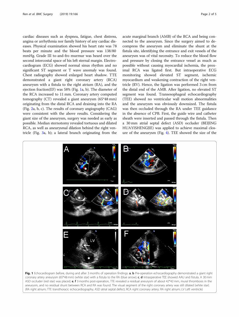
cardiac diseases such as dyspnea, fatigue, chest distress,angina or arrhythmia nor family history of any cardiac dis-eases. Physical examination showed his heart rate was 78beats per minute and the blood pressure was 138/80mmHg. Grade III to-and-fro murmur was heard over thesecond intercostal space of his left sternal margin. Electro-cardiogram (ECG) showed normal sinus rhythm and nosignificant ST segment or T wave anomaly was found.Chest radiography showed enlarged heart shadow. TTEdemonstrated a giant right coronary artery (RCA)aneurysm with a fistula to the right atrium (RA), and theejection fraction(EF) was 58% (Fig. 1a, b). The diameter ofthe RCA increased to 11mm. Coronary artery computedtomography (CT) revealed a giant aneurysm (65*48mm)originating from the distal RCA and draining into the RA(Fig. 2a, b, c). The results of coronary angiography (CAG)were consistent with the above results. Considering thegiant size of the aneurysm, surgery was needed as early aspossible. Median sternotomy revealed tortuous and dilatedRCA, as well as aneurysmal dilation behind the right ven-tricle (Fig. 3a, b); a lateral branch originating from the
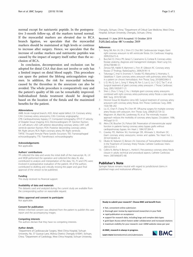
acute marginal branch (AMB) of the RCA and being con-nected to the aneurysm. Since the surgery aimed to de-compress the aneurysm and eliminate the shunt at thefistula site, identifying the entrance and exit vessels of theaneurysm was of vital necessity. To reduce the blood flowand pressure by closing the entrance vessel as much aspossible without causing myocardial ischemia, the prox-imal RCA was ligated first. But intraoperative ECGmonitoring showed elevated ST segment, ischemicmyocardium and weakening contraction of the right ven-tricle (RV). Hence, the ligation was performed 3 cm fromthe distal end of the AMB. After ligation, no elevated STsegment was found. Transesophageal echocardiography(TEE) showed no ventricular wall motion abnormalitiesand the aneurysm was obviously downsized. The fistulawas then occluded through the RA under TEE guidancein the absence of CPB. First, the guide wire and cathetersheath were inserted and passed through the fistula. Thena 30mm atrial septal defect (ASD) occluder (BEIJINGHUAYISHENGJIE) was applied to achieve maximal clos-ure of the aneurysm (Fig. 4). TEE showed the size of the
Fig. 1 Echocardiogram before, during and after 3 months of operation findings: a, b Pre-operation echocardiography demonstrated a giant rightcoronary artery aneurysm (65*48mm) (white star) with a fistula to the RA (blue arrow); c, d Intraoperative TEE showed AAU and fistula. A 30 mmASD occluder (red star) was placed; e, f 3 months post-operation, TTE revealed a residual aneurysm of about 42*42 mm, mural thrombosis in theaneurysm, and no residual shunt between RCA and RA was found. The visual segment of the right coronary artery was still dilated (white star).(RA right atrium; TTE transthoracic echocardiography; ASD atrial septal defect; RCA right coronary artery; RA right atrium; LV Left ventricle)
Ren et al. BMC Surgery (2019) 19:166 Page 2 of 5
aneurysm was obviously reduced, and no blood flow sig-nals, ventricular wall motion abnormalities or shunt wereobserved (Fig. 1c, d). The patient was discharged 9 daysafter operation. Aspirin and clopidogrel were administeredfor 3months. The follow-up revealed favorable outcomesand an improved quality of life for the patient.
Discussion and conclusionAlthough conventional CAA surgery under CPB hassome possible complications, including intraoperative
ischemia, impaired hemostasis and mechanical traumato blood cells [8, 9], many surgeons still believe it is the“gold-standard” operation for this type of pathology. Inthe present case, the surgery was performed in the ab-sence of CPB procedure not only to avoid the CPB- as-sociated complications [10], but also to monitor cardiacischemia in “real-time” and facilitate repair.In the present case, the giant CAA was formed at the
distal right posterior descending coronary artery (PDA).Since PDA is connected to the left anterior descending
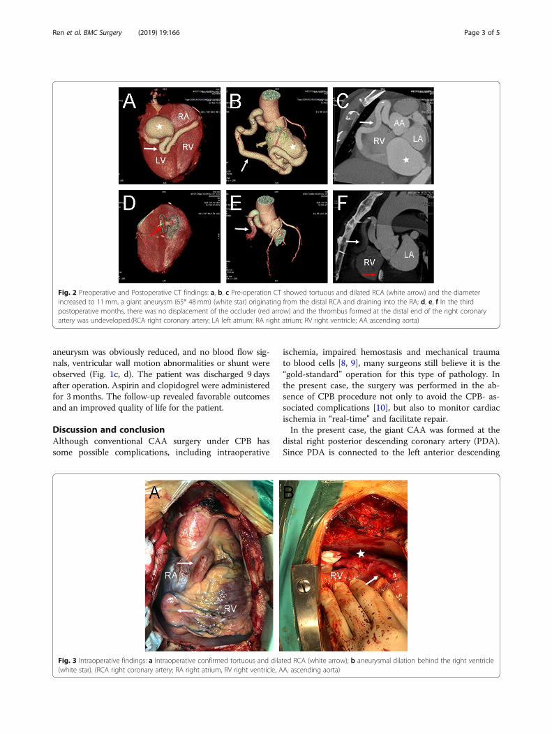
Fig. 2 Preoperative and Postoperative CT findings: a, b, c Pre-operation CT showed tortuous and dilated RCA (white arrow) and the diameterincreased to 11mm, a giant aneurysm (65* 48mm) (white star) originating from the distal RCA and draining into the RA; d, e, f In the thirdpostoperative months, there was no displacement of the occluder (red arrow) and the thrombus formed at the distal end of the right coronaryartery was undeveloped.(RCA right coronary artery; LA left atrium; RA right atrium; RV right ventricle; AA ascending aorta)
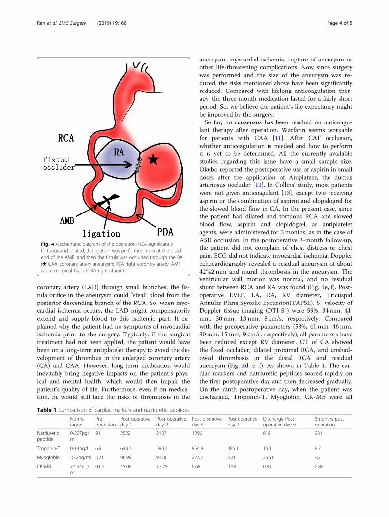
Fig. 3 Intraoperative findings: a Intraoperative confirmed tortuous and dilated RCA (white arrow); b aneurysmal dilation behind the right ventricle(white star). (RCA right coronary artery; RA right atrium, RV right ventricle, AA, ascending aorta)
Ren et al. BMC Surgery (2019) 19:166 Page 3 of 5
coronary artery (LAD) through small branches, the fis-tula orifice in the aneurysm could “steal” blood from theposterior descending branch of the RCA. So, when myo-cardial ischemia occurs, the LAD might compensatorilyextend and supply blood to this ischemic part. It ex-plained why the patient had no symptoms of myocardialischemia prior to the surgery. Typically, if the surgicaltreatment had not been applied, the patient would havebeen on a long-term antiplatelet therapy to avoid the de-velopment of thrombus in the enlarged coronary artery(CA) and CAA. However, long-term medication wouldinevitably bring negative impacts on the patient’s phys-ical and mental health, which would then impair thepatient’s quality of life. Furthermore, even if on medica-tion, he would still face the risks of thrombosis in the
aneurysm, myocardial ischemia, rupture of aneurysm orother life-threatening complications. Now since surgerywas performed and the size of the aneurysm was re-duced, the risks mentioned above have been significantlyreduced. Compared with lifelong anticoagulation ther-apy, the three-month medication lasted for a fairly shortperiod. So, we believe the patient’s life expectancy mightbe improved by the surgery.So far, no consensus has been reached on anticoagu-
lant therapy after operation. Warfarin seems workablefor patients with CAA [11]. After CAF occlusion,whether anticoagulation is needed and how to performit is yet to be determined. All the currently availablestudies regarding this issue have a small sample size.Okubo reported the postoperative use of aspirin in smalldoses after the application of Amplatzer, the ductusarteriosus occluder [12]. In Collins’ study, most patientswere not given anticoagulant [13], except two receivingaspirin or the combination of aspirin and clopidogrel forthe slowed blood flow in CA. In the present case, sincethe patient had dilated and tortuous RCA and slowedblood flow, aspirin and clopidogrel, as antiplateletagents, were administered for 3 months, as in the case ofASD occlusion. In the postoperative 3-month follow-up,the patient did not complain of chest distress or chestpain. ECG did not indicate myocardial ischemia. Dopplerechocardiography revealed a residual aneurysm of about42*42 mm and mural thrombosis in the aneurysm. Theventricular wall motion was normal, and no residualshunt between RCA and RA was found (Fig. 1e, f). Post-operative LVEF, LA, RA, RV diameter, TricuspidAnnular Plane Systolic Excursion(TAPSE), S′ velocity ofDoppler tissue imaging (DTI-S′) were 59%, 34 mm, 41mm, 30 mm, 13 mm, 8 cm/s, respectively. Comparedwith the preoperative parameters (58%, 41 mm, 46mm,30mm, 15 mm, 9 cm/s, respectively), all parameters havebeen reduced except RV diameter. CT of CA showedthe fixed occluder, dilated proximal RCA, and unshad-owed thrombosis in the distal RCA and residualaneurysm (Fig. 2d, e, f). As shown in Table 1. The car-diac markers and natriuretic peptides soared rapidly onthe first postoperative day and then decreased gradually.On the ninth postoperative day, when the patient wasdischarged, Troponin-T, Myoglobin, CK-MB were all
Fig. 4 A schematic diagram of the operation: RCA significantlytortuous and dilated, the ligation was performed 3 cm at the distalend of the AMB, and then the fistula was occluded through the RA.(★ CAA, coronary artery aneurysm; RCA right coronary artery, AMBacute marginal branch, RA right atrium)
Table 1 Comparison of cardiac markers and natriuretic peptides
Normalrange
Pre-operation
Post-operativeday 1
Post-operativeday 2
Post-operativeday 5
Post-operativeday 7
Discharge Post-operative day 9
3months post-operation
Natriureticpeptide
0-227pg/ml
91 2522 2137 1296 618 231
Troponin-T 0-14ng/L 6.9 648.7 590.7 954.9 485.1 15.3 8.7
Myoglobin <72ng/ml <21 98.99 91.86 22.57 <21 25.51 <21
CK-MB <4.94ng/ml
0.64 45.08 12.25 0.68 0.58 0.89 0.89
Ren et al. BMC Surgery (2019) 19:166 Page 4 of 5
normal except for natriuretic peptide. In the postopera-tive 3-month follow-up, all the markers turned normal.If the myocardial markers are elevated due to RCAbranch ligation, we speculate that the myocardialmarkers should be maintained at high levels or continueto increase after surgery. Hence, we speculate that theincrease of cardiac markers and natriuretic peptides wascaused by the impact of surgery itself rather than the oc-clusion of RCA.In conclusion, decompression and exclusion can be
adopted for distal CAA that does not involve or only hasa limited impact on distal blood supply. This procedurecan spare the patient the lifelong anticoagulation regi-men. In addition, the risk for myocardial ischemiacaused by the thrombus in the aneurysm can also beavoided. The whole procedure is comparatively easy andthe patient’s quality of life can be remarkably improved.Individualized fistula treatment should be designedbased on the location of the fistula and the maximizedbenefits for the patient.
AbbreviationsAMB: Acute marginal branch; ASD: Atrial septal defect; CA: Coronary artery;CAA: Coronary artery aneurysms; CAG: Coronary angiography;CPB: Cardiopulmonary bypass; CT: Computed tomography; DTI-S′: S′ velocityof Doppler tissue imaging; ECG: Electrocardiogram; EF: Ejection fraction;LA: Left atrium; LAD: Left anterior descending coronary artery; LVEF: Leftventricle ejection fraction; PDA: Posterior descending coronary artery;RA: Right atrium; RCA: Right coronary artery; RV: Right ventricle;TAPSE: Tricuspid Annular Plane Systolic Excursion; TEE: Transesophagealechocardiography; TTE: Transthoracic echocardiography
AcknowledgementsNot applicable.
Authors’ contributionsYR collected the data and wrote the initial draft of the manuscript. KL, LXand WQR performed the operation and collected the data. KL alsocontributed to analysis and interpretation of the data. YJL, PJ and CPG wereinvolved in postoperative evaluation of the patient. All of the authorscontributed to drafting and critically revising the paper and gave finalapproval of the version to be published.
FundingThis study received no financial support.
Availability of data and materialsThe datasets used and analyzed during the current study are available fromthe corresponding author on reasonable request.
Ethics approval and consent to participateNot applicable.
Consent for publicationWritten informed consent was obtained from the patient to publish this casereport and the accompanying images.
Competing interestsThe authors declare that they have no competing interests.
Author details1Department of Cardiovascular Surgery, West China Hospital, SichuanUniversity, No. 37 Guoxue Lane, Wuhou District, Chengdu 610041, Sichuan,China. 2Department of Cardiology, West China Hospital, Sichuan University,
Chengdu, Sichuan, China. 3Department of Critical Care Medicine, West ChinaHospital, Sichuan University, Chengdu, Sichuan, China.
Received: 11 June 2019 Accepted: 15 October 2019
References1. Kang SM, Kim JH, Oh J, Shim CY, Choi BW. Cardiovascular images. Giant
right coronary aneurysm to left ventricular fistula. Circ Cardiovasc Imaging.2009;2(3):e15–6.
2. Buccheri D, Chirco PR, Geraci S, Caramanno G, Cortese B. Coronary arteryfistulae: anatomy, diagnosis and management strategies. Heart Lung Circ.2018;27(8):940–51.
3. Zenooz NA, Habibi R, Mammen L, Finn JP, Gilkeson RC. Coronary arteryfistulas: CT findings. RadioGraphics. 2009;29:781–9.
4. Tokunaga C, Imai A, Enomoto Y, Tanaka YO, Matsushita S, Hiramatsu Y,Sakakibara Y. Giant coronary artery aneurysm with pulmonary artery fistulain a patient on chronic hemodialysis. Ann Thorac Surg. 2010;89(3):963–5.
5. Li D, Wu Q, Sun L, Song Y, Wang W, Pan S, Luo G, Liu Y, Qi Z, Tao T, et al.Surgical treatment of giant coronary artery aneurysm. J Thorac CardiovascSurg. 2005;130(3):817–21.
6. Shen J, Zhou Y, Fang Z, Hu J. Multiple giant coronary artery aneurysmscombined with right coronary artery-pulmonary artery fistula: a case report.BMC Surg. 2019;19(1):80.
7. Hironori Inoue M, Masahiro Ueno MD. Surgical treatment of coronary arteryaneurysm with coronary artery fistula. Ann Thorac Cardiovasc Surg. 2009;15(3):198–202.
8. Liu JC, Chan P, Chang TH, Chen RF. Off-pump surgery for multiple coronaryartery fistulas with aneurysm. Ann Thorac Surg. 2006;81(2):729–32.
9. Magovern JA, Mack MJ, Landreneau RJ, et al. The minimally invasiveapproach reduces the morbidity of coronary artery bypass. Circulation. 1996;94(Suppl I):I–51.
10. Akins CW, Boucher CA, Pohost GM. Preservation of interventricular septalfunction in patients having coronary artery bypass grafts withoutcardiopulmonary bypass. Am Heart J. 1984;107:304–9.
11. Crawley PD, Mahlow WJ, Huntsinger DR, Afiniwala S, Wortham DC.Giant coronary artery aneurysms: review and update. Tex Heart Inst J.2014;41(6):603–8.
12. Okubo M, Nykanen D, Benson LN. Outcomes of Transcatheter Embolizationin the Treatment of Coronary Artery Fistulas. Catheter Cardiovasc Interv.2001;52:510–7.
13. Collins N, Mehta R, Benson L, Horlick E. Percutaneous coronary artery fistulaclosure in adults: technical and procedural aspects. Catheter CardiovascInterv. 2007;69(6):872–80.
Publisher’s NoteSpringer Nature remains neutral with regard to jurisdictional claims inpublished maps and institutional affiliations.
Ren et al. BMC Surgery (2019) 19:166 Page 5 of 5