Fisher research report FINAL.pdf
87
Indigenous Salvia species - an investigation of the antimicrobial activity, antioxidant activity and chemical composition of leaf extracts Vanessa Louise Fisher Student number: 9604111J A research report submitted to the faculty of Health Sciences, University of the Witwatersrand, Johannesburg, in partial fulfillment of the requirements for the degree of Master of Science in Medicine (Pharmacotherapy). Johannesburg, 2005
Transcript of Fisher research report FINAL.pdf

Indigenous Salvia species - an investigation of the
antimicrobial activity, antioxidant activity and chemical
composition of leaf extracts
Vanessa Louise Fisher
Student number: 9604111J A research report submitted to the faculty of Health Sciences, University of the
Witwatersrand, Johannesburg, in partial fulfillment of the requirements for the degree of
Master of Science in Medicine (Pharmacotherapy).
Johannesburg, 2005

Declaration
I, Vanessa Louise Fisher declare that this research report is my own work. It is being
submitted for the degree of MSc (Med) Pharmacotherapy in the University of the
Witwatersrand, Johannesburg. It has not been submitted before for any degree or
examination at this or any other university.
……………………..
……… day of ……………….., 2005
2

Acknowledgements
I would like to thank the following:
My supervisors Prof. A. M. Viljoen and Ms S. F. van Vuuren.
Prof. Başer, Drs Betűl Demirci and Temel Őzek from the Medicinal and Aromatic Plant
and Drug Research Centre, University of Anadolu, Turkey for their help with the GC-MS
analysis during my visit to the Research Centre.
The staff at the Walter Sisulu Botanical Garden for access to plant material and Mr. J.
Vlok and Dr. J. Manning for collecting and identifying plant material.
The National Research Foundation, University of the Witwatersrand Research Committee
and Faculty of Health Sciences Research Endowment Fund for financial support.
3

TABLE OF CONTENTS
LIST OF FIGURES 6
LIST OF TABLES 9
ABSTRACT 10
CHAPTER 1:
INTRODUCTION
11
CHAPTER 2:
MATERIALS AND METHODS
19
2.1 Collection of plant material 19
2.1.1 Essential oil isolation 19
2.1.2 Preparation of methanol and acetone extracts 19
2.2 Gas chromatography mass spectroscopy (GC/MS) 20
2.2.2 Thin layer chromatography 20
2.3 Antimicrobial studies 21
2.3.1 Microbial strains 21
2.3.2 Disc diffusion method 22
2.3.3 Determination of minimum inhibitory concentration (MIC) 22
CHAPTER 3:
MONOGRAPHS OF SALVIA SPECIES STUDIES
25
Salvia africana-caerulea 26
Salvia africana-lutea 30
Salvia chamelaeagnea 34
Salvia disermas 35
Salvia dolomitica 38
Salvia lanceolata 42
4

Salvia namaensis 45
Salvia runcinata 49
Salvia stenophylla 52
Salvia verbenaca 55
CHAPTER 4:
RESULTS AND DISCUSSION
57
4.1 Analytical chemistry 57
4.2 Antimicrobial activity 61
4.3 Antioxidant activity 75
CHAPTER 5:
CONCLUSION
79
CHAPTER 6:
REFERENCES
81
5

LIST OF FIGURES Figure 1: Process of hydrodistillation. 24
Figure 2: Preparing discs for disc diffusion assay. 24
Figure 3: Preparing microplate. 24
Figure 4: TLC performed on extracts and rosmarinic acid. 24
Figure 5: S. africana-caerulea. 26
Figure 6: Geographical distribution of S. africana-caerulea. 26
Figure 7: Gas chromatography profile of S. africana-caerulea collected on the 27
West Coast, Dynefontein.
Figure 8: Chemical structure of the major compounds identified in S. africana- 29
caerulea essential oil.
Figure 9: S. africana-lutea. 30
Figure 10: Geographical distribution of S. africana-lutea. 30
Figure 11: Gas chromatography profile of S. africana-lutea collected on the West 31
Coast, Dynefontein.
Figure 12: Chemical structure of the major compounds identified in S. africana- 32,33
lutea essential oil.
Figure 13: S. chamelaeagnea. 34
Figure 14: Geographical distribution of S. chamelaeagnea. 34
Figure 15: S. disermas. 35
Figure 16: Geographical distribution of S. disermas. 35
Figure 17: Gas chromatography profile of S. disermas collected at Mossel Bay. 36
Figure 18: Chemical structure of the major compounds identified in S. disermas 37
essential oil.
Figure 19: S. dolomitica. 38
Figure 20: Geographical distribution of S. dolomitica. 38
Figure 21: Gas chromatography profile of S. dolomitica collected at the Walter 39
Sisulu Botanical Garden.
Figure22: Chemical structure of the major compounds identified in S. dolomitica 41
essential oil.
6

Figure 23: S. lanceolata. 42
Figure 24: Geographical distribution of S. lanceolata. 42
Figure 25: Gas chromatography profile of S. lanceolata collected on the West 43
Coast, Dynefontein.
Figure 26: Chemical structure of the major compounds identified in S. lanceolata 44
essential oil.
Figure 27: S. namaensis. 45
Figure 28: Geographical distribution of S. namaensis. 45
Figure 29: Gas chromatography profile of S. namaensis collected on Swartberg 46
Pass.
Figure 30: Chemical structure of the major compounds identified in S. namaensis 48
essential oil.
Figure 31: S. runcinata. 49
Figure 32: Geographical distribution of S. runcinata. 49
Figure 33: Gas chromatography profile of S. runcinata collected at Tarlton. 50
Figure 34: Chemical structures of the major compounds identified in S. runcinata 51
essential oil.
Figure 35: S. stenophylla. 52
Figure 36: Geographical distribution of S. stenophylla. 52
Figure 37: Gas chromatography profile of S. stenophylla collected at Lady Grey. 53
Figure 38: Chemical structure of the major compounds identified in S. stenophylla 54
essential oil.
Figure 39: S. verbenaca. 55
Figure 40: Geographical distribution of S. verbenaca. 56
Figure 41: TLC plate of Salvia methanol extracts and rosmarinic acid. 60
Figure 42: Disc diffusion plate of S. aureus. 64 – S. dolomitica (methanol); 68 – 67
S. namaensis (acetone); 69 – S. namaensis (methanol); S. africana-
lutea (essential oil). Neomycin (control) in middle of plate.
Figure 43: Disc diffusion plate of S. aureus. 78 - S. africana-caerulea (acetone); 67
81 – S. runcinata (acetone); 83 – S. stenophylla (methanol); 84 –
S. stenophylla (acetone). Neomycin (control) in middle of plate.
7

Figure 44: Chemical structures of the major antioxidant compounds in Salvia. 76
8

LIST OF TABLES Table 1: Traditional uses of Salvia in southern Africa. 12,13
Table 2: Table of study samples with voucher number and locality. 19
Table 3: Table of test organisms included in the study. 21
Table 4: GC-MS results of S. africana-caerulea. 27
Table 5: GC-MS results of S. africana-lutea. 31
Table 6: GC-MS results of S. disermas. 36
Table 7: GC-MS results of S. dolomitica. 39
Table 8: GC-MS results of S. lanceolata. 43
Table 9: GC-MS results of S. namaensis. 46
Table 10: GC-MS results of S. runcinata. 50
Table 11: GC-MS results of S. stenophylla. 53
Table 12: Salvia species analysed with the aid of GC-MS and the percentage 57
oil yield after distillation.
Table 13: The major compounds identified in the Salvia species. 58
Table 14: Antibacterial screening results from the disc diffusion assay. Results 61
expressed in millimeters (mm) from disc edge.
Table 15: Minimum Inhibitory Concentration (MIC) of Salvia against B. cereus. 65
9

ABSTRACT The genus Salvia, commonly known as the sages is a member of the mint family
(Lamiaceae). In Latin, ‘sage’ means "to save" and the Romans called it "sacred herb".
Throughout history it has been used for depression, fever, respiratory infections, women's
complaints, sleep inducer, diuretic, gargles and sick room use. The essential oil is
reported to have anti-inflammatory, antimicrobial, antioxidant, antiseptic, antispasmodic,
astringent, diuretic, antihypertensive and insecticidal properties. Of the 900 species
recorded worldwide, 30 are indigenous to South Africa where they are used extensively
in traditional healing.
The aerial parts of twelve samples were hydrodistilled and the essential oil analysed by
GC-MS. The essential oil composition varied quantitatively and qualitatively within the
different Salvia species analysed. Linalool was the only compound that was present in all
the essential oils. β-caryophyllene and caryophyllene oxide were present in all essential
oil with the exception of S. stenophylla.
The essential oil as well as methanol and acetone extracts were tested for antimicrobial
activity on a number of bacteria and fungi. No species showed activity against
Cryptococcus neoformans, Candida albicans nor Alternaria alternata. All test samples
studied demonstrated variable degrees of antibacterial activity with the exception of four
test samples; S. disermas (methanol and acetone) from the Walter Sisulu Botanical
Garden; S. disermas (methanol) from Mossel Bay and S. lanceolata (methanol). Gram-
positive organisms were more sensitive to the test samples than the Gram-negative
organisms. In general, the extracts were far more active than the essential oils.
Thin layer chromatography indicated that all methanol extracts possess antioxidant
activity. All methanol extracts contain the antioxidant compound, rosmarinic acid. It is
evident that, in addition to rosmarinic acid, other polar and non-polar compounds are
present in all Salvia species that also act as antioxidants.
10

CHAPTER 1: INTRODUCTION
Salvia is a large and polymorphous genus of the Lamiacea family, comprising 900
species with a cosmopolitan distribution (Ulubelen, 1994). The genus name Salvia is
derived from the Latin salvare which means “to be saved” or “to cure”. This name was
corrupted to sauge in French and sawge in Old English and eventually to sage, as we
commonly know it today (Blumenthal et al., 1998). Salvia has been prized since ancient
times and has enjoyed a reputation for being a panacea. The Romans referred to it as the
“sacred herb” and the Spanish have referred to it as ierba buena or “good herb”
(Baricevic and Bartol, 2000) in reference to its curative and healing properties. Salvia is
an important genus that is widely cultivated and has been used for centuries in foods for
flavouring and seasoning in addition to being used for the treatment of various medical
conditions. Throughout history Salvia has been used for depression, fever, respiratory
infections, women’s complaints, sleep inducer, diuretic, gargles, night sweats, memory
enhancer, disinfectant and sick room use. The essential oil is reported to have anti-
inflammatory, antimicrobial, antioxidant, antiseptic, antispasmodic, astringent, anti-
hypertensive and insecticidal properties.
The distribution of the genus extends all over northern Africa from west to east and
southwards to the east African highlands. The genus is absent from most of western and
central tropical Africa. This means that the species in southern Africa are geographically
isolated from the species on the northern part of the continent. Few species are common
to both northern and southern Africa (Jäger and van Staden, 2000). Southern Africa is
home to 30 species of the genus Salvia. In South Africa, Salvia only occurs in sparse
populations. A few species, such as S. africana-lutea and S. chamelaeagnea are relatively
common in the Knysna and the Cape regions. Salvia sclarea, S. coccinea, S. reflexa, and
S. tiliifolia have been introduced into southern Africa and are thus not endemic to the
region. However, the latter three species have become naturalized in parts of South
Africa. S. verbenaca is more than likely indigenous to the Mediterranean countries and
the Canary Islands and has spread into Europe and Asia. If it is an introduced plant in our
11

Flora, it is now widely distributed and considered indigenous to South Africa (Codd,
1985).
In southern Africa traditional medicine plays an important role in health care. In South
Africa it has been estimated that eighty percent of the total black population consults with
traditional healers (Jäger and van Staden, 2000). These traditional healers and herbalists
have centuries of experience and a vast knowledge in using plants for medicinal and
healing purposes. Traditional usages of some indigenous Salvia species and their
ethnomedicinal uses are summarised in Table 1. The table includes the uses of Salvia
across all the ethnic groups in South Africa as well as the European settlers.
Table 1: Traditional uses of Salvia in southern Africa (summarised from Jäger and van
Staden, 2000).
Species Medicinal use
Salvia africana-caerulea • Used as a remedy for coughs, colds and chest troubles
• Prepared as a tea and used as a remedy for colic,
diarrhoea, heartburn and indigestion
Salvia africana-lutea • Used for colds and coughs • Used to treat a variety female ailments
Salvia chamelaeagnea • Infusion of the dry leaf used for convulsions
• Used for coughs, colds, fever, bronchitis, whooping
cough, flu and general chest complaints • Also used for head-, ear- and stomach pains • Used for the treatment of burns • Helpful in the treatment of a variety of female ailments • Used in the treatment of diarrhoea • Has caused livestock death
Salvia coccinea • Used in the treatment of kidney diseases • Used to relieve the cough associated with pulmonary tuberculosis
12

Species Medicinal use
Salvia disermas • In the form of a lotion it is used to treat sores • Used as an anti-hypertensive • Used to treat rheumatism
Salvia reflexa • Has caused livestock death
Salvia repens • Is added to the bath water for treating sores on the body • A decoction of the root is administered before meals for stomach ache and diarrhoea • The smoke of the burning plant is used to disinfect houses
Salvia runcinata • A decoction of the root, leaf and stem taken for the relief of urticaria
• The smoke of the burning plant is used to disinfect houses • A paste of the leaf is used as a purgative in infants
Salvia scabra • A paste of the leaf is made with mother’s milk and given as the first medicine to infants
• A water extract of the roots is also given daily to infants
until they are two months old
Salvia sclarea • Used to treat diarrhoea and stomach aches
• A milk decoction is used for the alleviation of a sore
throat • A water decoction is used to treat sores and swellings
Salvia stenophylla • The smoke of the burning plant is used to disinfect houses
Very little phytochemical work has been done on indigenous Salvia species (Jäger and
van Staden, 2000). Diterpenoids are the main compounds present in the roots of Salvia
plants whilst aerial parts usually contain flavonoids, which as in many Lamiaceae, are
flavones substituted in the 6-position. Triterpenoids, derived from ursane (chiefly ursolic
acid) as well as essential oils with volatile compounds such as monoterpenoids (Baricevic
and Bartol, 2000) are also present in the genus. Salvia is also an abundant source of
phenolic acids. The majority of the phenolic acids found in Salvia are those of caffeic
acid derivatives, which are unique to Salvia except for rosmarinic acid and lithospermic
13

acid (Lu and Foo, 2002). Some of these compounds have been isolated and their structure
elucidated, however, many compounds are still scientifically challenging and
pharmacological studies with isolated constituents are still lacking. In addition to this,
there is much variety in bioactivity within the genus due to the diversity of species
present in the genus. More studies on structure-activity relationships within Salvia are
needed in order to explain mechanisms of biological activity.
There has been a renaissance of interest in natural occurring therapeutic agents (Carson
and Riley, 1995). The essential oils and extracts of many plant species have become the
subjects of research in the past few years, and attempts to characterize their bioactive
principles have recently gained momentum in many pharmaceutical and food processing
applications (Vardar-Űnlű et al., 2003). This increased level of interest in natural product
research can be attributed to the following factors; unmet therapeutic needs, the
remarkable diversity of both chemical structures and biological activities of naturally
occurring secondary metabolites, the utility of bioactive natural products as biochemical
and molecular probes, the development of novel and sensitive techniques to detect
biologically active natural products, improved techniques to isolate, purify, and
structurally characterize these active constituents and advances in solving the demand for
supply of complex natural products (Clark, 1996).
The potential use of higher plants as a source of new drugs is still poorly explored. Only a
small percentage of the estimated plant species has been investigated phytochemically
and an even smaller percentage has been properly studied in terms of their
pharmacological properties. In most cases those plant species that have been investigated
have only been screened or alternatively had preliminary studies carried out on them.
About twenty five percent of the drugs prescribed worldwide come from plants, 121 such
active compounds being in current use. Of the 252 drugs considered as basic and
essential by the World Health Organisation (WHO), eleven percent are exclusively of
plant origin and a significant number are synthetic drugs obtained from natural precursors
(Rates, 2001). Natural compounds can be lead compounds, allowing the design and
rational planning of new drugs, biomimetic synthesis development and the discovery of
14

new therapeutic properties not yet attributed to known compounds from plants. The
negative side is that the research and development of these compounds from plant origin
is a difficult and expensive task with each new drug requiring vast sums of money and
many years of hard work.
Plants can be used as therapeutic resources in several ways. They can be used as herbal
teas or other home made remedies when they are considered as medicinal plants. They
can be used as crude extracts or standard enriched fractions in preparations such as
tinctures, fluid extracts, powder, pills and capsules, when they are considered as
phytopharmaceutical preparations or herbal medicines. Finally, the plants can be
subjected to successive extraction and purification procedures to isolate compounds of
interest, which can themselves be active and used directly as a drug or they can be
precursors in the processes or synthesis of a drug (Rates, 2001). Traditionally, indigenous
Salvia is used in the form of water and milk decoctions, extracts, tinctures, lotions,
infusions, pastes and teas. This research report aims to determine the biological activity
of methanol and acetone extracts as well as the essential oils of selected indigenous
Salvia species. The biological activities investigated will include the assessment of the
antimicrobial and antioxidant activity.
Essential oils are accepted and recognised in academia, in the chemical industry and,
more recently, in everyday life. Essential oils are aromatic substances produced by
certain plants. They have been termed essential because it was once thought that each oil
represented the essence of the original plant (Nakatsu et al., 2000). Plant essential oils are
a potentially useful source of antimicrobial compounds (Friedman et al., 2002) and their
antimicrobial activities have been recognised, albeit empirically, for centuries (Faleiro et
al., 2003). This antimicrobial activity could act as a chemical defense mechanism against
plant pathogenic diseases. Damage or “wounding” of leaves, which are covered with
volatile oil glands, results in the rupture of glands causing the oil to flow over the area or
“wound” (Svoboda and Hampson, 1999). The existence of antimicrobial activity in the
essential oil would be of substantial benefit to the plant in such circumstances. These
antimicrobial compounds are therefore important in the control of plant and human
15

diseases of microbial origin. Antimicrobials are among the most important classes of
therapeutic agents and have had an enormous impact on both life expectancy and the
quality of life (Clark, 1996).
Natural antimicrobial compounds are needed and highly sought after as there is an
increased need for safer antimicrobial food preservatives which are more acceptable than
the present preservatives whose safety in food is sometimes questionable. Food
processors, food safety researchers and regulatory agencies have been increasingly
concerned with the growing number of food borne illness outbreaks caused by certain
pathogens. Several reports on the antimicrobial effectiveness of essential oils in foods
suggest that the use of these essential oils may improve food safety (Friedman et al.,
2002).
Salvia is also of particular interest because it may be used for the production of raw
materials or preparations that are in need of phytochemicals with significant antioxidant
capacity (Exarchou et al., 2002). Antioxidants play a role in protecting the body against
the damage caused by free radicals. The importance of free radicals in the aetiology of
certain diseases has been increasingly recognised. In fact, free radicals have been
implicated in more than one hundred diseases. They are produced in the body through
normal metabolic pathways. Exogenous sources of free radicals include tobacco smoke,
ionizing radiation, certain pollutants, organic solvents and even pesticides. When
produced in excess, free radicals have the ability to cause tissue injury. Additional dietary
supplies of natural antioxidants act as protective agents against damage by free radicals
and can ultimately act as efficient scavengers of free radicals before any tissue damage is
done.
The family Lamiaceae is considered to be a promising source of natural antioxidants
(Hohmann et al., 1999). It is believed that the antioxidant properties of Salvia can be
largely attributed to the rosmarinic acid, (Exarchou et al., 2002; Petersen and Simmonds,
2003) carnosic acid and carnosol content (Wang et al., 1999). In addition to these three
main compounds, the antioxidant activity of Salvia may also be partly due to the presence
16

of other phenolic compounds such as flavonoids (Exarchou et al., 2002) as well as
diterpenes and triterpenes (Wang et al., 1999). However, investigative studies have
concluded that the flavonoids have a rather small contribution to the total antioxidant
activity of extracts (Exarchou et al., 2002).
Within the Lamiaceae, rosmarinic acid is restricted to the sub-family Nepetoideae
however, it has also been found in other plant families (Petersen and Simmonds, 2003).
This distribution shows that the occurrence of rosmarinic acid cannot be used as a
chemotaxonomical marker to differentiate among different plant families. Therefore
extracts that are rich in rosmarinic acid have high radical scavenging activity. In saying
this, Salvia is an ideal candidate to investigate phytochemically as a potential, useful,
natural antioxidant.
The process of researching and developing a therapeutic compound or material species in
South Africa from plant origin is a multi-disciplinary process. There is an integration of
sciences including botany, pharmacology and chemistry which all need to be carefully
and thoroughly investigated. The use of Salvia therefore, has been largely empirical. The
aim of this report is to further investigate the antimicrobial and the antioxidant activities
of Salvia species in South Africa and to scientifically validate the appropriate use of
Salvia in African traditional healing and also in today’s modern world.
17

Study Objectives:
• To analyse the composition of the essential oils using gas chromatography-mass
spectroscopy.
• To determine the antimicrobial properties of the essential oils, methanol and acetone
extracts of selected species.
• To determine the preliminary antioxidant properties of the methanol extracts of
selected species.
• To scientifically validate the traditional use of Salvia especially in traditional herbal
preparations.
18

CHAPTER 2: MATERIALS AND METHODS
2.1 Collection of plant material
Ten Salvia species (twelve samples) were collected from the wild or the Walter Sisulu
Botanical Garden. Voucher specimens are housed in the Department of Pharmacy and
Pharmacology, University of the Witwatersrand.
Table 2: Table of study samples with voucher number and locality.
Species Locality Voucher S. africana-caerulea West Coast – Dynefontein AV 570 S. africana-lutea West Coast – Dynefontein AV 572 S. chamelaeagnea Walter Sisulu Botanical Garden AV584 S. disermas Walter Sisulu Botanical Garden AV 583 S. disermas Mossel Bay AV502 S. disermas Kokerboomwoud AV 250 S. dolomitica Walter Sisulu Botanical Garden AV 602 S. lanceolata West Coast – Dynefontein AV 571 S. namaensis Swartberg Pass AV 501 S. runcinata Tarlton AV 632 S. stenophylla Lady Grey AV 618 S. verbenaca De Rust AV 631
2.1.1 Essential oil isolation
Essential oils were extracted, from the aerial parts of individual Salvia species, by
hydrodistillation in a Clevenger-type apparatus for three hours. (Figure 1) The plant
material was weighed before distillation and the amount of oil weighed after distillation
to determine the yield of oil. Due to the volatile nature of these essential oils, they were
stored in tightly sealed amber vials and refrigerated. All oils were recovered without
solvent and were less dense than water.
2.1.2 Preparation of methanol and acetone extracts
The oven dried (30 °C) and finely ground plant material was weighed out into five-gram
samples. From each Salvia species a methanol and acetone extract was prepared. Extracts
were extracted and filtered twice and reconstituted to 128.00 mg/ml, unlike the essential
oils that were used without any dilution. Previous studies within the microbiology
19

laboratory Department of Pharmacy and Pharmacology at The University of the
Witwatersrand have confirmed no antimicrobail activity for acetone independently. This
is in support of literature (Eloff, 1998, NCCLS, 2003).
2.2 Analytical chemistry
2.2.1 Gas chromatography mass spectroscopy (GC/MS)
The analysis of the essential oil was performed using a Hewlett-Packard GCD system
equipped with a HP-Innowax column (60 m x 0.25 mm ∅., with 0.25 μm film thickness).
Temperature at the injection port was 250 °C. Column temperature was initially 60 °C for
ten minutes then gradually increased to 220 °C at a 4 °C per minute rate. Temperatures
were held at 220 °C for ten minutes until it was finally raised to 240 °C at a rate of 1 °C
per minute. The time in the column added up to a total of 80 minutes. Helium was the
carrier gas at a flow rate of 0.7 ml/minute. An electron ionization system with an
ionization energy of 70 eV was used with a split ratio of 50:1. The mass range was
between 35 and 425 m/z. 0.9 μl of hexane was added to 0.1 μl of the essential oil and was
injected. Data was recorded after a 5 minute lag phase. Identification took place using the
TBAM’S database libraries by matching both retention indices and mass spectral
fragmentation patterns.
2.2.2 Thin layer chromatography
All thin layer chromatography was performed using silica plates (silica gel 60 with
fluorescent indicator UV254. Layer: 0.20 mm. Macherey-Nagel, Germany). The mobile
phase used:
1. Chloroform-ethyl acetate-benzene-formic acid (2,4:2:1:0,6) was used for the
methanol extracts and a rosmarinic acid standard. (Figure 4)
After development the TLC plates (10 cm) were air-dried and detection was made
possible with the use of a spray reagent, vanillin-sulphuric acid (10 % acid). Colour
development took place after a short heating period at 100 ºC.
20

Rosmarinic acid (Fluka 435285/1 52902), in its pure form, was obtained and used as a
standard. Rosmarinic acid was subjected to thin layer chromatography on silica gel plates
along with the other methanol extracts of the test samples. The volume applied (spotted)
for the methanol extract (of a 128.00 mg/ml dilution) and rosmarinic acid was 3 μl, in
order to keep the same concentration of test samples for standardization. For
identification and visulization of antioxidant properties the plates were sprayed with a
DPPH (0.038/100 ml of methanol) spray reagent.
2.3 Antimicrobial studies
2.3.1 Microbial strains
Salvia has traditionally been used to treat diarrhoea, stomach complaints and other minor
digestive disturbances, therefore Escherichia coli, Bacillus cereus and Yersinia
enterocolitica were selected as reference test strains. Certain species have been used to
treat kidney infections and gynecological ailments therefore Candida albicans and
Escherichia coli were included in the study. Due to the fact that Salvia has been used as a
remedy for general respiratory infections Klebsiella pneumoniae has been selected. In
addition to this, Salvia has been used as an antiseptic and a remedy to treat skin infections
and inflammation therefore Staphylococcus aureus has been selected. All antimicrobial
tests were carried out under sterile conditions. All assays were undertaken in duplicate
and where necessary confirmed with a third test.
Table 3: Table of test organisms included in the study.
Gram-negative test organisms Escherichia coli ATCC 11775 Yersinia enterocolitica ATCC 23715 Klebsiella pneumoniae ATCC 13883
Gram-positive test organisms Staphylococcus aureus ATCC 12600
Bacillus cereus ATCC 11778 Yeast test organisms Cryptococcus neoformans ATCC 90112
Candida albicans ATCC 10231 Fungal test organism Alternaria alternata Clinical strain
21

2.3.2 Disc diffusion method
The bacterial and fungal cultures were harvested from stock agar plates. The bacterial
cultures were inoculated in Mueller Hinton (Oxoid) broth, whilst the fungal cultures were
harvested from Sabourauds Dextrose (Oxoid) agar. Base layers of autoclaved Mueller
Hinton (Oxoid) agar was prepared for bacterial cultures and Sabourauds Dextrose
(Oxoid) agar was prepared for fungal and yeast cultures. This method was employed
whereby yeast, bacterial or fungal spore suspensions yielding an approximate inoculum
size of 1 x 10⁶ CFU/ml was incorporated into Tryptone Soya (Oxoid) agar. With aseptic
manipulation, sterile 6 mm discs with approximately 15-20 μl either plant essential oil,
plant methanol extract or plant acetone extract (Figure 2) were introduced onto the
seeded agar plate. Positive bacterial controls, in the form of antibiotic discs, were
included in the assay. For the Gram-positive and Gram-negative bacteria Neomycin (30
μg, Oxoid) was used as a control and for the mould and yeasts, Nystatin (100 IU, Oxoid)
was used as a control. The plates were initially placed in the refrigerator for one hour to
allow diffusion of the disc contents into the agar with minimal bacterial or fungal growth,
while this takes place. Thereafter the bacterial plates were incubated at 37 °C for 24
hours. The yeasts were also incubated at 37 °C but for 48 hours. Alternaria alternata was
incubated at 25 °C for four days. The zones of inhibition were thereafter examined.
Measurements were recorded from the edge of the disc to the end of the zone of
inhibition and recorded in millimeters.
2.3.3 Determination of minimum inhibitory concentration (MIC)
A minimum inhibitory concentration (MIC) was determined for those test samples that
displayed antimicrobial activity from the disc diffusion assay (NCCLS, 2003). The MIC
was determined using the ρ-Iodonitrotetrazolium (INT) microplate method (Eloff, 1998).
Bacillus cereus was inoculated in Tryptone Soya Broth and incubated for 24 hours at 37
°C. Two microplates were prepared (Figure 3) so as to cover the necessary test samples.
An inoculum of 1 ml was transferred into 100 ml sterile broth to give a 1:100 stock
culture. Methanol and acetone test samples were prepared starting at a concentration of
16.000 mg/ml in the first well. The series of dilutions were carried out so that a final
concentration of 0.125 mg/ml was obtained in the eighth and final well. For the essential
22

oils, 51.200 mg of each essential oil was weighed out and 400 μl of acetone was added.
This gave the desired concentration of 32.000 mg/ml in the first well, taking into account
the sterile water and 100 μl culture added. The microplates were aseptically prepared
whereby 100 μl of sterile water was added to each well in the microplates. An amount of
100 μl of extract / essential oil was added to the first row of wells and a series of dilutions
(diluting 100 μl each time) was performed. An amount of 100 μl of prepared Bacillus
cereus stock culture was added to all the wells. Both negative and positive controls were
included. Ciprofloxacin (Oxoid) was included as a positive control. Stock solutions of
0.01 mg/ml were prepared and serially diluted as in assays performed with test samples.
The addition of the test samples to the wells and the serial dilutions took place under
laminar airflow so as to minimize the risk of contamination. The microplates were sealed
to prevent mixing or contamination of wells and were incubated at 37 °C. After 24 hours,
INT solution was prepared at a concentration of 0.2 mg/ml and 40 μl INT was added to
each well in both microplates. The compound INT is used to indicate biological activity
because the colourless compound acts as an electron acceptor and is reduced to a
coloured product by biologically active organisms (Eloff, 1998). The contents of the well
turned to red or purple if any bacterial growth was present. The microplates were visually
examined after 6 hours and the MIC’s were determined.
23

Figure 1: Process of hydrodistillation. Figure 2: Preparing discs for disc diffusion assay.
Figure 3: Preparing microplate. Figure 4: TLC performed on extracts and rosmarinic acid.
24

CHAPTER 3: MONOGRAPHS OF SALVIA SPECIES STUDIED Salvia africana-caerulea
Salvia africana-lutea
Salvia chamelaeagnea
Salvia disermas
Salvia dolomitica
Salvia lanceolata
Salvia namaensis
Salvia runcinata
Salvia stenophylla
Salvia verbenaca
25

Salvia africana-caerulea L. E. Codd 1. Botanical description Shrub 0,6 – 1,5 m tall, often branching at the base with several erect, usually sparingly
branched stems. Leaves: blade simple, petiolate, greenish and vary in size and shape.
Flowers often dense, light blue to bluish purple or pinkish in colour.
Figure 5: S. africana-caerulea. 2. Distribution In coastal fynbos and on rocky slopes. Distributed from Vanrhynsdorp district to Cape
Town and eastwards to Montagu and Caledon.
Figure 6: Geographical distribution of S. africana-caerulea*
26

* All distribution maps have been purchased and included with permission from the South African National Biodiversity Institute. 3. Essential oil composition
10.00 15.00 20.00 25.00 30.00 35.00 40.00 45.00 50.00 55.00 60.00 65.00 70.00 75.00
Figure 7: Gas chromatography profile of S. africana-caerulea collected on the West
Coast, Dynefontein. For all chromatograms the X axis represents time in minutes and the
Y axis the attenuation.
4. GC-MS results Table 4: GC-MS results of Salvia africana-caerulea.
RRI * Retention time Area percentage Compound (minutes)
1032 8.62 9.7 α-pinene 1076 10.22 0.1 Camphene 1118 12.15 4.1 β-pinene 1132 12.74 0.3 Sabinene 1159 14.08 0.1 δ-3 carene 1174 14.84 2.0 Myrcene 1203 16.53 2.7 Limonene 1213 16.95 5.2 1,8-cineole 1246 18.23 2.0 (Z)-β-ocimene 1266 19.00 5.2 (E)-β-ocimene 1280 19.87 5.2 ρ-cymene 1348 22.70 0.2 6-methyl-5-hepten-2-one 1384 24.21 0.2 α-pinene oxide 1386 25.03 1.4 (Z)-3-hexen-1-ol 1452 26.88 0.3 1-octen-3-ol 1467 27.33 0.5 6-methyl-5-hepten-2-ol 1474 27.49 0.1 trans-sabinene hydrate 1476 27.64 0.2 (Z)-β-ocimene epoxide
27

RRI * Retention time Area percentage Compound (minutes)
1498 28.36 0.5 (E)-β-ocimene epoxide 1497 28.67 0.2 α-copaene 1536 29.51 0.1 β-bourbonene 1532 29.51 0.1 Camphor 1553 30.24 0.1 Linalool 1571 30.86 0.04 trans-p-menth-2-en-1-ol 1589 31.47 0.1 Isocaryophyllene 1597 31.57 0.4 Bornylacetate 1594 31.73 0.7 trans-α-bergamotene 1602 32.04 0.1 6-methyl-3,5-heptadien-2-one 1612 32.25 17.3 β-caryophyllene 1648 33.26 0.2 Myrtenal 1661 33.75 0.1 trans-pinocarvyl acetate 1664 33.86 0.2 trans-pinocarveol 1668 34.16 0.2 (Z)-β-farnesene 1687 34.54 3.2 α-humulene 1698 34.93 0.3 Myrtenyl acetate 1729 35.72 0.2 cis-1,2-epoxy-terpin-4-ol 1727 35.90 0.5 7-epi-1,2-dehydrosesquicineole 1741 36.06 1.6 β-bisabolene 1773 37.00 0.2 δ-cadinene 1776 37.13 0.1 γ-cadinene 1796 37.81 0.8 Selina-3,7(11)-diene 1830 33.48 0.2 2,6-dimethyl.3(E),5(E),7-octatrien-2-ol 1845 38.98 0.02 trans-carveol 1853 39.18 0.1 cis-calamenene 1864 39.33 0.2 ρ-cymen-8-ol 1868 39.61 1.0 (E)-geranyl acetone 1900 40.57 0.04 Epicubebol 1969 42.12 0.03 cis-jasmone 1984 42.86 0.9 (E)-12-norcarophyll-5-en-2-one 2001 43.14 5.0 Isocarophyllene oxide 2008 43.48 18.7 Caryophyllene oxide 2071 44.78 2.0 Humulene epoxide II 2081 45.12 0.1 Humulene epoxide III 2104 45.65 0.9 Viridiflorol 2144 46.55 0.6 Spathulenol 2232 48.65 0.7 α-bisabolol 2324 50.58 0.6 Caryophylladienol II 2389 51.47 0.2 Caryophyllenol I 2392 52.42 0.3 Caryophyllenol II
Total 98.43 RRI* – relative retention indices calculated against n-alkanes
28

The major compounds in Salvia africana-caerulea are caryophyllene oxide (18.7%), β-
caryophyllene (17.3%), α-pinene (9.7%), 1,8-cineole (5.2%), (E)-β-ocimene (5.2%) and
p-cymene (5.2%).
O
O caryophyllene oxide β−caryophyllene α-pinene 1,8-cineole E-β-ocimene p-cymene
Figure 8: Chemical structure of the major compounds identified in Salvia africana-
caerulea essential oil.
29

Salvia africana-lutea L. E. Codd 1. Botanical description Branched shrub growing up to 2 m tall. Stems densely leafy. Leaves are petiolate, blade
simple. Flowers are large and golden brown, reddish brown, khaki or occasionally
purplish in colour.
Figure 9: S. africana-lutea. 2. Distribution Found on coastal sand dunes, adjoining hills and in arid fynbos on rocky slopes to 800 m
in altitude. Distributed from Namaqualand to the Cape Peninsula and eastwards to Port
Alfred.
Figure 10: Geographical distribution of S. africana-lutea.
30

3. Essential oil composition
10.00 15.00 20.00 25.00 30.00 35.00 40.00 45.00 50.00 55.00 60.00 65.00 70.00 75.00 Figure 11: Gas chromatography profile of S. africana-lutea collected on the West Coast,
Dynefontein.
3. GC-MS results Table 5: GC-MS results of Salvia africana-lutea.
RRI * Retention time Area percentage Compound
(minutes) 1000 7.69 0.1 Decane 1032 8.62 1.2 α-pinene 1100 11.63 0.03 Undecane 1118 12.15 0.5 β-pinene 1132 12.74 0.1 Sabinene 1159 14.08 2.2 δ-3 carene 1174 14.84 0.9 Myrcene 1187 15.44 0.1 ο-cymene 1188 15.59 0.1 α-terpinene 1205 16.35 0.2 Sylvestrene (m-mentha-1(6),8-diene) 1203 16.53 1.4 Limonene 1213 16.95 0.6 1,8-cineole 1280 19.87 5.2 ρ-cymene 1348 22.70 0.3 6-methyl-5-hepten-2-one 1429 25.89 0.03 Perillen 1452 26.88 0.3 1-octen-3-ol 1348 27.33 0.2 6-methyl-5-hepten-2-ol 1497 28.67 0.1 α-copaene 1544 29.95 0.1 α-gurjunene 1553 30.24 0.5 Linalool 1568 31.19 0.1 trans-α-bergamotene
31

RRI * Retention time Area percentage Compound (minutes)
1612 32.25 3.8 β-caryophyllene 1628 32.56 0.1 Aromadendrene 1661 33.77 0.4 Allo aromadendrene 1687 34.54 3.1 α-humulene 1704 35.05 0.3 γ-muurolene 1709 35.18 0.3 α-terpinylacetate 1727 35.90 0.2 7-epi-1,2-dehydrosesquicineole 1741 36.06 1.0 β-bisabolene 1743 37.13 0.5 γ−cadinene 1786 37.44 2.0 ar-curcumene 1853 39.18 0.2 cis-calamenene 1864 39.33 0.1 ρ-cymen-8-ol 1868 39.61 0.1 (E)-geranyl acetone 1949 41.70 0.8 Piperitone 2001 43.14 2.6 Isocaryophyllene oxide 2008 43.48 13.3 Caryophyllene oxide 2045 44.15 0.3 Humulene epoxide I 2071 44.78 5.0 Humulene epoxide II 2144 46.55 0.8 Spathulenol 2185 47.61 5.8 γ-eudesmol 2204 47.83 4.6 Eremoligenol 2232 48.65 0.3 α-bisabolol 2250 48.90 10.1 α-eudesmol 2257 49.06 19.8 β-eudesmol 2324 50.58 0.7 Caryophylladieneol II 2392 52.42 0.5 Caryophyllenol II 2923 74.79 0.9 Carissone
Total 91.86 RRI* – relative retention indices calculated against n-alkanes
The major compounds in Salvia africana-lutea are p-cymene (5.2%), caryophyllene
oxide (13.3%), humulene epoxide II (5.0%), γ−eudesmol (5.8%), α-eudesmol (10.1%)
and β-eudesmol (19.8%).
O O
p-cymene caryophyllene oxide humulene epoxide II
32

OH OH OH
γ−eudesmol α-eudesmol β-eudesmol
Figure 12: Chemical structure of the major compounds identified in Salvia africana-lutea
essential oil.
33

Salvia chamelaeagnea L. E. Codd 1. Botanical description Branched shrub 0,6-2 m tall. Leaves are petiolate, blade simple. Flowers are blue or
purplish blue often with white on the lower lip.
Figure 13: S. chamelaeagnea. 2. Distribution Distributed from Clanwilliam to Cape Town and eastwards to Ladysmith and Riversdale
districts. Found in fynbos along watercourses, in sandy soil among rocks and along
roadsides.
Figure 14: Geographical distribution of S. chamelaeagnea. No essential oil was obtained for Salvia chamelaeagnea.
34

Salvia disermas L. E. Codd 1. Botanical description Soft shrub, between 0,3 and 1,0 m tall with one or more stems from a woody rootstock.
Leaves often crowded and larger near the base of the plant, petiolate or subsessile, blade
ovate to oblong-lanceolate. Flowers are whitish, pale blue or mauve and denser above.
Figure 15: S. disermas.
2. Distribution Distributed from the North-West Province to the Northern Cape Province through the
western Free State, Karoo, Namaqualand and southwards to Uniondale and Swellendam.
Found on sandy soil in water-courses, on limestone formations and rocky hillsides. Tends
to spread as a weed along roadsides and overgrazed veld.
Figure 16: Geographical distribution of S. disermas.
35

3. Essential oil composition
10.00 15.00 20.00 25.00 30.00 35.00 40.00 45.00 50.00 55.00 60.00 65.00 70.00 75.00 Figure 17: Gas chromatography profile of S. disermas collected at Mossel Bay.
4. GC-MS results Table 6: GC-MS results of Salvia disermas.
RRI * Retention time Area percentage Compound (minutes)
1174 14.84 0.2 Myrcene 1266 19.00 0.1 (E)-β-ocimene 1450 26.72 0.2 trans-linalool-oxide 1480 27.77 0.1 Nerol oxide 1544 30.01 3.7 α-gurjunene 1553 30.24 8.9 Linalool 1565 30.78 34.5 Linalyl acetate 1600 31.93 0.9 Achillene 1612 32.25 1.0 β-caryophyllene 1687 34.54 0.3 α-humulene 1682 35.07 2.8 α-terpineol 1733 35.91 0.7 Neryl acetate 1765 36.80 1.4 Geranylacetate 1773 37.00 0.3 δ-cadinene 1776 37.13 0.3 γ-cadinene 1808 37.95 0.3 Nerol 1857 39.20 1.1 Geraniol 1871 39.78 4.4 Diepishyobunone 1900 40.62 0.7 Isoshuyobunone 1916 40.99 10.7 Shyobunone 1931 41.55 6.2 Epi-isoshyobunone 1953 42.99 0.2 Shyobunol 2001 43.14 0.1 Isocaryophyllene oxide 2069 44.78 1.0 Germacrene D 4-ol
36

RRI * Retention time Area percentage Compound (minutes)
2187 47.62 1.1 T-cadinol 2255 49.05 0.2 α−cadinol 2312 50.50 0.3 9-geranyl-p-cymene 2376 52.11 0.1 Manoyl oxide
Total 81.8
RRI* – relative retention indices calculated against n-alkanes
The major compounds in Salvia disermas are linalool (8.9%), linayl acetate (34.5),
shyobunone (10.7%) and epi-isoshyobunone (6.2%).
O-AcOH
O
linalool linayl acetate shyobunone epi-isoshyobunone Figure 18: Chemical structure of the major compounds identified in Salvia disermas
essential oil.
37

Salvia dolomitica L. E. Codd 1. Botanical description Shrub 1-2 m tall, branched from the base. Leaves petiolate. Flowers are light pink or lilac
with cream or yellow markings on the lower lip.
Figure 19: S. dolomitica.
2. Distribution Restricted to the Northern Province and northern Mpumalanga. Usually found on
dolomitic outcrops between 1000 and 1500 m in altitude.
Figure 20: Geographical distribution of S. dolomitica.
38
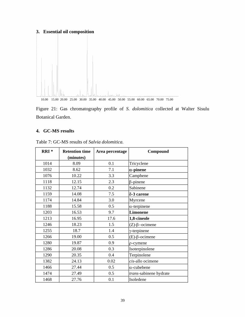
3. Essential oil composition
10.00 15.00 20.00 25.00 30.00 35.00 40.00 45.00 50.00 55.00 60.00 65.00 70.00 75.00 Figure 21: Gas chromatography profile of S. dolomitica collected at Walter Sisulu
Botanical Garden.
4. GC-MS results Table 7: GC-MS results of Salvia dolomitica.
RRI * Retention time Area percentage Compound (minutes)
1014 8.09 0.1 Tricyclene 1032 8.62 7.1 α-pinene 1076 10.22 3.3 Camphene 1118 12.15 2.3 β-pinene 1132 12.74 0.2 Sabinene 1159 14.08 7.5 δ-3 carene 1174 14.84 3.0 Myrcene 1188 15.58 0.5 α-terpinene 1203 16.53 9.7 Limonene 1213 16.95 17.6 1,8-cineole 1246 18.23 1.5 (Z)-β−ocimene 1255 18.7 1.4 γ-terpinene 1266 19.00 0.5 (E)-β-ocimene 1280 19.87 0.9 ρ-cymene 1286 20.08 0.3 Isoterpinolene 1290 20.35 0.4 Terpinolene 1382 24.13 0.02 cis-allo ocimene 1466 27.44 0.5 α-cubebene 1474 27.49 0.5 trans-sabinene hydrate 1468 27.76 0.1 Isoledene
39

RRI * Retention time Area percentage Compound (minutes)
1497 28.67 1.2 α-copaene 1544 29.95 0.3 α-gurjunene 1553 30.24 0.1 Linalool 1556 30.35 0.3 cis-sabinene hydrate 1571 30.86 0.1 trans-p-menth-2-en-1-ol 1589 31.30 0.1 Aristolene 1589 31.47 0.04 Isocaryophyllene 1612 32.25 17.4 β-caryophyllene 1628 32.56 3.0 Aromadendrene 1617 32.84 0.6 Selina-5,11-diene 1634 33.33 0.02 Cadina-3,5-diene 1661 33.77 0.3 Allo aromadendrene 1674 34.04 0.03 γ−gurjunene 1677 34.17 0.1 Epizonarene 1687 34.54 1.9 α-humulene 1704 35.05 0.4 γ-muurolene 1719 35.22 8.5 Borneol 1740 35.91 0.2 Valencene 1740 36.05 0.5 α-muurolene 1773 37.00 2.1 δ-cadinene 1776 37.13 1.5 γ-cadinene 1799 37.78 1.2 Cadina-1,4-diene (cubenene) 1853 39.18 0.2 cis-calamenene 1900 40.57 0.1 Epicubebol 1941 41.47 0.02 α-calacorene 1957 41.90 0.1 Cubebol 1969 42.12 0.04 cis-jasmone 2008 43.48 0.8 Caryophyllene oxide 2033 43.84 0.1 Epiglobulol 2050 44.24 0.1 (E)-nerolidol 2057 44.40 0.2 Ledol 2071 44.78 0.1 Humulene epoxide II 2088 45.18 0.1 1-epi-cubenol 2098 45.44 0.3 Globulol 2104 45.65 0.03 Viridiflorol 2144 46.14 0.1 Rosifoliol 2144 46.55 0.08 Spathulenol 2219 47.62 0.4 T-cadinol 2204 47.83 0.08 Eremoligenol 2209 48.01 0.3 T-muurolol 2218 48.23 0.06 Torreyol 2228 48.58 0.1 Pogostol 2250 48.90 0.3 α-eudesmol 2257 49.06 0.4 β-eudesmol 2324 50.58 0.06 Caryophylladieneol II
40

RRI * Retention time Area percentage Compound (minutes)
2389 51.47 0.1 Caryophyllenol I Total 99.77
RRI* – relative retention indices calculated against n-alkanes
The major compounds in Salvia dolomitica are α-pinene (7.1%), δ-3 carene (7.5%),
limonene (9.7%), 1,8-cineole (17.6%), β-caryophyllene (17.4%) and borneol (8.5%).
O
α-pinene δ-3 carene limonene 1,8-cineole
H
HOH
β-caryophyllene borneol Figure 22: Chemical structure of the major compounds identified in Salvia dolomitica
essential oil.
41

Salvia lanceolata L. E. Codd 1. Botanical description Branched shrub 1-2 m tall, stems often reddish brown. Leaves: blade simple, petiolate,
thick textured. Flowers vary in colour from dull rose to brownish crimson or grey-blue.
Figure 23: S. lanceolata. 2. Distribution Distributed from Namaqualand to the Cape Peninsula and eastwards to Montagu. Found
mainly in coastal sandveld or arid fynbos, on sandy soil and rocky hillsides at altitudes of
0-300m.
Figure 24: Geographical distribution of S. lanceolata.
42
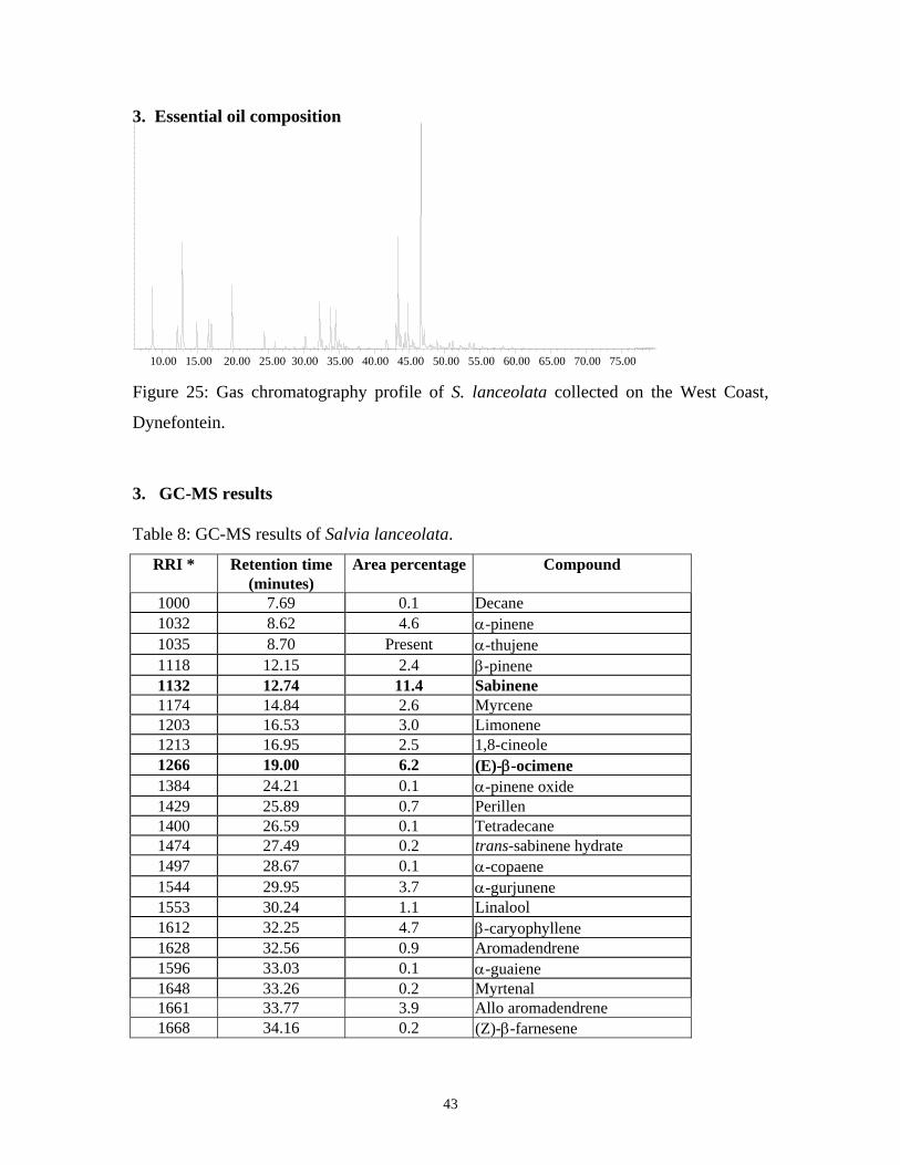
3. Essential oil composition
10.00 15.00 20.00 25.00 30.00 35.00 40.00 45.00 50.00 55.00 60.00 65.00 70.00 75.00 Figure 25: Gas chromatography profile of S. lanceolata collected on the West Coast,
Dynefontein.
3. GC-MS results Table 8: GC-MS results of Salvia lanceolata.
RRI * Retention time Area percentage Compound (minutes)
1000 7.69 0.1 Decane 1032 8.62 4.6 α-pinene 1035 8.70 Present α-thujene 1118 12.15 2.4 β-pinene 1132 12.74 11.4 Sabinene 1174 14.84 2.6 Myrcene 1203 16.53 3.0 Limonene 1213 16.95 2.5 1,8-cineole 1266 19.00 6.2 (E)-β-ocimene 1384 24.21 0.1 α-pinene oxide 1429 25.89 0.7 Perillen 1400 26.59 0.1 Tetradecane 1474 27.49 0.2 trans-sabinene hydrate 1497 28.67 0.1 α-copaene 1544 29.95 3.7 α-gurjunene 1553 30.24 1.1 Linalool 1612 32.25 4.7 β-caryophyllene 1628 32.56 0.9 Aromadendrene 1596 33.03 0.1 α-guaiene 1648 33.26 0.2 Myrtenal 1661 33.77 3.9 Allo aromadendrene 1668 34.16 0.2 (Z)-β-farnesene
43

RRI * Retention time Area percentage Compound
(minutes) 1687 34.54 3.6 α-humulene 1706 35.07 1.1 α-terpineol 1709 35.18 1.1 α-terpinylacetate 1708 35.28 0.3 Ledene 1755 36.03 0.2 Bicyclogermacrene 1751 36.45 0.1 Carvone 1804 37.83 0.3 Myrtenol 1864 39.33 0.1 ρ-cymen-8-ol 2001 43.14 2.1 Isocaryophyllene oxide 2008 43.48 9.4 Caryophyllene oxide 2045 44.15 0.4 Humulene epoxide I 2057 44.40 1.3 Ledol 2071 44.78 3.8 Humulene epoxide II 2081 45.12 0.1 Humulene epoxide III 2098 45.44 0.6 Globulol 2104 45.65 0.3 Viridiflorol 2144 46.55 20.9 Spathulenol 2247 48.90 0.5 α-bergamotol 2389 51.47 0.5 caryophyllenol I
Total 95.5
RRI* – relative retention indices calculated against n-alkanes
The major compounds in Salvia lanceolata are sabinene (11.4%), E-β-ocimene (6.2%),
caryophyllene oxide (9.4%) and spathulenol (20.9%).
O
H
H
OH
sabinene E-β-ocimene caryophyllene oxide spathulenol Figure 26: Chemical structure of the major compounds identified in Salvia lanceolata
essential oil.
44

Salvia namaensis L. E. Codd 1. Botanical description Bushy shrub 0,3-1,2 m tall, often herbaceous above and woody below. Leaves shortly
petiolate. White, mauve or blue flowers that are more crowded above.
Figure 27: S. namaensis. 2. Distribution Found on rocky slopes, in watercourses and on surface limestone. Distributed in northern
and central Cape as far south as Oudtshoorn and Willowmore, and western Free State.
Figure 28: Geographical distribution of S. namaensis.
45

3. Essential oil composition
10.00 15.00 20.00 25.00 30.00 35.00 40.00 45.00 50.00 55.00 60.00 65.00 70.00 75.00 Figure 29: Gas chromatography profile of S. namaensis collected at Swartberg Pass.
4. GC-MS results Table 9: GC-MS results of Salvia namaensis.
RRI * Retention time Area percentage Compound (minutes)
1014 8.09 0.6 Tricyclene 1032 8.62 9.3 α-pinene 1076 10.22 14.7 Camphene 1118 12.15 3.3 β-pinene 1132 12.74 0.1 Sabinene 1174 14.84 0.5 Myrcene 1203 16.53 2.2 Limonene 1213 16.95 8.2 1,8-cineole 1246 18.23 0.1 (Z)-β-ocimene 1255 18.70 0.6 γ-terpinene 1280 19.87 1.3 ρ-cymene 1290 20.35 0.2 Terpinolene 1452 26.66 0.02 α,p-dimethylstyrene (p-cymenene) 1451 26.82 0.2 β-thujone 1474 27.49 0.3 trans-sabinene hydrate 1435 28.63 0.02 α-campholene aldehyde 1497 28.67 0.6 α-copaene 1532 29.51 33.5 Camphor 1553 30.24 0.1 Linalool 1556 30.35 0.1 cis-sabinene hydrate 1571 30.86 0.1 trans-p-menth-2-en-1-ol
46

RRI * Retention time Area percentage Compound (minutes)
1586 31.29 0.1 Pinocarvone 1597 31.57 6.8 Bornylacetate 1612 32.25 4.8 β-caryophyllene 1628 32.56 1.2 Aromadendrene 1617 32.84 0.02 Selina-5,11-diene 1648 33.26 0.3 Myrtenal 1661 33.77 1.7 Allo aromadendrene 1682 34.31 0.1 δ-terpineol 1687 34.54 0.2 α-humulene 1700 34.78 0.02 Limonen-4-ol 1706 35.07 0.2 α-terpineol 1719 35.22 2.1 Borneol 1725 35.66 0.02 Verbenone 1755 36.03 0.1 Bicyclogermacrene 1773 37.00 0.5 δ-cadinene 1804 37.83 0.1 Myrtenol 1804 38.98 0.02 trans-carveol 1864 39.33 0.1 ρ-cymen-8-ol 1882 39.81 0.1 cis-carveol 1953 41.80 0.1 Palustrol 1948 42.14 0.1 trans-jasmone 2008 43.48 0.6 Caryophyllene oxide 2057 44.40 0.2 Ledol 2071 44.78 0.1 Humulene epoxide II 2098 45.44 0.1 Globulol 2104 45.65 0.2 Viridiflorol 2144 46.14 0.02 Rosifoliol 2144 46.55 0.2 Spathulenol 2198 47.27 0.03 Thymol 2185 47.61 0.3 γ-eudesmol 2204 47.83 0.1 Eremoligenol 2239 48.41 0.1 Carvacrol 2250 48.90 1.1 α-eudesmol 2257 49.06 0.8 β-eudesmol 2304 50.44 0.1 Cetylacetate 2389 51.47 0.1 Caryophyllenol I 2384 52.26 0.2 1-hexadecanol
Total 98.97 RRI* – relative retention indices calculated against n-alkanes
47
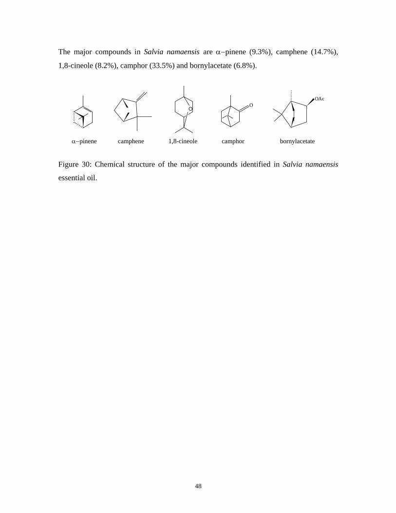
The major compounds in Salvia namaensis are α−pinene (9.3%), camphene (14.7%),
1,8-cineole (8.2%), camphor (33.5%) and bornylacetate (6.8%).
O
OAc O
α−pinene camphene 1,8-cineole camphor bornylacetate Figure 30: Chemical structure of the major compounds identified in Salvia namaensis
essential oil.
48

Salvia runcinata L. E. Codd 1. Botanical description Perennial erect herb 0,15 - 0,5 m tall with several stems from a taproot or less often a
creeping rootstock. Leaves shortly petiolate. Flowers denser above and more widely
spaced below, white or pale blue to mauve or purplish with the lower lip slightly longer.
Figure 31: S. runcinata. 2. Distribution Variable species extending from northern Gauteng to the Free State. Also widespread
throughout the northern, eastern and southern Cape as far as Bredasdorp district but rare
in Kwazulu-Natal and Lesotho. Found in a variety of habitats, usually on heavy soils and
locally common on disturbed places or overgrazed veld.
Figure 32: Geographical distribution of S. runcinata.
49

3. Essential oil composition
10.00 15.00 20.00 25.00 30.00 35.00 40.00 45.00 50.00 55.00 60.00 65.00 70.00 75.00 Figure 33: Gas chromatography profile of S. runcinata collected at Tarlton.
4. GC-MS results Table 10: GC-MS results of Salvia runcinata.
RRI * Retention Time Area percentage Compound (minutes)
1032 8.62 0.1 α-pinene 1118 12.15 0.1 β-pinene 1174 14.84 0.04 Myrcene 1203 16.53 0.1 Limonene 1213 16.95 0.2 1,8-cineole 1246 18.23 0.1 (Z)-β-ocimene 1266 19.00 0.1 (E)-β-ocimene 1280 19.87 0.3 ρ-cymene 1400 25.02 0.04 Nonanal 1553 30.24 0.5 Linalool 1612 32.25 7.6 β-caryophyllene 1668 34.16 1.1 (Z)-β-farnesene 1687 34.54 2.1 α-humulene 1706 35.07 0.3 α-terpineol 1727 35.90 0.1 7-epi-1,2-dehydrosesquicineole 1741 36.06 0.8 β-bisabolene 1783 37.36 0.5 β -sesquiphellandrene 2008 43.48 1.5 Caryophyllene oxide 2050 44.24 0.2 (E)-nerolidol 2071 44.78 0.3 Humulene epoxide II 2161 47.01 1.3 α-bisabolol oxide 2232 48.65 71.7 α-bisabolol
50

RRI * Retention Time Area percentage Compound
(minutes) 2324 50.58 0.3 Caryophylladienol 2518 55.80 6.2 cis-lanceol
Total 95.58 RRI* – relative retention indices calculated against n-alkanes
The major compounds in Salvia runcinata are β-caryophyllene (18.7%), α-humulene
(2.1%), α-bisabolol (71.7%) and cis-lanceol (6.2%).
HO
H
HO
H
H β-caryophyllene α-humulene α-bisabolol cis-lanceol Figure 34: Chemical structure of the major compounds identified in Salvia runcinata
essential oil.
51

Salvia stenophylla L. E. Codd 1. Botanical description Perennial, erect, bushy herb 0,2-0,4 m tall, usually branched from a woody taproot.
Leaves shortly petiolate or subsessile. Flowers are pale blue or mauve in colour.
Figure 35: S. stenophylla. 2. Distribution Distributed from Gauteng, Free State and Lesotho to northern, north-eastern and eastern
Cape. Rarely found in Kwazulu Natal. Found in grassland, open woodland and semi-arid
shrub, often on calcareous or brackish soil, sandy soil in watercourses or damp areas.
Sometimes considered a semi-weed of disturbed places.
Figure 36: Geographical distribution of S. stenophylla.
52

3. Essential oil composition
10.00 15.00 20.00 25.00 30.00 35.00 40.00 45.00 50.00 55.00 60.00 65.00 70.00 75.00 Figure 37: Gas chromatography profile of S. stenophylla collected at Lady Grey.
4. GC-MS results Table 11: GC-MS results of Salvia stenophylla.
RRI * Retention Time Area percentage Compound (minutes)
1032 8.62 0.3 α-pinene 1076 10.22 0.3 Camphene 1118 12.15 0.1 β-pinene 1159 14.08 7.0 δ-3 carene 1174 14.84 18.8 Myrcene 1205 16.35 0.5 Sylvestrene (m-mentha-1(6),8-diene) 1203 16.53 1.6 Limonene 1218 16.95 1.7 β-phellandrene 1246 18.23 0.1 (Z)-β-ocimene 1255 18.70 0.2 γ-terpinene 1265 19.14 0.1 5-methyl-3-heptanone 1278 19.82 0.1 m-cymene 1280 19.87 0.2 ρ-cymene 1290 20.35 0.1 Terpinolene 1348 22.70 0.1 6-methyl-5-hepten-2-one 1532 29.51 1.4 Camphor 1553 30.24 1.1 Linalool 1568 31.73 0.7 trans-α-bergamotene 1611 32.20 0.3 Terpinen-4-ol 1668 34.16 0.9 (Z)-β-farnesene 1682 35.07 0.3 α-terpineol 1719 35.22 0.2 Borneol 1727 35.90 0.1 7-epi-1,2-dehydrosesquicineole
53

RRI * Retention Time Area percentage Compound
(minutes) 1755 36.03 1.4 Bicyclogermacrene 1784 37.36 0.5 (E)-bisabolene 1804 37.83 0.1 Myrtenol 2050 44.24 0.2 (E)-nerolidol 2161 47.01 0.6 α-bisabolol oxide 2219 47.62 0.03 T-cadinol 2232 48.65 47.6 α-bisabolol 2518 55.80 0.2 cis-lanceol 2544 56.96 0.2 Bisabola-2,7,10-trien-13-ol 2676 62.51 8.6 Manool
Total 95.63
RRI* – relative retention indices calculated against n-alkanes
The major compounds in Salvia stenophylla are δ-3 carene (7.00%), mycrene (18.8%), α-
bisabolol (47.6%) and manool (8.6%).
OH
H
H
HO manool α-bisabolol mycrene δ-3 carene Figure 38: Chemical structure of the major compounds identified in Salvia stenophylla
essential oil.
54

Salvia verbenaca L. E. Codd 1. Botanical description Perennial, short-lived shrub with stems arising from a woody taproot. Stems erect, 0,15-
0,4 m tall. Leaves shortly petiolate to subsessile. Flowers are spaced below and denser
above and are range from light blue to purple in colour, lower lip usually slightly shorter.
Figure 39: S. verbenaca.
2. Distribution
Salvia verbenaca is an introduced plant in our Flora area. It is indigenous in the countries
around the Mediterranean and on the Canary Islands and has also spread further afield in
Europe and Asia. It has also become naturalized in Australia and the United States. In
South Africa it is now widely distributed, mainly in the drier, western half of the country,
in northern, central and western Cape and the western Free State with fewer plants
reaching the south western Cape.
55

Figure 40: Geographical distribution of S. verbenaca.
Salvia verbenaca does not produce essential oil and therefore the analysis with the aid of
GC-MS was not possible.
56

CHAPTER 4: RESULTS AND DISCUSSION
4.1 Analytical chemistry
The GC-MS results have been included under the monographs of each species and the
major compounds of the individual species have been identified. S. verbenaca does not
produce any essential oil and the analysis with the aid of GC-MS was therefore not
possible. No essential oil was obtained for S. chamelaeagnea. The species that were
analysed with the aid of GC-MS are included in Table 12.
Table 12: Salvia species analysed by of GC-MS and the percentage oil yield (wet weight) after distillation.
Species Plant material weight Percentage Yield S. africana-caerulea 335g 0.17% S. africana-lutea 167g 0.32% S. disermas (Mossel Bay) 369g 1.24% S. dolomitica 463g 1.02% S. lanceolata 314g 0.17% S. namaensis 249g 1.64% S. runcinata 90g 0.15% S. stenophylla 398g 0.79% The majority of the compounds identified by GC-MS are common to all the species
studied with the exception of a few. Camphor was only identified in three species. In S.
africana-caerulea and S. stenophylla it is found in very small concentrations whereas in
S. namaensis it exists as a major compound with a concentration of 33.50%. Camphene,
also present in low concentrations in a few species and is another major compound in S.
namaensis. The compounds β-caryophyllene and caryophyllene oxide are present in all
species with the exception of S. stenophylla. Terpinen-4-ol is present in low
concentrations in S. stenophylla. Spathulenol is found in high concentrations in S.
lanceolata when compared to the other species. Carvacrol is only present in S.
namaensis, in very low concentrations. α-Bisabolol is present as a major compound, in S.
runcinata and S. stenophylla. α-Eudesmol and β-eudesmol are both found in high
57

concentrations in S. africana-lutea. S. stenophylla is the only species to contain the
compound manool.
The compounds present in S. disermas vary considerably when compared with the other
species. S. disermas lacks compounds such us α-pinene and limonene that are present in
the other species. S. disermas also contains certain compounds that are not present in
other species. These include compounds such as linalyl acetate (34.50%),
diepishyobunone (4.40%), isoshuyobunone (0.70%), shyobunone (10.70%), epi-
isoshyobunone (6.20%) and shyobunol (0.20%).
Table 13: The major compounds identified in the Salvia species.
Species Major Compounds Salvia africana-caerulea α-pinene, 1,8-cineole, (E)-β-ocimene, ρ-cymene, β-caropyllene. Salvia africana-lutea ρ-cymene, caropyllene oxide, humulene epoxide II, γ-eudesmol, α-
eudesmol, β-eudesmol. Salvia disermas linalool, linalyl acetate,shyobunone, epi-isoshyobunone. Salvia dolomitica α-pinene, δ-3 carene, limonene, 1,8-cineole, β-caryophyllene, borneol Salvia lanceolata sabinene, (E)-β-ocimene, caryophyllene oxide, spathulenol. Salvia namaensis α-pinene, camphene, 1,8-cineole, camphor, bornylacetate. Salvia runcinata β-caryophyllene, α-humulene, α-bisabolol, cis-lanceol. Salvia stenophylla δ-3 carene, myrcene, α-bisabolol, manool.
Little phytochemical work has been done on the Salvia species occurring in southern
Africa and thus there is not an abundance of literature on the chemical constituents of
indigenous Salvia species. However, the essential oil of S. stenophylla collected on the
‘High Veld’ of the Free State has been investigated by GC-MS (Jäger and van Staden,
2000). It was found that the compounds mainly responsible for the persistent wood odour
of S. stenophylla, α-bisabolol and manool, were the most abundant. 1,8-Cineole and
camphor were present at very low levels and the species lacked α-thujone and β-thujone.
A similar study on S. stenophylla has been undertaken (Jäger and van Staden, 2000).
Most of the compounds were well known natural substances. The oil contained 29.80%
α-bisabolol, which was isolated by column chromatography and identified as (+)-epi-α-
bisabolol. This was the first time this isomer had been shown to occur in nature. The
58

results from these two independent studies are congruent with the results obtained in this
study.
Thin layer chromatography was employed on the methanol extracts, essential oils and on
rosmarinic acid, which was included as a standard. Chromatography is an analytical
method widely used for the separation, identification and determination of the
components in complex mixtures. The application of various chromatographic techniques
for the scientific validation of traditional medicine is inevitable. A specific solvent
system is used as a mobile phase, which allows separation of different components
present in the “mixture” based on their polarity. This system is not as sensitive as high
performance liquid chromatography, but its strength lies in the relatively cheap and rapid
assessment of the quality of extracts and actives present. For isolation, quantifying the
purity of the sample and evaluating the concentration of compounds in a sample, high
performance liquid chromatography provides an accurate method.
Literature states that the main compound responsible for the anti-oxidant activity of
Salvia is rosmarinic acid. Rosmarinic acid was subjected to thin layer chromatography on
silica gel plates along with the other methanol extracts of the test samples (Figure 41).
When the methanol extracts are compared to the rosmarinic acid standard a common
compound with Rf = 0.38 is observed. S. namaensis, S. chamelaeagnea, S. dolomitica
and S. stenophylla all have a common non-polar compound (Rf = 0.91). Various other
polar and non-polar compounds are present in the methanol extracts.
The antioxidant assay involves using a stable free radical 2,2-diphenyl-1-picrylhydrazyl
(DPPH) with a dark violet colour, whereby antioxidants are allowed to react with the
stable radical in methanol solution. Antioxidant compounds donate electrons to DPPH,
resulting in decolourization which is stoichiometric with respect to the number of
electrons captured by DPPH. The reduction in the concentration of the DPPH radical is
followed by monitoring the decrease in its absorbance at a characteristic wavelength
during the reaction. In its radical form, DPPH absorbs at 515 nm, but upon reduction by
antioxidant or a radical species, the absorption disappears. Results show that the
59

methanol extracts have both polar and non-polar compounds, in addition to rosmarinic
acid that are responsible for the antioxidant activity.
1 2 3 4 5 6 7 8 9 10 11 12 13
Figure 41: TLC plate of Salvia methanol extracts and rosmarinic acid after been
sprayed with DPPH reagent. White spots are indicative of compounds showing
antioxidant activity.
1 Salvia rigida 2 Salvia namaensis 3 Salvia disermas
(Mossel Bay) 4 Salvia disermas
(Walter Sisulu Botanical Garden) 5 Salvia chamelaeagnea 6 Salvia africana-caerulea 7 Rosmarinic acid 8 Salvia lanceolata 9 Salvia africana-lutea 10 Salvia dolomitica 11 Salvia stenophylla 12 Salvia runcinata 13 Salvia verbenaca
60

4.2 Antimicrobial activity The disc diffusion method is useful for the preliminary examination of the antimicrobial
properties of the test samples. The lack of reproducibility of the disc diffusion method is,
however, a distinct disadvantage and it should therefore only be used as a preliminary
screening. Extensive literature on the antimicrobial and antifungal potency of the Salvia
genus reveals a broad variability with regard to micro-organisms sensitivity as well as to
the efficiency (measured as MIC) of tested compounds, when different species within the
genus are considered (Baricevic and Bartol, 2000).
Table 13: Antibacterial screening results from the disc diffusion assay. Results expressed in millimeters (mm) from disc edge.
S. aureus B. cereus E. coli Y. enterocolitica K. pneumoniae Test Sample ATCC12600 ATCC11778 ATCC11775 ATCC23715 ATCC13883
S. africana-caerulea 2.0 <1.0 R 1.0 <1.0 Essential oil S. africana-caerulea <1.0 4.0 R R R Methanol extract S. africana-caerulea 3.0 5.0 R R R Acetone extract S. africana-lutea R 2.0 R R R Essential oil S. africana-lutea R 2.0 R R R Methanol extract S. africana-lutea 1.0 4.0 R R R Acetone extract S. chamelaeagnea 4.0 5.0 R R R Methanol extract S. chamelaeagnea 6.0 7.0 R R R Acetone extract S. disermas (WSBG) R R R R R Methanol extract S. disermas (WSBG) R R R R R Acetone extract S. disermas (Mossel Bay) <1.0 3.0 R R R Essential oil S. disermas (Mossel Bay) R R R R R Methanol extract S. disermas (Mossel Bay) R 1.5 R R R Acetone extract
61

S. aureus B. cereus E. coli Y. enterocolitica K. pneumoniae
Test Sample ATCC12600 ATCC11778 ATCC11775 ATCC23715 ATCC13883
S. disermas (Kokerboom- 2.0 2.0 R R R woud) Essential oil S. dolomitica R 1.0 R R R Essential oil S. dolomitica 4.0 7.0 R R R Methanol extract S. dolomitica 7.0 7.0 <1.0 R R Acetone extract S. lanceolata <1.0 R R R R Essential oil S. lanceolata R R R R R Methanol extract S. lanceolata <1.0 R R R R Acetone extract S. namaensis <1.0 <1.0 <1.0 <1.0 <1.0 Essential oil S. namaensis 5.0 7.0 R R R Methanol extract S. namaensis 6.0 8.0 R R R Acetone extract S. runcinata R 1.0 R R R Essential oil S. runcinata <1.0 4.0 R R R Methanol extract S. runcinata 4.0 8.0 R R R Acetone extract S. stenophylla <1.0 2.0 R R R Essential oil S. stenophylla 6.0 8.0 R R R Methanol extract S. stenophylla 7.0 9.0 R R R Acetone extract S. verbenaca <1.0 <1.0 R R R Methanol extract S. verbenaca R 2.0 R R R Acetone extract
Control – Neomycin 6.0 6.0 2.0 4.5 5.0
R = Resistant
62

The results of the disc diffusion assay are summarised in Table 13. No zones of inhibition
were evident on any of the fungal test organisms used in this assay and they are therefore
not included in the table. Measurements were recorded from the edge of the disc to the
end of the zone of inhibition and recorded in millimeters (Figure 42 and 43). All test
samples studied demonstrated variable degrees of antibacterial activity with the exception
of four test samples; S. disermas methanol and acetone extracts from the Walter Sisulu
Botanical Garden; S. disermas methanol extract from Mossel Bay and the S. lanceolata
methanol extract, which showed no activity.
The acetone extract of S. africana-caerulea showed more activity than the essential oil.
Antibacterial activity was noted against S. aureus, B. cereus and interestingly the
essential oil was one of two test samples that showed minimal activity against Y.
enterocolitica and K. pneumoniae. Salvia africana-lutea showed activity against B.
cereus but to a much lesser extent when compared with the activity of S. africana-
caerulea extract. The acetone extract of S. africana-lutea also showed little activity
against S. aureus. The extracts of S. chamelaeagnea showed good activity against both S.
aureus and B. cereus.
In general, S. disermas and S. lanceolata exhibited poor antibacterial activity. Both the
essential oils of S. disermas showed activity against S. aureus and B. cereus and only one
extract showed activity against B. cereus, the acetone extract from Mossel Bay. The
essential oil and acetone extract of S. lanceolata showed slight activity against S. aureus
and the methanol extract showed no activity.
Both methanol and acetone extracts of S. dolomitica demonstrated substantial activity
against B. cereus and S. aureus whilst the essential oil showed slight activity only against
B. cereus. S. dolomitica acetone extract showed minimal activity against E. coli. The
essential oil of S. namaensis showed activity, albeit very weak, against all test organisms
studied. In addition to this, both extracts showed significant activity against S. aureus and
B. cereus. The essential oil of S. runcinata exhibited little activity against B. cereus. The
63

acetone extract of S. runcinata showed activity against S. aureus and B. cereus and the
methanol extract demonstrated similar activity but to a lesser degree.
The methanol extract of S. verbenaca showed minimal activity against S. aureus and B.
cereus whilst the acetone extract only inhibited the growth of B. cereus. The methanol
and acetone extracts of S. stenophylla showed excellent activity against B. cereus and S.
aureus. The essential oil showed activity against the same bacteria but to a much lesser
degree. The acetone extract of S. stenophylla had the largest zone of inhibition of 9.00
mm against B. cereus.
The microplate bioassay method was chosen to determine the MIC of selected test
samples. Two microplates were prepared. The Gram-positive organism B. cereus was
chosen in this assay as a result of the high antimicrobial activity shown by test samples in
the disc diffusion assay towards this organism. A positive control, ciprofloxacin, was
included to ensure the antimicrobial sensitivity of the organism. Bacillus cereus with no
test samples or antimicrobial is included as a negative control. The MIC results have been
summarized in Table 14.
The minimum inhibitory concentration is the lowest concentration of an antimicrobial,
and in this case our test sample, found to inhibit the growth of a particular test organism,
namely B. cereus. These results reflect that the concentration of S. disermas (Mossel Bay-
essential oil) and S. africana-lutea (acetone) needed to inhibit the growth of B. cereus is
8.00 mg/ml. The concentration of S. disermas (Kokerboomwoud - essential oil,
S. dolomitica (essential oil), S. africana-lutea (essential oil) and S. stenophylla (essential
oil) are all required at a concentration of 4.00 mg/ml to inhibit B. cereus.
64

Table 14: Minimum Inhibitory Concentration (MIC) of Salvia against B. cereus.
Microplate Test Sample MIC
(mg/ml)
A1 S. disermas (Mossel Bay) essential oil 8.00 A2 S. disermas (Mossel Bay) acetone extract 1.00 A3 S. disermas (Kokerboomwoud) essential oil 4.00 A4 S. chamelaeagnea methanol extract < 0.50 A5 S. chamelaeagnea acetone extract < 0.50 A6 S. dolomitica essential oil 4.00 A7 S. dolomitica methanol extract 1.00 A8 S. dolomitica acetone extract < 0.50 A9 S. verbenaca acetone extract < 0.50
A10 S. namaensis methanol extract 1.00 A11 S. namaensis acetone extract < 0.50 B1 S. africana-lutea essential oil 4.00 B2 S. africana-lutea methanol extract 1.00 B3 S. africana-lutea acetone extract 8.00 B4 S. africana-caerulea methanol extract 1.00 B5 S. africana-caerulea acetone extract 1.00 B6 S. runcinata essential oil 2.00 B7 S. runcinata methanol extract 2.00 B8 S. runcinata acetone extract < 0.50 B9 S. stenophylla essential oil 4.00
B10 S. stenophylla methanol extract < 0.50 B11 S. stenophylla acetone extract < 0.50
The MIC for the essential oil and methanol extract of S. runcinata is 2.00 mg/ml. For the
following test samples a concentration of 1.00 mg/ml is required to inhibit the growth of
B. cereus: S. disermas (Mossel Bay – acetone), S. dolomitica (methanol), S. namaensis
(methanol), S. africana-lutea (methanol) and S. africana-caerulea (methanol and
acetone). It can thus be seen that in general the essential oils are required at much higher
concentrations than the extracts to inhibit B. cereus.
For those test samples that showed no colour change with INT solution we can conclude
that B. cereus is inhibited at all of the above concentrations and the MIC is smaller than
the last dilution. In order to obtain the exact MIC of such test samples, a series of
65

dilutions would have to be performed that are smaller than the ones already used at the
particular range identified.
A MIC of less than 0.50 mg/ml was determined for the following test samples: S.
chamelaeagnea (methanol and acetone), S. dolomitica (acetone), S. verbenaca (acetone),
S. namaensis (acetone), S. runcinata (acetone) and S. stenophylla (acetone and methanol).
S. chamelaeagnea (acetone), S. dolomitica (methanol and acetone), S. namaensis
(methanol and acetone), S. runcinata (acetone) and S. stenophylla (methanol and acetone)
all had relatively low MIC values with corresponding large zones of inhibition in the disc
diffusion assay. Although S. verbenaca had a small zone of inhibition (2 mm), the MIC
value was very low. It would have been anticipated that the MIC value would have been
larger.
In general, there seemed to be an overall agreement between the size of inhibition zones
obtained in the disc diffusion assay and the minimum inhibitory concentration values.
The larger zones of inhibition correlated with lower minimum inhibitory concentration
values and vice versa however, some variations did occur.
66

Figure 42: Disc diffusion plate of S. aureus 64 - S. dolomitica (methanol); 68 - S. namaensis (acetone); 69 - S. namaensis (methanol); 76 - S. africana-lutea (essential oil). Neomycin (control) in middle of plate.
Figure 43: Disc diffusion plate of S. aureus 78 - S. africana-caerulea (acetone); 81 - S. runcinata (acetone); 83 - S. stenophylla (methanol); 84 - S. stenophylla (acetone). Neomycin (control) in middle of plate.
67

The extracts and essential oils were screened against a varied but small selection of
bacterial and fungal organisms. It should be noted that those test samples that showed
little or no antimicrobial activity may be active against other yeasts, bacteria or moulds,
which were not included in this particular study. All test samples lacked activity against
the fungal organisms tested. Due to the fact that the controls for the fungal tests
demonstrated inhibition it is clear that the failure of the test samples was due to a lack of
potency and activity, and not as a result of other internal or external factors such as media
composition, incubation conditions or organism resistance.
There has been extensive literature published on the anti-fungal properties of the genus
Salvia and various oil components have been identified as being responsible for the
activity. Viollon and Chaumont (1994) studied the anti-fungal properties of a variety of
essential oils and their main components upon Cryptococcus neoformans. The conclusion
was clear that among the phenolic compounds, thymol and carvacrol are the most
fungitoxic. Velickovic et al., (2002) expanded the list of fungitoxic oil constituents to
include α-bisabolol and Tzakou et al., (2001) demonstrated that 1,8-cineole and α-pinene
are particularly active against Candida albicans. Peana et al., (1999) concluded that α-
terpineol and linalool have the strongest candidicidal activity. When Baricevic and Bartol
(2000) measured the antimicrobial property of the essential oil of Salvia against
Alternaria alternata and Aspergillus parasiticus, a strong fungistatic effect was observed.
The antifungal activity in that study was thought to be due to the high content of thujone
in the essential oil samples that were tested. In comparison with these findings the lack of
anti-fungal activity for all test samples is therefore surprising. Thymol, carvacrol and
thujone are found in very small concentrations in S. namaensis and are possibly of little
significance. According to the GC-MS results for S. stenophylla and S. runcinata, α-
bisabolol has an area percentage of 47.6 % and 71.7 % respectively and yet they both
show no anti-fungal activity. In addition to this 1,8-cineole, α-pinene, α-terpineol, and
linalool are present in many other test samples in varying and often quite substantial
concentrations and yet no anti-fungal activity is evident. It is likely that test samples may
be required in much higher concentrations to produce similar results to that found in the
literature. Alternatively, the concentrations of identified compounds may be needed in
68

higher concentrations. It could also be possible that antagonism may be taking place
between compounds within a particular species.
In contrast to the anti-fungal properties it is clear that certain Salvia species definitely
possess antimicrobial activity. Although research is lacking on indigenous Salvia species,
much time and research has gone into studies on the antimicrobial properties of Salvia
worldwide. Promising results have been documented and in one study Salvia has even
shown to be the most potent inhibitor of oral bacteria when compared to peppermint and
tea tree essential oils (Shapiro et al., 1994).
Generally, Gram-negative bacteria proved not to be sensitive or in some cases, only
slightly sensitive when compared with the sensitivity of Gram-positive bacteria. This is in
agreement with the observation of Baricevic and Bartol (2000). Because of the difference
in cell wall structure between Gram-positive and Gram-negative bacteria, it was
anticipated that the Gram-positive bacteria would be more sensitive to the test samples.
Gram-negative bacteria have an outer membrane which makes penetration of any
antibiotic, and in this case the test samples, a difficult task (Velickovic et al., 2002).
However, certain test samples did demonstrate a small amount of inhibition against some
Gram-negative organisms and it may be probable that, for realization of antimicrobial
action, larger quantities of oils may be required to inhibit Gram-negative bacteria.
The methanol and acetone extracts were far more active than the essential oils. This is
probably due to the lipophillic and hydrophilic properties of the essential oils and extracts
respectively. The disc diffusion method depends on the aqueous solubility of the test
samples in order to facilitate diffusion through the solid medium, the agar. Essential oils
are generally hydrophobic and do not readily diffuse or penetrate through an aqueous
medium as well as the methanol and acetone extracts do. Therefore, the potency of the
essential oils may be decreased due to decreased permeability. It is because of this that
the prevalence of false negatives or reduced activity might be anticipated in a disc
diffusion assay (Nakatsu et al., 2000). However, in practice, the hydrophobicity of
essential oils enables them to partition into the lipids of the cell membrane and
69

mitochondria, rendering them permeable and leading to leaking of cell contents. Physical
conditions that improve the activity of the essential oils are low pH, low temperature and
low oxygen levels (Burt, 2004). Theoretically, it would therefore be desirable to develop
a system that would allow lipophilic materials to disperse in an aqueous environment
using a solvent that is safe for consumption, lacks toxicity and does not interfere with
antimicrobial activity of the test sample.
A significant difficulty in the study of biologically active essential oils is that the
biological activity does not depend on only one active compound or constituent. In
addition, the essential oils are usually mixtures of structurally very similar compounds
and, therefore, the biological activity of essential oils cannot easily be explained with a
simple, single molecule or compound. In fact, the action is possibly the result of the
combined effect of both active and inactive compounds. The nature as well as the
proportion of the individual compounds will have a direct influence on the antimicrobial
activity (Pattnaik et al., 1997). In some instances, the inactive compounds may influence
resorption, rate of reactions and the bioavailability of the active compounds either
positively or negatively (Svoboda and Hampson, 1999). However, the ultimate goal is to
identify as accurately as possible the active compounds, the structure activity
relationships of compounds and the synergism that take place between these compounds
in order to be able to use this information in developing future medicinal compounds.
Literature studied on essential oils from a variety of plant species show similarities in
antimicrobial activity with the compounds found in the majority of the Salvia species.
The essential oil of Melaleuca alternifolia, or tea tree oil, has been used medicinally, for
its antimicrobial activity for many years. A study was done by Carson and Riley (1995)
on the antimicrobial activity of the major components of tea tree oil. The components
identified in the essential oil and tested included: 1,8-cineole, 1-terpinen-4-ol, ρ-cymene,
linalool, α-terpinene, γ-terpinene, α-terpineol and terpinolene. The test organisms
included B. subtilis, C. albicans, E. coli, P. aeruginosa and S. aureus. It was found that
terpinen-4-ol was active against all the test organisms while linalool and α-terpineol were
70

active against all organisms with the exception of P. aeruginosa. S. stenophylla contains
terpinen-4-ol in low concentrations. Linalool is present as a major compound in S.
disermas and as a minor compound in every species. α-Terpineol is present as a minor
compound in more than fifty percent of the species studied. In another study by Carson et
al., (2002) the mechanism of action of tea tree oil and three of its components, 1,8-
cineole, terpinen-4-ol and α-terpineol, against S. aureus was investigated. Loss of 260-
nm-absorbing material occurred after treatment with 1,8-cineole and α-terpineol. When
the agents were tested at one-half the MIC, only 1,8-cineole significantly reduced the
tolerance of S. aureus to sodium chloride. Salvia africana-caerulea, S. namaensis and S.
dolomitica (methanol and acetone) all inhibited S. aureus to some degree and all contain
1,8-cineole. The antimicrobial activity of linalool and 1,8-cineole has been demonstrated
separately and it has also been observed that the antimicrobial activity is increased when
they are used in combination (Faleiro et al., 2003).
Ulubelen et al., (1994) studied the terpenoids from Salvia sclarea. The diterpenoids and
the sesquiterpenoids were tested for antimicrobial activity against standard bacterial
strains. Manool, spathulenol and caryophyllene oxide were found to be active against S.
aureus. Salvia stenophylla is the only species that has manool present, in addition to this
it is present as a major compound. Salvia stenophylla showed very good activity against
S. aureus in the disc diffusion assay in this report. Spathulenol is present as a major
compound in S. lanceolata and is present as a minor compound in some of the other
species. S. lanceolata exhibited weak activity against S. aureus. Caryophyllene oxide is
present in all species with the exception of S. stenophylla, all of which showed some
degree of inhibition towards S. aureus. No direct relationship can be found between
caryophyllene oxide and the test samples.
The composition and antimicrobial activity of the essential oil of Hypericum rumeliacum
have been researched by Couladis et al., (2003). The major compounds reported to be
responsible for the antimicrobial activity are: α-pinene, β-pinene and α-copaene. α-
pinene and β-pinene are present in all species except S. disermas. In this research report
S. disermas showed negligible activity.
71

Űnlű et al., (2002) have researched the compositions and the antimicrobial activities of
the essential oils of Achilla setacea and Achilla teretifolia. The essential oils exhibited
antimicrobial activity against C. perfringes, S. aureus, S. pneumoniae and B. cereus. The
major compounds identified were camphor, 1,8-cineole and borneol and can be
considered as the antimicrobial compounds of the essential oils. Borneol is present in S.
namaensis, S. stenophylla and S. dolomitica. Camphor is present as a major compound in
S. namaensis. When comparing their data with the results generated in this study it can be
seen that these three species show excellent antimicrobial activity towards B. cereus, both
in the disc diffusion assay and the microplate assay. The antimicrobial activity observed
in these three species that contain borneol is actually greater than the neomycin control
included in this assay.
Camphene has been identified in the literature as an antimicrobial compound. In a study
by Tirillini et al., (1996) on the chemical composition and the antimicrobial activity of
the essential oil of Piper angustifolium, camphor and camphene were identified as the
main compounds. Conclusions were drawn that the oil inhibited the growth of E. coli,
among others, due to the presence of camphene and camphor. Camphene is present as a
major compound in S. namaensis and as minor compounds in S. stenophylla, S. africana-
lutea and S. dolomitica. Although very weakly, S. namaensis is the only essential oil that
inhibited the growth of all organisms including E. coli. Salvia dolomitica has the second
highest concentration of camphene present and is the only other essential oil to inhibit the
growth of E. coli. S. africana-lutea and S. stenophylla do not inhibit the growth of E. coli
in this study but they do show remarkable antimicrobial activity toward S. aureus and B.
cereus and they contain camphene in small concentrations.
The bacterial susceptibility of E. coli, S. aureus, B. subtilis and K. pneumoniae to the
essential oils of Thymus kotschyanus and Thymus persicus was investigated by Rasooli
and Mirmostafa, (2003). The results from their study showed that carvacrol, thymol and
ρ-cymene contributed to more than 75 % of the chemical composition of thyme oils with
strong antibacterial properties. ρ-cymene is present in all species besides S. lanceolata
72

and S. disermas. It is found in concentrations of 5.20 % in both S. africana-lutea and S.
africana-caerulea. K. pneumoniae was slightly inhibited by the essential oil of S.
africana-caerulea but not by S. africana-lutea.
The chemical composition of the essential oil of Salvia desoleana from Italy was
analysed by Peana et al., (1999). S. desoleana had a high content of monoterpenic esters
including linalyl acetate and α-terpinyl acetate. Weak antimicrobial activity was noted
against S. aureus, E. coli and S. epidermidis. However, inhibition increased progressively
with increased contact time. Salvia disermas showed poor antibacterial activity. It is the
only species in this study to have the compound linalyl acetate in a concentration of 34.50
%. α-terpinyl acetate is found in small concentrations in three different species. The
contact time of our test samples and the organism was only 24 hours, if the parameters of
this study were altered and the contact time increased different results may have been
noticed.
Costantin et al., (2001) has identified the main compounds of Piper cernuum essential oil
as being bicyclogermacrene (21.88 %) and β-caryophyllene (20.69 %). The main
compounds of Piper regnellii essential oil have been identified as mycrene (52.60 %) and
linalool (15.89 %). Both essential oils presented growth inhibitory activities against S.
aureus. Mycrene is present in all species and is of particular importance in S. stenophylla
as it is present in a concentration of 18.80 %. S. stenophylla shows good inhibitory
activity toward S. aureus in this study. Besides S. stenophylla, β-caryophyllene is present
in all species. The concentrations of β-caryophyllene within each species does not
correspond to the degree of inhibition exhibited towards S. aureus in the disc diffusion
assay.
The chemical composition and antibacterial activity tests were carried out on the essential
oil of Senecio graveolens by Perez et al., (1999). The essential oil proved that it has
antibacterial effects on oxacillin-sensitive and oxacillin-resistant S. aureus. The chemical
composition of the essential oil was analysed by GC-MS and some of the compounds
identified were: α-pinene, α-terpinene, ρ-cymene, sabinene, terpinen-4-ol and α- and β-
73

eudesmol. Sabinene is found in many of the species and present as a major compound in
S. lanceolata. Our results are not in agreement with this study because S. lanceolata
showed very weak antibacterial activity. The compounds α- and β- eudesmol are present
in large concentrations in S. africana-lutea. This species showed moderate activity in the
disc diffusion assay.
A fact that may seem paradoxical, but may very well explain the lack of anticipated
activity from certain Salvia species, is the interaction that may take place between two
constituents within a species. While two major constituents in a particular species would
demonstrate and have antimicrobial activity by themselves, combining the two would
eliminate their activity. This has been observed with the compounds eugenol and linalool,
both active singularly, but show an antagonistic effect toward one another. Many
combinations of compounds may give rise to this phenomenon that may have not, as yet,
been researched (Nakatsu et al., 2000).
Published studies have also indicated the high degree of variability in quantity and
composition of essential oils within species that have been collected from different
locations. This geographical variation was proved in a study on Salvia fruticosa collected
from different parts on the island of Crete, Greece (Skoula et al., 2000). The three
samples, in this study, exhibited significant differences in essential oil yield and
composition. Variability in yield and content of essential oils in the Lamiaceae is known
to be affected by environmental factors such as light, nutrient availability and season.
This study has not taken in to account the enantiomers that exist for different compounds.
Demirci et al., (2002) discusses the enantiomeric distribution of some monoterpenes in
the essential oils of certain Salvia species found in Turkey. It is known that both
enantiomers of camphor are found in nature, but the (-) – form is said to be less common
than the (+) – form. (-)-Camphor has been shown to have antibacterial activity whilst (+)-
camphor is inactive. The antimicrobial activity of the enantiomers of borneol have been
studied by Tabanca et al., (2001). Both enantiomers showed antibacterial activity towards
all bacteria included in the study however, the MIC values varied.
74

The methanol and acetone extracts displayed promising antibacterial activity against
Gram-positive bacteria. This is consistent with a screening programme undertaken by
Izzo et al., (1995) in which it was found that plant extracts and essential oils readily
inhibit Gram-positive rather than Gram-negative bacteria. It is possible that the
antibacterial properties of the extracts in this study could be attributed to the presence of
carnosol derivatives that are reported to have strong antibacterial activity as well as
antioxidant properties. In a study undertaken by Lee et al., (1999) it was found that the
ability of the compounds to generate superoxide radicals also inhibited bacterial growth
by non-selective inhibitory action on DNA, RNA and protein synthesis.
4.3 Antioxidant activity
Within the last decade, the importance of free radicals in the aetiology of disease has
been increasingly recognized and has lead to the development of new approaches in
biochemical evaluation of events associated with mutagenisis, tumorigenesis and / or
cancer promotion (Baricevic and Bartol, 2000).
The reason that antioxidants are important to human physical well being comes from the
fact that oxygen is a potentially toxic element since it can be transformed by metabolic
activity into more reactive forms such as superoxide, hydrogen peroxide, singlet oxygen
and hydroxyl radicals, collectively known as active oxygen. These molecules are formed
in living cells by various metabolic pathways. Specific molecules, pollution from tobacco
smoke and burning of fossil fuels, together with UV radiation and pollutants such as
ozone, nitrogen oxide and sulphur dioxide, add to the formation of free radicals.
Superoxide is converted by an enzyme superoxide dismutase, into H2O2. This compound
is able to cross all biological membranes. Much of the damage done by these compounds
is thought to be due to the conversion to highly reactive oxidants such as the hydroxyl
radical. This radical is very reactive as it combines with almost all molecules found in
living cells. Proteins, lipids, carbohydrates and DNA in living cells represent oxidizable
substrate. The secondary events include changes in membrane structure, permeability and
fluidity, lysomal destabilization and stimulation of apoptosis. Lipid peroxidation finally
75

leads to loss of membrane function and integrity leading to cell necrosis and death
(Svoboda and Hampson, 1999).
The antioxidant activity of Salvia has been extensively studied. The antioxidant
properties of water extracts obtained from herbs of the species Lamiaceae, was
investigated by Triantaphyllou et al., (2001). The extracts were examined for their effect
against lipid oxidation in comparison to a tea (water) extract. Salvia was one of three
extracts found to have a remarkable capacity in retarding lipid oxidation. Examination by
thin layer chromatography showed that the extracts were rich in compounds such as
hydroxycinnamic acids, flavonoids, caffeic acids and rosmarinic acid. The antioxidant
activity of a synthetic antioxidant and Salvia has also been compared (Bandoniene et al.,
2001) and the data obtained showed that all the Salvia extracts had a very strong
antioxidant activity.
An antioxidant screening study in Yugoslavia (Malencic et al., 2000) on the wild
growing Salvia species isolated rosmarinic acid as the dominant naturally occurring
compound. Wang et al., (1999) reports that the antioxidative effects of Salvia relate to the
presence of carnosic acid, carnosol and rosmarinic acid (Figure 44). Exarchou et al.,
(2002) found that the major component of all Salvia ethanol extracts was rosmarinic acid,
determined by thin layer chromatography and nuclear magnetic resonance spectroscopy.
Furthermore, the extracts that are rich in rosmarinic acid were found to have a higher
radical scavenging activity. It was concluded that the flavanoids have a rather small
contribution to the total antioxidant activity of the extracts and the main ingredient is
rosmarinic acid, which is indeed a strong radical scavenger.
H
OH
OH
O
HCOOHO OH
O
O
O
H
H
HO
O
OHOH
HO
H carnosic acid rosmarinic acid carnosol
76

Figure 44: Chemical structures of the major antioxidant compounds in Salvia.
Rosmarinic acid is an ester of caffeic acid and 3,4-dihydroxyphenyllactic acid. It is
commonly found in species of the Boraginaceae and the subfamily Nepetoideae of the
Lamiaceae. However, it is also found in other higher plant families and in some fern and
hornwort species (Petersen and Simmonds, 2003). Rosmarinic acid is the most abundant
caffeic acid dimer found in Salvia species and has been reported to be the major phenolic
compound responsible for the high antioxidant activity of Salvia species (Lu and Foo,
2002); (Baricevic and Bartol, 2000). When measuring the radical scavenger effect on 1,
1-diphenyl-2-picrylhydrazyl (DPPH) free radical, the antioxidant effect of rosmarinic
acid has been shown to be comparable to that of ascorbic acid (Baricevic and Bartol,
2000). In plants, rosmarinic acid is believed to act as a preformed constitutively
accumulated defence compound. Rosmarinic acid has a number of interesting biological
activities that include antiviral, anti-inflammatory and antioxidant properties. (Petersen
and Simmonds, 2003). It has been found that plant cell cultures accumulate rosmarinic
acid in amounts much higher than in the plant itself (up to 36% of the cell dry weight).
For this reason a biotechnological production of rosmarinic acid with plant cell cultures
has been proposed (Petersen and Simmonds, 2003).
Interestingly, the results of an experimental study by Liu et al., (2001) indicate that
certain Salvia species have anti-tumor potential. This potential is thought to be mediated
through dual mechanisms, one of which is the antioxidant property of Salvia. Certain
Salvia species investigated by Du et al., (1997) showed results that indicate that there
were protective effects on learning and memory impairment caused by cerebral ischemia
reperfusion and these results may be related to its antioxidant activity. There is also the
possible efficacy for the application of the oils from such medicinal plants, through their
antioxidant capacities, in the prevention of age-related macular degeneration (Nakatsu et
al., 2000). Many reports on the relationship between the skin aging induced by UV-
irradiation and the active oxygen suggest that the prevention of skin aging can be
achieved by active-oxygen scavengers. Salvia is reported to contain chain-breaking
antioxidants, which are able to inhibit peroxidation chain reactions (Masaki et al., 1995).
77

Oxygen free radicals appear to be an important factor in chronic inflammatory joint
disease such as rheumatoid arthritis. There are several known reactions of oxygen
centered free radicals which are relevant to tissue injury in inflamed joint conditions.
Traditionally S. disermas has been used to treat such inflammatory conditions especially
rheumatism.
The conclusion that Salvia has antioxidant properties is a result of a qualitative screening
of certain indigenous Salvia species. It is desirable that this is followed up with a
quantitative assay. Although certain compounds have been pointed out to contribute
largely to the antioxidant activity, further studies are required to reveal whether Salvia
contains other antioxidant compounds that may contribute to their activity. Further work
on the characterization of specific components needs to be performed in order to establish
the connection between antioxidant activity and chemical composition.
The importance of researching traditional uses of Salvia is clear. It is therefore beneficial
and desirable to obtain natural, non-toxic and easily consumable dietary supplements
because these natural antioxidants will act as protective agents against free radicals and
can ultimately, in sufficient amounts, act as efficient scavengers of free radicals before
any tissue damage occurs (Baricevic and Bartol, 2000).
78

CHAPTER 5: CONCLUSION
• The essential oil composition varied quantitatively and qualitatively within the
different Salvia species analysed. Some species lacked certain compounds that were
present as major chemical compounds in other species. This observation was
illustrated by GC-MS analysis of the essential oils.
• Many Salvia species demonstrated varied antibacterial activity although no antifungal
activity was observed in this study. In general, S. chamelaeagnea, S. dolomitica, S.
namaensis, S. runcinata and S. stenophylla showed good antimicrobial activity. S.
disermas and S. lanceolata exhibited poor antimicrobial activity. Gram-positive
bacteria were more sensitive than Gram-negative bacteria.
• The methanol and acetone extracts were more active than the essential oils.
• The chemical compounds found in the highest concentrations in the essential oils are
not necessarily responsible for the antimicrobial activity. The possibility of synergy
and / or antagonism existing between compounds within a species should be
considered. Geographical variation and the enantiomeric distribution of chemical
compounds are also factors that should be considered in a study of this nature.
• The antimicrobial activity exhibited by the investigated species, especially their
greater activity against B. cereus and S. aureus may provide a scientific basis for the
ethnomedicinal use of the indigenous Salvia species. Traditionally Salvia has been
used to treat a host of infections, including skin infections.
• The antioxidant properties if Salvia can be largely attributed to the presence of
rosmarinic acid, carnosic acid and carnosol. Rosmarinic acid was found to be present
in all Salvia methanol extracts.
79

• The antioxidant activity of Salvia species may provide a scientific basis for the
ethnomedicinal use of the indigenous Salvia species, especially treating conditions
such as rheumatism.
• Therefore it can be concluded that the antimicrobial and antioxidant therapeutic
applications for Salvia are supported based on its history of use in well established
systems of traditional medicine and the assays carried out in this study.
RECOMMENDATIONS:
• It is recommended that further studies be carried out on the extracts due to the fact
that they are far more active than the essential oils.
• Antimicrobial studies can be carried out incorporating more Gram-positive bacteria.
• Autobiographic assays can be carried out to determine the actual compounds
responsible for the antimicrobial activity.
• Qualitative screening demonstrated that Salvia possesses antioxidant properties, this
should be followed up with quantitative studies.
• The DPPH assay is one of several antioxidant assays. It was used as a qualitative
assay in this study as it can be easily visualised on TLC. Other in vitro assays (lipid
peroxidase, ABTS) should be carried out on the leaf extracts.
80

CHAPTER 6: REERENCES
Bandoniene D., Gruzdiene D. and Venskutonis P.R. 2001. Antioxidant activity of sage
extracts in rapeseed oil irradiated with UV-rays. Nahrung 45: 105-108.
Baricevic D. and Bartol T. 2000. The biological / pharmacological activity of the Salvia
genus in S.E. Kintzios (Ed) Sage. The genus Salvia. Harwood academic publishers. 143-
184.
Blumenthal M., Busse W.R., Goldberg A., Gruenwald J., Hall T., Riggins C.W., Rister
R.S. (Ed), Klein S. and Rister R.S. (Translator). 1998. Austin: American Botanical
Council; Boston: Integrative Medicine Communications 330-333.
Burt S. 2004. Essential oils: their antibacterial properties and potential applications in
foods—a review. International Journal of Food Microbiology 3: 223-253.
Carson C.F. and Riley T.V. 1995. Antimicrobial activity of the major components of the
essential oil of Melaleuca alternifolia. Journal of Applied Bacteriology 78: 264-269.
Carson C.F., Mee B.J. and Riley T.V. 2002. Mechanism of action of Melaleuca
alternifolia (tea tree) oil on Staphylococcus aureus determined by time-kill, lysis,
leakage, and salt tolerance assays and electron microscopy. Antimicrobial Agents and
Chemotherapy 46: 1914-1920.
Clark A.M. 1996. Natural products as a resource for new drugs. Pharmaceutical Research
13: 1133-1140.
Codd L.E. 1985. Flora of Southern Africa: Part 4 Lamiaceae. National Botanical
Institute, Pretoria.
81

Constantin M.B., Sartorelli P., Limberger R., Henriques A.T., Steppe M., Ferreira M.J.,
Ohara M.T., Emerenciano V.P. and Kato M.J. 2001. Essential oils from Piper cernuum
and Piper regnellii: antimicrobial activities and analysis by GC/MS and 13C-NMR.
Planta Medica 67: 771-773.
Couladis M., Chinou I.B., Tzakou O. and Petrakis P.V. 2003. Composition and
antimicrobial activity of the essential oil of Hypericum rumeliacum subsp. apollinis
(Boiss. & Helder.). Phytotherapy Research 17: 152-154.
Demirci B., Tabanca N. and Başer K.H.C. 2002. Enantiomeric distribution of some
monoterpenes in the essential oils of some Salvia species. Flavour and Fragrance Journal
17: 54-58.
Du G. and Zhang J. 1997. Protective effects of salvianolic acid A against impairment of
memory induced by cerebral ischemia – reperfusion in mice. Chinese Medical Journal
(England) 110: 65-68.
Eloff J.N. 1998. A sensitive and quick microplate method to determine the minimal
inhibitory concentration of plant extracts for bacteria. Planta Medica 64: 711-713.
Exarchou V., Nenadis N., Tsimidou M., Gerothanassis I.P., Troganis A. and Boskou D.
2002. Antioxidant activities and phenolic composition of extracts from Greek oregano,
Greek sage, and Summer savory. Journal of Agricultural and Food Chemistry 50: 5294-
5299.
Faleiro M.L., Miguel M.G., Ladeiro F., Venancio F., Tavares R., Brito J.C., Figueiredo
A.C., Barroso J.G. and Pedro L.G. 2003. Antimicrobial activity of essential oils isolated
from Portuguese endemic species of Thymus. Letters in Applied Microbiology 36: 35-40.
Friedman M., Henika P.R. and Mandrell R.E. 2002 Bactericidal activities of plant
essential oils and some of their isolated constituents against Campylobacter jejuni,
82

Escherichia coli, Listeria monocytogenes and Salmonella enterica. Journal of Food
Protection 65: 1545-1560.
Hohmann J., Zupko I., Redei D., Csanyi M., Falkay G., Mathe I. and Janicsak G. 1999.
Protective effects of the aerial parts of Salvia officianalis, Melissa officinalis and
Lavendula angustifolia and their constituents against enzyme-dependent and enzyme-
independent lipid peroxidation. Planta Medica 65: 576-578.
Izzo A.A., Di Carlo G., Biscardi D., De Fusco R., Mascolo N., Borelli F., Capasso M.F.,
Fasulo M.P. and Autore G. 1995. Biological screening of Italian medicinal plants for
antibacterial activity. Phytotherapy Research 9: 281-286.
Jäger A.K. and van Staden J. 2000. Salvia in southern Africa in S.E. Kintzios (Ed). Sage.
The genus Salvia. Harwood academic publishers.
Lee D.S., Lee S.H., Noh J.G. and Hong S.D. 1999. Antibacterial activities of
cryptotanshinone and dihydrotanshinone I from medicinal herb, Salvia miltiorrhiza
Bunge. Bioscience Biotechnology and Biochemistry 12: 2236-2239.
Liu J., Yang C.F., Wasser S., Shen H.M., Tan C.E. and Ong C.N. 2001. Protection of
Salvia miltiorrhiza against aflatoxin-B1-induced hepatocarcinogenesis in Fischer 344 rats
dual mechanisms involved. Life Science 69: 309-326.
Lu Y. and Foo Y. 2002. Polyphenolics of Salvia – a review. Phytochemistry 59: 117-140.
Malencic D., Gasic O., Popovic M. and Boza P. 2000. Screening for antioxidant
properties of Salvia reflexa. Phytotherapy Research 14: 546-548.
Masaki H., Sakaki S., Atsumi T. and Sakurai H. 1995. Active-oxygen screening of plant
extracts. Biology Pharmaceutical Bulletin 18: 162-166.
83

Nakatsu T., Lupo A.T., Chinn J.R. and Kang R.K.L. 2000. Biological activity of essential
oils and their constituents. Studies in Natural Products Chemistry 21: 571-621.
NCCLS. 2003. Methods for dilution Antimicrobial Susceptibility tests for bacteria.
Approved Standard Edition ISBN 1-56238-486-4, USA.
Pattnaik S., Subramanyam V.R., Bapaji M. and Kole C.R. 1997. Antibacterial and
antifungal activity of aromatic constituents of essential oils. Microbiology 89: 39-46.
Peana A.T., Moretti M.D., and Juliano C. 1999. Chemical composition and antimicrobial
action of the essential oils of Salvia desoleana and Salvia sclarea. Planta Medica 8: 785-
754.
Perez C., Agnese A.M. and Cabrera J.L. 1999. The essential oil of Senecio graveolens
(Compositae): chemical composition and antimicrobial activity tests. Journal of
Ethnopharmacology 66: 91-96.
Petersen M. and Simmonds M.S. 2003. Rosmarininc acid. Phytochemistry 62: 121-125.
Rasooli I. and Mirmostafa S.A. 2003. Bacterial susceptibility to and chemical
composition of essential oils from Thymus kotschyanus and Thymus persicus. Journal of
Agricultural and Food Chemistry 51: 2200-2205.
Rates S.M.K. 2001. Plants as a source of drugs. Toxicon 39: 603-613.
Shapiro S., Meier A. and Guggenheim B. 1994. The anitmicrobial activity of essential
oils and essential oil components towards oral bacteria. Oral Microbiology and
Immunology 9: 202-208.
84

Skoula M., Abbes J.E. and Johnson C.B. 2000. Genetic variation of volatiles and
rosmarinic acid in populations of Salvia fruticosa growing in Crete. Biochemical
Systematics and Ecology 28: 551-561.
Svoboda K.P. and Hampson J.B. 1999. Bioactivity of essential oils of selected temperate
aromatic plants: antibacterial, antioxidant, anti-inflammatory and other related
pharmacological activities. Plant Biology Department, SAC Auchincruive, Ayr, Scotland,
UK., KA65HW.
Tabanca N., Kirimer N., Demirci B., Dermirci F. and Başer K.H.C. 2001. Composition
and antimicrobial activity of the essential oils of Micromeria cristata subsp. phrygia and
the enantiomeric distribution of borneol. Journal of Agricultural and Food Chemistry 49:
4300-4303.
Trillini B., Velasquez E.R. and Pellegrino R. 1996. Chemical composition and
antimicrobial activity of essential oil of Piper angustifolium. Planta Medica 62: 372-373.
Triantaphyllou K., Blekas G. and Boskou D. 2001. Antioxidative properties of water
extracts obtained from herbs of the Lamiaceae. International Journal of Food Sciences
and Nutrition 52: 313-317.
Tzakou O., Pitarokili D., Chinou I.B. and Harvala C. 2001. Composition and
antimicrobial activity of the essential oil of Salvia ringens. Planta Medica 67: 81-83.
Űnlű M., Daferera D., Dőnmez E., Polissiou M., Tepe. B. and Sőkmen A. 2002.
Compositions and the in vitro antimicrobial activities of the essential oils of Achillea
setacea and Acillea teretifolia (Compositae). Journal of Ethnopharmacology 83: 117-121.
Ulubelen A., Topcu G., Eeris C., Sonmez U., Kartal M., Kurucu S. and Bozok-Johansson
C. 1994. Terpinoids from Salvia sclarea. Phytochemistry 36: 971-974.
85

Vardar-Űnlű G., Candan F., Sőkmen A., Daferera D., Polissiou M., Sőkmen M., Dőnmez
E. and Tepe. B. 2003. Antimicrobial and antioxidant activity of the essential oil and
methanol extracts of Thymus pectinatus Fisch. et Mey. var. pectinatus (Lamiaceae).
Journal of Agricultural and Food Chemistry 51: 63-67.
Velickovic D.T., Ristic M.S., Randjelovic N.V. and Smelcerovic A.A. 2002. Chemical
composition and antimicrobial characteristic of the essential oils obtained from the
flower, leaf and stem of Salvia officinalis L. originating from southeast Serbia. Journal
of Essential Oil Research 14: 453-458.
Viollon C. and Chaumont J.P. 1994. Antifungal properties of essential oils and their main
components upon Cryptococcus neoformans. Mycopathologia 128: 151-153.
Wang M., Shao Y., Li J., Zhu N., Rangarajan M., LaVoie E.J. and Ho C.T. 1999.
Antioxidative phenolic glycosides from sage (Salvia officinalis). Journal of Natural
Products 62: 454-456.
86

87