Firing relations of lateral septal neurons to the hippocampal theta rhythm in urethane anesthetized...
5
Exp Brain Res (1990) 79:92-96 ~ Springer-Verlag1990 Firing relations of lateral septal neurons to the hippoeampal theta rhythm in urethane anesthetized rats M. Stewart and S.E. Fox Department of Physiology, Box 31, SUNY Health Science Center, 450 Clarkson Avenue, Brooklyn, NY 11203. USA Summary. The firing of lateral septal neurons was examined in relation to the hippocampal theta rhythm in urethane anesthetized rats. In general, the firing rates of these cells were low during both theta and non-theta EEG states. There was no significant change in firing rate between the two states (theta: 8.5 + 9.9 spks/sec; non-theta: 6.0 + 5.3). Sixty-four of 68 cells fired simple spikes and 4 cells were found to fire bursts of action potentials (complex-spikes). Ap- proximately 30% (21/65) of the cells showed a significant phase relation to the hippocampal theta rhythm. The preferred phases of firing of these 21 cells were broadly distributed. The possibility that the phase-locked firing of LSN cells is due to the phase-locked firing of hippocampal projection cells is discussed. Key words: Theta rhythm - Lateral septum - Phase histogram - Rats Introduction The septal nuclei are intimately connected with the hippocampus. Projections into the hippocampal formation from the medial septal nuclei (medial septal nucleus and vertical limb of the diagonal band nucleus) have been well described (e.g. Amaral and Kurz 1985; Swanson and Cowan 1979). Many parts of the hippocampal formation have been shown to project back into the lateral septal nucleus (e.g. Meibach and Siegel 1977; Swanson and Cowan 1977). Completing a septo-hippocampo-septal "loop" are the lateral septal inputs to the medial septal nuclei (e.g. Swanson and Cowan 1979). The medial septal neurons, in particular the rhythmically Offprint requests to." M. Stewart (address see above) bursting neurons, have received a great deal of experimental attention. Much less is known about the firing patterns of cells in the lateral septal nucleus, the principal subcortical target of the hip- pocampal pyramidal cells (Swanson and Cowan 1977). In spite of the fact that most pyramidal neurons in Ammon's horn bear a clear phase relation to the hippocampal theta rhythm (Fox et al. 1986), no such relation has been detected for lateral septal cells (Ranck 1973, 1976) t. We have examined the firing of lateral septal cells in relation to the hippo- campal EEG in urethane anesthetized rats in an effort to determine if a phase-relation exists between lateral septal cells and the hippocampal theta rhythm. A portion of these results has been pre- viously published in abstract form (Stewart and Fox 1987). Material and methods Fourteen male Sprague-Dawley albino rats (310-450 g) were used in this study. Detailed descriptions of the experimental preparation and analytical methods have been published (Stewart and Fox 1989a, b). Briefly, each animal was ether- anesthetized and fitted with tracheal and intra-jugular cannulae. Urethane (1.0 g/'kg) was administered intravenously to replace the ether. Small supplemental doses of urethane were given, as needed, during the experiments. Animals were held in a Kopf stereotaxic frame and maintained at 37~ with an isothermal heating pad (Braintree Scientific, Braintree, MA) and/or a shielded heating lamp. All implants were made using stereotaxic coordinates (Paxinos and Watson 1982). Microelectrodes (1 2 M.Q teflon- insulated stainless steel, Microprobe Inc., Clarksburg, MD) were lowered through the septal nuclei (AP level 9.2-9.8 mm anterior to interaaral line) at an angle of 20 ~ off the vertical in the coronal plane. Monopolar recordings of the hippocampal EEG (AP 4.7, i Ranck's studies (1973, 1976) are the only reported efforts to detect a phase relation between the hippocampal theta rhythm and the firing of lateral septal cells. They were done in freely moving rats and were not quantitative
Transcript of Firing relations of lateral septal neurons to the hippocampal theta rhythm in urethane anesthetized...
Exp Brain Res (1990) 79:92-96
~ Springer-Verlag 1990
Firing relations of lateral septal neurons to the hippoeampal theta rhythm in urethane anesthetized rats
M. Stewart and S.E. Fox Department of Physiology, Box 31, SUNY Health Science Center, 450 Clarkson Avenue, Brooklyn, NY 11203. USA
Summary. The firing of lateral septal neurons was examined in relation to the hippocampal theta rhy thm in urethane anesthetized rats. In general, the firing rates of these cells were low during both theta and non-theta E E G states. There was no significant change in firing rate between the two states (theta: 8.5 + 9.9 spks/sec; non-theta: 6.0 + 5.3). Sixty-four of 68 cells fired simple spikes and 4 cells were found to fire bursts of action potentials (complex-spikes). Ap- proximately 30% (21/65) of the cells showed a significant phase relation to the hippocampal theta rhythm. The preferred phases of firing of these 21 cells were broadly distributed. The possibility that the phase-locked firing of LSN cells is due to the phase-locked firing of hippocampal projection cells is discussed.
Key words: Theta rhy thm - Lateral septum - Phase histogram - Rats
Introduction
The septal nuclei are intimately connected with the hippocampus. Projections into the h ippocampal format ion f rom the medial septal nuclei (medial septal nucleus and vertical limb o f the diagonal band nucleus) have been well described (e.g. Amaral and Kurz 1985; Swanson and Cowan 1979). Many parts of the hippocampal format ion have been shown to project back into the lateral septal nucleus (e.g. Meibach and Siegel 1977; Swanson and Cowan 1977). Complet ing a septo-hippocampo-septal " l o o p " are the lateral septal inputs to the medial septal nuclei (e.g. Swanson and Cowan 1979). The medial septal neurons, in part icular the rhythmically
Offprint requests to." M. Stewart (address see above)
bursting neurons, have received a great deal of experimental attention. Much less is known about the firing patterns of cells in the lateral septal nucleus, the principal subcortical target o f the hip- pocampal pyramidal cells (Swanson and Cowan 1977). In spite o f the fact that most pyramidal neurons in Ammon's horn bear a clear phase relation to the hippocampal theta rhythm (Fox et al. 1986), no such relation has been detected for lateral septal cells (Ranck 1973, 1976) t. We have examined the firing of lateral septal cells in relation to the hippo- campal E E G in urethane anesthetized rats in an effort to determine if a phase-relation exists between lateral septal cells and the hippocampal theta rhythm. A port ion of these results has been pre- viously published in abstract form (Stewart and Fox 1987).
Material and methods
Fourteen male Sprague-Dawley albino rats (310-450 g) were used in this study. Detailed descriptions of the experimental preparation and analytical methods have been published (Stewart and Fox 1989a, b). Briefly, each animal was ether- anesthetized and fitted with tracheal and intra-jugular cannulae. Urethane (1.0 g/'kg) was administered intravenously to replace the ether. Small supplemental doses of urethane were given, as needed, during the experiments. Animals were held in a Kopf stereotaxic frame and maintained at 37~ with an isothermal heating pad (Braintree Scientific, Braintree, MA) and/or a shielded heating lamp.
All implants were made using stereotaxic coordinates (Paxinos and Watson 1982). Microelectrodes (1 2 M.Q teflon- insulated stainless steel, Microprobe Inc., Clarksburg, MD) were lowered through the septal nuclei (AP level 9.2-9.8 mm anterior to interaaral line) at an angle of 20 ~ off the vertical in the coronal plane. Monopolar recordings of the hippocampal EEG (AP 4.7,
i Ranck's studies (1973, 1976) are the only reported efforts to detect a phase relation between the hippocampal theta rhythm and the firing of lateral septal cells. They were done in freely moving rats and were not quantitative

93
ME 3.0) were taken from each of a pair of varnish-insulated stainless steel wires (125/~m diameter) that were referred to a screw over cerebellum. Theta rhythm recorded from the deeper electrode, located near the hippocampal fissure, will be referred to as "'dentate" theta rhythm. Theta rhythm recorded from the more superficial electrode, located dorsal to the pyramidal cell layer of CA1, will be referred to as "CAI" theta rhythm. Stimulating electrodes were parallel bipolar (200/~m diameter stainless steel 0.5 mm tip exposure, 0.7 mm tip separation) or concentric bipolar (0.5 mm tip exposure, 1.0 mm tip separation) and placed in the ventral hippocampal commissure or the fimbria/fornix.
During experiments, extracellularly recorded cells from the lateral septal nucleus (LSN) were identified on the basis of spontaneous firing patterns and stereotaxic coordinates. When stimulating electrodes were placed in the fimbria/fornix, LSN cells could also be identified by orthodromic driving at short and/or long latency (DeFrance et al. 1973, 1976; Gumick and Feldman 1977; Jorls and Urban 1985; McLennan and Miller 1974). Electrode locations for the last neuron on each pass were marked at the ends of the experiments by passing current through the recording electrodes and identifying the lesions with the Prussian blue reaction. Cells not at the lesion site were located by measuring back from a lesion in Nissl-stained 40/~m frozen sections. All cells were confirmed to be within the lateral septal nuclei. Because of the orientation of the electrode passes, all lateral septal neurons appeared to be confined to the inter- mediate part, as defined by Swanson and Cowan (1979).
Neuronal activity and EEG recordings were amplified, fil- tered (action potentials: bandpass=500 Hz to 10kHz; - 2 4 d B / o c t a v e high pass, - 6 dB/octave low pass. EEG: bandpass = 0.1 Hz to 10 kHz; - 6 dB/octave rolloffs), and stored on tape for off-line analysis. Phase histograms were constructed using either a DEC PDP 11/45 or a Hewlett Packard 9836U computer system. Detailed descriptions of the construction of phase histograms have been published (Fox et al. 1986; Stewart and Fox 1989b). Estimates of rhythmic quality based on autocor- relations of 1 second epochs of EEG made every 64 msec were used to identify periods of rhythmic theta frequency activity. Peaks of identified theta cycles were detected, and acceptable theta cycles were those whose periods fell within pre-set time windows (typically 176-272 msec). Cycles were averaged by normalizing slow wave voltage data and spike data collected during each theta cycle, redistributing them into 32 histogram bins. Significant phase-locking of cells to the theta rhythm was determined by the Runs and Rayleigh tests (Batschelet 1981; Fox et al. 1986). The mean phase for the dentate theta rhythm was used as the reference for summarizing the mean phases of the spike histograms in this paper. Although it is nearly identical to the positive peak, the mean phase is better for summarizing neuronal phase relations since it tends to minimize variations in the asymmetry of the averaged theta cycle across animals (Ste- wart and Fox 1989b~.
Results
A total of 68 lateral septal nucleus (LSN) neurons were recorded from 14 rats. The number of cells with signal to noise ratios of greater than 3:1 that were recorded from a single rat ranged from 1 to 10. Compared to recordings within the medial septal nuclei, it was relatively easy to isolate single LSN cells. In fact, it was rare to find a recording location where two different LSN cells could be recorded simultaneously from the same electrode. Con-
sequently, recordings from 16 pairs of LSN cells came only from cells on two independent microelec- trodes. Under these circumstances, short latency (< 10 msec) interactions were never identified.
Based on their firing patterns, lateral septal cells appeared to be a relatively homogeneous group. With the exception of 4 cells, all fired simple spikes with an apparently irregular pattern. Four cells discharged in bursts similar to complex-spikes seen in hippocampal pyramidal cells, but with a much higher intra-burst frequency (nearly 1000 Hz com- pared to approximately 250 Hz). No cells, however, were observed to burst rhythmically in phase with the hippocampal theta rhythm. Responses to stimulation of the fimbria/fornix helped to confirm the identities of LSN cells during 6 of the experi- ments. Seventeen of 27 cells were orthodromically driven from the firnbria/fornix. Short latency activa- tion at 6-7 msec was seen in 3 cells. Longer latency activation at 15-20 msec was seen in 7 cells, and 7 other cells responded at both latencies. Seven of the 10 cells that did not respond to the stimuli were accompanied by clear population activity at the above latencies.
Sufficient data were collected for 65 of the 68 cells to permit analysis of their firing in relation to the hippocampal theta rhythm. Three cells fired much too slowly during theta rhythm for an accurate estimate of firing rate to be made. The distribution of firing rates during theta rhythm for the remaining 65 LSN cells was unimodal, but positively skewed. The mean firing rate (_+ std. dev.) for all 65 cells was 8.5 +9.9 spikes per second during theta rhythm. The median rate during theta rhythm was 4.8 spikes/sec (range: 0.8-58.6 spikes/sec). In terms of the number of spikes fired on an averaged theta rhythm cycle, 30 of the 65 cells fired fewer than one spike/cycle and 47 of the 65 cells fired fewer than 2 spikes/cycle. Stable non-theta rates were obtained for 10 cells. The average non-theta firing rate for these cells was 6.0 _+ 5.3 spikes/sec. The median firing rate during non- theta was 4.8 spikes/sec (range: 0.3-19.4 spikes/sec), identical to the median rate during theta rhythm. No significant change in firing rates could be detected between the non-theta and theta EEG states. Five cells increased their firing rate from non-theta to theta (avg. increase: 176%) and 5 cells decreased their rates from non-theta to theta (avg. decrease: 35%).
Twenty-one of the 65 LSN cells showed a statist- ically significant phase relation to the hippocampal theta rhythm, as determined by both the Runs and Rayleigh tests. The percentage of cells from one animal exhibiting a phase-locked firing pattern ranged from 0% to 100% (mean percentage = 45%;

94
PHASE-LOCKED NOT PHASE-LOCKED
LSN UNIT
CA1 EEG ' T X P W ' v ~ ' * ~
DT EEG
8~ SPIKESIL
PER J] j BIlL .... o
110 ~
. . . . . ~ 0
TIME (msec) 2000 0 TIME (msec) 500
81 ..] SP,KES
PER Ih_ n, alllllllfl r4 seclq l 0 ~ .......... ' " ' " " ' ! �9 0 . . . . .
0 9 0 180 2 7 0 3 6 0 0 9 0 180 2 7 0 3 6 0
PHASE (deg) PHASE (deg)
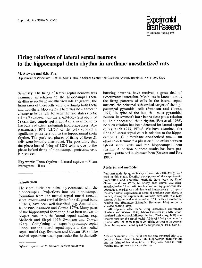
Fig. 1. Firing of two lateral septal units and their relations to the hippocampal theta rhythm. Each column shows three seconds of spontaneous firing of a representative cell along with the hippocampal EEG from the "CAI" and "'dentate" (DT) recording sites, an interspike interval distribution (IID) for that cell, and a phase histogram for that cell. The liDs reflect activity of the cells largely during hippocampal theta rhythm. Note that variability in spike amplitude is due to computer digitization of action potentials at 100/~sec intervals. A phase-locked lateral septal cell is illustrated in the left column. This cell was driven at 15 msec latency from the fimbria/fornix. The firing rate of this cell during theta rhythm was 3.3 spikes/sec. This cell fired faster during non-theta EEG, but a stable non-theta firing rate could not be determined. Notice that in spite of the nearly Poisson (pseudo-random) distribution of intervals, there is a clear phase relation of the unit to the hippocampal theta rhythm (middle). When the cell fires, it is most likely to fire on the positive-going phase of the dentate theta rhythm cycle (bottom). The positive peak of the dentate theta cycle is 0 ~ Calibrations for top panel: 0.5 mV for the cell, 1.0 mV for CA1 and DT EEG; 0.5 sec. IID (middle panel): number of spikes=861, firing rate = 4.7/sec; bin width = 20 msec. Phase histogram (bottom panel): number of spikes = 455. number of theta cycles = 572. A lateral septal cell that was not apparently phase-locked to the theta rhythm is illustrated in the right column. The firing rate for this cell during theta rhythm was 15.5 spikes/sec. Note the higher firing rate of this cell allowed the shorter time axis for the lID (middle). The distribution of intervals is also positively skewed. The total time displayed is 500 milliseconds and not 2 seconds as had been used for the left column. Calibrations for top panel: 0.4 mV for the cell, 1.0 mV for CA1 EEG and 0.6 mV for dentate EEG; 0.5 sec. IID (middle panel): number of spikes = 1399, firing rate = 16. l spikes/sec; bin width = 5 msec. Phase histogram (bottom panel): number of spikes = 289; number of theta cycles = 85
m e d i a n p e r c e n t a g e = 28%). E x e m p l a r y p h a s e - l o c k e d a n d n o n - p h a s e - l o c k e d cells a re i l l u s t r a t ed in Fig . 1. A s u m m a r y h i s t o g r a m of the m e a n phases o f f ir ing for the 21 p h a s e - l o c k e d L S N cells re la t ive to the d e n t a t e t he t a r h y t h m m e a n p h a s e is s h o w n in Fig . 2. T h e d e n t a t e t he t a r h y t h m m e a n p h a s e (pos i t iv i ty) is def ined in the f igure as 0 ~ M e a n vec to r l eng ths for these cells (0 .20+0 .09) were s ign i f ican t ly g r e a t e r t h a n those for the r e m a i n i n g 44 cells (0 .12+0 .08) tha t were no t p h a s e - l o c k e d : t = 3 . 3 9 2 , one- t a i l ed , P < 0.001.
T h e p re fe r red phases of f i r ing of the p h a s e - l o c k e d L S N cells were n o t d e p e n d e n t u p o n successful o r t h o -
d r o m i c d r i v ing f rom the f imbr ia / fo rn ix . O f the 17 o r t h o d r o m i c a l l y d r iven cells, 7 were p h a s e - l o c k e d , a n d 4 of the 10 cells t ha t c o u l d n o t be d r iven were p h a s e - l o c k e d . The cha rac t e r i s t i c s of p h a s e - l o c k e d a n d n o n - p h a s e - l o c k e d L S N cells a re s u m m a r i z e d in T a b l e 1.
Discussion
The a b o v e ana lys i s of the f i r ing of l a t e r a l sep ta l cells in r e l a t i on to the h i p p o c a m p a l E E G has r evea l ed t ha t s o m e L S N cells are, in fact, p h a s e - l o c k e d to the the ta r h y t h m . A g r o u p of s lowly f i r ing m e d i a l sep ta l

10
L E [ - 0-
-180 1 3 5 - 9 0 - 4 5 0 45 90 135 180
PHASE (degrees~
Fig. 2. Summary distribution of the preferred phases of firing for 21 lateral septal neurons that were phase-locked to the hippocampal theta rhythm. Each cell is plotted according to the lag or lead of the unit 's mean phase relative to the dentate theta rhythm mean phase (near the positive peak). The dentate theta rhythm mean phase is defined as 0 ~ in this figure
cells with an irregular firing pattern has been de- scribed in a previous paper (Stewart and Fox 1989b). Most of these "irregular" medial septal cells were phase-locked to the hippocampal theta rhythm and had their maximum probability of firing near the positive peak of the dentate theta rhythm. The pyramidal cells of CA 1, whose afferents to the lateral septum terminate more medially than those arising in CA3 (e.g. Swanson and Cowan 1977), also tend to fire on the positive peak of the dentate theta rhythm. This match between the firing phases of the "irregu- lar" medial septal cells and those of the CA1 pyramidal cells suggests that the dendrites of the "irregular" cells might extend into the more medial parts of the lateral septum to receive afferents from
95
CA1. The lateral septal cells described in this paper probably receive afferents from both CA1 and CA3. The broad distribution of peak firing phases for these LSN cells is consistent with a greater fraction of their inputs originating in CA3, where the firing phases of the pyramidal cells are more scattered than in CA1 (Fox et al. 1986).
Perhaps more remarkable than the phase-locked firing by some LSN cells is that the majority of LSN cells bear no relation to the hippocampal theta rhythm in spite of the clear phase-locked firing of hippocampal projection cells. This might be the result of convergence of hippocampal inputs with varying phases, such as from area CA3, onto individ- ual LSN cells (DeFrance et al. 1971, 1976). Another possible explanation for the variation in the degree of phase-locked firing of LSN cells is suggested by the extreme variability in the proportion of phase- locked LSN cells seen across animals. Some aspect of the general condition of the animal preparation might play a major role in determining the degree of phase-locked firing seen in these LSN cells, since they are a minimum of two (and probably at least three) synapses away from the rhythmically bursting cells of the medial septal nuclei. Finally, the lack of robust periodic firing by lateral septal cells helps to emphasize the, at best, weak role of a septo-hippo- campo-septal loop for the maintenance of rhythmic firing by medial septal neurons and the consequent production of the hippocampal theta rhythm.
Acknowledgements. Supported by National Institutes of Health grants NS 17095 (to S. E. F.) and NS 07117 (Institutional Neurophysiology training grant).
Table 1. Summary of characteristics of lateral septal (LSN) neurons a
Phase-locked Not phase-locked
Total cells 21 44 Firing rate (mean + S.D.)
during theta (N)
during non-theta (N)
10.2+12.8 spks/sec (21)
8.6 spks/sec (l)
Firing pattern (numbers of cells) simple spikes 20 bursts (complex-spikes) b 1
Orthodromic driving (numbers of cells) total driven/number tested 7/11 c short latency only (6~7 msec) 1 long latency only (15 20 msec) 2 both latencies 4
7.5 + 8.4 spks/sec (44) 5.7 + 5.5 spks/sec (9)
41 3
10/16 2 5 3
"Three LSN cells fired too slowly to be classified as phase-locked or not phase-locked b The bursts (complex-spikes) of this cell, while phase-locked to the hippocampal theta rhythm, did not resemble the rhythmic bursts of medial septal cells c The actual firing phases for these cells were as follows: short latency only ( - 149:); long latency only ( - 6 C ' , -53~); both latencies (-f58 ~, - 8 4 ~, - 1 3 4 ~ - 167:); not driven (+342 , +49 ~ - 7 0 = , - 140:)

96
References
Amaral DG, Kurz J (1985) An analysis of the origins of the cholinergic and noncholinergic septal projections to the hip- pocampal formation of the rat. J Comp Neurol 240:37-59
Batschelet E (1981) Circular statistics in biology. Academic Press, London
DeFrance JF, Shimono T, Kitai ST (1971) Anatomical distribu- tion of the hippocampal fibers afferent to the lateral septal nucleus. Brain Res 34:176-180
DeFrance JF, Kitai ST, Shimono T (1973) Electrophysiological analysis of the hippocampal-septal projections. I. Response and topographical characteristics. Exp Brain Res 17:447 462
DeFrance JF, Yoshihara H, Chronister RB (1976) Electro- physiological studies of the septal nuclei. I. The lateral septal nuclei. Exp Neurol 53:399-419
Fox SE, Wolfson S, Ranck JB Jr (1986) Hippocampal theta rhythm and the firing of neurons in walking and urethane anesthetized rats. Exp Brain Res 62:495 508
Gutnick M J, Feldman S (1977) Effects of hippocampal and hypothalamic afferents on neuronal activities in the rat septum. Exp Neurol 57:212-230
JoWls M, Urban IJA (1985) Topographic organization of tim- bria/fornix fibers projecting to the lateral septum of rats: a single and field response analysis. Exp Neurol 87:474 486
McLennan H, Miller JJ (1974) The hippocampal control of neuronal discharges in the septum of the rat. J Physiol (Lond) 237:607 624
Meibach RC, Siegel A (1977) Efferent connections of the hippo- campal formation in the rat. Brain Res 124:197-224
Paxinos G, Watson C (1982) The rat brain in stereotaxic coordinates. Academic Press, Sydney
Ranck JB Jr (1973) Studies on single neurons in dorsal hippocam- pal formation and septum in unrestrained rats. I. Behavioral correlates and firing repertoires. Exp Neurol 41:461-531
Ranck JB Jr (1976) Behavioral correlates and firing repertoires of neurons in septal nuclei in unrestrained rats. In: DeFrance JF (ed) The septal nuclei. Plenum Press, New York, pp 423 462
Stewart M, Fox SE (1987) Phases of firing of medial and lateral septal neurons and the importance of hippocampal feedback. Soc Neurosci Abstr 13:1331
Stewart M, Fox SE (198%) Two populations of rhythmically bursting neurons in the rat medial septum are revealed by atropine. J Neurophysiol 61:982 993
Stewart M, Fox SE (1989b) Firing relations of medial septal neurons to the hippocampal theta rhythm in urethane anes- thetized rats. Exp Brain Res 77:507 516
Swanson LW, Cowan WM (1977) An autoradiographic study of the organization of the efferent connections of the hippocam- pal formation in the rat. J Comp Neurol 172:49-84
Swanson LW, Cowan WM (1979) The connections of the septal region in the rat. J Comp Neurol 186:621-656
Received April 20, 1989 / Accepted July 21, 1989