Finite Element Analysis of Soccer Headingumpir.ump.edu.my/12719/1/fkp-2015-hasnun-Finite... ·...
6
Procedia Engineering 112 (2015) 46 – 51 1877-7058 © 2015 The Authors. Published by Elsevier Ltd. This is an open access article under the CC BY-NC-ND license (http://creativecommons.org/licenses/by-nc-nd/4.0/). Peer-review under responsibility of the the School of Aerospace, Mechanical and Manufacturing Engineering, RMIT University doi:10.1016/j.proeng.2015.07.174 ScienceDirect Available online at www.sciencedirect.com 7th Asia-Pacific Congress on Sports Technology, APCST 2015 Finite Element Analysis of Soccer Heading Mohd Hasnun Arif Hassan*, Zahari Taha Innovative Manufacturing, Mechatronics & Sports Laboratory, Universiti Malaysia Pahang, 26600 Pekan, Pahang, Malaysia Abstract Studies have shown compelling evidence that suggests heading soccer ball might lead to brain trauma. Researchers have attempted to experimentally quantify head acceleration induced by soccer ball heading. A series of linear accelerometers as well as angular accelerometer were used to measure head accelerations. This method however is limited to measuring only head acceleration during the impact. However, it is essential to analyse the acceleration of the brain in addition to the acceleration of the head that takes place during a soccer-heading manoeuvre. Since the motion of the brain is almost impossible to be quantified experimentally, this work focuses on performing finite element (FE) analysis of soccer heading to study the motions of both the head and the brain during the impact. FE model of soccer ball was developed and validated against published experimental data as well as a more detailed model. Moreover, FE model of human head that consists of the skull, facial bones, cerebrospinal fluid (CSF) layer and the brain was also developed and validated against experimental data of blunt impact on human cadaver. Both validated models were assembled to perform the soccer heading simulations. Linear and angular accelerations of the skull and the brain generated are comparable to those of experimental data. However, it has underestimated the angular acceleration due to the absence of neck in the model, but with comparable acceleration profile. Linear and angular accelerations of the brain were found to be almost similar to those of the head, which is contradictory to our initial hypothesis. Further study such as ball impact on instrumented dummy skull is required to corroborate the findings. Nonetheless, the FE models were able to replicate the head accelerations sustained during soccer ball heading satisfactorily. The simulation results show that the models can be employed in finding protective materials that can reduce the accelerations, thus minimising the probability of suffering from long-term brain trauma due to soccer ball heading. Keywords: Soccer heading; finite element analysis; head injury; brain injury; soccer ball model; head model. * Corresponding author. Tel.: +609-424-6358; fax: +609-424-5888. E-mail address: [email protected] © 2015 The Authors. Published by Elsevier Ltd. This is an open access article under the CC BY-NC-ND license (http://creativecommons.org/licenses/by-nc-nd/4.0/). Peer-review under responsibility of the the School of Aerospace, Mechanical and Manufacturing Engineering, RMIT University
Transcript of Finite Element Analysis of Soccer Headingumpir.ump.edu.my/12719/1/fkp-2015-hasnun-Finite... ·...
Procedia Engineering 112 ( 2015 ) 46 – 51
1877-7058 © 2015 The Authors. Published by Elsevier Ltd. This is an open access article under the CC BY-NC-ND license (http://creativecommons.org/licenses/by-nc-nd/4.0/).Peer-review under responsibility of the the School of Aerospace, Mechanical and Manufacturing Engineering, RMIT Universitydoi: 10.1016/j.proeng.2015.07.174
ScienceDirectAvailable online at www.sciencedirect.com
7th Asia-Pacific Congress on Sports Technology, APCST 2015
Finite Element Analysis of Soccer Heading
Mohd Hasnun Arif Hassan*, Zahari Taha Innovative Manufacturing, Mechatronics & Sports Laboratory, Universiti Malaysia Pahang, 26600 Pekan, Pahang, Malaysia
Abstract
Studies have shown compelling evidence that suggests heading soccer ball might lead to brain trauma. Researchers have attempted to experimentally quantify head acceleration induced by soccer ball heading. A series of linear accelerometers as well as angular accelerometer were used to measure head accelerations. This method however is limited to measuring only head acceleration during the impact. However, it is essential to analyse the acceleration of the brain in addition to the acceleration of the head that takes place during a soccer-heading manoeuvre. Since the motion of the brain is almost impossible to be quantified experimentally, this work focuses on performing finite element (FE) analysis of soccer heading to study the motions of both the head and the brain during the impact. FE model of soccer ball was developed and validated against published experimental data as well as a more detailed model. Moreover, FE model of human head that consists of the skull, facial bones, cerebrospinal fluid (CSF) layer and the brain was also developed and validated against experimental data of blunt impact on human cadaver. Both validated models were assembled to perform the soccer heading simulations. Linear and angular accelerations of the skull and the brain generated are comparable to those of experimental data. However, it has underestimated the angular acceleration due to the absence of neck in the model, but with comparable acceleration profile. Linear and angular accelerations of the brain were found to be almost similar to those of the head, which is contradictory to our initial hypothesis. Further study such as ball impact on instrumented dummy skull is required to corroborate the findings. Nonetheless, the FE models were able to replicate the head accelerations sustained during soccer ball heading satisfactorily. The simulation results show that the models can be employed in finding protective materials that can reduce the accelerations, thus minimising the probability of suffering from long-term brain trauma due to soccer ball heading. © 2015 The Authors. Published by Elsevier Ltd. Peer-review under responsibility of the School of Aerospace, Mechanical and Manufacturing Engineering, RMIT University.
Keywords: Soccer heading; finite element analysis; head injury; brain injury; soccer ball model; head model.
* Corresponding author. Tel.: +609-424-6358; fax: +609-424-5888.
E-mail address: [email protected]
© 2015 The Authors. Published by Elsevier Ltd. This is an open access article under the CC BY-NC-ND license (http://creativecommons.org/licenses/by-nc-nd/4.0/).Peer-review under responsibility of the the School of Aerospace, Mechanical and Manufacturing Engineering, RMIT University
47 Mohd Hasnun Arif Hassan and Zahari Taha / Procedia Engineering 112 ( 2015 ) 46 – 51
1. Introduction
Soccer is a unique sports, in which the purposeful use of the head to direct the ball in the game is allowed. There have been different opinions on whether heading soccer ball might lead to brain trauma. In the past decades, researchers have tried to quantify the impact of soccer heading through neuropsychological evaluations. This is a set of tests that measures the ability of one’s brain to function properly. These studies have revealed that heading soccer ball contributes to cognitive dysfunction [1,2]. Moreover, the cognitive function of soccer players was also studied using a tablet-based approach. It was found that heading soccer ball might cause symptoms typically found in patients with mild traumatic brain injury (TBI) of the frontal lobes [3].
In addition, the brain of soccer players have also been evaluated by means of magnetic resonance imaging (MRI) technique known as diffusion tensor imaging (DTI). Both amateur [4] and professional soccer players [5] were examined using this method. These studies have found abnormalities in the brain of frequent headers similar to those found in patients with mild TBI. Furthermore, the former study on amateur soccer players has found compelling evidence that exceeding the threshold level of approximately 885 to 1,550 is associated with lower white matter fractional anisotropy (which indicates pathological changes in the brain tissue) and memory problems [4]. These findings suggest that heading soccer ball poses a long-term threat to the brain.
Studies have been conducted to experimentally quantify the impact of heading soccer ball. Linear and angular accelerations of the head were measured using accelerometers. The experiments were conducted on both human subjects as well as dummy headform [6–8]. Moreover, mathematical models were also employed to predict the head acceleration sustained during soccer ball heading [9,10]. All these methods were able to measure or predict the acceleration of the head as a single unit. However, the concern of possible brain injury is in fact associated with the acceleration of the brain. Thus, it is vital to know the amount of accelerations sustained by the brain during soccer heading in addition to head acceleration.
Measuring brain acceleration in vivo is almost impossible. Hence, finite element (FE) analysis is one of the promising methods that can be used to predict the brain responses due to soccer ball heading. FE analysis has been mostly used to study head injuries occurred in car accidents as well as those occurred in contact sports. However, to authors’ knowledge, no FE analysis has been done to quantify brain accelerations endured during soccer ball heading. This study focuses on the development and validation of FE models of soccer ball and human head. Both validated models will be used to perform FE analysis of soccer heading. Head and brain accelerations are the parameters of interest that will be measured in the analysis. Head accelerations obtained from the simulation will be compared with experimental data from literature [7] to evaluate the reliability of the model. The following section describes the development of the required FE models as well as the analysis done. The subsequent section discusses the results obtained from the simulations.
2. Method
2.1. Finite element modelling of soccer ball
Soccer balls are normally made of 32 manually stitched composite panels that are pressurised through an internal latex bladder. However, to simplify the FE model of the ball, the model was developed using a hollow spherical shell that possesses isotropic material properties instead of modelling the 32 panels separately. The surface of the shell was discretised into 2,400 linear quadrilateral elements in Abaqus/CAE. The use of composite shell has greatly increased computational efficiency compared to three-dimensional continuum elements. The composite shell element includes two layers, namely an inner bladder that is 0.2 mm thick, and an outer panel that is 2.2 mm thick.
Tensile response for each layer was extracted from [11] and were applied to the model by fitting a hyperelastic reduced polynomial strain energy potential equation against the tensile test data. The reduced polynomial strain energy potential form is given by following equation:
U = Ci0 I1 − 3( )i=1
N
∑i
+ 1
Di
J el −1( )i=1
N
∑2i
(1)
48 Mohd Hasnun Arif Hassan and Zahari Taha / Procedia Engineering 112 ( 2015 ) 46 – 51
where U is the strain energy per unit of reference volume, I1 is the first strain invariant, J el is the elastic volume ratio, whilst Ci0 and Di are the material constants. The density of the inner bladder and the outer panel was defined as 1,175 kg/m3 and 900 kg/m3, respectively. This has resulted in a total mass of 0.444 kg.
Pressurisation of the ball was done using surface-based fluid cavity technique. This technique supersedes the element-based hydrostatic fluid cavity capability in terms of functionality and eliminates the need for fluid or fluid link elements definition [12]. It is assumed that the cavity of the ball is completely filled by fluid with uniform properties and state. A cavity reference node, which has a single degree of freedom, was defined at the centre of gravity of the ball to represent the pressure inside the cavity.
Since the model developed is based on the material properties taken from Price et al. [11], we have validated the model against their models and experimental data. The ball was pressurised to 0.9 bar. Dynamic impact tests were simulated with inbound velocities of 11, 15, 20, 22 and 28 m/s. Comparison of the coefficient of restitution (COR), contact time and longitudinal deformation between both models as well as experimental data have shown a very good agreement as shown in Fig. 1. This suggests that our FE model of soccer ball is validated and can be used in soccer heading analysis.
(a) (b) (c) Fig. 1. Validation of the soccer ball FE model against Price et al. (2006) [11].
2.2. Finite element modelling of human head
The aim was to develop a human head FE model that is capable of producing responses similar to that of an actual human brain upon impacts. The geometry of the skull was obtained online at www.anatomium.com. A human skull consists of two main parts: the cranium and the facial bones. The sutures were removed and all eight bones were merged to form a unified human skull. To further reduce the complexity of the model, the lower jaw and the teeth were also removed.
The cranium and facial bones were defined as linear elastic single-layered bone with an elastic modulus of 6.5 GPa and Poisson’s ratio of 0.2 [13]. The densities of both components were defined as 3,000 kg/m3 whilst the density of the facial bones was defined as 8,000 kg/m3 to match the average weight of human head. The cerebrospinal fluid (CSF) is a 1.3 mm thick layer that separates the skull and the brain. The CSF protects the brain from various closed head injuries by acting as a natural shock absorber. It was modelled as linear elastic with an elastic modulus of 150 kPa and Poisson’s ratio of 0.49886 [14] with a density of 1,004 kg/m3.
The brain was modelled as solid, which fills the area beneath the CSF. It was defined as viscoelastic with a linear elastic material model. The elastic modulus, E of the brain was computed from the bulk modulus, K and shear modulus, G from [15] using following equation:
E = 9KG
3K +G (2)
which gives a value of 5.04 MPa. The Poisson’s ratio was defined as 0.4996 with a density of 1,040 kg/m3 [15].
This results in a mass of the brain of 1.56 kg. In addition, the shear characteristics of the viscoelastic behaviour of the brain were expressed by following equation:
49 Mohd Hasnun Arif Hassan and Zahari Taha / Procedia Engineering 112 ( 2015 ) 46 – 51
G t( ) =G∞ + G0 −G∞( )e−βt (3) where G0 is the short-term shear modulus with a value of 0.528 MPa, G∞ is the long-term shear modulus with a
value of 0.168 MPa, β is the decay factor that was defined as 35 s-1 and t is time, expressed in second [16]. These characteristics were implemented in ABAQUS 6.13 as the time domain viscoelastic material model that is
given by a Prony series expansion of the following dimensionless relaxation modulus [12]: gR t( ) =1− gP 1− e− t/τG( ) (4)
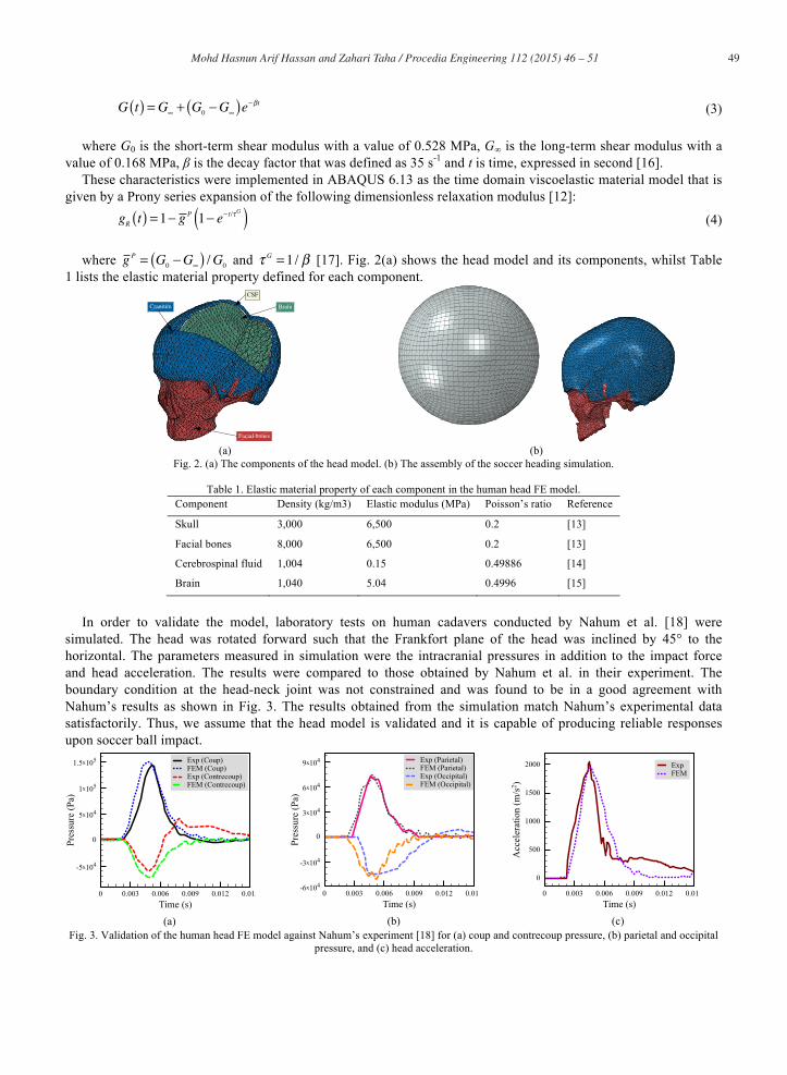
where gP = G0 −G∞( ) /G0 and τ G =1/ β [17]. Fig. 2(a) shows the head model and its components, whilst Table
1 lists the elastic material property defined for each component.
Facial bones
Cranium
CSF
Brain
(a) (b)
Fig. 2. (a) The components of the head model. (b) The assembly of the soccer heading simulation.
Table 1. Elastic material property of each component in the human head FE model. Component Density (kg/m3) Elastic modulus (MPa) Poisson’s ratio Reference
Skull 3,000 6,500 0.2 [13]
Facial bones 8,000 6,500 0.2 [13]
Cerebrospinal fluid 1,004 0.15 0.49886 [14]
Brain 1,040 5.04 0.4996 [15]
In order to validate the model, laboratory tests on human cadavers conducted by Nahum et al. [18] were
simulated. The head was rotated forward such that the Frankfort plane of the head was inclined by 45° to the horizontal. The parameters measured in simulation were the intracranial pressures in addition to the impact force and head acceleration. The results were compared to those obtained by Nahum et al. in their experiment. The boundary condition at the head-neck joint was not constrained and was found to be in a good agreement with Nahum’s results as shown in Fig. 3. The results obtained from the simulation match Nahum’s experimental data satisfactorily. Thus, we assume that the head model is validated and it is capable of producing reliable responses upon soccer ball impact.
(a) (b) (c) Fig. 3. Validation of the human head FE model against Nahum’s experiment [18] for (a) coup and contrecoup pressure, (b) parietal and occipital
pressure, and (c) head acceleration.
50 Mohd Hasnun Arif Hassan and Zahari Taha / Procedia Engineering 112 ( 2015 ) 46 – 51
2.3. FE analysis of soccer heading
Both validated models of the soccer ball and human head were assembled in one model as shown in Fig. 2(b). The ball was placed very close (approximately 40 mm) to the head to minimise the computing time. The head was rotated 22.5° forward about the axis on the coronal plane to replicate the position of the head during soccer heading manoeuvre. The velocity boundary condition was defined on the ball that signifies its inbound velocity. The interface between the skull and CSF as well as the CSF and the brain were defined using tangential sliding contact with coefficient of friction of 0.2. Linear and angular accelerations were measured at two nodes: one at the mouth area, another one at the centre of gravity of the brain. Accelerations of the node at the mouth area were transformed to the centre of gravity of the head to obtain the head accelerations.
3. Results and Discussion
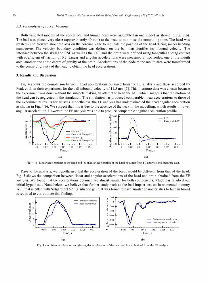
Fig. 4 shows the comparison between head accelerations obtained from the FE analysis and those recorded by Funk et al. in their experiment for the ball inbound velocity of 11.5 m/s [7]. This literature data was chosen because the experiment was done without the subjects making an attempt to head the ball, which suggests that the motion of the head can be neglected in the simulation. The simulation has produced comparable linear accelerations to those of the experimental results for all axes. Nonetheless, the FE analysis has underestimated the head angular acceleration as shown in Fig. 4(b). We suspect that this is due to the absence of the neck in the modelling, which results in lower angular acceleration. However, the FE analysis was able to produce comparable angular acceleration profile.
(a) (b)
Fig. 4. (a) Linear accelerations of the head and (b) angular acceleration of the head obtained from FE analysis and literature data.
Prior to the analysis, we hypothesise that the acceleration of the brain would be different from that of the head. Fig. 5 shows the comparison between linear and angular accelerations of the head and brain obtained from the FE analysis. We found that the accelerations obtained are almost similar for both components, which has falsified our initial hypothesis. Nonetheless, we believe that further study such as the ball impact test on instrumented dummy skull that is filled with Sylgard gel 527 (a silicone gel that was found to have similar characteristics to human brain) is required to corroborate this finding.
(a) (b)
Fig. 5. (a) Linear acceleration and (b) angular acceleration of the head and brain obtained from the FE analysis.

51 Mohd Hasnun Arif Hassan and Zahari Taha / Procedia Engineering 112 ( 2015 ) 46 – 51
4. Conclusion
FE models of soccer ball and human head were successfully developed and validated. Both models were assembled to perform soccer heading simulation. Linear and angular accelerations of the head and brain were recorded. Comparison with experimental data from literature shows that the model is capable of generating comparable linear head acceleration. However, the FE analysis underestimates the angular acceleration of the head, but with comparable acceleration profile. This might due to the absence of neck in the head model. Thus, we believe that both models can be used in the search of protective materials that can reduce the head accelerations during soccer ball heading, hence minimise the probability of sustaining long-term brain trauma. We also found that both linear and angular accelerations of the brain and head are almost similar, which is not as what we have expected. Further study of ball impact on instrumented dummy skull is required to corroborate this finding. Future works include the inclusion of neck in the head model and ball impact test on instrumented dummy skull.
References
[1] A. Tysvaer, E. Løchen, Soccer injuries to the brain: A neuropsychologic study of former soccer players, Am. J. Sports Med. 19 (1991) 56–60.
[2] E. Matser, A. Kessels, M. Lezak, B. Jordan, J. Troost, Neuropsychological Impairment in Amateur Soccer Players, JAMA J. Am. Med. Assoc. 282 (1999) 971–973.
[3] M.R. Zhang, S.D. Red, A.H. Lin, S.S. Patel, A.B. Sereno, Evidence of cognitive dysfunction after soccer playing with ball heading using a novel tablet-based approach., PLoS One. 8 (2013) e57364. doi:10.1371/journal.pone.0057364.
[4] M. Lipton, N. Kim, M. Zimmerman, Soccer Heading Is Associated with White Matter Microstructural and Cognitive Abnormalities, Radiology. (2013).
[5] I. Koerte, B. Ertl-Wagner, White matter integrity in the brains of professional soccer players without a symptomatic concussion, JAMA J. Am. Med. Assoc. 308 (2012) 2006–2008. http://jama.jamanetwork.com/article.aspx?articleID=1391907 (accessed June 24, 2013).
[6] R.S. Naunheim, P. V Bayly, J. Standeven, J.S. Neubauer, L.M. Lewis, G.M. Genin, Linear and angular head accelerations during heading of a soccer ball., Med. Sci. Sports Exerc. 35 (2003) 1406–12. doi:10.1249/01.MSS.0000078933.84527.AE.
[7] J.R. Funk, J.M. Cormier, C.E. Bain, H. Guzman, E. Bonugli, Validation and application of a methodology to calculate head accelerations and neck loading in soccer ball impacts, SAE Tech. Pap. 01 (2009). http://www.brconline.com/files/consultants/105/article_12.pdf (accessed July 29, 2013).
[8] N. Shewchenko, C. Withnall, M. Keown, R. Gittens, J. Dvorak, Heading in football. Part 1: development of biomechanical methods to investigate head response., Br. J. Sports Med. 39 Suppl 1 (2005) i10–25. doi:10.1136/bjsm.2005.019034.
[9] P.E. Riches, A dynamic model of the head acceleration associated with heading a soccer ball, Sport. Eng. 9 (2006) 39–47. doi:10.1007/BF02844261.
[10] Z. Taha, M.H.A. Hassan, I. Hasanuddin, Analytical Modelling of Soccer Heading, Sadhana. In Press (2015). doi:http://dx.doi.org/10.1007/s12046-015-0383-5.
[11] D.S. Price, R. Jones, A.R. Harland, Computational modelling of manually stitched soccer balls, Proc. Inst. Mech. Eng. Part L J. Mater. Des. Appl. 220 (2006) 259–268. doi:10.1243/14644207JMDA83.
[12] Dassault Systèmes, Abaqus 6.13 Documentation, (2013). [13] M. Claessens, Finite Element Modeling of the Human Head under Impact Conditions, TU Eindhoven, 1997. [14] Y. Chen, M. Ostoja-Starzewski, MRI-based finite element modeling of head trauma: spherically focusing shear waves, Acta Mech. 213
(2010) 155–167. doi:10.1007/s00707-009-0274-0. [15] J.S. Ruan, T. Khalil, A.I. King, Dynamic response of the human head to impact by three-dimensional finite element analysis., J. Biomech.
Eng. 116 (1994) 44–50. [16] J. Ruan, T. Khalil, A. King, Finite element modeling of direct head impact, in: Stapp Car Crash Conf., 1993. [17] S.P.C. Marques, G.J. Creus, Solution with Abaqus, in: Comput. Viscoelasticity, Springer Berlin Heidelberg, Berlin, Heidelberg, 2012: pp.
103–111. doi:10.1007/978-3-642-25311-9. [18] A.M. Nahum, R. Smith, C.C. Ward, Intracranial Pressure Dynamics During Head Impact, (1977). doi:10.4271/770922.