Finite Element Analysis of Radiofrequency Ablation Abirvab Deb- BME M.Eng ‘14 Brice Lekavich- BME...
17
Finite Element Analysis of Radiofrequency Ablation Abirvab Deb- BME M.Eng ‘14 Brice Lekavich- BME M.Eng ‘14 Cristian Vilorio- BME M.Eng ‘14
-
Upload
rudolf-tate -
Category
Documents
-
view
222 -
download
0
Transcript of Finite Element Analysis of Radiofrequency Ablation Abirvab Deb- BME M.Eng ‘14 Brice Lekavich- BME...
Finite Element Analysis of
Radiofrequency Ablation
Abirvab Deb- BME MEng lsquo14Brice Lekavich- BME MEng lsquo14Cristian Vilorio- BME MEng lsquo14
Background
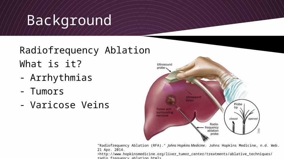
Radiofrequency AblationWhat is it- Arrhythmias- Tumors- Varicose Veins
Radiofrequency Ablation (RFA) Johns Hopkins Medicine Johns Hopkins Medicine nd Web 21 Apr 2014 lthttpwwwhopkinsmedicineorgliver_tumor_centertreatmentsablative_techniques radio_frequency_ablationhtmlgt
FEA Motivation
bull Lesion has to be deep enough into targeted tissue yet limit the amount of irreversible damage occurring to normal cardiac tissue
bull The ablated tissue area has to be large enough to compensate for the uncertainties in the electrical mapping that is used to localize the target tissue
bull RF energy is effective in ablating some arrhythmias bull Multiple attempts often have to be made prior to successfully
destroying the targeted tissue bull Lesions produced are not deep enough to eliminate the target
electrical pathway bull Lesion depth is a problem in the left ventricle where the heart wall
is thick
Objective
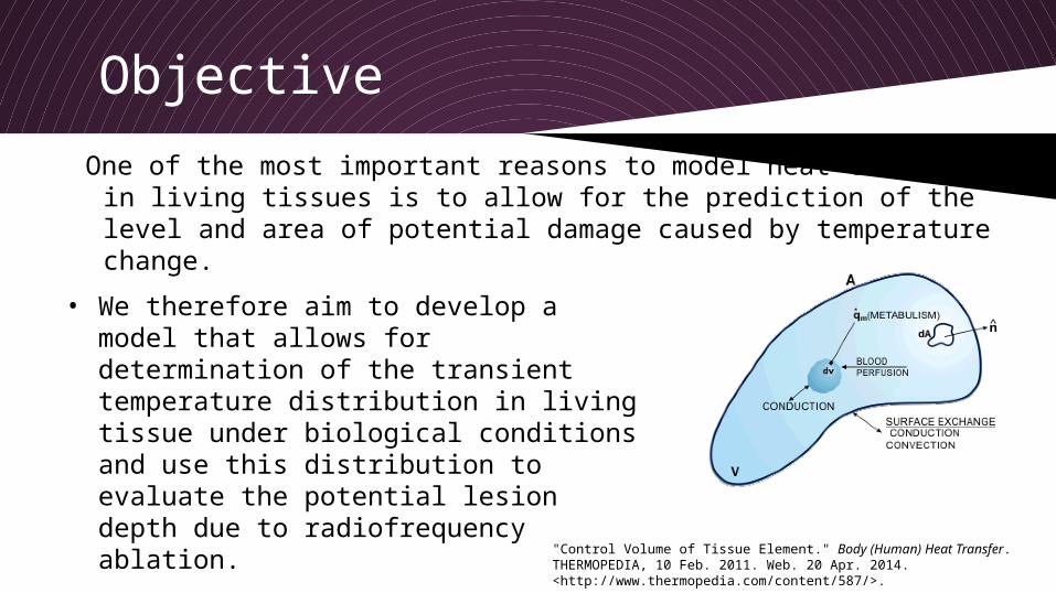
One of the most important reasons to model heat transfer in living tissues is to allow for the prediction of the level and area of potential damage caused by temperature change
bull We therefore aim to develop a model that allows for determination of the transient temperature distribution in living tissue under biological conditions and use this distribution to evaluate the potential lesion depth due to radiofrequency ablation
Control Volume of Tissue Element Body (Human) Heat Transfer THERMOPEDIA 10 Feb 2011 Web 20 Apr 2014 lthttpwwwthermopediacomcontent587gt
FEA Methodology
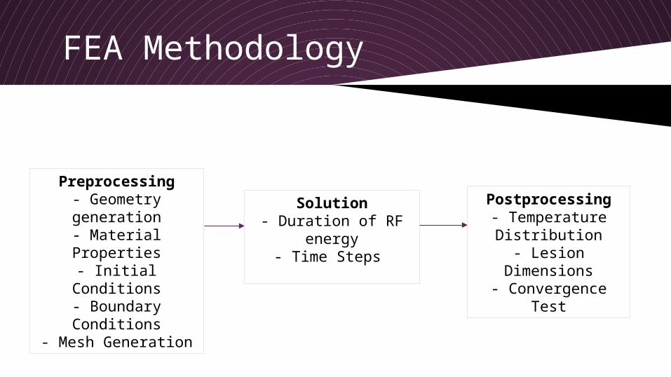
Preprocessing- Geometry generation
- Material Properties- Initial Conditions
- Boundary Conditions
- Mesh Generation
Solution- Duration of RF
energy- Time Steps
Postprocessing- Temperature Distribution
- Lesion Dimensions
- Convergence Test
Bioheat Transfer amp Pennesrsquo Equation
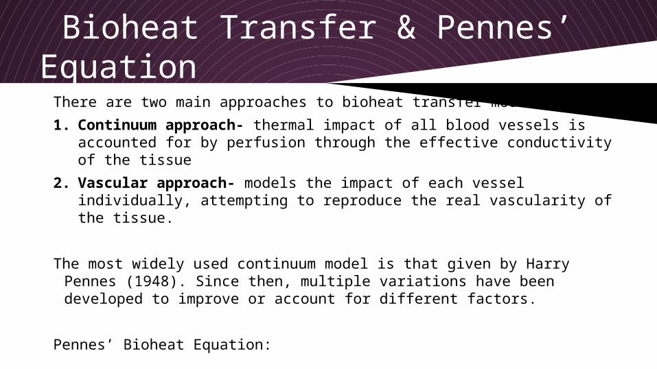
There are two main approaches to bioheat transfer models
1 Continuum approach- thermal impact of all blood vessels is accounted for by perfusion through the effective conductivity of the tissue
2 Vascular approach- models the impact of each vessel individually attempting to reproduce the real vascularity of the tissue
The most widely used continuum model is that given by Harry Pennes (1948) Since then multiple variations have been developed to improve or account for different factors
Pennesrsquo Bioheat Equation
Assumptions of Pennesrsquo Equation 1) Pre-arteriolepost-venule heat transfer between
the tissue and blood is neglected
2) Blood flow in small capillaries is assumed to be isotropic (ignores blood flow directionality)
3) Does not consider local vascular geometry (role of larger blood vessels near capillary beds is neglected)
4) Blood is assumed to reach arterioles supplying the capillary beds at the body core temperature (assumed instantaneous exchange of energy and equilibrium with local tissue temperature)
Sakaguchi et al In Vitro Engineering of Vascularized Tissue Surrogates Scientific Reports 3 1316 1-7 (2013)
Cardiac cells
vascularization
Modifying Pennes Bioheat Equation
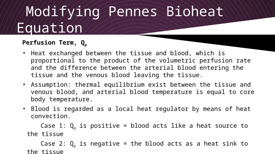
Perfusion Term Qp
bull Heat exchanged between the tissue and blood which is proportional to the product of the volumetric perfusion rate and the difference between the arterial blood entering the tissue and the venous blood leaving the tissue
bull Assumption thermal equilibrium exist between the tissue and venous blood and arterial blood temperature is equal to core body temperature
bull Blood is regarded as a local heat regulator by means of heat convection
Case 1 Qp is positive = blood acts like a heat source to the tissue
Case 2 Qp is negative = the blood acts as a heat sink to the tissue
In our case the core body temperature (Tb) is lower than the tissue temperature (T) and therefore Qp is negative and Case 2 is satisfied
Modifying Pennes Bioheat Equation
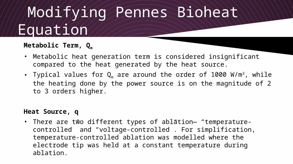
Metabolic Term Qm
bull Metabolic heat generation term is considered insignificant compared to the heat generated by the heat source
bull Typical values for Qm are around the order of 1000 Wm3 while the heating done by the power source is on the magnitude of 2 to 3 orders higher
Heat Source q
bull There are two different types of ablationmdash ldquotemperature-controlledrdquo and ldquovoltage-controlledrdquo For simplification temperature-controlled ablation was modelled where the electrode tip was held at a constant temperature during ablation
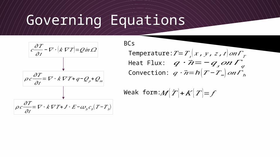
Governing Equations
BCs
Temperature
Heat Flux
Convection
Weak form
119888120597119879120597119905minus120571 ∙ (119896120571 119879 )=119876119894119899 120570
120588 119888120597119879120597119905
=120571 ∙119896120571119879 + 119869 ∙119864minus120596119887119888119887(119879 minus119879119887)
120588 119888120597119879120597119905
=120571 ∙119896120571119879 +119902minus119876119901+119876119898
119879=119879 119904 ( 119909 119910 119911 119905 ) 119900119899 120548119879
119902 ∙ =minus119902119904 119900119899120548 119902119902 ∙ =h (119879 minus119879infin )119900119899120548 h
119872 +119870 119879 = 119891
Time Integration
Have equation of the form
θ-method time integration
Where
Plugging and into the θ-family of approximation and rearranging terms to be of the equivalent form of
Where
To allow for unconditional stability
(Crank-Nicolson Method)
Solve for at each time step
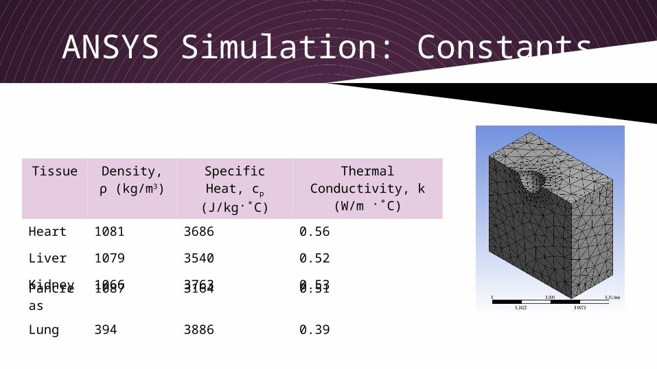
ANSYS Simulation Constants
Tissue Density ρ (kgm3)
Specific Heat cp (Jkg˚C)
Thermal Conductivity k (Wm
˚C)
Heart 1081 3686 056
Liver 1079 3540 052
Kidney 1066 3763 053Pancreas
1087 3164 051
Lung 394 3886 039
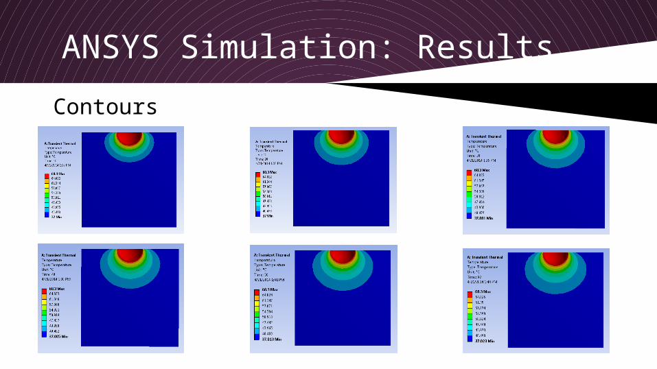
ANSYS Simulation Results
Contours
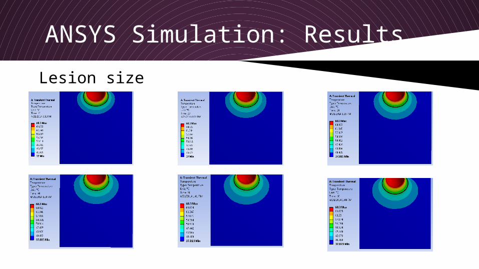
ANSYS Simulation Results
Lesion size
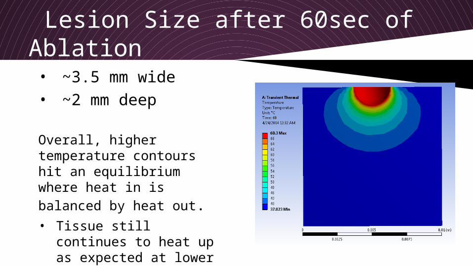
Lesion Size after 60sec of Ablation
bull ~35 mm widebull ~2 mm deep
Overall higher temperature contours hit an equilibrium where heat in is balanced by heat outbull Tissue still continues to
heat up as expected at lower temperatures
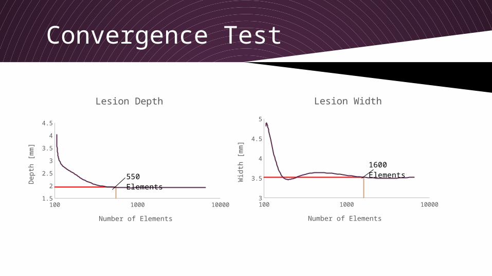
Convergence Test
100 1000 1000015
2
25
3
35
4
45
Lesion Depth
Number of Elements
Dep
th [
mm
]
550 Elements
100 1000 100003
32343638
442444648
5
Lesion Width
Number of Elements
Wid
th [
mm
]
1600 Elements
Lesion Size by Tissue Type
Heart Kidney Liver Pancreas Lung195
2
205
21
215
22
225
23
235
Lesion Depth
Dep
th [
mm
]
Lung Pancreas Heart Liver Kidney3
31
32
33
34
35
36
37
Lesion Width
Wid
th [
mm
]
- Finite Element Analysis of Radiofrequency Ablation
- Background
- FEA Motivation
- Objective
- FEA Methodology
- Bioheat Transfer amp Pennesrsquo Equation
- Assumptions of Pennesrsquo Equation
- Modifying Pennes Bioheat Equation
- Modifying Pennes Bioheat Equation (2)
- Governing Equations
- Time Integration
- ANSYS Simulation Constants
- ANSYS Simulation Results
- ANSYS Simulation Results (2)
- Lesion Size after 60sec of Ablation
- Convergence Test
- Lesion Size by Tissue Type
-
Background
Radiofrequency AblationWhat is it- Arrhythmias- Tumors- Varicose Veins
Radiofrequency Ablation (RFA) Johns Hopkins Medicine Johns Hopkins Medicine nd Web 21 Apr 2014 lthttpwwwhopkinsmedicineorgliver_tumor_centertreatmentsablative_techniques radio_frequency_ablationhtmlgt
FEA Motivation
bull Lesion has to be deep enough into targeted tissue yet limit the amount of irreversible damage occurring to normal cardiac tissue
bull The ablated tissue area has to be large enough to compensate for the uncertainties in the electrical mapping that is used to localize the target tissue
bull RF energy is effective in ablating some arrhythmias bull Multiple attempts often have to be made prior to successfully
destroying the targeted tissue bull Lesions produced are not deep enough to eliminate the target
electrical pathway bull Lesion depth is a problem in the left ventricle where the heart wall
is thick
Objective
One of the most important reasons to model heat transfer in living tissues is to allow for the prediction of the level and area of potential damage caused by temperature change
bull We therefore aim to develop a model that allows for determination of the transient temperature distribution in living tissue under biological conditions and use this distribution to evaluate the potential lesion depth due to radiofrequency ablation
Control Volume of Tissue Element Body (Human) Heat Transfer THERMOPEDIA 10 Feb 2011 Web 20 Apr 2014 lthttpwwwthermopediacomcontent587gt
FEA Methodology
Preprocessing- Geometry generation
- Material Properties- Initial Conditions
- Boundary Conditions
- Mesh Generation
Solution- Duration of RF
energy- Time Steps
Postprocessing- Temperature Distribution
- Lesion Dimensions
- Convergence Test
Bioheat Transfer amp Pennesrsquo Equation
There are two main approaches to bioheat transfer models
1 Continuum approach- thermal impact of all blood vessels is accounted for by perfusion through the effective conductivity of the tissue
2 Vascular approach- models the impact of each vessel individually attempting to reproduce the real vascularity of the tissue
The most widely used continuum model is that given by Harry Pennes (1948) Since then multiple variations have been developed to improve or account for different factors
Pennesrsquo Bioheat Equation
Assumptions of Pennesrsquo Equation 1) Pre-arteriolepost-venule heat transfer between
the tissue and blood is neglected
2) Blood flow in small capillaries is assumed to be isotropic (ignores blood flow directionality)
3) Does not consider local vascular geometry (role of larger blood vessels near capillary beds is neglected)
4) Blood is assumed to reach arterioles supplying the capillary beds at the body core temperature (assumed instantaneous exchange of energy and equilibrium with local tissue temperature)
Sakaguchi et al In Vitro Engineering of Vascularized Tissue Surrogates Scientific Reports 3 1316 1-7 (2013)
Cardiac cells
vascularization
Modifying Pennes Bioheat Equation
Perfusion Term Qp
bull Heat exchanged between the tissue and blood which is proportional to the product of the volumetric perfusion rate and the difference between the arterial blood entering the tissue and the venous blood leaving the tissue
bull Assumption thermal equilibrium exist between the tissue and venous blood and arterial blood temperature is equal to core body temperature
bull Blood is regarded as a local heat regulator by means of heat convection
Case 1 Qp is positive = blood acts like a heat source to the tissue
Case 2 Qp is negative = the blood acts as a heat sink to the tissue
In our case the core body temperature (Tb) is lower than the tissue temperature (T) and therefore Qp is negative and Case 2 is satisfied
Modifying Pennes Bioheat Equation
Metabolic Term Qm
bull Metabolic heat generation term is considered insignificant compared to the heat generated by the heat source
bull Typical values for Qm are around the order of 1000 Wm3 while the heating done by the power source is on the magnitude of 2 to 3 orders higher
Heat Source q
bull There are two different types of ablationmdash ldquotemperature-controlledrdquo and ldquovoltage-controlledrdquo For simplification temperature-controlled ablation was modelled where the electrode tip was held at a constant temperature during ablation
Governing Equations
BCs
Temperature
Heat Flux
Convection
Weak form
119888120597119879120597119905minus120571 ∙ (119896120571 119879 )=119876119894119899 120570
120588 119888120597119879120597119905
=120571 ∙119896120571119879 + 119869 ∙119864minus120596119887119888119887(119879 minus119879119887)
120588 119888120597119879120597119905
=120571 ∙119896120571119879 +119902minus119876119901+119876119898
119879=119879 119904 ( 119909 119910 119911 119905 ) 119900119899 120548119879
119902 ∙ =minus119902119904 119900119899120548 119902119902 ∙ =h (119879 minus119879infin )119900119899120548 h
119872 +119870 119879 = 119891
Time Integration
Have equation of the form
θ-method time integration
Where
Plugging and into the θ-family of approximation and rearranging terms to be of the equivalent form of
Where
To allow for unconditional stability
(Crank-Nicolson Method)
Solve for at each time step
ANSYS Simulation Constants
Tissue Density ρ (kgm3)
Specific Heat cp (Jkg˚C)
Thermal Conductivity k (Wm
˚C)
Heart 1081 3686 056
Liver 1079 3540 052
Kidney 1066 3763 053Pancreas
1087 3164 051
Lung 394 3886 039
ANSYS Simulation Results
Contours
ANSYS Simulation Results
Lesion size
Lesion Size after 60sec of Ablation
bull ~35 mm widebull ~2 mm deep
Overall higher temperature contours hit an equilibrium where heat in is balanced by heat outbull Tissue still continues to
heat up as expected at lower temperatures
Convergence Test
100 1000 1000015
2
25
3
35
4
45
Lesion Depth
Number of Elements
Dep
th [
mm
]
550 Elements
100 1000 100003
32343638
442444648
5
Lesion Width
Number of Elements
Wid
th [
mm
]
1600 Elements
Lesion Size by Tissue Type
Heart Kidney Liver Pancreas Lung195
2
205
21
215
22
225
23
235
Lesion Depth
Dep
th [
mm
]
Lung Pancreas Heart Liver Kidney3
31
32
33
34
35
36
37
Lesion Width
Wid
th [
mm
]
- Finite Element Analysis of Radiofrequency Ablation
- Background
- FEA Motivation
- Objective
- FEA Methodology
- Bioheat Transfer amp Pennesrsquo Equation
- Assumptions of Pennesrsquo Equation
- Modifying Pennes Bioheat Equation
- Modifying Pennes Bioheat Equation (2)
- Governing Equations
- Time Integration
- ANSYS Simulation Constants
- ANSYS Simulation Results
- ANSYS Simulation Results (2)
- Lesion Size after 60sec of Ablation
- Convergence Test
- Lesion Size by Tissue Type
-
Modifying Pennes Bioheat Equation
Metabolic Term Qm
bull Metabolic heat generation term is considered insignificant compared to the heat generated by the heat source
bull Typical values for Qm are around the order of 1000 Wm3 while the heating done by the power source is on the magnitude of 2 to 3 orders higher
Heat Source q
bull There are two different types of ablationmdash ldquotemperature-controlledrdquo and ldquovoltage-controlledrdquo For simplification temperature-controlled ablation was modelled where the electrode tip was held at a constant temperature during ablation
Governing Equations
BCs
Temperature
Heat Flux
Convection
Weak form
119888120597119879120597119905minus120571 ∙ (119896120571 119879 )=119876119894119899 120570
120588 119888120597119879120597119905
=120571 ∙119896120571119879 + 119869 ∙119864minus120596119887119888119887(119879 minus119879119887)
120588 119888120597119879120597119905
=120571 ∙119896120571119879 +119902minus119876119901+119876119898
119879=119879 119904 ( 119909 119910 119911 119905 ) 119900119899 120548119879
119902 ∙ =minus119902119904 119900119899120548 119902119902 ∙ =h (119879 minus119879infin )119900119899120548 h
119872 +119870 119879 = 119891
Time Integration
Have equation of the form
θ-method time integration
Where
Plugging and into the θ-family of approximation and rearranging terms to be of the equivalent form of
Where
To allow for unconditional stability
(Crank-Nicolson Method)
Solve for at each time step
ANSYS Simulation Constants
Tissue Density ρ (kgm3)
Specific Heat cp (Jkg˚C)
Thermal Conductivity k (Wm
˚C)
Heart 1081 3686 056
Liver 1079 3540 052
Kidney 1066 3763 053Pancreas
1087 3164 051
Lung 394 3886 039
ANSYS Simulation Results
Contours
ANSYS Simulation Results
Lesion size
Lesion Size after 60sec of Ablation
bull ~35 mm widebull ~2 mm deep
Overall higher temperature contours hit an equilibrium where heat in is balanced by heat outbull Tissue still continues to
heat up as expected at lower temperatures
Convergence Test
100 1000 1000015
2
25
3
35
4
45
Lesion Depth
Number of Elements
Dep
th [
mm
]
550 Elements
100 1000 100003
32343638
442444648
5
Lesion Width
Number of Elements
Wid
th [
mm
]
1600 Elements
Lesion Size by Tissue Type
Heart Kidney Liver Pancreas Lung195
2
205
21
215
22
225
23
235
Lesion Depth
Dep
th [
mm
]
Lung Pancreas Heart Liver Kidney3
31
32
33
34
35
36
37
Lesion Width
Wid
th [
mm
]
- Finite Element Analysis of Radiofrequency Ablation
- Background
- FEA Motivation
- Objective
- FEA Methodology
- Bioheat Transfer amp Pennesrsquo Equation
- Assumptions of Pennesrsquo Equation
- Modifying Pennes Bioheat Equation
- Modifying Pennes Bioheat Equation (2)
- Governing Equations
- Time Integration
- ANSYS Simulation Constants
- ANSYS Simulation Results
- ANSYS Simulation Results (2)
- Lesion Size after 60sec of Ablation
- Convergence Test
- Lesion Size by Tissue Type
-
ANSYS Simulation Results
Lesion size
Lesion Size after 60sec of Ablation
bull ~35 mm widebull ~2 mm deep
Overall higher temperature contours hit an equilibrium where heat in is balanced by heat outbull Tissue still continues to
heat up as expected at lower temperatures
Convergence Test
100 1000 1000015
2
25
3
35
4
45
Lesion Depth
Number of Elements
Dep
th [
mm
]
550 Elements
100 1000 100003
32343638
442444648
5
Lesion Width
Number of Elements
Wid
th [
mm
]
1600 Elements
Lesion Size by Tissue Type
Heart Kidney Liver Pancreas Lung195
2
205
21
215
22
225
23
235
Lesion Depth
Dep
th [
mm
]
Lung Pancreas Heart Liver Kidney3
31
32
33
34
35
36
37
Lesion Width
Wid
th [
mm
]
- Finite Element Analysis of Radiofrequency Ablation
- Background
- FEA Motivation
- Objective
- FEA Methodology
- Bioheat Transfer amp Pennesrsquo Equation
- Assumptions of Pennesrsquo Equation
- Modifying Pennes Bioheat Equation
- Modifying Pennes Bioheat Equation (2)
- Governing Equations
- Time Integration
- ANSYS Simulation Constants
- ANSYS Simulation Results
- ANSYS Simulation Results (2)
- Lesion Size after 60sec of Ablation
- Convergence Test
- Lesion Size by Tissue Type
-
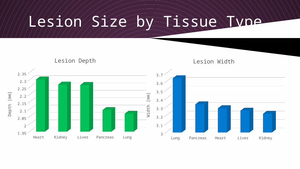