FINDINGS IN COMPUTED TOMOGRAPHY BRAIN SCANS OF …
58
FINDINGS IN COMPUTED TOMOGRAPHY BRAIN SCANS OF PATIENTS REFERRED WITH FIRST-EPISODE PSYCHOSIS Matthys Johannes van Wyk Dissertation submitted to the Faculty of Health Sciences, University of the Witwatersrand, in partial fulfilment of the requirements for the degree of Master of Medicine in the Division of Diagnostic Radiology Johannesburg 2012
Transcript of FINDINGS IN COMPUTED TOMOGRAPHY BRAIN SCANS OF …

FINDINGS IN COMPUTED TOMOGRAPHY BRAIN SCANS
OF PATIENTS REFERRED WITH FIRST-EPISODE
PSYCHOSIS
Matthys Johannes van Wyk
Dissertation submitted to the Faculty of Health Sciences, University of the
Witwatersrand, in partial fulfilment of the requirements for the degree
of
Master of Medicine
in the Division of
Diagnostic Radiology
Johannesburg
2012
ii
DECLARATION
I, Matthys Johannes van Wyk, declare that this dissertation is my own work. It is being
submitted for the degree of Master of Medicine in the branch of Diagnostic Radiology at the
University of the Witwatersrand, Johannesburg. It has not been submitted before for any
degree or examination at this or any other university.
_______________________
22 July 2012

iii
DEDICATION
In memory of my father
Thys van Wyk
1932-2007

iv
PRESENTATIONS ARISING FROM THIS STUDY
The results of this study were delivered as an oral presentation at the Radiological Society of
Southern Africa’s MDCT Essentials course (26 – 28 August 2011) at Sandton Convention
Centre, Johannesburg.
v
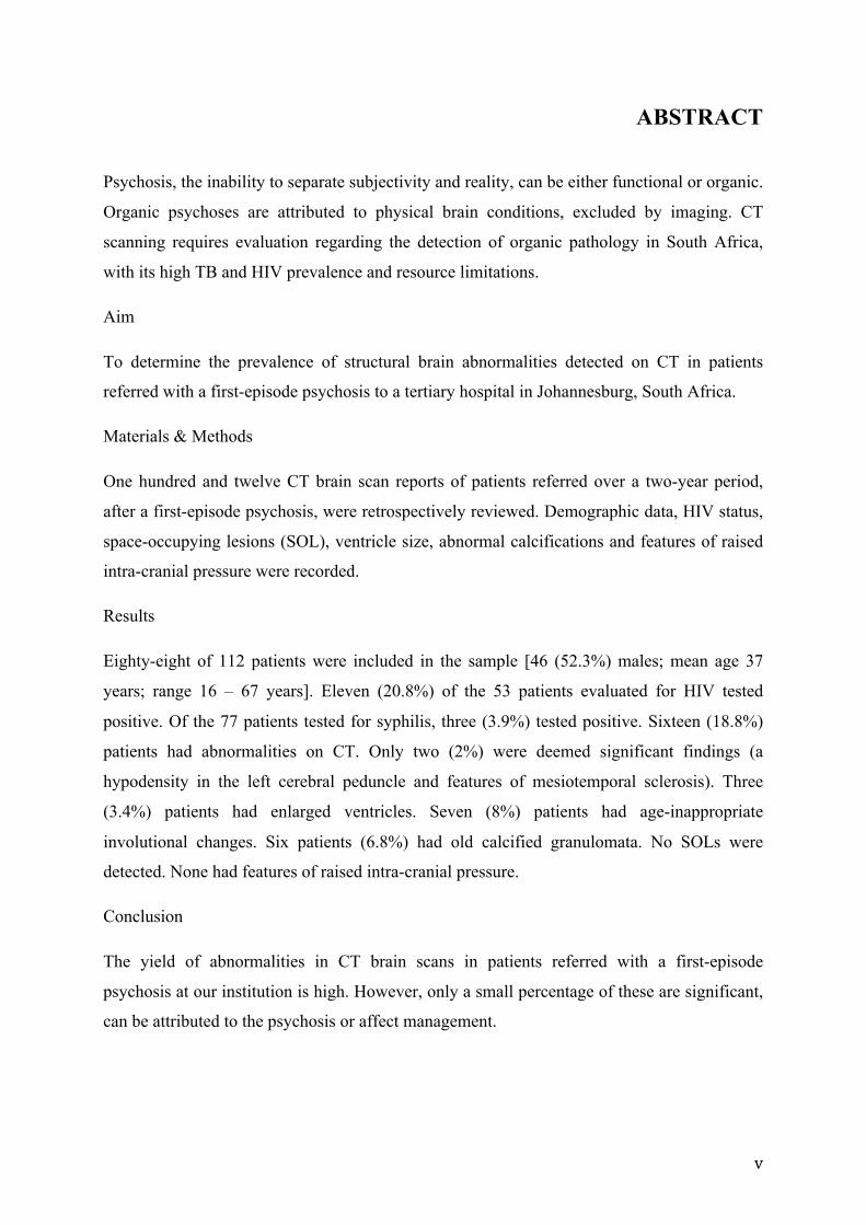
ABSTRACT
Psychosis, the inability to separate subjectivity and reality, can be either functional or organic.
Organic psychoses are attributed to physical brain conditions, excluded by imaging. CT
scanning requires evaluation regarding the detection of organic pathology in South Africa,
with its high TB and HIV prevalence and resource limitations.
Aim
To determine the prevalence of structural brain abnormalities detected on CT in patients
referred with a first-episode psychosis to a tertiary hospital in Johannesburg, South Africa.
Materials & Methods
One hundred and twelve CT brain scan reports of patients referred over a two-year period,
after a first-episode psychosis, were retrospectively reviewed. Demographic data, HIV status,
space-occupying lesions (SOL), ventricle size, abnormal calcifications and features of raised
intra-cranial pressure were recorded.
Results
Eighty-eight of 112 patients were included in the sample [46 (52.3%) males; mean age 37
years; range 16 – 67 years]. Eleven (20.8%) of the 53 patients evaluated for HIV tested
positive. Of the 77 patients tested for syphilis, three (3.9%) tested positive. Sixteen (18.8%)
patients had abnormalities on CT. Only two (2%) were deemed significant findings (a
hypodensity in the left cerebral peduncle and features of mesiotemporal sclerosis). Three
(3.4%) patients had enlarged ventricles. Seven (8%) patients had age-inappropriate
involutional changes. Six patients (6.8%) had old calcified granulomata. No SOLs were
detected. None had features of raised intra-cranial pressure.
Conclusion
The yield of abnormalities in CT brain scans in patients referred with a first-episode
psychosis at our institution is high. However, only a small percentage of these are significant,
can be attributed to the psychosis or affect management.

vi
ACKNOWLEDGEMENTS
Prof Victor Mngomezulu – supervisor
Prof Savvas Andronikou for his critical and creative analysis of the text
Mrs Barbara Dadswell for retrieving the reports
Prof Elena Libhaber for her patient and kind help with the statistical analysis
My loving wife, Carol, for her unwavering love and support

vii
TABLE OF CONTENTS
DECLARATION……………………………………………………………………..…...ii
DEDICATION……………………………………………………………………………iii
PRESENTATION……………………………………………….…………………..……iv
ABSTRACT……………………………………………………………………………….v
ACKNOWLEDGEMENTS……………………………………………………………....vi
TABLE OF CONTENTS……………………………………………………...……..…..vii
LIST OF FIGURES……………………………………………………………………….x
LIST OF TABLES………………………………………………………………………...xi
ACRONYMS…...……………………………………………………………………...…xii
PREFACE..........................................................................................................................xiv
1.0 LITERATURE REVIEW……………………………………………………………...1
1.1 Psychosis………………………………………………………………………1
1.1.1 Psychosis classification……………………………………………2
1.1.2 Demographics……………………………………………………..2
1.1.3 Etiology……………………………………………………………2
1.2 First-episode psychosis……………………………………………………...…3
1.2.1 Definition………………………………………………………….3
1.2.2 Diagnostic work-up for first-episode psychosis…………………...4
1.3 Brain imaging…………………………………………………………………4
1.3.1 CT…………………………………………………………………4
1.3.2 MRI………………………………………………………………12
1.3.3 Yield of brain imaging…………………………………………...12
1.4 International psychosis work-up guidelines……………..................………...14

viii
1.5 Infection and psychosis………………..……………………………..………17
1.5.1 HIV ………………………………………………………………17
1.5.2 Tuberculosis…………………………………………..………...18
1.5.3 Syphilis……………………………………………..…………...18
2.0 INTRODUCTION…………………………………………………….…………….19
2.1 Rationale……………………………………………….……………………19
2.2 Aim………………………………………………………………………….19
2.3 Objectives…………………………………………………………………..20
2.4 Scope of the research study………………………………………………....20
3.0 METHODOLOGY…………………………………………………………………21
3.1 Study design...................................................................................................21
3.2 Sample……………………………………………………………………….21
3.3 Inclusion and exclusion criteria…………………..………………………….21
3.4 Method………………………………………………………………………22
3.5 Data analysis and statistical methods………………………………………..22
4.0 RESULTS…………………………………………………………………………...26
4.1 Index episode………………………………………………………………...26
4.2 Demographics………………………………………………………………..26
4.3 HIV status……………………………………………………………………29
4.4 Syphilis serology…………………………………………………………….29
4.5 CT brain findings……………………………………………………………30
4.5.1 Raised intra-cranial pressure……………………………………..30
4.5.2 Ventricular size and shape………………………………………30
4.5.3 Involutional changes……….……………………………………30
4.5.4 Abnormal calcifications………………………………………….30
ix
4.5.5 Space-occupying lesions…………………………………………31
4.5.6 Other findings……………………………………………………31
4.6 Change in management……………………………………………………...31
5.0 DISCUSSION…………………………...…………..………………………………33
5.1 Demographics………………………………………………………………...33
5.2 Index episode………………………………………………………………...33
5.3 HIV status…………………………………………………………………….33
5.4 Syphilis serology……………………………………………………………..34
5.5 CT brain findings…………………………………………………………….34
5.5.1 Raised intracranial pressure……………………………………...34
5.5.2 Ventriculomegaly………………………………………………...35
5.5.3 Involutional changes……………………………………………..35
5.5.4 Abnormal calcifications………………………………………….35
5.5.5 Space-occupying lesions………………………………………....36
5.6 Impact of CT brain scans …………………………………………………...36
5.7 Limitations of the study………………………………………………………37
6.0 CONCLUSION……………………………………………………………………….38
7.0 REFERENCES……………………………………………………………………….39
8.0 APPENDICES………………………………………………..……………………...43
8.1 Data collection sheet……………………………………..…………………..44
8.2 Ethics approval clearance certificate ...………………………………………45

x
LIST OF FIGURES
Figure Page
4.1 Gender distribution of findings………………………………………....27
4.2 Distribution of findings by age………………………………………….28
xi
LIST OF TABLES
Table Page
1.1 Compilation and summary of studies evaluating the CT brain findings
in patients with first-episode psychosis.………………………..………….. 6
1.2 Inclusion criteria for CT brain of patients with psychosis. ………………...16
3.1 Original and modified classification of Goulet……………………………. 25
4.1 Summary of demographic results of 88 patients. …………………………..26
4.2 The influence of HIV status on scans results……………………………..... 29
4.3 Stratification of the impact of scans results according to the modified
Goulet classification………………….………………………...………….. 31

xii
ACRONYMS
AC0P0 Abnormal finding on CT brain with unlikely relation to clinical picture or
psychosis
AC+P0 Abnormal finding on CT brain with a likely relation to clinical picture but not
to the psychosis
AC+P+ Abnormal finding on CT brain with likely relation to clinical picture and
psychosis
CMJAH Charlotte Maxeke Johannesburg Academic Hospital
CNS Central Nervous System
CSF Cerebrospinal fluid
CT Computed Tomography
CTB Computed Tomography of the Brain
EEG Electroencephalogram
FBC Full blood count
FEP First-episode psychosis
HIV Human Immunodeficiency Virus
LFT Liver Function Test
MRI Magnetic Resonance Imaging
MSE Mental State Examination
NCC Neurocysticercosis
RANZCP Royal Australian and New Zealand College of Psychiatrists
RPR Rapid plasma reagent test
TB Tuberculosis
TFT Thyroid Function Test

xiii
TLE Temporal Lobe Epilepsy
TPHA Treponema pallidum haem-agglutination assay
U&E Urea and creatinine
UK United Kingdom

1
CHAPTER 1:
LITERATURE REVIEW
This review will define and classify psychosis. It will give an outline of its demographics,
etiology and investigation. We will critically evaluate the use of CT and MRI and discuss the
CT brain imaging findings in psychotic patients. The current international guidelines on the
diagnostic work-up of a first-episode psychosis (FEP) will be discussed as well as the yield of
brain imaging in this group of patients. We will briefly discuss the role that HIV, syphilis and
tuberculosis play in patients with FEP.
1.1 PSYCHOSIS
There are different definitions of psychosis documented in the literature.
According to Kaplan and Sadock’s Synopsis of Psychiatry, the most common psychiatric use
of the term psychosis refers to the “severe impairment of social and personal functioning
characterised by social withdrawal and inability to perform the usual and occupational roles”.
They further elaborate to define psychosis as “[the] inability to distinguish reality from
fantasy; impaired reality testing, with the creation of a new reality (as opposed to neurosis:
mental disorder in which reality testing is intact; behaviour may not violate gross social
norms, but is relatively enduring or recurrent without treatment).”(1)
Stedman’s Medical Dictionary, on the other hand, defines psychosis as,
“A mental disorder causing gross distortion or disorganization of a person’s mental
capacity, affecting response, and capacity to recognize reality, communicate, and
relate to others to the degree of interfering with his capacity to cope with the ordinary
demands of everyday life” (2).

2
1.1.1 Psychosis classification
Psychosis is a symptom and not a diagnosis in itself. The psychoses are divided into two
major groups depending on their origin: 1) those related to organic brain syndromes and 2)
those that have some functional component and are not clearly organic by nature (3).
1.1.2 Demographics
In the United Kingdom (UK) most psychotic episodes (83.3%) occur in the 15-59 year age
group followed by 16.5% in the age group older than 60 years. The least percentage (0.2%)
occurs in those younger than 14 years of age (4). The majority of the patients in the UK were
male (59%) (4). South African data of mentally ill inpatients scanned that presented either
with (i) a first episode psychosis or (ii) psychosis and features of a delirium, focal physical or
neurological signs, and/or abnormal special investigation results,correlate with these age and
gender trends (5).
1.1.3 Etiology
Etiology differs by gender and age. In younger patients the cause is more commonly
functional in nature whilst in the elderly organic causes are more prevalent (4, 5).
Although there is renewed debate about the classification of the psychoses (6) functional
psychoses include schizophrenia, bipolar mood disorder and mania, and form the majority of
the psychoses (3).
In the UK the incidence of organic pathology resulting in psychosis is estimated to be
between five and ten percent (4).
Organic psychoses can be attributed to physical medical conditions such as previous head
trauma, encephalitis or a space-occupying brain lesion (for example, a tumour) (7).

3
A viral etiology for psychosis has been suggested. Psychotic symptoms may be caused by
limbic encephalitis secondary to Epstein-Barr, cytomegalovirus, rubella, herpes simplex,
measles and HIV viruses (8).
In Alzheimer’s disease, psychosis is often a non-cognitive condition that accompanies
dementia whereas in Parkinson’s disease, treatment with anti-parkinsonian drugs is the most
frequent cause of psychotic symptoms (9).
People with multiple sclerosis rarely develop psychotic symptoms (8).
Psychosis in epilepsy does occur and is episodic rather than continuing with normal interictal
periods (10).
Organic psychoses are commonly of acute onset and where a space-occupying lesion is the
cause associated, neurological manifestations are usually found. These might include cranial
nerve paralyses, motor neuron impairment, sensory loss as well as speech and hearing
difficulties (7).
Unusual features such as an acute onset and delirium characterise organic psychoses and may
lead the clinician to suspect an organic rather than a functional cause. Some of these patients
presenting with psychosis may have obvious underlying psychiatric pathology, may have
used psychosis-inducing agents or have a history of previous head trauma (7).
For many of these patients, however, the cause of their psychosis will be unknown.
1.2 FIRST-EPISODE PSYCHOSIS
1.2.1 Definition
Also known as an index-episode psychosis, it refers to the first time that an individual
experiences a psychotic episode. It is sometimes difficult to determine the exact time of onset.
4
In practice this translates into an individual who presents to healthcare with psychosis and
who has never previously presented to healthcare with psychosis (11). There is however no
consensus operational definition for first episode psychosis (11).
Studies revealed that early intervention (within three to five years) resulted in symptom
improvement, lower relapse rates and promoted functional recovery (12).
1.2.2 Diagnostic work-up for first-episode psychosis
In addition to the routine physical, mental and neurological assessment, laboratory
examinations are also conducted. These include: full blood count (FBC); renal function and
electrolytes (U&E); liver functions (LFT); thyroid function tests (TFT); human
immunodeficiency virus (HIV) status; and syphilis (TPHA and RPR). Furthermore
cerebrospinal fluid (CSF) is evaluated biochemically and cytologically. Some cases may
require an encephalogram (EEG) (13).
1.3 BRAIN IMAGING
Pathology may sometimes be missed when relying solely on the standard tests
(misidentification syndrome) and thus CT and MRI may prove useful to diagnose organic
pathology where it has been missed in the initial standard examinations. If all these tests yield
no positive findings, it is assumed that the patient has functional psychosis (4).
1.3.1 CT
CT of the brain adds information on the structure of the brain, meninges and soft tissues of
the head. Computed tomography uses ionising radiation to produce multiple axial two-
dimensional slices through the brain recorded by an array of detectors. The resultant images
can be reconstructed into various planes of visualisation. It produces superior images of the
bones and hard tissues compared to other imaging modalities. It may reveal pathologies such

5
as space-occupying lesions, abnormal calcifications, involutional changes, cerebrospinal fluid
space abnormalities and even meningitis.
a) Findings on CT in patients with psychosis
A number of studies have described CT changes noted in the brains of patients with first-
episode psychosis (5, 14-20) (Table 1.1). Most of these changes are equivocal and non-
specific (20).
These findings are likely to be influenced by factors such as age, gender, age of onset of
psychosis, clinical symptoms, length and stage of illness and exposure to neuroleptics (21).

6
Table 1.1: Compilation and summary of studies evaluating the CT brain findings in
patients with first-episode psychosis (constructed from references 3, 11-17).
Study Normal n(%)
AC0P0* n(%)
AC+P0* n(%)
AC+P+* n(%)
Beresford (16) 1986
106 (68) 22 (14.1) 6 cerebral atrophy 2 cerebral and cerebellar atrophy 2 ventricular enlargement 2 superior cerebellar vermis atrophy 1 bifrontal atrophy 1 enlarged third ventricle 1 old surgical lesion 2 frontal lobe atrophy 1 old craniotomy 1 orbit mucocoele 1 midfrontal hypodensity left 1 parietal calcification left 1 focal Sylvian fissure atrophy
18 (11.5) 2 ventricular outflow obstruction 1 right frontal and left occipital infarcts 1 patchy post fossa encephalomalacia 1 small frontal haematomas 1 subdural haematoma 1 middle cerebral artery distribution encephalomalacia right 1 putamen infarct right 1 lacunar infarcts in putamen and internal capsule 1 occipital infarct right 2 putamen infarct left 2 MCA territory atrophy left 1 left frontal infarct 1 frontal encephalomalacia 1 left occipital infarct 1 cerebral and cerebellar encephalomalacia
10 (6.4) 1 focal parietal atrophy 1 encephalomalacia next to right lateral ventricle 1 parietal and temporal lobe atrophy right 1 right head of caudate infarct 1 frontal and temporal hypodensity left 1 temporal lobe stroke left 1 temporal lobe atrophy 1 metastatic lesion to left parietal lobe 1 anterior parietal encephalomalacia left 1 brain stem stroke
Battaglia and Spector (15) 1988
42 (93.3)
2 (4.4) 2 atrophy (mild)
1 (2.2) 2 possible frontal white matter infarct
1 (2.2) 1 possible left caudate infarct
Ananth (17) 1993
25 (73.5) 8 (23.5) 1 mild central atrophy 1 mild cerebral atrophy 1 right frontal area of density 1 generalised atrophy 1 bifrontal atrophy 1 prominent sulci 1 asymmetrical Sylvian fissures 1 mild atrophy left frontal lobe
0 (0) 1 (2.9) 1 post parietal and occipital attenuation
Gerwitz et al. (14) 1994
93 (55.4) 71 (42.3) 67 cortical atrophy (diffuse) 2 arachnoid cysts 1 ventricular enlargement 1 venous angioma
2 (1.2) 1 right subcortical parieto-temporal infarct 1 ischaemic changes (diffuse)
4 (2.4) 1 moderately large right temporal arachnoid cyst 1 parietal and subinsular infarcts (bilateral) 1 parietal ischaemic changes (bilateral) 1 colloid cyst obstructing foramen of Munro
Bain (20) 1998
123 (96.9) 3 (2.4) 1 subcortical right frontal calcification 2 arachnoid cysts
1 (0.8) 1 possible pineal tumour
0 (0)
Agzarian (18) 2006
377 (95) 14 (3.5) 5 cortical atrophy 5 arachnoid cysts 1 calcified choroid plexus in 4th ventricle 3 non specific abnormalities disproved on MRI
6 (1.5) 3 small vessel ischaemic changes 1 small right cerebellar cavernoma 1 small calcified meningeoma 1 colloid cyst without hydrocephalus
0 (0)
Hirano (19) 2006
88 (75.2) 18 (15.4) 10 old infarcts 3 age indeterminate infarcts 2 lipoma 1 hygroma 1 chronic subdural 1 post traumatic calcification
11 (9.4) 3 acute infarct 3 intracranial haemorrhage 1 subdural haematoma 3 meningeoma 1 pituitary adenoma
0 (0)
Jeenah (5) 2007
35 (63.6) 8 (14.4) 7 global cerebral atrophy 1 blow-out fracture of the orbit
6 (11) 6 old infarct +/- calcification
6 (11) 6 pituitary adenoma, TB granuloma, neurocysticercosis
*(AC0P0) Abnormal finding on CT brain with unlikely relation to clinical picture or psychosis,
(AC+P0) Abnormal finding on CT brain with a likely relation to clinical picture but not to the psychosis
(AC+P+) Abnormal finding on CT brain with likely relation to clinical picture and psychosis

7
Ventriculomegaly
Malla et al. found mild enlargement of the ventricles, the right temporal horn being larger
than the left (21). Ventriculomegaly on CT was also documented by Hoffler et al. (22). Third
ventricle prominence was noted in patients with schizophrenia but not in other psychotic
disorders (23). Ichimiya and others, however, describe third ventricle enlargement as an
inconsistent finding (21, 24).
In refractory temporal lobe epilepsy (TLE), mesial temporal sclerosis is the most common
pathologic entity found and is seen in as many as 60% to 80% of cases (25). Shukla et al.
compared two groups of 62 with TLE and 90 with generalised epilepsy respectively and
found that there was a statistically significant higher incidence of psychosis in the TLE group
(26). Wyler describes atrophy and chronic herniation of the mesial temporal structures over
the free edge of the tentorium in seventeen patients who had a CTB prior to temporal
lobectomy (27).
Cerebral atrophy
Malla et al. found mild enlargement of cortical sulci in psychotic patients but noted that
younger patients did not exhibit a significantly greater level of cerebral atrophy (21). Variance
in the findings was mostly explained by age and not by variables such as clinical syndrome
types, duration of illness or gender (21). Tsai and Tsuang studied 135 psychiatric patients’
brain scans. Of these nearly fifteen had atrophy (28). In a study by Evans on 100 consecutive
psychiatric in- and outpatients, 66% had atrophy (29). Larson et al. found that 35% of a group
of 123 patients with psychosis had atrophy (30). Some authors found the atrophy in the
frontal and central brain regions to be progressive over a five-year period but Malla et al.
found, if after controlling for the effect of age, the atrophy did not progress as a result of
disease evolution (21, 31). They did, however, report progressive enlargement of the sylvian

8
fissure due to chronic illness (21). In first-episode psychosis the left sylvian fissure appeared
more prominent than the right (21). Other inconsistent findings reported include focal
reduction in the size of the frontal lobes and involvement of the cerebellum (24).
Infarcts
In the elderly, organic pathology such as infarcts may result in psychotic behaviour. Anderson
et al. reported on two cases of elderly females presenting with acute psychosis with occipital
and subcortical infarcts respectively (32). They also noted that patients with a psychiatric
history are more prone to psychosis following a stroke and that it is rarely reported owing to
under recognition (32).
Space-occupying lesions
Asselman et al. noted that thirteen percent of 75 HIV positive patients with new neurological
and psychiatric symptoms presented with a space-occupying lesion; one related to
toxoplasmosis and nine of uncertain etiology (33). In a post-mortem study in 1978, Cole
reported that in South African psychiatric patients the prevalence of space-occupying lesions
was 15.5% (34). These lesions included: subdural haematomas, primary CNS neoplasms, a
metastatic neoplasm, a hamartoma, tuberculomas, parasitic tapeworm cysts and an abscess
(34).
Brain tumours causing psychosis are rare and their prevalence on CT in the psychiatric
population is about 1.2% (35). Three percent of the necropsy material studied by Cole had
intracranial neoplasms (34).

9
Traumatic brain injury
In a group of brain-injured troops from Finland, nearly 10% developed psychotic symptoms
within 5 years (8). Only one patient in the series studied by Cole had evidence of head injury
– a recent fall, resulting in a subdural haemorrhage (34).
Arachnoid cysts
Some anecdotal reports of patients presenting with first-episode psychosis and arachnoid cyst
have been published (36, 37). The authors either did not ascribe the psychosis to the presence
of the arachnoid cyst (36), or had difficulty in deciding whether or not the lesion was
significant (37).
Meningitis
Cryptococcal meningitis is a known cause of psychosis (38). It was the cause of
neuropsychiatric deterioration in 24% of 75 patients who were HIV positive and had been
recently started on antiretrovirals (33).
b) Advantages of CT
Availability
CT scanners are relatively widely available in the world (39). In South Africa it is the case in
larger cities where secondary and tertiary hospitals are found. There is, however, a
discrepancy between public and private distribution of these scanners.
During 2005/2006 in the Western Cape Province of South Africa there were seven CT
scanners available in 32 public hospitals and 24 CT scanners for 29 private hospitals. This
means that a CT scanner was available for every 4.5 public hospitals and 1.2 for private
10
hospitals. During this period a total of 28 281 CT examinations were requested in the public
sector while 36 519 CT examinations were ordered in the private sector (40).
Cost
The cost is less than that of MRI but remains relatively high. In 2009 a non-contrasted CT
brain cost around R1 681.70; a contrasted CT of the brain cost in the order of R2 470.90 and a
pre- and post-contrast scan cost the patient R3 005.50. An MRI cost between R4 736.90 and
R7 724.60 depending on whether or not contrast was used (41).
Quickness
It is a relatively quick procedure, compared to MRI and takes between 15 to 30 minutes to
acquire brain images depending on whether or not contrast was given. This results in fewer
movement artifacts (39, 42).
Additional findings
Additional findings or pathologies in the surrounding tissues of the head might be
serendipitously picked up in imaging the brain.
Non-invasiveness
Unlike a biopsy, a CT is a non-invasive method to exclude pathology. Moreover, unlike MRI,
it is not contra-indicated in patients with pacemakers or metallic implants (39).
c) Limitations and disadvantages of CT
Patient factors
For optimal visualisation, the patient must be able to lie still.

11
The average radiation dose to which people in the US are exposed has doubled over the past
30 years (43). Ionising radiation is a well-known cause of deleterious effects in high doses
and may even be carcinogenic. The life-time risk of cancer from a single brain CT is low (1:
10 000 for those older than fifteen but younger than 20) but higher in the young (42). For this
reason it is contra-indicated in pregnancy.
CT has poorer soft-tissue contrast ability and fails to diagnose lesions with the same density
as the surrounding tissue (39). In order to visualise these lesions and diagnose infection,
iodinated contrast medium is often injected intravenously. It is not without any risk however.
These compounds may cause permanent renal impairment and damage (39).
Some patients may be allergic and can react fatally to the iodine in these contrast agents (44).
Equipment factors
There are a number of equipment related factors that can affect the quality of the scan and
some that result in artifacts (45).
There is 90% - 100% accuracy for mass lesions greater than 2.5 cm in diameter (46, 47).
CT is also unable to diagnose some of the causes of psychosis such as dementia and epilepsy.
In dementia, however, CT scanning is done to screen such patients for potentially treatable
conditions such as hydrocephalus, meningeoma and subdural haematoma (46).
The number of CT scans of the brain conducted annually in South Africa is unknown (48).
In our setting we are overburdened by requests for CT of various parts of the body. The great
demand for CT results in long waiting lists and sometimes an unfortunate delay in an urgent
CT. Complicating this matter, the old and overused equipment requires regular servicing and
sometimes replacement of a faulty component.

12
1.3.2 MRI
Compared to CT, MRI is a relatively new imaging modality.
a) Advantages
MRI has exquisite grey-white matter resolution. Unlike CT, it is possible to image in various
planes. The coronal views are especially valuable in evaluating the frontal and limbic regions.
3-D reconstruction of brain structures is possible (49). MRI is useful in, among others,
morphometric brain studies, evaluation of brain tissue and metabolic function as well as in
examining the cerebral blood flow (49).
No ionising radiation is used. MRI poses minimal risk to the patient (49).
b) Disadvantages
Compared to CT, MRI is quite expensive (41). Its availability is limited to larger medical
centres and cities. It is estimated that there were 2 MRI scanners for 32 public hospitals
compared to 16 MRI scanners available to the 29 private hospitals during 2005/2006 in the
Western Cape Province of South Africa (40).
Patients with claustrophobia find the scanning unbearable (42, 49).
MRI is contra-indicated in patients with pacemakers, certain implants, metallic foreign bodies
and aneurysm clips (49).
1.3.3 Yield of brain imaging
Several studies have been conducted on imaging to determine the incidence of organic
pathology that could be the cause of psychosis (5, 14, 16-20).

13
In an exploratory study of 55 psychiatric patients by Jeenah and Moosa (see Table 1) at Chris
Hani Baragwanath Hospital in South Africa, 20 (36.4%) patients had abnormal CT brain
scans (5). The abnormalities detected included blow-out fracture of the orbit (1%), old
infarcts (6%), global cerebral atrophy (7%) and mass lesions (6%). These masses included:
pituitary adenoma, TB granuloma and neurocysticercosis. There was a significant correlation
between abnormalities and advancing patient age. Fifteen of the abnormal scans were of
patients with a first-episode psychosis (5). The authors recommended further study with a
larger sample size to determine the necessity for scanning all first-episode psychotic patients
(5).
The systematic review by Albon et al. of 25 studies found that in most studies, structural
neuro-imaging identified very little that would influence patient management that was not
suspected, based on a medical history and/or physical examination and there were more
incidental findings (35). They found that in these studies only around 0,5% of patients had
findings on CT that would have affected clinical management (35). In a study by Strahl et al.
reports of 237 consecutive patients who provided a history of first-episode psychosis revealed
no focal brain lesion that could be responsible for the psychosis. Unrelated findings included
small vessel ischaemic changes, arachnoid cysts, cerebral atrophy and normal variants in
17.6% of patients (36). None of these findings led to a change in management (36). Ebdrup et
al. reviewed eight papers, including a total of 625 CT and 214 MRI scans. On CT, the
incidence of potentially causal brain abnormalities was 0.8% and on MRI 3.3% (50). The
findings had limited clinical consequences, however, and thus they concluded that there is no
support for performing routine CT scans of first-episode psychotic patients (50). Hirano et al.
reported that the overall yield of CT brain scans in elderly unselected hospitalised patients is
low (19). They, however, did not consider elderly psychotic patients. The same conclusions
were made by, among others, Bain (20) and Agzarian (18). The tendency noted from these
14
studies is that most of the CT scans conducted for first-episode psychosis are normal. This is
followed by findings of an abnormality without any impact on the clinical or psychiatric
manifestations. Findings that were believed to influence either the clinical or psychiatric
picture ranged between 0 and 11.5%. The highest number of abnormal imaging findings
related to psychosis was observed in the study by Jeenah and Moosa (5). They did, however,
have one of the smaller sample sizes (i.e. 55). The study with the greatest sample size (i.e.
397) was conducted by Agzarian (18). These findings confirmed the hypothesis that the
abnormal findings in patients with a first-episode psychosis is very low and that the majority
of scans are normal (18).
1.4 INTERNATIONAL PSYCHOSIS WORK-UP GUIDELINES
Guidelines for the work-up of first-episode psychosis vary among countries. The Royal
Australian and New Zealand College of Psychiatrists (RANZCP) advocates a neuro-imaging
of the new onset psychotic patient, while Britain is more conservative in their approach and
do not suggest an initial CT of the brain (4, 51). Although the RANZCP advocates neuro-
imaging in first-episode psychosis, they acknowledge that currently no evidence is available
that routine imaging alters outcome or clinical management (4, 51). They furthermore
acknowledge that CT has a low yield for screening psychotic patients. They highlight the fact
that MRI has been reported to have significant yield detecting clinically important findings
but that no yield is available for patients that underwent screening MRI for psychosis (51).
The American group of Hollister suggested the following as sound, high yield indications for
brain imaging in psychiatric patients: 1) history of previous head trauma, stroke, neurological
disease, Alzheimer’s disease or multi-infarct dementia; 2) abnormal neurological signs or
organic mental signs (e.g. cognitive decline or confusion); or 3) a first psychotic episode after
the age of 50 years (52). They found brain scanning not to be rewarding in patients with a

15
history of alcohol or substance abuse, mental disorders lacking additional neuropsychiatric
abnormalities, or for the evaluation of seizure disorders or abnormal EEG patterns (52). The
study dates back to 1996, relying on old CT technology and no MRI at their disposal.
Moreover, the sample consisted of only 68 patients and did not study first-episode psychotic
patients per se.
Tsai et al. recommended that those psychiatric patients who had normal mental state
examination (MSE); neurological examination (NE); EEG and Bender Gestalt test (BIP),
could safely be excluded from having organic pathology requiring imaging by CT. Those who
had normal ME and NE but abnormal BIP or EEG could also be excluded if they were 40
years or younger with no history of head injury (28). Once again the study evaluates general
psychiatric patients and did not exclusively investigate patients with FEP.
In a review article by Goodstein in 1986, broad inclusion criteria for CT scanning of the brain
in general psychiatric patients are suggested: 1) when symptoms of a significantly disabling
psychiatric disease do not fit the classical descriptions for the illness; 2) where there is a high
index of suspicion of a space-occupying brain lesion due to specific evaluation findings; and
3) If the CT findings, whether positive or normal will potentially influence management,
rather than merely satisfy the clinician’s academic curiosity (47).
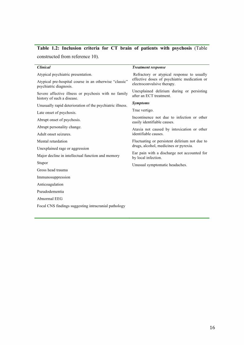
Goodstein suggested numerous specific inclusion criteria for CT of the brain (Table 2.2).
16
Table 1.2: Inclusion criteria for CT brain of patients with psychosis (Table
constructed from reference 10).
Clinical
Atypical psychiatric presentation.
Atypical pre-hospital course in an otherwise “classic” psychiatric diagnosis.
Severe affective illness or psychosis with no family history of such a disease.
Unusually rapid deterioration of the psychiatric illness.
Late onset of psychosis.
Abrupt onset of psychosis.
Abrupt personality change.
Adult onset seizures.
Mental retardation
Unexplained rage or aggression
Major decline in intellectual function and memory
Stupor
Gross head trauma
Immunosuppression
Anticoagulation
Pseudodementia
Abnormal EEG
Focal CNS findings suggesting intracranial pathology
Treatment response
Refractory or atypical response to usually effective doses of psychiatric medication or electroconvulsive therapy.
Unexplained delirium during or persisting after an ECT treatment.
Symptoms
True vertigo.
Incontinence not due to infection or other easily identifiable causes.
Ataxia not caused by intoxication or other identifiable causes.
Fluctuating or persistent delirium not due to drugs, alcohol, medicines or pyrexia.
Ear pain with a discharge not accounted for by local infection.
Unusual symptomatic headaches.

17
1.5 INFECTION AND PSYCHOSIS
Infectious diseases remain the leading cause of death in South Africa. The chief infective
disease culprits are HIV and TB (53). Due to the high prevalence of these diseases, a closer
look at their relation to psychosis and the imaging findings thereof is necessitated.
1.5.1 HIV
It has been demonstrated that HIV per se may be associated with psychosis (54-56). First-
episode psychosis in HIV positive patients occurred in 0.2%-15% of patients. Säll et al.
reviewed the files of 38 South African black male, HIV positive mine workers and found the
incidence of psychosis to be 29% (57). CT scans were only conducted in 24% of these
patients and only in 4 were some kind of pathological finding found. Three patients, who had
concurrent features of dementia, demonstrated cerebral atrophy, wide ventricles and sulci on
CT. The fourth patient had a lesion in the pituitary fossa (57). In another South African
prospective observational cohort study of 75 HIV positive patients, reported by Asselman et
al., 12% presented with psychosis (33). In the majority (five patients) Efavirenz (an
antiretroviral agent) was thought to be the cause (33) as it is known to cause psychotic side-
effects (58). The highest incidence of psychosis was reported among patients in later stages of
HIV disease and with HIV associated dementia. In one study 15% of 46 patients with HIV
associated dementia presented with psychosis (59). This suggests that psychosis is a direct
effect of the HIV infection on the CNS. Various hypotheses have been constructed to explain
the pathogenesis of first-episode psychosis in HIV positive patients: subcortical
neurodegeneration due to HIV itself or in the presence of other viral infections; psychosis
secondary to HIV encephalopathy; brain damage from another underlying opportunistic
infection, or an underlying dementia (60). The following factors contributed to the

18
development of psychosis in HIV positive patients: untreated HIV infection, cognitive
impairment, dementia and a history of psychiatric disease or substance abuse (58).
1.5.2 Tuberculosis
In the study by Asselman, 36% of the 75 HIV positive patients started on antiretrovirals had
neurological deterioration secondary to TB. All patients diagnosed with tuberculoma had a
negative serum immunoglobulin G (IgG) analysis for Toxoplasma species (33) and thus
immunological studies were not an accurate assessment of CNS infection with tuberculosis.
In a case report, Woodroof ascribed a patient’s psychotic symptoms to structural damage from
an intracranial tuberculoma (62). There is, however, a paucity of data in the literature on the
association between patients suffering from first-episode psychosis and cerebral tuberculosis.
1.5.3 Syphilis
CNS involvement with concurrent syphilis infection is a common cause for neuropsychiatric
symptoms (57). However, in a group of South African mine workers reviewed by Säll et al.,
53% of the HIV positive patients were tested for syphilis and only two tested positive (57).
The RPR test is used for screening and a confirmatory TPHA test is conducted (61).

19
CHAPTER 2:
INTRODUCTION
2.1 RATIONALE
The general opinion of CT brain scans done on first-episode psychotic patients, in order “to
rule out organic pathology”, is that it does not yield a great amount of information. This
creates a preconceived notion that these scans, on the whole, will be normal and therefore
may impact on the bias of the reader. New imaging techniques e.g. magnetic resonance
imaging (MRI) may be more appropriate but are not cost effective in our resource limited
setting. The use of CT, when the yield of significant pathology is low, is even less appropriate
as it exposes patients to high doses of radiation.
2.2 AIM
The purpose of this research study is to determine the utility of CT brain scans in first episode
psychosis patients (referred from a tertiary hospital psychiatry service in Johannesburg, South
Africa) based on abnormal findings.

20
2.3 OBJECTIVES
The objectives of this study are divided in to primary and secondary objectives.
Primary objectives:
To:
2.3.1 determine the prevalence of abnormalities on CT brain scans;
2.3.2 categorise the abnormalities as either: i) bearing no clinical significance, ii)
with clinical significance, but unlikely to cause the patient’s psychosis or iii)
with clinical significance, and possibly contributing to the patient’s psychosis.
Secondary objectives:
To:
2.3.3 determine the prevalence of human immunodeficiency virus (HIV) and
syphilis in the study population;
2.3.4 stratify the abnormalities according to age, gender and HIV status.
2.4 SCOPE OF THE RESEARCH STUDY
The study will only focus on patients referred as a first-episode psychosis from the psychiatric
service at the Charlotte Maxeke Johannesburg Academic Hospital (CMJAH).
The CMJAH is a tertiary hospital serving not only the immediate Johannesburg population
but also patients from around the Gauteng Province.

21
CHAPTER 3:
METHODOLOGY
In this chapter we will explain the type of study and discuss the study material and methods
used to gather and analyse the data. Inclusion and exclusion criteria will be mentioned and the
descriptors of the dataset will be defined.
3.1 STUDY DESIGN
This is a retrospective descriptive review of reports and patient records in one South African
tertiary referral centre (CMJA Hospital in Johannesburg).
3.2 SAMPLE
A consultant radiologist reviewed the CT brain scan reports of patients referred with a first-
episode psychosis. One hundred and twelve of these reports in conjunction with patient
records and laboratory results review were dealt with.
3.3 INCLUSION AND EXCLUSION CRITERIA
The study included CT reports spanning a period of fourteen months (January 2009 to March
2010) of patients referred after a first episode of psychosis at the Charlotte Maxeke
Johannesburg Academic hospital (CMJAH) for the purposes of ruling out organic pathology.
At the time of the original reading, the radiologists were blind to the clinical details with the
exception of the history provided by the referring clinician on the request form.
Only patients who had a true index psychotic episode were included. False referrals were
excluded from the study. One hundred and twelve patients’ reports and folders were
reviewed. Twenty-four patients had to be excluded owing to the fact that they did not present

22
with a true first-episode psychotic episode. A patient was regarded to have a first psychotic
episode if folder or discharge summary review revealed no previous admission for psychosis
was made, the patient received no antipsychotics in the past and collateral history revealed no
prior psychotic episode.
3.4 METHOD
All the scans reviewed in this study were conducted in a standardised manner, as is routine
practice in the department. Non-contrast, computed tomograms were performed on one of two
CT machines in the department and reported by various radiology consultants and registrars.
3.5 DATA ANALYSIS AND STATISTICAL METHODS
Data was recorded in an Excel spreadsheet and statistical analysis was performed using the
Statistica 8.0 package. Data was summarised as mean (SAD) or median (range) when
distribution was normal or not normal respectively and variables were measured in a
continuous scale. Categorical data is shown as frequencies, percentages and with a 95%
Confidence Interval.
For comparison between categorical variables a Chi-square or Fisher Exact Test when
adequate was used. Significance was set at 0.05.
The following data was extracted:
a) Demographics
The patients’ gender and age were recorded.
b) Index episode
From the record or discharge summary it was ascertained whether the current psychotic
episode was indeed the index or first episode
23
c) HIV status
The HIV status was recorded either as a) positive, b) negative or c) not tested.
If the patient tested positive for HIV, the CD4 count (cells/µL) was also evaluated.
d) Syphilis status
Both the RPR and TPHA test results were documented and recorded either as a) positive, b)
negative or c) not tested.
e) CT brain findings
i) Features of raised intra-cranial pressure
If the surface sulci, ventricles and/or basal cisterns were attenuated and/or the foramen
magnum was crowded, the patient was deemed to have raised intra-cranial pressure. It was
recorded as either a) present or b) absent.
ii) Ventricular size and shape
Ventricular size was recorded as either a) normal; b) attenuated; c) enlarged but not
hydrocephalic; d) hydrocephalic; e) asymmetric or f) in keeping with normal pressure
hydrocephalus. If the ventricles were asymmetric, the location of the asymmetry was noted if
described (e.g. posterior horn of the left lateral ventricle).
iii) Involutional changes
Involutional changes were recorded as either absent or present. If present, they were classified
as either being age appropriate or age inappropriate.
The principles of Le May (63) were applied retrospectively to the report findings recorded
regarding the following features: From the fourth decade there is gradual widening of the

24
third ventricle, sylvian and interhemispheric fissures, superficial sulci and basal cisterns (63).
After the sixth decade, enlargement of the lateral ventricles is the most striking feature (63).
Regressional changes are a normal aging process but highly variable (63).
Involution occurring before the age of 50 years was regarded as being age inappropriate.
Thereafter the changes were deemed to be age appropriate depending on their degree. The
consultant radiologist reviewing the scan subjectively assessed the degree of involution.
iv) Abnormal calcification
Calcifications that are not considered as forming part of the normal or normal ageing brain
were recorded. These were further characterised as being either a) granulomata (TB or old
Neurocysticercosis); b) post traumatic or c) uncharacterised.
v) Space-occupying lesions
A space-occupying lesion was considered present if it occupied space and impinged on
adjacent structures. Abnormal calcifications did not form part of this category and were
recorded separately.
f) Change in management
The study of Goulet (42) served as the basis for our modified classification of abnormal CT
brains (Table 3.1).

25
Table 3.1: Original and modified classification of Goulet
Original classification of Goulet (42) Modified classification of Goulet 1. Normal. 2. Abnormal, with no clinical impact: benign or non- specific findings with no implication on diagnosis, management, or treatment. We included in this category all findings such as atrophy, fissure enlargement, benign cysts, and other similar matters. 3. Abnormal, with implication on management or
treatment, but an unlikely causal link to psychotic symptoms. This group included findings warranting neurological investigation or treatment, such as the unexpected finding of an arterio-venous malformation, but with a high unlikelihood that treatment would influence the course of the psychosis.
4. Abnormal, with implication on management or treatment, and a possible causal link to psychotic symptoms. This group included the neurological syndromes mentioned by Weinberger, lesions in key cerebral regions already mentioned (temporal lobes, diencephalon, and basal ganglia), and demyelinating diseases.
1. Normal. 2. Abnormal, benign or non-specific findings
bearing no influence on the diagnosis, management or treatment. In this category, amongst others, the following is included: atrophy, fissure prominence, benign cysts and the like. (AC0P0)
3. Abnormal, with ramifications for management or treatment, but an unlikely causal link to psychotic symptoms. This group included findings requiring further neurological investigation or treatment, but with a low likelihood that treatment would affect the course of the psychosis. (AC+P0)
4. Abnormal, with implication on management or treatment and a possible causal link to psychotic symptoms. This group included lesions in key cerebral regions such as the temporal lobes, diencephalon and basal ganglia. (AC+P+)

26
CHAPTER 4:
RESULTS
This chapter will present the results of the data collection according to index episode,
demographics, HIV status, syphilis serology and CT brain findings.
4.1 INDEX EPISODE
Only 88 patients (78.6%) actually presented with an index episode while 24 (21.4%) had a
history of previous psychotic episodes.
4.2 DEMOGRAPHICS
The total of 88 patients included in the sample is comprised 46 (52.3%) males and 42 (47.7%)
females (Table 4.1). No statistically significant difference was found between the prevalence
of abnormalities when correlated with gender (χ2 test p=0.8405) (Figure 4.1).
Table 4.1: Summary of the demographic results of 88 patients
Variable Total (n=88) Abnormal (n=16) Normal (n=72)
Male 46 (52.3%) 8 (17.4%) 38 (82.6%) Gender
Female 42 (47.7%) 8 (19%) 34 (81%)
16-30 34 (38.6%) 3 (8.8%) 31 (91.2%)
31-60 50 (56.8%) 12 (24%) 38 (76%) Age group
(years)
>60 4 (4.6%) 1 (25%) 3 (75%)

27
Figure 4.1: Gender distribution of findings
The mean age of these patients was 37 years (SD ± 13) and ranged from 16 to 67 years. There
was no statistically significant difference between the prevalence of abnormalities when
correlated with age (Fisher’s Exact Test1 p = 0.1433) (Figure 4.2).
1 Fisher’s Exact Test was used, instead of the χ2 test, owing to the small sample numbers.

28
Figure 4.2: Distribution of findings by age
In order to evaluate the relationship between age group and abnormalities without the
influence of HIV, we evaluated the HIV negative group. Once again no statistically
significant difference between age groups where CT abnormalities were found (Fisher’s Exact
Test p=0.3598).
Age group (years)

29
4.3 HIV STATUS
Only 53 patients were submitted to HIV testing.
Eleven (20.8%) patients tested HIV positive; 42 (79.2%) tested HIV negative.
Of the eleven HIV positive patients, the mean CD4 count was 301 cells/µL (SD ± 230) and
ranged between 41 cells/µL and 707 cells/µL. The median CD4 count was 231 cells/µL. The
highest prevalence of HIV is in the age group 31 – 60. The relationship between the
prevalence of abnormalities and HIV appears to be slightly higher in this group, but not
statistically significant (Fisher’s Exact Test p = 0.6634) (Table 4.2).
Table 4.2: The influence of HIV status on scans results
Scan result
HIV status Normal Abnormal
HIV + (n = 11) 8 3
HIV – (n = 42) 37 5
Only 53 patients were submitted to HIV testing. The table reflects the scan results of the tested patients only.
4.4 SYPHILIS SEROLOGY
Seventy-seven patients were tested for syphilis. The remaining 11 were not submitted to
testing.
Three patients (3.9%) tested RPR positive. Seventy-four (96.1%) tested negative. There was
no statistically significant correlation between the RPR positive patients and abnormal CT
brain scans (Fisher’s Exact Test p = 0.4496)
Six patients (7.8%) had a positive TPHA reaction; 71 (92.2%) negative.

30
4.5 CT BRAIN FINDINGS
4.5.1 Raised intracranial pressure
None of the patients’ scans displayed features of raised intracranial pressure.
4.5.2 Ventricular size and shape
The ventricles were normal in 85 (96.6%) patients. Three (3.4%) patients had enlarged but not
hydrocephalic ventricles.
4.5.3 Involutional changes
Most of the patients, 80 (90.9%), had no involutional changes reported. Age appropriate
involutional changes were described in one (1.1%) patient while seven (8%) patients had age
inappropriate involutional changes. In most (five) of the patients with involutional changes,
the affected area was not specified. In two patients, focal parietal and in another one left
hemi-atrophy were described.
4.5.4 Abnormal calcifications
Six patients (6.8%) had abnormal intracranial calcifications. Of these, two were regarded as
old neurocysticercosis and four calcifications were not specified.
The location of the old neurocysticerci calcifications was not specified. The non-specified
calcifications were located in the left basal ganglia, the right frontal and temporal lobes as
well as in the left frontal lobe.

31
4.5.5 Space-occupying lesions
Only one (1.1%) patient demonstrated a space-occupying lesion. This was described as a
hypodense lesion in the left cerebral peduncle and further imaging via MRI was
recommended.
4.5.6 Other findings
Scalp nodules were noted on the scan of one patient and a working diagnosis of possible
neurofibromatosis was made. A biopsy was suggested. No intracranial features of
neurofibromatosis were documented.
4.6 CHANGE IN MANAGEMENT
The brain scans were normal in 72 (81.8%) cases.
Abnormalities were described in 16 (18.2%) patients’ scans.
These abnormalities and their clinical importance are listed in Table 4.3.
Table 4.3: Stratification of the impact of scans results according to the modified Goulet classification Classification Number (%) Findings [number of patients]
Normal 72 (82%)
AC0P0 14 (16%) Old neurocysticercosis [2] Age inappropriate involution changes [7]
Enlarged ventricles [3] Unspecified granulomata [4]
AC+P0 1 (1%) Hypodensity in the left cerebral peduncle [1]
AC+P+ 1 (1%) CT features of left mesiotemporal sclerosis [1]
Some patients had more than one anomaly, which explains why the sum of the findings results in
a total slightly greater than the total number of abnormalities. Percentages, however, express
proportion of subjects, not a proportion of all abnormalities.

32
Fourteen (15.9%) of the 16 abnormalities were graded to be without any clinical impact and
thought to have no implication on the diagnosis, management or treatment of the patient.
These abnormalities were thought not to contribute to the patient’s psychosis.
One (1.1%) patient had abnormalities that may have had possible implications on
management and treatment but were not deemed to have a causal link to the patient’s
psychosis. This abnormality was a hypodense lesion in the left cerebral peduncle.
One (1.1%) patient had an abnormality (left mesiotemporal sclerosis suggestive of temporal
lobe epilepsy) that most likely impacted on further management and was thought possibly to
contribute to the patient’s psychotic state.

33
CHAPTER 5:
DISCUSSION
This chapter discusses the results of the research presented in the previous chapter. These are
viewed in conjunction with the preceding literature review in order to provide answers to the
stated aim and objectives.
5.1 DEMOGRAPHICS
The gender distribution was nearly equal and matched that of similar studies elsewhere (5,
64).
Age distribution also matched international samples. The youngest patient in our population
was 16 years of age. Most of the abnormalities occurred in the groups younger than 60 years
of age.
5.2 INDEX EPISODE
An important observation is that 21.4% of all the cases referred as a first-episode psychosis
turned out to have had a prior psychotic episode.
We postulate that reasons for this might include improper or incomplete history taking by the
examining clinician, inappropriate imaging referral or deliberate falsifying of the history in
order to obtain a CT brain that would otherwise be denied. A MEDLINE search however, did
not reveal any existing literature on the falsification of history in order to obtain medical
imaging. This referral practice might be a topic for future investigation.
5.3 HIV STATUS
Although only 11 patients tested HIV positive, 39.8% of the 88 patients were not subjected to
HIV testing. The reasons for this might include refusal by the patient to be tested, a low
clinical suspicion by the examining clinician or non-adherence to work-up protocols.

34
The mean CD4 count of the HIV positive patients was not particularly low (301 cells/µL).
Whether or not the patients received antiretroviral medication was not investigated.
It does not seem, at least from this data, that an HIV positive patient has a proclivity to the
development of an acute first-episode psychosis as noted in a Nigerian study where HIV
positive patients had significantly higher rates of affective-, anxiety and psychotic disorders
compared to their healthy controls (64). The authors found that both the lack of social support
as well as the stage of disease contributed to the psychiatric manifestations (64).
5.4 SYPHILIS SEROLOGY
There was discordance between the RPR and TPHA tests as seven patients tested positive on
RPR but ten patients on TPHA. Discrepant syphilis results require further confirmatory tests.
None such tests were recorded in the NHLS laboratory database. As the RPR test is the first
screening test that would prompt further confirmatory testing, concordant positive RPR and
TPHA tests were regarded as positive for syphilis. Three (3.4%) patients tested positive for
both RPR and TPHA and were deemed to have syphilis. This is in agreement with the
findings of Säll (57).
Eleven patients (12.5%) were not submitted to syphilis testing. Once again the reasons for this
might include: patient refusal, a low clinical index of suspicion by the examining clinician or
non-adherence to departmental work-up protocols.
5.5 CT BRAIN FINDINGS
5.5.1 Raised intracranial pressure
None of the patients displayed structural features of raised intracranial pressure. Frequently
scans are requested to exclude features of raised intracranial pressure so that the clinician can
perform a lumbar puncture without the risk of brain herniation.

35
5.5.2 Ventriculomegaly
Although ventriculomegaly (65) and ventricular asymmetry (36) is a known pathologic
feature of CT brain scans in patients with psychosis, in our population only 3.4% of the
patients had abnormal ventricular configuration. Reasons for this low figure might include:
subtle ventriculomegaly, which might be missed; an apparently insignificant finding that is
disregarded or not mentioned in the report. General radiologists might not be aware of the
significance of ventriculomegaly in this subset of patients.
5.5.3 Involutional changes
Our findings are similar to those of Malla et al. (21). Only nine percent of patients had
involutional changes and it is per se not a predictor or marker for psychosis.
The CT diagnosis of mesiotemporal sclerosis is doubtful, as computed tomography is an
insensitive tool for the workup of medically refractory epilepsy. Surrounding bone and beam
hardening artifact contributes to the poor visualisation of this area. Bronen and co-workers
(66) concluded that CT is not useful in the diagnostic imaging evaluation of refractory
epilepsy compared to MRI. In their study, the sensitivity of MRI was superior (95%)
compared to that of CT (32%). The ability of MRI to locate mesial temporal sclerosis was
uncontested when compared to CT (98% vs 2%).
5.5.4 Abnormal calcifications
Of the six patients with abnormal calcifications, two were deemed to be old neurocysticerci
granulations whilst the other calcifications were not ascribed to any specific etiology.
Tuberculosis is prevalent in South Africa and thus a possible cause; however, no active
tuberculomas were recorded. The relationship between calcifications and psychosis is not
established.

36
5.5.5 Space-occupying lesions
The hypodense lesion in the left cerebral peduncle was the only non-calcific mass lesion
noted in the brain. The precise nature of this lesion was not clear and further imaging via MRI
was suggested. This lesion was regarded as clinically significant but the causality to the
patient’s psychosis was not proven.
5.6 IMPACT OF CT BRAIN SCANS
CT has established itself as an invaluable and relatively affordable tool in the initial
evaluation of intracranial pathology. It is not without shortcomings however. It is unable to
match the superb soft tissue differentiation of MRI. In a setting where the prevalence of
clinically significant pathology is low, the question should be asked whether this practice
should continue unaltered.
This research makes a contribution by validating findings from numerous previous studies
(18-20) that found a low yield of clinically significant findings that might also be related to
the patient’s psychosis. The majority of the abnormalities included ventricular prominence
and involutional changes that would not necessarily affect the patient’s management.
In this study the sample size is small, albeit nearly double the numbers of similar studies
conducted in a nearby institution (5). It is a retrospective analysis of various consultant-
reviewed reports that begs the question about the consistency and standardised manner in
which these scans were reported.
This study at least supports the recommendations by Hollister for CT brain imaging (52). The
following are regarded as sound indications for a CT of the brain in a patient with a first-
episode psychosis: history of previous head trauma, stroke, neurological disease, Alzheimer’s

37
disease or multi-infarct dementia; abnormal neurological signs or organic mental signs (e.g.
cognitive decline or confusion); or a first psychotic episode after the age of 50 years.
Furthermore, this study did not support the general idea that patients infected with HIV or
syphilis have a higher prevalence of psychosis.
5.7 LIMITATIONS OF THE STUDY
Study design
The significance of this study is hampered by its retrospective design. It could not have taken
account for the multiple readers, different scanners used, inconsistent scan settings and scan
protocols.
System or practice limitations
These include poor record keeping and insufficiently detailed reports as well as inconsistency
in the grading of involution and ventricle size.
Sample size
All these factors contributed to the small sample size. Several hundred patients should be
included owing to the relative infrequency of true-positive findings.
Population
The population only includes patients referred from one tertiary hospital’s psychiatric service.
38
CHAPTER 6:
CONCLUSION
The yield of abnormalities in CT brain scans in patients referred with a first-episode
psychosis at our institution is high. However, only a small percentage of these are significant,
can be attributed to the psychosis or affect management.
Is further research needed after all the published evidence? The relevance of further study
seems valid if future research consists of several hundred patients, are designed as
prospective, multicentre and double blind evaluated studies. Attempts at classifying
abnormalities should be made along the groups described by Goulet (42).
If ventricle size is to be described, objective evaluation of ventriculomegaly must be made, as
the descriptive approach is vague and non-specific.
The clinical significance of findings should be evaluated in consultation with a psychiatrist,
neurologist and/or neurosurgeon.
A comparison should be made between the findings on CT and MRI. We believe that MRI
might shed more light on the underlying pathology of first-episode psychosis than CT.
The cost-effectiveness of imaging patients with FEP should be evaluated.
An interesting observation made was that of the apparent falsification of the patient’s history
in order to obtain a CT scan. This practice is not described in the literature and warrants
further investigation.

39
7. REFERENCES
1. Typical Signs and Symptoms of Psychiatric Illness Defined. In: Kaplan HI, Sadock BJ, editors. Kaplan and Sadock's Synopsis of Psychiatry. 8th ed. Baltimore, Maryland: Williams & Wilkins; 1998. p. 281 -‐ 302.
2. Stedman's medical dictionary. 25th ed: Williams & Wilkins. Stedman's medical dictionary.
3. Wong KE. A practical approach to the management of psychoses. Singapore Med J. 1993 Aug;34(4):346-‐8.
4. Excellence NIfHaC. Structural neuroimaging in first-‐episode psychosis. NICE technology appraisal guidance 136. London2011. p. 1-‐24.
5. Jeenah FY, Moosa MY. CT scans in psychiatric patients -‐ an exploratory study at Chris Hani Baragwanath Hospital. S Afr J Psych. 2007 February;13(1):22-‐5.
6. Allardyce J, Gaebel W, Zielasek J, van Os J. Deconstructing Psychosis conference February 2006: the validity of schizophrenia and alternative approaches to the classification of psychosis. Schizophr Bull. 2007 Jul;33(4):863-‐7.
7. Dilsaver SC. Differentiating organic from functional psychosis. Am Fam Physician. 1992 Mar;45(3):1173-‐80.
8. Falkai P. Differential diagnosis in acute psychotic episode. Int Clin Psychopharmacol. 1996 May;11 Suppl 2:13-‐7.
9. Mintzer J, Targum SD. Psychosis in elderly patients: classification and pharmacotherapy. J Geriatr Psychiatry Neurol. 2003 Dec;16(4):199-‐206.
10. Tucker GJ, Price TR, Johnson VB, McAllister T. Phenomenology of temporal lobe dysfunction: a link to atypical psychosis-‐-‐a series of cases. J Nerv Ment Dis. 1986 Jun;174(6):348-‐56.
11. Breitborde NJ, Srihari VH, Woods SW. Review of the operational definition for first-‐episode psychosis. Early Interv Psychiatry. 2009 Nov;3(4):259-‐65.
12. Shiers D, Lester H. Early intervention for first episode psychosis. BMJ. 2004 Jun 19;328(7454):1451-‐2.
13. Shefrin A, Puddester D, Greenham S, Bisnaire L, Gandy H. What Investigations Are Ordered in Patients with First-‐episode Psychosis? Jefferson Journal of Psychiatry. 2006;Volume 20(Number 1):4 -‐ 12.
14. Gewirtz G, Squires-‐Wheeler E, Sharif Z, Honer WG. Results of computerised tomography during first admission for psychosis. Br J Psychiatry. 1994 Jun;164(6):789-‐95.
15. Battaglia J, Spector IC. Utility of the CAT scan in a first psychotic episode. Gen Hosp Psychiatry. 1988 Nov;10(6):398-‐401.
16. Beresford TP, Blow FC, Hall RC, Nichols LO, Langston JW. CT scanning in psychiatric inpatients: clinical yield. Psychosomatics. 1986 Feb;27(2):105-‐12.
17. Ananth J, Gamal R, Miller M, Wohl M, Vandewater S. Is the routine CT head scan justified for psychiatric patients? A prospective study. J Psychiatr Neurosci. 1993;18(2):69 -‐ 73.
18. Agzarian MJ, Chryssidis S, Davies RP, Pozza CH. Use of routine computed tomography brain scanning of psychiatric patients. Australas Radiology. 2006;50:27-‐8.
19. Hirano LA, Bogardus ST, Jr., Saluja S, Leo-‐Summers L, Inouye SK. Clinical yield of computed tomography brain scans in older general medical patients. J Am Geriatr Soc. 2006 Apr;54(4):587-‐92.

40
20. Bain BK. CT scans of first-‐break psychotic patients in good general health. Psychiatr Serv. 1998 Feb;49(2):234-‐5.
21. Malla AK, Mittal C, Lee M, Scholten DJ, Assis L, Norman RM. Computed tomography of the brain morphology of patients with first-‐episode schizophrenic psychosis. J Psychiatry Neurosci. 2002 Sep;27(5):350-‐8.
22. Hoffler J, Braunig P, Kruger S, Ludvik M. Morphology according to cranial computed tomography of first-‐episode cycloid psychosis and its long-‐term-‐course: differences compared to schizophrenia. Acta Psychiatr Scand. 1997 Sep;96(3):184-‐7.
23. Iacono WG, Smith GN, Moreau M, Beiser M, Fleming JA, Lin TY, et al. Ventricular and sulcal size at the onset of psychosis. Am J Psychiatry. 1988 Jul;145(7):820-‐4.
24. Ichimiya T, Okubo Y, Suhara T, Sudo Y. Reduced volume of the cerebellar vermis in neuroleptic-‐naive schizophrenia. Biol Psychiatry. 2001 Jan 1;49(1):20-‐7.
25. Puppala P, Thakore H, Edelman MJ. Case report of mesial temporal sclerosis with seizures and psychosis: an interface between psychiatry and neurology. Prim Care Companion J Clin Psychiatry. 2009;11(1):37-‐8.
26. Shukla GD, Katiyar BC, Nigam P. Temporal lobe epilepsy. (A phenomenological study of 70 cases). J Indian Med Assoc. 1979 Oct;73(7-‐8):118-‐23.
27. Wyler AR, Bolender NF. Preoperative CT diagnosis of mesial temporal sclerosis for surgical treatment of epilepsy. Ann Neurol. 1983 Jan;13(1):59-‐64.
28. Tsai L, Tsuang MT. How can we avoid unnecessary CT scanning for psychiatric patients. J Clin Psychiatry. 1981 Dec;42(12):452-‐4.
29. Evans NJ. Cranial computerized tomography in clinical psychiatry: 100 consecutive cases. Compr Psychiatry. 1982 Sep-‐Oct;23(5):445-‐50.
30. Larson EB, Mack LA, Watts B, Cromwell LD. Computed tomography in patients with psychiatric illnesses: advantage of a "rule-‐in" approach. Ann Intern Med. 1981 Sep;95(3):360-‐4.
31. Madsen AL, Karle A, Rubin P, Cortsen M, Andersen HS, Hemmingsen R. Progressive atrophy of the frontal lobes in first-‐episode schizophrenia: interaction with clinical course and neuroleptic treatment. Acta Psychiatr Scand. 1999 Nov;100(5):367-‐74.
32. Anderson KE, Starr JM, Lindley RI. Acute psychosis in two elderly patients. Postgrad Med J. 1998 Sep;74(875):569-‐70.
33. Asselman V, Thienemann F, Pepper DJ, Boulle A, Wilkinson RJ, Meintjes G, et al. Central nervous system disorders after starting antiretroviral therapy in South Africa. AIDS. 2010 Nov 27;24(18):2871-‐6.
34. Cole G. Intracranial space-‐occupying masses in mental hospital patients: necropsy study. J Neurol Neurosurg Psychiatry. 1978 Aug;41(8):730-‐6.
35. Albon E, Tsourapas A, Frew E, Davenport C, Oyebode F, Bayliss S, et al. Structural neuroimaging in psychosis: a systematic review and economic evaluation. Health Technol Assess. 2008 May;12(18):iii-‐iv, ix-‐163.
36. Strahl B, Cheung YK, Stuckey SL. Diagnostic yield of computed tomography of the brain in first episode psychosis. J Med Imaging Radiat Oncol. 2010 Oct;54(5):431-‐4.
37. da Silva JA, Alves A, Talina M, Carreiro S, Guimaraes J, Xavier M. Arachnoid cyst in a patient with psychosis: Case report. Ann Gen Psychiatry. 2007;6:16.
38. Sa'adah MA, Araj GF, Diab SM, Nazzal M. Cryptococcal meningitis and confusional psychosis. A case report and literature review. Trop Geogr Med. 1995;47(5):224-‐6.

41
39. Gupta A, El-‐Hais M, Pansari K, Syed M. An observational study of CT scanning in psychiatric patients. Progress in Neurology and Psychiatry. 2007:24-‐31.
40. Strachan M. Imbalance between public and private radiological services in the Western Cape of South Africa [Dissertation]. Cape Town: University of Stellenbosch; 2007.
41. National reference price list for radiologists. In: Health Do, editor. Pretoria: Government publishers; 2009.
42. Goulet K, Deschamps B, Evoy F, Trudel JF. Use of brain imaging (computed tomography and magnetic resonance imaging) in first-‐episode psychosis: review and retrospective study. Can J Psychiatry. 2009 Jul;54(7):493-‐501.
43. Brenner DJ, Hricak H. Radiation exposure from medical imaging: time to regulate? JAMA. 2010 Jul 14;304(2):208-‐9.
44. Namasivayam S, Kalra MK, Torres WE, Small WC. Adverse reactions to intravenous iodinated contrast media: a primer for radiologists. Emerg Radiol. 2006 Jul;12(5):210-‐5.
45. Barrett JF, Keat N. Artifacts in CT: recognition and avoidance. Radiographics. 2004 Nov-‐Dec;24(6):1679-‐91.
46. Naidich TP, Solomon S, Leeds NE. Computerized tomography in neurological evaluations. JAMA. 1978 Aug 11;240(6):565-‐8.
47. Goodstein RK. Guide to CAT scanning in hospital psychiatry. Overview of clinical practice and criteria for use. Gen Hosp Psychiatry. 1985 Oct;7(4):367-‐76.
48. Rabie JA, Otto S, le Roux AJ. Is computed tomography of the brain necessary in patients with clinically suspected depressed skull fracture and no focal neurological deficit? S Afr J Radiol. 2010;14(2):28-‐30.
49. Andreasen NC. Brain Imaging: Applications in Psychiatry. Andreasen NC, editor. Washington, DC: American Psychiatric Press Inc; 1989.
50. Ebdrup BH, Lublin H, Akeson P, Glenthoj B. [Patients with first-‐episode psychosis should not be scanned routinely]. Ugeskr Laeger. 2011 Feb 14;173(7):484-‐9.
51. Royal Australian and New Zealand College of Psychiatrists clinical practice guidelines for the treatment of schizophrenia and related disorders. Aust N Z J Psychiatry. 2005 Jan-‐Feb;39(1-‐2):1-‐30.
52. Hollister LE, Shah NN. Structural brain scanning in psychiatric patients: a further look. J Clin Psychiatry. 1996 Jun;57(6):241-‐4.
53. Bradshaw D, Pillay-‐Van Wyk V, Laubscher R, Nojilana B, Groenewald P, Nannan N, et al. Cause of death statistics for South Africa: Challenges and possibilities for improvement: Medical Reseasrch council, unit Bod;2010.
54. Hinkin CH, Castellon SA, Atkinson JH, Goodkin K. Neuropsychiatric aspects of HIV infection among older adults. J Clin Epidemiol. 2001 Dec;54 Suppl 1:S44-‐52.
55. Sewell DD. Schizophrenia and HIV. Schizophr Bull. 1996;22(3):465-‐73. 56. de Ronchi D, Faranca I, Forti P, Ravaglia G, Borderi M, Manfredi R, et al.
Development of acute psychotic disorders and HIV-‐1 infection. Int J Psychiatry Med. 2000;30(2):173-‐83.
57. Sall L, Salamon E, Allgulander C, Owe-‐Larsson B. Psychiatric symptoms and disorders in HIV infected mine workers in South Africa. A retrospective descriptive study of acute first admissions. Afr J Psychiatry (Johannesbg). 2009 Aug;12(3):206-‐12.
58. Owe-‐Larsson B, Sall L, Salamon E, Allgulander C. HIV infection and psychiatric illness. Afr J Psychiatry (Johannesbg). 2009 May;12(2):115-‐28.

42
59. Navia BA, Price RW. The acquired immunodeficiency syndrome dementia complex as the presenting or sole manifestation of human immunodeficiency virus infection. Arch Neurol. 1987 Jan;44(1):65-‐9.
60. Dolder CR, Patterson TL, Jeste DV. HIV, psychosis and aging: past, present and future. AIDS. 2004 Jan 1;18 Suppl 1:S35-‐42.
61. Egglestone SI, Turner AJL. Serological diagnosis of syphilis. Commun Dis Public Health. 2000 September;3(3):158-‐62.
62. Woodroof A, Gleason O. Psychiatric symptoms in a case of intracranial tuberculosis. Psychosomatics. 2002 Jan-‐Feb;43(1):82-‐4.
63. LeMay M. Radiologic changes of the aging brain and skull. AJR Am J Roentgenol. 1984 Aug;143(2):383-‐9.
64. Adewuya AO, Afolabi MO, Ola BA, Ogundele OA, Ajibare AO, Oladipo BF. Psychiatric disorders among the HIV-‐positive population in Nigeria: a control study. J Psychosom Res. 2007 Aug;63(2):203-‐6.
65. Fannon D, Tennakoon L, Sumich A, O'Ceallaigh S, Doku V, Chitnis X, et al. Third ventricle enlargement and developmental delay in first-‐episode psychosis: preliminary findings. Br J Psychiatry. 2000 Oct;177:354-‐9.
66. Bronen RA, Fulbright RK, Spencer DD, Spencer SS, Kim JH, Lange RC, et al. Refractory epilepsy: comparison of MR imaging, CT, and histopathologic findings in 117 patients. Radiology. 1996 Oct;201(1):97-‐105.

43
8. APPENDICES

44
8.1 DATA COLLECTION SHEET
NO PT ID AGE GENDER RPR HIV CD4 RICP SOL Ventricle size Involution Location Calc Calc Nature ABN GRADE1 = M 1 = + 1 = + 0 = no 0 = no 1=normal 1=none 0 = N/A 0 = no 1=NCC 0=normal2 = F 2 = - 2 = - 1 = yes 1 = yes 2=attenuated 2=AGE + 1=frontal 1 = yes 2=TB 1=A+C0P0
3 = N/T 3 = N/T 3=enlarged 3=AGE - 2=parietal 3=N/S 2=A+C+P04=HC 3=temporal 0=N/A 3=A+C+P+5=Asymmetry 4=occipital6=NP HC 5=general
6=N/S7= L hemi-A8= R hemi-A
1 1 22 1 2 2 0 0 1 1 0 0 0 02 2 17 2 2 2 0 0 1 1 0 0 0 03 3 22 1 2 3 0 0 1 1 0 0 0 04 5 40 2 3 2 0 0 1 1 0 0 0 05 6 22 1 2 3 0 0 1 1 0 0 0 06 7 43 2 2 1 510 0 0 1 1 0 0 0 07 8 41 2 2 3 0 0 1 1 0 0 0 08 9 24 1 2 2 0 0 1 1 0 0 0 09 10 48 2 2 2 0 0 1 1 0 0 0 010 11 32 2 2 2 0 0 1 1 0 1 1 111 12 33 1 2 1 115 0 1 1 1 0 0 0 212 13 20 1 3 3 0 0 1 1 0 0 0 013 15 51 2 1 2 0 0 1 1 0 0 0 014 18 41 1 2 3 0 0 1 3 2 0 0 115 19 23 2 2 2 0 0 1 1 0 0 0 016 21 36 2 3 3 0 0 1 1 0 0 0 017 22 26 2 3 2 0 0 1 1 0 0 0 018 23 40 2 2 2 0 0 1 1 0 0 0 019 24 42 1 2 2 0 0 1 1 0 0 0 020 25 25 1 3 3 0 0 1 1 0 0 0 021 26 22 2 2 2 0 0 1 1 0 0 0 022 27 27 2 2 2 0 0 1 1 0 0 0 023 28 33 1 3 3 0 0 1 1 0 0 0 024 29 46 1 2 2 0 0 1 1 0 0 0 025 30 45 1 2 3 0 0 1 1 0 0 0 026 31 46 1 2 2 0 0 1 1 0 0 0 027 32 53 1 2 2 0 0 1 1 0 0 0 028 33 49 2 1 2 0 0 1 1 0 1 3 129 34 46 2 3 3 0 0 3 1 0 0 0 330 35 49 1 3 3 0 0 1 1 0 0 0 031 36 23 1 2 2 0 0 1 1 0 0 0 032 37 21 2 2 1 231 0 0 1 1 0 0 0 033 38 47 2 2 3 0 0 1 1 0 0 0 034 40 38 2 2 1 179 0 0 1 3 6 1 3 135 41 49 1 2 3 0 0 3 1 0 0 0 136 42 16 1 2 3 0 0 1 1 0 0 0 037 43 22 2 2 2 0 0 1 1 0 0 0 038 44 50 1 2 3 0 0 1 3 7 0 0 139 45 42 2 2 2 0 0 1 1 0 0 0 040 46 45 1 2 3 0 0 1 1 0 0 0 041 47 31 2 2 1 179 0 0 1 1 0 0 0 042 48 18 2 2 2 0 0 1 1 0 0 0 043 49 19 1 2 2 0 0 1 1 0 0 0 044 50 42 2 2 2 0 0 1 1 0 0 0 045 51 39 2 2 3 0 0 3 1 0 0 0 146 52 32 1 3 3 0 0 1 1 0 0 0 047 53 30 2 2 2 0 0 1 1 0 0 0 048 54 47 1 2 3 0 0 1 1 0 0 0 049 55 47 1 2 3 0 0 1 1 0 0 0 050 56 42 2 2 2 0 0 1 1 0 1 3 151 57 60 2 2 2 0 0 1 1 0 0 0 052 59 25 1 2 3 0 0 1 1 0 1 1 153 61 42 1 2 3 0 0 1 1 0 0 0 054 62 31 1 2 3 0 0 1 1 0 0 0 055 63 23 1 2 3 0 0 1 1 0 0 0 056 64 45 2 2 1 230 0 0 1 1 0 0 0 057 65 53 2 2 2 0 0 1 1 0 0 0 058 66 21 1 2 2 0 0 1 1 0 0 0 059 68 47 2 2 1 41 0 0 1 1 0 0 0 060 69 40 1 2 3 0 0 1 1 0 0 0 061 70 62 1 2 3 0 0 1 1 0 0 0 062 71 28 2 3 3 0 0 1 1 0 0 0 063 72 45 2 2 3 0 0 1 1 0 0 0 064 73 50 1 2 2 0 0 1 1 0 0 0 065 74 65 2 2 2 0 0 1 1 0 0 0 066 75 64 1 2 2 0 0 1 1 0 0 0 067 76 31 1 2 3 0 0 1 1 0 0 0 068 77 20 1 2 2 0 0 1 1 0 0 0 069 78 41 1 2 3 0 0 1 1 0 0 0 070 80 48 1 2 3 0 0 1 1 0 0 0 071 81 21 1 2 3 0 0 1 3 6 0 0 172 82 22 1 2 1 621 0 0 1 1 0 0 0 073 83 20 1 2 2 0 0 1 1 0 0 0 074 84 33 2 2 1 109 0 0 1 3 6 1 3 175 85 26 2 2 2 0 0 1 1 0 0 0 076 88 33 1 2 1 707 0 0 1 1 0 0 0 077 89 47 2 1 2 0 0 1 1 0 0 0 078 92 25 2 3 3 0 0 1 1 0 0 0 079 93 24 2 2 1 264 0 0 1 1 0 0 0 080 94 67 2 2 3 0 0 1 2 6 0 0 181 95 40 1 2 3 0 0 1 1 0 0 0 082 97 22 2 2 2 0 0 1 1 0 0 0 083 100 27 1 2 2 0 0 1 3 2 0 0 184 101 50 1 2 2 0 0 1 1 0 0 0 085 103 53 1 2 2 0 0 1 3 6 0 0 186 104 23 2 2 2 0 0 1 1 0 0 0 087 109 25 2 2 2 0 0 1 1 0 0 0 088 111 25 1 2 2 0 0 1 1 0 0 0 0
N/T = not tested, HC = hydrocephalus, NP HC = normal pressure hydrocephalus, AGE + = age appropriate involution,AGE - = age inappropriate involution, L hemi-A = L sided hemi-atrophy, R hemi-A = R sided hemi-atrophy, NS = not specified,N/A = not assessed, NCC = neurocysticercosis, TB = tuberculosis

45
8.2 ETHICS APPROVAL CLEARANCE CERTIFICATE