Final Co-Management of Refractive surgery
22
CLINICAL PRACTICE RECOMMENDATIONS Optometric Co-Management of Refractive Surgery: • LASER ASSISTED IN SITU KERATOMILEUSIS • ADVANCED SURFACE ABLATION • CONDUCTIVE KERATOPLASTY FOR HYPEROPIA American Optometric Association OVERVIEW The field of corneal modification for the correction of refractive errors (myopia, hyperopia, and astigmatism) is a developing and rapidly evolving area of eye care. This report reviews three currently available technologies and the role optometrists play in initial evaluation, consultation, and co-management with an ophthalmic surgeon of patients electing to undergo these procedures. Laser Assisted In Situ Keratomileusis (Lasik) – a procedure using an excimer laser to remove tissue from within the middle layer (stroma) of the cornea after the surface tissue has been folded back resulting in a change in corneal thickness and curvature. A device called a microkeratome is used to cut and temporarily fold back and then create a flap of surface tissue that is replaced after the laser has been used to remove some of the middle layer of the cornea. It is used for treating refractive errors from +4.00 to –12.00 with or without astigmatism. ADVANCED SURFACE ABLATION (Lasek, PRK, E-Lasik) – a procedure which removes the surface tissue of the cornea with alcohol or by debridement, rather than with a microkeratome. An excimer laser is then used to remove central corneal tissue as in LASIK. Since no corneal flap is created in surface ablation, the potential for microkeratome malfunctions is eliminated and the risk of intraoperative complications is reduced. CONDUCTIVE KERATOPLASTY (CK) – a procedure using radio frequency energy to shrink the tissue in the peripheral cornea in a series of bands resulting in a reshaping of the cornea. This procedure has been approved for use in the reduction of low hyperopia (+0.75D to +3.00D) and astigmatism (<0.75 D), and has also been successfully used in treating some patients with presbyopia and keratoconus.
Transcript of Final Co-Management of Refractive surgery

CLINICAL PRACTICE RECOMMENDATIONS Optometric Co-Management of Refractive Surgery: • LASER ASSISTED IN SITU KERATOMILEUSIS • ADVANCED SURFACE ABLATION • CONDUCTIVE KERATOPLASTY FOR HYPEROPIA American Optometric Association
OVERVIEW
The field of corneal modification for the correction of refractive errors (myopia, hyperopia, and astigmatism) is a developing and rapidly evolving area of eye care. This report reviews three currently available technologies and the role optometrists play in initial evaluation, consultation, and co-management with an ophthalmic surgeon of patients electing to undergo these procedures. Laser Assisted In Situ Keratomileusis (Lasik) – a procedure using an excimer laser to remove tissue from within the middle layer (stroma) of the cornea after the surface tissue has been folded back resulting in a change in corneal thickness and curvature. A device called a microkeratome is used to cut and temporarily fold back and then create a flap of surface tissue that is replaced after the laser has been used to remove some of the middle layer of the cornea. It is used for treating refractive errors from +4.00 to –12.00 with or without astigmatism. ADVANCED SURFACE ABLATION (Lasek, PRK, E-Lasik) – a procedure which removes the surface tissue of the cornea with alcohol or by debridement, rather than with a microkeratome. An excimer laser is then used to remove central corneal tissue as in LASIK. Since no corneal flap is created in surface ablation, the potential for microkeratome malfunctions is eliminated and the risk of intraoperative complications is reduced. CONDUCTIVE KERATOPLASTY (CK) – a procedure using radio frequency energy to shrink the tissue in the peripheral cornea in a series of bands resulting in a reshaping of the cornea. This procedure has been approved for use in the reduction of low hyperopia (+0.75D to +3.00D) and astigmatism (<0.75 D), and has also been successfully used in treating some patients with presbyopia and keratoconus.

PATIENT EDUCATION
Prior to considering refractive surgery, patients need to be fully informed about the benefits and risks of available procedures and be counseled on all available treatment options. The optometrist, as a primary provider of eye and vision care services, plays a key role in this process. Factors considered in patient education include the following:
Realistic Expectations Elective procedure and costs Risks vs. benefits Enhancement potential
Alternative Corrections Spectacles Contact lenses Other surgical procedures (e.g., Phakic IOLs or refractive lens exchange procedures
with accommodation (Crystalens) or multifocal IOLs)
Normal Symptoms and Side Effects
Discomfort Dry eye (3−9 months, depending on pre-existing condition) Fluctuating vision (from 4−6 weeks to 3 months, depending on dry eye and other
factors) Halos and glare at night (4−6 weeks) Foreign body sensation (lasting 24-48 hours)
Risk for Complications Intraoperative problems Abnormal healing Corneal haze Loss of Best Corrected Visual Acuity (BCVA) Higher-order aberrations Infection Other surgical complications like corneal ectasia
Presbyopia Increased dependence on reading glasses in later years when both eyes are surgically corrected for distance
Optional slight undercorrection of non-dominant eye for reading in patients of presbyopic and prepresbyopic age
Intraoperative Role of Patient Maintain fixation on target
Postoperative Eye Care Lubrication, instillation of drops Oral medication, including precautions for use of oral narcotic analgesics and
avoidance of alcoholic beverages Do not rub eyes and wear protective shield at night for first week Follow-up visits Reporting of symptoms
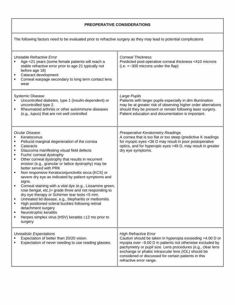
PREOPERATIVE CONSIDERATIONS
The following factors need to be evaluated prior to refractive surgery as they may lead to potential complicatons
Unstable Refractive Error Age <21 years (some female patients will reach a
stable refractive error prior to age 21 typically not before age 18)
Cataract development Corneal warpage secondary to long term contact lens
wear
Corneal Thickness Predicted post-operative corneal thickness <410 microns (i.e. <~300 microns under the flap)
Systemic Disease Uncontrolled diabetes, type 1 (insulin-dependent) or
uncontrolled type 2. Rheumatoid arthritis or other autoimmune diseases
(e.g., lupus) that are not well controlled
Large Pupils Patients with larger pupils especially in dim illumination may be at greater risk of observing higher order aberrations should they be present or remain following laser surgery. Patient education and documentation is important.
Ocular Disease Keratoconus Pellucid marginal degeneration of the cornea Cataracts Glaucoma manifesting visual field defects Fuchs’ corneal dystrophy Other corneal dystrophy that results in recurrent
erosion (e.g., granular or lattice dystrophy) may be better served with PRK
Non responsive Keratoconjunctivitis sicca (KCS) or severe dry eye as indicated by patient symptoms and signs.
Corneal staining with a vital dye (e.g., Lissamine green, rose bengal, etc.)> grade three and not responding to dry eye therapy or Schirmer tear tests <5 mm.
Untreated lid disease, e.g., blepharitis or meibomitis High positioned scleral buckles following retinal
detachment surgery Neurotrophic keratitis Herpes simplex virus (HSV) keratitis ≤12 mo prior to
surgery
Preoperative Keratometry Readings A cornea that is too flat or too steep (predictive K readings for myopic eyes <36 D may result in poor postoperative optics, and for hyperopic eyes >49 D, may result in greater dry eye symptoms.
Unrealistic Expectations Expectation of better than 20/20 vision. Expectation of never needing to use reading glasses.
High Refractive Error Caution should be taken in hyperopia exceeding +4.00 D or myopia over –9.00 D in patients not otherwise excluded by pachymetry or pupil size. Lens procedures (e.g., clear lens exchange or phakic intraocular lens (IOL) should be considered or discussed for certain patients in this refractive error range.

PRE-OPERATIVE EVALUATION
The pre-operative evaluation procedures for patients considering refractive surgery may include, but are not limited to:
Pachymetry (corneal thickness measurement)
Postoperative corneal thickness ≥410 microns. Ablation depth is based on optical zone (OZ) diameter, blend zone (BZ), and
refractive error ~15 microns per diopter.
Pupil Diameter The patient with larger pupils may be more likely than one with smaller pupils to notice any existing higher-order aberration, which may manifest as night vision problem.
Measure pupils under both photopic (or mesopic) and scotopic conditions and document for dim illumination.
Topography Evaluation of surface contour of cornea to determine the potential of early keratoconus.
Keratometry Preoperative corneal curvature (K readings) and dioptric value of refractive error predict postoperative K readings.
Manifest/ Cycloplegic refraction
Measurement of refractive error (myopia, hyperopia, astigmatism) needed to ensure against overcorrection.
Phorometry A cover test should be performed to rule out strabismus, since patients with intermittent strabismus may not tolerate monovision corrections.
Tear Film Assessment Significant dry eye may delay healing and decrease visual acuity during early healing.
Reduced tear quantity: Schirmer’s tear test <5 mm. Corneal staining: Rose Bengal dye, Lissamine green dye or Fluoresceine dye. Conjunctival staining alone: Rose Bengal dye, Lissamine green dye. Treat pre- and post-operatively with artificial tears or punctal occlusion.
Slit Lamp Examination (anterior segment evaluation)
Patients with blepharitis and meibomitis are more likely to experience dry eye and have more post-operative symptoms.
The presence of staphylococcal bacteria in these conditions poses a risk for infection during refractive surgery.
Patients with blepharitis should be treated with lid scrubs, antibiotics, etc. Intrastromal scars may result in an irregular ablation rate. Larger pingueculae may increase difficulty of proper suction of microkeratome.
Dilated Fundus Examination
A thorough peripheral fundus examination is required to rule out retinal thinning, holes, or partial detachments that could lead to potential problems during refractive surgery.

FOLLOW-UP VISITS FOR CO-MANAGEMENT OF REFRACTIVE SURGERY PATIENTS
Following refractive surgery, the co-managing optometrist should examine the patient. A typical schedule for follow-up evaluation is: 1 day 1 week 1 month 3 months Additionally, the patient may be seen at 9-12 months, or as needed
Components of the post-operative visit will include:
1 day Patient history Uncorrected visual acuity Biomicroscopy
1 week
Patient history Uncorrected visual acuity Biomicroscopy Tonometry
1 month Patient history Uncorrected visual acuity Manifest refraction/best corrected visual acuity Biomicroscopy Tonometry
3 months Patient history Topography/keratometry Uncorrected visual acuity Manifest refraction/best corrected visual acuity Biomicroscopy Cycloplegic refraction Tonometry Dilated fundus examination

Potential Post-operative Signs/Symptoms and Their Management for LASIK Patient
Sign/Symptom Management
Flap slipped or wrinkled Refer to surgeon for refloating and smoothing
Epithelial defect Monitor daily, consider contact lens bandage
Dryness Loose epithelium Flap edema
Prescribe preservative-free tears; Monitor; consider bandage contact lens Monitor; measure IOP
Diffuse lamellar keratitis (DLK) Prescribe antibiotic and corticosteroid. Inform or refer to surgeon.
Early epithelial ingrowth Monitor
Overcorrection or undercorrection Prescribe extended wear contact lens or, topical NSAID to speed regression, discuss enhancement, part time spectacles, contact lenses
Infection Refer to surgeon

Potential Post-operative Signs/Symptoms and Their Management for Advanced Surface Ablation Patient
Sign/Symptom Management
Excessive movement of loose-fitting bandage contact lens or blinked out lens
Replace with properly fitted contact lens.
Intermittent pain, corneal edema, conjunctival injection, blurred vision from tight-fitting contact lens
Use cool compresses, artificial tears, and oral analgesics rather than attempt to remove and replace a tight-fitting bandage lens.
Infiltrates arising from combination of bandage lens and topical NSAIDS
Remove bandage contact lens, discontinue topical NSAID, maintain antibiotic and steroid.
Filamentary keratitis Prescribe use of Mucomyst (acetylcysteine).
Overcorrection Prescribe extended wear contact lens; reassess overcorrection monthly.
Mild to marked (grade 2-4) subepithelial edema
Prescribe steroid drops q.i.d. haze on cornea.
Potential Post-operative Signs/Symptoms and Their Management for Conductive Keratoplasty Patient
Sign/Symptoms Management
Foreign body sensation Photophobia Tearing
Prescribe artificial tears, topical steroids with less risk of increasing IOP (e.g., NSAIDS or loteprednol 0.5%) and over-the-counter analgesics.
Fluctuating vision Educate patient to expect gradual increase in vision.
Overcorrection Usually resolves without treatment.
Undercorrection Refer to surgeon to correct residual hyperopia with additional CK spots to increase corneal steepening provided additional treatment does not put patient at risk.
Induced cylinder Refer to surgeon for treatment of persistent induced cylinder by placement of additional CK treatment spot in flattest hemi-meridian to steepen it.

POSSIBLE COMPLICATIONS OF LASIK
Intraoperative Perforation of the eye and loss of intraocular contents, when plate depth is excessive, resulting from incomplete or incorrect assembly of certain types of microkeratomes.
Incomplete flap, created by microkeratome sticking or losing power.
Free cap, created when insufficient cornea presents through the suction ring or when microkeratome continues past the stop point.
Too thin or irregular cut, created when suction is lost, diminished, or fluctuates during passage of the microkeratome. Donut-shaped flap, created when the blade breaks through the epithelium to the surface in the center of the cornea, then returns to the stroma.
Decentered ablation, resulting from patient’s loss of fixation or head movement, or from the laser tracker’s improper centration can lead to unfavorable visual outcome, including irregular astigmatism and loss of BCVA.
Early postoperative Normal early healing symptoms that should not be confused with complications: Slight overcorrection of refractive error likely during first month post-LASIK, due to
calculated laser nomograms that anticipate a natural regression of effect. Induced astigmatism, due to corneal remolding or tear film disruption.
LASIK-induced neurotrophic keratitis (LINK, a.k.a. neurotrophic epitheliopathy), extremely rare severe dry eye effect or keratitis, believed to result from microkeratome transection of the long ciliary nerve which hinders sensory corneal feedback with delayed nerve regeneration. Irregular regrowth of the nerve may be a contributing factor.
Late postoperative Central islands are defined as central steepening greater than 1.50 D covering a 1.5 mm area, seen postoperatively after 1 month. Classic symptoms are ghosting or monocular diplopia. The surgeon may retreat these areas after three months with 30 pulses within a 2.5 mm zone. NOTE: With new pretreatment software, this complication has virtually disappeared.
Epithelial ingrowth, most commonly detected at the 1-month visit as white, milky deposits at the level of the interface and can become an aggressive complication. Treatment is required only if: It obstructs the visual axis. It causes irregular astigmatism (even though not in visual axis; see Topography, below). It is progressive, suggesting risk for corneal melt or recurrent erosion.
Persistent stromal or flap edema (rare beyond 1 week post-LASIK) can occur in the patient with Fuch’s corneal dystrophy (a contraindication for LASIK), in the patient with high IOP (e.g., steroid responder), or in those on medications that slow endothelial pumping function (e.g., carbonic anhydrase inhibitors).
Progressive corneal ectasia, a progressive myopia or ectatic condition thought to be due to weakened corneal tissue (genetic predisposition) or to not maintaining 250 microns of tissue under the flap after ablation, demonstrates the essential need for pachymetry in high myopes.

Refer patient with advanced ectasia to surgeon for partial lamellar keratoplasty or full-thickness penetrating keratoplasty.
Irregular astigmatism or higher-order uncorrectable astigmatism and associated loss of BCVA, among the most common complications of LASIK,* induced by irregular flap cut or misalignment, epithelial ingrowth or from laser ablation irregularities (decentration and the resulting central islands), or by the patient’s preoperative corneal irregularity and healing response.
Regression of effect, overcorrection, undercorrection, and regular astigmatism. Treat temporarily with spectacle correction until adequate refractive stabilization. Earliest recommended retreatment at 3 months. Many patients achieve stability at 6 months.

POSSIBLE COMPLICATIONS AFTER ADVANCED SURFACE ABLATION
Intraoperative Lack of a stromal flap in surface ablation eliminates the potential for microkeratome malfunction, greatly reducing the risk for intraoperative complications.
Decentered ablation, resulting from patient’s loss of fixation on specified target, from head movement, or from improper fixation by laser eye tracker. Symptoms: irregular astigmatism, high-order aberations, loss of BCVA.
Early postoperative Lack of a stromal flap eliminates many postoperative complications. Neither discomfort, the most common symptom following surface ablation, nor refractive errors are complications during this period. Infection is rare, owing to sterile technique and effective antibiotics. It may be present at day 1 though symptoms may not be evident until day 2.
Emergency examination is indicated if patient notices blurry vision, redness, pain, or discharge.
Neurotrophic epitheliopathy or keratitis (dry eye) usually resulting from microkeratome transection of the long ciliary nerve, is minimal in surface ablation, which insults only the most anterior layers of the cornea, affecting only buds of corneal nerves and causing temporary loss of sensation.
Late postoperative Central islands or central steepening >1.50 D covering a small central area of the cornea, seen postoperatively as ghosting or monocular diplopia. Symptom largely eliminated by use of new pretreatment software. Central islands may be simply higher order aberrations such as secondary astigmatism.
The management is the fitting of a rigid gas permeable contact lens or referal to a surgeon or wavefront topography guided ablation.
Persistent stromal edema, a rare occurrence beyond 1 week postsurgery, can occur in patients with Fuchs’ corneal dystrophy (a contraindication for surface ablation), in the patient with high IOP (e.g., steroid responder) or in those on medications that slow endothelial pump function (e.g., carbonic anhydrase inhibitors).
Check IOP – (if elevated, consider site-specific steroids such as loteprednol 0.5% as well as considering IOP lowering agents), discontinue topical CAIs or begin hyperosmotic ointment treatment if Fuchs’ corneal dystrophy is present.
Regression of effect, overcorrection, and regular astigmatism.
Treat temporarily with spectacle correction. Refractive stabilization is achieved in practically all patients at 6 months. Consider retreatment to enhance vision after 3 months, and examine carefully via slit lamp biomicroscopy. Even minimal corneal haze contraindicates further laser ablation, which can reactivate corneal keratocytes and cause severe haze. Schedule borderline cases for reevaluation in 3 months.

Progressive corneal ectasia, increasingly prevalent among high myopes, attributed to weakening of corneal tissue or postoperatively decreased corneal thickness. The requirement of projected postoperative corneal thickness > 400 microns prevents most cases of iatrogenic ectasia. The percentage of tissue removed is critical: patients with a posterior surface elevation or difference of >0.050 mm Hg on corneal topography may be at greater risk for progressive ectasia. Pachymetry is required during the pre-op evaluation to reduce this risk, although theoretically there is less risk of progressive ectasia with PRK compared to LASIK in myopes.
Irregular astigmatism or higher-order uncorrectable astigmatism with an associated loss of BCVA is among the most common complications of refractive surgery.* Causes include irregularities of laser ablation due to decentration and resulting central islands, preoperative corneal irregularity, and healing response.
If extensive, postpone treatment and consider customized or wavefront-guided ablation technology targeting high-order aberrations included in the category of irregular astigmatism.
Long-term High myopes: retinal tears, detachments, glaucoma, and other risks of the myopic-shaped eye (despite correction of refractive error).
High hyperopes: still have same risks for angle closure. Changes in corneal thickness artificially lower IOP ~3mm Hg.
To approximate the IOP after myopic PRK procedures, add 3 mm to all IOP measurements (e.g., postablation IOP 18 mm Hg ~ or = 21 mm Hg.

POSSIBLE COMPLICATIONS OF CONDUCTIVE KERATOPLASTY
Intraoperative Misplaced treatment spots, which can occur when a patient does not properly fixate on specified target during corneal treatment marking (minimal risk). Proper remarking of cornea helps to ensure treatment according to the appropriate nomogram.
Postoperative, 1 day Noninvasive CK procedure conveys only minimal risk for the following: Early loss of BCVA Induced cylinder Overcorrection greater than –1.00 D Undercorrection greater than +1.00 D
Monitor
Postoperative, 1 week Pending refraction, risk for Early loss of BCVA Induced cylinder Overcorrection Undercorrection
Monitor
Postoperative, 1 month Induced cylinder, regular or irregular, without loss of BCVA, resulting from Centration method (centering treatment on the entrance pupil instead of
corneal apex) Preoperative corneal topographic asymmetry (decentered corneal apex,
peripheral or asymmetric astigmatism) Asymmetric treatment Asymmetric pachymetry
The induced changes may decrease over time without loss of hyperopic correction.
Place additional CK treatment spots along flat meridian of the cornea to steepen it, making corneal curvature more symmetrical and reducing or eliminating astigmatism treatment of persistent induced cylinder or in undercorrection, consider adding additional treatment spots once stability is achieved.
Undercorrection (low risk)
Correct residual hyperopia with additional CK spots to increase corneal steepening, provided additional treatment does not put the patient at risk.
Overcorrection Average –0.50 D. Resolves in 3-6 months without treatment. Significant. Usually resolves without treatment in early postoperative period.
Prescribe spectacles (temporary).
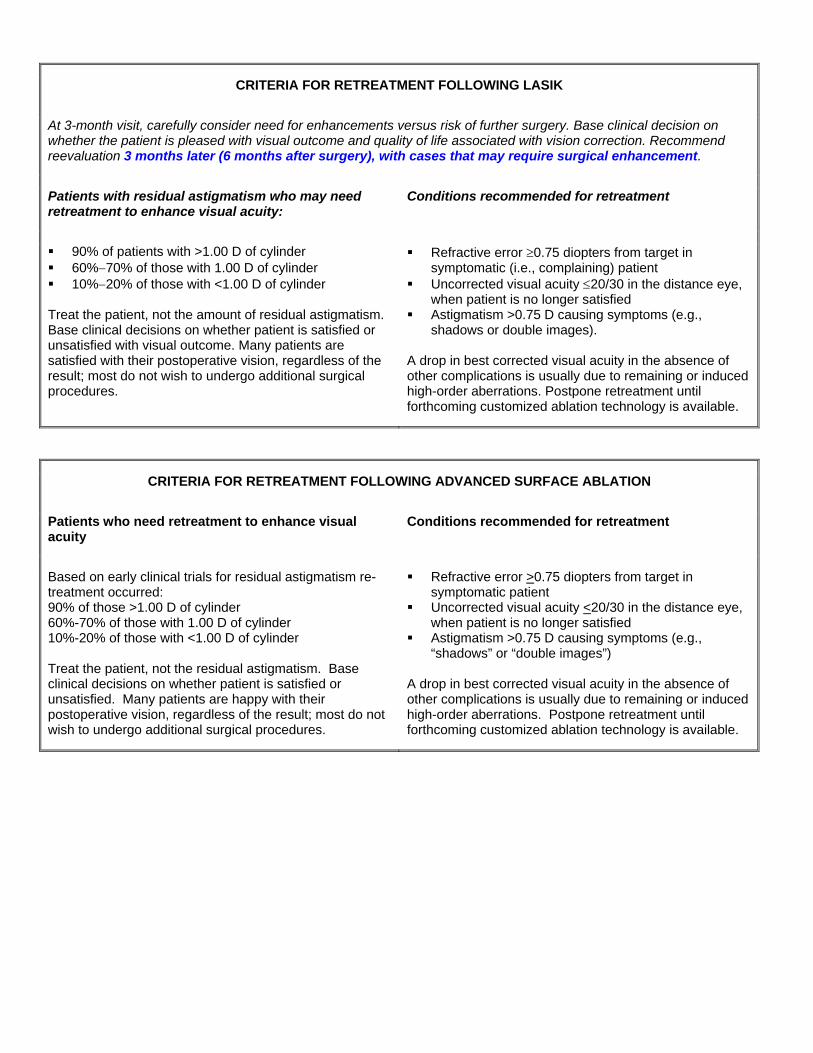
CRITERIA FOR RETREATMENT FOLLOWING LASIK
At 3-month visit, carefully consider need for enhancements versus risk of further surgery. Base clinical decision on whether the patient is pleased with visual outcome and quality of life associated with vision correction. Recommend reevaluation 3 months later (6 months after surgery), with cases that may require surgical enhancement.
Patients with residual astigmatism who may need retreatment to enhance visual acuity:
Conditions recommended for retreatment
90% of patients with >1.00 D of cylinder 60%−70% of those with 1.00 D of cylinder 10%−20% of those with <1.00 D of cylinder
Treat the patient, not the amount of residual astigmatism. Base clinical decisions on whether patient is satisfied or unsatisfied with visual outcome. Many patients are satisfied with their postoperative vision, regardless of the result; most do not wish to undergo additional surgical procedures.
Refractive error ≥0.75 diopters from target in symptomatic (i.e., complaining) patient
Uncorrected visual acuity ≤20/30 in the distance eye, when patient is no longer satisfied
Astigmatism >0.75 D causing symptoms (e.g., shadows or double images).
A drop in best corrected visual acuity in the absence of other complications is usually due to remaining or induced high-order aberrations. Postpone retreatment until forthcoming customized ablation technology is available.
CRITERIA FOR RETREATMENT FOLLOWING ADVANCED SURFACE ABLATION
Patients who need retreatment to enhance visual acuity
Conditions recommended for retreatment
Based on early clinical trials for residual astigmatism re-treatment occurred: 90% of those >1.00 D of cylinder 60%-70% of those with 1.00 D of cylinder 10%-20% of those with <1.00 D of cylinder Treat the patient, not the residual astigmatism. Base clinical decisions on whether patient is satisfied or unsatisfied. Many patients are happy with their postoperative vision, regardless of the result; most do not wish to undergo additional surgical procedures.
Refractive error >0.75 diopters from target in symptomatic patient
Uncorrected visual acuity <20/30 in the distance eye, when patient is no longer satisfied
Astigmatism >0.75 D causing symptoms (e.g., “shadows” or “double images”)
A drop in best corrected visual acuity in the absence of other complications is usually due to remaining or induced high-order aberrations. Postpone retreatment until forthcoming customized ablation technology is available.

ABBREVIATION KEY
BCVA Best corrected visual acuity BZ Blend zone CK Conductive keratoplasty DLK Diffuse lamellar keratitis HSV Herpes simplex virus IOP Intraocular pressure KCS Keratoconjunctivitis sicca LASEK Laser epithelial keratomileusis LASIK Laser assisted in situ keratomileusis NSAID Nonsteroidal anti-inflammatory drug
OZ Optical zone PMMA Polymethyl methacrylate PRK Photorefractive keratectomy
q. i. d. Four times daily (quarter in die) RGP Rigid gas permeable

Herpes Simplex Keratitis
• Dendritic Epithelial keratitis with classic terminal end bulbs
Keratoconus

Fuchs’ Dystrophy
Anterior Membrane Dystrophy

Pellucid Marginal Degeneration
Corneal Haze?

Epithelial Ingrowth
Epithelial Ingrowth

Epithelial Erosion - mild sloughing
Bowman’sStriae or folds
?

Striae/Folds
Flap Folds
DLK
Diffuse lamellar keratitis

DLK
Diffuse lamellar keratitis
Flap / Stromal Edema
?
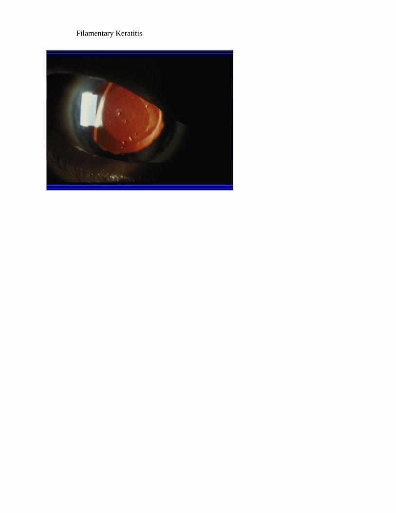
Filamentary Keratitis