Final-Brochure- SpineFEST 2021
54
REGISTRATION
Transcript of Final-Brochure- SpineFEST 2021

��������������������������������������
�������������������������������
�������
���������� ���� ������
REGISTRATION
CONTENT
☞ ABOUT SPINEFEST 2021
☞ - Learning Objectives and Accreditation
☞ - Previous Visiting Professors
☞ ABOUT UNIVERSITY OF TORONTO SPINE PROGRAM
☞ - Vision and Integration
☞ - Faculty
☞ REMARKS FROM PROGRAM CO-DIRECTORS
☞ AGENDA
☞ TATOR & HALL
☞ PROGRAM CO-DIRECTORS
☞ VISITING PROFESSOR & SPEAKERS
☞ SCIENTIFIC ABSTRACTS
☞ ACKNOWLEDGMENTS

SPINEFEST 2021 Brochure
P 2
ABOUT SPINEFEST 2021 ☝
SpineFEST, which was first established 13 years ago, is the Annual Academic Spine Day and the key
spine event at the University of Toronto (U of T). SpineFEST brings together the U of T spine community
to disseminate knowledge of advances in spine surgery, spine care management, and spine research.
The day serves as a unique educational platform for clinicians and researchers from a broad spectrum
of disciplines including neurosurgery, orthopaedic surgery, and a multidisciplinary group of clinicians and
academic professionals from a variety of disciplines including chiropractic, physiatry, physical therapy,
nursing, family medicine, pain medicine, biomedical engineering, and basic/clinical and translational
science.
LEARNING OBJECTIVES OF SPINEFEST 2021: • Recognize the dynamic nature of progress in MIS deformity surgery;
• Learn the indications for MIS correction of deformity;
• Understand the advantages of MIS surgery for correction of deformity;
• Learn how to incorporate MIS techniques in the management of spine oncology.
ACCREDITATION Royal College of Physicians and Surgeons of Canada – Section 1: This event is an Accredited Group
Learning Activity (Section 1) as defined by the Maintenance of Certification Program of the Royal College
of Physicians and Surgeons of Canada, approved by Continuing Professional Development, Temerty
Medicine, University of Toronto up to a maximum of 2.5 credits.
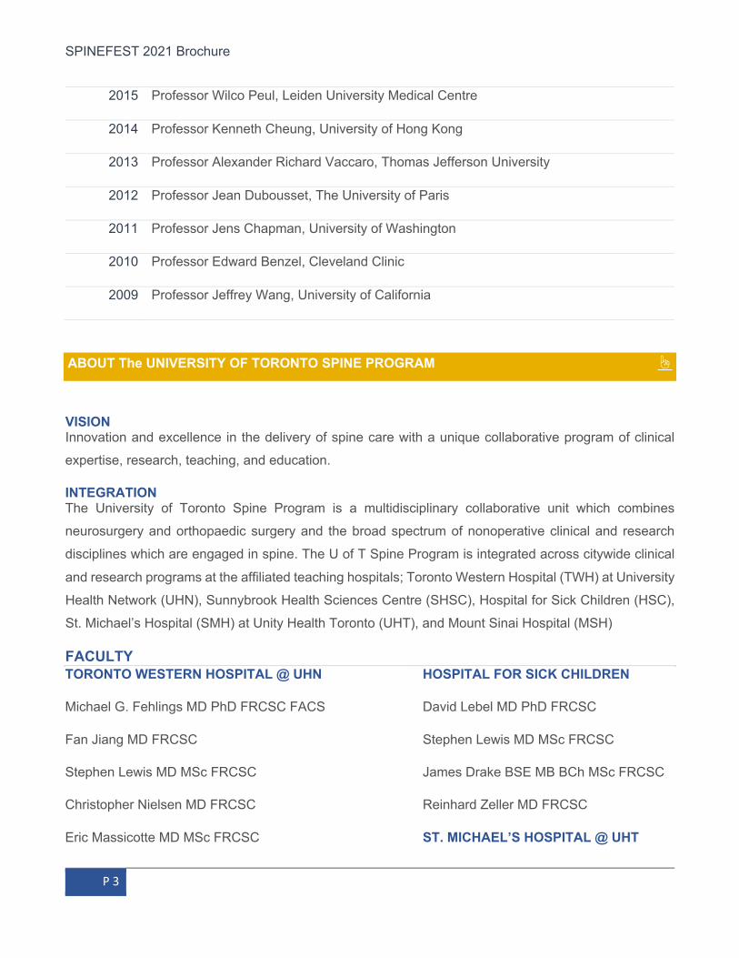
PREVIOUS VISITING PROFESSORS AT THE TATOR – HALL LECTURE
2020 Professor Marcus Stoodley, Macquarie University, Sydney, Australia
2019 Professor Praveen Mummaneni, The University of California, San Francisco
2018 Professor Sanford Emery, West Virginia University
2017 Professor Zoher Ghogawala, Tufts University School of Medicine
2016 Professor Daniel Riew, Columbia University Medical Center
SPINEFEST 2021 Brochure
P 3
2015 Professor Wilco Peul, Leiden University Medical Centre
2014 Professor Kenneth Cheung, University of Hong Kong
2013 Professor Alexander Richard Vaccaro, Thomas Jefferson University
2012 Professor Jean Dubousset, The University of Paris
2011 Professor Jens Chapman, University of Washington
2010 Professor Edward Benzel, Cleveland Clinic
2009 Professor Jeffrey Wang, University of California
ABOUT The UNIVERSITY OF TORONTO SPINE PROGRAM ☝
VISION Innovation and excellence in the delivery of spine care with a unique collaborative program of clinical
expertise, research, teaching, and education.
INTEGRATION The University of Toronto Spine Program is a multidisciplinary collaborative unit which combines
neurosurgery and orthopaedic surgery and the broad spectrum of nonoperative clinical and research
disciplines which are engaged in spine. The U of T Spine Program is integrated across citywide clinical
and research programs at the affiliated teaching hospitals; Toronto Western Hospital (TWH) at University
Health Network (UHN), Sunnybrook Health Sciences Centre (SHSC), Hospital for Sick Children (HSC),
St. Michael’s Hospital (SMH) at Unity Health Toronto (UHT), and Mount Sinai Hospital (MSH)
FACULTY TORONTO WESTERN HOSPITAL @ UHN HOSPITAL FOR SICK CHILDREN
Michael G. Fehlings MD PhD FRCSC FACS David Lebel MD PhD FRCSC
Fan Jiang MD FRCSC Stephen Lewis MD MSc FRCSC
Stephen Lewis MD MSc FRCSC James Drake BSE MB BCh MSc FRCSC
Christopher Nielsen MD FRCSC Reinhard Zeller MD FRCSC
Eric Massicotte MD MSc FRCSC ST. MICHAEL’S HOSPITAL @ UHT
SPINEFEST 2021 Brochure
P 4
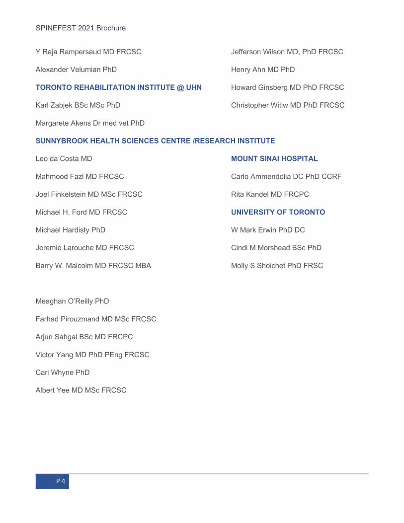
Y Raja Rampersaud MD FRCSC Jefferson Wilson MD, PhD FRCSC
Alexander Velumian PhD Henry Ahn MD PhD
TORONTO REHABILITATION INSTITUTE @ UHN Howard Ginsberg MD PhD FRCSC
Karl Zabjek BSc MSc PhD Christopher Witiw MD PhD FRCSC
Margarete Akens Dr med vet PhD
SUNNYBROOK HEALTH SCIENCES CENTRE /RESEARCH INSTITUTE
Leo da Costa MD MOUNT SINAI HOSPITAL
Mahmood Fazl MD FRCSC Carlo Ammendolia DC PhD CCRF
Joel Finkelstein MD MSc FRCSC Rita Kandel MD FRCPC
Michael H. Ford MD FRCSC UNIVERSITY OF TORONTO
Michael Hardisty PhD W Mark Erwin PhD DC
Jeremie Larouche MD FRCSC Cindi M Morshead BSc PhD
Barry W. Malcolm MD FRCSC MBA Molly S Shoichet PhD FRSC
Meaghan O’Reilly PhD
Farhad Pirouzmand MD MSc FRCSC
Arjun Sahgal BSc MD FRCPC
Victor Yang MD PhD PEng FRCSC
Cari Whyne PhD
Albert Yee MD MSc FRCSC

SPINEFEST 2021 Brochure
P 5
REMARKS FROM PROGRAM CO-DIRECTORS ☝
Colleagues,
We are almost there! With the easing of COVID related restrictions, we look forward to resuming a more
normal situation at our hospitals and University in the coming months. We want to extend our appreciation
to the University of Toronto Department of Surgery Spine Program Council, administrative staff,
educators, and trainees for the continued dedication and professionalism in making this past academic
calendar a significant success during challenging pandemic times. The Program’s academic calendar of
2020/2021 has been a productive one as the Program fosters meaningful citywide collaborations within
the University and participates and leads on several key regional and international initiatives. Our
Program has grown a respected academic footprint locally, nationally, and globally. Collaboration, inter-
professional, and inter-disciplinary knowledge exchange remain the key element to our success.
This U of T Spine Program celebrates its 13th Annual Spine Academic Day “SpineFEST.” At this time of
the year, we gather to highlight our spinal community’s accomplishments and disseminate recent clinical
and scientific advances. As the restrictive measures continue, SpineFEST continues to be held virtually,
again, this year. We are pleased to have had Dr. Richard Fessler, a world-renowned spine surgeon and
Professor of Neurosurgery at Rush University Medical Center, visit us virtually on Monday evening June
14th, to provide his keynote address as our Tator-Hall Lecturer. Professor Fessler will discuss the
management of minimally invasive (MIS) correction of adult spinal deformity. Please join us in welcoming
Professor Fessler to SpineFEST 2021! The New Faculty talk will be presented by Dr. Christopher Witiw,
who will discuss MIS techniques in spinal oncology. The meeting will continue to highlight spine research
from the faculty and trainee. In addition, there will be a follow-up from the 12th Annual meeting with an
update on clinical care and translational research being done on craniocervical junction disorders and
Ehlers Danlos Syndrome. Oral presentations will be provided by Best Abstract winners from both clinical
and basic science perspectives. SpineFEST this year received around 30 excellent scientific abstracts (
☞ abstract), most of which have been presented online on (☞ VoiceThread). All participants are
welcome to communicate with trainees online until the day of the event on June 14th.
Recent activities have leveraged our education platform to create a national spine surgery fellowship
training curriculum for cognitive and procedural competencies. Building on this, our program, over many
years, has established and enhanced Neurosurgery and Orthopaedic Surgery spinal training
opportunities between Toronto Academic Health Sciences Network (TAHSN) teaching hospitals (Toronto

SPINEFEST 2021 Brochure
P 6
Western Hospital (TWH-UHN); Sunnybrook Health Sciences Centre (SHSC); Saint Michael’s Hospital
(SMH) and Hospital for Sick Children (HSC). We have built a top-tier academic hub that attracts 12-15
national and international clinical fellows and many additional visiting surgeons each year.
Over the past years, our program continues to offer both a one-year core fellowship training experience
and a two-year fellowship program with a first-year comprehensive spine training experience followed by
a second year focused on advanced subspecialty exposure. While the fellowships are primarily focused
at one of the TAHSN hospitals, great options exist for a citywide experience. Many thanks to Drs. Albert
Yee, Michael Fehlings, Stephen Lewis, Eric Massicotte, Joel Finkelstein, Howard Ginsberg, Henry Ahn,
and Reinhard Zeller for their valued help shaping our citywide fellowship training opportunities. Building
on our national fellowship curriculum, our Program also continues with the surgical case-log for our
citywide spine fellows with over 2000 cases and procedures recorded. We thank Drs. Jeremie Larouche,
Dr. Tony Bateman, and Ms. Nadia Jaber for creating a successful case-log program for our fellows.
We are excited to announce that an application to the Royal College of Physicians and Surgeons of
Canada has been submitted by the Canadian Spine Society towards establishing an RCPSC Area of
Focused Competence (AFC) Diploma for Spine Surgery. The University of Toronto Spine Program has
partnered closely with the Canadian Spine Society and other centres across the country to advance this
effort. Thanks to Drs. Albert Yee, Jeremie Larouche, Michael Fehlings, Scott Paquette, Hamilton Hall
and Ms. Nadia Jaber for taking the lead in engaging several university spine programs and fellowship
directors across Canada in this initiative. Several members in our Program Education Subcommittee
have expressed keen interest in being involved as the initiative develops; a terrific opportunity for our
Program to continue developing materials that will shape the future of spine surgical education in Canada.
It will provide a valued competence-based model for our international community of surgical educators
as well.
Each year we launch our academic calendar of events with a welcome dinner for our incoming fellows.
This past year, the event was organized virtually to provide an update on our citywide research
opportunities. Thanks to Dr. Carlo Ammendolia and Dr. Karl Zabjek for keeping us updated on the
progress of spine research in Toronto. We also organize a mini bootcamp course in the fall for our fellows
and senior residents to discuss Traumatic Spinal Cord Injury, the ASIA neurological assessment,
surgical/non-surgical management, and current clinical trials. Thanks to Dr. Sukhvinder Kalsi-Ryan for
coordinating the course with Drs. Fehlings, Yee, Jeremie Larouche and Jeff Wilson. Each year, Dr.
Stephen Lewis chairs a citywide fellow surgical skills course, introducing advanced anatomy of the spine
with fellows performing anterior and posterior surgical approaches as well as spinal instrumentation. Over
the past several years, Dr. Lewis extended this course to include advanced complex procedures including
SPINEFEST 2021 Brochure
P 7
deformity osteotomy, minimally invasive surgery, and trauma techniques. The course encompasses a
combination of wet lab, simulation, faculty lectures and case-based discussions throughout the day. It
was unfortunate that the third wave of COVID-19 peaked in May; the course has been rescheduled to
the Fall. Each year, we continue to complement the residents’ surgical training with our Royal College
Mock Oral on Spine course Co-Chaired by Drs. Fehlings and Yee. On March 15th our citywide spine
fellows took a key leadership role in teaching the senior residents and organizing a selection of
representative case scenarios in Royal College examination format. The fellows also provided valuable
tips and updated literature reviews on several spine disorders in this virtual course,. We thank Drs. Julia
Bowes, Nandan Marathe, Ohad Einav, Dora Pelletier, and Carolyn Lai, also our alumni Dr. Mario Ganau
for taking the lead in teaching our residents. We also host a citywide Fellow Journal Club several times
a year to discuss recent and controversial spine articles with a collection of relevant cases. This year,
journal clubs were conducted virtually and hosted by our faculty from several hospitals. We thank Drs.
Fehlings and Yee for hosting a Journal Club on frailty in spine disorder and spine surgery, and Dr. Jeff
Wilson for hosting one on sports-related spinal injury.
The Program invites several world-renowned Professors each year to a Hospital-Based Visiting
Professorship. A few previously scheduled lectures have been postponed to resume when the pandemic
restrictions are lifted and larger in-person meetings are permitted. Meanwhile, our Program hosted a
virtual Visiting Professorship on April 9th jointly with the Department of Surgery, Division of Orthopaedic
Surgery, and Division of Anatomy. The event featured Professor Sigurd Berven from the University of
California San Francisco as our Harland-Smith Lecturer. He provided a very informative and thoughtful
lecture on the use of interbody implant and advanced minimally invasive techniques in spine surgery.
The importance of understanding human anatomy as relevant to advance surgical techniques was
highlighted. Following the lecture, our Program hosted Dr. Berven in a special case-based session with
citywide fellow presentations to discuss complex interbody implant cases and complex deformity. Thanks
to Dr. Berven for his insightful input and our citywide fellows, Drs. Nandan Marathe, Brett Rocos, Julia
Bowes, and Isaaq Carenno for providing exciting and thought-provoking cases. We also held a virtual
Tator-Turnbull Spinal Cord Injury (SCI) Symposium on October 23rd. This event was hosted jointly with
the TWH Spinal Cord Injury Program and the Collaborative Program in Neuroscience to pay tribute to
the enormous contribution of Dr. Charles Tator and Ms. Barbara Turnbull in driving advances in SCI
research and related advocacy. We were delighted to have had Dr. Wolfram Tetzlaff provided an exciting
keynote presentation on the role of the ketogenic diet in cell therapy and other preclinical strategies in
SCI. Dr. Fehlings provided an overview of the U of T Spine Program and the Krembil Brain Institute
Research. Dr. Cathy Craven also provided an update on the Lyndhurst SCI Rehab Program.

SPINEFEST 2021 Brochure
P 8
With the challenges imposed by the pandemic restrictions, our Program has been keen on bringing
together citywide surgeons and trainees in multiple virtual activities. A series of Case-Based Forum has
been initiated to present controversial and complex cases, and to discuss best practices in surgical
approaches and treatment management. We thank Drs. David Lebel, Reinhard Zeller and Stephen Lewis
from HSC, and Drs. Joel Finkelstein and Leo Da Costa from SHSC for organizing excellent presentations.
We have also made hospital-based spine weekly rounds available to citywide surgeons fellows and
residents. These rounds typically discuss weekly on-call, pre-op and post-op case planning and
management, including reviewing relevant literatures on the topics. Thanks to Drs. Fehlings, Stephen
Lewis, Raja Rampersaud and Eric Massicotte for providing this outstanding opportunity. A special thanks
to Dr. Arjun Sahgal for his continued valued input on oncology cases.
On the advocacy level, the Program continues to be proactive to raise awareness of spine conditions.
Efforts in raising awareness and promoting best practices are being undertaken. The Ontario
Degenerative Cervical Myelopathy Summit, which was organized and Co-Chaired by Dr. Fehlings and
Dr. James Milligan (a family physician from the Mobility Clinic in Kitchener-Waterloo) in November 2020,
has brought together a team of Canadian health professionals and federal representatives to discuss the
topic to develop a white paper with a set of priorities to tackle relevant healthcare challenges. It aims to
advance an Ontario-based DCM health care strategy and knowledge translation. Regarding
Craniocervical Junction (CCJ) disorders, including Ehlers Danlos Syndrome (EDS), Dr. Fehlings and
colleagues have engaged the local spine community in attending to the challenges around this disorder.
There remains a lack of evidence-based practice and research. Dr. Fehlings is leading research efforts
which address this knowledge gap. His team is currently undertaking systematic reviews on the
diagnostic criteria for CCJI in EDS to develop diagnostic and management pathways; efforts are being
made in collaboration with the EDS Clinic Program at UHN.
We want to take this moment and celebrate the graduation of our 2020/2021 citywide spine fellows.
Congratulations to Drs. Nandan Marathe, Isaac Aguirre Carreno, Brett Rocos, Julia Bowes, Hari
Ramakonar, Ohad Einav, Laura-Nanna Lohkamp, Dora Pelletier, Jérémie Nallet, Peter Prömmel, Kelechi
Eseonu, Dhawi Aali Alotaibi, and Manuel Fuetsch. We acknowledge their relentless efforts and dedication
in completing advanced fellowship training during this challenging year. We wish them all the best for a
successful and rewarding professional career. We look forward to a continued future engagement in our
Program’s activities.
We wish to recognize the support from the U of T Department of Surgery and Divisions of Neurosurgery
and Orthopedic Surgery. We also would like to thank all our Program faculty members and industry
partners, Medtronic, Zimmer Biomet, De Puy Synthes and Stryker, for their continued support over many
SPINEFEST 2021 Brochure
P 9
years and particularly during the past year. We thank our Program members; we are privileged to benefit
from their diverse and specialized knowledge. Special thanks to Ms. Nadia Jaber, our Program Manager,
for her outstanding expertise and valued Information and Communication Technology skills. They have
been invaluable towards moving forward our collaborative agenda and virtual academic activities during
this rapidly evolving time.
Sincerely,
Michael Fehlings & Albert Yee, Co-Directors Nadia Jaber, Program Manager
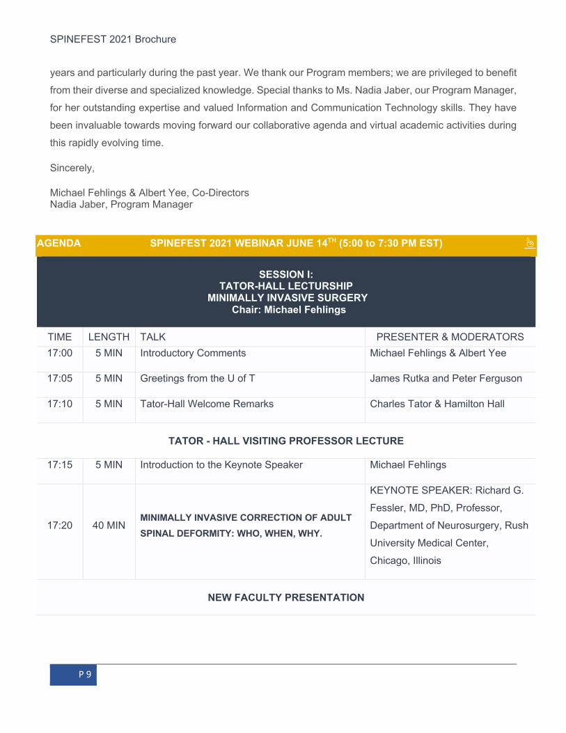
AGENDA SPINEFEST 2021 WEBINAR JUNE 14TH (5:00 to 7:30 PM EST) ☝
SESSION I: TATOR-HALL LECTURSHIP
MINIMALLY INVASIVE SURGERY Chair: Michael Fehlings
TIME LENGTH TALK PRESENTER & MODERATORS 17:00 5 MIN Introductory Comments Michael Fehlings & Albert Yee
17:05 5 MIN Greetings from the U of T James Rutka and Peter Ferguson
17:10 5 MIN Tator-Hall Welcome Remarks Charles Tator & Hamilton Hall
TATOR - HALL VISITING PROFESSOR LECTURE
17:15 5 MIN Introduction to the Keynote Speaker Michael Fehlings
17:20 40 MIN MINIMALLY INVASIVE CORRECTION OF ADULT SPINAL DEFORMITY: WHO, WHEN, WHY.
KEYNOTE SPEAKER: Richard G.
Fessler, MD, PhD, Professor,
Department of Neurosurgery, Rush
University Medical Center,
Chicago, Illinois
NEW FACULTY PRESENTATION

SPINEFEST 2021 Brochure
P 10
18:00 10 MIN
INCORPORATING MINIMALLY INVASIVE
TECHNIQUES IN THE MANAGEMENT OF SPINAL
ONCOLOGY
Chris Witiw, Saint Michael's
Hospital
18:10 Panel Discussion
18:30
20 MIN
End of Session I
5 MIN BREAK
E-POSTER ON VOICETHREAD AVAILABLE ALL DAY HERE
SESSION II: RESEARCH TRAINEE PRESENTATIONS
Chair: Albert Yee
INVITED TRAINEE PRESENTATOIN
18:35 2 MIN Introduction Albert Yee & Michael Fehlings
18:37 7 MIN BUSINESS TECHNIQUES TO IMPROVE HEALTH
SYSTEM DELIVERY OF SPINE CARE
Jay Toor, Resident, Orthopaedic
Surgery
18:44 7 MIN
BIOENGINEERED HUMAN STEM CELL
STRATEGIES TO REGENERATE THE INJURED
SPINAL CORD
Chris Ahuja, resident,
Neurosurgery
18:51 7 MIN
SPINAL MANIFESTATIONS IN EHLERS DANLOS SYNDROME- A SYSTEMATIC REVIEW OF
DIAGNOSTIC CRITERIA FOR CRANIOCERVICAL
INSTABILITY
A follow up presentation from the 12th
Annual SpineFEST Day
Laura- Nanna Lohkamp, spine
fellow, TWH
19:01 10 MIN Panel Discussion
BEST ABSTRACT ORAL PRESENTATION
19:11 5 MIN
1st Place Best Abstract (Basic Science)
ENHANCED OUTCOMES AND REDUCED
PERIOPERATIVE NEUROLOGICAL
COMPLICATIONS IN THE SURGICAL
James Hong - Post Doc - Fehlings
Lab - Krembil Research Institute

SPINEFEST 2021 Brochure
P 11
MANAGEMENT OF DEGENERATIVE CERVICAL
MYELOPATHY: EXAMINING THE IMPACT OF
REMOTE ISCHEMIC PRECONDITIONING
19:16 5 MIN
1st Place Best Abstract (Clinical)
SEQUENTIAL ROD ROLLING FOR SURGICAL
CORRECTION OF LENKE TYPE 2 ADOLESCENT
IDIOPATHIC SCOLIOSIS: A 3D ANALYSIS Jérémie Nallet - Spine Fellow, HSC
19:21 5 MIN Panel Discussion
19:26 4 MIN AWARD PRESENTATIONS
19:30 Wrap up
TATOR & HALL ☝
Dr. Charles Tator is a Professor in the Department of Surgery, at the
University of Toronto, and a neurosurgeon at the Toronto Western Hospital.
He is the former Chair of Neurosurgery at the University of Toronto. He started
the first Acute Spinal Cord Injury Unit in Canada in 1974, and has reported on
the epidemiology, prevention and treatment of spinal cord injury. He has
undertaken seminal translational and clinical research in spinal cord injury. In
1992, he founded ThinkFirst, Canada, a national brain and spinal cord injury
foundation whose mission is to reduce the incidence of catastrophic injuries
in Canada. In 2012, ThinkFirst merged with three other charities to form
Parachute Canada, the country’s foremost injury prevention agency, of which he is a founding Director.
In 2008, the University of Toronto Press published his book “Catastrophic Injuries in Sports and
Recreation, Causes and Prevention-a Canadian Study.” He has held two research chairs at the University
of Toronto, the Dan Family Chair in Neurosurgery and the Campeau Family-Charles Tator Chair in Brain
and Spinal Cord Research. In 2000, he received the Order of Canada, and in 2009 he was inducted into
the Canadian Medical Hall of Fame. In 2017, he was promoted to Officer within the Order of Canada,
and was also inducted into Canada’s Sports Hall of Fame for his work on prevention of sports injuries.

SPINEFEST 2021 Brochure
P 12
Dr. Hamilton Hall is a Professor in the Department of Surgery at the University
of Toronto and on the orthopaedic staff at the Sunnybrook Health Sciences
Centre. He completed his medical degree at the University of Toronto then
joined CARE and was stationed at a rural hospital in Malaysia. Dr. Hall returned
to Toronto for his orthopaedic residency which concluded with a fellowship in
medical education at the University of Dundee, Scotland. In 1974, because of
his interest in patient education and rehabilitation, Dr. Hall founded the
Canadian Back Institute which expanded into the CBI Health, now the largest
home care and rehabilitation company in Canada. He is co-founder and Executive Director of the Canadian Spine Society and has served on the editorial boards of Spine, The
Spine Journal and The BackLetter.
Dr. Hall has received Outstanding Paper and Poster awards from the North American Spine Society and
the International Society for the Study of the Lumbar Spine. He is a recipient of the Laurie Chute Award
for Best Undergraduate Clinical Lecturer Award at the University of Toronto, the NASS Henry Farfan
Award for outstanding contributions to the field of spine care and two Lifetime Achievement Awards, one
from Stryker Spine and the other from the Canadian Spine Society. In 2019 he was inducted into the
Toronto Orthopaedic Hall of Fame. Dr. Hall’s concept of a syndrome approach to classifying
mechanical back pain is an essential component of several Canadian provincial initiatives to improve
spine care. In addition to over 140 published articles and book chapters and over 1200 invited
presentations, many as Visiting Professor, to universities in North America, Europe and Asia, he is
author of the best-selling Back Doctor series of books for the lay public.
CO-DIRECTORS ☝
Dr. Michael Fehlings is a Professor of Neurosurgery, Co-Director of the
Spine Program and Vice Chairman (Research) in the Department of Surgery
at the University of Toronto. He holds the Halbert Chair in Neural Repair and
Regeneration and combines an active clinical practice in complex spinal
surgery at the Toronto Western Hospital with a translationally oriented
research program focused on discovering novel treatments for the injured
brain and spinal cord. He has authored over 1000 peer-reviewed articles (h-
index 100) chiefly in the area of central nervous system injury and complex
spinal surgery. His work has been featured in Nature, Nature Neuroscience,

SPINEFEST 2021 Brochure
P 13
Science Translational Medicine, Nature Reviews Neurology, JAMA, Lancet Neurology, and the New
England Journal of Medicine. Dr. Fehlings has held a number of prominent leadership roles, including
current President of the International Neurotrauma Society, the Chair of the AO Foundation Clinical
Investigation and Documentation Advisory Committee, past Chair of the AOSpine International Spinal
Cord Injury Knowledge Forum, past President of the Cervical Spine Research Society, and leader of
several international clinical research trials. Dr. Fehlings is a Fellow of the Royal Society (Canada) and
a Fellow of the Canadian Academy of Health Sciences. He has received numerous international
recognitions including the Royal College Gold Medal, Olivecrona Award, Ryman Prize, Magnus Medal in
Neurosurgery and the Jonas Salk Award.
Dr. Albert Yee is the Holland Bone and Joint Program Chief and the Head of
the Division of Orthopaedic Surgery at Sunnybrook Health Sciences Centre,
where he holds the Marvin Tile Chair in Orthopaedic Surgery. Dr. Yee is an
Orthopaedic Spine Surgeon at Sunnybrook Health Sciences Centre, an
Associate Scientist (Physical Sciences Platform) at Sunnybrook Research
Institute and a Consultant in Surgical Oncology at the Odette Cancer Centre.
He is a Full Professor at the University of Toronto, Department of Surgery and
Full Member of the Institute of Medical Sciences with a cross appointment in
the Institute of Biomaterials and Biomedical Engineering. He is the Vice Chair of Research in the Division
of Orthopaedic Surgery and Co-Director of the University of Toronto’s Department of Surgery Spine
Program. Dr. Yee is the Past President of the Canadian Orthopaedic Research Society as well as
Canadian Spine Society, and is a Co-Chair of Bone & Joint Canada. He is the Canadian Lead for the
Young Investigators Initiative (YII) of Bone & Joint Canada, and the US Bone & Joint Initiative, a grant
mentorship and career development program. Dr. Yee has over 100 peer reviewed publications and has
received academic honours including the American British Canadian (ABC) International Travelling
Fellowship (American Orthopaedic Association / Canadian Orthopaedic Association, 2013), the Charles
H. Tator Surgeon-Scientist Mentoring Award (2012), and the Canadian Orthopaedic Foundation J.
Edouard Samson Award (2011). In 2019, he was awarded the distinction of Fellow of International
Research (FIOR) by the International Combined Orthopaedic Research Society (ICORS). Dr. Yee’s
laboratory focuses on translational orthopaedic research utilizing pre-clinical surgical models to evaluate
novel minimally invasive vertebral metastatic therapies (e.g. Photodynamic Therapy, Radiofrequency
Ablation). His work has led to first in human clinical trials and FDA approval with commercialization of
new minimally invasive spine technology. He has interest in understanding mechanisms of disease in
cancer invasiveness to bone with an aim towards identifying potential new promising therapeutic targets.

SPINEFEST 2021 Brochure
P 14
SESSION I - SPEAKERS ☝
VISITING PROFESSOR & KEYNOTE SPEAKER
Dr. Richard G. Fessler is Professor of Neurosurgery at the Rush University
Medical Center and former Vice Chair of Neurosurgery at the Feinberg
School of Medicine of Northwestern University, and the John Harper Seeley
Professor and Chief of Neurosurgery at the University of Chicago Hospitals
and Clinics. He was the founder and Director of the Institute for Spine Care
at the Chicago Institute of Neurosurgery and Neuroresearch (CINN),
Director of Clinical Services and Education at the University of Florida Brain
Institute, and the Dunspaugh-Dalton Chair of Brain and Spinal Surgery. Dr.
Fessler completed his Medical Doctorate with honors, and Surgical and
Neurosurgical residencies at the University of Chicago, a Doctorate of
Philosophy in Pharmacology and Physiology, and a Master of Science in Psychology. His undergraduate
degree was from Lawrence University, Appleton, WI, also in Psychology, where he also earned a
certificate of education.
Dr. Fessler is internationally known for his contributions to endoscopic and microendoscopic surgical
developments. He has been instrumental in developing many of the current minimally invasive surgical
techniques. He received the Kambin Foundation Annual Research award for his research in MIS. Dr.
Fessler is known for his pioneering research into human embryonic spinal cord transplantation for the
treatment of spinal cord injury. He also led and co-led studies including first study on the human transplant
to evaluate the safety and efficacy of human embryonic spinal cord transplantation for the treatment of
syringomyelia, and other studies to evaluate the safety of transplantation of the stem cell GRNOPC-
1/ASTOPC-1 into humans suffering acute spinal cord injury.
Dr. Fessler took leadership in several neurosurgical organizations and societies and served on several
government federal health committees and missions including serving as Medical Specialist and Flight
Surgeon for NASA/Space Shuttle. Dr Fessler is well published with over 240 peer-reviewed publications,
and 37 books and over 200 book chapters in medical texts. He sat on several Editorial Boards including
Neurosurgery, Spine Surgery, and Neuro-Orthopaedics, and is frequently invited for visiting
professorships worldwide.

SPINEFEST 2021 Brochure
P 15
SPEAKER - NEW FACULTY
Dr. Christopher Witiw entered the neurosurgery residency program at the
University of Toronto after completing his MD at the University of Manitoba in
2012. During his residency he completed a MS degree with a focus on Health
Economics at The University of Chicago after receiving an award from the
Canadian Institutes of Health Research. His thesis on the value of surgery for
Degenerative Cervical Myelopathy was awarded the prestigious Outstanding
Paper Award from the North American Spine Society in 2016. He has
received numerous other awards including the Shafie S. Fazel Outstanding
Resident Surgeon and Investigator Award from the University of Toronto Department of Surgery and the
Alan R. Hudson Neurosurgery Resident Teaching Award from the University of Toronto Division of
Neurosurgery. After obtaining his FRCSC in Neurosurgery in 2018, Chris undertook a subspecialty
fellowship in Complex and Minimally Invasive Spine Surgery at Rush University Medical Center in
Chicago. Chris returns to Toronto as a Surgeon Investigator at St. Michael's Hospital where his clinical
work is directed toward treating the full spectrum of spinal disorders. He has a specific interest in
minimally invasive approaches to spinal conditions. Chris’ research work is centered on Health
Economics and Health Services pertinent to spinal pathology and he is especially interested in ‘big data’
analytics as a means to optimize efficiency and quality of spine surgery.
SESSION II - SPEAKERS ☝
INVITED RESEARCH TRANEES
Dr. Chris Ahuja is a PGY 5 neurosurgery resident. He was in the SSTP
working towards his PhD with Dr. Michael Fehlings, studying bioengineered
human neural stem cell therapies for traumatic spinal cord injury in Dr.
Fehlings lab at UHN. He completed his medical training at Queen's University
in Kingston before joining the Division of Neurosurgery at the University of
Toronto. He served on the University of Toronto Department of Surgery's
Research Committee and Translational Research Committee, as well as the
University's Medical Innovation Toronto (MiTO) Executive Committee. His
work focused on strategies to modify the extracellular matrix to generate an environment that is more
conducive to cell-based regeneration.

SPINEFEST 2021 Brochure
P 16
INVITED RESEARCH TRANEES
Dr. Jay Toor is entering his final year of Orthopaedic Surgery residency and
graduated the Surgeon Scientist Training Program with a MBA specializing
in Supply Chain Management. His academic interest is optimizing hospital
efficiency and translating business techniques to improve healthcare
delivery. He founded a software and consulting company that has
successfully overhauled surgical device inventory at several hospitals
leading to significant financial savings. His research work is also focused on
deploying Artificial Intelligence to optimize hospital resource allocation to
address surgical backlogs, improve surgical patient throughput rates and
generate cost savings.
FOLLOW-UP ON 12TH ANNUAL SPINEFEST
Dr. Laura-Nanna Lohkamp completed her Neurosurgery residency at the
Charite Berlin, followed by pediatric subspecialisation in Lyon, France and at
the Hospital for Sick Children, including 6 months of pediatric orthopaedic
spine training. She also completed her Master's degree from the
International University Dresden/Harvard. Laura is currently completing her
spine fellowship training at the Toronto Western Hospital/University of
Toronto Spine Program. Her Clinical focus is adult and pediatric spine
surgery.
BEST ABSTRACT WINNER (BASIC SCIENCE)
Dr. James Hong is a post-doctoral fellow at the Fehlings laboratory at the
Krembil Research Institute, Toronto Western Hospital. His doctoral thesis
focused on the temporal profiling of local and peripheral changes after
traumatic cervical and thoracic injury. He is the author of 11 articles in the field,
and currently works on the development of therapies and next-generation
sequencing analysis of degenerative cervical myelopathy and traumatic
cervical spinal cord injury. He will present the unpublished results of his most
recent collaborative work with Dr. Hiroyuki Katoh investigating the efficacy of

SPINEFEST 2021 Brochure
P 17
a non-invasive strategy for enhancing functional recovery following surgical decompression of
degenerative cervical myelopathy.
BEST ABSTRACT WINNER (CLINICAL)
Dr. Jérémie Nallet completed his medical studies at the University of
Bourgogne Franche-comté, France. He started his residency in general surgery
in Besançon in 2013. In 2015, he decided to specialize in pediatric orthopaedic
surgery. In 2018, he got the French Board Certification in Orthopaedic and
Traumatology Surgery. Currently, he is completing his fellowship training in
Pediatric Spine Surgery at Hospital for Sick Children and set to complete his
Master degree in Biomechanic engineering at Ecole Nationale des arts et
métiers (ENSAM), Paris. Jérémie is involved in humanitarian activities for
Pediatric Orthopaedic surgery with « la chaine de l’espoire », for which he completed an assignment in
Jordan.

SPINEFEST 2021 Brochure
P 18
SCIENTIFIC ABSTRACTS ☝
VIEW E-POSTER ON VOICETHREAD HERE
# AUTHOR TITLE
1 Jonathon Chon, Teng Chio, Jian Wang, Vithushan Surendran, Lijun Li, Mohammad-Masoud Zavvarian, Kataryzna Pieczonka, Michael G. Fehlings
Drug Repurposing: Delayed Administration of High Dose Human Immunoglobulin G for Treatment of Traumatic Cervical Spinal Cord Injury
2 E Crawford P Balasuberamaniam, A Wasim, M Shrikumar, T Chen, T Anthony, A Philips, A Nathens, M Chapman, J Larouche, J Finkelstein
Prolonged Duration of Norepinephrine Infusions is Associated with Sacral Ulcers (SU) in Adults with Complete Spinal Cord Injuries (SCI)
3 Ali Moghaddamjou, Alex B. Bak, Francois Mathieu, Jerry Ku, Michael G. Fehlings
Familial Arachnoiditis with Syringomyelia: Analysis of a Family of 15 Affected Individuals and a Systematic Review
4 Ali Moghaddamjou, Jefferson R. Wilson, Michael G. Fehlings.
Natural History and Spontaneous Recovery of Neurological Function in Patients with an ASIA A Spinal Cord Injury: Analysis of Multicentre Data in 943 Cases
5 Geoff Klein, Isaac Carreno, Joel Finkelstein, Young Lee Arjun Sahgal, Cari Whyne, Anne Martel, Michael Hardisty
Using Convolutional Neural Networks to Predict Scoliosis from 3D Spine CT Scans

SPINEFEST 2021 Brochure
P 19
6 Katarzyna Pieczonka, Mohamad Khazaei and Michael G. Fehlings
Determining the Mechanisms of Transplanted Oligodendrogenically-Biased Neural Progenitor Cells
7 Christopher R. Pasarikovski, Jerry C. Ku, Joel Ramjist, Yuta Dobashi, Stefano M. Priol, Leodante da Costa, AshishKumar, Victor XD. Yang
Minimally Invasive Intrathecal Spinal Cord Imaging with Optical Coherence Tomography
8 Kelly Fullerton, Geoff Klein, Urban Emmenegger, Joel Finkelstein, Frank Lyons, Cari Whyne, Michael Hardisty
Automatic 3D Prostate Cancer Induced Sarcopenia Segmentation
9 William Brett McIntyre, Mohammad Khazaei, Michael G. Fehlings
Regional Identity of Neural Stem Cells is Maintained Throughout the Cell Transplantation Process
10 Lohkamp LN, Marathe N, Fehlings MG
Craniocervical Instability in Ehlers-Danlos Syndrome – A Systematic Review of Diagnostic andTherapeutic Approaches
11 Cari Whyne, Michael Hardisty, and Abdalrahman Alfakir
Measure Patient Adherence to Back Pain Physiotherapy with Artificial Intelligence
12 Mohammedayaz Rangrez, Margarete K. Akens, Michael Hardisty, and Cari Whyne
Temporal Effect of Docetaxel on Tumor Growth and Bone Quality in Rat Model of Vertebral Metastases
13 Brett Rocos, Masayoshi Machida, Karl Zabjek, Reinhard Zeller, David E. Lebel
A comparison of the Reliability and Vulnerability of 3D SterEOS and 2D EOS when Measuring the Sagittal Spinal Alignment of Patients with Adolescent Idiopathic Scoliosis
14 Brett Rocos, Jérèmie Nallet, Reinhard Zeller, Stephen Lewis, David E. Lebel
A comparison of 3 Rod and 2 Rod Constructs in the Correction of Severe Paediatric Scoliosis
15 Brett Rocos, Luke Reda, David E. Lebel, Michael K. Dodds, Reinhard Zeller
The Use of Halo gravity Traction in Severe, Stiff Scoliosis
16 Brett Rocos, David E. Lebel, Reinhard Zeller.
Congenital Kyphosis: Progressive Correction with an Instrumented Posterior Epiphysiodesis. A preliminary Report.
17 Brett Rocos, Y. Raja Rampersaud, Stephen J. Lewis
The Role of Early Wound Contamination on Deep Wound Infections in Lumbosacral Fusions
18 Allison Tolgyesi, Normand Robert, Cari M. Whyne, Michael Hardisty
Enhanced μCT Imaging Enables High Resolution 3D Visualization of Microdamage in Rat Vertebrae.
19 William Luong, Mohamed Khazaei, Christopher S. Ahuja, and Michael G. Fehlings
Investigating Human Neural Precursor Response to Chondroitin Sulfate Proteoglycan
20 Tiffany Lung, James Y. Lee Jessica Widdifield, Ruth Croxford, Jeremie S.
Rate of Revision and Acute Complications of Lumbar Disc Replacement vs Fusion: A population Based Study

SPINEFEST 2021 Brochure
P 20
Larouche, Bheeshma Ravi, J. Michael Paterson, Joel A. Finkelstein
21 Carlo Ammendolia, Corey Hofkirchner, Joshua Plener, André Bussières, Michael Schneider, James J Young, Andrea D Furlan, Kent Stuber, Aksa Ahmed, Carol Cancelliere, Aleisha Adeboyejo, and Joseph Ornelas
Nonoperative Treatment for Lumbar Spinal Stenosis with Neurogenic Claudication. An Updated Systematic Review
22 Nayaab Punjani, Svetlana Altamentova, Jonathon Chio, Jian Wang, Sighild Lemarchant, Yann Godfrin, Michael G. Fehlings
Enhancing Neural Regeneration and Locomotor Recovery with NX Peptide Administration in a Cervical Spinal Cord Injury Rat Model
23 Sydney Brockie, James Hong, Michael Fehlings
Genetic Inhibition of CX3CR1 to Improve Surgical Outcomes in Degenerative Cervical Myelopathy
24 James Hong, Hiroyuki Katoh, Michael Fehlings, Toronto Western Hospital, Tokai University
Enhanced Outcomes and Reduced Perioperative Neurological Complications in the Surgical Management of Degenerative Cervical Myelopathy: Examining the Impact of Remote Ischemic Preconditioning
25 Mandana Movahed, James Hong, Hiroyuki Katoh, Michael Fehlings
Transcriptional Footprint of Ischemia Reperfusion
Injury after DCM
26 Jeremie Nallet, Brett Rocos, David Eduard Lebel, Reinhard Zeller
Sequential Rod Rolling for Surgical Correction of Lenke Type 2 Adolescent Idiopathic Scoliosis: a 3D Analysis
27 Marathe N, Lohkamp LN, , Massicotte EM
Surgical repair for dural leaks causing Spontaneous Intracranial Hypotension – A Case Series and review of literature
SPINEFEST 2021 Brochure
P 21
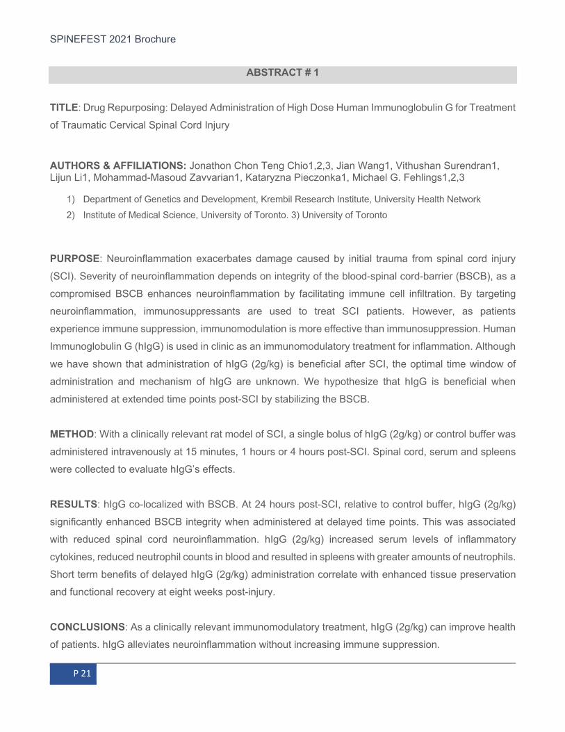
ABSTRACT # 1 TITLE: Drug Repurposing: Delayed Administration of High Dose Human Immunoglobulin G for Treatment
of Traumatic Cervical Spinal Cord Injury
AUTHORS & AFFILIATIONS: Jonathon Chon Teng Chio1,2,3, Jian Wang1, Vithushan Surendran1, Lijun Li1, Mohammad-Masoud Zavvarian1, Kataryzna Pieczonka1, Michael G. Fehlings1,2,3
1) Department of Genetics and Development, Krembil Research Institute, University Health Network 2) Institute of Medical Science, University of Toronto. 3) University of Toronto
PURPOSE: Neuroinflammation exacerbates damage caused by initial trauma from spinal cord injury
(SCI). Severity of neuroinflammation depends on integrity of the blood-spinal cord-barrier (BSCB), as a
compromised BSCB enhances neuroinflammation by facilitating immune cell infiltration. By targeting
neuroinflammation, immunosuppressants are used to treat SCI patients. However, as patients
experience immune suppression, immunomodulation is more effective than immunosuppression. Human
Immunoglobulin G (hIgG) is used in clinic as an immunomodulatory treatment for inflammation. Although
we have shown that administration of hIgG (2g/kg) is beneficial after SCI, the optimal time window of
administration and mechanism of hIgG are unknown. We hypothesize that hIgG is beneficial when
administered at extended time points post-SCI by stabilizing the BSCB.
METHOD: With a clinically relevant rat model of SCI, a single bolus of hIgG (2g/kg) or control buffer was
administered intravenously at 15 minutes, 1 hours or 4 hours post-SCI. Spinal cord, serum and spleens
were collected to evaluate hIgG’s effects.
RESULTS: hIgG co-localized with BSCB. At 24 hours post-SCI, relative to control buffer, hIgG (2g/kg)
significantly enhanced BSCB integrity when administered at delayed time points. This was associated
with reduced spinal cord neuroinflammation. hIgG (2g/kg) increased serum levels of inflammatory
cytokines, reduced neutrophil counts in blood and resulted in spleens with greater amounts of neutrophils.
Short term benefits of delayed hIgG (2g/kg) administration correlate with enhanced tissue preservation
and functional recovery at eight weeks post-injury.
CONCLUSIONS: As a clinically relevant immunomodulatory treatment, hIgG (2g/kg) can improve health
of patients. hIgG alleviates neuroinflammation without increasing immune suppression.

SPINEFEST 2021 Brochure
P 22
ABSTRACT # 2
TITLE: Prolonged Duration of Norepinephrine Infusions is Associated with Sacral Ulcers (SU) in Adults
with Complete Spinal Cord Injuries (SCI)
AUTHORS & AFFILIATIONS: E Crawford1,2, P Balasuberamaniam1, A Wasim1, M Shrikumar1, T Chen1, T Anthony1, A Philips1, A Nathens1,2, M Chapman1,2, J Larouche1,2, J Finkelstein1,2
1) Sunnybrook Health Sciences Centre. 2) University of Toronto
PURPOSE: Complete SCI remains a devastating injury, made worse by preventable complications.
Sacral ulcers (SU) are frequently reported within this population. Standard treatment in this population is
to receive norepinephrine to maintain minimum mean arterial pressure (MAP) targets and ensure spinal
cord perfusion. This is achieved, in part by peripheral vasoconstriction and reduced blood flow. This led
us to our research question: Are norepinephrine infusions associated with SU in patients with complete
SCI?
METHOD: Adults with an ASIA A SCI presenting to a level-one trauma centre from 2014-18 were
reviewed retrospectively. Patient and injury variables (age, gender, location of SCI [cervical vs.
thoracolumbar], Injury Severity Score [ISS]) and treatment factors (surgery, MAP targets, vasopressor
treatment) were recorded along with the presence/absence of SU. A multivariable logistic regression
analyses was used to determine potential associations with SU. Model fit and accuracy to correctly predict
patients with SU, were assessed with Hosmer-Lemeshow test and C-statistic, respectively.
RESULTS: Of the 103 patients identified, 35 (34%) developed SU. Patient age (Mean: 48; SD:21.8),
location of SCI (54 cervical, 52.4%) and ISS (Mean 34.6; SD:13.8) did not differ by the development of
SU. Eighty-six patients (83.5%) were treated with norepinephrine. There was no difference in the
proportion of SU between patients who received norepinephrine versus those who did not. For patients
treated with norepinephrine, a multivariable logistic regression analysis found that norepinephrine
infusion durations >100 hours (3.41 [1.35-16.37]; OR [95%CI]; p=0.046), and hospital LOS > 3 weeks
(4.70 [1.5 to 16.36]; p=0.015) were significantly associated with SU. This model was found to fit the data
well and had a c-statistic of 0.805.

SPINEFEST 2021 Brochure
P 23
CONCLUSIONS: This study reported a prevalence rate of 34% for SU in patients with complete SCI, with
patients receiving >100hrs of norepinephrine infusions having 3.41 times the odds of developing a SU,
compared to those with shorter durations. Additionally, SU are associated with prolonged hospital LOS.
Future research in this area should include prospective, randomized controlled trails and economic
analyses.
ABSTRACT # 3
TITLE: Familial Arachnoiditis with Syringomyelia: Analysis of a Family of 15 Affected Individuals and a
Systematic Review
AUTHORS & AFFILIATIONS: Ali Moghaddamjou (1), MD; Alex B. Bak (1); Francois Mathieu, MD (1);
Jerry Ku (1), MD; Michael G. Fehlings, PhD, MD (1)
1) University of Toronto, Toronto, Ontario, Canada.
PURPOSE: We report a case of a 61-year-old male with non-communicating thoracic syringomyelia living
in Canada of Japanese descent with 15 known family members with the same condition (Figure 1). Most
cases of idiopathic syringomyelia are sporadic, with no family history of the disease. Familial
syringomyelia is an extremely rare form of presentation and is defined by the presence of syringomyelic
cavities in 2 or more patients within the same family.
Our patient has been diagnosed in 2011 and after an initial progressive phase causing significant lower
extremity spasticity, has remained clinically and radiologically stable. He has no history of trauma, Chiari
malformation or associated scoliosis. He has been started on RILUZOLE 50 mg b.i.d. as an off-label use
since 2017.
METHOD: On May 2nd, 2020, 10 keywords relating to familial trait and 23 keywords relating to
syringomyelia were used as search terms on MEDLINE, EMBASE and Cochrane libraries. Abstracts
were screened by two reviewers following the PRIMSA checklist. Papers reporting cases of familial
syringomyelia in English were included for quantitative analysis.

SPINEFEST 2021 Brochure
P 24
RESULTS: The search revealed 476 results of which 25 were included in the qualitative analysis (Figure
2). Overall, there are 131 cases reported of which 16% had associated scoliosis and 65% associated
Chari Malformation (Table 1). Most of the patients had surgery as a treatment (71.74%).
CONCLUSIONS: After a systematic review of the current English literature we can conclude that our
case is one of the largest known family clusters of syringomyelia not associated with a Chiari
Malformation in North America. While its difficult to demonstrate efficacy, we propose the off-label use of
RILUZOLE as a potential conservative therapy. Apart from these results, there is basic science support
for the use RILUZOLE for arachnoiditis-associated secondary injury. Given the autosomal dominant
pattern in our case, whole exome sequencing would be an interesting avenue of investigation into the
pathophysiology of this condition. Currently, there are no known genetic causes.
ABSTRACT # 4
TITLE: Natural History and Spontaneous Recovery of Neurological Function in Patients with an ASIA A
Spinal Cord Injury: Analysis of Multicentre Data in 943 Cases
AUTHORS & AFFILIATIONS: Ali Moghaddamjou1, MD; Jefferson R. Wilson1, MD PhD; Michael G.
Fehlings1, MD PhD.
1) University of Toronto, Toronto, Ontario, Canada.
PURPOSE: Predicting spontaneous recovery after traumatic Spinal Cord Injury (tSCI) is important for
expectation setting of patients and clinical trial design. Spontaneous recovery needs to be accounted for
in trial design to prevent type 1 errors through erroneous randomization. It is recognized that the
prediction of patients with an American Spinal Injury Association (ASIA) Impairment Scale (AIS) A that
convert is challenging. Better understanding of the natural history of ASIA A patients is required to
understand disease trajectory and to decipher treatment effect in trials from spontaneous recovery.
METHOD: Patients with ASIA A injury were identified from 3 prospective, multi-center datasets (NACTN,
STASCIS, and SYGEN). All follow-up examinations for each patient were included and transitions with
their respective time from injury in hours were tabulated. A-priori we identified age (<60yrs and =>60),

SPINEFEST 2021 Brochure
P 25
injury region (cervical, thoracic, and lumbar), and early surgery (surgery <24hrs vs >24hrs) as covariates
in our analyses. We also tabulated the number of muscle groups below the neurological level of injury
from baseline at each examination point. The mstate statistical package in R was utilized to develop
flexible Markov models of disease progression. Covariate effects were estimated using Cox regression
without any proportionality assumption.
RESULTS: We identified 943 patients with 384 total transitions. On average patients recovered 2.7
muscle groups below their level of injury at the 52 week mark. The cumulative hazard ratio plot overtime
reveals an exponential relationship in all transition groups illustrating the time-dependent impact on
transition intensities. Dynamic prediction probabilities revealed a total conversion of 34.36% from ASIA
A. Cervical injuries showed statistically significant increase in spontaneous transition probabilities.
CONCLUSIONS: We demonstrate that multi-state models can successfully be applied to the progression
of tSCI as measured by the transition of AIS grades of AIS A patients. The combined predictive factor of
our on AIS conversion is time-dependent requiring comprehensive models incorporating all prediction
timepoints. These results also support early aggressive treatment for ASIA A patients and consideration
of patient trajectories in decision making.
ABSTRACT # 5
TITLE: Using Convolutional Neural Networks to Predict Scoliosis from 3D Spine CT Scans AUTHORS & AFFILIATIONS: Geoff Klein1,2, Isaac Carreno5,6, Joel Finkelstein4,5,6, Young Lee3,
Arjun Sahgal1,3, Cari Whyne1,4, Anne Martel1,2, Michael Hardisty1,4
1Physical Sciences, 5Division of Spine Surgery, 6Orthopaedic Surgery, Sunnybrook Research Institute;
Department of 2Medical Biophysics, 3Radiation Oncology, 4Surgery, University of Toronto
PURPOSE: Vertebral metastases can lead to biomechanical instability, pain, and neurological
compromise. Stereotactic body radiation therapy (SBRT) delivers high-dose focal treatment to tumours.
A significant side effect of SBRT is vertebral compression fracture, occurring in 10% to 40% of patients
following SBRT. Spinal malalignment (scoliotic deformity) has been shown to be related to vertebral
fracture risk following SBRT. However, current evaluation of spinal malalignment can be time consuming

SPINEFEST 2021 Brochure
P 26
with significant inter-observer variation. As such, an automated algorithm to evaluate Cobb angle in 3D
CT scans was developed and applied to patients with spinal metastasis treated with SBRT.
METHOD: A 3D U-Net model which determined a Gaussian heatmap for spine localization was used to
extract a spline following the curvature of the spine projected in the coronal plane. The gradient of the
spline was determined, and the Cobb angle was calculated from the spline. To account for varying voxel
spacing and the number of vertebrae in the field-of-view, we used the median angle from an axially sliding
window. Ground truth and predicted angles above 10° were classified as scoliotic.
RESULTS: The model was able to predict scoliosis with accuracy of 79.5% and 76.2% on the diagnostic
imaging and SBRT planning datasets, respectively. The mean ground truth and predicted Cobb angles
in the SBRT treatment planning were 8.8° ± 7.0° (ranging from 0.8° to 28.0°) and 9.5° ± 7.5° (ranging
from 1.2° to 35.6°), respectively. The mean ground truth and predicted Cobb angles in the diagnostic
imaging dataset were 8.5° ± 6.7° (ranging from 0.2° to 28.6°) and 12.0° ± 12.6° (ranging from 0.4° to
51.4°), respectively.
CONCLUSIONS: A fully automated model was constructed to predict scoliotic spinal curvature in 3D CT
spine scans by evaluating the Cobb angle. Spinal curvature is contributing parameter for the SINS
classification to determine instability. This algorithm can be used in clinical decision making to aid in
spinal curvature classification and scoliosis severity assessment. Future work will focus on improving
accuracy, expansion to kyphotic deformity, and combing with other image features related to fracture
risk.
ABSTRACT # 6 TITLE: Determining the Mechanisms of Transplanted Oligodendrogenically-Biased Neural Progenitor
Cells
AUTHORS & AFFILIATIONS: Katarzyna Pieczonka, Mohamad Khazaei and Michael G. Fehlings
Krembil Research Institute, Institute of Medical Science, University of Toronto
PURPOSE: Myelin structure is particularly susceptible to dysregulation after spinal cord injury (SCI),
ultimately contributing to impaired signal conductivity in the central nervous system and devastating
behavioural symptoms. Neural progenitor cell (NPC) transplantation represents a potential regenerative

SPINEFEST 2021 Brochure
P 27
approach for promoting remyelination following SCI, however the injury microenvironment predominantly
directs NPCs to differentiate into astrocytes as opposed to myelinating oligodendrocytes. Our lab has
successfully developed a protocol for priming human NPCs into oligodendrogenically-biased NPCS
(oNPCs), which effectively differentiate into a greater ratio of oligodendrocytes. Importantly, oNPCs have
been found to promote remyelination and to ultimately contribute to functional recovery following
transplantation into the injured rat spinal cord. However, a detailed analysis of the mechanisms of these
cells post-transplantation has not been conducted to date. We aim to utilize RNA sequencing approaches
in order to determine the mechanisms by which oNPCs promote recovery following SCI. We hypothesize
that oNPC transplantation decreases the expression of negative myelin molecules following SCI.
METHOD: Female immunodeficient Rowett Nude (RNU) rats were subjected to a cervical SCI, and half
of the rats were transplanted with oNPCs 1 week post-injury. The oNPCs were prepared from human
NPCs by mimicking oligodendrogenic developmental cues in vitro. The animals were sacrificed 9 weeks
following injury and RNA was isolated from the injury epicenter for subsequent bulk RNA sequencing and
analysis.
RESULTS: It is expected that oNPC transplantation reduces the expression of negative myelin molecules
such as myelin associated glycoprotein, polysialylated neural cell adhesion molecule, oligodendrocyte-
myelin glycoprotein and neurite outgrowth inhibitor A when compared to the non-transplanted injury
group.
CONCLUSIONS: This project will provide us with a greater understanding of oNPC mechanisms, which
will help us optimize NPC interventions for SCI in the future.
ABSTRACT # 7
TITLE: Minimally Invasive Intrathecal Spinal Cord Imaging with Optical Coherence Tomography
AUTHORS & AFFILIATIONS: Christopher R. Pasarikovski, MD1, Jerry C. Ku, MD1, Joel Ramjist2, Yuta
Dobashi2, Stefano M. Priola MD3, Leodante da Costa, MD, MSc2, Ashish Kumar, MCh2, Victor XD.
Yang, MD, PhD2, 4

SPINEFEST 2021 Brochure
P 28
1Division of Neurosurgery, Department of Surgery, University of Toronto, Toronto, Ontario, Canada;
2Division of Neurosurgery, Sunnybrook Hospital, University of Toronto, Toronto, Ontario, Canada;
3Division of Neurosurgery, Department of Surgery, Health Sciences North, Sudbury, Ontario, Canada;
4Hurvitz Brain Sciences Research Program, Sunnybrook Research Institute, Sunnybrook Health Sciences Centre, University of Toronto, Toronto, Ontario, Canada.
PURPOSE: Imaging of the spinal cord is challenging due to the surrounding bony anatomy, physiologic
motion, and the small diameter of the spinal cord. This precludes the use of non-invasive imaging
techniques in assessing structural changes related to trauma and evaluating residual function. The
purpose of this research was to apply endovascular technology and techniques and construct a
minimally-invasive preclinical animal model of intrathecal spinal cord imaging using optical coherence
tomography (OCT).
METHOD: Five animals (2 Yorkshire Swine and 3 New Zealand Rabbits) were utilized. A dedicated
animal interventional radiology suite equipped with a single-plane C-Arm was used for all procedures.
Intrathecal access was gained using a 16-guage Tuohy, and an OCT catheter was advanced under
roadmap guidance technique into the cervical thecal sac. The OCT device generates cross-sectional
images with spatial resolution of 10µm. Imaging frequency is 100 frames per second, with a total of 540
cross sectional images generated per pullback. The OCT catheter has a motorized pullback, and a total
length of 54mm of the spinal canal is imaged with one pullback.
RESULTS: Image acquisition was successful for all animals. There were no instances of difficult catheter
navigation, enabling OCT imaging rostrally to C2. The thecal sac provided excellent thoroughfare for the
OCT catheter. The clear cerebrospinal fluid also provided an excellent medium for image acquisition,
with no detectable artifact from the contents of the cerebrospinal fluid. The anatomical space of the spinal
canal could be readily appreciated including: dural lining of the thecal sac, epidural veins, pial lining of
the spinal cord, arachnoid bands, dentate ligaments, and nerve rootlets/roots.
CONCLUSIONS: Minimally invasive intrathecal imaging using endovascular OCT was feasible in this
preclinical animal study. Using OCT, excellent visualization of the dura, subarachnoid space, epidural
vessels, dentate ligaments, and nerve roots and rootlets was achieved. This technology and imaging
technique may allow for high resolution, minimally invasive imaging for pathologies such as structural
changes related to spinal cord trauma, spinal neoplasms, vascular malformations, arachnoid webs,
arachnoiditis, and cysts.
SPINEFEST 2021 Brochure
P 29
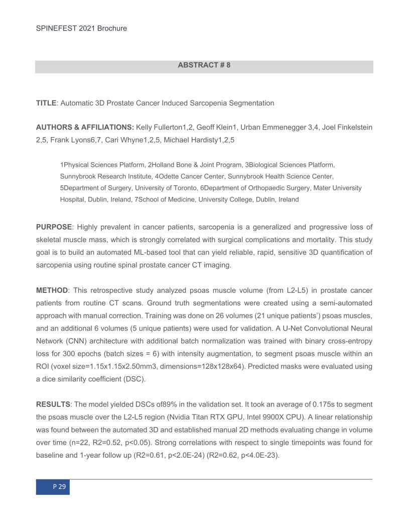
ABSTRACT # 8
TITLE: Automatic 3D Prostate Cancer Induced Sarcopenia Segmentation
AUTHORS & AFFILIATIONS: Kelly Fullerton1,2, Geoff Klein1, Urban Emmenegger 3,4, Joel Finkelstein
2,5, Frank Lyons6,7, Cari Whyne1,2,5, Michael Hardisty1,2,5
1Physical Sciences Platform, 2Holland Bone & Joint Program, 3Biological Sciences Platform,
Sunnybrook Research Institute, 4Odette Cancer Center, Sunnybrook Health Science Center,
5Department of Surgery, University of Toronto, 6Department of Orthopaedic Surgery, Mater University
Hospital, Dublin, Ireland, 7School of Medicine, University College, Dublin, Ireland
PURPOSE: Highly prevalent in cancer patients, sarcopenia is a generalized and progressive loss of
skeletal muscle mass, which is strongly correlated with surgical complications and mortality. This study
goal is to build an automated ML-based tool that can yield reliable, rapid, sensitive 3D quantification of
sarcopenia using routine spinal prostate cancer CT imaging.
METHOD: This retrospective study analyzed psoas muscle volume (from L2-L5) in prostate cancer
patients from routine CT scans. Ground truth segmentations were created using a semi-automated
approach with manual correction. Training was done on 26 volumes (21 unique patients’) psoas muscles,
and an additional 6 volumes (5 unique patients) were used for validation. A U-Net Convolutional Neural
Network (CNN) architecture with additional batch normalization was trained with binary cross-entropy
loss for 300 epochs (batch sizes = 6) with intensity augmentation, to segment psoas muscle within an
ROI (voxel size=1.15x1.15x2.50mm3, dimensions=128x128x64). Predicted masks were evaluated using
a dice similarity coefficient (DSC).
RESULTS: The model yielded DSCs of89% in the validation set. It took an average of 0.175s to segment
the psoas muscle over the L2-L5 region (Nvidia Titan RTX GPU, Intel 9900X CPU). A linear relationship
was found between the automated 3D and established manual 2D methods evaluating change in volume
over time (n=22, R2=0.52, p<0.05). Strong correlations with respect to single timepoints was found for
baseline and 1-year follow up (R2=0.61, p<2.0E-24) (R2=0.62, p<4.0E-23).

SPINEFEST 2021 Brochure
P 30
CONCLUSIONS: This automated ML-based 3D method yielded accuracy, speed and promise for greater
sensitivity to initial development of sarcopenia will enable future study of large datasets. Accurate and
precise measurement of sarcopenia will allow better disease and treatment monitoring and allow for
better prediction of patient outcom.
ABSTRACT # 9
TITLE: Regional Identity of Neural Stem Cells is Maintained Throughout the Cell Transplantation Process
AUTHORS & AFFILIATIONS: William Brett McIntyre1,2, Mohammad Khazaei2, Michael G. Fehlings1,2.
1) Institute of Medical Science, University of Toronto. 2) Krembil Research Institute, Department of
Surgery, University Health Network.
PURPOSE: Traumatic spinal cord injury (SCI) elicits damage to the neural circuitry of the spinal cord
(SC), which directly translates to impaired motor/sensory functions. Neural Stem Cell (NSC)
transplantation is a promising regenerative strategy to treat SCI because they can replenish lost cells
and restore motor/sensory deficits, thus improving these patients’ quality of life. However, NSCs exhibit
limited success to treat SCI when the identity of the NSC does not match the site of transplantation in the
spinal cord. NSC identity is conferred through the expression of Homeobox (Hox) transcription factors,
which regulate where the NSCs localize within the brain and spinal cord during development and
throughout adulthood. This segmentation process can then promote the formation of appropriate
neuronal circuits necessary to perform motor/sensory functions. If identity is maintained in NSCs post-
transplantation, this may suggest their developmental role is recapitulated to promote regenerative
success within the Central Nervous System (CNS). Thus, it is hypothesized that NSCs from the brain &
SC will maintain regionally-specific Hox gene expression.
METHOD: Brain and SC NSCs were dissected, expanded, and differentiated in culture. Next, NSCs were
transplanted into either the adult brain and SC (2 cell lines; 2 regions of interest; 4 groups). RT-qPCR
and immunohistochemistry markers of regional Hox markers were be used to confirm NSC identity.
RESULTS: After in vitro characterization & in vivo transplantation, B-NSCs and SC-NSCs maintained a
greater proportion of region specific Hox expression (SC: Hox4-Hox10; Brain: Otx2, Emx2). Each cell

SPINEFEST 2021 Brochure
P 31
type also exhibited equal tripotency potential, meaning they differentiated into the three major cell types
of the CNS: Neurons, Oligodendrocytes, and Astrocytes.
CONCLUSIONS: These results further support that NSCs derived from the brain and SC retain their
identity following proliferation and maturation both in vivo and in vitro. B-NSCs and SC-NSCs are also
equally suited to replenish the injured CNS as they exhibit similar differentiation potentials. This suggests
the correlation between identity and optimal regenerative success may be due to the maintenance of Hox
expression. This work presents correlation evidence, however future work will involve evaluating the
efficacy of B-NSCs and SC-NSCs in the context of SCI.
ABSTRACT # 10 TITLE: Craniocervical Instability in Ehlers-Danlos Syndrome – A Systematic Review of Diagnostic and Therapeutic Approaches
AUTHORS & AFFILIATIONS: Lohkamp LN1, Marathe N1, Fehlings MG1
1 Division of Neurosurgery, Department of Surgery, University of Toronto, Toronto, Ontario, Canada;
Division of Neurosurgery, Spinal Program, Toronto Western Hospital, University Health Network, Toronto,
Ontario, Canada.
PURPOSE: Ehlers-Danlos Syndrome (EDS) comprises a spectrum of connective tissue disorders
affecting 1/5000 patients. Due to associated ligamentous laxity some subtypes of EDS may be associated
with craniocervical instability (CCI). Patients with EDS and suspected CCI present with a constellation of
functional abnormalities and there is a lack of consensus on the best imaging parameters to achieve the
diagnosis. Herein the clinical research goals are to summarize results of a systematic literature review in
order to identify knowledge gaps and to provide a scientific overview of the current standard of care in
these patients.
METHOD: A systematic literature review was performed using the databases Ovid Medline, EMbase,
Cochrane Library and PubMed based on the PRISMA guidelines. Articles were included if they described
diagnostic or treatment criteria for CCI in EDS patients. These criteria and identified knowledge gaps are
summarized.

SPINEFEST 2021 Brochure
P 32
RESULTS: Seven out of 93 articles reporting on a total of 42 EDS patients met the inclusion criteria. All
of these patients were diagnosed with CCI and subsequently underwent surgical intervention. The main
diagnostic measures were lateral flexion and extension x-rays of the cervical spine as well as dynamic
CT imaging. Thirteen different radiographic parameters were reported pertaining to CCI of which four
were the most frequently applied: the atlantodental interval (ADI), the basion-axis interval (BAI), the clivo-
axial angle (CXA), and the angular displacement of C1 to C2.
CONCLUSIONS: There is a significant lack of evidence for the choice and application of diagnostic
methods and criteria for CCI in EDS patients. A standardized, validated algorithm describing the
sequential imaging measures for the correct diagnosis of CCI is lacking. Furthermore, the radiographic
parameters to assess CCI warrant assessment in larger cohorts and prospective, controlled studies.
Currently, based on the best available evidence we would recommend that patients with EDS and
suspected CCI be evaluated for abnormalities in the ADI, BA, CXA and angular displacement of C1-C2.
Surgical fixation for suspected CCI should only be used in cases with clear objective evidence on imaging
of CCI and concordant symptoms and/or physical signs.
ABSTRACT # 11
TITLE: Measure Patient Adherence to Back Pain Physiotherapy with Artificial Intelligence AUTHORS & AFFILIATIONS: Cari Whyne & Michael Hardisty & Abdalrahman Alfakir
University of Toronto and & Sunnybrook Research Institute
PURPOSE: The objective of this project is to develop an in home deployable objective quantitative
assessment system for posture and lower back pain (LBP) exercises performed (quality and quantity)
using wearable sensors and machine learning (ML) algorithms.
METHOD: Aim 1: Develop an Inertia Movement Unit (IMU) based wearable sensor arrangement for
measuring LBP physiotherapy exercise performance and posture. Initial development will focus on the
number and positioning of IMU sensors required for LBP physiotherapy adherence and posture
assessment. Eight (8) sensors will be placed on test subjects directly with adhesive. The specific
exercises and posture which will be considered were chosen based on consultations with an advanced

SPINEFEST 2021 Brochure
P 33
practiced physiotherapist focused on spinal disorders. For this purpose, we will extend a pre-existing
software platform developed by our laboratory (SPARS) to enable multiple IMU data acquisition, pre-
processing, and storage using a pre-existing cloud-based infrastructure. Aim 2: To train the SPARS-LBP
system to classify LBP physiotherapy exercises and grade posture. Forty healthy volunteers will be
instructed to place the sensors at specific locations on their body while they perform the prescribed
exercises for LBP and posture poses. ML algorithms using the SPARS-LBP system will be trained to
classify exercises and postures from the IMU time series sensor data. The system architecture will then
be optimized by considering the trade offs between accuracy, efficiency, and deployability. This will allow
for the possibility of sensor reduction and/or rearrangement in the final configuration.
RESULTS: To date, the first iteration of aim 1 has been completed where a full IMU recording system
has been constructed. As for aim 2, utilizing the system designed, recording sessions are underway and
eighteen sessions have been completed thus far. The analysis of the data is being worked on and thus
fare the results seem promising.
CONCLUSIONS: This project will yield an objective system for LBP physiotherapy monitoring using a
multi-IMU wearable garment and the SPARS ML platform. SPARS-LBP has the potential to improve the
effectiveness and efficiency of physiotherapy delivery, allowing problems with performance to be
identified early, enable remote monitoring of patient progress, and may lead to improved participation
and associated improvements in outcome.
ABSTRACT # 12
TITLE: Temporal Effect of Docetaxel on Tumor Growth and Bone Quality in Rat Model of Vertebral
Metastases
AUTHORS & AFFILIATIONS: Mohammedayaz Rangrez1,2, Margarete K. Akens2,3,4, Michael
Hardisty1,4 and Cari Whyne1,4,5
1Sunnybrook Research Institute, 2Techna Institute, University Health Network, 3Departments of Medical
Biophysics, 4 Surgery and 5Institute of Biomedical Engineering, University of Toronto; Toronto, ON,
Canada.

SPINEFEST 2021 Brochure
P 34
PURPOSE: Bone is one of the three common sites of metastasis due to its high remodeling rate and
nutrient rich microenvironment. The treatment for bone metastases is often multimodal, requiring
chemotherapy such as docetaxel in addition to focal therapy. Despite proven clinical efficacy in treating
bone metastases, little is known about the impact of docetaxel on bone quality. This study evaluated the
temporal effects of docetaxel on vertebral bone with osteolytic metastasis in a pre-clinical animal model.
METHOD: Osteolytic (OL) bone metastases were introduced in athymic nude rats. Tumour burden was
evaluated with bioluminescent imaging (BLI) on day 14 (d14) and d21 post inoculation. Docetaxel
(5mg/kg) was injected (i.v) in the early (d7, n=8) or late (d14, n=8) stages of metastases, and compared
to untreated controls (n=5). After sacrifice on d21, bone architecture was assessed via stereologic
evaluation of µCT imaging of L2 vertebrae and immunohistochemistry was used to visualize tumour
burden in T11 and L5 vertebrae.
RESULTS: Animals with OL metastases showed a significant decrease in body weight that was avoided
by early docetaxel treatment (EDT) but not by late docetaxel treatment (LDT). The EDT group showed
(p<0.01) less tumour burden (BLI and immunohistochemistry) and improved trabecular bone volume
fraction compared to both untreated OL and LDT groups. Trabecular number was higher (p<0.001) and
trabecular spacing was lower (p<0.001) in both EDT and LDT groups compared to the untreated OL
group. Despite large tumor burden in the LDT group seen in histology, overall bone histoarchitecture was
well preserved with less trabecular damage than the untreated OL group.
CONCLUSIONS: This study demonstrates the ability of early docetaxel treatment in preventing tumor
metastases and subsequent bone loss; later treatment was not nearly as effective. These findings align
with the clinical observation of poor docetaxel response in advanced stages of cancer. A better
understanding of the impact of cancer treatment on bone quality can help to guide treatments for
osteolytic bone metastases.
ABSTRACT # 13
TITLE: A comparison of the Reliability and Vulnerability of 3D SterEOS and 2D EOS when Measuring
the Sagittal Spinal Alignment of Patients with Adolescent Idiopathic Scoliosis
SPINEFEST 2021 Brochure
P 35
AUTHORS & AFFILIATIONS: Brett Rocos MD FRCS (Tr & Orth) 1, Masayoshi Machida MD1, Karl
Zabjek MSc1, Reinhard Zeller MD MSC FRCSC1, David E. Lebel MD PhD1
1Department of Orthopaedic Surgery, The Hospital for Sick Children, 555 University Ave., Toronto,
Canada M5G 1X8
PURPOSE: An essential component of making the diagnosis of adolescent idiopathic scoliosis (AIS) is
standing anteroposterior (AP) and lateral radiographs. Two dimensional (2D) radiographs inevitably fail
to reflect every plane of the three dimensional (3D) deformity in scoliosis and as a result have the potential
to misrepresent the exact nature of the deformity. In this retrospective cohort study, we have tested the
hypothesis that there is no difference in the assessment of the sagittal plane deformity in patients being
treated for AIS when measured with either 2D or 3D EOS radiography.
METHOD: A retrospective radiographic analysis was performed on patients with AIS. The cohort was
then subdivided into three groups according to the coronal angular deformity (mild group: 45- 69°,
moderate group 70- 89°, and severe group 90°+). The sagittal parameters were compared between
manual measurement with 2D sterEOS and 3D reconstruction. Descriptive measures were used to
summarize the distribution of numeric variables. Inter-study reliability was assessed using Bland-Altman
plots and single measure two-way mixed intra-class correlation coefficient (ICC).
RESULTS: 52 patients were included in each group. The inter-study reliability when measuring the TK
and LL between the two study modalities was excellent in in mild group (ICC:0.90, 95%CI: 0.82~0.94
and ICC:0.84 95%CI: 0.74~0.91), excellent in TK and fair in LL in moderate group (ICC:0.76, 95%CI:
0.61~0.85 and ICC:0.70 95%CI: 0.53~0.81), and fair in TK and LL in severe group respectively (ICC:0.74,
95%CI: 0.57~0.84 and ICC:0.65 95%CI: 0.46~0.84). A Bland-Altman plot showed proportional bias in TK
measurements in each group and LL in the moderate group. The difference between 95% upper and
lower limits were increased as the coronal angular deformity increased.
CONCLUSIONS: 3D sterEOS is less vulnerable to the influence of coronal plane deformity than 2D EOS
in evaluating the sagittal spinal parameters of patients with a coronal deformity exceeding 70°.
ABSTRACT # 14

SPINEFEST 2021 Brochure
P 36
TITLE: A comparison of 3 Rod and 2 Rod Constructs in the Correction of Severe Paediatric Scoliosis
AUTHORS & AFFILIATIONS: Brett Rocos MD FRCS (Tr & Orth) 1, Jérèmie Nallet MD 1, Reinhard Zeller
MD FRCSC 1, Stephen Lewis MD MSc FRCSC 1, David E. Lebel MD PhD 1
1Department of Orthopaedic Surgery, The Hospital for Sick Children. 555 University Avenue, Toronto ON
M5G 1X8
PURPOSE: Treating severe scoliosis is challenging and risky with a significant complication rate
regardless of treatment strategy. In this retrospective comparative study, we report our results using a
three rod (3R) compared to two rod (2R) construct in the treatments of severe, stiff pediatric spine
deformities, and hypothesize that the former is safer, provides superior radiological outcomes and is
associated with a lower rate of complication.
METHOD: There were 21 patients in the 3R group and 25 in the 2R group. Both groups were comparable
in their baseline demographics and radiographic deformity. The mean preoperative major coronal
deformity was 100°±9 and 102°±10 in the 3R and 2R respectively (P=0.6). The average major curve
correction was 51% and 59% in 3R and 2R groups respectively (p=0.03). The postoperative thoracic
kyphosis was 30°±11 and 21°±12 in the 3R and the 2R groups respectively (p=0.01). The surgical time
was 476±52 min and 387±84 min in 3R and 2R respectively (p<0.01). One patient in the 2R cohort
showed permanent postoperative sensory deficit. There were three unplanned returns to operating
theatre in the 2R group.
RESULTS: There were 21 patients in the 3R group and 25 in the 2R group. Both groups were comparable
in their baseline demographics and radiographic deformity. The mean preoperative major coronal
deformity was 100°±9 and 102°±10 in the 3R and 2R respectively (P=0.6). The average major curve
correction was 51% and 59% in 3R and 2R groups respectively (p=0.03). The postoperative thoracic
kyphosis was 30°±11 and 21°±12 in the 3R and the 2R groups respectively (p=0.01). The surgical time
was 476±52 min and 387±84 min in 3R and 2R respectively (p<0.01). One patient in the 2R cohort
showed permanent postoperative sensory deficit. There were three unplanned returns to operating
theatre in the 2R group.
CONCLUSIONS: Coronal correction was better with 2R whereas sagittal balance was superior with 3R.
Both techniques achieved balanced spine and found to be safe treating severe scoliosis. The 2R
technique was associated with a higher likelihood of requiring revision surgery.
SPINEFEST 2021 Brochure
P 37
ABSTRACT # 15
TITLE: The Use of Halo gravity Traction in Severe, Stiff Scoliosis
AUTHORS & AFFILIATIONS: Brett Rocos MD FRCS (Tr & Orth) 1, Luke Reda MD MSc 1, David E.
Lebel MD PhD 1, Michael K. Dodds FRCS (Tr & Orth) 2, Reinhard Zeller MD MSc FRCSC 1
1 Department of Orthopaedic Surgery, Hospital for Sick Children, 555 University Avenue, Toronto, ON,
Canada M5G 1XB. 2 Department of Trauma and Orthopaedic Surgery, Mater Misericordiae University
Hospital and Children’s University Hospital, Temple St, Rotunda, Dublin 1, Ireland D01 XD99
PURPOSE: The correction of severe, stiff scoliosis in children is challenging. One method used to reduce
the risk is pre-operative halo gravity traction (HGT). In this study, we sought to define the efficiency and
safety of HGT and characterize the chronology of the correction seen.
METHOD: A consecutive group of paediatric patients with severe spinal deformities were treated with
HGT prior to definitive correction. A standard protocol with the daily addition of weight to 50% of
bodyweight at 3 weeks was used. Traction remained in place until signs of impending neurological
complication or 6 weeks, whichever was sooner.
RESULTS: Twenty-four patients were included with a mean age of 11.8 years. The mean coronal
deformity was 123°, with a T1-L5 height of 234 mm. The mean duration of traction was 42 days with a
mean improvement in height of 72 mm with 82% occurring over the first 3 weeks. 100% of the angular
and 98% of T1-L5 height correction was reached by 6 weeks. One patient showed early signs of a cranial
nerve palsy prompting early surgery and 8 patients showed pin loosening, 1 of which required revision of
their halo. One patient underwent a slower progression of traction due to transitory urinary disturbance.
Following fusion, angular correction of the major curve was 49%.
CONCLUSIONS: HGT is a safe treatment for severe, stiff scoliosis because it can respond to early signs
of impending neurological impairment. The first 3 weeks of treatment, reaching 50% of bodyweight as a
traction force accounts for 80% of correction, with the remaining 20% in the following 2 weeks. At least 4
weeks of traction is recommended when following this protocol.

SPINEFEST 2021 Brochure
P 38
ABSTRACT # 16
TITLE: Congenital Kyphosis: Progressive Correction with an Instrumented Posterior Epiphysiodesis. A
preliminary Report.
AUTHORS & AFFILIATIONS: Brett Rocos MD FRCS (Tr & Orth) 1, David E. Lebel MD PhD 1, Reinhard
Zeller MD MSc FRCSC 1.
1 Department of Orthopaedic Surgery, Hospital for Sick Children, 555 University Avenue, Toronto, ON,
Canada M5G 1XB
PURPOSE: Congenital kyphosis is a rare condition. In this case series we sought to identify the outcomes
and complications of posterior instrumented fusion and the resultant epiphysiodesis effect in uniplanar
congenital kyphosis in paediatric patients.
METHOD: Paediatric patients were included if treated for a uniplanar congenital kyphotic deformity
treated with posterior instrumented spinal fusion between October 2006 and August 2017, with a
minimum of 2 years of follow up. Patients were excluded if a coronal deformity greater than 10° was
present.
RESULTS: Six patients met the inclusion criteria. Mean age at surgery was 3.6 years. The mean kyphotic
deformity prior to surgery was 49.7°. All patients underwent posterior instrumented fusion with
autogenous iliac crest graft and a cast or brace postoperatively. One patient showed a loss of motor
evoked potential on prone positioning which returned to normal on supine positioning. No patient showed
any post-operative neurological deficits. One patient was diagnosed with a wound infection which was
successfully treated with oral antibiotics. By a follow up of 5.4 years (range 2.2- 10.9 years) there was no
failure of instrumentation. An epiphysiodesis effect (a difference of ≥ 5° in the kyphotic deformity
measured between the immediate post- operative and final follow up lateral whole spine XR) of 16.2°
(range 7.2- 30.9°) was seen in 5 patients. The mean annual epiphysiodesis effect was 2.7° (95% CI, 1.4-
4.1°). No kyphosis proximal to the instrumentation was observed for the duration of follow up.

SPINEFEST 2021 Brochure
P 39
CONCLUSIONS: Posterior instrumented fusion and epiphysiodesis is safe and effective. The
epiphysiodesis effect occurs in 5/6 of cases, and our data suggests that the procedure is associated
with an acceptable blood loss and a low incidence of neurological complications.
ABSTRACT # 17
TITLE: The Role of Early Wound Contamination on Deep Wound Infections in Lumbosacral Fusions
AUTHORS & AFFILIATIONS: Brett Rocos MD FRCS (Tr & Orth) 1, Y. Raja Rampersaud MD FRCSC
1, Stephen J. Lewis MD MSc FRCSC 1
Department of Orthopaedic Surgery, Toronto Western Hospital, University Health Network, 399 Bathurst
Street, Toronto, Ontario, Canada M5T 2S8
PURPOSE: Infection in patients treated with lumbosacral fusion has usually been considered to be
caused by intraoperative contamination or hematogenous dissemination. It is not clear if this is the
case, or if in these patients with low incisions external inoculation (out-to-in) are more often responsible
for early deep post-operative infection requiring surgical debridement.
METHOD: We conducted a retrospective review of all adult patients treated with lumbosacral fusion
between January 2014 and January 2021. Patients were included if they underwent at least 1 surgical
debridement for infection or dehiscence requiring debridement. Cases of primary tumor were excluded.
RESULTS: 363 eligible cases were identified. 14 patients underwent at least 1 debridement (4.1%) for
dehiscence or deep infection. The mean BMI was 32.5 (20.7-44.4) and median number of levels fused
was 5. Mean surgical time was 369 mins (195-600) and blood loss 1800 mls (250- 4150).
3/14 patients showed no growth on culture, 1 of which sustained durotomy at the primary procedure.
2/14 showed S. aureus infection requiring debridement at a mean of 97 days. 9/14 showed infection
with intestinal or urogenital pathogens requiring debridement at a mean of 15 days. Infection with
intestinal or urogenital pathogens required significantly earlier debridement (p= 0.03) than S. aureus
and dehiscence (p= 0.037).

SPINEFEST 2021 Brochure
P 40
CONCLUSIONS: Local contamination was the principal cause of deep wound infection in this series.
These present significantly earlier than hematogenous infections. Proximity to the bowel and bladder
puts this region at risk. We should focus on barrier dressing and urinary diversion with a foley catheter
to keep these pathogens away from the wound at a critical healing time.
ABSTRACT # 18
TITLE: Enhanced μCT Imaging Enables High Resolution 3D Visualization of Microdamage in Rat
Vertebrae.
AUTHORS & AFFILIATIONS: Allison Tolgyesi1,3, Normand Robert1, Cari M. Whyne1,2,3, Michael
Hardisty1,2
1Orthopaedic Biomechanics Laboratory, Sunnybrook Research Institute; 2Division of Orthopaedics, Department of Surgery, 3Biomedical Engineering, University of Toronto
PURPOSE: Microdamage accumulation in metastatic bone can increase fracture risk. Experimentally,
barium sulfate (BaSO4) can be used to stain bone to visualize cracks and microdamage. Backscattered
electron imaging (BSE) provides very high resolution 2D images of bone with excellent contrast of
BaSO4 stained microdamage.1 The objective of this study was to evaluate the ability of enhanced
micro-computed tomography (μCT) imaging of whole rat vertebrae stained with BaSO4 to enable
microdamage visualization that is spatially correlated to visualization using BSE imaging.
METHOD: First lumbar (L1) vertebrae from nine 8-9 week old athymic rats (Hsd:RH-Foxn1rnu, Envigo,
IN, USA) (3 healthy, 3 osteolytic, 3 mixed) were mechanically loaded (50N for 3hrs) and stained with
BaSO4. Twelve slides from the L1 vertebrae (6 healthy, 3 osteolytic, 3 mixed) were prepared for BSE
imaging (2μm/pixel, Philips/FEI). The slides were imaged using μCT (μCT100, Scanco) under varied
protocols for high contrast of the BaSO4. The μCT and BSE images were aligned, resampled,
registered (affine) and label fields of the BaSO4 were generated. Spatial correlation, g(r), was used to
evaluate agreement between damage in the μCT and BSE images.1 Convolution of the μCT label field
(kernel r=0.02mm) created an inflated region. The distance between voxels in the two images were
considered to be correlated if g(r)>1.

SPINEFEST 2021 Brochure
P 41
RESULTS: By controlling the μCT parameters (focal spot size, tube voltage, filtration, and dose), the
contrast to noise ratio was enhanced. Increasing data averaging reduced the grainy texture of the
images and trabeculae were more clearly distinguished with low current. The enhanced scan
parameters were 90kVp, 44uA, 200ms integration time, 8 data averaging and a 4.9µm voxel size.
Spatial correlation (g(r)=3.88-12.28) was found between the μCT and BSE images. Examination of the
μCT and BSE images shows microdamage that is obscured by noise in standard μCT images.
CONCLUSIONS: Enhancement of μCT scanning parameters allows for rapid high-resolution 3D
imaging able to quantify bone microdamage. This is important for understanding post-yield bone tissue
mechanics and quantifying 3D microdamage in healthy and metastatically involved vertebrae and
effects of cancer treatments on bone quality.
ABSTRACT # 19
TITLE: Investigating Human Neural Precursor Response to Chondroitin Sulfate Proteoglycan
AUTHORS & AFFILIATIONS: William Luong1,2, Mohamed Khazaei1, Christopher S. Ahuja1,2, and
Michael G. Fehlings1,2,3
1Division of Genetics and Development, Krembil Research Institute; 2Institute of Medical Science,
University of Toronto; 3Division of Neurosurgery, Department of Surgery, University of Toronto
PURPOSE: Human neural precursor cell (hNPC) transplant for chronic spinal cord injury can be
optimized for better recovery by understanding interactions with the glial scar. Our aim is to uncover the
transcriptional profile of hNPCs in their response to a key glial scar molecule, chondroitin sulfate
proteoglycans (CSPGs), and identify genes that can be utillilzed to degrade the glial scar.
METHOD: Human induced pluripotent stem cell-derived NPCs were developed and plated on different
levels of CSPG. RNA was extracted from the cells and analyzed by RNA-sequencing to find
differentially expressed genes. Gene ontology analysis was performed to investigate potential
mechanisms and processes by which CSPGs affect hNPCs. Differentially expressed genes were
validated by at the RNA level by qRT-PCR and protein level by Western blot. Differentiation profiles of

SPINEFEST 2021 Brochure
P 42
hNPCs in response to CSPG were then compared by staining for neuronal, oligodendroglial and
astrocytic markers.
RESULTS: RNA-seq analysis reveal that hNPCs upregulate key genes associated with negatively
regulating neurogenesis and neurodevelopment in response to CSPGs exposure. CSPGs inhibit
neuronal differentiation of hNPC. The overall transcriptional activity of hNPCs is decreased and hNPCs
downregulate genes associated with immune response after CSPGs.
CONCLUSIONS: These differentially expressed genes can be utilized to optimize the degradation of
scar tissue in combination of hNPC transplant and differentiation of these cells remains a key barrier to
overcome in treating chronic spinal cord injury after cell transplant. Ultimately, these results further our
understanding of the chronic injury microenvironment and reveal strategies to improve regeneration of
the spinal cord.
ABSTRACT # 20
TITLE: Rate of Revision and Acute Complications of Lumbar Disc Replacement vs Fusion: A
population Based Study
AUTHORS & AFFILIATIONS: Tiffany Lung MD, BKin., James Y. Lee MD, BMSc., Jessica Widdifield,
PhD., Ruth Croxford, MSc., Jeremie S. Larouche, MD, MSc, FRCS(C)., Bheeshma Ravi MD, PhD,
FRCS(C)., J. Michael Paterson, MSc., Joel A. Finkelstein, MD, MSc, FRCS(C)
PURPOSE: The primary objective is to compare revision rates for lumbar disc replacement (LDR) and
fusion at the same or adjacent levels in Ontario, Canada. The secondary objectives include acute
complications during hospitalization and in 30 days, and length of hospital stay.
METHOD: A population-based cohort study was conducted using health administrative databases
including patients undergoing LDR or single level fusion between October 2005 to March 2018.
Patients receiving LDR or fusion were identified using physician claims recorded in the Ontario Health
Insurance Program database. Additional details of surgical procedure were obtained from the Canadian
Institute for Health Information hospital discharge abstract. Primary outcome measured was presence

SPINEFEST 2021 Brochure
P 43
of revision surgery in the lumbar spine defined as operation greater than 30 days from index procedure.
Secondary outcomes were immediate/ acute complications within the first 30 days of index operation.
RESULTS: A total of 42, 024 patients were included. Mean follow up in the LDR and fusion groups
were 2943 and 2301 days, respectively. The rates of revision surgery at the same or adjacent levels
were 4.7% in the LDR group and 11.1% in the fusion group (P=.003). Multivariate analysis identified
risk factors for revision surgery as being female, hypertension, and lower surgeon volume. More
patients in the fusion group had dural tears (p<.001), while the LDR group had more “other”
complications (p=.037). The LDR group had a longer mean hospital stay (p=.018).
CONCLUSIONS: In this study population, the LDR group had lower rates of revision compared to the
fusion group. Caution is needed in concluding its significance due to lack of clinical variables and
possible differences in indications between LDR and posterior decompression and fusion.
ABSTRACT # 21 TITLE: Nonoperative Treatment for Lumbar Spinal Stenosis with Neurogenic Claudication. An Updated
Systematic Review
AUTHORS & AFFILIATIONS: Carlo Ammendolia, DC, PhD,1,2,3 Corey Hofkirchner, DC 4,, Joshua
Plener, DC 5, André Bussières, DC, PhD6,7, Michael Schneider, DC, PhD8, James J Young DC, MSc
9,5, Andrea D Furlan, MD, PhD10,11, Kent Stuber, DC, MSc5, Aksa Ahmed, DC12, Carol Cancelliere,
DC, PhD13,14, Aleisha Adeboyejo, DC3,14, and Joseph Ornelas, DC, PhD15
1) Department of Health Policy, Management and Evaluation, University of Toronto, Canada 2) Department of Medicine, Mount Sinai Hospital, Toronto, Canada 3) Department of Surgery, University of Toronto, Toronto, Canada 4) Private Practice, Toronto, Canada 5) Department of Graduate Education and Research, Canadian Memorial Chiropractic College, Toronto, Canada 6) Faculty of Medicine and Health Sciences, McGill University, Montreal Canada 7) Département Chiropratique, Université du Québec à Trois-Rivières, Trois-Rivières, Canada 8) Department of Physical Therapy, University of Pittsburgh, Pittsburgh, PA USA 9) Centre for Muscle and Joint Health, Department of Sports Science and Clinical Biomechanics, University of Southern Denmark, Odense, Denmark 10) Institute for Work & Health, Toronto, Canada 11) Toronto Rehabilitation Institute, Toronto, Canada 12) The Rebecca MacDonald Centre for Arthritis and Autoimmune Diseases, Sinai Health, Toronto, Canada 13) Faculty of Health Sciences, Ontario Tech University

SPINEFEST 2021 Brochure
P 44
14) Centre for Disability Prevention and Rehabilitation, Ontario Tech University and Canadian Memorial Chiropractic College 15) Department of Health Systems Management. Rush University, Chicago, Illinois, USA
PURPOSE: Lumbar spinal stenosis (LSS) causing neurogenic claudication is a rapidly growing public
health problem that can significantly impact quality of life in older adults. Despite most people receiving
a course of nonoperative care, our original 2013 review found low or very low-quality evidence that
limited our ability to make conclusions on effective nonoperative treatments. The purpose of this study
is to systematically review and update the evidence on the effectiveness of nonoperative treatment of
LSS with neurogenic claudication.
METHOD: We updated our search in CENTRAL, MEDLINE, EMBASE, CINAHL, and ICL databases
from February 2012 to September 2020 for randomized controlled trials in which at least one arm
provided data on nonoperative treatments. Risk of bias in each study was independently assessed by
two reviewers. Quality of the evidence was evaluated using GRADE.
RESULTS: From the 13,817 citations screened, 156 were assessed and 23 new trials were identified
and added to the original 21 trials. Of the 44 total trials identified in both reviews, only 9 were
considered low risk of bias. Our current review demonstrates moderate quality evidence from 3 trials
that: Manual therapy and exercise provides superior and clinically important short-term improvement in
symptoms and function compared to medical care or community-based group exercise; Manual
therapy, education and exercise delivered using a cognitive-behavioural approach, demonstrates
superior and clinically important improvements in walking distance in the immediate to long-term
compared to self-directed home exercises; Glucocorticoid plus lidocaine injection is statistically more
effective than lidocaine alone in improving pain and function in the short-term, but not with clinically
important improvements. The remaining 20 new trials assessing oral medications, other physical
therapy/multimodal treatment, epidural interventions, acupuncture, and spinal manipulation
demonstrated low or very low-quality evidence for all comparisons and outcomes, similar to the findings
of our original review.
CONCLUSIONS: There is moderate quality evidence that a multimodal approach including manual
therapy and exercise, with or without education is an effective treatment, and that epidural steroids are
not effective for the management of LSS with neurogenic claudication. The results of this review can
inform clinical practice guidelines and aid in clinical decision-making.

SPINEFEST 2021 Brochure
P 45
ABSTRACT # 22
TITLE: Enhancing Neural Regeneration and Locomotor Recovery with NX Peptide Administration in a
Cervical Spinal Cord Injury Rat Model
AUTHORS & AFFILIATIONS: Nayaab Punjani1,2, Svetlana Altamentova1, Jonathon Chio1,2, Jian
Wang1, Sighild Lemarchant3, Yann Godfrin3, Michael G. Fehlings1,2
1 Genetics and Development, Krembil Research Institute, University Health Network
2 University of Toronto
3 Axoltis Pharma, Lyon, France
PURPOSE: Initial physical trauma in spinal cord injury (SCI) is followed by secondary cascades which
involve further cell death in the central nervous system, upregulation of inflammatory cytokines, and scar
formation. NX is a peptide derived from a conserved region of sub-commissural organ (SCO)-spondin, a
protein proposed to be involved in spinal cord regeneration in vertebrates. NX has demonstrated
increased neural growth in vitro and exhibits anti-inflammatory properties. The purpose of this study is to
examine the neuroprotective and regenerative properties of NX in a pre-clinical SCI model, through two
main aims. Aim 1: assessing locomotor recovery and bladder function and aim 2: determining cellular
anatomical changes at the lesion site. It is hypothesized that NX will reduce inflammation, allowing for
enhanced neural repair and regeneration at and across the injury site, and improved neurobehavioural
recovery.
METHOD: Female adult Wistar rats will receive a clip compression-contusion SCI at the C6/C7 level of
the spinal cord, which is a clinically relevant model of traumatic SCI in humans. 72 injured rats will be
randomized into 4 groups, in a blinded manner, to receive one daily dose of either NX (8mg/kg) or sterile
water, starting 4 hours (h) or 8 h post-SCI. NX or water will be administered intraperitoneally over 8
weeks. 12 sham rats will only receive a laminectomy with no clip-induced SCI, and water treatment
beginning at 4 h post-surgery. Neurobehavioral assessments will be performed at numerous intervals
until 8 weeks post-SCI, where animals will be sacrificed for histological assessments.
EXPECTED RESULTS: As heightened inflammation is associated with chronic pain; it is predicted that
the anti-inflammatory properties of NX will reduce neuropathic pain. Furthermore, NX reduces the
expression of markers involved in astroglial scar formation. Hence, reduced scar formation near the
lesion site is anticipated, allowing for enhanced neural survival and improved motor function.

SPINEFEST 2021 Brochure
P 46
CONCLUSIONS: Compared to other proposed treatments for SCI, NX provides a multi-faceted approach
that mitigates various aspects of SCI. By reducing inflammation and improving regeneration, NX
treatment will enhance functional recovery, reduce neuropathic pain, and improve SCI patients’ quality
of life.
ABSTRACT # 23
TITLE: Genetic Inhibition of CX3CR1 to Improve Surgical Outcomes in Degenerative Cervical
Myelopathy
AUTHORS & AFFILIATIONS: Sydney Brockie – University of Toronto, James Hong – University
Health Network, Michael Fehlings – University of Toronto
PURPOSE: Degenerative cervical myelopathy (DCM) is the most common form of spinal cord
impairment worldwide and entails one or a combination of degenerative changes that compress the
spinal cord. As the population ages, DCM is becoming increasingly prevalent and there is an urgent
need for effective treatment approaches. Currently, DCM can be treated with surgical decompression
(DEC) but functional recovery is limited by ischemia reperfusion injury (IRI), whereby restoration of
blood flow perpetuates inflammation, causing activation of microglia and their subsequent release of
nitric oxide, pro-inflammatory cytokines and interleukin factors that contribute to cytotoxic cell death. In
the central nervous system, the fractalkine receptor CX3CR1 is expressed predominantly by microglia
and plays a critical role in modulating neuroinflammation. We hypothesize that CX3CR1 is elevated
following DEC and that its inhibition will attenuate inflammation and improve functional recovery.
METHOD: DCM is induced in C57BL/6 wildtype (WT) and CX3CR1-knockout (KO) mice at 8 weeks of
age through the insertion of an ossification-inducing polymer under C5-6 that gradually compresses the
cord. After 12 weeks of DCM progression, animals will be treated with DEC and then sacrificed at one
of four time points, 24 hours, 1, 2, or 5 weeks post-DEC (n=20 per group per time). Neurobehavioral
assessment of motor and sensory function in the fore- and hind-limbs will begin 4 weeks after DCM
induction and continue weekly until the experimental endpoint.

SPINEFEST 2021 Brochure
P 47
RESULTS: Preliminary data from Western blotting and immunohistochemical analysis of wildtype
samples after DEC indicate increased expression of CX3CR1 and CX3CR1-expressing cells relative to
DCM controls (p<0.05), suggesting a role for fractalkine signaling in IRI.
CONCLUSIONS: Determining the role of CX3CR1 in DEC will provide novel insight into the mechanism
of DEC IRI and evaluate its inhibition as a therapeutic target. This study investigates a novel, clinically-
relevant approach to improve functional recovery in DCM patients and paves the way for further
research on IRI and inflammation-focused therapy.
ABSTRACT # 24
TITLE: Enhanced Outcomes and Reduced Perioperative Neurological Complications in the Surgical
Management of Degenerative Cervical Myelopathy: Examining the Impact of Remote Ischemic
Preconditioning
AUTHORS & AFFILIATIONS: James Hong*, Hiroyuki Katoh#, Michael Fehlings*, *Toronto Western
Hospital, #Tokai University
PURPOSE: Degenerative cervical myelopathy (DCM) is caused by progressive compression of the
cervical spinal cord. Surgical decompression (DEC), while effective in most cases, results in ischemia
reperfusion injury (IRI) and hinders a return to baseline function. Remote ischemic preconditioning (RIPC)
is a non-invasive intervention that uses transient ischemia distal to the site of injury to protect the host
from ischemic insult. In this study, we posit that RIPC prior to DEC will enhance neurological recovery
through the amelioration of DEC-induced IRI.
METHOD: DCM was induced in mice and at 12-weeks they either underwent: 1) RIPC prior to DEC; or
2) DEC alone (n = 50, respectively). Acute (24h post-DEC) and chronic (5wk post-DEC) cohorts were
subjected to molecular and Catwalk gait analysis.
RESULTS: Acutely, RIPC resulted in a significant decrease of nearly all proinflammatory markers relative
to DEC alone (p < 0.05) and markedly reduced astrogliosis. Chronically, RIPC animals significantly
outperformed both DEC and DCM groups in nearly all gait metrics and returned to pre-DCM baselines (p
< 0.05). RNA-seq revealed that RIPC negated the change of thousands of DEC-associated genes and
SPINEFEST 2021 Brochure
P 48
combined with Western blotting we show that RIPC upregulates PPARγ, an inhibitor of STAT3, which is
a critical activator of IRI-mediated astrogliosis.
CONCLUSIONS: In conclusion, RIPC when performed prior to DEC, reduces neuroinflammation and
confers robust long-term neurological recovery relative to DEC alone. We are currently planning to move
into a Phase I/II clinical trial.
ABSTRACT # 25
TITLE: Transcriptional Footprint of Ischemia Reperfusion Injury after DCM
AUTHORS & AFFILIATIONS: Mandana Movahed, James Hong, Hiroyuki Katoh, Michael Fehlings
Genetics and Development Division, Krembil Research Tower, Toronto Western Hospital, University Health Network and University of Toronto
BACKGROUND: Degenerative Cervical Myelopathy (DCM) is a chronic compression on the spinal cord
that causes both pathobiological and functional complications. Decompression surgery (DEC) currently
is the recommended treatment for this disorder. Although it is effective in most of the cases, 44% of
patients still show functional impairments within 6 months post-DEC. We hypothesized that the DEC-
related complications are due to the secondary injury to the spinal cord known as Ischemia Reperfusion
Injury (IRI), and deciphering IRI cellular mediators can minimize the adverse patient outcomes.
PURPOSE: This project has three different aims: the first is to determine whether there is a defined
transcriptomic hallmark for IRI induced by DEC relative to other IRIs. The second is to decipher the role
of neuroinflammation and specifically reactive astrocytes in mediating DEC-induced IRI. The third is to
investigate the role of STAT3-mediated astrogliosis on functional recovery after DEC.
METHOD: DCM induction was performed on 8-weeks old C57BL/6J mice. 12-weeks post-DCM, These
animals underwent either DEC or sham surgery. Then, they were sacrificed at acute (24-hours post-DEC)
or chronic (5-weeks post-DEC) time-points for the examination of protein and immunohistochemical
readouts. The same analyses will also be done for STAT3 knock-out mice to reduce DEC-related
behavioral and pathobiological complications.
SPINEFEST 2021 Brochure
P 49
RESULTS: By cross-referencing the differentially-expressed genes from a previously performed RNA-
Sequencing between DCM (sham DEC) and DEC animals, with those found in other bona fide IRIs (e.g.
cerebral and myocardial IRI), we found several common astrocytic-expressed genes. This outcome
suggests post-DEC complications may be classified as an IRI and proposes the critical involvement of
astrocytes in this process. Western blotting and immunostaining for astrocytic markers also confirmed
the significant increase of astrogliosis in the DEC group relative to DCM.
CONCLUSIONS: This study implicates astrogliosis as a critical mediator of IRI and may suggest STAT3
as a crucial acute therapeutic target for the attenuation of DEC-associated IRI. Understanding the cellular
mediators of IRI will help more effective management of DEC-related complications and reduces the
incidence of incomplete functional recovery in DCM patients after DEC.
ABSTRACT # 26
TITLE: Sequential Rod Rolling for Surgical Correction of Lenke Type 2 Adolescent Idiopathic Scoliosis:
a 3D Analysis
AUTHORS & AFFILIATIONS: Jeremie Nallet (co-investigator), Brett Rocos (Co-investigator), David
Eduard Lebel (Co-investigator), Reinhard Zeller (Principal investigator)
Sickkids Hospital for Sick Children, 555 University avenue, Toronto
PURPOSE: Many options have been described to restore balance and create stable fusion in severe
adolescent idiopathic scoliosis (AIS) including preoperative gravity halo traction, posterior vertebral
column resection and three column osteotomies. Unfortunately, each of these come with risks of excess
bleeding or neurological injury. The sequential rod rolling (SRR) technique uses a short stiff rod to distract
and derotate the main thoracic curve (MT), followed by a second full length rod on the opposite side to
distract and derotate the proximal thoracic (PT) curve and finally a short rod on the convexity of the PT
to offer a controlled correction of rigid deformities. The aim of this investigation is to describe the
technique, its indications, the rotational correction achieved and the complications observed when it is
used in the treatment of severe pediatric AIS.

SPINEFEST 2021 Brochure
P 50
METHOD: A retrospective study was carried out to include including all patients treated with SRR to
manage a Lenke 2 curve between 2006 and 2018 in whom a 3D EOS reconstruction was available. The
primary objective of this study was to measure the derotation of the apical vertebra of the PT achieved
by the sequential rod technique. The secondary objectives include defining the morbidity and
complications observed.
RESULTS: Sixteen patients with a mean age of 15 years were included. The mean pre-operative coronal
angular deformity was 53° for the PT and 76° for the MT. The mean post-operative coronal angular
deformity was 19° for the PT, 22° for the MT. The mean rotation pre operatively was 10° for the apical
vertebra of the PT and 23° for the MT. The mean rotation post operatively was 3° for the apical vertebra
of the PT and 8° for the MT. Twelve patients had a 2 years post operative follow up. No proximal junctional
kyphosis or complications were reported at the 2 year follow up.
CONCLUSIONS: This data shows that SRR achieves a mean coronal PT correction of 66%, and 72%
for the MT curve. The average derotation is 7° for the PT and 15° for the MT. No complications were
encountered. The SRR technique for Lenke 2 type AIS is a safe and effective technique.
ABSTRACT # 27
TITLE: Surgical repair for dural leaks causing Spontaneous Intracranial Hypotension – A Case Series
and review of literature
AUTHORS & AFFILIATIONS: Marathe N, Lohkamp LN, , Massicotte EM.
Univeristity of Toronto, University Helth Network
PURPOSE: Spontaneous intracranial hypotension (SIH) is an important cause of new daily persistent
headache. The predominant clinical symptom is orthostatic headache (OH) caused by a spontaneous
cerebrospinal fluid (CSF) leak at the spine. It was recently shown, through an extensive diagnostic
protocol including microsurgical exploration, that a 5- to 8-mm long slit of the dura, caused mainly by
discogenic micro-spurs, represents the anatomic etiological explanation of SIH in patients refractory to
conservative treatment. Herein, we summarize the applied diagnostic algorithm and surgical techniques
for CSF leak repair based on their anatomical classification in a case series. Patient outcomes including
clinical and imaging parameters are reported.

SPINEFEST 2021 Brochure
P 51
METHOD: A single-center retrospective chart review of patients with SIH and an identified CSF leak or
CSF-venous fistula, who underwent surgical treatment between 01/01/2018 and 30/03/2021, was
performed. Records were accessed for clinical, surgical and follow up data as well as imaging results
were reviewed.
RESULTS: Seventeen patients underwent surgical repair of a CSF leak, including 8 females and 9 males.
The mean age at surgery was 49 years (range 35-62 years). All patients presented with long-standing
orthostatic headaches and 4 with symptomatic subdural collections. Diagnostic workup included cranial
and spinal MRI, CT-myelography, as well as dynamic digital subtraction myelogram. Six patients were
diagnosed CSF-venous fistulas and 11 with anterior or lateral dural tears, all of them located in the
thoracic spine. The most common underlying pathology was degenerative disc disease (8 patients),
followed by CSF-venous fistulas (6 patients) and no detectable morphological cause (4 patients). Surgery
was performed after unsuccessful EBP and included either primary closure of the leak or disconnection
of the CSF-venous fistula. The mean follow-up was 8,2 months (range 0-24 months) and demonstrated
resolution or improvement of symptoms in 14 patients (82%). Two patients experienced a symptom-free
interval followed by recurrence of moderate symptoms, one being diagnosed with a second CSF venous
fistula and requirement of a second intervention. Worsening of symptoms was documented in one patient
and one patient suffered from a transient post-operative neurological deficit.
CONCLUSIONS: Spontaneous CSF leaks remain a challenging pathology, warranting a dedicated
diagnostic and therapeutic approach. A tailored surgical repair is the most reliable treatment option when
EBP was unsuccessful and may include open repair combined with or without instrumented fusion or
disconnection of CSF-venous fistulas via a minimal invasive surgery.
ACKNOWLEDGMENET ☝
The University of Toronto Spine Program gratefully acknowledges the strong support of the U of T
Department of Surgery, Division of Orthopedic Surgery, and Division of Neurosurgery. The Program
also thanks Medtronic, DePuy Synthes, Stryker, and Zimmer Biomet for their continued support.

SPINEFEST 2021 Brochure
DONATION
Donations to the University of Toronto Spine Program support educational events, research, clinical
fellowship, and ongoing programmatic initiatives and efforts towards advances of spine care in Canada
and worldwide.
Contact: Nadia Jaber
Program Manager [email protected]
Tel: (416)978 8468 Room #: 503, 5th Floor Stewart Building 149 College St Toronto, ON, M5T 1P5
***