No. 6 1. Small Intestine 1. Small Intestine 2. Great Intestine 2. Great Intestine.
Upload
alaina-hillCategory
view
212download
0
EXTRACELLULAR MATRIX/ADHESION
Reading: Molecular Biology of the Cell Chapter 19 isrequired reading, except for information pertaining tononmammalian cells, and to embryology.
The lecture is based on this chapter, but will onlycover a portion of the required material, plusadditional information not included in the text.
The extracellular matrix and cellular adhesiveevents have fundamental roles in embryonicdevelopment. Time and effort considerations resultin the exclusion of this area.

Fig. 19-1 Simplified drawing of a cross-section throughpart of the wall of the intestine. Each tissue is an organized assembly of cells held together by cell-cell adhesions, extracellularmatrix, or both.

CELL-CELL ADHESION
Largely self-study: types of cell-cell adhesive interactions
[junctional: occluding (tight), adherens, desmosomes & gap; +
nonjunctional] constituents (plasma membrane and
cytoskeletal, if any) and structures of each type of cell-cell interaction
functional roles of each type of cell-cell adhesion

A Summary of Junctional and Nonjunctional Adhesion
Fig. 19-28

Cell-Cell Adhesion: MajorPlasma Membrane Proteins
Junction Protein family
Tight Occludins & claudins (not in text)
Adhesion Cadherins, e.g., E-cadherin
Desmosome Cadherins
Gap Connexins
nonjunct. Ig-like, e.g., N-CAM
nonjunct. Selectins*, e.g., E-selectin
nonjunct. Integrins*
* cell-cell adhesion in a limited number of cell types

Functions of Tight Junctions Restricts movement
of plasma membrane proteins & lipids between the apical and basolateral membranes
Barrier to diffusion of molecules across the epithelial cell sheet
Fig. 19-3A

EXTRACELLULAR MATRIX (ECM)
Extracellular network of macromolecules
Components secreted locally; organized by cells associated with the matrix
Fig. 19-31

BASAL LAMINA
Flexible, thin mat of specialized ECM
Often termed basement membrane
In close association with cells, as illustrated at right, Fig.19-53.

MAJOR ECM CONSTITUENTS
Hyaluronan Proteoglycans Collagens Elastin Fibronectin Laminin Enactin/
nidogenFig. 19-57 Protein in green, glycosaminoglycan in red.

Plasma Membrane Constituents which Bind to the ECM
Integrins Proteoglycans
Fig. 19-28

INTEGRINS Plasma membrane
heterodimers Have ECM or
surface membrane binding domain(s)
Have divalent cation binding sites
Many participate in linking the ECM to the cytoskeleton
Fig. 19-60

Circ Res 89:211. 2001

Integrins and theirligands
J Biol Chem 275:21785, 2000

J Biol Chem 275:21785, ‘00

PLASMA MEMBRANE PROTEOGLYCANS
Annu Rev Biochem 68:729,’99

HYALURONAN
Fig. 19-35
Disaccharide structure
Fig. 19-34 Relative volumes
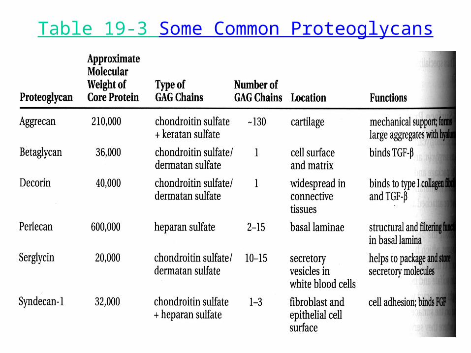
Table 19-3 Some Common Proteoglycans

COLLAGENS
A helix comprised of homotrimer & heterotrimer polypeptides (alpha chains)
Major proteins of ECMs Many different alpha chains Multiple structures (involves
cross-linking of chains)– fibrils– network forming– fibril-associated
Fig. 19-40

Table 19-4 Some types of collagen & their properties

Formation of CollagenFIBRILS and FIBERS
Fig. 19-43

Formation of Collagen Networks
Fig. 19-52

COLLAGEN ASSEMBLIES
Ann Med 33:7, 2001

ELASTIC FIBERS Elastin, main constituent Fibers crosslinked to form a network Fibers & network can extend and recoil
Fig. 19-50

MICROFIBRILS
Cover elastin core of elastic fibers Also found in other extracellular matrices Contain glycoproteins including fibrillin

FIBRONECTIN (FN) Extracellular dimeric
glycoprotein Differential splicing Multiple functional
domains– cell binding
• RGD sequence of FN• other specificities
– heparin binding – collagen binding – fibrin binding
Organized into a matrix
Fig. 19-51 A,C

LAMININ Heterotrimeric glycoprotein Basal lamina constituent Multiple binding domains
Fig. 19-55

Binding Domains of Laminin
Self assembly Type IV collagen Heparan sulfate Enactin/nidogen Cell Surface
– integrin– nonintegrin
J. Anat. 193:1, ‘98
Cell Suface Binding Sites

BASAL LAMINA
Fig. 19-56

HEMIDESMOSOMES
Junctions linking the intermediate filaments to the ECM
Constituents include – integrins– other plasma membrane
proteins– cytosolic proteins
Functions include– adhesion– intracellular signaling
Bioessays 20:488, ‘98

Focal Adhesion (Focal Contact or Adhesion Plaque)
Junctions linking actin filaments to the ECM
Constituents include – integrins– cytosolic proteins
Functions include– adhesion– cell signaling
Fig. 16-75B
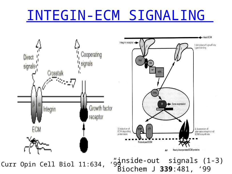
INTEGIN-ECM SIGNALING
“inside-out” signals (1-3) Biochem J 339:481, ‘99
Curr Opin Cell Biol 11:634, ‘99

Diversity in Adhesive Interactions
Diversity in the major components due to:– differential splicing– post-translational modifications– different gene products
Participation of additional molecules

Matrix Metalloproteases Also termed matrix metalloproteinases,
matrixins, MMPs Enzymes which cleave ECM
constituents, including collagenases Contain a propeptide which must be
cleaved for enzyme activity Multiple mechanisms to regulate MMP
activity, including inhibition by tissue inhibitors of metalloproteases (TIMPs)
Other functional domains

Genes Dev 14:2123,’00
Matrix Metalloproteases

Genes Dev 14:2123,’00

ADAM A Disintegrin And Metalloprotease
Trends Genet. 16:83, ‘00
A disintegrin is a molecule that binds to an integrin.
T = transmembrane domain

Trends Genet. 16:83, ‘00