Fibrous and Fibrohistiocytic Proliferations of the Skin P1
161
FIBROUS AND FIBROHISTIOCYTIC PROLIFERATIONS OF THE SKIN P.I WWW.FACEBOOK.COM/GROUPS/DERMATOLOGYCOURSEONLINE
-
Upload
ibrahim-mohammed -
Category
Health & Medicine
-
view
522 -
download
1
Transcript of Fibrous and Fibrohistiocytic Proliferations of the Skin P1
FIBROUS AND FIBROHISTIOCYTIC PROLIFERATIONS OF THE SKIN P.IWWW.FACEBOOK.COM/GROUPS/DERMATOLOGYCOURSEONLINE

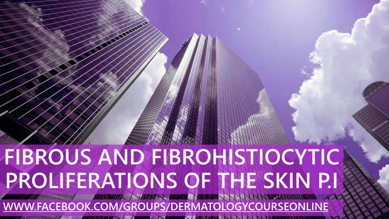
CLASSIFICATION OF FIBROUS AND FIBROHISTIOCYTIC PROLIFERATIONS OF THE SKIN
1. Skin tags
2. Cutaneous angiofibroma
3. Dermatofibroma
4. Dermatomyofibroma
5. Superficial acral fibromyxoma
6. Sclerotic fibroma of the skin
7. Pleomorphic fibroma of the skin
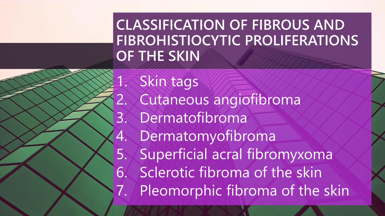
CLASSIFICATION OF FIBROUS AND FIBROHISTIOCYTIC PROLIFERATIONS OF THE SKIN
8. Nodular fasciitis
9. Multinucleate cell angiohistiocytoma
10. Epithelioid fibrous histiocytoma
11. Connective tissue nevus
12. Infantile digital fibroma
13. Infantile myofibromatosis
14. Calcifying aponeurotic fibroma
15. Keloids and hypertrophic scars
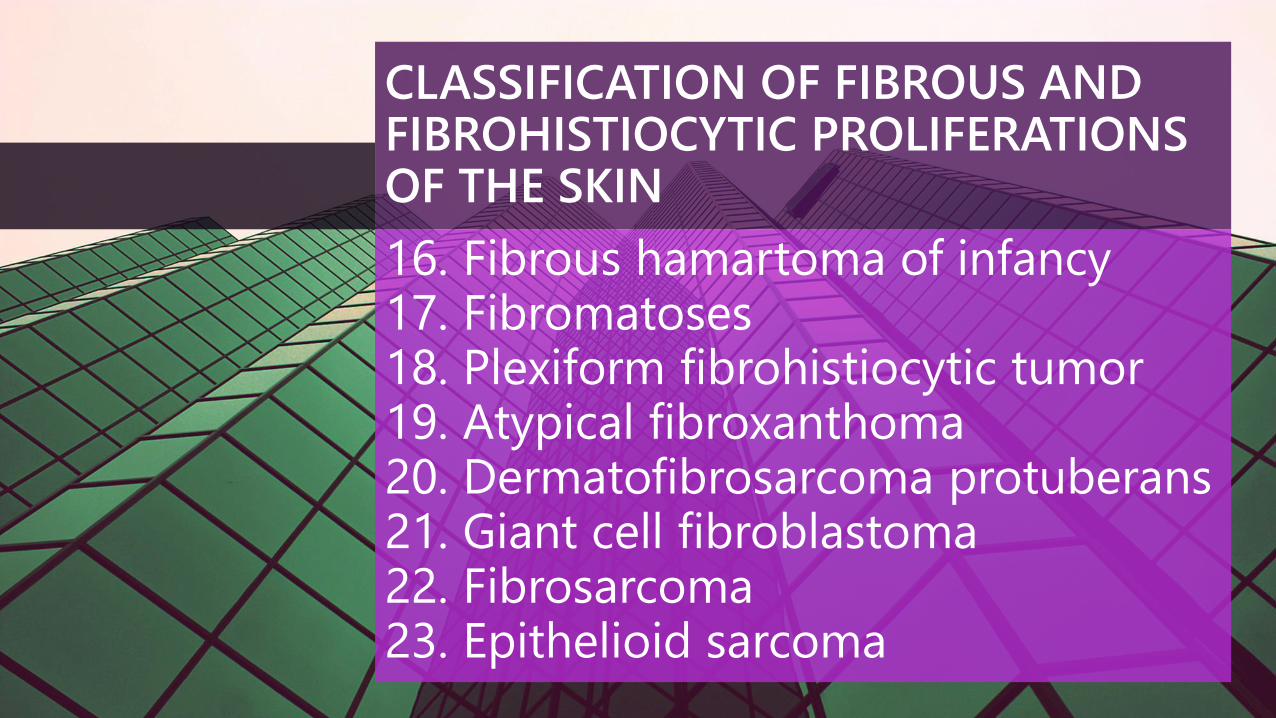
CLASSIFICATION OF FIBROUS AND FIBROHISTIOCYTIC PROLIFERATIONS OF THE SKIN
16. Fibrous hamartoma of infancy17. Fibromatoses18. Plexiform fibrohistiocytic tumor 19. Atypical fibroxanthoma20. Dermatofibrosarcoma protuberans21. Giant cell fibroblastoma22. Fibrosarcoma23. Epithelioid sarcoma

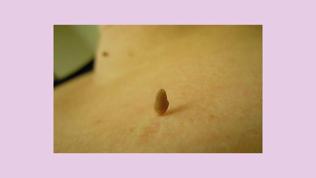
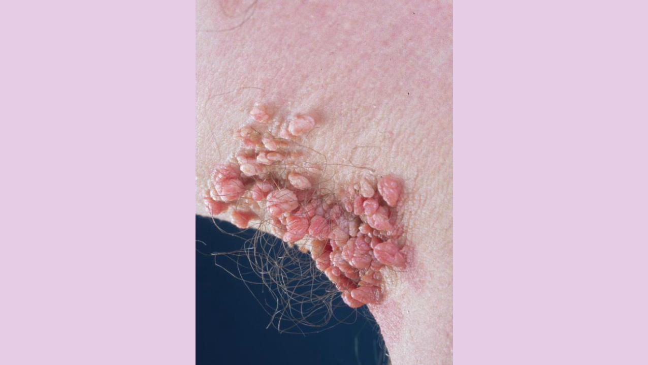
SKIN TAGS
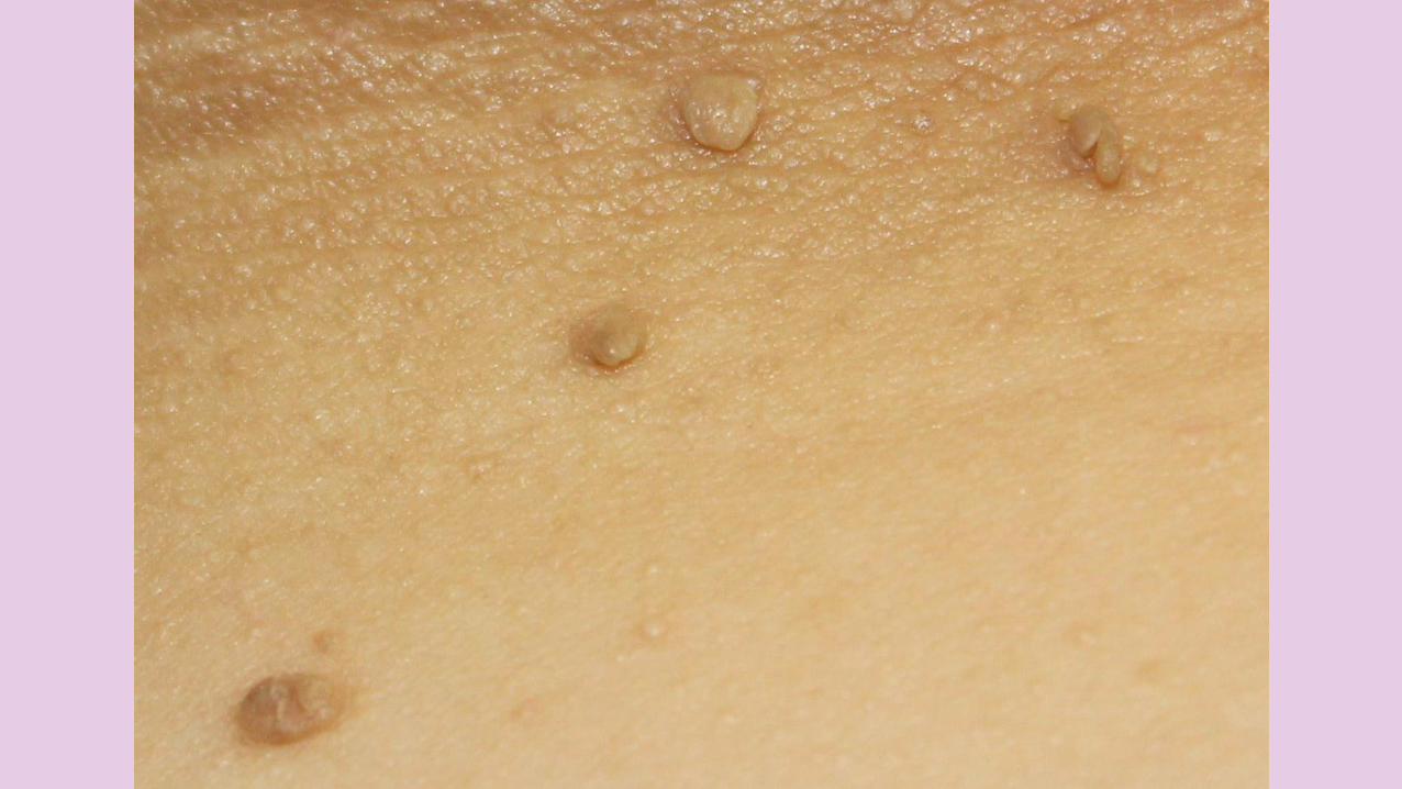
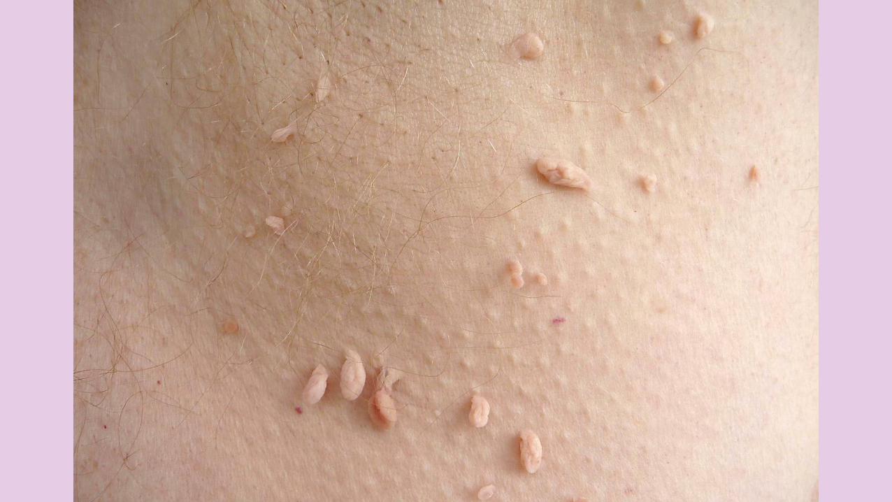
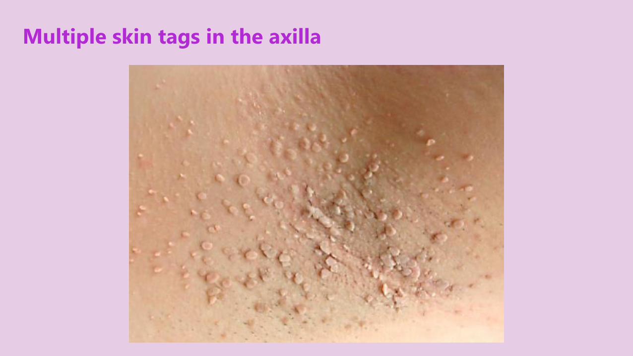
Multiple skin tags in the axilla
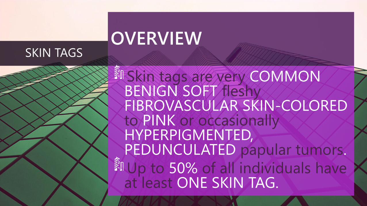
OVERVIEW
Skin tags are very COMMONBENIGN SOFT fleshyFIBROVASCULAR SKIN-COLOREDto PINK or occasionallyHYPERPIGMENTED, PEDUNCULATED papular tumors.
Up to 50% of all individuals haveat least ONE SKIN TAG.
SKIN TAGS
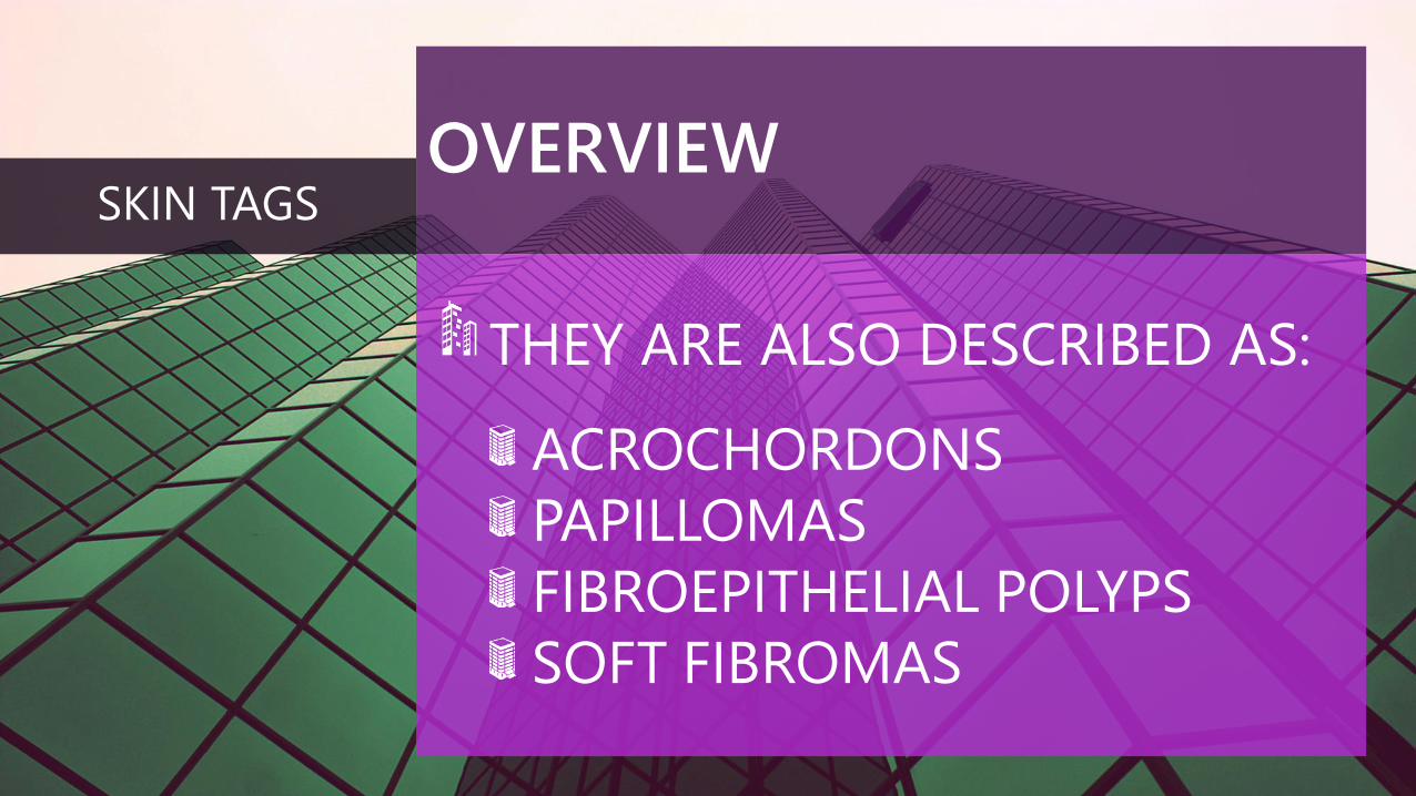
OVERVIEW
THEY ARE ALSO DESCRIBED AS:
ACROCHORDONS
PAPILLOMAS
FIBROEPITHELIAL POLYPS
SOFT FIBROMAS
SKIN TAGS
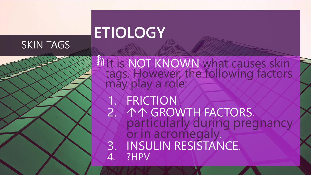
ETIOLOGY
It is NOT KNOWN what causes skintags. However, the following factorsmay play a role:
1. FRICTION2. GROWTH FACTORS,
particularly during pregnancyor in acromegaly.
3. INSULIN RESISTANCE.4. ?HPV
SKIN TAGS
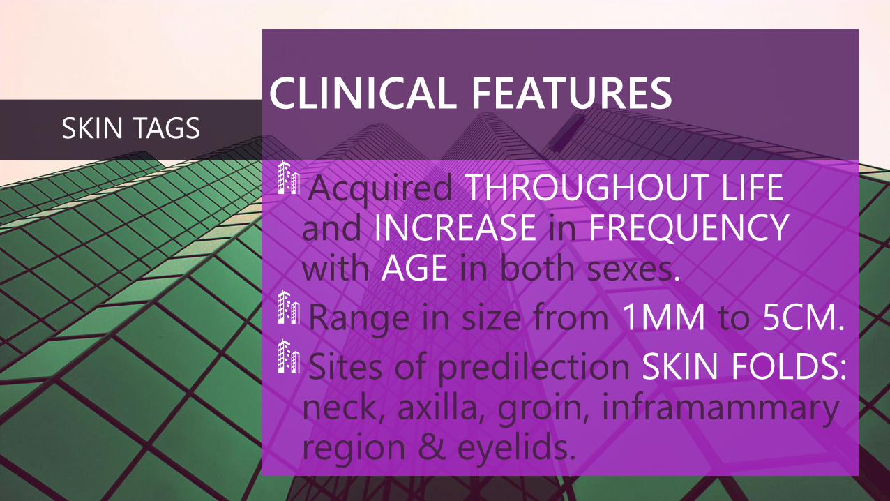
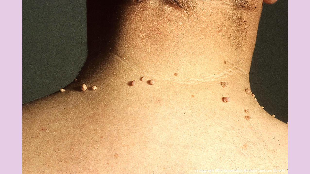
CLINICAL FEATURES
Acquired THROUGHOUT LIFEand INCREASE in FREQUENCYwith AGE in both sexes.
Range in size from 1MM to 5CM.
Sites of predilection SKIN FOLDS: neck, axilla, groin, inframammary region & eyelids.
SKIN TAGS
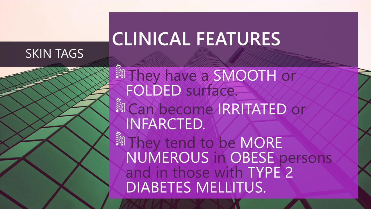
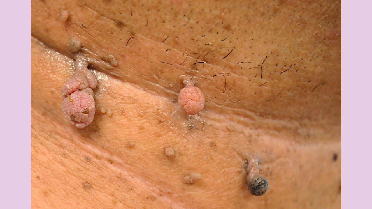
CLINICAL FEATURES
They have a SMOOTH orFOLDED surface.
Can become IRRITATED orINFARCTED.
They tend to be MORENUMEROUS in OBESE personsand in those with TYPE 2DIABETES MELLITUS.
SKIN TAGS
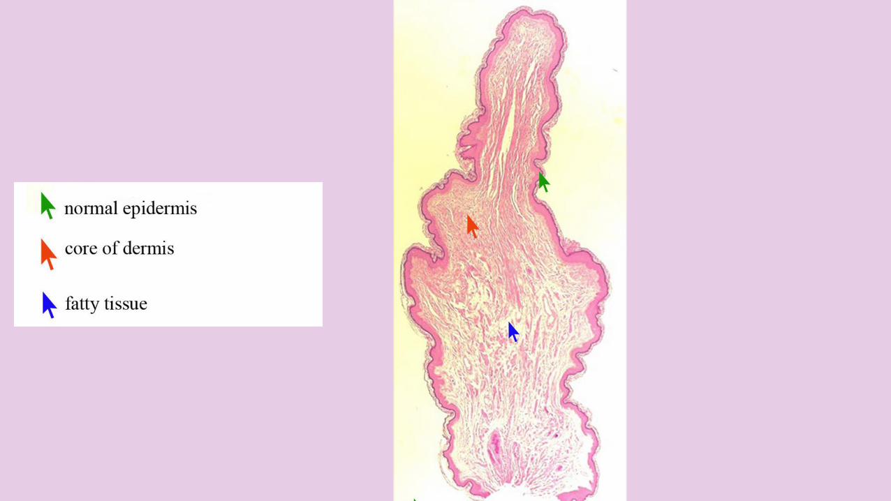
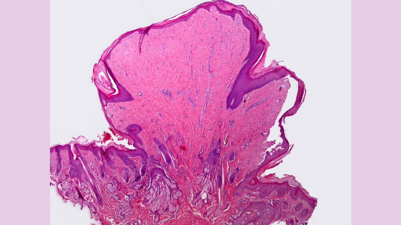
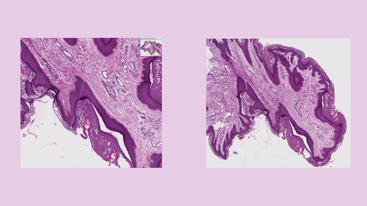
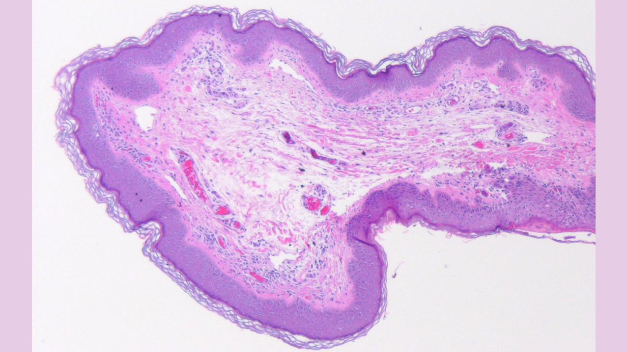
HISTOPATHOLOGY
Benign FIBROVASCULAR tumorsdue to NORMAL EPIDERMIS orEPIDERMAL HYPERPLASIAoverlying a dermal stalk.
SKIN TAGS
HISTOPATHOLOGY
A CORE of DERMIS made up ofloosely arranged COLLAGENFIBERS and BLOOD VESSELSand/or SUBCUTANEOUS FATTYTISSUE.
SKIN TAGS
TREATMENT
Skin tags are benign & can be removed only for COSMETICREASONS.
SKIN TAGS
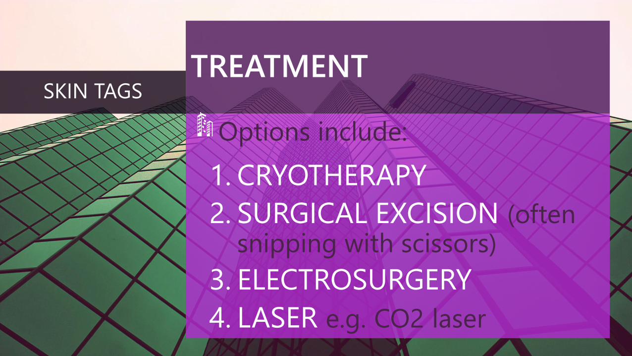
TREATMENT
Options include:
1. CRYOTHERAPY
2. SURGICAL EXCISION (oftensnipping with scissors)
3. ELECTROSURGERY
4. LASER e.g. CO2 laser
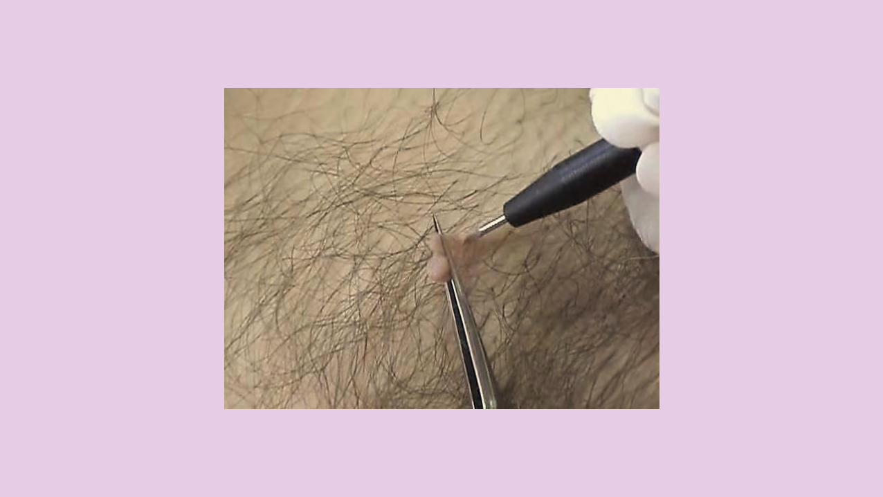
SKIN TAGS
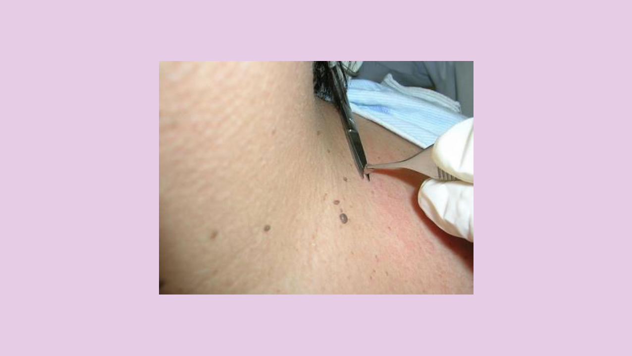
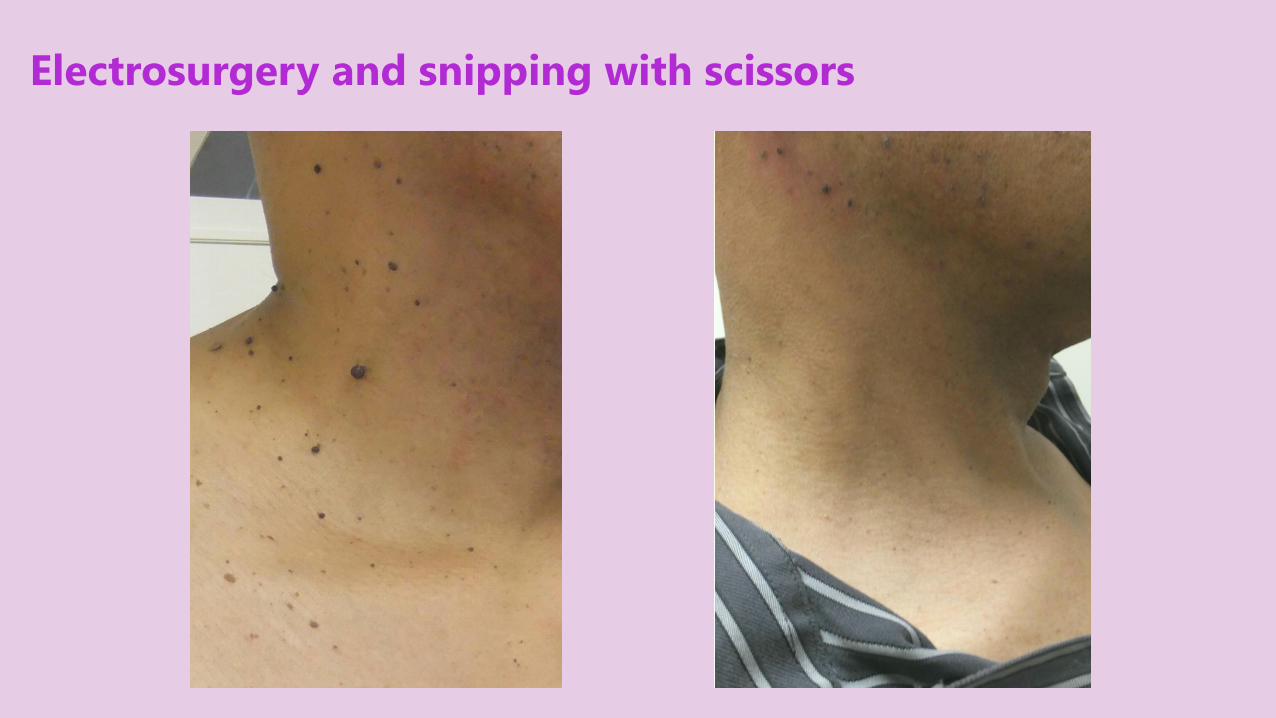
Electrosurgery and snipping with scissors
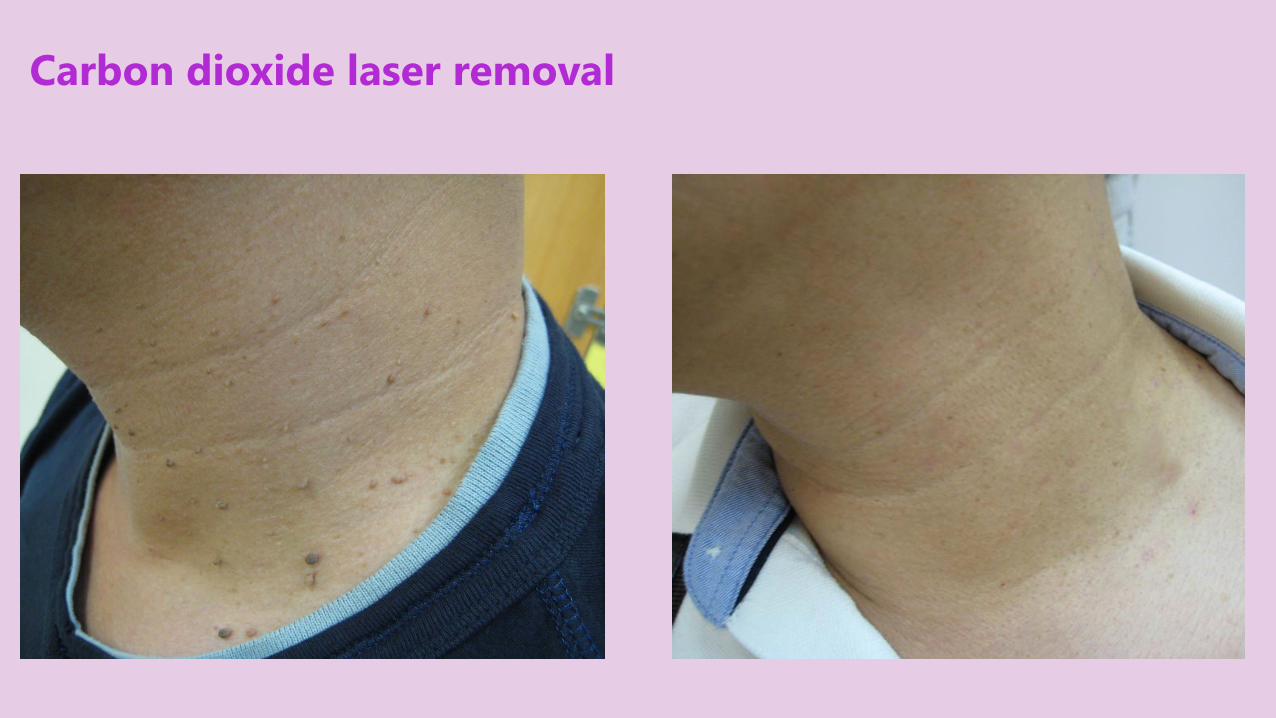
Carbon dioxide laser removal


ANGIOFIBROMAS
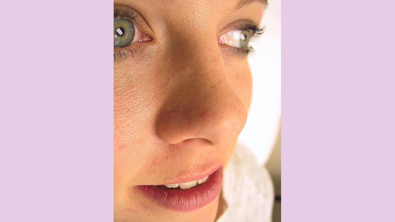
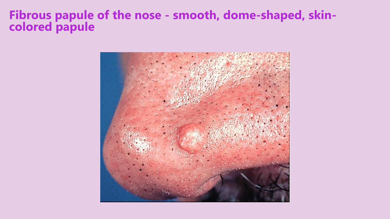
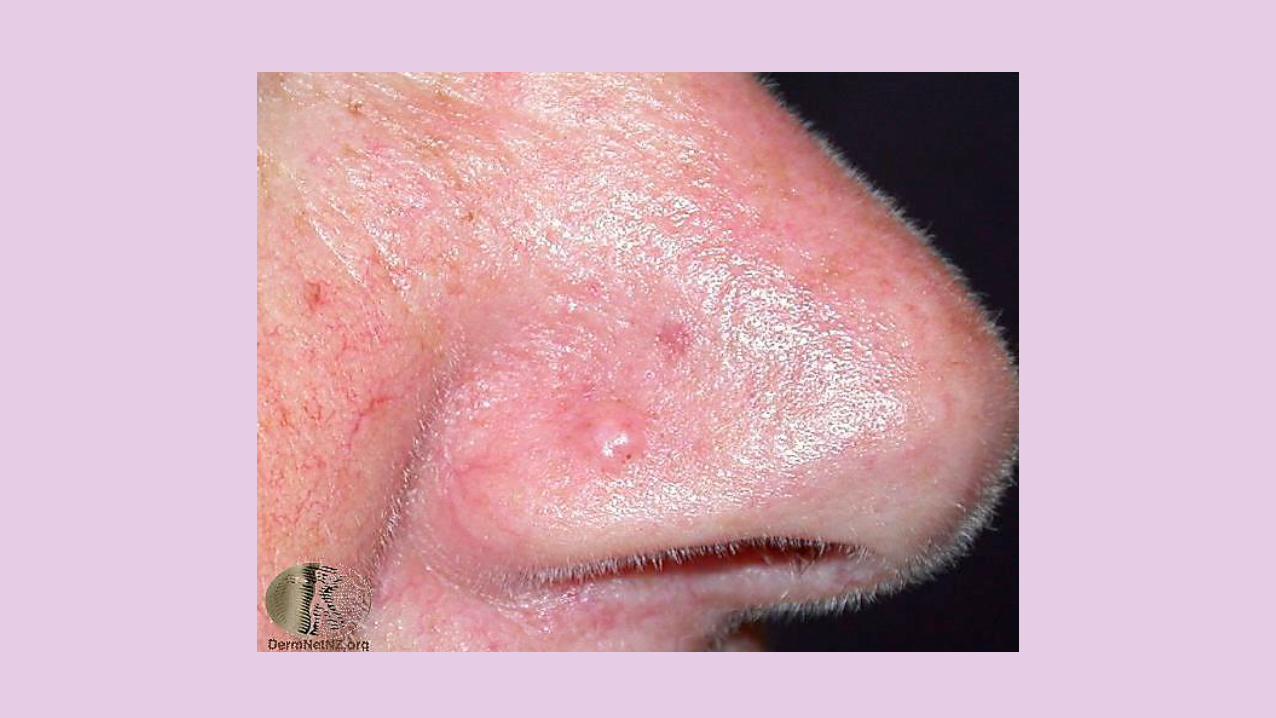
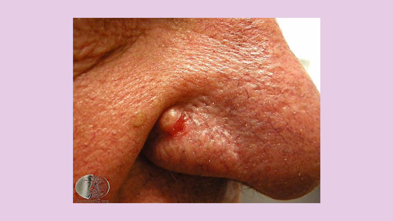
Fibrous papule of the nose - smooth, dome-shaped, skin-colored papule
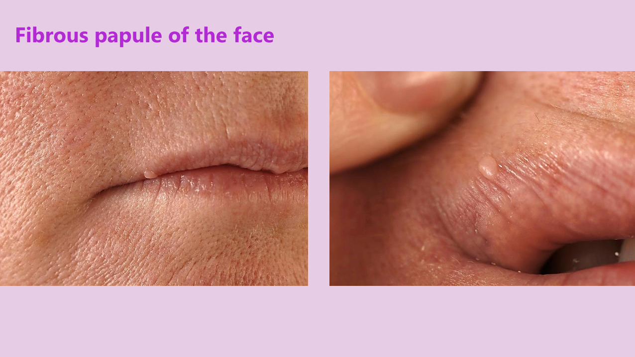
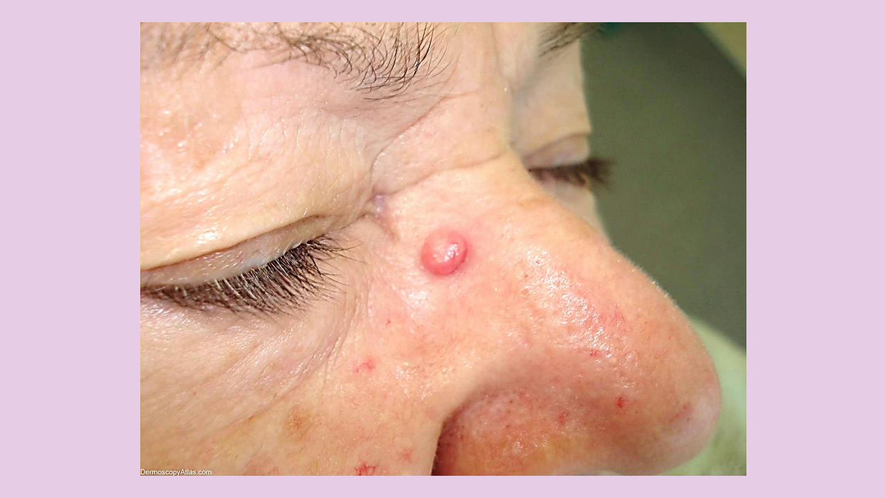
Fibrous papule of the face
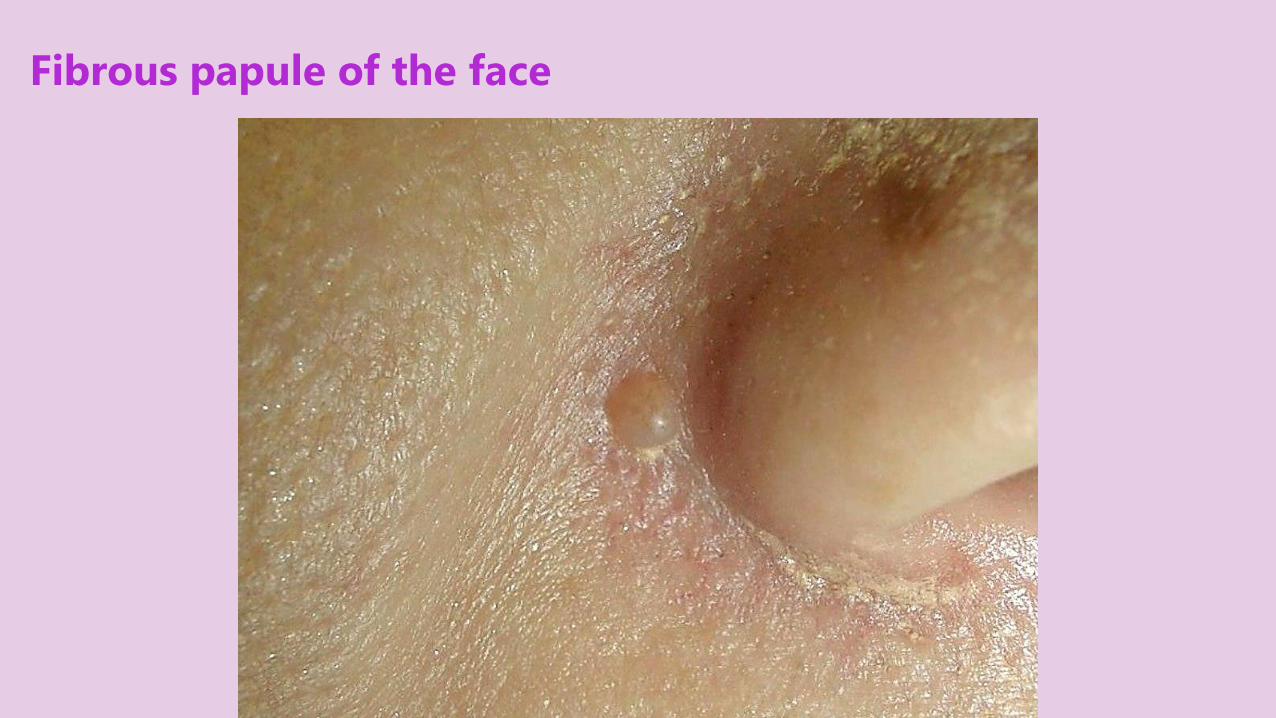
Fibrous papule of the face
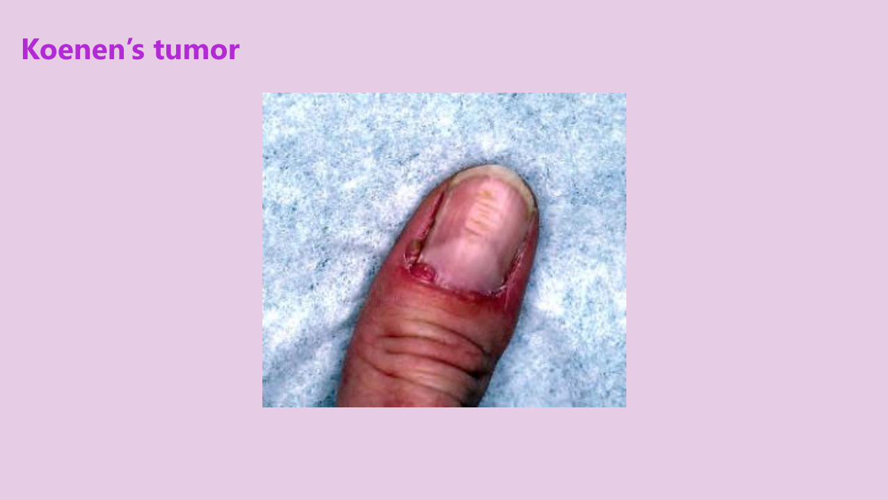
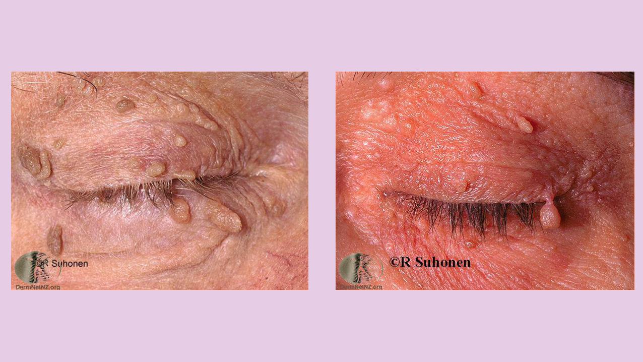
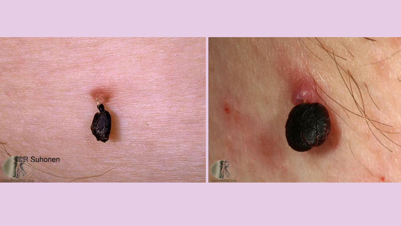
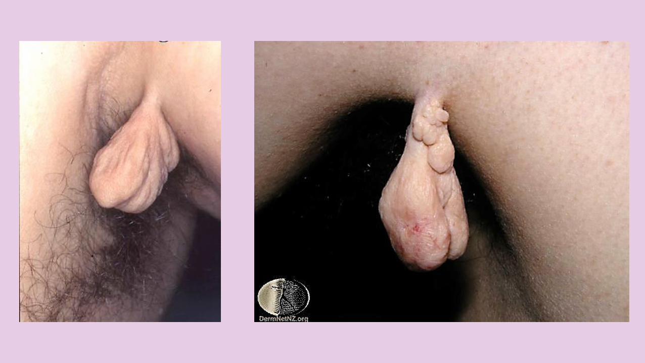
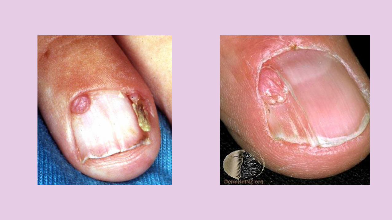
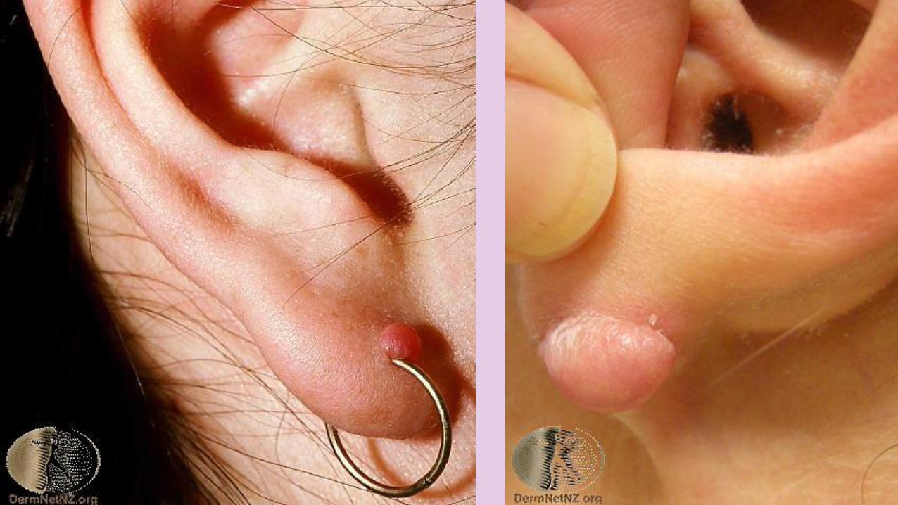
Koenen’s tumor

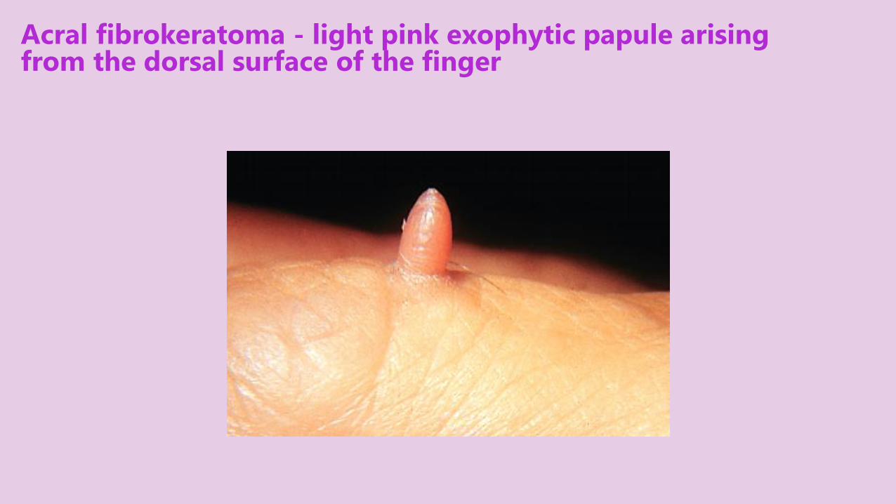
Acral fibrokeratoma - light pink exophytic papule arising from the dorsal surface of the finger
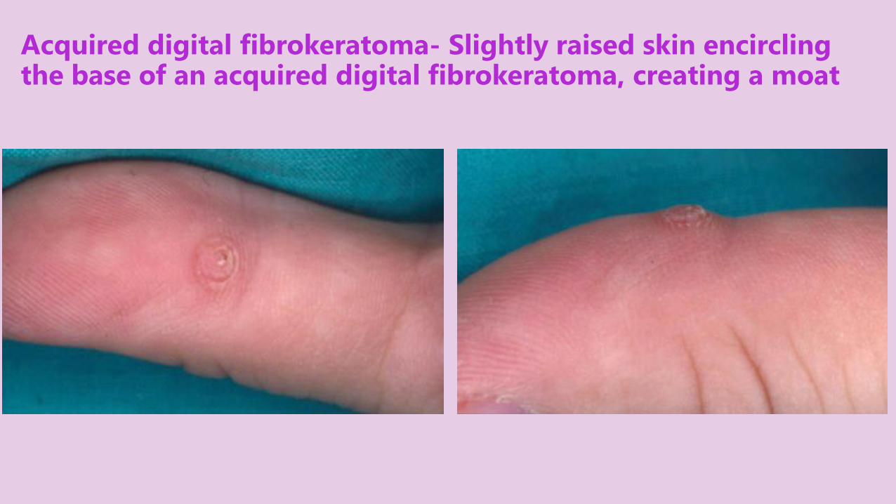
Acquired digital fibrokeratoma- Slightly raised skin encircling the base of an acquired digital fibrokeratoma, creating a moat
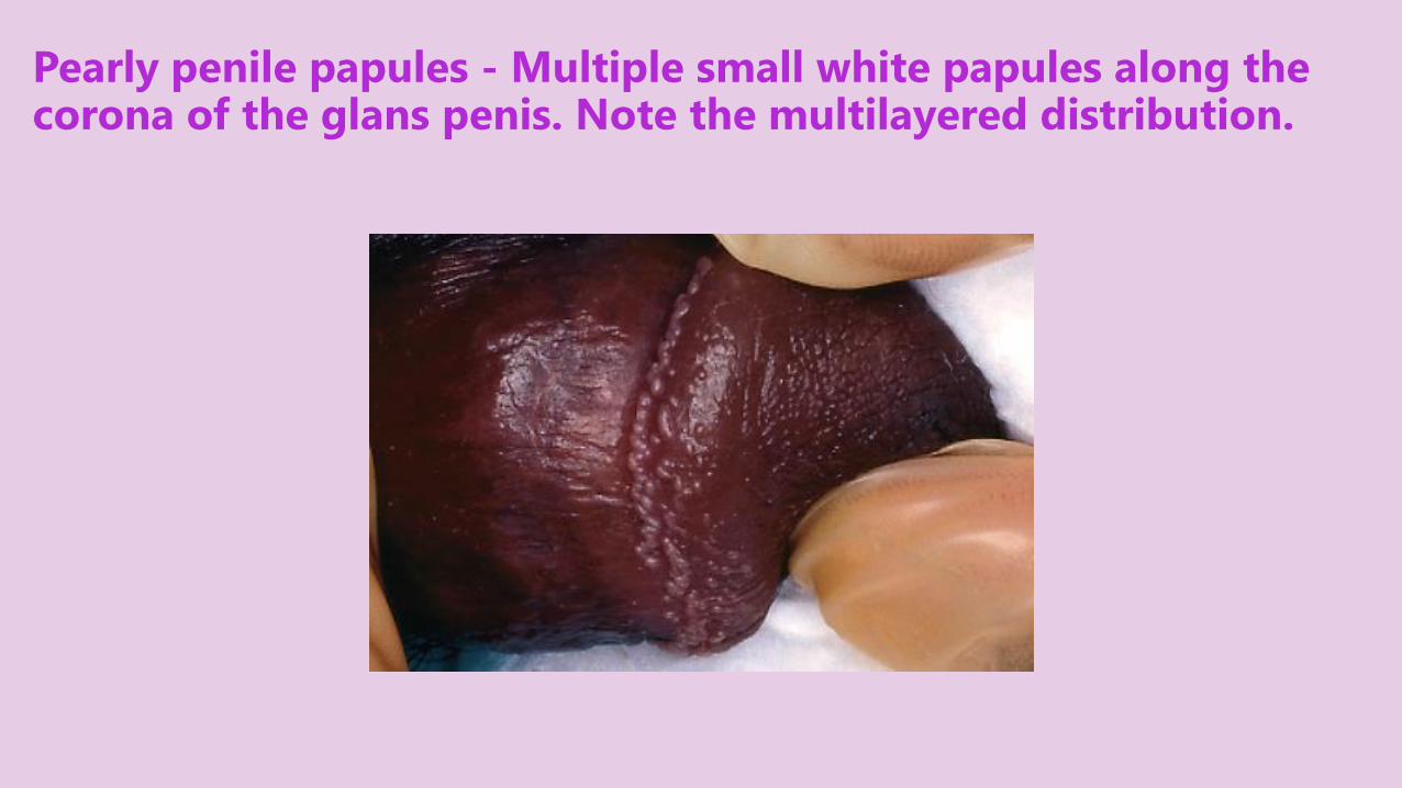
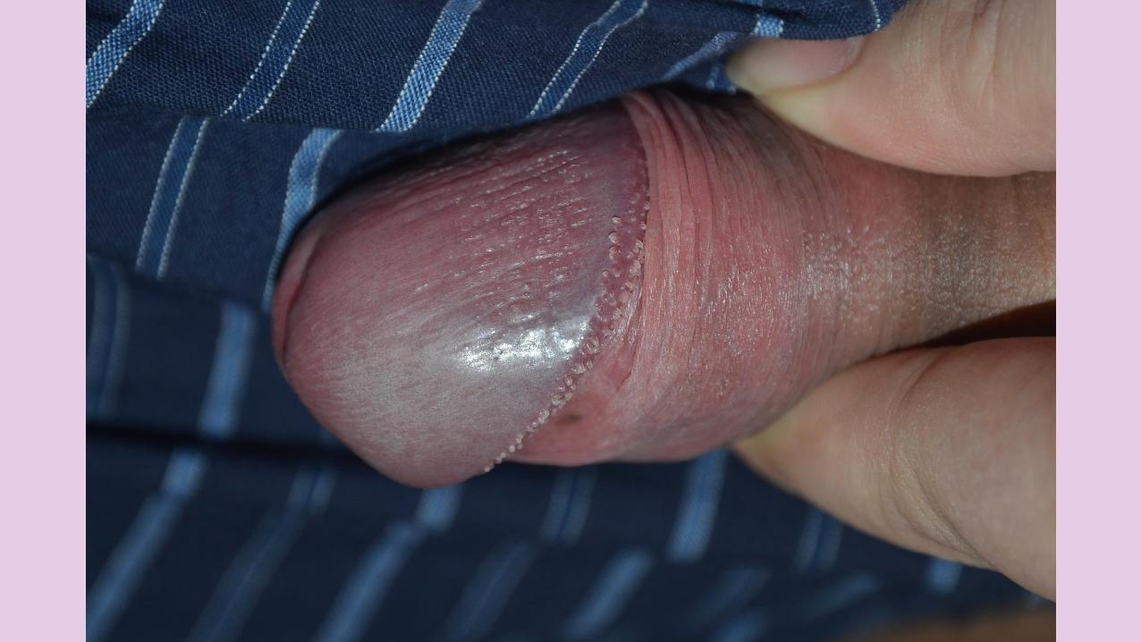
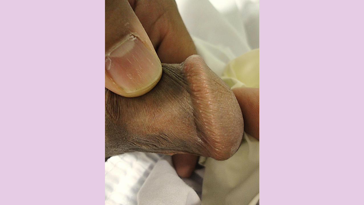
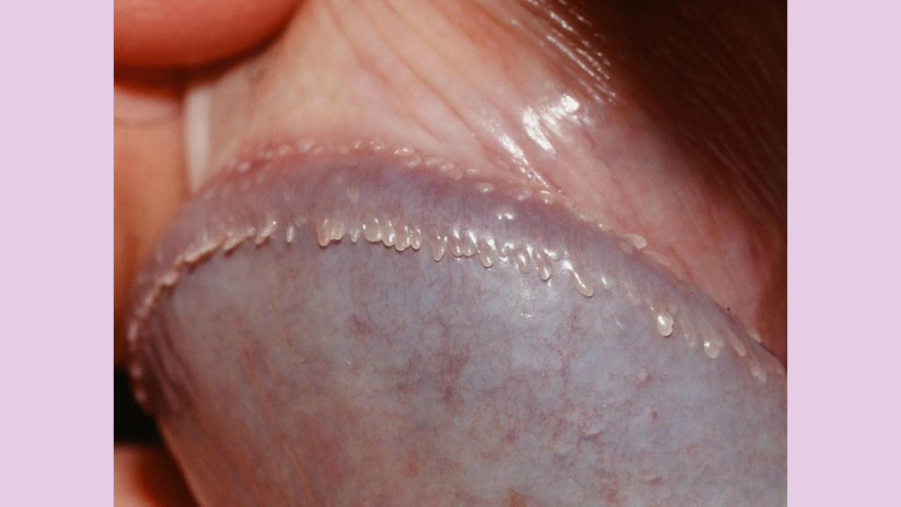
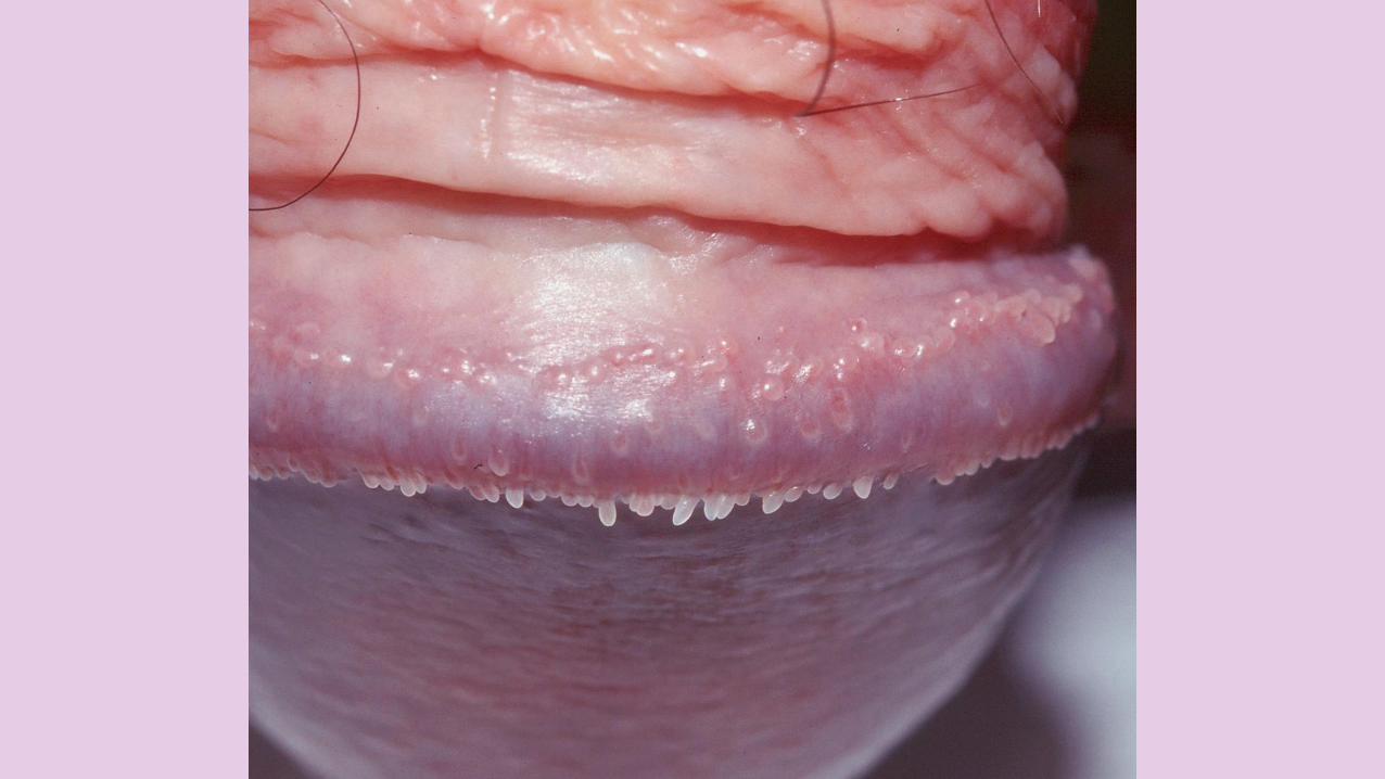
Pearly penile papules - Multiple small white papules along the corona of the glans penis. Note the multilayered distribution.
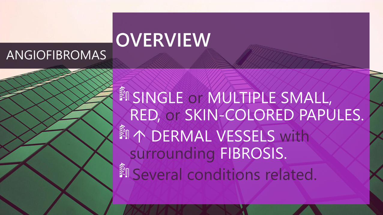
OVERVIEW
SINGLE or MULTIPLE SMALL, RED, or SKIN-COLORED PAPULES.
DERMAL VESSELS withsurrounding FIBROSIS.
Several conditions related.
ANGIOFIBROMAS
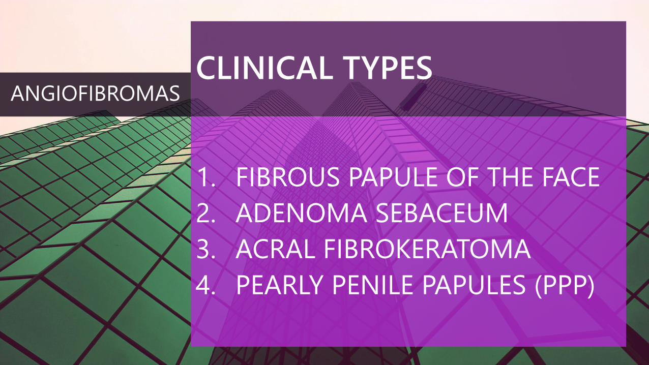
CLINICAL TYPES
1. FIBROUS PAPULE OF THE FACE
2. ADENOMA SEBACEUM
3. ACRAL FIBROKERATOMA
4. PEARLY PENILE PAPULES (PPP)
ANGIOFIBROMAS
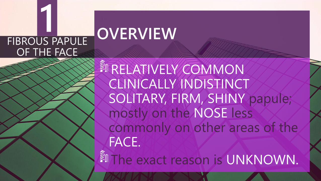
OVERVIEW
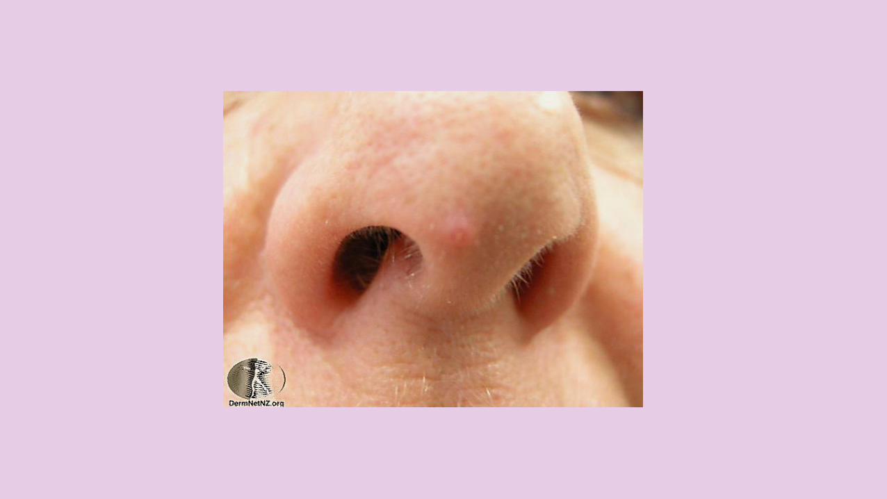
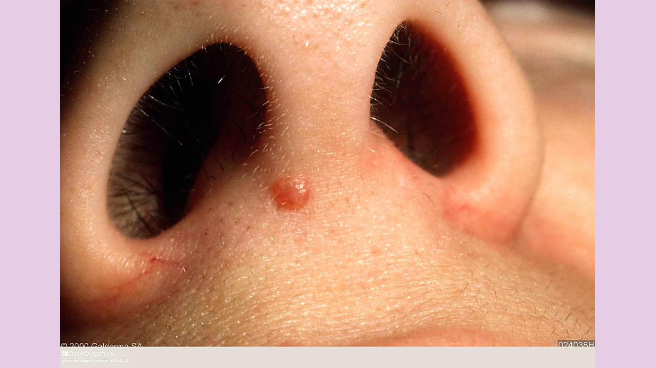
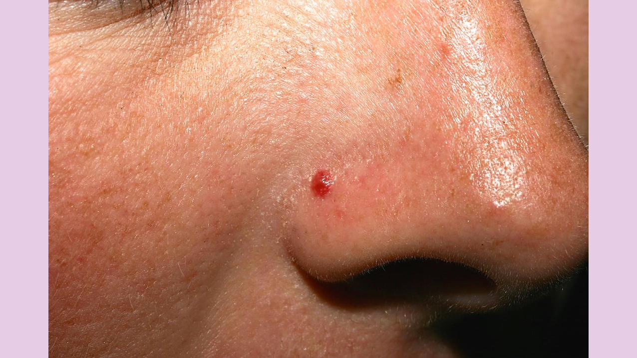
RELATIVELY COMMONCLINICALLY INDISTINCTSOLITARY, FIRM, SHINY papule;mostly on the NOSE less commonly on other areas of the FACE.
The exact reason is UNKNOWN.
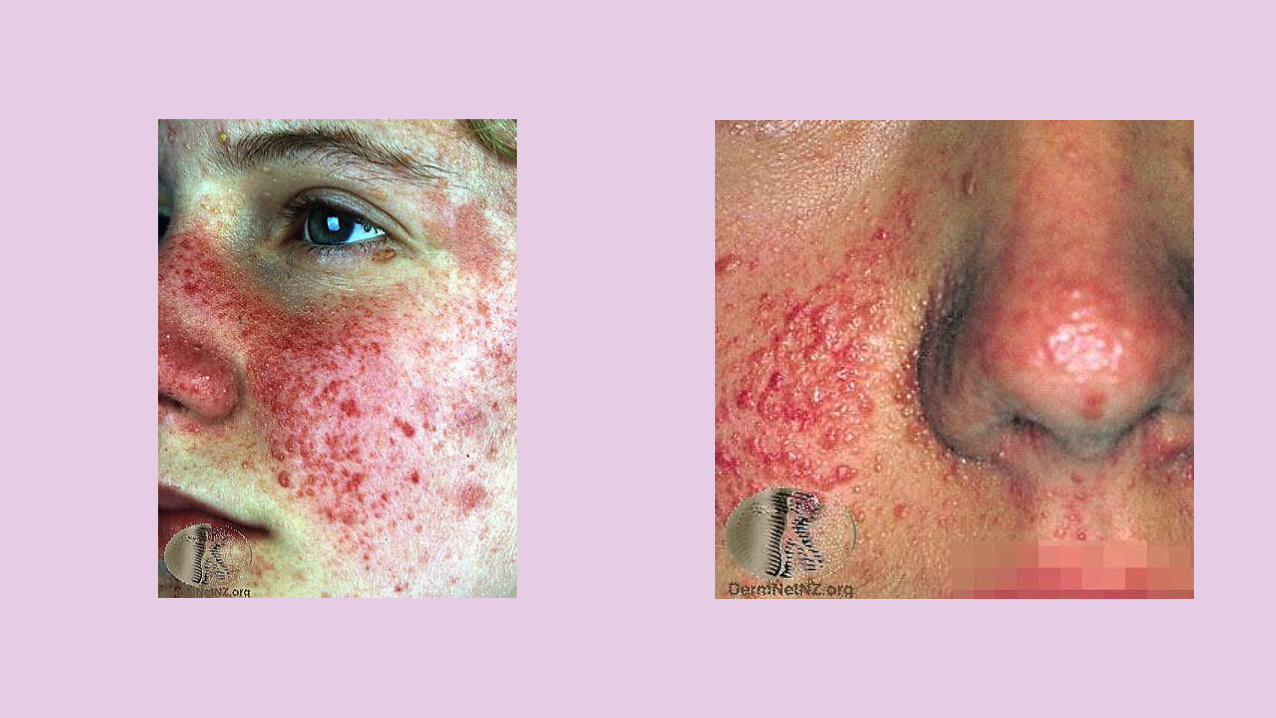
FIBROUS PAPULE OF THE FACE
1
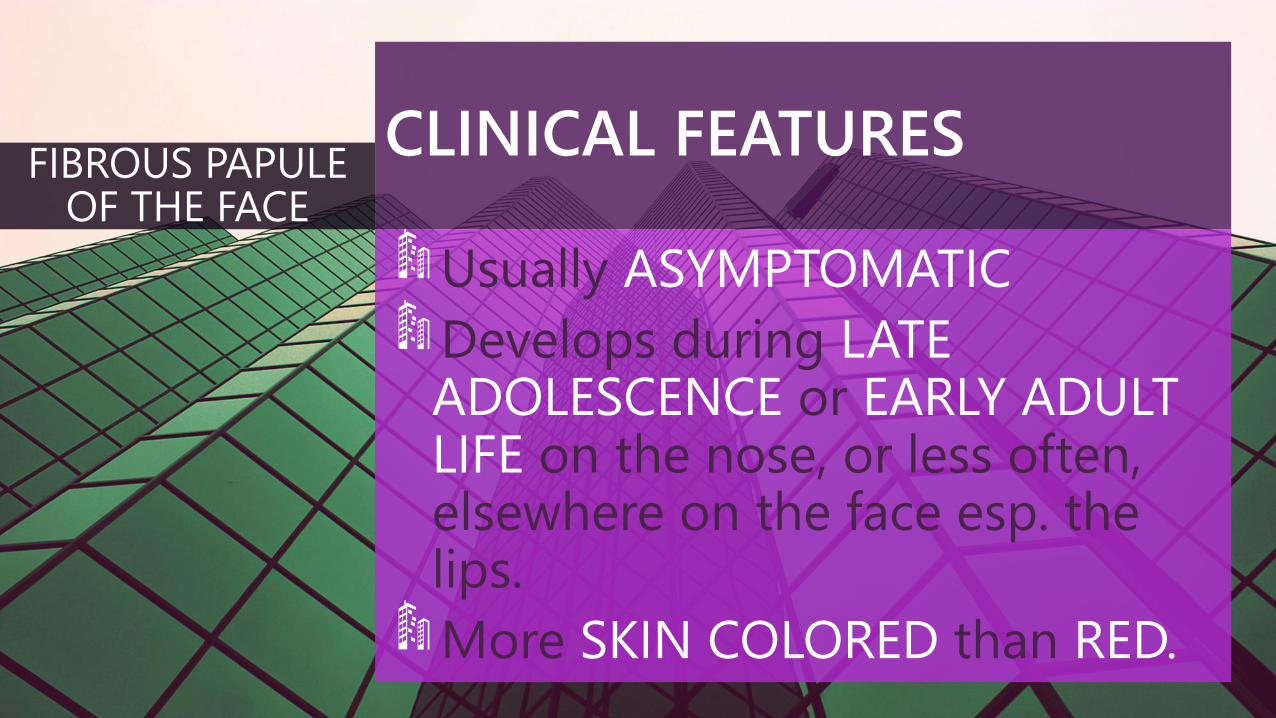
CLINICAL FEATURES
Usually ASYMPTOMATIC
Develops during LATEADOLESCENCE or EARLY ADULTLIFE on the nose, or less often, elsewhere on the face esp. the lips.
More SKIN COLORED than RED.
FIBROUS PAPULE OF THE FACE
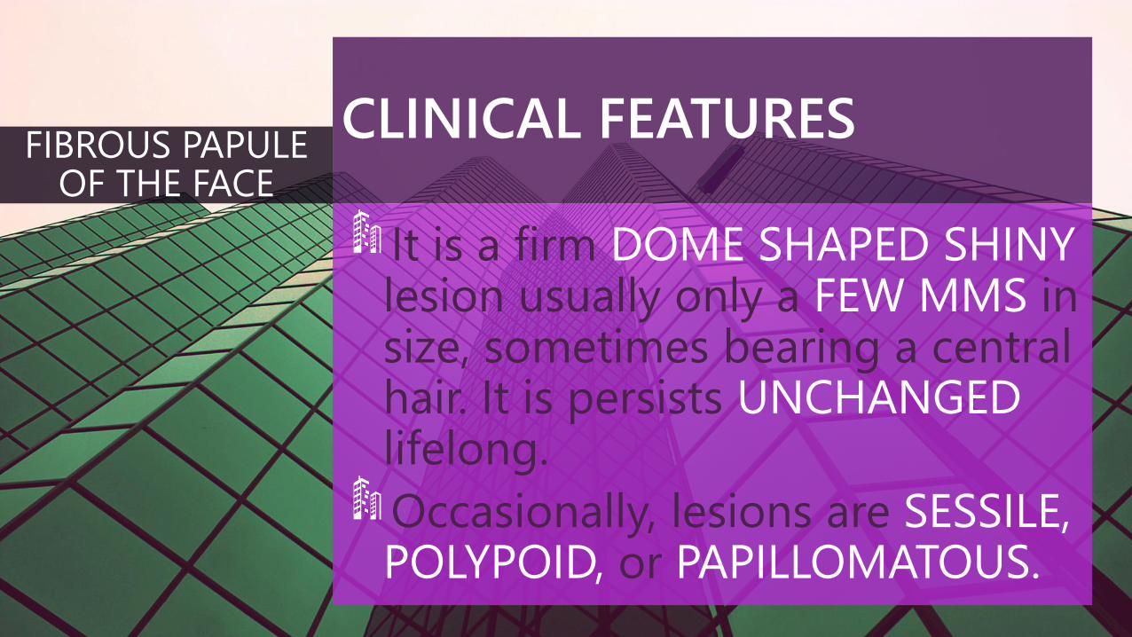
CLINICAL FEATURES
It is a firm DOME SHAPED SHINYlesion usually only a FEW MMS insize, sometimes bearing a centralhair. It is persists UNCHANGEDlifelong.
Occasionally, lesions are SESSILE, POLYPOID, or PAPILLOMATOUS.
FIBROUS PAPULE OF THE FACE
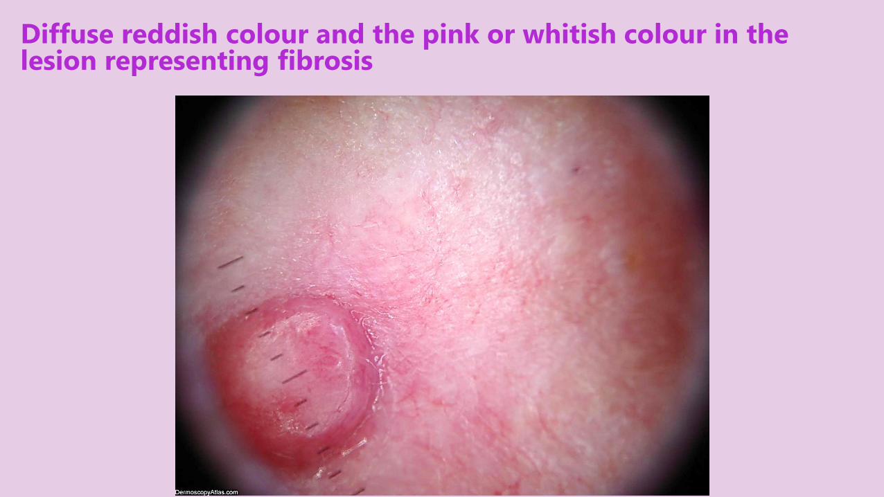
Diffuse reddish colour and the pink or whitish colour in the lesion representing fibrosis
DDx
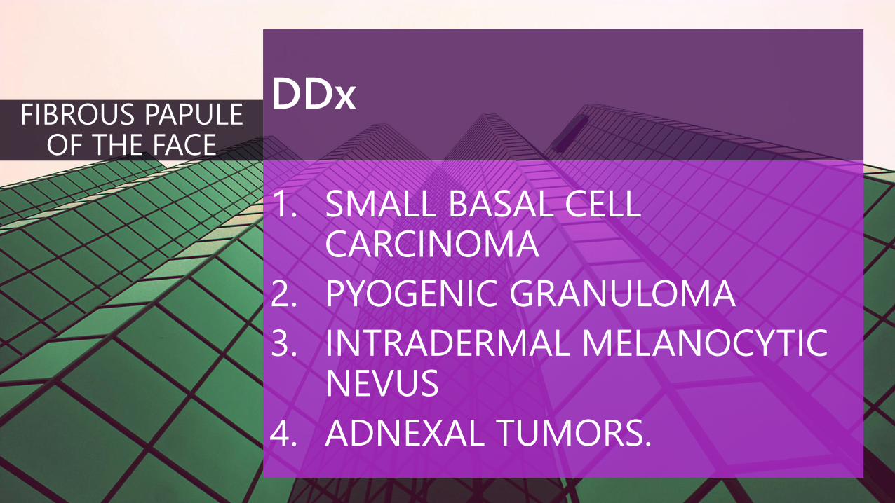
1. SMALL BASAL CELLCARCINOMA
2. PYOGENIC GRANULOMA
3. INTRADERMAL MELANOCYTICNEVUS
4. ADNEXAL TUMORS.
FIBROUS PAPULE OF THE FACE
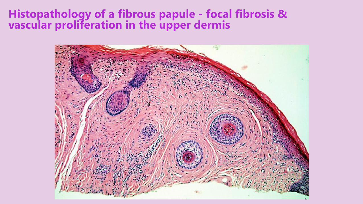
Histopathology of a fibrous papule - focal fibrosis & vascular proliferation in the upper dermis
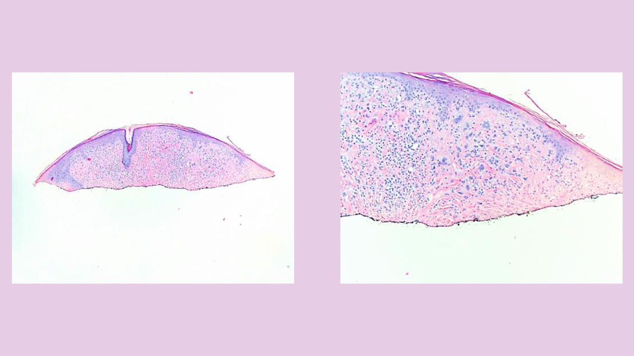
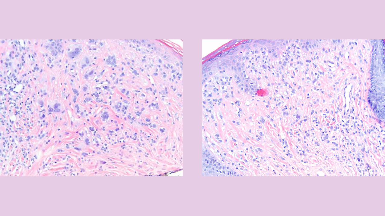
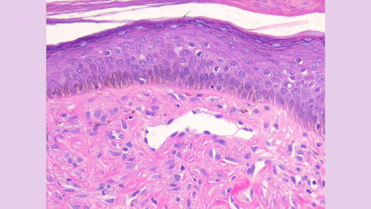
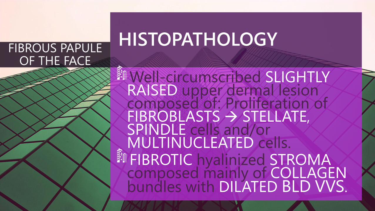
HISTOPATHOLOGY
Well-circumscribed SLIGHTLYRAISED upper dermal lesioncomposed of: Proliferation of FIBROBLASTS STELLATE, SPINDLE cells and/orMULTINUCLEATED cells.FIBROTIC hyalinized STROMAcomposed mainly of COLLAGENbundles with DILATED BLD VVS.
FIBROUS PAPULE OF THE FACE
HISTOPATHOLOGY
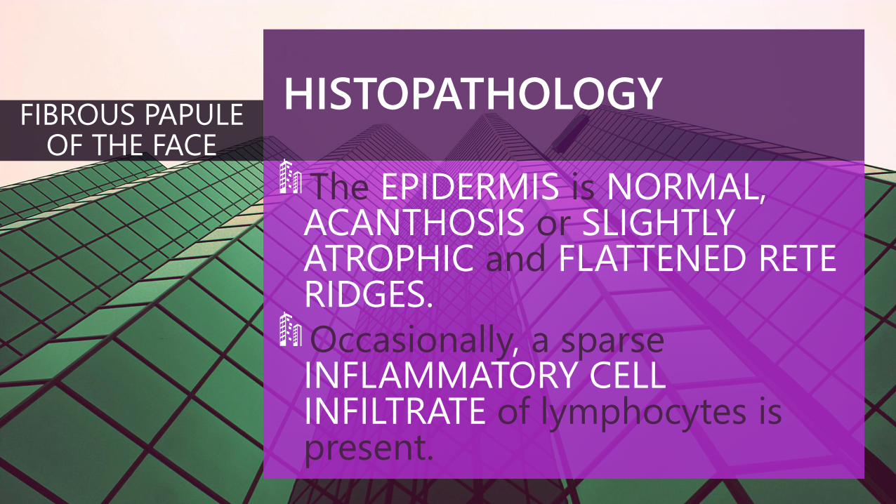
The EPIDERMIS is NORMAL, ACANTHOSIS or SLIGHTLYATROPHIC and FLATTENED RETERIDGES.
Occasionally, a sparseINFLAMMATORY CELLINFILTRATE of lymphocytes is present.
FIBROUS PAPULE OF THE FACE
IMMUNOHISTOCHEMISTRY
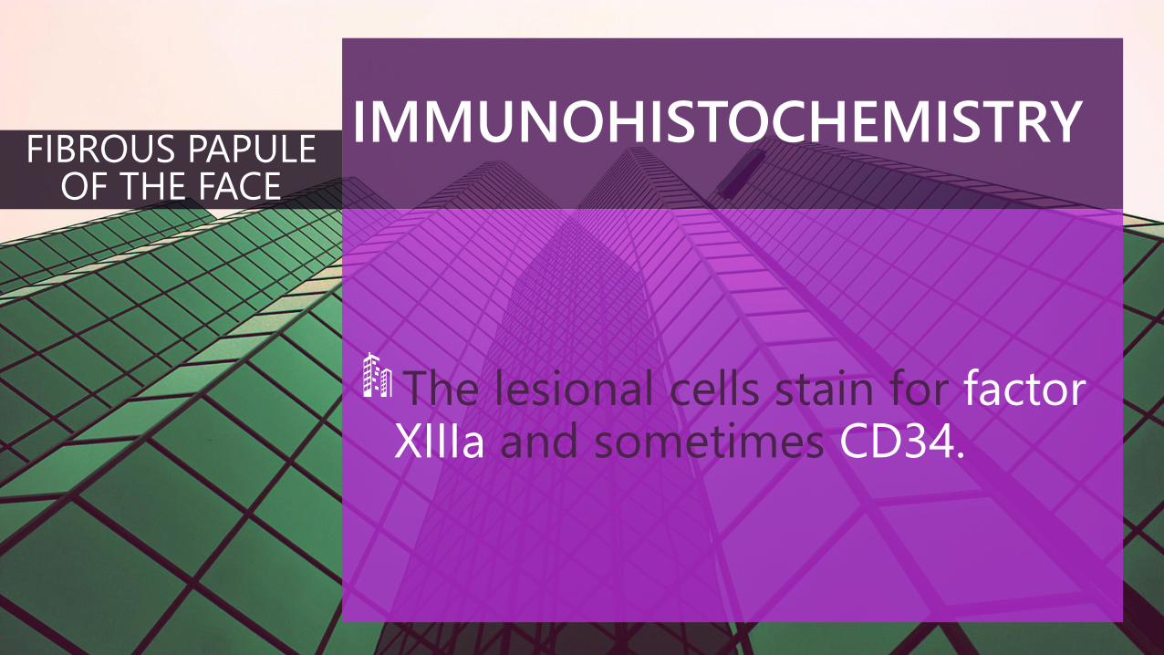
The lesional cells stain for factor XIIIa and sometimes CD34.
FIBROUS PAPULE OF THE FACE
TREATMENT
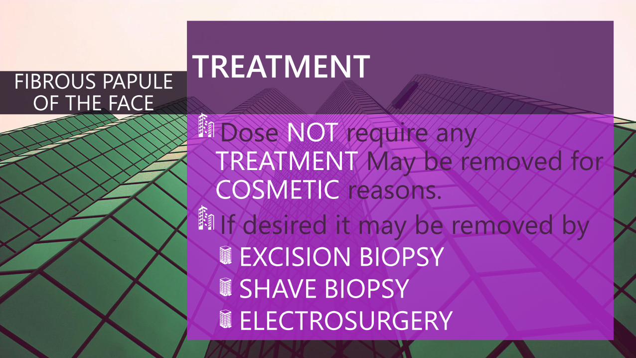
Dose NOT require anyTREATMENT May be removed forCOSMETIC reasons.
If desired it may be removed by
EXCISION BIOPSY
SHAVE BIOPSY
ELECTROSURGERY
FIBROUS PAPULE OF THE FACE
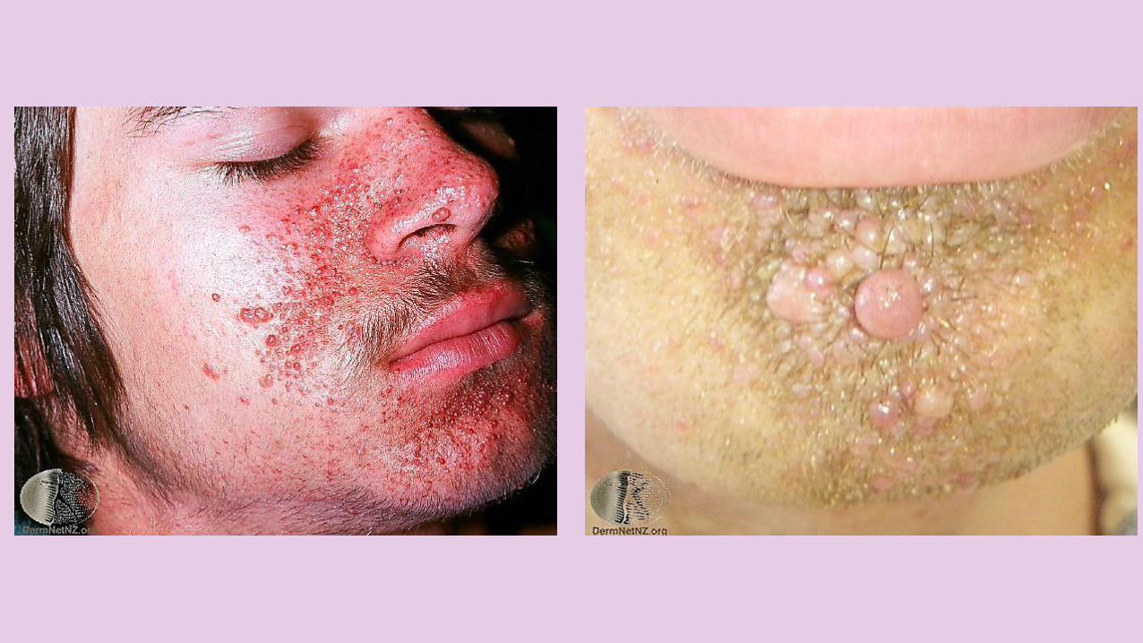
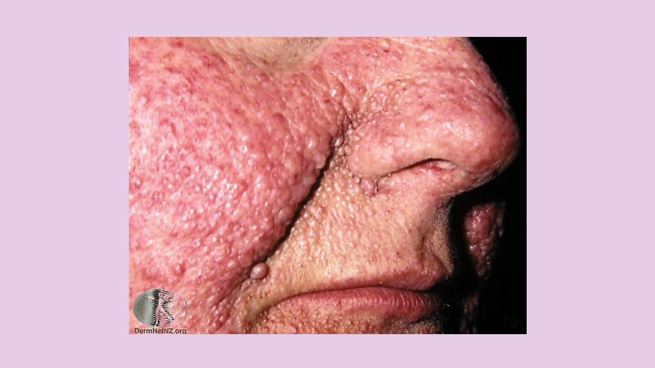
OVERVIEW
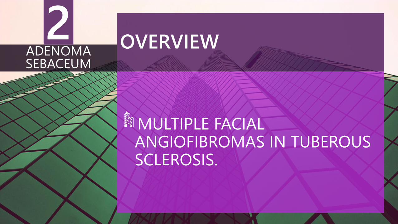
MULTIPLE FACIALANGIOFIBROMAS IN TUBEROUS SCLEROSIS.
ADENOMA SEBACEUM
2
OVERVIEW
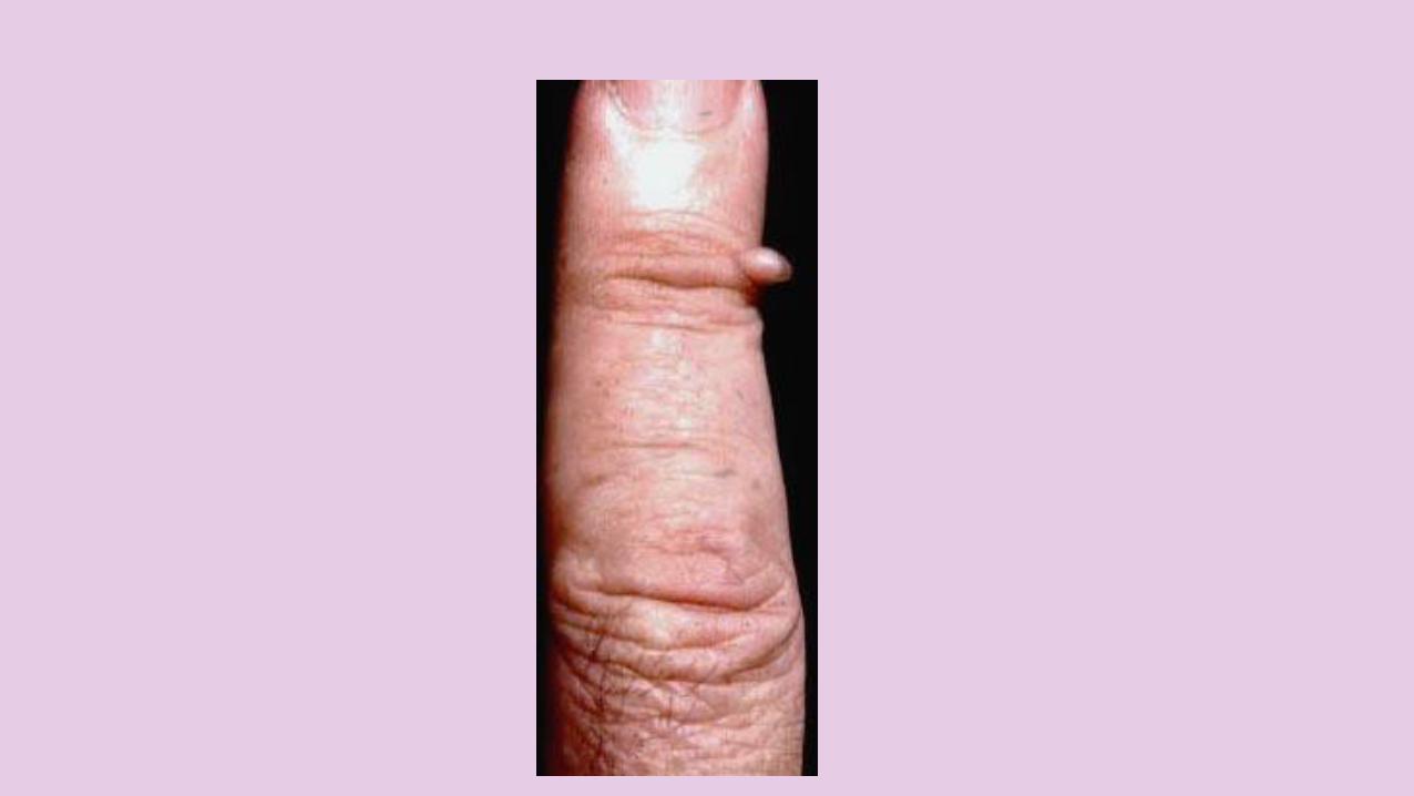
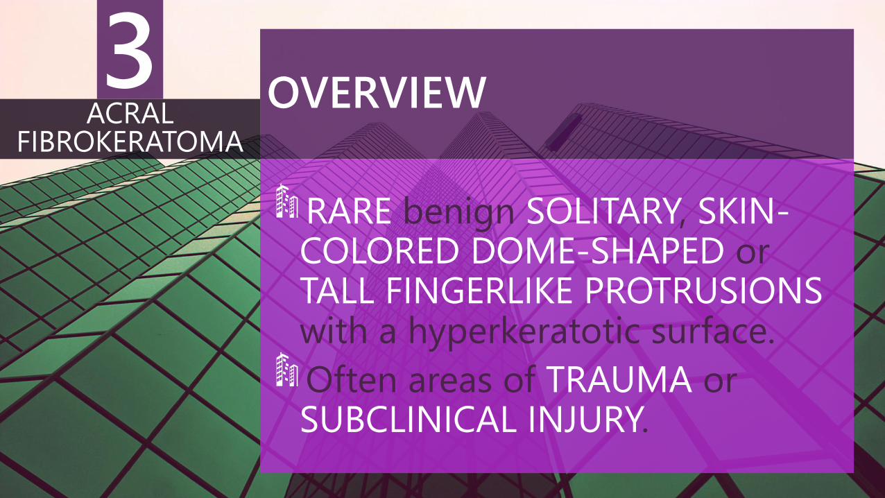
RARE benign SOLITARY, SKIN-COLORED DOME-SHAPED or TALL FINGERLIKE PROTRUSIONSwith a hyperkeratotic surface.
Often areas of TRAUMA or SUBCLINICAL INJURY.
ACRAL FIBROKERATOMA
3
VARIANTS
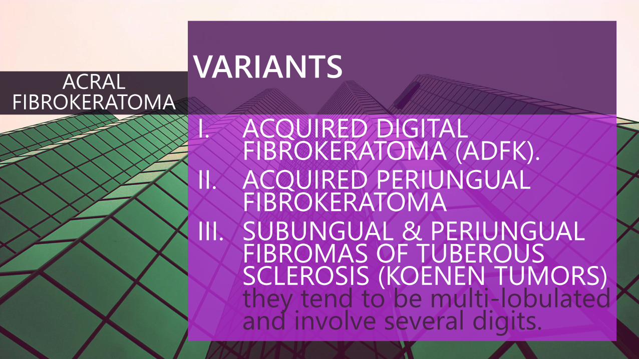
I. ACQUIRED DIGITAL FIBROKERATOMA (ADFK).
II. ACQUIRED PERIUNGUAL FIBROKERATOMA
III. SUBUNGUAL & PERIUNGUAL FIBROMAS OF TUBEROUS SCLEROSIS (KOENEN TUMORS) they tend to be multi-lobulated and involve several digits.
ACRAL FIBROKERATOMA
CLINICAL FEATURES
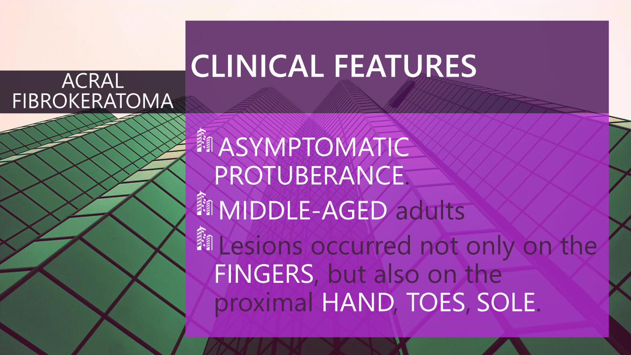
ASYMPTOMATICPROTUBERANCE.
MIDDLE-AGED adults
Lesions occurred not only on the FINGERS, but also on the proximal HAND, TOES, SOLE.
ACRAL FIBROKERATOMA
CLINICAL FEATURES
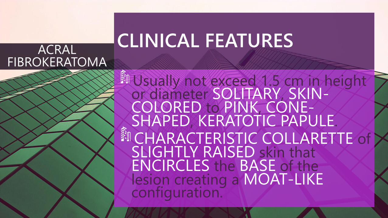
Usually not exceed 1.5 cm in height or diameter SOLITARY, SKIN-COLORED to PINK, CONE-SHAPED, KERATOTIC PAPULE.
CHARACTERISTIC COLLARETTE of SLIGHTLY RAISED skin that ENCIRCLES the BASE of the lesion creating a MOAT-LIKEconfiguration.
ACRAL FIBROKERATOMA
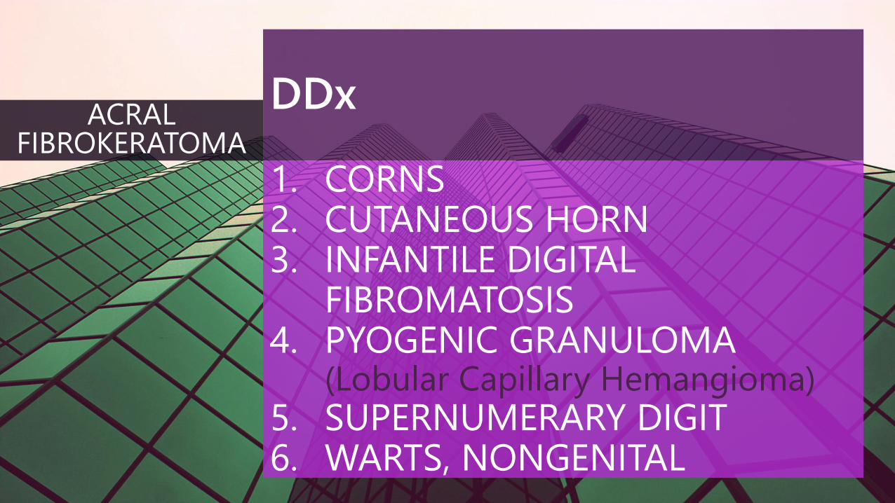
DDx
1. CORNS2. CUTANEOUS HORN3. INFANTILE DIGITAL
FIBROMATOSIS4. PYOGENIC GRANULOMA
(Lobular Capillary Hemangioma)5. SUPERNUMERARY DIGIT6. WARTS, NONGENITAL
ACRAL FIBROKERATOMA
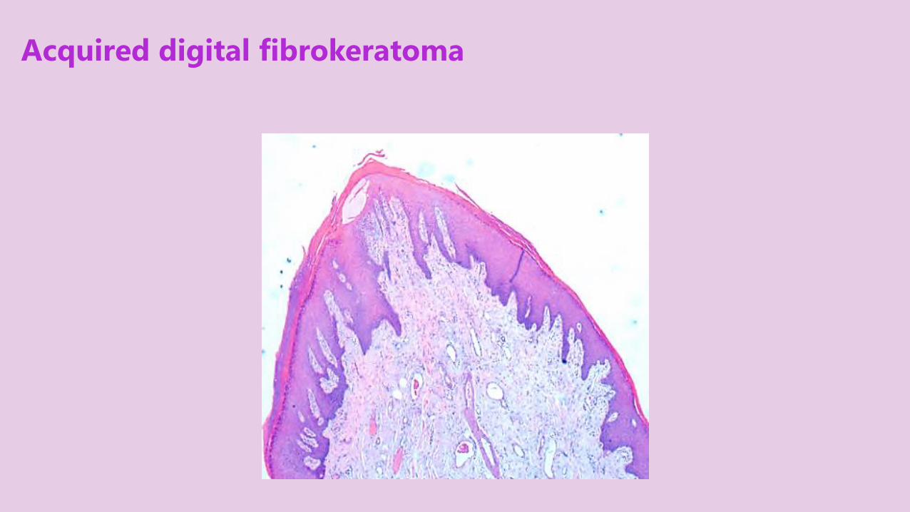
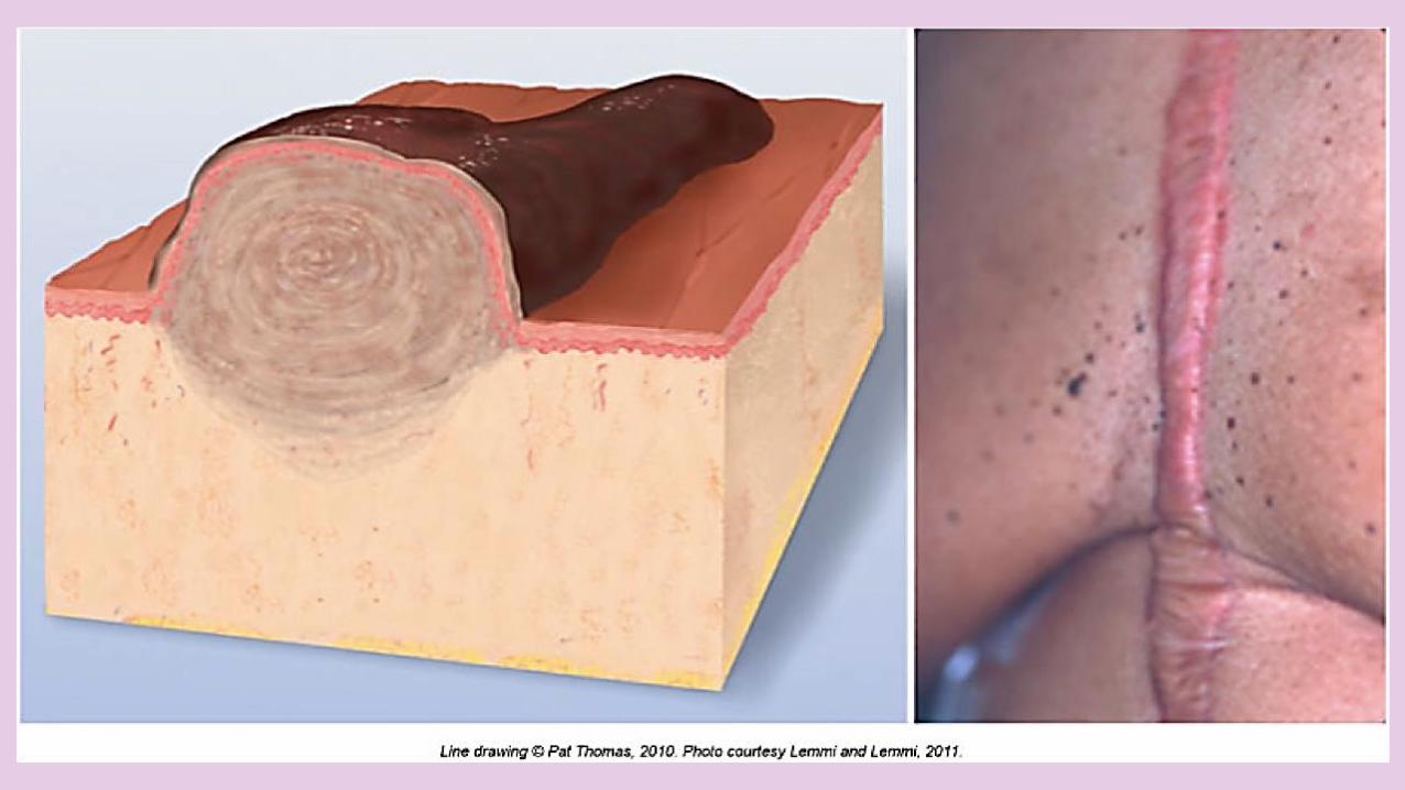
Acquired digital fibrokeratoma
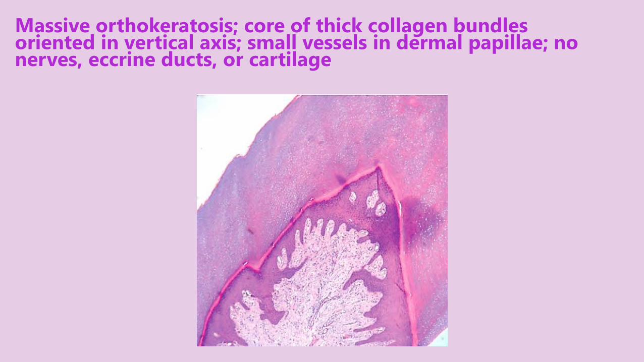
Massive orthokeratosis; core of thick collagen bundles oriented in vertical axis; small vessels in dermal papillae; no nerves, eccrine ducts, or cartilage
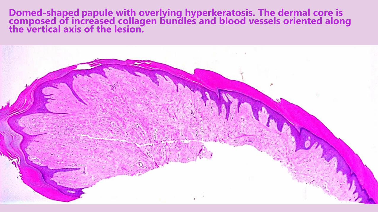
Domed-shaped papule with overlying hyperkeratosis. The dermal core is composed of increased collagen bundles and blood vessels oriented along the vertical axis of the lesion.
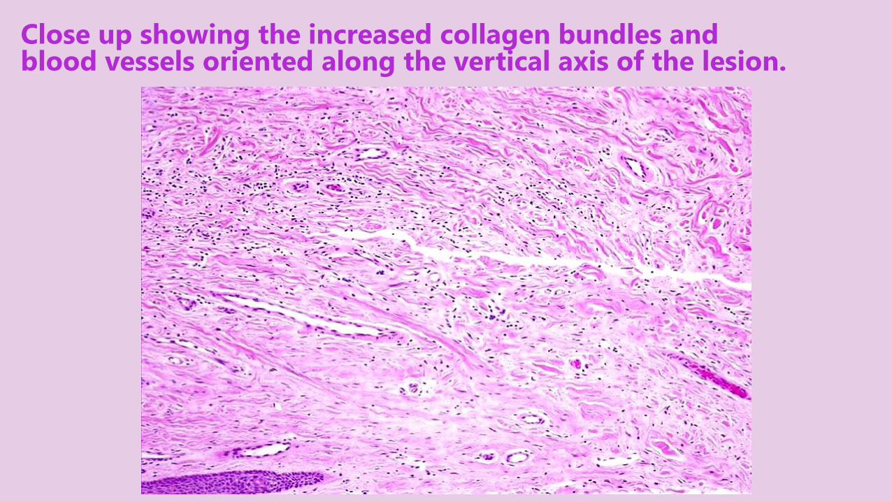
Close up showing the increased collagen bundles and blood vessels oriented along the vertical axis of the lesion.
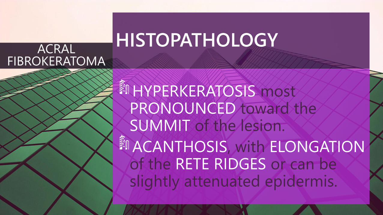
HISTOPATHOLOGY
HYPERKERATOSIS most PRONOUNCED toward the SUMMIT of the lesion.
ACANTHOSIS, with ELONGATIONof the RETE RIDGES or can be slightly attenuated epidermis.
ACRAL FIBROKERATOMA
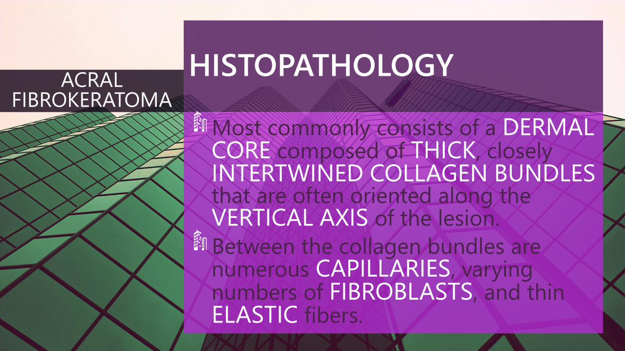
HISTOPATHOLOGY
Most commonly consists of a DERMALCORE composed of THICK, closely INTERTWINED COLLAGEN BUNDLESthat are often oriented along the VERTICAL AXIS of the lesion.
Between the collagen bundles are numerous CAPILLARIES, varying numbers of FIBROBLASTS, and thin ELASTIC fibers.
ACRAL FIBROKERATOMA

TREATMENT
SIMPLE EXCISION is curative; recurrence is rare.
ACRAL FIBROKERATOMA
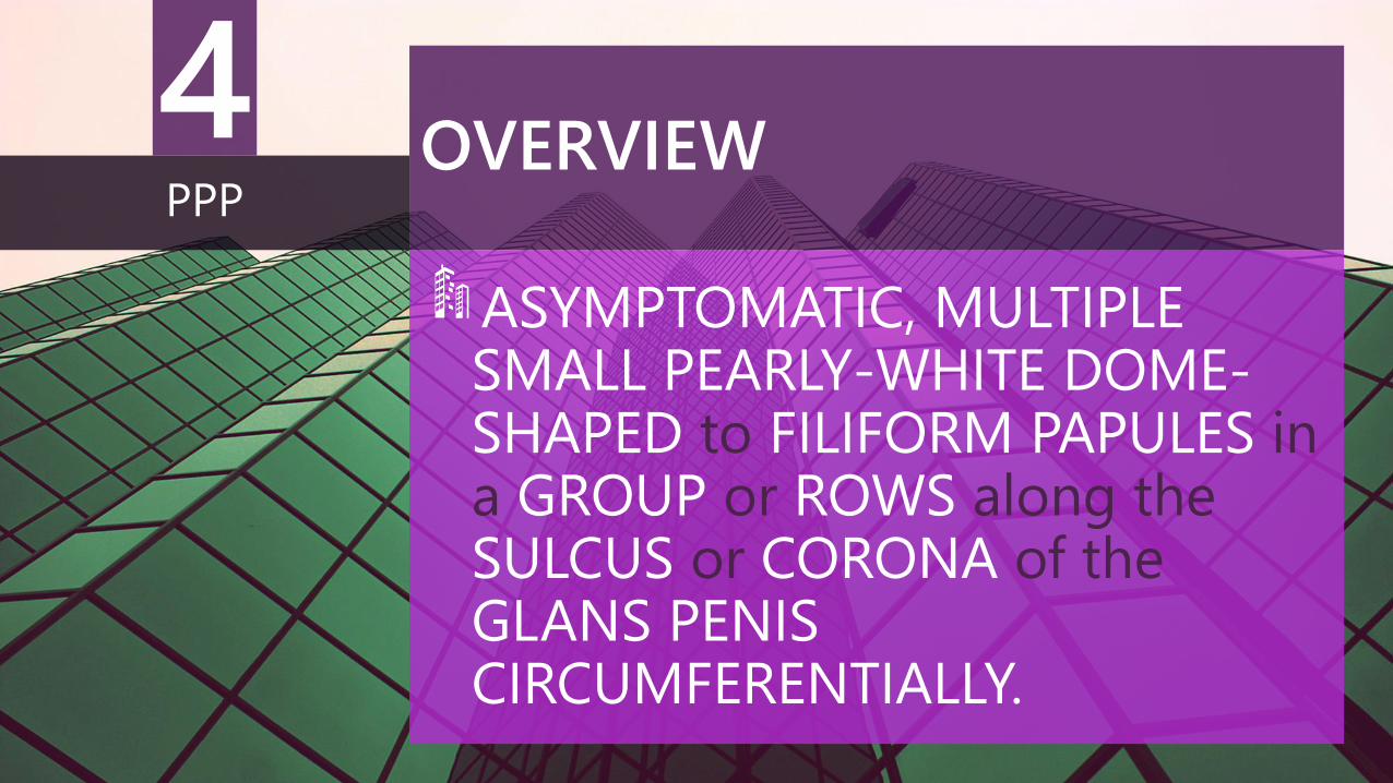
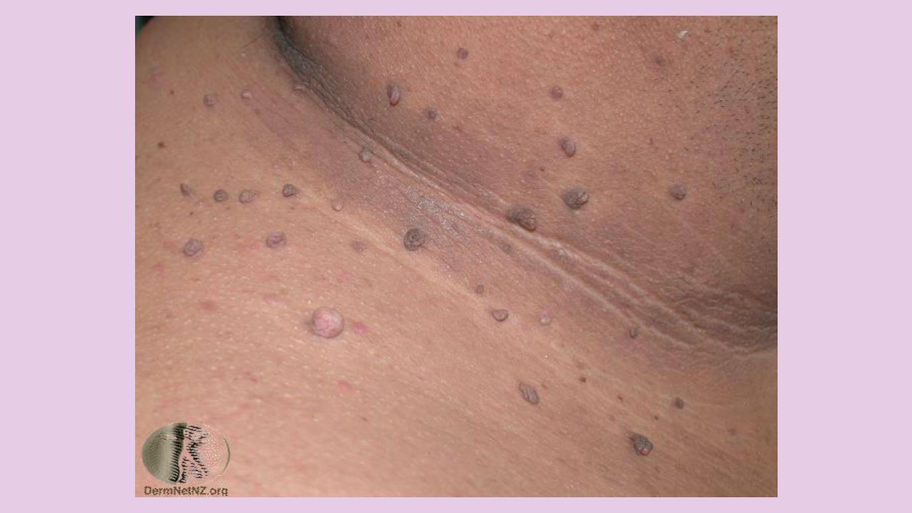
OVERVIEW
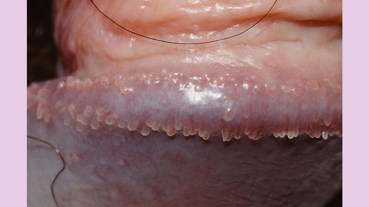
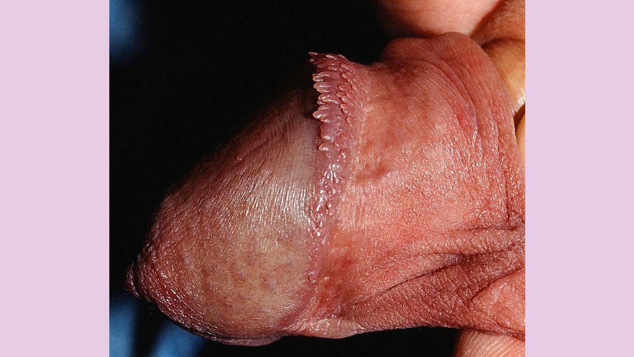
ASYMPTOMATIC, MULTIPLESMALL PEARLY-WHITE DOME-SHAPED to FILIFORM PAPULES ina GROUP or ROWS along the SULCUS or CORONA of theGLANS PENIS CIRCUMFERENTIALLY.
PPP
4

OVERVIEW
They're NORMAL ANATOMICvariant present in between 8% and 43% of men.
More common in UNCIRCUMCISED men.
Often, lesions cause GREATANXIETY to patients until their BENIGN NATURE is clarified.
PPP
CLINICAL FEATURES
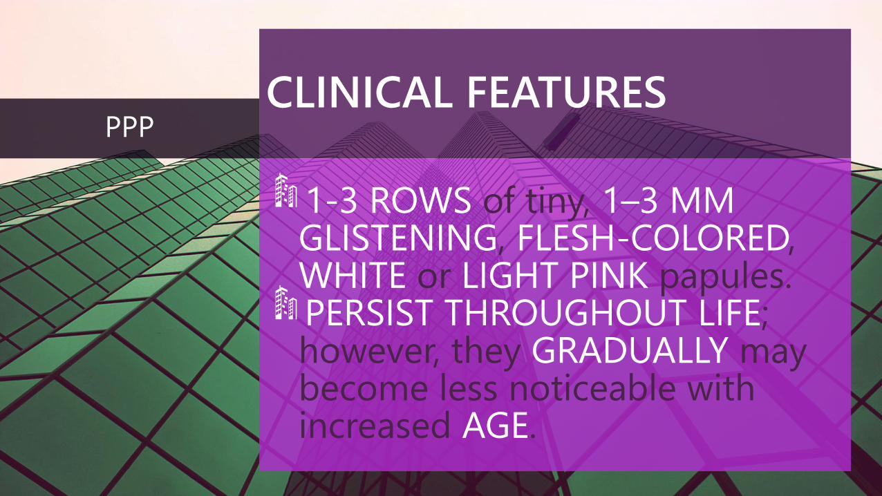
1-3 ROWS of tiny, 1–3 MMGLISTENING, FLESH-COLORED, WHITE or LIGHT PINK papules.PERSIST THROUGHOUT LIFE; however, they GRADUALLY may become less noticeable with increased AGE.
PPP

DDx
1. GENITAL WARTS
2. MOLLUSCUM CONTAGIOSUM
3. ECTOPIC SEBACEOUS GLANDS (FORDYCE SPOTS)
PPP

PPP- Papule with fibroblasts, rich vascularity, dense connective tissue
HISTOPATHOLOGY
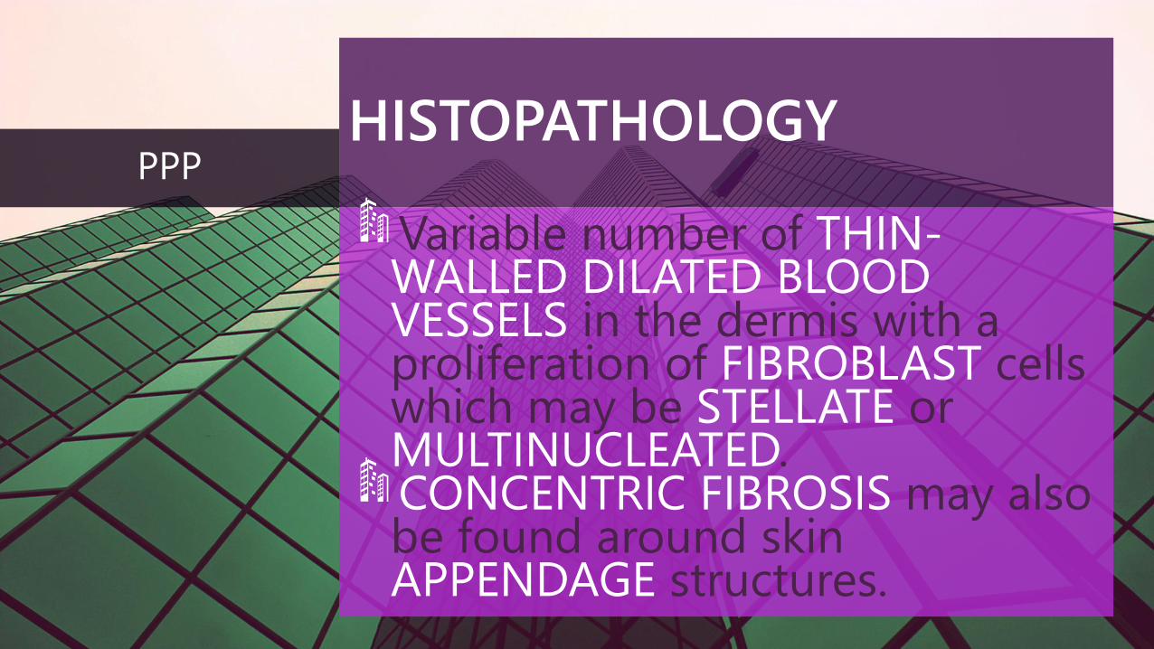
Variable number of THIN-WALLED DILATED BLOODVESSELS in the dermis with a proliferation of FIBROBLAST cells which may be STELLATE or MULTINUCLEATED. CONCENTRIC FIBROSIS may also be found around skin APPENDAGE structures.
PPP
TREATMENT
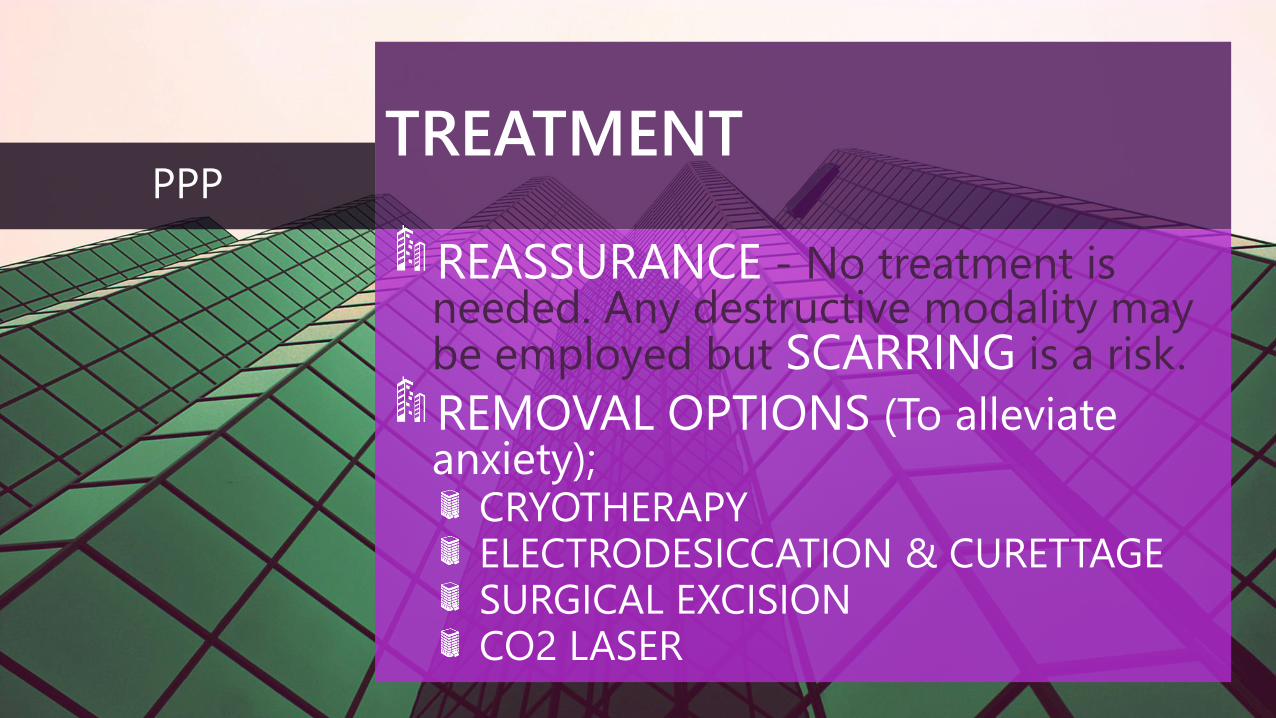
REASSURANCE - No treatment is needed. Any destructive modality may be employed but SCARRING is a risk.
REMOVAL OPTIONS (To alleviate anxiety);
CRYOTHERAPYELECTRODESICCATION & CURETTAGESURGICAL EXCISION CO2 LASER
PPP


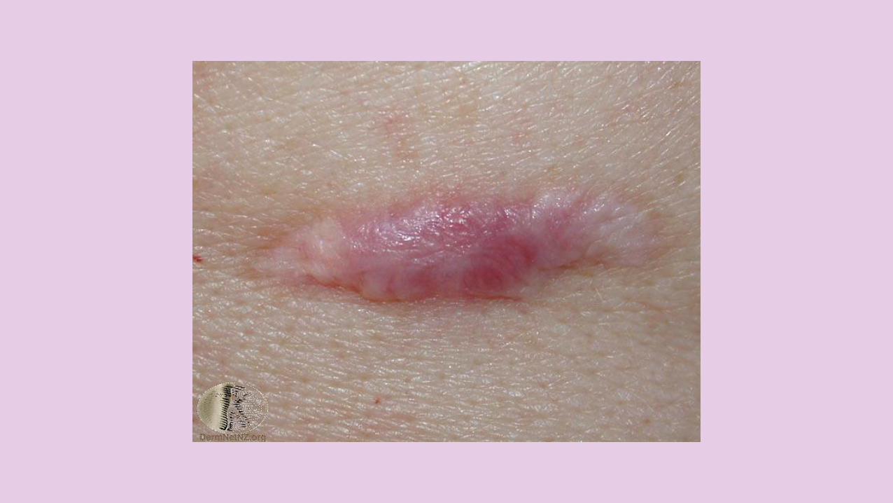
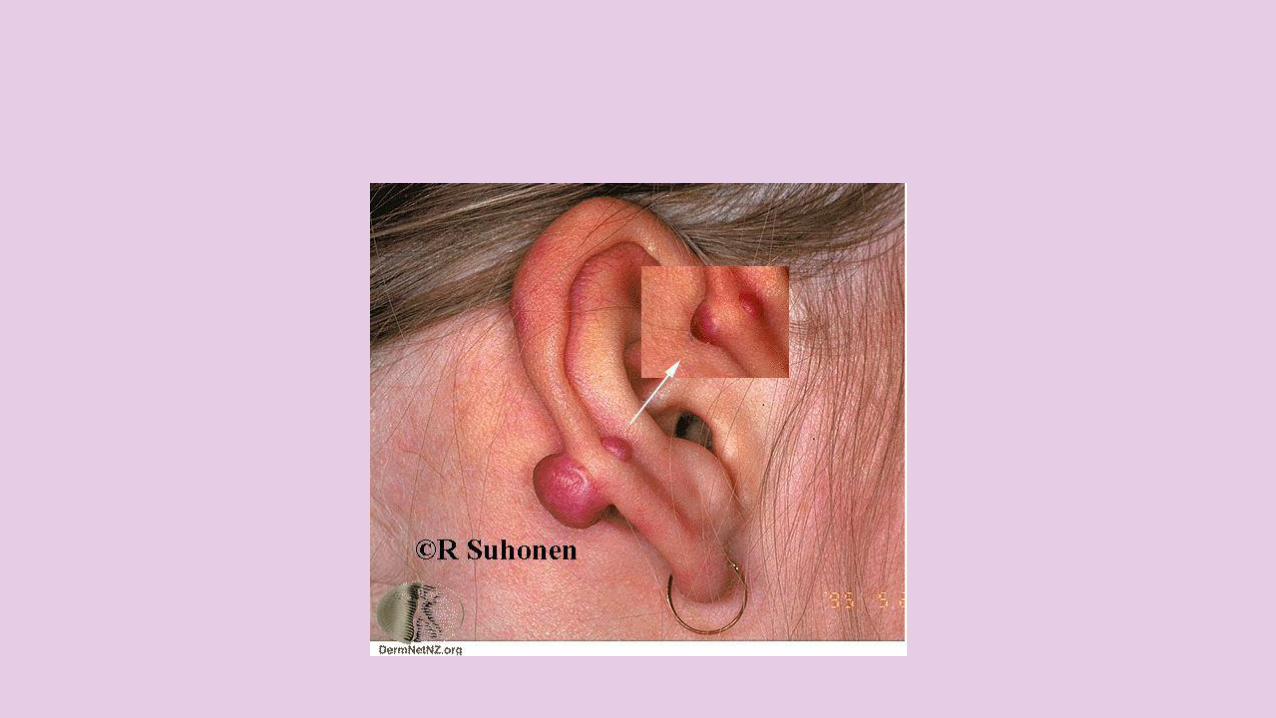
KELOIDS AND HYPERTROPHIC SCARS
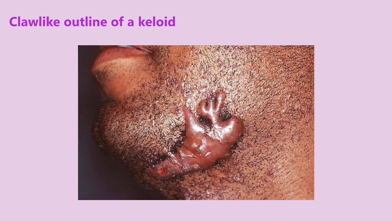
Clawlike outline of a keloid

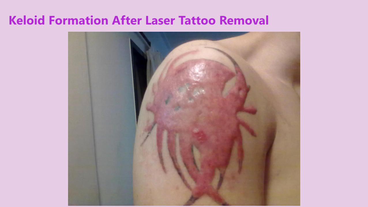
Keloid Formation After Laser Tattoo Removal
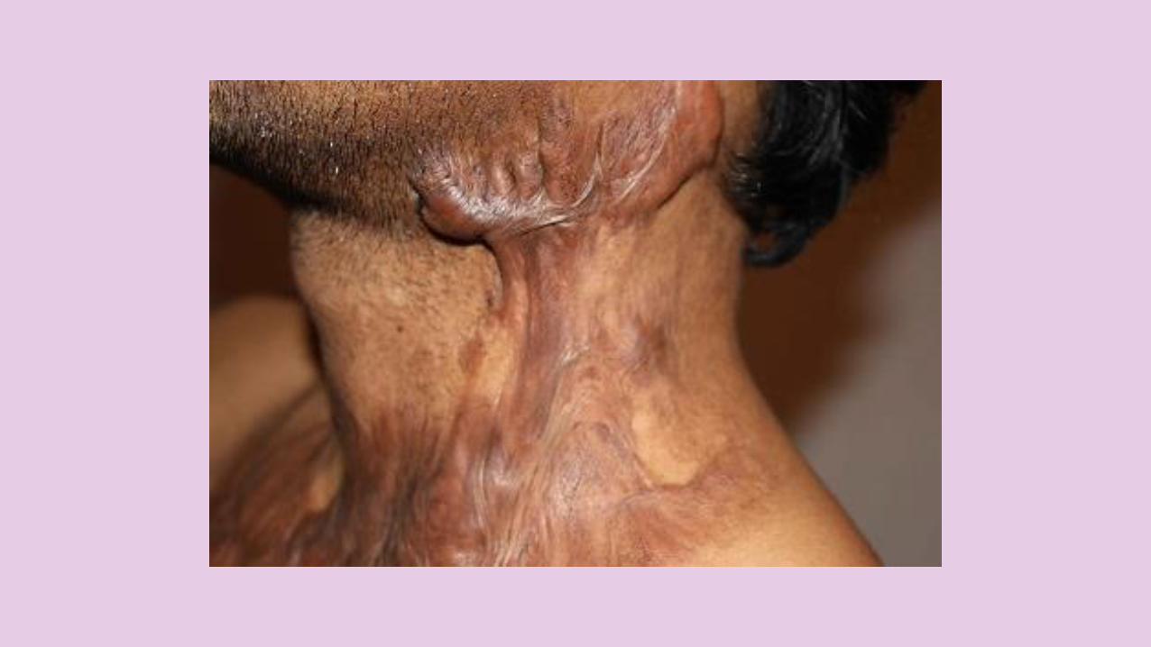
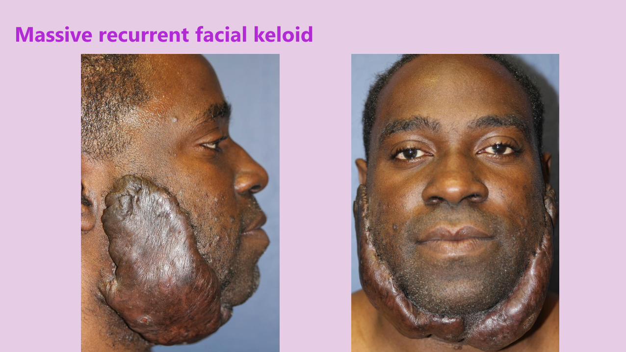
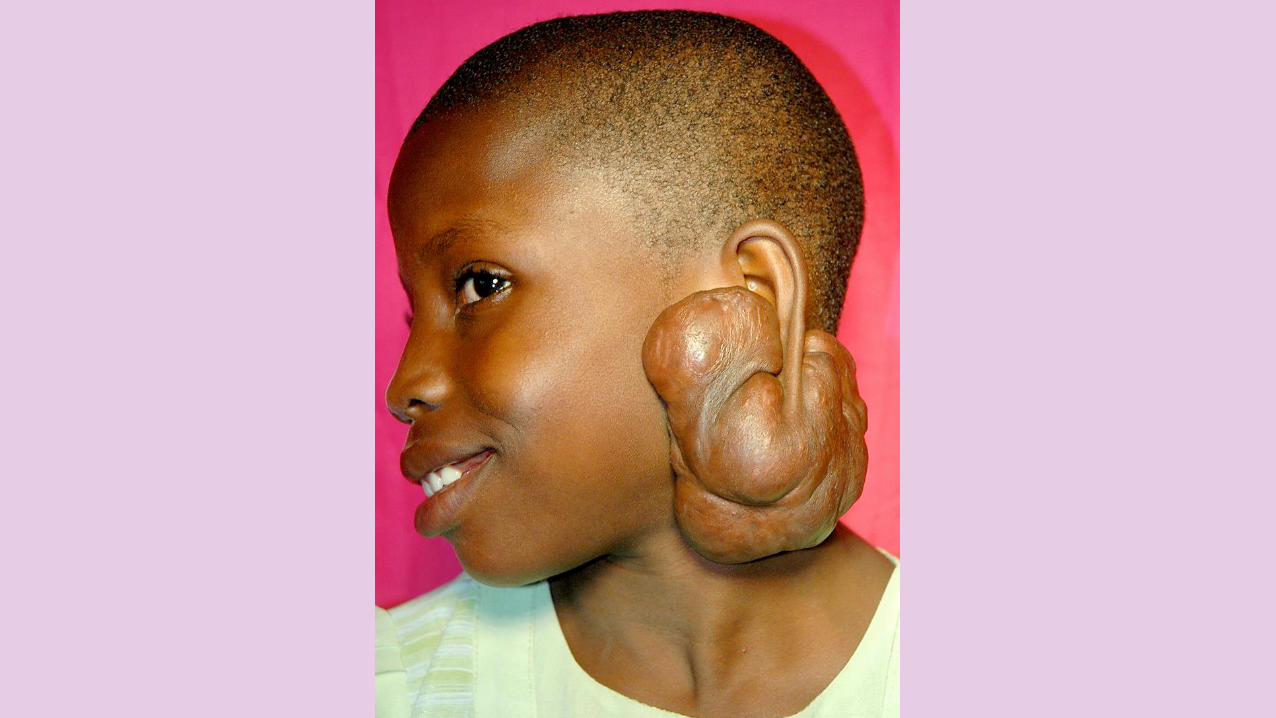
Massive recurrent facial keloid
OVERVIEW
Keloid are FIRM to HARD, SMOOTH, GROWTHS due to SPONTANEOUS SCARFORMATION and much LARGERthan the ORIGINAL WOUND.
KELOID & HYPERTROPHIC SCARS
OVERVIEW
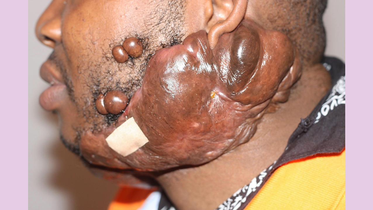
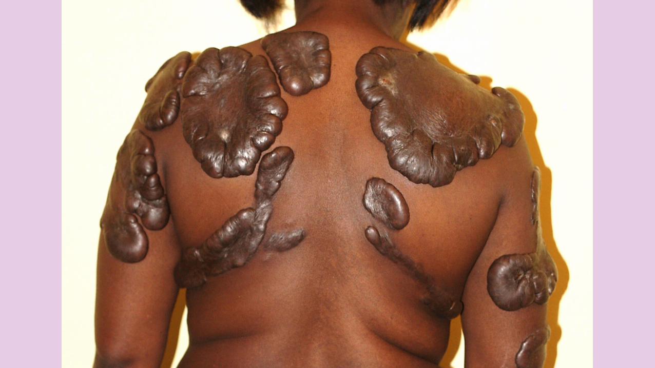
Keloids may form on ANY PART of the body, although the EARS, UPPERCHEST & shoulders are especially prone.While most people never form keloids, others develop them after MINORINJURIES, burns, insect bites and acne. DARK SKINNED people form keloids more easily than Caucasians.
KELOID & HYPERTROPHIC SCARS
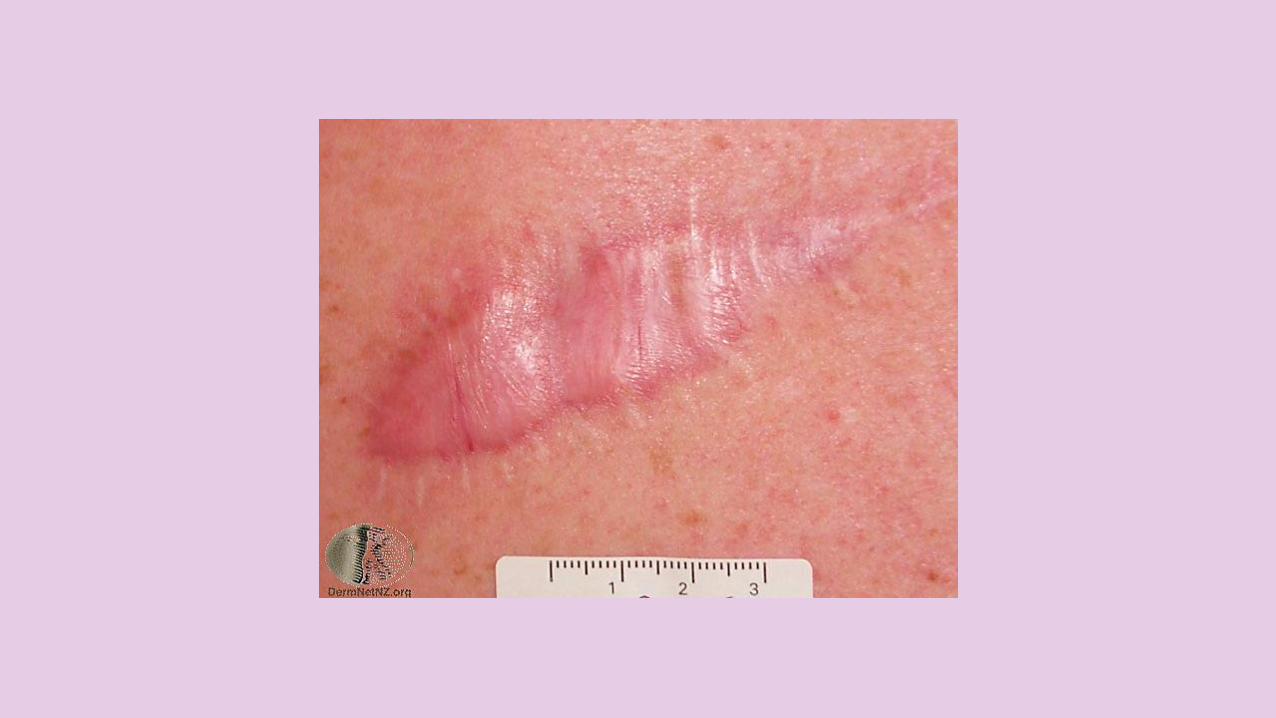
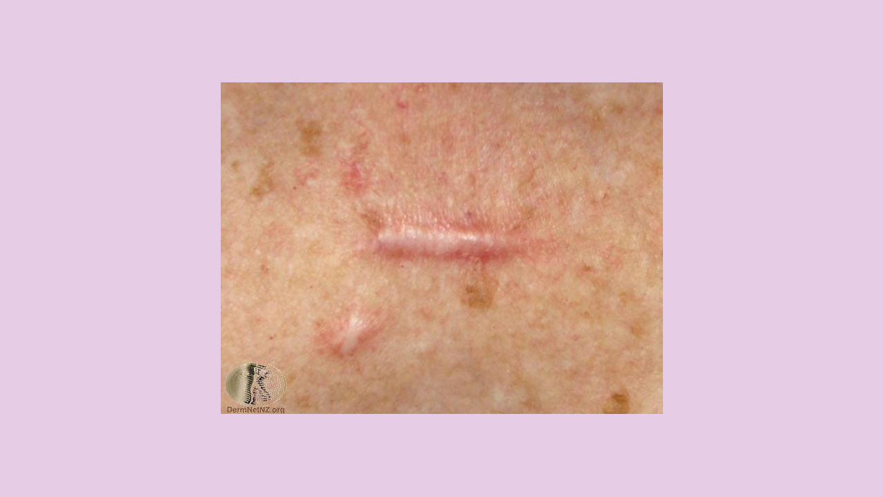
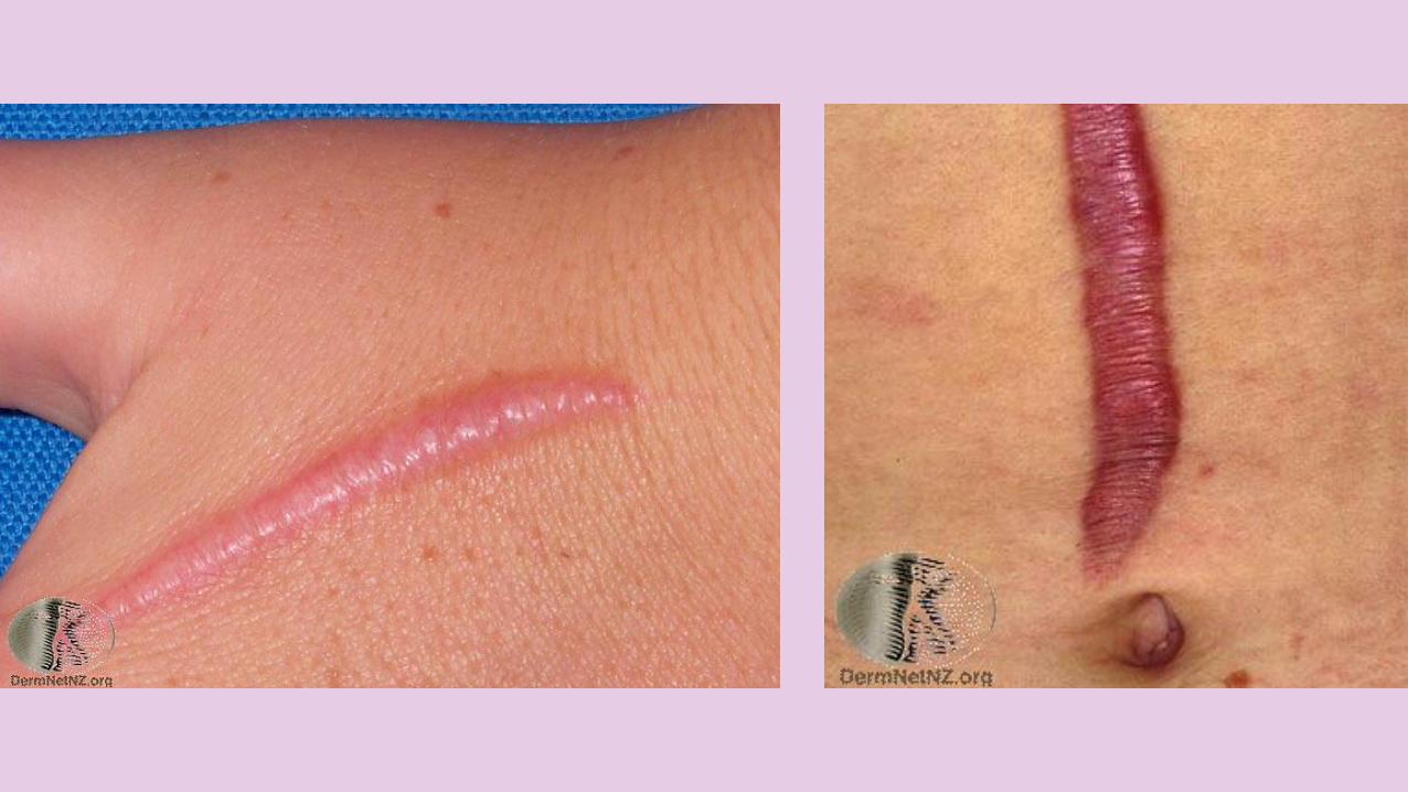
WHAT ARE HYPERTROPHIC SCARS?
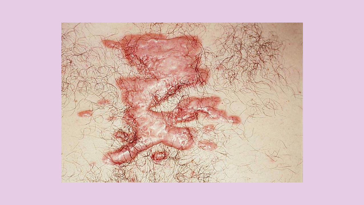
As wounds heal, SCAR TISSUE forms, which at first is often ERYTHEMATOUS and somewhat PROMINENT. Over several MONTHS, a scar usually becomes FLAT and PALE. If there is a LOT of TENSION on a healing wound, the healing area is RATHER THICKER than USUAL. This is known as a HYPERTROPHIC SCAR.
KELOID & HYPERTROPHIC SCARS
WHAT ARE HYPERTROPHIC SCARS?
Hypertrophic scars remain LIMITED to the TRAUMATIZEDAREA.
Regress SPONTANEOUSLY within 12-24 months, although regression may NOT necessarily be COMPLETE.
KELOID & HYPERTROPHIC SCARS
PREVALENCE
The prevalence has been reported to be HIGHER in YOUNG FEMALESthan in young males, probably reflecting the greater frequency of EARLOBE PIERCING among females.
Keloids and hypertrophic scars affect BOTH SEXES equally in OTHERAGE GROUPS.
KELOID & HYPERTROPHIC SCARS
ETIOLOGY
The EXACT MECHANISMS of keloid and hypertrophic scar pathogenesis continue to be an ENIGMA for physicians and researchers alike.
The INCREASED PREVALENCE of keloids paralleling increased CUTANEOUS PIGMENTATIONsuggests a GENETIC BASIS or, at least, a genetic linkage.
KELOID & HYPERTROPHIC SCARS
ETIOLOGY
TRAUMA TO THE SKIN, both physical (e.g. earlobe piercing, surgery) and pathological (e.g. acne, chickenpox), is the primary cause of keloids. THE PRESENCE of FOREIGNMATERIAL, INFECTION, HEMATOMA, or increased SKIN TENSION can also lead to keloid or hypertrophic scar formation in susceptible individuals.
KELOID & HYPERTROPHIC SCARS
CLINICAL FEATURES
Keloids and hypertrophic scars do NOT usually cause SYMPTOMS, but they MAY be TENDER, PAINFUL, or PRURITIC or they may cause a burning sensation.
In addition to symptomatic relief, COSMETIC CONCERN is the primary reason patients seek medical intervention.
KELOID & HYPERTROPHIC SCARS
CLINICAL FEATURES
Keloids manifest as EXAGGERATEDGROWTHS of SCAR TISSUE, USUALLYin areas of PREVIOUS TRAUMA.
Keloids extend BEYOND the areas of trauma, PROJECTING ABOVE the LEVEL of the SURROUNDING SKIN, but they rarely extend into underlying subcutaneous tissue.
KELOID & HYPERTROPHIC SCARS
CLINICAL FEATURES
Keloids range in CONSISTENCY from SOFT and DOUGHY to RUBBERY and HARD. Early lesions are often ERYTHEMATOUS. Lesions become BROWNISH RED and then PALE as they age. Lesions are usually DEVOID of HAIRFOLLICLES and other functioning ADNEXAL GLANDS.
KELOID & HYPERTROPHIC SCARS
CLINICAL FEATURES
MOST lesions CONTINUE to GROWfor WEEKS to MONTHS and OTHERSgrow for YEARS. Growth is usually SLOW, but keloids occasionally enlarge rapidly, tripling in size within months. Once they stop growing, keloids do not usually cause symptoms and REMAINSTABLE or INVOLUTE SLIGHTLY.
KELOID & HYPERTROPHIC SCARS
CLINICAL FEATURES
In WHITE persons, keloids tend to be present, in decreasing order of frequency, on the FACE (with CHEEK and EARLOBESPREDOMINATING), UPPER EXTREMITIES, CHEST, PRESTERNAL AREA, NECK, BACK, LOWER EXTREMITIES, breasts & abdomen.In BLACK persons, the descending order of frequency tends to be EARLOBES, FACE, NECK, LOWER EXTREMITIES, breasts, chest, back, and abdomen.
KELOID & HYPERTROPHIC SCARS
CLINICAL FEATURES
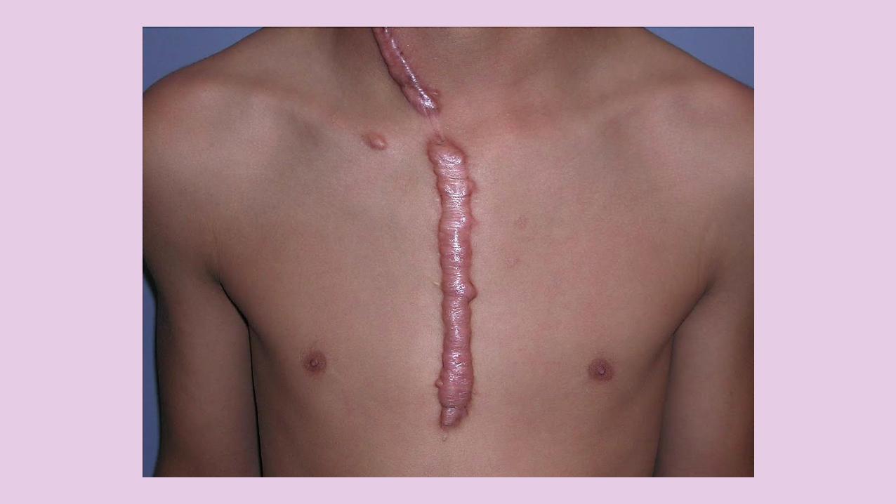
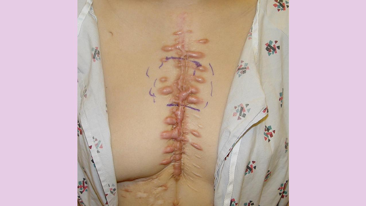
Keloids on the EARS, NECK, and ABDOMEN tend to be PEDUNCULATED. Keloids on the CENTRAL CHEST and extremities are usually RAISED with a FLAT surface, and the base is often wider than the top.
Keloids OVERLYING a JOINT can CONTRACT and restrict movement.
KELOID & HYPERTROPHIC SCARS
CLINICAL FEATURES
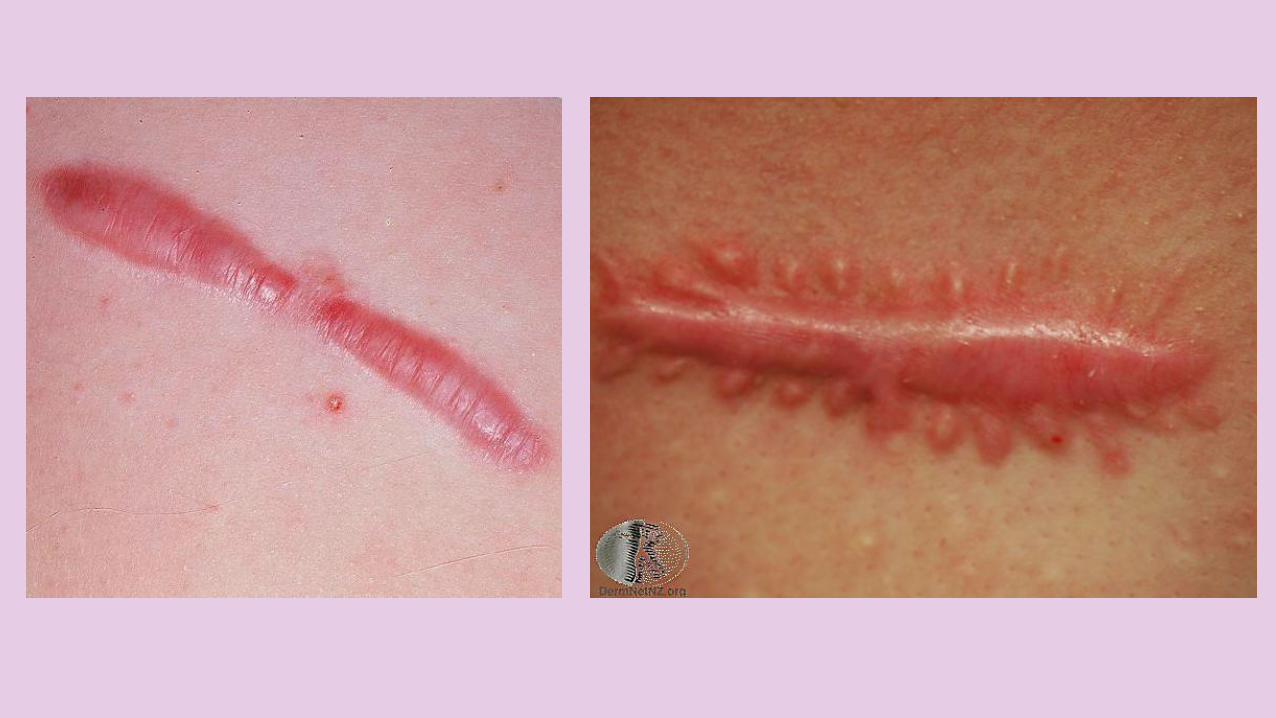
Most keloids are ROUND, OVAL, or OBLONG with REGULAR MARGINS; however, some have CLAWLIKEconfigurations with irregular borders. KELOIDS may be DISTINGUISHEDfrom HYPERTROPHIC SCARS by their CLAWLIKE PROJECTIONS, which are absent in the hypertrophic scar.
KELOID & HYPERTROPHIC SCARS
CLINICAL FEATURES
MOST patients present with 1 OR 2 keloids; however, a FEWpatients, especially patients with spontaneous keloids, have MULTIPLE LESIONS, as do patients who develop keloids as a consequence of acne or chickenpox.
KELOID & HYPERTROPHIC SCARS
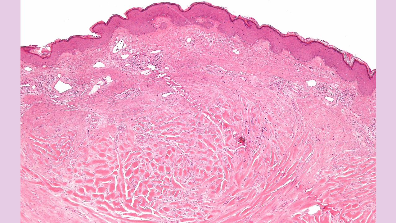
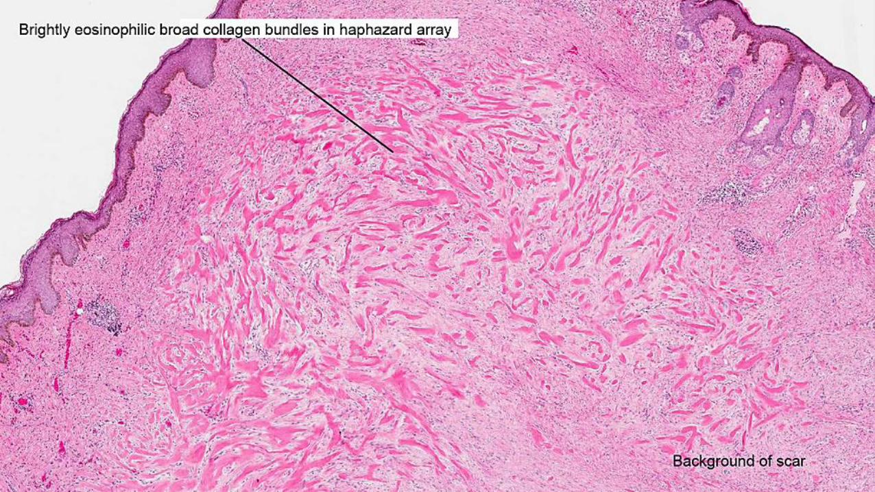
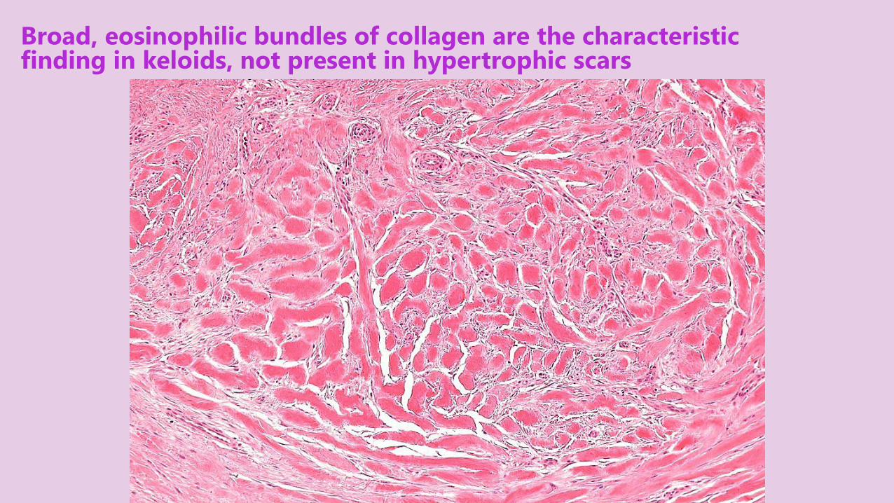
Broad, eosinophilic bundles of collagen are the characteristic finding in keloids, not present in hypertrophic scars
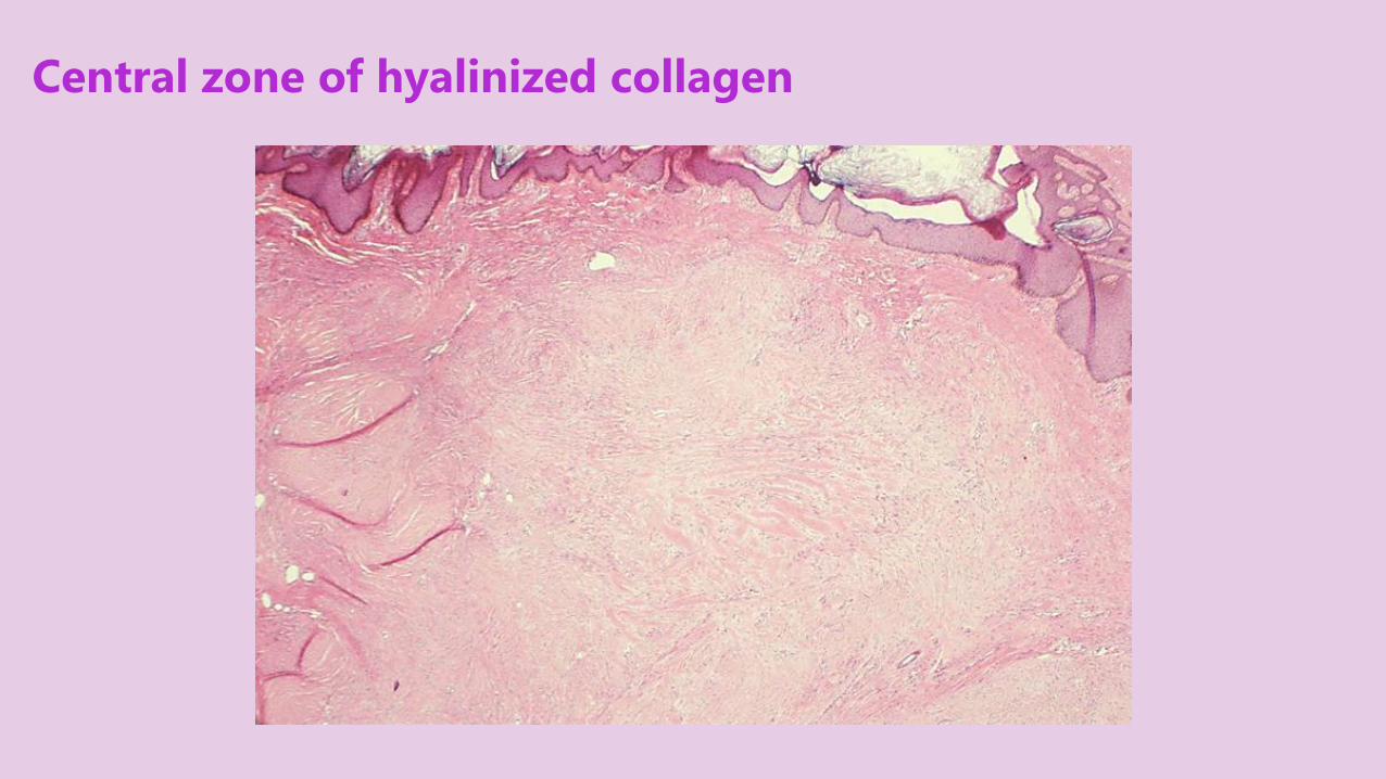
Central zone of hyalinized collagen
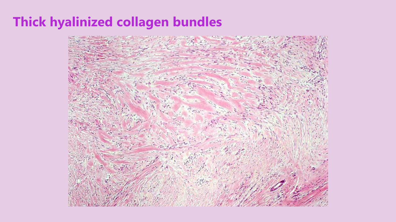
Thick hyalinized collagen bundles
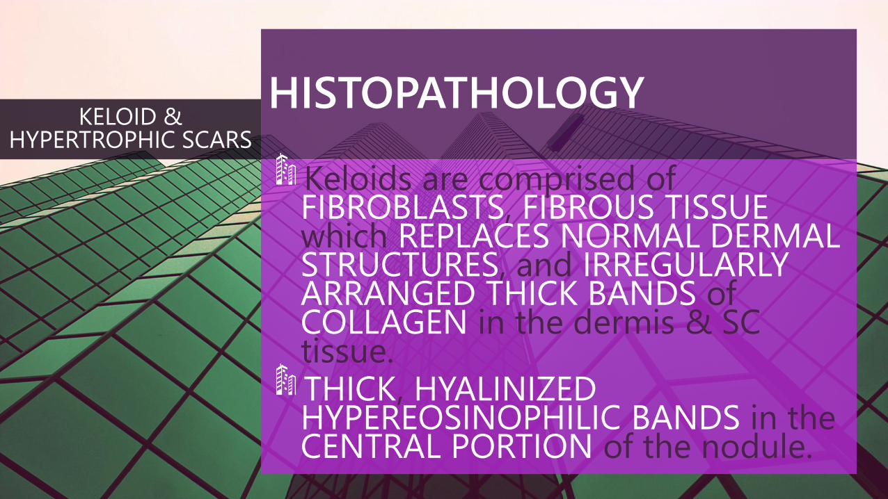
HISTOPATHOLOGY
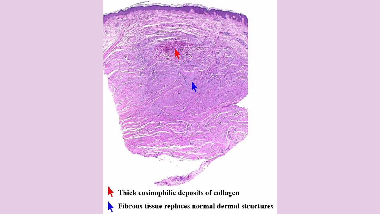
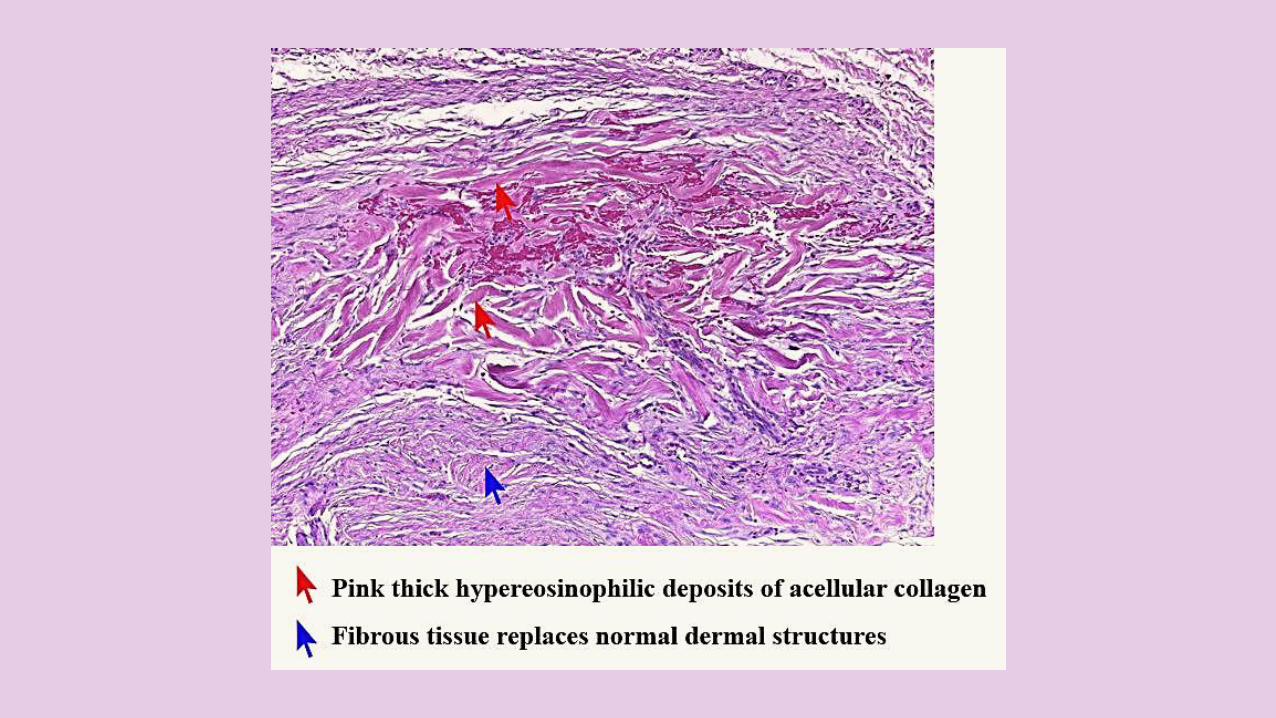
Keloids are comprised of FIBROBLASTS, FIBROUS TISSUEwhich REPLACES NORMAL DERMALSTRUCTURES, and IRREGULARLYARRANGED THICK BANDS of COLLAGEN in the dermis & SC tissue.THICK, HYALINIZEDHYPEREOSINOPHILIC BANDS in the CENTRAL PORTION of the nodule.
KELOID & HYPERTROPHIC SCARS
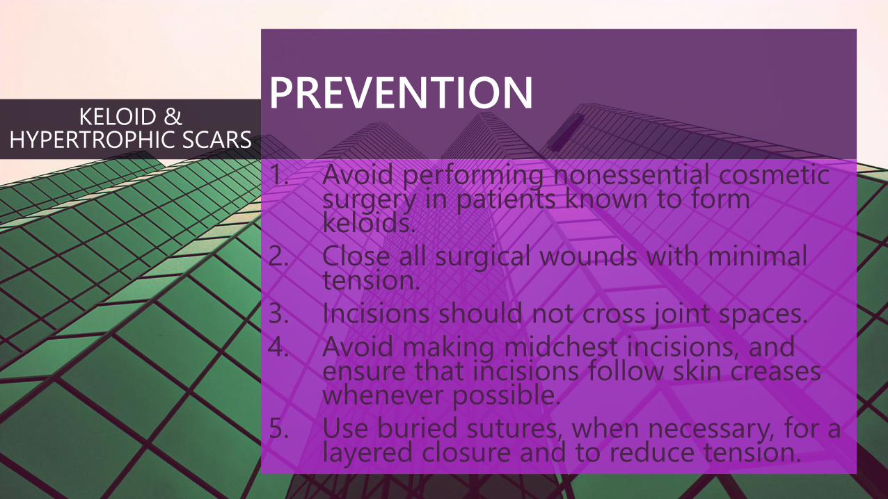
PREVENTION
1. Avoid performing nonessential cosmetic surgery in patients known to form keloids.
2. Close all surgical wounds with minimal tension.
3. Incisions should not cross joint spaces. 4. Avoid making midchest incisions, and
ensure that incisions follow skin creases whenever possible.
5. Use buried sutures, when necessary, for a layered closure and to reduce tension.
KELOID & HYPERTROPHIC SCARS
TREATMENT
HYPERTROPHIC SCARS generally SETTLE in TIME OR with TREATMENT, but KELOIDS may PERSIST and prove resistant to treatment.
KELOID & HYPERTROPHIC SCARS
STANDARD TREATMENTS
These include OCCLUSIVEDRESSINGS, COMPRESSIONTHERAPY, and INTRALESIONALCORTICOSTEROID INJECTION.
KELOID & HYPERTROPHIC SCARS
TREATMENT MEASURES
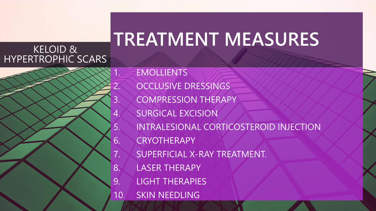
1. EMOLLIENTS
2. OCCLUSIVE DRESSINGS
3. COMPRESSION THERAPY
4. SURGICAL EXCISION
5. INTRALESIONAL CORTICOSTEROID INJECTION
6. CRYOTHERAPY
7. SUPERFICIAL X-RAY TREATMENT.
8. LASER THERAPY
9. LIGHT THERAPIES
10. SKIN NEEDLING
KELOID & HYPERTROPHIC SCARS
TREATMENT MEASURES
OCCLUSIVE DRESSINGS, include silicone gel sheets and dressings, nonsilicone occlusive sheets, dressings should be worn for 12 to 24 hours per day.
KELOID & HYPERTROPHIC SCARS
TREATMENT MEASURES
COMPRESSION THERAPY Mechanoreceptors induce apoptosis also extracellular matrix. Rigidity caused by compression may also inhibit the differentiation and proliferation of scar fibroblasts(button compression, pressure earrings, ACE bandages, elastic adhesive bandages, compression wraps, spandex or elastane (Lycra) bandages, and support bandages)
KELOID & HYPERTROPHIC SCARS
TREATMENT MEASURES
SURGICAL EXCISION (but in keloids, excision may result in a new keloid even larger than the original one) Excisional surgery alone has been shown to yield a 45-100% recurrence rate.
KELOID & HYPERTROPHIC SCARS
TREATMENT MEASURES
INTRALESIONAL CORTICOSTEROID INJECTION, as a single modality and as an adjunct to excision repeated every few weeks reducing collagen synthesis, altering glucosaminoglycan synthesis, and reducing production of inflammatory mediators and fibroblast proliferation during wound healing. The most commonly used corticosteroid is triamcinolone acetonide(TAC) in concentrations of 10-40 mg/mL administered intralesionally with a 25- to 27-gauge needle at 4- to 6-week intervals.
KELOID & HYPERTROPHIC SCARS
TREATMENT MEASURES
CRYOTHERAPY - 2, or 3 freeze-thaw cycles lasting up to 30 seconds repeated every 20-30 days. Insertion of a lumbar puncture needle through the long axis of the keloid, from one side to the other, passing the liquid nitrogen.
KELOID & HYPERTROPHIC SCARS

TREATMENT MEASURES
LASER THERAPY Ablative lasers Carbon dioxide, argon laser, and Nd:YAG laser (1064 nm)Nonablative lasers Pulsed dye laser because of its efficacy, safety, and relatively low cost, the PDL remains the laser treatment of choice for hypertrophic scars.
KELOID & HYPERTROPHIC SCARS
MEDICAL TREATMENTS
1. Retinoic acid
2. Intralesional interferon (IFN)
3. Intralesional 5-fluorouracil (5-FU)
4. Intralesional calcium channel blockers verapamil
5. Doxorubicin
6. Bleomycin
7. Imiquimod 5% cream
8. Tacrolimus
9. Tamoxifen
10. Botulinum toxin type A,
11. Over-the-counter treatments (e.g. onion extract; combination of hydrocortisone, silicon, and vitamin E).
KELOID & HYPERTROPHIC SCARS
LIGHT THERAPIES
1. Photodynamic therapy [PDT]
2. Intense pulsed light (IPL)
3. UVA-1
4. Narrowband UVB
5. Broadband UVB
KELOID & HYPERTROPHIC SCARS
REFERENCES
BOLONGIA DERMATOLOGY ESSENTIALS
BOLONGIA 3rd ed
WEEDON’S SKIN PATHOLOGY ESSENTIALS
GOOGLE IMAGES
DERMNETNZ.ORG
EMEDICINE.MEDSCAPE.COM
THANK YOU
WWW.FACEBOOK.COM/GROUPS/DERMATOLOGYCOURSEONLINE