Fiber Optic Microarray Detection of Freshwater...
20
Fiber Optic Microarray Detection of Freshwater Cyanobacteria Shonda Gaylord PhD Candidate TCSVM Walt Laboratory Woods Hole Oceanographic Institute
Transcript of Fiber Optic Microarray Detection of Freshwater...

Fiber Optic Microarray Detection
of Freshwater Cyanobacteria
Shonda Gaylord
PhD Candidate TCSVM
Walt Laboratory
Woods Hole Oceanographic Institute
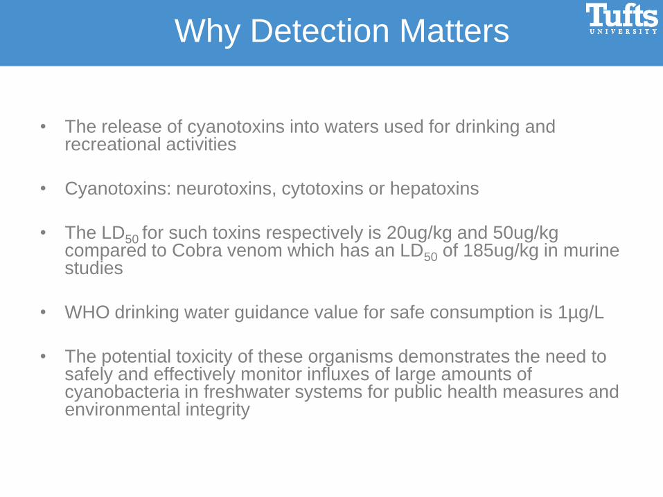
• The release of cyanotoxins into waters used for drinking and recreational activities
• Cyanotoxins: neurotoxins, cytotoxins or hepatoxins
• The LD50 for such toxins respectively is 20ug/kg and 50ug/kg compared to Cobra venom which has an LD50 of 185ug/kg in murine studies
• WHO drinking water guidance value for safe consumption is 1µg/L
• The potential toxicity of these organisms demonstrates the need to safely and effectively monitor influxes of large amounts of cyanobacteria in freshwater systems for public health measures and environmental integrity
Why Detection Matters

• In collaboration with WHOI, Tufts Chemistry Department and Ahura
Technologies …the overall project goal is to adapt and validate a rapid and
accurate optical fiber-based technology for CyanoHAB cell detection and
enumeration in both laboratory and field settings.
Cyanobacteria

C A C C G A T G T T C T T C C C A A T C
C G C T G G T G T T C T T C A G A A T A
C A G A C C C T T T A C G C C C A A T C
C G G A C C C T T T A C G C C C A A T C
C A G C A G A C T T T C A G T T C C
C A G C A G A C T T A C A T G G C C
C A G C C A C A C C T T C C G G T A
C A G C A G A C T T A C A A T G C C
Microcystis probe #1
S. elongates
C. raciborskii
A. flos-aquae
Cylindro probe #2
A. cylindrica
M. aeruginosa
A. flos-aquae
Signal probe #1
M. aeruginosa
C. raciborskii
A. flos-aquae
C T G A G A C A C G G C C C A G A C
C T G A G A C A C G G C C C A G A C
C T G A G A C A C G G C C C A G A C
C T G A G A C A C G G C C C A G A C
Base pair mismatches for capture probe
development are highlighted in yellow
Capture & Signal Probes

Cylindrospermopsis Microcystis
Cylindro probe 1 Micro probe 11
20x: Few Dozen 20x: ~100
Fluorescent In Situ Hybridization
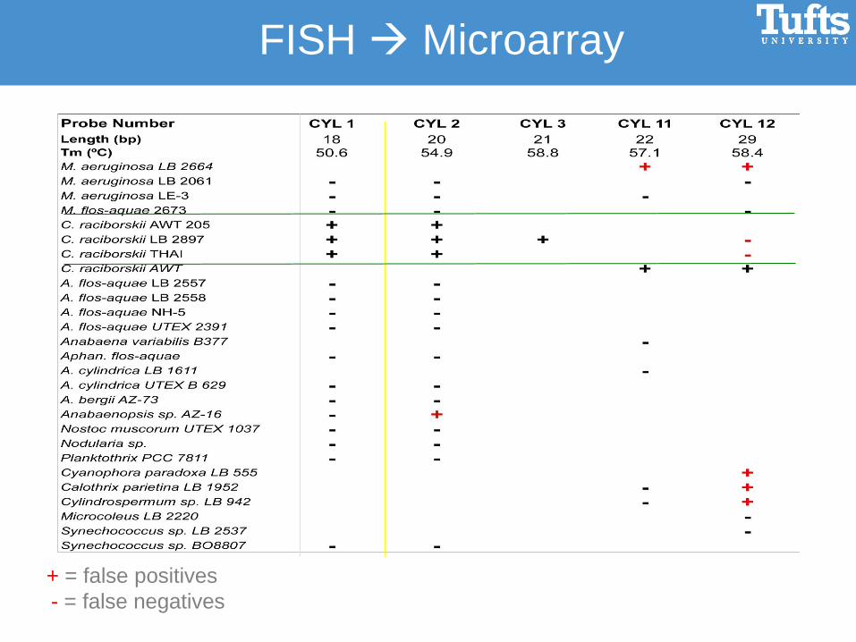
+ = false positives
- = false negatives
FISH Microarray
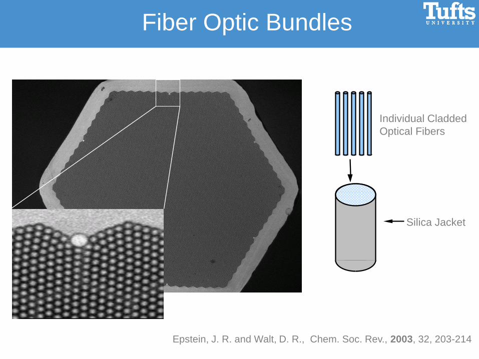
Individual Cladded
Optical Fibers
Silica Jacket
Epstein, J. R. and Walt, D. R., Chem. Soc. Rev., 2003, 32, 203-214
Fiber Optic Bundles

Wet etching with HCl
Pantano, P.; Walt, D.R. Chem. Mater. 1996, 8, 2832-2835
Microspheres in Etched Wells

A
T
G
Chttp://upload.wikimedia.org/wikipedia/en/f/f0/DNA_Overview.png
A
T
G
CA
T
G
C A
T
Base Pairing
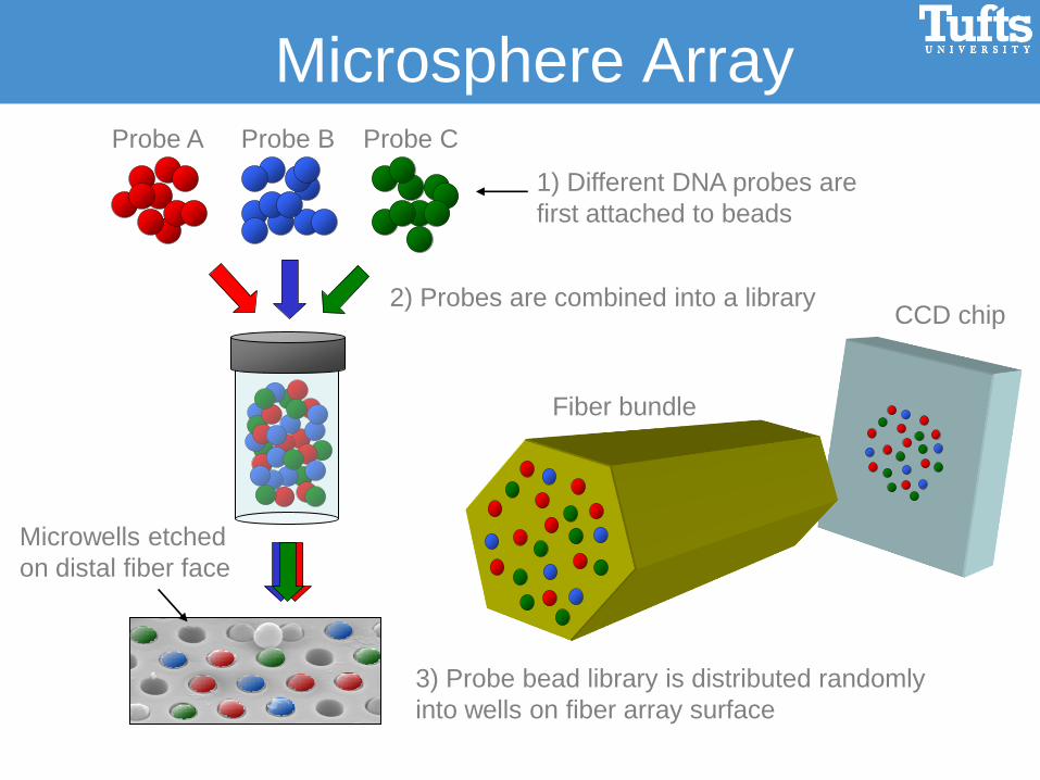
Probe A Probe B Probe C
Microwells etched
on distal fiber face
CCD chip2) Probes are combined into a library
1) Different DNA probes are
first attached to beads
3) Probe bead library is distributed randomly
into wells on fiber array surface
Fiber bundle
Microsphere Array

Cy5
o
Cy3
o
Direct hybridization
Sandwich
hybridization
assay
Background
Direct hybridization
Encoding
Microarray Assay
Cy3
o
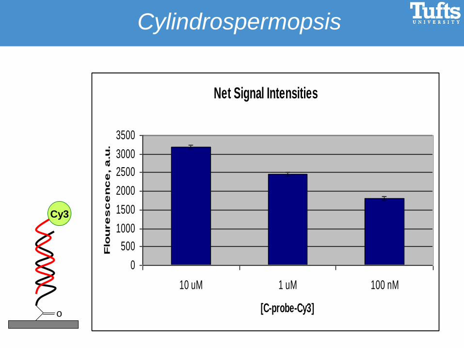
Cylindrospermopsis
Net Signal Intensities
0
500
1000
1500
2000
2500
3000
3500
10 uM 1 uM 100 nM
[C-probe-Cy3]
Flo
ure
sc
en
ce
, a
.u.
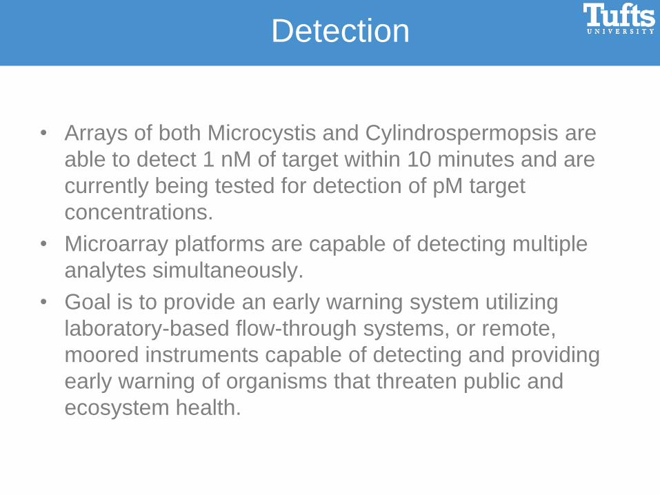
• Arrays of both Microcystis and Cylindrospermopsis are
able to detect 1 nM of target within 10 minutes and are
currently being tested for detection of pM target
concentrations.
• Microarray platforms are capable of detecting multiple
analytes simultaneously.
• Goal is to provide an early warning system utilizing
laboratory-based flow-through systems, or remote,
moored instruments capable of detecting and providing
early warning of organisms that threaten public and
ecosystem health.
Detection

Barcode
scannercomputer
Touch screen
display
CCD camera
actuators
•28x Magnifications
•Field of view: 350 x 280 micron
•Over 2000 three-µm beads
in field of view
•NA: 0.55
•Depth of focus: ~1 micron
•Two-color measurement, Cy3
and Cy5 dyes
•Folded to reduce height and
width
Ahura

SAC Committee
Dr. Saul Tzipori
Dr. David Walt
Dr. Dan Milner
Dr. Don Anderson
Groups
Walt Lab
WSSS Program
Funding
NIH
EPA STAR
Acknowledgements

Anatoxin–a Anatoxin–a(s)
ZACCARONI et al, 2008
Neurotoxins
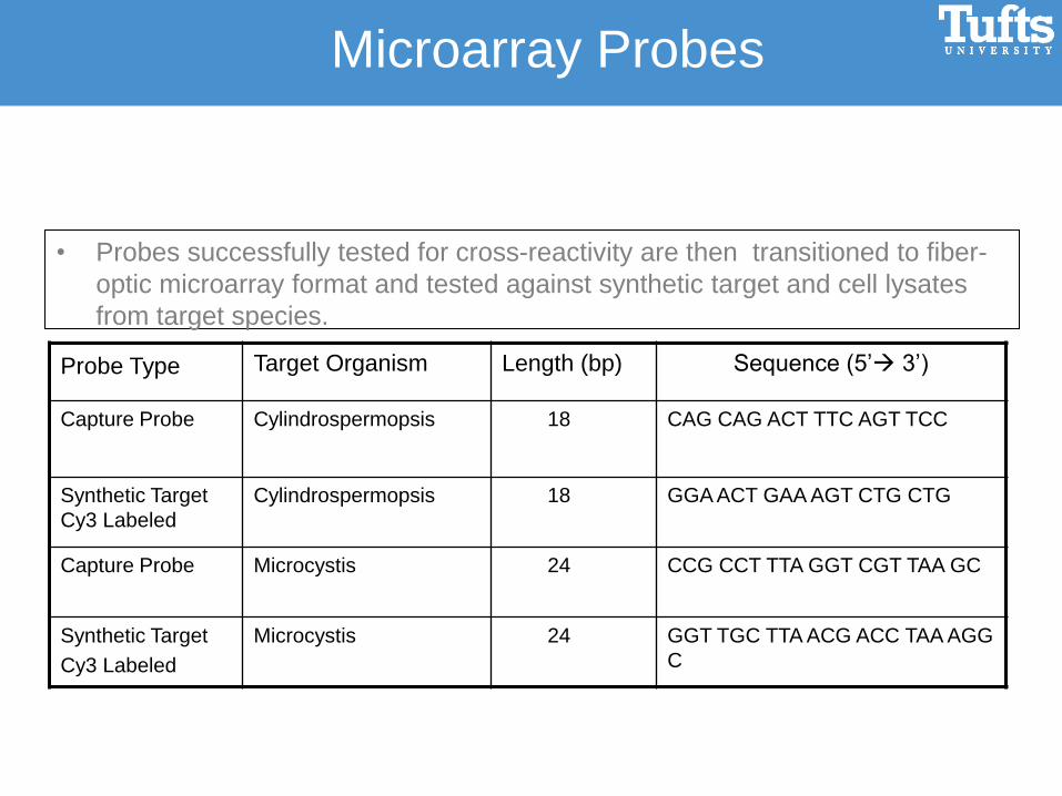
Probe Type Target Organism Length (bp) Sequence (5’ 3’)
Capture Probe Cylindrospermopsis 18 CAG CAG ACT TTC AGT TCC
Synthetic Target
Cy3 Labeled
Cylindrospermopsis 18 GGA ACT GAA AGT CTG CTG
Capture Probe Microcystis 24 CCG CCT TTA GGT CGT TAA GC
Synthetic Target
Cy3 Labeled
Microcystis 24 GGT TGC TTA ACG ACC TAA AGG
C
• Probes successfully tested for cross-reactivity are then transitioned to fiber-
optic microarray format and tested against synthetic target and cell lysates
from target species.
Microarray Probes
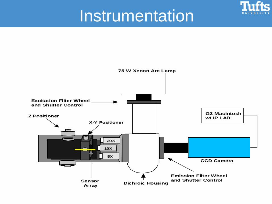
CCD Camera
75 W Xenon Arc Lamp
Emission Filter Wheel and Shutter Control
Dichroic Housing
X-Y Positioner
10X
20X
Z Positioner
Excitation Fliter Wheel and Shutter Control
G3 Macintosh w/ IP LAB
5X
Sensor Array
Instrumentation

Imaging System

Analytical Measurements
•Using the software in IPLab overlay the ROI obtained form the Eu
image onto the background and hybridization images
•Using the Measurements/ROI option measure the mean intensities of
the beads and the standard deviation of the signals