
Fgf15-mediated control of neurogenic and proneural gene expression regulates dorsal midbrain...
15
Fgf15-mediated control of neurogenic and proneural gene expression regulates dorsal midbrain neurogenesis Thomas Fischer a , Theresa Faus-Kessler a , Gerhard Welzl a , Antonio Simeone b , Wolfgang Wurst a,c,d, ⁎, Nilima Prakash a, ⁎ a Institute of Developmental Genetics, Helmholtz Centre Munich, German Research Centre for Environmental Health (GmbH), and Technical University Munich, Ingolstaedter Landstr. 1, 85764 Neuherberg, Germany b CEINGE Biotecnologie Avanzate, SEMM European School of Molecular Medicine, and Institute of Genetics and Biophysics “A. Buzzati-Traverso”, CNR, Via P. Castellino 111, 80131 Naples, Italy c Max-Planck-Institute of Psychiatry, Kraepelinstr. 2, 80804 Munich, Germany d Deutsches Zentrum für Neurodegenerative Erkrankungen (DZNE) Standort München, Schillerstr. 44, 80336 Munich, Germany abstract article info Article history: Received for publication 31 August 2010 Revised 14 November 2010 Accepted 13 December 2010 Available online 21 December 2010 Keywords: Id1/3 Hes5 Neurog1/2 Cell cycle exit Neural progenitors Mouse The balanced proliferation and cell cycle exit of neural progenitors, by generating the appropriate amount of postmitotic progeny at the correct time and in the proper location, is required for the establishment of the highly ordered structure of the adult brain. Little is known about the extrinsic signals regulating these processes, particularly in the midbrain. Fibroblast growth factor (Fgf) 15, the mouse ortholog of FGF19 and member of an atypical Fgf subfamily, is prominently expressed in the dorsolateral midbrain of the midgestational mouse embryo. In the absence of Fgf15, dorsal midbrain neural progenitors fail to exit the cell cycle and to generate the proper amount of postmitotic neurons. We show here that this is due to the altered expression of inhibitory/neurogenic and proneural/neuronal differentiation helix-loop-helix transcription factor (TF) genes. The expression of Id1, Id3, and Hes5 was strongly increased and ectopically expanded, whereas the expression of Ascl1 (Mash1), Neurog1 (Ngn1) and Neurog2 (Ngn2) was strongly decreased and transcription of Neurod1 (NeuroD) was completely abolished in the dorsolateral midbrain of Fgf15 -/- mice. These abnormalities were not caused by the mis-expression of cell cycle regulatory proteins such as cyclin-dependent kinase inhibitors or retinoblastoma proteins. Furthermore, human FGF19 promotes cell cycle exit of murine dorsal neural progenitors in vitro. Therefore, our data suggest that Fgf15 is a crucial signaling molecule regulating the postmitotic transition of dorsal neural progenitors and thus the initiation and proper progression of dorsal midbrain neurogenesis in the mouse, by controlling the expression of neurogenic and proneural TFs. © 2010 Elsevier Inc. All rights reserved. Introduction The establishment of the complex and highly ordered structure of the adult mammalian brain also requires a precisely orchestrated interplay of extrinsic and intrinsic signals during development, that regulate the spatial and temporal balance between self-renewal and cell cycle exit of neural progenitors and the generation of the appropriate numbers of postmitotic progeny. Among the intrinsic signals are TFs of the helix– loop–helix (HLH) family, playing a particularly prominent role in the control of progenitor proliferation, cell cycle exit and neuronal differen- tiation (Guillemot, 2007). These TFs are therefore classified in three groups: Proneural TFs (Ascl1 (Mash1) and Neurogenins (Neurog, Ngn)) promote the cell cycle exit of neural progenitors and initiation of neurogenesis, and the activation of Notch signaling in adjacent progeni- tors. Neuronal differentiation TFs (Neurod1 (NeuroD)) are induced by the proneural TFs in postmitotic cells and control the neuronal differentiation program. Inhibitory or neurogenic TFs (Id and Hes) directly inhibit proneural TFs or repress proneural gene expression, thereby maintaining the proliferative, undifferentiated state of neural progenitors. Cell cycle proteins, such as the cyclin-dependent kinase inhibitor (cdki) Cdkn1b (p27 Kip1 ) and the retinoblastoma (Rb) proteins Rb1, Rbl1 (p107) and Rbl2 (p130), are another type of intrinsic factors promoting cell cycle exit and differentiation of neural progenitors (Galderisi et al., 2003; Nguyen et al., 2006). In contrast to the intrinsic factors, the extrinsic signals acting upstream of these TFs or cell cycle proteins are not yet fully established, particularly in the murine midbrain. These are mostly secreted factors, such as bone morphogenetic protein (BMP)/transforming growth factor β (Tgfβ) family members promoting cell cycle exit of neural progenitors (Roussa Developmental Biology 350 (2011) 496–510 ⁎ Corresponding authors. Prakash is to be contacted at Institute of Developmental Genetics, Helmholtz Centre Munich, German Research Centre for Environmental Health (GmbH), and Technical University Munich, Ingolstaedter Landstr. 1, 85764 Neuherberg, Germany. Fax: +49 89 3187 3099. E-mail addresses: [email protected] (W. Wurst), [email protected] (N. Prakash). 0012-1606/$ – see front matter © 2010 Elsevier Inc. All rights reserved. doi:10.1016/j.ydbio.2010.12.017 Contents lists available at ScienceDirect Developmental Biology journal homepage: www.elsevier.com/developmentalbiology
-
Upload
thomas-fischer -
Category
Documents
-
view
212 -
download
0
Transcript of Fgf15-mediated control of neurogenic and proneural gene expression regulates dorsal midbrain...

Developmental Biology 350 (2011) 496–510
Contents lists available at ScienceDirect
Developmental Biology
j ourna l homepage: www.e lsev ie r.com/deve lopmenta lb io logy
Fgf15-mediated control of neurogenic and proneural gene expression regulatesdorsal midbrain neurogenesis
Thomas Fischer a, Theresa Faus-Kessler a, Gerhard Welzl a, Antonio Simeone b,Wolfgang Wurst a,c,d,⁎, Nilima Prakash a,⁎a Institute of Developmental Genetics, Helmholtz Centre Munich, German Research Centre for Environmental Health (GmbH), and Technical University Munich, Ingolstaedter Landstr. 1,85764 Neuherberg, Germanyb CEINGE Biotecnologie Avanzate, SEMM European School of Molecular Medicine, and Institute of Genetics and Biophysics “A. Buzzati-Traverso”, CNR, Via P. Castellino 111,80131 Naples, Italyc Max-Planck-Institute of Psychiatry, Kraepelinstr. 2, 80804 Munich, Germanyd Deutsches Zentrum für Neurodegenerative Erkrankungen (DZNE) Standort München, Schillerstr. 44, 80336 Munich, Germany
⁎ Corresponding authors. Prakash is to be contactedGenetics, Helmholtz Centre Munich, German Research Ce(GmbH), and Technical University Munich, IngolstaedterGermany. Fax: +49 89 3187 3099.
E-mail addresses: [email protected] ([email protected] (N. Prakash).
0012-1606/$ – see front matter © 2010 Elsevier Inc. Aldoi:10.1016/j.ydbio.2010.12.017
a b s t r a c t
a r t i c l e i n f oArticle history:Received for publication 31 August 2010Revised 14 November 2010Accepted 13 December 2010Available online 21 December 2010
Keywords:Id1/3Hes5Neurog1/2Cell cycle exitNeural progenitorsMouse
The balanced proliferation and cell cycle exit of neural progenitors, by generating the appropriate amount ofpostmitotic progeny at the correct time and in the proper location, is required for the establishment of thehighly ordered structure of the adult brain. Little is known about the extrinsic signals regulating theseprocesses, particularly in the midbrain. Fibroblast growth factor (Fgf) 15, the mouse ortholog of FGF19 andmember of an atypical Fgf subfamily, is prominently expressed in the dorsolateral midbrain of themidgestational mouse embryo. In the absence of Fgf15, dorsal midbrain neural progenitors fail to exit the cellcycle and to generate the proper amount of postmitotic neurons. We show here that this is due to the alteredexpression of inhibitory/neurogenic and proneural/neuronal differentiation helix-loop-helix transcriptionfactor (TF) genes. The expression of Id1, Id3, and Hes5 was strongly increased and ectopically expanded,whereas the expression of Ascl1 (Mash1), Neurog1 (Ngn1) and Neurog2 (Ngn2) was strongly decreased andtranscription of Neurod1 (NeuroD) was completely abolished in the dorsolateral midbrain of Fgf15−/− mice.These abnormalities were not caused by the mis-expression of cell cycle regulatory proteins such ascyclin-dependent kinase inhibitors or retinoblastoma proteins. Furthermore, human FGF19 promotes cellcycle exit of murine dorsal neural progenitors in vitro. Therefore, our data suggest that Fgf15 is a crucialsignaling molecule regulating the postmitotic transition of dorsal neural progenitors and thus the initiationand proper progression of dorsal midbrain neurogenesis in the mouse, by controlling the expression ofneurogenic and proneural TFs.
at Institute of Developmentalntre for Environmental HealthLandstr. 1, 85764 Neuherberg,
W. Wurst),
l rights reserved.
© 2010 Elsevier Inc. All rights reserved.
Introduction
The establishment of the complex and highly ordered structure of theadult mammalian brain also requires a precisely orchestrated interplay ofextrinsic and intrinsic signals during development, that regulate thespatial and temporal balance between self-renewal and cell cycle exit ofneural progenitors and the generation of the appropriate numbers ofpostmitotic progeny. Among the intrinsic signals are TFs of the helix–loop–helix (HLH) family, playing a particularly prominent role in thecontrol of progenitor proliferation, cell cycle exit and neuronal differen-tiation (Guillemot, 2007). These TFs are therefore classified in three
groups: Proneural TFs (Ascl1 (Mash1) and Neurogenins (Neurog, Ngn))promote the cell cycle exit of neural progenitors and initiation ofneurogenesis, and the activation of Notch signaling in adjacent progeni-tors. Neuronal differentiation TFs (Neurod1 (NeuroD)) are induced by theproneural TFs in postmitotic cells and control the neuronal differentiationprogram. Inhibitory or neurogenic TFs (Id and Hes) directly inhibitproneural TFs or repress proneural gene expression, thereby maintainingthe proliferative, undifferentiated state of neural progenitors. Cell cycleproteins, such as the cyclin-dependent kinase inhibitor (cdki) Cdkn1b(p27Kip1) and the retinoblastoma (Rb) proteins Rb1, Rbl1 (p107) andRbl2(p130), are another type of intrinsic factors promoting cell cycle exit anddifferentiation of neural progenitors (Galderisi et al., 2003; Nguyen et al.,2006).
In contrast to the intrinsic factors, the extrinsic signals acting upstreamof these TFs or cell cycle proteins are not yet fully established, particularlyin the murine midbrain. These are mostly secreted factors, such as bonemorphogenetic protein (BMP)/transforming growth factor β (Tgfβ)family members promoting cell cycle exit of neural progenitors (Roussa

497T. Fischer et al. / Developmental Biology 350 (2011) 496–510
et al., 2006; Siegenthaler and Miller, 2005); Wnt family membersmaintaining the proliferative state of neural progenitors, repressingtheir differentiation, or promoting neurogenesis and neuronal fatespecification in a time- and context-dependent manner (Hirabayashiet al., 2004; Kuwabara et al., 2009;Megason andMcMahon, 2002;Wexleret al., 2009); and members of the fibroblast growth factor (Fgf) family,which so far have been mostly implicated in the maintenance of theproliferative neural progenitor state (Mason, 2007).
The Fgf family comprises 22 members in mammals, classified inseven subfamilies (Itoh andOrnitz, 2008;Mason, 2007). Fgf15/19, Fgf21and Fgf23 constitute an atypical Fgf subfamily based on theirevolutionary relationship and on the facts that these Fgfs aretranscriptionally regulated by members of the nuclear receptor classof ligand activated TFs, possess low-affinity heparin-binding sites andtherefore act in an endocrine manner, and require Klotho/β-Klothotransmembrane proteins for efficient signaling via Fgf receptors (Itohand Ornitz, 2008; Jones, 2008). Mouse Fgf15, despite its low amino acididentity (between 32% and 51%), is considered the structural andfunctional ortholog of human, chick and zebrafish FGF19/Fgf19 (Miyakeet al., 2005;Nishimura et al., 1999;Wright et al., 2004). Fgf15 expressionis confined mostly to the developing central nervous system (CNS)(Gimeno et al., 2003; Ishibashi and McMahon, 2002; McWhirter et al.,1997), but its functionwasfirst described in non-neural tissues,where itcontrols bile acid homeostasis, gall bladder filling and proper morpho-genesis of the cardiacoutflow tract (Choi et al., 2006; Inagaki et al., 2005;Vincentz et al., 2005). However, a recent report indicated that Fgf15suppresses proliferation and promotes neural differentiation duringneocortical development, although the mechanism of this proneuralactivity of Fgf15 remained unclear (Borello et al., 2008). Here we showthat extrinsic Fgf15 is a crucial signaling molecule regulating theexpression of intrinsic inhibitory/neurogenic and proneural HLH TFs inthe mouse midbrain, thereby controlling the cell cycle exit of dorsalneural progenitors and their differentiation into neurons.
Materials and methods
Mouse strains
CD-1 mice were purchased from Charles River (Kisslegg/Germany).Generation and genotyping of Fgf15+/−mice in amixed C57BL/6-129/Svbackground was described by Wright et al. (2004). These mice wereoutcrossed with CD-1 mice for 12 generations (F12). Fgf15−/− embryoswere analyzed from F1 onwards and phenotypic differences were notdetected between F1 and F12 embryos. Collection of embryonic stageswas done from timed-pregnant females of heterozygous (Fgf15+/−)intercrosses, noon of the day of vaginal plug detectionwas designated asembryonic day0.5 (E0.5). Embryoswere additionally staged according toTheiler (1989). Mutant embryos were always compared to their wild-type (Fgf15+/+) littermates and at least 3 embryos were analyzed foreach probe, genotype and stage, if not otherwise indicated in the text.Animal treatment was conducted under federal guidelines and wasapproved by the HMGU Institutional Animal Care and Use Committee.
Radioactive in situ hybridization (ISH)
Paraffin sections (8 μm)were processed for radioactive ([α-35S]UTP,Amersham/UK) ISH as described in Fischer et al. (2007). Riboprobesused were Fgf15 (McWhirter et al., 1997), Fgf8, Shh, Pax6 and En1(Puelles et al., 2003), Wnt1 (Fischer et al., 2007), Wnt3a (Parr et al.,1993), Id1 (Benezra et al., 1990), Id3 (Christy et al., 1991), Hes5(Akazawa et al., 1992),Hes3 (Hirata et al., 2001),Ascl1 (Mash1),Neurog1(Ngn1), Neurog2 (Ngn2) and Neurod1 (NeuroD) (Cau et al., 1997;Ma et al., 1997), Helt (Mgn) (Guimera et al., 2006a),Dll1 (Bettenhausenet al., 1995), Dusp6 (Mkp3) (Echevarria et al., 2005), Fgfr1-3 (Blak et al.,2005), Spry2 (Minowada et al., 1999), Etv4 (Pea3) and Etv5 (Erm) (Blaket al., 2007). Images were taken with an Axioplan2 microscope or
StemiSV6 stereomicroscope using bright- and dark-field optics,AxioCam MRc camera and Axiovision 4.6 software (Zeiss/Germany),and processed with Adobe Photoshop 7.0 or CS software (Adobe SystemsInc./USA).
Immunohistochemistry (IHC)
Antigenswere detected onparaffin (8 μm)or cryosections (16 μm)asreported by Brodski et al. (2003) and Puelles et al. (2004). Primarymonoclonal antibodies usedweremouse anti- 5-bromo-2′-deoxyuridine(BrdU) (1:10; Roche Diagnostics/Germany) and βIII-Tubulin (Tubb3,TuJ1) (1:5000; Chemicon/USA), and rat anti-Nestin (1:3; BD Pharmin-gen/USA). Polyclonal antisera used were rabbit anti-cleaved Caspase 3(cCasp3) (1:200; Cell Signaling/USA), phosphorylated Histone H3 (pH3)(1:1000; Upstate/USA), Ki-67 (1:100; Vision BioSystems/UK) andDoublecortin (Dcx) (1:80, gift from O. Reiner, Weizmann Institute ofScience/Israel). Secondary antibodies were either fluorescently labelled(Cy3/Cy2) or coupled to biotin/streptavidin-horseradish-peroxidase(Jackson ImmunoResearch Laboratories/USA), and detected using theVECTOR® M.O.M.™ and Vectastain ABC System (Vector Laboratories/USA). Fluorescent images were taken with an Axiovert 200 M invertedmicroscope (Zeiss) and processed with Adobe Photoshop 7.0 or CSsoftware.
BrdU treatments
Intraperitoneal (i.p.) injections of pregnant dams with 31 μg BrdU(Sigma/Germany)/g body weight were performed on E11.5 for single(10 min) or cumulative (3× every 2 hours (hrs)) labeling. Embryoswere dissected 10 min (single labeling) or 2 hrs (cumulative labeling)after the last injection and processed for immunodetection of BrdU.
Cell countings
Ki67+ and pH3+ cells were counted on serial coronal sections fromE11.5 embryos using the Neurolucida 6 software (MBF Bioscience/USA).Cell numbers were averaged for each genotype and subjected to tests forthe estimationof statistical significance asdescribed in Statistical analyses.
Cell cycle exit assay
Pregnant dams from Fgf15+/− intercrosses were injected i.p. withBrdU (31 μg/g body weight) on E10.5 (t0). Embryos were dissected24 hrs later (t24) at E11.5 and processed for fluorescent immunodetec-tion of BrdU and Ki-67. Stained sections were evaluated with a confocallaser scanning microscope (LSM 510 META, Zeiss). A z-stack imageseries in intervals of 1 μm was recorded from each section comprisingthe entire thickness of the tissue. The index Tc of cell cycle re-entry after24 hrs was calculated by dividing the number of Ki-67+/BrdU+ double-labeled cells by the total number of BrdU+ cells.
Immunoblotting
The anterior neural tube (fore-/midbrain and rhombomere 1) ofE11.5 embryos from Fgf15+/− intercrosses was dissected free ofnon-neural tissues and eye cups in ice-cold tissue lysis buffer (20 mMTris pH7.5, 150 mM NaCl, 1 mM EDTA, 1 mM EGTA, 1% Triton X-100,protease and phosphatase inhibitors (complete Mini and PhosSTOP,Roche)) and homogenized in 100 μl tissue lysis buffer. The remainingembryonic tissue was used for PCR genotyping. Total protein contentwas determined by Bradford assay (Sigma-Aldrich/Germany). Equalamounts of total protein from tissue lysates were separated in 4–12%Criterion XT Bis-Tris Precast gels (Bio-Rad/Germany) together withcontrols and a biotinylated protein ladder following themanufacturer'sinstructions (PhosphoPlus Rb antibody kit, Cell Signaling), and blottedonto nitrocellulose membranes (Hybond-ECL, GE Healthcare Europe/

498 T. Fischer et al. / Developmental Biology 350 (2011) 496–510
Germany). Blots were probed with rabbit anti-phosphorylated RB1(Ser807/811) (1:1000; Cell Signaling) and anti-RBL1 (p107) (1:2000;Santa Cruz Biotechnology/USA); and mouse anti-Cdkn1b (p27Kip1)(1:2500; BD Pharmingen), anti-RB1 (p110) (1:500; BD Pharmingen)and anti-active (dephosphorylated) β-Catenin (1:1000; Upstate) anti-bodies. Membranes were stripped in 0.2 M Glycine-HCl pH2.5, 0.1%Tween-20, and reprobed with mouse anti-β-Actin antibody (1:5000;Abcam/UK) as a loading control. Densitometric analysis of theimmunoblots was performed with a GS-800 Calibrated Densitometer(Bio-Rad) and ImageJ 1.34 s software (NIH/USA). Variations in totalprotein contentwere correctedwithin each blot by using the amount ofβ-Actin protein as standard.
Primary cell cultures and clonal analyses
Primary cortical progenitor cell cultures were prepared from E12.5CD-1 embryos as described inHartfuss et al. (2001). Cellswereplated at adensity of 800,000 cells/well on poly-D-lysine coated coverslips andinfected with a green fluorescent protein (GFP)-expressing retrovirus2 hrs after platingas reportedbyMalatesta et al. (2000). Threehours afterretroviral infection and with each medium change, recombinant humanFGF19 protein (R&D Systems/USA) was added to a final concentrationof 5 ng/ml. After 7 days in culture, cells were fixed and processed forimmunocytochemical detection of GFP and Tubb3 as described inBerninger et al. (2007). GFP+ cell clones were evaluated with anAxioplan2microscope/AxioCamHRmcamera (Zeiss), and the number of
Fig. 1. Fgf15 is expressed indorsal regionsof themousemidbrain. (a–t) Representative sagittal (coronal level indicatedby broken lines ina, f, k, p)ofwild-type (CD-1)mouseembryos atE9.5 (ai, s), respectively. Insets in (c, h, m, r, o) are pseudo-colored overlays of consecutive sectionsoverlapping expression domains appear in yellow. Fgf15 is expressed in the dorsal di-, mes-arrowheads ing and insets inh,m, r), and sparing theRP (white arrowhead ine, j, t) and the Shh+
Di, diencephalon; DT, dorsal thalamus; FP, floor plate; Mes, mesencephalon; Met, metencepha
GFP+ andGFP+/Tubb3+double-labeled cellswas counted for each cloneusing Axiovision 4.6 software (Zeiss). The mean was calculated fromthe total number of clones analyzed with or without FGF19 treatment±s.e.m. Data were derived from four independent experiments.
Statistical analyses
For analysis of the effect of an experimental factor (genotype ortreatment) with data from different brain regions or severalindependent experiments, two-way ANOVA was applied. First, anANOVA with interaction between factor and experiment was fitted. Ifthe interaction effect was significant, t-tests were performed for theindividual experiments. Otherwise a two-way ANOVA withoutinteraction effect was fitted, and post-hoc t-tests with P-valueadjustment according to Holm (1979) were performed. For cellcycle experiments with several sections per embryo, genotypes werecompared by one-way ANOVA for repeated measurements. For allcalculations, the R software with the nlme package was used(Pinheiro et al., 2008; R Development Core Team, 2009).
Results
Fgf15 is expressed in dorsal regions of the mouse midbrain
We first mapped the expression of Fgf15 in relation to Fgf8 and Shh,two crucial signaling molecules controlling antero-posterior (A/P) and
a–c, f–h, k–m,p–r) andcoronalmidbrain (d, e, i, j, n, o, s, t) sections (anterior left, dorsal top;–e), E10.5 (f-j), E11.5 (k-o) andE12.5 (p–t). (b, g, l, q, e, j, t) aredarkfield views of (a, f ,k, p, d,hybridized with probes for Fgf15 (red) and Fgf8 (green in c, h, m, r) or Shh (green in o);and metencephalon, with a gap in the rostral Fgf8+ domain at the MHB (white arrow/BPand FP (inset ino). Abbreviations:AP, alarplate; BP, basal plate; CbA, cerebellar anlage;
lon; POA, preoptic area; RP, roof plate; SA, septal area; T, tectum; Tel, telencephalon.

499T. Fischer et al. / Developmental Biology 350 (2011) 496–510
dorso-ventral (D/V) patterning of the brain (Fig. 1). Between E9.5 andE12.5, Fgf15 expressionwas confinedmostly to dorsal regions of the di-,mes- and metencephalon and exhibited a decreasing gradient at bothsides of the isthmic constriction (Fig. 1). Metencephalic Fgf15transcription overlapped with the caudal Fgf8+ domain, but mesence-phalic Fgf15 expression showed a sharp, non-overlapping border withthe rostral Fgf8+domain at themid-/hindbrain boundary (MHB). As themidbrain was one of the most prominent sites of Fgf15 expressionwithin the neural tube of themidgestationalmouse embryo,we focusedour analysis on this brain region. From E9.5 to E11.5, Fgf15 wasexpressed throughout the neuroepithelium in the dorsal two thirds ofthe mesencephalon, sparing the roof plate (RP) and abutting the Shh+
floor (FP) and basal plate (BP) (Fig. 1). Notably, Fgf15 expression refinedtodiscrete sites at both sides of theRPandof the BP atE12.5,with lowestFgf15 transcription in an intermediate zone locatedapproximately at thelevel of the dorsolateral sulcus of the midbrain (Fig. 1). Thus, Fgf15expression is confined to dorsolateral regions of the midgestationalmouse midbrain and does not overlap with Fgf8 and Shh.
Embryonic lethality and CNS defects in outbred Fgf15−/− mice
The lack of a CNS phenotype in Fgf15−/− mice on a mixed C57BL/6-129/Sv and inbred C57BL/6 genetic background (Vincentz et al., 2005;Wright et al., 2004) (our ownobservations), despite theprominent neuralexpression of Fgf15 in themidgestational mouse embryo, prompted us tooutcross the Fgf15+/−mice into a CD-1 genetic background. The genotypeand morphologically abnormal phenotype distribution of embryosobtained from Fgf15+/− intercrosses in the first five (F1-F5), tenth (F10)and twelfth (F12)outcrossgenerationsare summarized inSupplementaryTable 1. In thefirst and second (F1–F2) outcross generation, the frequencyof Fgf15−/− embryos recovered at E13.5 or later was much lower thanexpected by the Mendelian ratios and, in particular, no Fgf15−/−mutants
Fig. 2.Morphological and CNS defects in outbred Fgf15−/− embryos. (a–d) Representative iman outbred (CD-1) genetic background. More than 30% of the outbred Fgf15−/− embryos dispretardation at this stage. (e–h) Representative sagittal (e) and coronal (f–h) Nissl-stained seFgf15−/− embryos, showing a massive overgrowth and infolding of dorsal neural tissues in
were found at E17.5 and E18.5. From the third (F3) outcross generationonwards, Fgf15−/− embryos were recovered at approximatelyMendelianratios up to E11.5, indicating that most mutant embryos died after thistime-point during development. The outer inspection of the Fgf15−/−
embryos showed that around 10% of the E11.5 mutants, 20% of the E12.5mutants, and more than 30% of the E13.5 mutants displayed mor-phological abnormalities such as a poorly vascularized chorion, paleappearance and a clear growth retardation (Suppl. Table 1 and Fig. 2).Sagittal and coronal sections of these morphologically abnormal mutantembryos revealed a massive overgrowth and infolding of dorsal neuraltissues (Fig. 2). Furthermore, apoptotic cell death was dramaticallyincreased in the Fgf15−/− embryosdisplaying an abnormalmorphology atE12.5, whereas it did not differ from their wild-type littermates at E11.5(Suppl. Fig. S1). All the other Fgf15−/− embryos were morphologicallyindistinguishable from their wild-type littermates (Suppl. Table 1). Theblood vessels of the perineural vascular plexus had developed normallyand were filled with erythrocytes in the E11.5 Fgf15−/− embryos(Suppl. Fig. S1), indicating that blood supply of neural tissues was intactin the mutant embryos up to this stage. These results showed that theFgf15 null mutation in an outbred (CD-1) genetic background wasembryonic lethal after E11.5, and that up to one tenth of the Fgf15−/−
embryos displayed severe CNS defects already at this stage.
Dorsal neural progenitors fail to exit the cell cycle in Fgf15−/− embryos
The massive overgrowth and infolding of dorsal neural tissues inmore than one third of the outbred Fgf15−/− embryos at E13.5suggested a more general defect of dorsal neural tube development inthe mutant embryos. Since the frequency of morphological CNSdefects increased after E11.5 in outbred Fgf15−/− embryos, and mostof these embryos appeared to die 2 days later, we focused our analyseson morphologically normal Fgf15−/− embryos at E11.5. The
ages of an E13.5 wild-type (Fgf15+/+) embryo (a, b) and its Fgf15−/− littermate (c, d) onlayed a poorly vascularized chorion membrane, pale appearance and manifest growth-ctions (anterior left, dorsal top; coronal level indicated by broken lines in e) from E13.5these mutant embryos.

Fig. 4. Increased Nestin expression and reduced amount of Dcx+/Tubb3+ postmitotic neurons in the Fgf15−/− dorsolateral midbrain. (a–h) Representative close-up views of thedorsal midbrain (pial surface top) on coronal sections from wild-type (Fgf15+/+) (a, c, e, g) and Fgf15−/− (b, d, f, h) embryos at E11.5 (a–f) and E12.5 (g, h). Nestin expression inthe Fgf15−/− dorsolateral midbrain was increased at E11.5 (a, b), and appeared distorted at E12.5 (g, h). Postmitotic Dcx+ and Tubb3+ neurons occupied only one cell layer withinthe mantle zone (MZ) of the mutants, as compared to the 2–3 cell layers in wild-type embryos (white brackets in c–f). (i, j) Representative coronal midbrain sections fromwild-type(Fgf15+/+) (i) and Fgf15−/− (j) embryos at E12.5. Nestin expression was strongly increased in the Fgf15−/− dorsolateral midbrain (j), where Fgf15 is highly expressed in the wild-type embryo (i). Nestin was very weakly expressed in the Fgf15−/− ventral midbrain at this stage, where Fgf15 is normally not expressed (arrows in i, j).
501T. Fischer et al. / Developmental Biology 350 (2011) 496–510
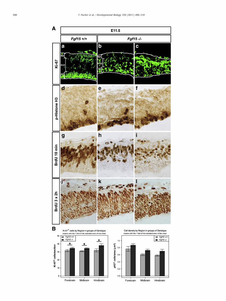
expression of Pax6, En1, Fgf8, Shh,Wnt1 andWnt3awas not affected inthe mutant embryos (Suppl. Figs. S2 and S3), suggesting that regionalidentities were properly induced in the absence of Fgf15. We noted,however, that all mutants exhibited a thinner dorsal neuroepithelium,and we thus assessed neural progenitor proliferation in theseembryos. The total number of proliferating Ki-67+ cells wassignificantly increased in the Fgf15−/− embryos, which in additionwere ectopically positioned throughout the dorsal midbrain neuroe-pithelium, although the numbers and distribution of phospho-HistoneH3+ (pH3+) and BrdU+ cells were not severely affected in themutants (Fig. 3). These data suggested that the number of cycling
Fig. 3. Increased numbers of proliferating dorsal neural progenitors in the Fgf15−/− embryos. (A)from E11.5 wild-type (Fgf15+/+) (a, d, g, j) and Fgf15−/− (b, c, e, f, h, i, k, l) embryos. The Fgf15−
ProliferatingKi-67+cellswere increased andectopically positioned in the Fgf15−/−dorsal neuroeBrdU+cellswas not severely affected in themutants (d–l). Note the thinner neuroepithelium in thembryos. (m) The number of Ki-67+ cells/sectionwas increased from 31.67±1.43 (Fgf15+/+) to(Fgf15−/−) in the dorsal midbrain, and from 32.0±1.95 (Fgf15+/+) to 38.13±2.06 (Fgf15−/−) isignificant in the two-wayANOVA (asterisk, P=0.035). In post-hoc t-testswith P-value adjustmewere increased from0.77±0.07 (Fgf15+/+) to 0.87±0.04 (Fgf15−/− in thedorsal forebrain, from(Fgf15+/+) to 0.70±0.05 (Fgf15−/−) in the dorsal hindbrain (n=4/genotype; mean±s.e.m.). TAbbreviations: MZ, mantle zone; SVZ, subventricular zone; VZ, ventricular zone.
dorsal neural progenitors was significantly increased in the Fgf15−/−
embryos.The thinning of the mutant dorsal neuroepithelium also suggested
that despite the increased numbers of cycling progenitors, these cellsdid not generate the proper numbers of postmitotic progeny. Indeed,Doublecortin+ (Dcx) and βIII-Tubulin+ (Tubb3, TuJ1) postmitoticneurons were remarkably reduced and occupied only 1 cell layer inthe mantle zone (MZ) of the mutant dorsal midbrain instead of the2–3 cell layers detected in wild-type embryos (Fig. 4). By contrast,expression of the intermediate filament Nestin, a marker for neuralstem and progenitor cells that is down-regulated as soon as these cells
Representative close-up views of the dorsal midbrain (pial surface top) on coronal sections/− embryos in (c, f, i, l) displayed a stronger phenotype than the ones shown in (b, e, h, k).pithelium(a–c), although thenumber anddistribution of phospho-HistoneH3+(pH3+)ande Fgf15−/− embryos. (B)Quantification of Ki-67+ and pH3+ cells inwild-type and Fgf15−/−
34.63±1.27 (Fgf15−/−) in the dorsal forebrain, from 30.89±0.63 (Fgf15+/+) to 34.36±0.9n the dorsal hindbrain (Fgf15+/+, n=3; Fgf15−/− n=4; mean±s.e.m.). This increase wasnt, therewasno significance for any of the brain regions separately. (n) pH3+ cells/1000 μm2
0.60±0.03 (Fgf15+/+) to 0.73±0.04 (Fgf15−/− in thedorsalmidbrain, and from0.58±0.02his increase was not significant (P=0.063 for the genotype effect in the two-way ANOVA).

502 T. Fischer et al. / Developmental Biology 350 (2011) 496–510
become postmitotic (Lendahl et al., 1990), was strongly increased inthe dorsal midbrain of E11.5 Fgf15−/− embryos (Fig. 4). The aberrantexpression of Nestin was even more pronounced in the mutantembryos at E12.5. Immunostaining for Nestin at this stage wasstrongest and appeared severely distorted in the Fgf15−/− dorsolat-eral midbrain, where Fgf15 is highly expressed in the midgestationalwild-type embryo, whereas it was very weak in the mutant ventralmidbrain, where Fgf15 is normally not expressed (Fig. 4). Theseresults suggested that Fgf15 directly controls the postmitotictransition of dorsal neural progenitors in the midbrain.
The increased numbers and ectopic distribution of Ki-67+ prolifer-ating cells, aswell as the corresponding reductionof Tubb3+postmitoticneurons, strongly suggested that dorsal neural progenitors failed to exitthe cell cycle in the Fgf15−/− embryos. Therefore, we injected BrdU intopregnant Fgf15+/− females at E10.5, 24 hrs before dissection of theembryos at E11.5. Double-staining for BrdU and Ki-67 should reveal thefraction of BrdU+/Ki67+ cells that had re-entered the cell cycle duringthis 24 hr-period (Fig. 5). Compared to E11.5 control embryos, thisfraction was significantly increased by 60% in the dorsal neuroepithe-
Fig. 5. Dorsal neural progenitors fail to exit the cell cycle and are ectopically positioned in Fgf1progenitors expressKi-67 (Ki-67+/BrdU− cells, greenovals). Cells in the S-phaseof thecell cycle infraction of the Ki-67+/BrdU+ progeny had exited the cell cycle (G0-phase) and initiated neuronacycle.A cell cycle exit defect in themutant embryos leads toanaccumulationofKi-67+/BrdU+prothe cell cycle and become postmitotic (Ki-67−/BrdU+, red), theymigrate out of theVZ into theMmostly to theVZ in thewild-type (Fgf15+/+), but are ectopically positioned in the SVZ/MZof the Fon coronal sections from wild-type (Fgf15+/+) (c) and Fgf15−/− (d, e) embryos at E11.5 (t24), trwhite arrows in c, d) are increased and ectopically positioned in the mutants. Approximately 2(percentageofKi-67+andBrdU+cells/total BrdU+cells) of cell cycle re-entry after 24 hrswas sign24.28±0.61, n=4; mean±s.e.m.) (f), whereas the percentage of Ki-67− and BrdU+ cells/toapproximately 11% in the Fgf15−/− embryos (Fgf15+/+, 84.86±0.41, n=3; Fgf15−/−, 75.72measurements ANOVA. Abbreviations: G0, G1, S, G2, M, phases of the cell cycle; MZ, mantle zone
lium of the Fgf15−/− embryos, whereas the complementary fraction ofBrdU+/Ki-67− cells that had exited the cell cycle during the 24 hr-periodwas significantly decreased by approximately 11% in themutants(Fig. 5). Moreover, BrdU+/Ki-67+ progenitors were ectopically posi-tioned in the SVZ of allmutant embryos analyzed, and even found in theMZ in about 20% of the Fgf15−/− embryos (Fig. 5). These data confirmedthat dorsal neural progenitors failed to exit the cell cycle and wereectopically located in the mesencephalic neuroepithelium of theoutbred Fgf15−/− embryos.
Neurogenic/inhibitory genes are upregulated and proneural genes aredownregulated in the Fgf15−/− dorsolateral midbrain
Dorsal neural progenitors might fail to exit the cell cycle in theFgf15−/− embryos due to the de-regulation of crucial cell cycleproteins, such as cdki or Rb proteins (Galderisi et al., 2003); or due tothe misexpression of neurogenic and proneural genes that in turnregulate cell cycle exit and neuronal differentiation (Bertrand et al.,2002). No significant variation in the levels of phosphorylated Rb1
5−/− embryos. (A) Scheme depicting the experimental paradigm. (a) Proliferating neuralcorporateBrdUat E10.5 (t0) (Ki-67+/BrdU+cells, yellowovals).After 24 hrs (at E11.5; t24), al differentiation (Ki-67−/BrdU+ cells, red ovals), while the remaining progeny continued toliferatingcellsandreductionof theKi-67−/BrdU+G0 fraction. (b)Asneuroepithelial cells exitZ. Ki-67+/BrdU− (green) and Ki-67+/BrdU+ (yellow) proliferating progenitors are confinedgf15−/− embryos. (B) Representative close-upviews of thedorsalmidbrain (pial surface top)eated with BrdU at E10.5 (t0). Ki-67+ (green)/BrdU+ (red) double-labeled cells (in yellow;0% of the Fgf15−/− embryos showed a stronger cell cycle exit defect (e). (C) The index Tcificantly increasedby60% in theFgf15−/− embryos (Fgf15+/+, 15.14±0.41, n=3; Fgf15−/−,tal BrdU+ cells that had exited the cell cycle after 24 hrs was significantly reduced by±0.61, n=4; mean±s.e.m.) (g); triple asterisks: Pb0.0001 in the one-way repeated-; SVZ, subventricular zone; VZ, ventricular zone.

503T. Fischer et al. / Developmental Biology 350 (2011) 496–510
(the inactive form of Rb1), Rbl1 (p107) and Cdkn1b (p27Kip1) wasobserved in the Fgf15−/− anterior neural tube at E11.5 (Suppl. Fig. S4),suggesting that the expression of these cell cycle proteins was notaffected by the lack of Fgf15. An increased activation of the Wnt/β-Catenin pathway was previously reported in the Fgf15−/− cortex(Borello et al., 2008), and the phenotype of the morphologicallyabnormal Fgf15−/− embryos indeed resembled a mouse mutant over-expressing a constitutively active form of β-Catenin (Chenn andWalsh, 2002). However, the protein levels of the activated (depho-sphorylated) form of β-Catenin were not changed between wild-typeand Fgf15−/− brains at E11.5 (Suppl. Fig. S4).
We then analyzed the expression patterns of different HLH TFs inthe Fgf15−/− embryos between E9.5 and E11.5. These TFs may begrouped in neurogenic factors sustaining proliferation (Hes3 andHes5; (Kageyama et al., 2007)), inhibitors of differentiation (Id1 andId3; (Ruzinova and Benezra, 2003)), or proneural factors promoting
Fig. 6. Strongly increased and ectopic expression of Id and Hes genes in the Fgf15−/− dorsolawild-type (Fgf15+/+) (a, b, e, f, i, j, m, n) and Fgf15−/− (c, d, g, h, k, l, o, p) embryos at E10.5 (higher magnifications of the boxed areas in (m, o). Images in (a–d) were pseudo-colored fo(a, c) in the ventrolateral midbrain and of Id3 (b, d) in the dorsolateral midbrain of the Fgf15−
and Id3 (i–l) expression was strongly increased and ectopically expanded (white arrowhearrowheads in p) of the dorsolateral midbrain neuroepithelium in mutant embryos. Abbrev
cell cycle exit and neuronal differentiation (Ascl1 (Mash1), Neurog1(Ngn1), Neurog2 (Ngn2) and Neurod1 (NeuroD); (Bertrand et al.,2002)). Remarkably, a strong increase and ectopic expansion of Hes5and Id3 transcription in the ventrolateral and dorsolateral Fgf15−/−
midbrain was first detected at E10.5, and at E11.5, Id1, Id3 and Hes5were strongly up-regulated and ectopically expressed in the mutantdorsal midbrain (Fig. 6). By contrast, the transcription of theproneural bHLH TFs Ascl1 and Neurog1 was diminished in themutant dorsolateral midbrain at E9.5 and E10.5, respectively,(Fig. 7). At E11.5, the expression of Ascl1, Neurog1 and Neurog2was heavily reduced, and that of Neurod1, acting downstream ofAscl1 and Neurog2 (Guillemot, 2007), was totally silenced in theFgf15−/− dorsolateral midbrain (Fig. 7). These findings indicated thatFgf15 controls the cell cycle exit and differentiation of dorsalmidbrain neural progenitors by suppressing the expression ofneurogenic and inhibitory Hes5 and Id3, and/or by activating the
teral midbrain. (a–p) Representative coronal midbrain hemisections (dorsal top) froma-d) and E11.5 (e–p). (f, h, j, l) are darkfield images of (e, g, i, k), respectively. (n, p) arer better visualization of the ISH signal. A strong increase and ectopic expression of Hes5/− embryos was already evident at E10.5 (black arrowheads in c, d). At E11.5, Id1 (e–h)ads in h, l), whereas Hes5 (m–p) expression extended ectopically into the MZ (whiteiations: MZ, mantle zone; SVZ, subventricular zone; VZ, ventricular zone.

504 T. Fischer et al. / Developmental Biology 350 (2011) 496–510
expression of proneural Ascl1, Neurog and neuronal differentiationNeurod1 HLH TFs.
Next, we analyzed the expression of two other genes involved indorsal midbrain neurogenesis in the Fgf15−/− embryos. Transcriptionof the bHLH TF Helt (Heslike, Megane), required for the generation ofdorsal midbrain GABAergic neurons (Guimera et al., 2006b; Nakataniet al., 2007), was strongly decreased in the mutant dorsolateralmidbrain at E11.5 (Fig. 8). Expression of the Notch ligand Delta-like 1(Dll1), which is activated by Ascl1 and Neurog2 and promotesneuronal differentiation (Bertrand et al., 2002), was also down-regulated in the Fgf15−/− dorsolateral midbrain (Fig. 8), suggestingthat dorsal midbrain neurogenesis was blocked along the proneuralpathway in the absence of Fgf15.
Fgfr3 transcription is reduced in the Fgf15−/− midbrain
The intracellular signal transduction pathway activated by Fgf15 inthe murine anterior neural tube is still unknown (Borello et al., 2008).We therefore analyzed the expression of Fgf signaling pathwaycomponents in the Fgf15−/− midbrain, including the Fgf receptors(Fgfr) 1, 2 and 3, the intracellular negative regulators and at the sametime target genes of the mitogen-activated protein kinase (MAPK)pathway Dual specificity phosphatase 6 (Dusp6, Mkp3) and Sprouty 2(Spry2), and the Fgf/MAPK target genes of the Ets family, Etv4 (Pea3)and Etv5 (Erm) (Mason, 2007). Expression of Dusp6 was ectopicallyexpanded in the mutant dorsolateral midbrain, whereas the tran-
Fig. 7. Reducedor lost expressionofproneural genes in the Fgf15−/−dorsolateralmidbrain. (a–l) Rg, i, k) and Fgf15−/− (b, d, f, h, j, l) embryos at E9.5 (a, b), E10.5 (c, d) and E11.5 (e–l). Ascl1 (Mashstrongly decreased in themutant dorsolateralmidbrain at E11.5 (open arrowheads in b, d, f, h). Trcompletely abolished in the Fgf15−/− dorsolateral midbrain at E11.5 (open arrowheads in j, l). A
scription of Fgfr3 was notably reduced in the VZ/SVZ of the Fgf15−/−
midbrain including ventral domains (Fig. 8). Expression of all othergenes (Fgfr1, Fgfr2, Spry2, Etv4 and Etv5) was not altered in themutant midbrain (Fig. 8). These results indicated that Fgf15 isrequired, directly or indirectly, for the regulation of Dusp6 and Fgfr3transcription in the mouse midbrain.
The human ortholog FGF19 promotes cell cycle exit of mouse corticalprogenitors in vitro
Based on the previous findings, we tested if recombinant humanFGF19 protein, the ortholog of mouse Fgf15, promotes cell cycle exitand neuronal differentiation of cultured neural progenitor cells. Wedecided to use primary cortical cultures from E12.5 wild-type (CD-1)embryos for two reasons: first, because most cortical cells at this stageare neural progenitors (Borello et al., 2008), whereas dorsal midbrainneurogenesis is already peaking at E11.5–12.5 in the mouse (Edwardset al., 1986); second, because the amount and viability of neuralprogenitor cells that can be isolated from the mouse dorsal midbrain atE10.5–11.5 is very limited (our own observations). To follow the fate ofproliferating progenitors, cultures were infected shortly after platingwith a GFP-expressing retrovirus that can only integrate in replicatingcells (Malatesta et al., 2000). Cell cultures were maintained in thepresence of recombinant human FGF19 protein for 7 days. At day 7, wedetermined the number of GFP+ cells (cells that were proliferating atthe time of retroviral infection and their progeny) and the proportion of
epresentativecoronalmidbrainhemisections (dorsal top) fromwild-type(Fgf15+/+) (a, c, e,1; a, b, e, f) andNeurog1 (Ngn1; c, d, g, h) expression was already reduced at E9.5–10.5 andanscription ofNeurog2 (Ngn2; i, j) was strongly decreased and ofNeurod1 (NeuroD; k, l)wasbbreviations: MZ, mantle zone; SVZ, subventricular zone; VZ, ventricular zone.

Fig. 8. Aberrant expression of other proneural genes and Fgf signal transduction components in the Fgf15−/−midbrain. (a–r) Representative coronal midbrain hemisections (dorsal top) fromE11.5wild-type (Fgf15+/+) (a, c, e, g, i, k,m,o, q) and Fgf15−/− (b, d, f, h, j, l, n, p, r) embryos. (a, b, k, l)brightfield images, (c–j,m–r)darkfield images.Helt (Mgn; a, b) andDll1 (c, d)expressionwasdecreased (open arrowheads in b, d), whereasDusp6 (Mkp3; e, f) transcriptionwas increased (white arrowheads in f) in themutant dorsolateral midbrain. Fgfr3 (k, l) transcriptionwas notablydecreased in the entire Fgf15−/−midbrain (open arrowheads in l). Expression of Fgfr1 (g, h), Fgfr2 (i, j), Spry2 (m, n), Etv4 (Pea3; o, p) and Etv5 (Erm; q, r)was not altered in themutantmidbrain.
505T. Fischer et al. / Developmental Biology 350 (2011) 496–510
these cells co-expressing Tubb3 (proliferating progenitors that haddifferentiated into neurons during the culture period) in the treatedversus untreated cultures (Fig. 9). The total number of GFP+ cells wassignificantly reduced after FGF19 treatment, indicating that FGF19promoted the cell cycle exit of proliferating cortical progenitors inculture (Fig. 9). Moreover, the proportion of Tubb3+/GFP+ double-labeled cells tended to be increased in the FGF19-treated cultures, but itdid not reach statistical significance (Fig. 9). We therefore concludedthat treatment of mouse cortical progenitors with FGF19 significantlyenhanced their cell cycle exit and tended to increase their neuronaldifferentiation.
Discussion
Here we show that Fgf15 controls the expression of neurogenicand proneural HLH TFs in the dorsolateral midbrain of the developingmouse embryo, thereby coordinating the postmitotic transition ofdorsal neural progenitors and the initiation and progression of dorsalmidbrain neurogenesis. In the absence of Fgf15, dorsal midbrainneural progenitors fail to exit the cell cycle and to generate the properamount of postmitotic neurons. This defect correlates with a failure tosuppress the expression of inhibitory Id and Hes HLH TFs and toactivate the expression of proneural bHLH genes, leading to a massiveovergrowth of dorsal neural tissues in some cases.
Fgf15 regulates the initiation and normal progression of dorsal midbrainneurogenesis by controlling the expression of neurogenic and proneural genes
Fgf15 exhibits a prominent, dynamic and graded expression in themidbrain of the midgestational mouse embryo (Gimeno et al., 2002,2003; Ishibashi and McMahon, 2002). However, Fgf15 is not required
for the regionalization of the midbrain as supported by the normalexpression of early patterning genes such as Fgf8, Shh, En1, Pax6,Wnt1andWnt3a in the Fgf15−/− embryos. This is apparently in contrast to aprevious study showing that Fgf15 is required to regulate rostralpatterning in themouse forebrain (Borello et al., 2008). The reason forthis discrepant role of Fgf15 in the fore- and midbrain is not known,but is most likely due to regional differences in the response to Fgf15signaling. Moreover, the Fgf15−/− embryos displayed rather subtlechanges in the expression of most cortical patterning genes analyzedby Borello et al. (2008), which could also reflect the deficits in corticalprecursor proliferation and differentiation observed in these mutants,as indicated by the same authors (Borello et al., 2008).
The first evidence of an aberrant phenotype in the Fgf15−/−
midbrain was detected at the beginning of the neurogenic period ataround E9.5–10.5. At this time-point, the expression of certainproneural and inhibitory HLH TFs initiates in the dorsal and ventralmidbrain (Andersson et al., 2006; Guillemot and Joyner, 1993;Hatakeyama and Kageyama, 2006; Kele et al., 2006). In the wild-type embryo, Fgf15 is strongly transcribed in the dorsolateralmidbrain where a few scattered Ascl1+ and Neurog1+ cells are firstdetected at E9.5–10.5, but Fgf15 is not expressed in the mesencephalicId3+ dorsal and ventral midline (RP and FP/BP) and Hes5 expressionhas not yet initiated (except for a few scattered Hes5+ cells) in themidbrain at these stages (compare Fig. 1 with Figs. 6 and 7). Thus,Fgf15 and proneural gene expression overlap in themidbrain, whereasthe transcription of Fgf15 and of inhibitory/neurogenic HLH TFs ismutually exclusive during this early neurogenic period. At laterembryonic stages (E11.5–12.5), when Fgf15 is transcribed in discreteregionswith highest levels between the dorsolateral and ventrolateralsulcus of the midbrain (Fig. 1), these distinctions are less clear. Atthese stages, all proneural/neuronal differentiation and inhibitory/

Fig. 9. The human ortholog FGF19 promotes cell cycle exit of mouse cortical progenitors in vitro. (A) Representative images of primary cortical cell cultures infected with aGFP-expressing retrovirus when plated and cultured for 7 days in vitro (DIV). Cultures were untreated (− FGF19, a–c), or maintained in the presence of recombinant human FGF19(+ FGF19, d–f). Cells were immunostained with antibodies against GFP (green; a, d) and βIII-Tubulin (Tubb3, red; b, e); merged images are shown in (c, f). (B) Quantification of thetotal number of GFP+ cells, and of Tubb3+/GFP+ double-labeled cells (white arrowheads in c, f) within each GFP+ clone. The data shown are from four independent experiments.The total number of GFP+ cells (proliferating progenitors at the time of retroviral infection and their progeny) was significantly reduced after FGF19 treatment (−FGF19: 70.0±8.3;+FGF19: 49.0±5.8, mean±s.e.m.; two-way-ANOVA: no significant interaction (F=0.347 with 3 and 8 df, P=0.79); significant FGF19-effect in the ANOVA without interaction:F=5.0 with 1 and 11 df, P=0.0464 (asterisk)) (g). The proportion of Tubb3+/GFP+ double-labeled cells (proliferating progenitors that had differentiated into neurons during theculture period) tended to be increased after FGF19 treatment, but this increase did not reach statistical significance (−FGF19: 0.183±0.027; +FGF19: 0.256±0.052, mean±s.e.m.;two-way-ANOVA: no significant interaction (F=0.373 with 3 and 8 df, P=0.77); FGF19-effect in the ANOVA without interaction: F=2.1 with 1 and 11 df, P=0.176) (h).
506 T. Fischer et al. / Developmental Biology 350 (2011) 496–510
neurogenic genes tested are broadly expressed in the wild-typemidbrain, including the Fgf15+ domain (Figs. 6 and 7). Notably, wefound that in the absence of Fgf15, the transcription of inhibitory/neurogenic Id3 and Hes5 genes was up-regulated and ectopicallyexpanded, whereas that of proneural Ascl1 and Neurog1 genes wasdown-regulated in the mutant midbrain already during the earlyneurogenic period. The aberrant expression of inhibitory/neurogenicand proneural TFs persisted at later embryonic stages and stillappeared to be most affected in the Fgf15−/−dorsolateral midbrain,where Fgf15 is highly expressed in wild-type embryos at earlierstages. This suggests that Fgf15 regulates the timing (initiation) andearly phases of dorsal midbrain neurogenesis in a mostly autocrine or
paracrine manner, by controlling the expression of inhibitory/neurogenic and proneural TF genes in the same or nearby cellsproducing Fgf15. In this context, Fgf15might act in threeways to controlthe expression of these genes in the mouse midbrain (Fig. 10): 1) Fgf15signaling might repress the transcription of inhibitory Id3 and/orneurogenicHes5HLHTFs, and thusde-repress the expressionof proneuralbHLHTFs (Bertrand et al., 2002; Kageyamaet al., 2005); 2) Fgf15 signalingmight repress only the transcription of Id3, which is required for thede-repression ofHes gene expression by inhibiting Hes binding to its ownpromoter (Bai et al., 2007), and thus lead to the activation of proneuralbHLHTF expression; 3) Fgf15 signalingmight activate the transcription ofproneural Ascl1 and/or Neurog1 bHLH TFs, while directly or indirectly

Fig. 10. Possible mechanisms of Fgf15 action in the murine dorsal midbrain. Fgf15 might promote the cell cycle exit and neuronal differentiation of neural progenitors by:1) Repressing the expression of inhibitory Id3 and/or neurogenic Hes5 HLH TFs, thereby de-repressing the transcription of proneural bHLH TFs; 2) Repressing the expression of Id3,thereby inhibiting Hes5 gene expression through negative autoregulation (Hes5 represses its own promoter) and de-repressing the transcription of proneural bHLH TFs;3) Activating the transcription of proneural Ascl1 and/or Neurog bHLH TFs, thereby directly or indirectly suppressing the Notch signaling pathway and Hes gene expression throughinduction of Dll1 in the same cell; 4) Activating the transcription of neuronal differentiation Neurod1 bHLH TF directly and/or indirectly through induction of Ascl1/Neurog. See textfor details. Abbreviations: Ascl1 (Mash1), Achaete–scute complex homolog 1; Dll1, Delta-like 1; Fgf15, Fibroblast growth factor 15; Hes5, Hairy and enhancer of split 5; Id3, Inhibitorof DNA binding 3; Neurod1 (NeuroD), Neurogenic differentiation 1; Neurog (Ngn), Neurogenin; Notch, Notch gene homolog.
507T. Fischer et al. / Developmental Biology 350 (2011) 496–510
suppressing the Notch signaling pathway and thereby expression of Hesgenes in the same cell (Kageyamaet al., 2005). In the liver, Fgf15 repressesthe transcription of the rate-limiting enzyme for bile acid synthesis,Cyp7a1, via an Fgfr4-mediated intracellular signaling pathway (Inagakiet al., 2005). It is therefore conceivable that a similar repressivemechanism might also operate in the CNS, supporting the idea of ade-repression of Id and Hes genes in the Fgf15−/− embryos.
Strikingly, the transcription of the neuronal differentiation bHLH TFNeurod1was completely abolished in the Fgf15−/− dorsolateralmidbrain,in contrast to the residual expression ofAscl1,Neurog1 andNeurog2 in thisregion of the mutant brain. The loss of Neurod1 transcription might be adirect consequence of the strongly reduced Ascl1 and Neurog1 expressionin the Fgf15−/− embryos, as Neurod1 is induced by these proneural TFs(Guillemot, 2007).Alternatively, Fgf15 signalingmightdirectly control thetranscription of this gene as well as of the upstream activators Ascl1 andNeurog; the lack of Fgf15 signaling and strong reduction of Ascl1/Neurogexpression would consequently lead to the complete abolishment ofNeurod1 transcription in the mutant dorsolateral midbrain (Fig. 10).Furthermore, proliferating neural progenitors were detected at ectopiclocations within the SVZ and MZ of the Fgf15−/− dorsolateral midbrain,suggesting that someof theneural progenitors and/or precursors undergonormal migration to their neuronal target sites in the mutants. It istherefore possible that these cells enter the neuronal differentiationprogram but are incapable of completing it due to the lack of Neurod1.Therefore, our findings also suggest that Fgf15 regulates the properprogression of dorsalmidbrain neurogenesis by controlling either directlyor indirectly the expression of neuronal differentiation bHLH TF genes.
Fgf15 does not appear to control the expression of cell cycle proteins
During bone development, Fgf signaling regulates chondrocytegrowth arrest and differentiation by a rapid dephosphorylation of Rbproteins and induction of cdki expression (Aikawa et al., 2001; Daileyet al., 2003; Laplantine et al., 2002). The Fgf15−/− phenotype indeedresembled the phenotype of Rb null mutants in certain aspects, suchas embryonic lethality between E11.5 and E15.5, arrested neuronaldifferentiation, delayed cell cycle exit and ectopic proliferation ofneural progenitors followed by massive apoptosis (Ferguson andSlack, 2001; Galderisi et al., 2003). We therefore hypothesized thatFgf15 might act via the activation/dephosphorylation of Rb and/or
cdki proteins in the control of cell cycle exit and differentiation ofneural progenitors. However, our data do not indicate a directinvolvement of Fgf15 signaling in the regulation of cell cycle proteinssuch as the cdki Cdkn1b (p27Kip1) or the Rb1/Rbl1 proteins, as theexpression or phosphorylation levels of these proteins were notaltered in the entire Fgf15−/− anterior neural tube.
Similarly, we did not detect an alteration in the levels of depho-sphorylated (active) β-Catenin protein in the Fgf15−/− anterior neuraltube (including forebrain, midbrain and rostral hindbrain), whichcontradicts previous findings by Borello et al. (2008) showing anincreased activation of the Wnt/β-Catenin pathway in the Fgf15−/−
cortex. There might be two reasons for these contradicting findings: i)Different experimental approaches were applied to determine Wnt/β-Catenin pathway activity in the Fgf15−/− embryos. Borello et al. (2008)used a Wnt/β-Catenin LacZ reporter (BATgal) allele to detect increasednumbers ofβ-galactosidase+ cells in themutants,whichmight be amuchmore sensitive approach as compared to the quantification of depho-sphorylated (active) β-Catenin protein levels in brain tissue lysates byWestern blot analyses used in this work. ii) Different embryonic stageswere analyzed to determine Wnt/β-Catenin pathway activity in theFgf15−/− embryos. The enhanced activation of the Wnt/β-Cateninpathway in the mutants might occur only after E11.5, which was thestage analyzed in the present work, and might thus be detected only inE12.5 and E14.5 embryos,whichwere the stages analyzed by Borello et al.(2008). Nevertheless, our own and previous data by Borello et al. (2008)suggest that the regulatory function of Fgf15 in the postmitotic transitionof dorsal neural progenitors is conserved between the fore- and themidbrain. The proliferation of neural progenitors was increased andreduced numbers of postmitotic (Tubb3+) neurons were generated inboth cortex and dorsal midbrain of the Fgf15−/− embryos (Borello et al.(2008) and this work).
Complementary expression of Fg15 and Fgf8 at the MHB—functionalimplications
Fgf15 and Fgf8 are expressed in a complementary pattern in thedorsal fore- and midbrain (Borello et al., 2008; Gimeno and Martinez,2007) (this work); in particular, the dorsal mesencephalic Fgf15+
region spares the Fgf8+ andHes3+ (Suppl. Fig. S5) domain at theMHB.Fgf8 and Hes3 are required for the maintenance of the isthmic

508 T. Fischer et al. / Developmental Biology 350 (2011) 496–510
organizer at the MHB, by promoting the proliferation/survival andinhibiting the premature differentiation, respectively, of thecorresponding neural precursor cells (Chi et al., 2003; Hirata et al.,2001). Fgf15 has an opposite function to Hes3, by promoting thepostmitotic transition and differentiation of neural precursors, andmight antagonize the transcription of this gene. Consistently, theexpression of Fgf8 at the MHB was not altered (Suppl. Fig. S2), butHes3 expression appeared to be broadened along the rostro-caudalextent of the isthmic constriction in the Fgf15−/− embryos (Suppl. Fig.S5). This suggests that the spatial restriction of Fgf8/Hes3 and Fgf15expression at the MHB is crucial for the proper maintenance of theisthmic organizer and consequently for the proper development of themid-/hindbrain region, by providing a precise balance betweenproliferation and cell cycle exit/differentiation of neural progenitorcells within this region of the brain.
The Fgf15−/− neural phenotype is only evident in an outbred geneticbackground
The Fgf15−/− mice displayed a neural phenotype only when thesemice were crossed into an outbred (CD-1) genetic background (thiswork and Borello et al. (2008)). A strong effect of genetic backgroundwas also shown for Rbl1 (p107) and Rbl2 (p130) null mutant mice,showing a phenotype only when backcrossed into the BALB/cJ but notin the C57BL/6 genetic background (LeCouter et al., 1998a,b). Thissuggests that the control of cell cycle progression and exit in neuraltissues is particularly sensitive to second-site geneticmodifiers havingepistatic relationships with Fgf15 or Rbl1/Rbl2, respectively.
The embryonic lethality of the outbred Fgf15−/− mutants is mostlikely not a direct consequence of the CNS defects but rather due to thepleiotropic effects of Fgf15, including the proper formation of thecardiac outflow tract (Vincentz et al., 2005). Notably, the establish-ment of and blood flow in the perineural vascular plexus was not yetaffected in the Fgf15−/− embryos at the stages analyzed here,indicating that the neural phenotype of the mutant embryos is notdirectly related to vascular or cardiac defects. The massive apoptosisof neural and non-neural tissues in around 20% of the Fgf15−/−
embryos at E12.5, and the lethality of these embryos between E13.5and E14.5, precluded the analysis of a possible later function of Fgf15in neuronal fate determination and glial differentiation. Tissue-specific conditional mutagenesis of the Fgf15 gene will be requiredto circumvent these problems.
The intracellular Fgf15 signal transduction pathway in the developingmouse CNS is still unknown but might partly differ from human andzebrafish FGF19/Fgf19
The neural phenotype of the Fgf15−/− mutants, including cell cycleirregularities and the mis-expression of neurogenic/inhibitory andproneural genes, was mostly restricted to the sites where Fgf15 isexpressed, such as the dorsolateral midbrain and forebrain (thisreport and Borello et al. (2008)), suggesting that Fgf15 acts locally inan autocrine or paracrine manner within the neural tube. This is instark contrast to the known endocrine function of intestinal Fgf15 forthe control of bile acid synthesis in the liver (Inagaki et al., 2005). Theprecise mode of action and the nature of the intracellular Fgf15 signaltransduction pathway in the brain, however, remain unknown. In fact,Fgfr4 and β-Klotho, the preferential receptor and necessary co-receptor for FGF19/Fgf15 (Inagaki et al., 2005; Kurosu et al., 2007;Tomiyama et al., 2010; Xie et al., 1999), are not expressed in thedeveloping brain at the stages analyzed here (Blak et al., 2005; Borelloet al., 2008; Ito et al., 2000). We detected a notable decrease of Fgfr3transcription in the entire Fgf15−/− midbrain, suggesting that Fgf15signaling directly and/or indirectly controls the expression of this Fgfreceptor. A similar, selective down-regulation of Fgfr expressionoccurs in the neocortex of the Fgf15−/− embryos (Borello et al., 2008).
It should be noted that Fgfr3−/− mice do not display a neuralphenotype in the midbrain (Blak et al., 2007); the down-regulation ofFgfr3 expression in the Fgf15−/− mutants is thus unlikely to be theprimary cause of the Fgf15−/− midbrain phenotype. Fgfr3 actsredundantly with other Fgfr to maintain the proliferative neuralprogenitor pool in the dorsal telencephalon and ventral midbrain(Kang et al., 2009; Maric et al., 2007; Saarimaki-Vire et al., 2007). Inthis context, loss of Fgfr function leads to the premature depletion ofneural progenitors due to their premature cell cycle exit anddifferentiation, which is the opposite phenotype of the Fgf15−/−
mutants. The down-regulation of Fgfr3 expression, however, mightcontribute additionally to the Fgf15−/− neurogenic phenotype. Wealso detected an up-regulation of Dusp6 (Mkp3), a phosphatase thatdephosphorylates Erk1/2 and thereby negatively regulates the Fgf/MAPK signaling pathway (Mason, 2007), in the Fgf15−/− dorsolateralmidbrain. This suggests that Fgf15 activates the MAPK signalingpathway by repressing the expression of Dusp6 in neural tissues. Theonly transient phosphorylation of Erk reported by Borello et al. (2008)after stimulation with Fgf15 of cortical progenitors in vitro, however,does not support this view.
Finally, treatment of mouse cortical progenitor cultures withrecombinant human FGF19 protein increased significantly their cellcycle exit but had no significant effect on their neuronal differenti-ation. This discrepancy could be due to differences in the in vitroversus in vivo activity of FGF19/Fgf15, as our findings are consistentwith the reduced proliferation of cortical progenitor cells after Fgf15treatment and functional equivalence of recombinant murine Fgf15and human FGF19 proteins in vitro reported by Borello et al. (2008).However, the failure to significantly enhance neurogenesis fromFGF19-treated cortical progenitor cells might also be due to inherentmechanistic differences between Fgf15 and FGF19/Fgf19 signaling inthe brain. Apart from their low homology, the developmentalexpression patterns of zebrafish and especially of chicken Fgf19 differsubstantially from mouse Fgf15 (Gimeno and Martinez, 2007; Miyakeet al., 2005). Moreover, zebrafish Fgf19 was reported to promote theproliferation of neural progenitors in the fore-, mid- and hindbrain ofthe fish (Miyake et al., 2005), in contrast to the cell cycle exit- anddifferentiation-inducing activity of murine Fgf15 in the fore- andmidbrain of the mouse (this report and Borello et al. (2008)).Elucidation of the intracellular signaling pathway(s) and downstreamtargets of Fgf15 and Fgf19 in the developing vertebrate brain shouldhelp in clarifying these issues.
Acknowledgments
We would like to thank C. Murre and S.L. Mansour for providingthe Fgf15−/−mice, M. Goetz for helpful comments and critical readingof themanuscript, A. Steiner for help with cell culture experiments, M.Costa for help with clonal analyses, O. Reiner for the Dcx antibody, S.Laass for excellent technical assistance, and Theresia Wandrowetz,Monika Nadler and Rosina Pfeiffer for animal husbandry. This workwas supported by mdDANEURODEV FP7-Health-2007-B-222999 andthe Italian Association for Cancer Research (AIRC) to A.S.; byBayerischer Forschungsverbund ‘ForNeuroCell II’ (F2-F2412.18/10086) and Deutsche Forschungsgemeinschaft (DFG, WU 164/4-1) toW.W. and N.P., and by the Initiative and Networking Fund in theframework of the Helmholtz Alliance of Systems Biology (CoReNe)and of Mental Health in an Ageing Society (HA-215), Federal Ministryof Education and Research (BMBF) NGFNPlus DiGtoP (FKZ 01GS0858),and European Union (mdDANEURODEV FP7-Health-2007-B-222999,EuTRACC LSHG-CT-2006-037445) to W.W.
Appendix A. Supplementary data
Supplementary data to this article can be found online atdoi:10.1016/j.ydbio.2010.12.017.

509T. Fischer et al. / Developmental Biology 350 (2011) 496–510
References
Aikawa, T., Segre, G.V., Lee, K., 2001. Fibroblast growth factor inhibits chondrocyticgrowth through induction of p21 and subsequent inactivation of cyclin E-Cdk2. J.Biol. Chem. 276, 29347–29352.
Akazawa, C., Sasai, Y., Nakanishi, S., Kageyama, R., 1992. Molecular characterization of arat negative regulator with a basic helix–loop–helix structure predominantlyexpressed in the developing nervous system. J. Biol. Chem. 267, 21879–21885.
Andersson, E., Tryggvason, U., Deng, Q., Friling, S., Alekseenko, Z., Robert, B., Perlmann,T., Ericson, J., 2006. Identification of intrinsic determinants of midbrain dopamineneurons. Cell 124, 393–405.
Bai, G., Sheng, N., Xie, Z., Bian, W., Yokota, Y., Benezra, R., Kageyama, R., Guillemot, F.,Jing, N., 2007. Id sustains Hes1 expression to inhibit precocious neurogenesis byreleasing negative autoregulation of Hes1. Dev. Cell 13, 283–297.
Benezra, R., Davis, R.L., Lockshon, D., Turner, D.L., Weintraub, H., 1990. The protein Id: anegative regulator of helix–loop–helix DNA binding proteins. Cell 61, 49–59.
Berninger, B., Costa, M.R., Koch, U., Schroeder, T., Sutor, B., Grothe, B., Gotz, M., 2007.Functional properties of neurons derived from in vitro reprogrammed postnatalastroglia. J. Neurosci. 27, 8654–8664.
Bertrand, N., Castro, D.S., Guillemot, F., 2002. Proneural genes and the specification ofneural cell types. Nat. Rev. Neurosci. 3, 517–530.
Bettenhausen, B., Hrabe de Angelis, M., Simon, D., Guenet, J.L., Gossler, A., 1995.Transient and restricted expression during mouse embryogenesis of Dll1, a murinegene closely related to Drosophila Delta. Development 121, 2407–2418.
Blak, A.A., Naserke, T., Weisenhorn, D.M., Prakash, N., Partanen, J., Wurst, W., 2005.Expression of Fgf receptors 1, 2, and 3 in the developing mid- and hindbrain of themouse. Dev. Dyn. 233, 1023–1030.
Blak, A.A., Naserke, T., Saarimaki-Vire, J., Peltopuro, P., Giraldo-Velasquez, M., VogtWeisenhorn, D.M., Prakash, N., Sendtner, M., Partanen, J., Wurst, W., 2007. Fgfr2and Fgfr3 are not required for patterning and maintenance of the midbrain andanterior hindbrain. Dev. Biol. 303, 231–243.
Borello, U., Cobos, I., Long, J.E., McWhirter, J.R., Murre, C., Rubenstein, J.L., 2008. FGF15promotes neurogenesis and opposes FGF8 function during neocortical develop-ment. Neural Dev. 3, 17.
Brodski, C., Weisenhorn, D.M., Signore, M., Sillaber, I., Oesterheld, M., Broccoli, V.,Acampora, D., Simeone, A., Wurst, W., 2003. Location and size of dopaminergic andserotonergic cell populations are controlled by the position of the midbrain–hindbrain organizer. J. Neurosci. 23, 4199–4207.
Cau, E., Gradwohl, G., Fode, C., Guillemot, F., 1997. Mash1 activates a cascade of bHLHregulators in olfactory neuron progenitors. Development 124, 1611–1621.
Chenn, A., Walsh, C.A., 2002. Regulation of cerebral cortical size by control of cell cycleexit in neural precursors. Science 297, 365–369.
Chi, C.L., Martinez, S., Wurst, W., Martin, G.R., 2003. The isthmic organizer signal FGF8 isrequired for cell survival in the prospectivemidbrain and cerebellum. Development130, 2633–2644.
Choi, M., Moschetta, A., Bookout, A.L., Peng, L., Umetani, M., Holmstrom, S.R., Suino-Powell, K., Xu, H.E., Richardson, J.A., Gerard, R.D., Mangelsdorf, D.J., Kliewer, S.A.,2006. Identification of a hormonal basis for gallbladder filling. Nat. Med. 12,1253–1255.
Christy, B.A., Sanders, L.K., Lau, L.F., Copeland, N.G., Jenkins, N.A., Nathans, D., 1991. AnId-related helix–loop–helix protein encoded by a growth factor-inducible gene.Proc. Natl Acad. Sci. USA 88, 1815–1819.
Dailey, L., Laplantine, E., Priore, R., Basilico, C., 2003. A network of transcriptional andsignaling events is activated by FGF to induce chondrocyte growth arrest anddifferentiation. J. Cell Biol. 161, 1053–1066.
Echevarria, D., Martinez, S., Marques, S., Lucas-Teixeira, V., Belo, J.A., 2005. Mkp3 is anegative feedback modulator of Fgf8 signaling in the mammalian isthmicorganizer. Dev. Biol. 277, 114–128.
Edwards, M.A., Caviness Jr., V.S., Schneider, G.E., 1986. Development of cell and fiberlamination in the mouse superior colliculus. J. Comp. Neurol. 248, 395–409.
Ferguson, K.L., Slack, R.S., 2001. The Rb pathway in neurogenesis. NeuroReport 12,A55–A62.
Fischer, T., Guimera, J., Wurst, W., Prakash, N., 2007. Distinct but redundant expressionof the Frizzled Wnt receptor genes at signaling centers of the developing mousebrain. Neuroscience 147, 693–711.
Galderisi, U., Jori, F.P., Giordano, A., 2003. Cell cycle regulation and neuraldifferentiation. Oncogene 22, 5208–5219.
Gimeno, L., Martinez, S., 2007. Expression of chick Fgf19 and mouse Fgf15 orthologs isregulated in the developing brain by Fgf8 and Shh. Dev. Dyn. 236, 2285–2297.
Gimeno, L., Hashemi, R., Brulet, P., Martinez, S., 2002. Analysis of Fgf15 expressionpattern in the mouse neural tube. Brain Res. Bull. 57, 297–299.
Gimeno, L., Brulet, P., Martinez, S., 2003. Study of Fgf15 gene expression in developingmouse brain. Gene Expr. Patterns 3, 473–481.
Guillemot, F., 2007. Spatial and temporal specification of neural fates by transcriptionfactor codes. Development 134, 3771–3780.
Guillemot, F., Joyner, A.L., 1993. Dynamic expression of the murine Achaete–Scutehomologue Mash-1 in the developing nervous system. Mech. Dev. 42, 171–185.
Guimera, J., Vogt Weisenhorn, D., Echevarria, D., Martinez, S., Wurst, W., 2006a.Molecular characterization, structure and developmental expression of MeganebHLH factor. Gene 377, 65–76.
Guimera, J., Weisenhorn, D.V., Wurst, W., 2006b. Megane/Heslike is required for normalGABAergic differentiation in the mouse superior colliculus. Development 133,3847–3857.
Hartfuss, E., Galli, R., Heins, N., Gotz, M., 2001. Characterization of CNS precursorsubtypes and radial glia. Dev. Biol. 229, 15–30.
Hatakeyama, J., Kageyama, R., 2006. Notch1 expression is spatiotemporally correlatedwith neurogenesis and negatively regulated by Notch1-independent Hes genes inthe developing nervous system. Cereb. Cortex 16 (Suppl 1), i132–i137.
Hirabayashi, Y., Itoh, Y., Tabata, H., Nakajima, K., Akiyama, T., Masuyama, N., Gotoh, Y.,2004. The Wnt/beta-catenin pathway directs neuronal differentiation of corticalneural precursor cells. Development 131, 2791–2801.
Hirata, H., Tomita, K., Bessho, Y., Kageyama, R., 2001. Hes1 and Hes3 regulatemaintenance of the isthmic organizer and development of the mid/hindbrain.EMBO J. 20, 4454–4466.
Holm, S., 1979. A simple sequentially rejectivemultiple test procedure. Scand. J. Stat. 6, 65–70.Inagaki, T., Choi, M., Moschetta, A., Peng, L., Cummins, C.L., McDonald, J.G., Luo, G., Jones,
S.A., Goodwin, B., Richardson, J.A., Gerard, R.D., Repa, J.J., Mangelsdorf, D.J., Kliewer,S.A., 2005. Fibroblast growth factor 15 functions as an enterohepatic signal toregulate bile acid homeostasis. Cell Metab. 2, 217–225.
Ishibashi, M., McMahon, A.P., 2002. A sonic hedgehog-dependent signaling relayregulates growth of diencephalic and mesencephalic primordia in the early mouseembryo. Development 129, 4807–4819.
Ito, S., Kinoshita, S., Shiraishi, N., Nakagawa, S., Sekine, S., Fujimori, T., Nabeshima, Y.I.,2000. Molecular cloning and expression analyses of mouse betaklotho, whichencodes a novel Klotho family protein. Mech. Dev. 98, 115–119.
Itoh, N., Ornitz, D.M., 2008. Functional evolutionary history of the mouse Fgf genefamily. Dev. Dyn. 237, 18–27.
Jones, S., 2008. Mini-review: endocrine actions of fibroblast growth factor 19. Mol.Pharm. 5, 42–48.
Kageyama, R., Ohtsuka, T., Hatakeyama, J., Ohsawa, R., 2005. Roles of bHLH genes inneural stem cell differentiation. Exp. Cell Res. 306, 343–348.
Kageyama, R., Ohtsuka, T., Kobayashi, T., 2007. The Hes gene family: repressors andoscillators that orchestrate embryogenesis. Development 134, 1243–1251.
Kang, W., Wong, L.C., Shi, S.H., Hebert, J.M., 2009. The transition from radial glial tointermediate progenitor cell is inhibited by FGF signaling during corticogenesis.J. Neurosci. 29, 14571–14580.
Kele, J., Simplicio, N., Ferri, A.L.M., Mira, H., Guillemot, F., Arenas, E., Ang, S.-L., 2006.Neurogenin 2 is required for the development of ventral midbrain dopaminergicneurons. Development 133, 495–505.
Kurosu, H., Choi, M., Ogawa, Y., Dickson, A.S., Goetz, R., Eliseenkova, A.V., Mohammadi,M., Rosenblatt, K.P., Kliewer, S.A., Kuro-o, M., 2007. Tissue-specific expression ofbetaKlotho and fibroblast growth factor (FGF) receptor isoforms determinesmetabolic activity of FGF19 and FGF21. J. Biol. Chem. 282, 26687–26695.
Kuwabara, T., Hsieh, J., Muotri, A., Yeo, G., Warashina, M., Lie, D.C., Moore, L., Nakashima,K., Asashima, M., Gage, F.H., 2009. Wnt-mediated activation of NeuroD1 and retro-elements during adult neurogenesis. Nat. Neurosci. 12, 1097–1105.
Laplantine, E., Rossi, F., Sahni, M., Basilico, C., Cobrinik, D., 2002. FGF signaling targetsthe pRb-related p107 and p130 proteins to induce chondrocyte growth arrest.J. Cell Biol. 158, 741–750.
LeCouter, J.E., Kablar, B., Hardy, W.R., Ying, C., Megeney, L.A., May, L.L., Rudnicki, M.A.,1998a. Strain-dependent myeloid hyperplasia, growth deficiency, and acceleratedcell cycle in mice lacking the Rb-related p107 gene. Mol. Cell. Biol. 18, 7455–7465.
LeCouter, J.E., Kablar, B., Whyte, P.F., Ying, C., Rudnicki, M.A., 1998b. Strain-dependentembryonic lethality in mice lacking the retinoblastoma-related p130 gene.Development 125, 4669–4679.
Lendahl, U., Zimmerman, L.B., McKay, R.D., 1990. CNS stem cells express a new class ofintermediate filament protein. Cell 60, 585–595.
Ma, Q., Sommer, L., Cserjesi, P., Anderson, D.J., 1997. Mash1 and neurogenin1expression patterns define complementary domains of neuroepithelium in thedeveloping CNS and are correlated with regions expressing notch ligands.J. Neurosci. 17, 3644–3652.
Malatesta, P., Hartfuss, E., Gotz, M., 2000. Isolation of radial glial cells by fluorescent-activated cell sorting reveals a neuronal lineage. Development 127, 5253–5263.
Maric, D., Fiorio Pla, A., Chang, Y.H., Barker, J.L., 2007. Self-renewing and differentiatingproperties of cortical neural stem cells are selectively regulated by basic fibroblastgrowth factor (FGF) signaling via specific FGF receptors. J. Neurosci. 27, 1836–1852.
Mason, I., 2007. Initiation to end point: the multiple roles of fibroblast growth factors inneural development. Nat. Rev. Neurosci. 8, 583–596.
McWhirter, J.R., Goulding,M.,Weiner, J.A., Chun, J.,Murre, C., 1997.Anovelfibroblast growthfactor gene expressed in the developing nervous system is a downstream target of thechimeric homeodomain oncoprotein E2A-Pbx1. Development 124, 3221–3232.
Megason, S.G., McMahon, A.P., 2002. A mitogen gradient of dorsal midline Wntsorganizes growth in the CNS. Development 129, 2087–2098.
Minowada, G., Jarvis, L.A., Chi, C.L., Neubuser, A., Sun, X., Hacohen, N., Krasnow, M.A.,Martin, G.R., 1999. Vertebrate Sprouty genes are induced by FGF signaling and cancause chondrodysplasia when overexpressed. Development 126, 4465–4475.
Miyake, A., Nakayama, Y., Konishi, M., Itoh, N., 2005. Fgf19 regulated by Hh signaling isrequired for zebrafish forebrain development. Dev. Biol. 288, 259–275.
Nakatani, T., Minaki, Y., Kumai, M., Ono, Y., 2007. Helt determines GABAergic overglutamatergic neuronal fate by repressing Ngn genes in the developing mesen-cephalon. Development 134, 2783–2793.
Nguyen, L., Besson, A., Roberts, J.M., Guillemot, F., 2006. Coupling cell cycle exit, neuronaldifferentiation and migration in cortical neurogenesis. Cell Cycle 5, 2314–2318.
Nishimura, T., Utsunomiya, Y., Hoshikawa, M., Ohuchi, H., Itoh, N., 1999. Structure andexpression of a novel human FGF, FGF-19, expressed in the fetal brain. Biochim.Biophys. Acta 1444, 148–151.
Parr, B.A., Shea, M.J., Vassileva, G., McMahon, A.P., 1993. Mouse Wnt genes exhibitdiscrete domains of expression in the early embryonic CNS and limb buds.Development 119, 247–261.
Pinheiro, J., Bates, D., DebRoy, S., Sarkar, D., R Development Core Team, 2008. nlme:Linear and Nonlinear Mixed Effects Models 2008.

510 T. Fischer et al. / Developmental Biology 350 (2011) 496–510
Puelles, E., Acampora, D., Lacroix, E., Signore, M., Annino, A., Tuorto, F., Filosa, S., Corte, G.,Wurst, W., Ang, S.L., Simeone, A., 2003. Otx dose-dependent integrated control ofantero-posterior and dorso-ventral patterning of midbrain. Nat. Neurosci. 6, 453–460.
Puelles, E., Annino, A., Tuorto, F., Usiello, A., Acampora, D., Czerny, T., Brodski, C., Ang, S.L.,Wurst, W., Simeone, A., 2004. Otx2 regulates the extent, identity and fate ofneuronal progenitor domains in the ventral midbrain. Development 131,2037–2048.
RDevelopment CoreTeam,2009. R: A languageandenvironment for statistical computing.Vienna, Austria: R Foundation for Statistical Computing. http://www.R-project.org.
Roussa, E., Wiehle, M., Dunker, N., Becker-Katins, S., Oehlke, O., Krieglstein, K., 2006.Transforming growth factor beta is required for differentiation of mousemesencephalic progenitors into dopaminergic neurons in vitro and in vivo: ectopicinduction in dorsal mesencephalon. Stem Cells 24, 2120–2129.
Ruzinova, M.B., Benezra, R., 2003. Id proteins in development, cell cycle and cancer.Trends Cell Biol. 13, 410–418.
Saarimaki-Vire, J., Peltopuro, P., Lahti, L., Naserke, T., Blak, A.A., Vogt Weisenhorn, D.M.,Yu, K., Ornitz, D.M., Wurst, W., Partanen, J., 2007. Fibroblast growth factor receptorscooperate to regulate neural progenitor properties in the developing midbrain andhindbrain. J. Neurosci. 27, 8581–8592.
Siegenthaler, J.A., Miller, M.W., 2005. Transforming growth factor beta 1 promotes cellcycle exit through the cyclin-dependent kinase inhibitor p21 in the developingcerebral cortex. J. Neurosci. 25, 8627–8636.
Theiler, K., 1989. The House Mouse. Atlas of embryonic development. Springer Verlag,New York.
Tomiyama, K., Maeda, R., Urakawa, I., Yamazaki, Y., Tanaka, T., Ito, S., Nabeshima, Y.,Tomita, T., Odori, S., Hosoda, K., Nakao, K., Imura, A., 2010. Relevant use of Klotho inFGF19 subfamily signaling system in vivo. Proc. Natl Acad. Sci. USA 107, 1666–1671.
Vincentz, J.W., McWhirter, J.R., Murre, C., Baldini, A., Furuta, Y., 2005. Fgf15 is requiredfor proper morphogenesis of the mouse cardiac outflow tract. Genesis 41, 192–201.
Wexler, E.M., Paucer, A., Kornblum, H.I., Plamer, T.D., Geschwind, D.H., 2009.Endogenous Wnt signaling maintains neural progenitor cell potency. Stem Cells27, 1130–1141.
Wright, T.J., Ladher, R., McWhirter, J., Murre, C., Schoenwolf, G.C., Mansour, S.L., 2004.Mouse FGF15 is the ortholog of human and chick FGF19, but is not uniquelyrequired for otic induction. Dev. Biol. 269, 264–275.
Xie, M.H., Holcomb, I., Deuel, B., Dowd, P., Huang, A., Vagts, A., Foster, J., Liang, J., Brush,J., Gu, Q., Hillan, K., Goddard, A., Gurney, A.L., 1999. FGF-19, a novel fibroblastgrowth factor with unique specificity for FGFR4. Cytokine 11, 729–735.