FGF signaling in the osteoprogenitor lineage non...
43
Development • Advance article © 2016. Published by The Company of Biologists Ltd. FGF signaling in the osteoprogenitor lineage non-autonomously regulates postnatal chondrocyte proliferation and skeletal growth Kannan Karuppaiah 1 , Kai Yu 1,2 , Joohyun Lim 3 , Jianquan Chen 3, # , Craig Smith 1 , Fanxin Long 3 , and David M. Ornitz 1* Departments of 1 Developmental Biology, 3 Orthopaedic Surgery, Washington University School of Medicine, St. Louis, MO 63110 USA. 2 Division of Craniofacial Medicine, Department of Pediatrics, University of Washington and Center for Developmental Biology and Regenerative Medicine, Seattle Children's Research Institute, Seattle, WA 98101 USA. # Current address: Orthopedic Institute, Soochow University, China Key words: FGF Signaling; PTHLH; IHH; Skeletal Development; Endochondral bone formation; Osteoblast; Chondrocyte *Corresponding author David M. Ornitz Washington University School of Medicine, Department of Developmental Biology Campus Box 8103, 660 S. Euclid Avenue, Saint Louis, MO 63110 USA Tel: 314-362-3908 Fax: 314-362-7058 Email: [email protected] http://dev.biologists.org/lookup/doi/10.1242/dev.131722 Access the most recent version at First posted online on 6 April 2016 as 10.1242/dev.131722
Transcript of FGF signaling in the osteoprogenitor lineage non...

Dev
elo
pmen
t • A
dvan
ce a
rtic
le
© 2016. Published by The Company of Biologists Ltd.
FGF signaling in the osteoprogenitor lineage non-autonomously regulates postnatal
chondrocyte proliferation and skeletal growth
Kannan Karuppaiah1, Kai Yu1,2, Joohyun Lim3, Jianquan Chen3, #, Craig Smith1, Fanxin Long3,
and David M. Ornitz1*
Departments of 1Developmental Biology, 3Orthopaedic Surgery, Washington University School
of Medicine, St. Louis, MO 63110 USA. 2Division of Craniofacial Medicine, Department of
Pediatrics, University of Washington and Center for Developmental Biology and Regenerative
Medicine, Seattle Children's Research Institute, Seattle, WA 98101 USA.
#Current address: Orthopedic Institute, Soochow University, China
Key words: FGF Signaling; PTHLH; IHH; Skeletal Development; Endochondral bone
formation; Osteoblast; Chondrocyte
*Corresponding author
David M. Ornitz
Washington University School of Medicine,
Department of Developmental Biology
Campus Box 8103,
660 S. Euclid Avenue,
Saint Louis, MO 63110 USA
Tel: 314-362-3908
Fax: 314-362-7058
Email: [email protected]
http://dev.biologists.org/lookup/doi/10.1242/dev.131722Access the most recent version at First posted online on 6 April 2016 as 10.1242/dev.131722

Dev
elo
pmen
t • A
dvan
ce a
rtic
le
ABSTRACT
Fibroblast Growth Factor (FGF) signaling is important for skeletal development; however,
cell-specific functions, redundancy, and feedback mechanisms regulating bone growth are poorly
understood. FGF receptors 1 and 2 (Fgfr1 and Fgfr2) are both expressed in the osteoprogenitor
lineage. Double conditional knockout mice (DCKO) mice, in which both receptors were
inactivated using an osteoprogenitor-specific Cre driver, appeared normal at birth; however,
DCKO mice showed severe postnatal growth defects that include an ~50% reduction in body
weight and bone mass, and impaired longitudinal bone growth. Histological analysis showed
reduced cortical and trabecular bone in DCKO mice, suggesting cell autonomous functions of
FGF signaling during postnatal bone formation. Surprisingly, DCKO mice also showed growth
plate defects and an arrest in chondrocyte proliferation. We provide genetic evidence that
revealed a non-cell autonomous feedback pathway regulating Fgf9, Fgf18, and Pthlh expression,
which together led to increased expression and signaling of Fgfr3 in growth plate chondrocytes
and suppression of chondrocyte proliferation. These observations show that FGF signaling in the
osteoprogenitor lineage is obligately coupled to chondrocyte proliferation and the regulation of
longitudinal bone growth.
SUMMARY STATEMENT
Conditional ablation of Fgfr1 and Fgfr2 in the osteoblast lineage reveals a feed-forward
loop controlling FGFR3 signaling and chondrocyte proliferation.

Dev
elo
pmen
t • A
dvan
ce a
rtic
le
INTRODUCTION
Human genetic disease and conditional gene inactivation experiments in mice have
demonstrated essential roles for FGFRs 1 and 2 in development of the appendicular and axial
skeleton (Ornitz and Marie, 2002; Ornitz and Marie, 2015; Su et al., 2014). Although both of
these receptors are expressed in the osteoprogenitor lineage, redundant functions of these FGFRs
and mechanisms that couple FGFR signaling in the osteoprogenitor lineage to chondrogenesis
and longitudinal bone growth are not known.
In mice, Fgfr1 has been targeted with T(brachyury)-Cre, Ap2-Cre, Prx1-Cre, Col2a1-Cre,
Col1-Cre, OC(osteocalcin)-Cre, and Dmp1-Cre (Jacob et al., 2006; Karolak et al., 2015; Li et al.,
2005; Verheyden et al., 2005; Xiao et al., 2014; Yu and Ornitz, 2008; Zhang et al., 2014). With
the exception of Col1-Cre, OC-Cre, and Dmp1-Cre, which target relatively late stages of
development, inactivation of Fgfr1 was in multiple cell lineages that include condensing
mesenchyme, chondrocytes, and osteoprogenitors. Observed phenotypes for Prx1-Cre and T-Cre
include impaired limb bud development, increased cell death, and reduced size of mesenchymal
condensations (Li et al., 2005; Verheyden et al., 2005; Yu and Ornitz, 2008). Col2a1-Cre targets
chondrocytes and osteoblasts, and inactivation of Fgfr1 resulted in an expanded hypertrophic
chondrocyte zone (Jacob et al., 2006; Karolak et al., 2015); however, whether this was a cell-
autonomous function of FGFR1 in hypertrophic chondrocytes or a non-cell-autonomous effect of
inactivation of Fgfr1 in the osteoblast lineage could not be determined from these experiments.
Use of Col1-Cre or OC-Cre to target Fgfr1 in mature osteoblasts resulted in increased bone mass
and osteoblast number and no reported effect on bone length (Jacob et al., 2006; Zhang et al.,
2014). Use of Dmp1-Cre to target Fgfr1 in osteocytes resulted in decreased osteocyte-specific
gene expression but no overt skeletal phenotype (Xiao et al., 2014).

Dev
elo
pmen
t • A
dvan
ce a
rtic
le
Mice in which the Fgfr2c splice variant has been inactivated (Fgfr2c-/-) were viable but
showed reduced postnatal growth (Eswarakumar et al., 2002). Fgfr2 has also been conditionally
targeted with Dermo1(Twist2)-Cre or suppressed using RNA interference in limb bud
mesenchyme. Inactivation of Fgfr2 with Dermo1-Cre, which effectively targets the chondrocyte
and osteoblast lineage, also showed that Fgfr2 is necessary for postnatal bone growth (Yu et al.,
2003). Suppression of Fgfr2 expression in limb bud mesenchyme in the Ap2-Cre lineage showed
that FGFR2 is important for digit and tarsal bone development and ossification (Coumoul et al.,
2005). None of the Fgfr2 gene inactivation studies provided a mechanism to explain decreased
bone growth.
Fgfr1 and Fgfr2 have considerable overlap in their expression patterns in developing limb
bud and bone (Orr-Urtreger et al., 1991; Peters et al., 1992; Yu et al., 2003). Inactivation of
Fgfr1 and Fgfr2 in limb mesenchyme with Prx1-Cre resulted in severe skeletal hypoplasia (Yu
and Ornitz, 2008). Analysis of phenotypes in distal limb bud mesenchyme identified a role for
FGFR signaling in regulating cell survival, but not proliferation (Yu and Ornitz, 2008). The
severity of the phenotype in the limb bud precluded analysis of embryonic or postnatal skeletal
development.
Fgfr3 is expressed in proliferating and prehypertrophic chondrocytes and functions to
inhibit postnatal chondrogenesis (Chen et al., 2001; Havens et al., 2008; Naski et al., 1998;
Ornitz and Marie, 2015; Su et al., 2014). Loss of function of FGFR3, either globally or
specifically in chondrocytes leads to skeletal overgrowth in mice, sheep, and humans (Beever et
al., 2006; Colvin et al., 1996; Deng et al., 1996; Makrythanasis et al., 2014; Ornitz and Marie,
2015; Toydemir et al., 2006; Zhou et al., 2015). The inhibitory activity of FGFR3 on growth
plate chondrocytes explains the pathogenic consequences of gain of function mutations in

Dev
elo
pmen
t • A
dvan
ce a
rtic
le
FGFR3 on suppressing pre-pubertal skeletal growth in Achondroplasia and related
chondrodysplastic disorders (Laederich and Horton, 2012; Naski et al., 1998; Naski et al., 1996).
The signaling mechanisms by which FGFR3 suppresses chondrogenesis involves activation of
STAT1, ERK1/2, and p38, increased expression of Snail1 (Snai1), decreased expression of AKT,
and activation of protein phosphatase 2a (PP2a), which dephosphorylates (activates) the
retinoblastoma family members, p107 and p130. Activation of p107 (and p130) and increased
expression of the cell cycle inhibitor, p21Waf1/Cip1 function to directly suppress chondrocyte
proliferation (Aikawa et al., 2001; Cobrinik et al., 1996; Dailey et al., 2003; de Frutos et al.,
2007; Kolupaeva et al., 2013; Kolupaeva et al., 2008; Kurimchak et al., 2013; Laplantine et al.,
2002; Legeai-Mallet et al., 2004; Priore et al., 2006; Raucci et al., 2004; Su et al., 1997).
Although much is known about signals downstream of FGFR3 in chondrocytes, the mechanisms
that regulate FGFR3 expression and activation and coordinate osteogenesis and chondrogenesis
are poorly understood.
Here we investigate cell-autonomous FGFR1 and FGFR2 signaling in the osteoprogenitor
lineage. We show that inactivation of FGFR1 and FGFR2 with Osx-Cre (Rodda and McMahon,
2006) results in decreased bone mass. Unexpectedly, we found that loss of FGFR1/2 in the
osteoprogenitor lineage has a profound effect on chondrogenesis and postnatal longitudinal bone
growth. The mechanism by which osteoprogenitor FGFR1/2 signaling regulates chondrogenesis
involves activation of FGFR3 expression and signaling in chondrocytes through reduction in the
expression of parathyroid hormone related peptide (Pthlh) and increased expression of Fgf9 and
Fgf18, ligands that normally regulate postnatal endochondral bone growth.

Dev
elo
pmen
t • A
dvan
ce a
rtic
le
RESULTS
Postnatal growth defects in mice lacking Fgfr1 and Fgfr2 in the osteoprogenitor lineage
Fgfr1 and Fgfr2 are expressed in the perichondrium and periosteum during skeletal
development (Yu et al., 2003). Both FGFR1 and FGFR2 have similar in vitro signaling potency
and ligand response profiles to FGF9 and FGF18 (Zhang et al., 2006), ligands that have key roles
in regulating skeletal development (Hung et al., 2016; Hung et al., 2007; Liu et al., 2007; Liu et
al., 2002; Ohbayashi et al., 2002). In several tissues, including the limb bud, palate, lung, kidney,
liver, cerebellum, epidermis, and inner ear, Fgfr1 and Fgfr2 show significant functional
redundancy (Bohm et al., 2010; Huh et al., 2015; Meyer et al., 2012; Ornitz and Itoh, 2015;
Poladia et al., 2006; Sims-Lucas et al., 2011; Smith et al., 2012; White et al., 2006; Yang et al.,
2010; Yu et al., 2015; Yu and Ornitz, 2008). To study the roles of FGFR signaling in the
osteoprogenitor-lineage the Osx-GFP::Cre (Osx-Cre) allele was crossed to floxed alleles of
Fgfr1 and Fgfr2 (Rodda and McMahon, 2006; Trokovic et al., 2003; Yu et al., 2003). Osx-Cre
efficiently targets the osteoprogenitor lineage (trabecular bone and cortical bone), bone marrow
stroma, a small percentage of chondrocytes, and some other non-skeletal cell types (Chen et al.,
2014a; Rodda and McMahon, 2006).
Osx-Cre;Fgfr1f/f;Fgfr2f/f double conditional knockout (Osx-Cre;DCKO), Fgfr1f/f;Fgfr2f/f
double floxed control (DFF), and Osx-Cre control mice appeared normal at birth. Body weight
was not significantly different between Osx-Cre;DCKO, DFF, and Osx-Cre control mice before
postnatal day 4 (P4) (Fig. 1A, supplementary material Fig. S1A). Inactivation of Fgfr1 and Fgfr2
in the Osx-Cre lineage was confirmed by quantitative PCR evaluation of mRNA isolated from
cortical bone from P21 DFF and Osx-Cre;DCKO mice (supplementary material Fig. S2).
Histological evaluation of embryonic day 18.5 (E18.5) Osx-Cre;DCKO proximal tibia showed
an increased height of the hypertrophic chondrocyte zone and narrowing of the growth plate and

Dev
elo
pmen
t • A
dvan
ce a
rtic
le
diaphysis, but no other changes in cortical, trabecular, or growth plate histology (Fig. 1B).
Furthermore, bone architecture of Osx-Cre;DCKO mice, as determined by Alizarin-Red and
Alcian Blue staining of P0 skeletons also showed slightly narrowed long bones, but normal
mineralized regions and cartilaginous growth plates (Fig. 1C).
Osx-Cre;DCKO mice failed to gain normal body weight when compared to DFF or Osx-
Cre control mice. This growth defect became statistically significant (p<0.05) after P4 (Fig. 1A).
By three weeks of age, DCKO mice were approximately half normal size but otherwise healthy
(Fig. 1A, D). Because Osx-Cre is active in some non-skeletal lineages, including stromal cells,
adipocytes, perivascular cells in the bone marrow, olfactory glomerular cells, and a subset of
gastric and intestinal epithelial cells (Chen et al., 2014a), we questioned whether inactivation of
Fgfr1 and Fgfr2 with Osx-Cre could influence growth by affecting the nutritional or hormonal
status of the mice. Analysis of bone density and total body fat content, using Dual Energy X-Ray
Absorptiometry (DEXA), showed a 29±2% (n=4, p<0.01) decrease in bone mineral content in
Osx-Cre;DCKO compared to DFF mice, but no significant change in body fat content (Fig. 1E).
Additionally, litters were placed on a high fat, high calorie diet at birth until 5 weeks of age. On
this diet, Osx-Cre;DCKO and DFF mice both showed an elevated (19±1%, n=4, p<0.05) body
fat content but Osx-Cre;DCKO mice still showed a decrease in bone mineral content (30±1%,
n=4, p<0.01). We conclude that the growth defect in Osx-Cre;DCKO mice is most likely a
consequence of impaired FGF signaling in Osx-Cre targeted cell lineages within skeletal tissue
and not a consequence of extrinsic hormonal or nutritional changes.
The Osx-Cre allele, by itself, has been reported to have variable effects on skeletal growth
that could depend on genetic background (Huang and Olsen, 2015; Wang et al., 2015). To
evaluate a potential contribution of the Osx-Cre allele in the mixed C57BL/6J; 129X1
Dev
elo
pmen
t • A
dvan
ce a
rtic
le
background used in these studies, wild type hybrid mice were compared to littermate Osx-Cre
mice by following growth and by endpoint skeletal micro CT and histological analysis. Growth
curves for wild type and Osx-Cre mice revealed a slight delay in Osx-Cre mice at P30 that
normalized after P36 (supplementary material Fig. S1A). Micro CT analysis of cortical and
trabecular bone showed no significant difference in the bone volume to total volume (BV/TV)
ratio or bone mineral density (BMD) between P21 wild type and Osx-Cre mice (supplementary
material Fig. S1B, C). Growth plate histology and Fgfr3 expression at P21 were also similar
between wild type and Osx-Cre mice (supplementary material Fig. S1D,E). These studies show
that the Osx-Cre allele has a minimal effect on bone growth on the genetic background used in
these studies.
Decreased bone formation in Osx-Cre;DCKO mice
Radiographic analysis of intact skeletons of three-month-old mice revealed that Osx-
Cre;DCKO mice had shorter bones and reduced bone density compared to control mice (Fig. 1F).
The overall shape of the bones was normal. MicroCT analysis of intact long bones (femur, tibia)
revealed that the Osx-Cre;DCKO mice had reduced trabecular and cortical bone (Fig. 1G). This
was reflected in a significantly reduced trabecular and cortical BV/TV ratio and BMD (Fig. 1H).
Consistent with the microCT analysis, von Kossa-stained histological sections of P21 tibia
revealed a reduced area of mineralized cortical bone, trabecular bone (primary spongiosa), and
secondary ossification centers in Osx-Cre;DCKO mice (Fig. 2A). Although Osx-Cre;DCKO
mice clearly have less mineralized trabecular and cortical bone and thus decreased numbers of
osteoblasts, histological analysis of the trabecular region revealed normal osteoblast density and
similar intensity of type I collagen (Col1) expression in osteoblasts (Fig. 2B, C). Consistent with
this, histomorphometric analysis revealed a normal number of osteoblasts (N.Ob) and osteoblast
surface area (Ob.S), when normalized to bone surface area (Fig. 2D).
Dev
elo
pmen
t • A
dvan
ce a
rtic
le
Decreased growth plate size in Osx-Cre;DCKO mice
Growth plate histology of P21 Osx-Cre;DCKO mice compared to DFF controls showed a
significant decrease in the overall length of the growth plate (GP) and the length of the
proliferating (columnar) chondrocyte zone (p) (24% and 36%, p< 0.02, respectively) (Fig. 3A,B).
At this stage of postnatal development, the hypertrophic chondrocyte zone (h), which was
expanded at E18.5, was not significantly different from controls. Normalization of the length of
the hypertrophic zone could be due to compensatory changes in the number of available input
cells (assessed by chondrocyte proliferation) and changes in distal loss of hypertrophic
chondrocytes through apoptosis, degradation of the extracellular matrix, or differentiation into
trabecular osteoblasts.
Chondrocyte proliferation in P21 mice was evaluated by BrdU labeling. Osx-Cre;DCKO
mice showed a 58% reduction in chondrocyte proliferation (Fig. 3C,D). Cell death, evaluated by
expression of activated caspase 3 immunostaining was decreased in distal hypertrophic
chondrocytes in Osx-Cre;DCKO mice (supplementary material Fig. S3A,B), and matrix
degradation potential, evaluated by measuring osteoclast number (N.Oc) and osteoclast surface
(Oc.S) per bone surface area was not significantly different in DFF and Osx-Cre;DCKO mice
(Fig. 3E, F). Collectively, these data suggest that normalization of the hypertrophic chondrocyte
zone in P21 Osx-Cre;DCKO mice results from both decreased chondrocyte proliferation that is
partially compensated for by decreased cell death in distal hypertrophic chondrocytes.
Decreased chondrocyte proliferation is due to effects of non cell-autonomous loss of Fgfr1
and Fgfr2.
Given that Osx-Cre targets a small percentage of prehypertrophic and hypertrophic
chondrocytes (Chen et al., 2014a) and Fgfr1 is expressed in hypertrophic chondrocytes, it was
necessary to determine if inactivation of Fgfr1 (and Fgfr2) in growth plate chondrocytes could

Dev
elo
pmen
t • A
dvan
ce a
rtic
le
contribute to the observed decrease in chondrocyte proliferation. The aggrecan enhancer-driven,
tetracycline-inducible Cre (ATC) transgene allele, which efficiently targets proliferating and
hypertrophic chondrocytes during embryonic development (Dy et al., 2012), was used to
inactivate floxed alleles of Fgfr1 and Fgfr2. Female mice carrying ATC;Fgfr1f/f;Fgfr2f/f
(ATC;DCKO) embryos were placed on doxycycline throughout gestation and pups were
maintained on doxycycline until P21. In situ hybridization shows Fgfr1 expression in
hypertrophic chondrocytes in DFF control mice and decreased expression in ATC;DCKO mice
(Fig. 3G). PCR analysis of isolated growth plates from P21 mice demonstrated inactivation of
Fgfr1 (Fig. 3H). However, at P21, DFF control mice and ATC;DCKO mice were of similar
weight, and showed no difference in growth plate histology (Fig. 3I), or chondrocyte
proliferation (Fig. 3J,K). We conclude from these data that FGFR1 (and FGFR2, which is not
expressed in chondrocytes) does not have a major cell-autonomous impact on embryonic or
postnatal chondrogenesis.
Increased expression of Fgf9 and Fgf18 in Osx-Cre;DCKO mice
We hypothesized that inactivation of Fgfr1 and Fgfr2 in the Osx-Cre lineage could lead to
a compensatory upregulation of Fgf9 or Fgf18, ligands that are each necessary for normal
embryonic skeletal development (Hung et al., 2007; Liu et al., 2007; Liu et al., 2002; Ohbayashi
et al., 2002) and together display marked redundancy in skeletal development (Hung et al., 2016).
Because FGF9 and FGF18 are also thought to function as ligands that signal to FGFR3 during
postnatal bone growth to negatively regulate chondrocyte proliferation, compensatory
upregulation of Fgf9 or Fgf18 expression due to loss of FGFR1/2 signaling in the
osteoprogenitor lineage could aberrantly activate FGFR3 in the growth plate and suppress
chondrocyte proliferation. To test this hypothesis, we performed in situ hybridization analysis of
paraffin fixed intact bone tissues and qRT-PCR on distal bone tissue from DFF and Osx-
Dev
elo
pmen
t • A
dvan
ce a
rtic
le
Cre;DCKO mice. In situ analysis revealed that Fgf9 expression was induced in perichondrial
tissue, adjacent connective tissue, reserve, proliferating, and prehypertrophic chondrocytes of
Osx-Cre;DCKO mice (Fig. 4A). Consistent with in situ expression, qRT-PCR analysis of distal
bone tissue showed a ~3.5 fold increased Fgf9 expression in tissue from Osx-Cre;DCKO
compared to DFF mice (Fig. 4B). Analysis of the expression pattern of Fgf18 by in situ
hybridization showed increased expression in reserve, proliferating, and prehypertrophic
chondrocytes in Osx-Cre;DCKO compared to DFF mice (Fig. 4C). Consistent with these data,
qRT-PCR showed a ~1.5 fold increase in Fgf18 expression in Osx-Cre;DCKO compared to DFF
distal bone tissue (Fig. 4D).
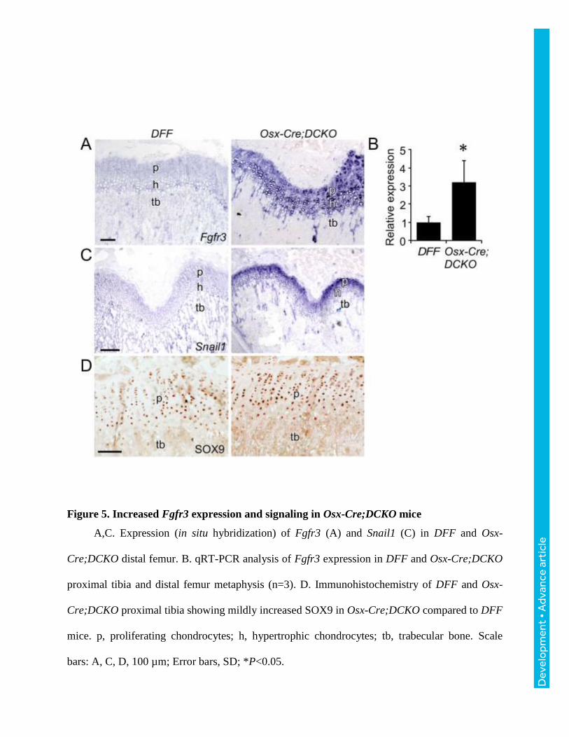
Increased Fgfr3 expression and signaling Osx-Cre;DCKO growth plate
Evaluation of Fgfr3 expression by in situ hybridization revealed a striking increase in
expression in Osx-Cre;DCKO compared to DFF mice in both proliferating and prehypertrophic
chondrocytes (Fig. 5A). This increase was confirmed by qRT-PCR analysis of distal bone tissue
from P21 distal femur and proximal tibia (Fig. 5B). The Snail1 transcription factor is induced by
FGFR3 and is required for the activation of both the STAT1 and MAPK branches of the FGFR3
signaling pathway (de Frutos et al., 2007). Consistent with increased FGFR3 expression and
signaling, Snail1 expression was highly increased in Osx-Cre;DCKO compared to DFF mice
(Fig. 5C). Immunostaining for the chondrocyte-specific transcription factor, SOX9, showed
mildly elevated levels of expression in Osx-Cre;DCKO compared to DFF mice (Fig. 5D).
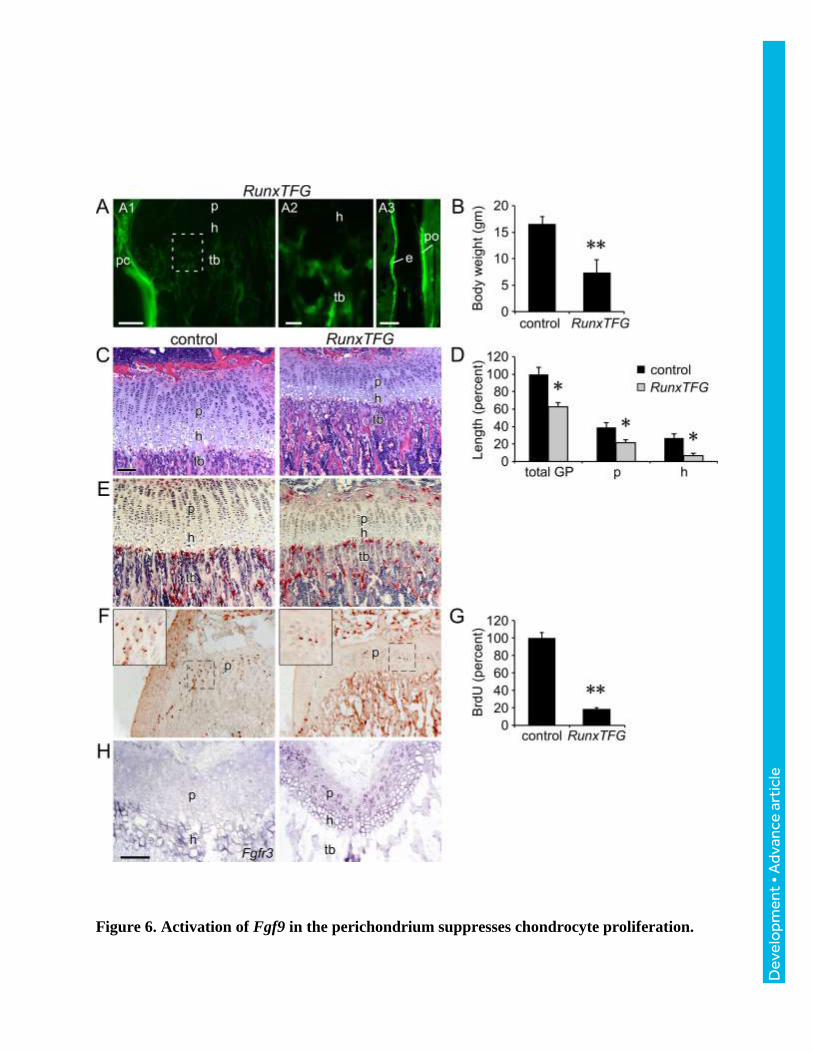
Activation of FGF9 in the perichondrium suppresses chondrocyte proliferation.
The ability of FGF9 to signal from perichondrial tissue to growth plate chondrocytes has
been inferred from phenotypes seen in Fgf9-/- embryos (Hung et al., 2007). Additionally,
transgenic mice that overexpressed FGF9 in chondrocytes (Col2a1-Fgf9) showed short limbs
and a smaller growth plate and died by 5 weeks of age (Garofalo et al., 1999). However, whether
Dev
elo
pmen
t • A
dvan
ce a
rtic
le
FGF9 has the capacity to signal from periosteal and trabecular osteoblasts to growth plate
chondrocytes during prepubertal growth was not known. To conditionally overexpress Fgf9 in
periosteal and trabecular osteoblasts, Runx2-rtTA (Chen et al., 2014b) and TRE-Fgf9-ires-eGFP
(White et al., 2006) transgenic mice were mated to generate biallelic Runx2-rtTA;TRE-Fgf9-ires-
eGFP (RunxTFG) mice. In the presence of doxycycline, GFP fluorescence was observed in the
perichondrium, periosteum, and trabecular bone of RunxTFG mice, but not in proliferating or
hypertrophic chondrocytes (Fig. 6A). Compared to control (single transgenic mouse), RunxTFG
transgenic mice showed a significantly (p<0.01) reduced body weight at P21 (Fig. 6B). Growth
plate histology revealed that compared to control, RunxTFG transgenic mice had significantly
(p<0.01) smaller proliferating and hypertrophic chondrocyte zones compared to controls (Fig. 6C,
D). The height of the trabecular zone in RunxTFG transgenic mice was reduced, but otherwise
histologically normal, and osteoclast numbers and morphology appeared normal (Fig. 6C, E).
Most notably, chondrocyte proliferation was significantly (p<0.001) reduced in RunxTFG
transgenic mice compared to control mice (Fig. 6F, G). Finally, in situ hybridization revealed
that activation of Fgf9 in the perichondrium/periosteum and trabecular bone induced the
expression of Fgfr3 in proliferating chondrocytes (Fig. 6H).
PTHLH links Osx-Cre lineage FGFR1/2 signaling to Fgfr3 expression and chondrocyte
proliferation in the postnatal growth plate.
Indian hedgehog (IHH) and parathyroid hormone-related peptide (PTHLH) are critical
regulators of endochondral bone growth (Kozhemyakina et al., 2015; Long and Ornitz, 2013).
IHH stimulates chondrocyte proliferation and Pthlh expression, while PTHLH suppresses
chondrocyte maturation and Ihh expression. Because we have observed apparent non-cell
autonomous effects of loss of Osx-Cre lineage FGFR1 and FGFR2 on chondrocyte growth, it
was important to examine the potential activity of other signaling pathways that regulate growth
Dev
elo
pmen
t • A
dvan
ce a
rtic
le
plate function. Compared to controls, Ihh was decreased in the P21 growth plate of Osx-
Cre;DCKO mice (Fig. 7A,B). Interestingly, we found that Pthlh expression was also reduced in
reserve chondrocytes in Osx-Cre;DCKO mice (Fig. 7C). Quantitative RT-PCR analysis of distal
bone tissue showed an overall reduction in Pthlh mRNA (Fig. 7D). Consistent with FGFR3
signaling suppressing Ihh-Pthlh expression (Chen et al., 2001; Li et al., 2010; Minina et al.,
2002; Naski et al., 1998), in mice induced to overexpress Fgf9, expression of Pthlh was reduced
in reserve zone chondrocytes (Fig. 7E).
Analysis of Fgfr3 promoter function in vitro shows that Fgfr3 expression could be directly
regulated (suppressed) by PTHLH activation of Protein Kinase A (PKA) (McEwen et al., 1999).
To test whether PTH signaling could suppress Fgfr3 expression in vivo in Osx-Cre;DCKO mice
that highly overexpress Fgfr3, Osx-Cre;DCKO mice were injected intermittently (daily) with
PTH from P15 to P21, a treatment regimen known to stimulate the anabolic effects of PTH
signaling on bone (Esen et al., 2015; Xie et al., 2012). Compared to control Osx-Cre;DCKO
mice that were only injected with PBS, PTH injected Osx-Cre;DCKO mice showed an increase
in the size of the growth plate, increased thickness of trabecular bone, decreased expression of
Fgfr3, and increased chondrocyte proliferation (Fig. 7F-J).
Dev
elo
pmen
t • A
dvan
ce a
rtic
le
DISCUSSION
The growth plate is a transient component of developing endochondral bone that mediates
longitudinal bone growth from late stages of embryonic development through puberty (Hunziker
and Schenk, 1989; Noonan et al., 1998). FGFR3 is a well-established negative regulator of
postnatal bone growth, functioning in the growth plate in proliferating and prehypertrophic
chondrocytes. Activating mutations in FGFR3 are responsible for Achondroplasia, the most
common form of dwarfism in humans (Horton et al., 2007; Ornitz and Marie, 2015). As
signaling pathways that function downstream of FGFR3 are well established,(Ornitz and Itoh,
2015) the identification of non-cell autonomous mechanisms that regulate FGFR3 expression
and signaling and postnatal growth plate function are essential for further elucidating complex
regulatory networks that control endochondral bone formation.
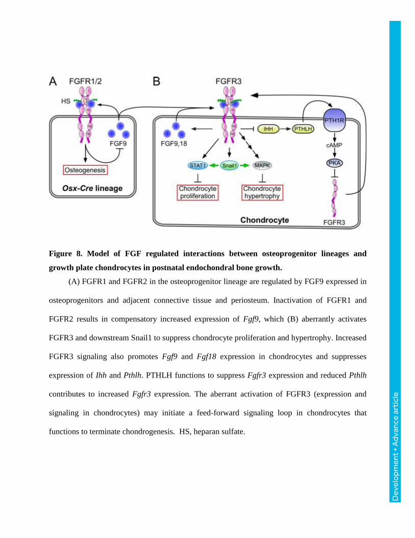
Inactivation of Fgfr1 and Fgfr2 in the Osx-Cre lineage disrupted a non cell-autonomous
feedback loop resulting in activation of FGFR3 signaling in growth plate chondrocytes and
suppression of chondrocyte proliferation and longitudinal bone growth (Fig. 8). The precise cell-
type(s) that maintains this feedback loop is not known; however, it is likely to be an immature
osteoprogenitor, as similar phenotypes are not observed when Fgfr1 and Fgfr2 are inactivated in
mature osteoblasts with the Osteocalcin-Cre allele (unpublished data). A likely early event
eliciting this phenotype is increased expression of Fgf9 in osteoprogenitor cells in the
perichondrium, resulting in increased signaling through FGFR3 in adjacent chondrocytes.
Activation of FGFR3 inhibits Ihh expression and signaling in prehypertrophic chondrocytes
(Naski et al., 1998), a factor that is required to maintain Pthlh expression in reserve and articular
chondrocytes (Hilton et al., 2005; Koziel et al., 2005; St-Jacques et al., 1999; Vortkamp et al.,
1996). Propagating events include increased Fgfr3 expression and signaling in the growth plate,
which may further suppress Ihh and Pthlh and increase Fgf9 and Fgf18 expression. Such non
Dev
elo
pmen
t • A
dvan
ce a
rtic
le
cell-autonomous cellular signaling pathways thus coordinate osteoprogenitor development and
longitudinal bone growth.
FGFR1/2 function in the osteoprogenitor lineage.
Although FGFR1 and FGFR2 signaling have robust functions in limb bud mesenchyme,
their function in the osteoprogenitor lineage during embryonic development is surprisingly mild.
Osx-Cre;DCKO mice were born alive and showed no patterning defects in the appendicular
skeleton. However, Osx-Cre;DCKO mice exhibited a calvarial ossification defect at birth (data
not shown) and a postnatal reduction in cortical bone growth that indicates that osteoprogenitor
lineage FGFR signaling is required for osteoblast growth and maturation that is independent of
chondrogenesis. The precise role of FGFR signaling in osteoblasts will require further
investigation.
FGFR signaling in osteoprogenitor cells indirectly affects growth plate activity.
The most striking feature of Osx-Cre;DCKO mice was the profound reduction in
chondrocyte proliferation and longitudinal bone growth. We posited that this phenotype resulted
from non-cell autonomous changes in chondrocytes that are secondary to loss of FGFR1 and
FGFR2 signaling in osteoprogenitor cells. Because Osx-Cre targets a small percentage of
chondrocytes (Chen et al., 2014a), there remained a possibility that the observed phenotype
could result from inactivation of Fgfr1 and Fgfr2 in chondrocytes. However, this is unlikely
because Fgfr1 expression is restricted to hypertrophic chondrocytes and Fgfr2 is not expressed in
proliferating or hypertrophic chondrocytes. Nevertheless, to rule out cell autonomous effects of
FGFR1 and FGFR2 in chondrocytes, these genes were inactivated specifically in chondrocytes
using the ATC-Cre allele. The normal development of ATC-Cre;DCKO mice demonstrated that
inactivation of Fgfr1 and Fgfr2 in proliferating and hypertrophic chondrocytes does not
significantly affect chondrogenesis or prepubertal longitudinal bone growth.

Dev
elo
pmen
t • A
dvan
ce a
rtic
le
A second feature of the Osx-Cre;DCKO phenotype was the prominent increase in Fgfr3
expression in proliferating and hypertrophic chondrocytes. In vitro analysis of the Fgfr3
promoter identified a regulatory sequence that results in decreased promoter activity in response
to cAMP (McEwen et al., 1999). This in vitro data suggested that the observed decrease in Pthlh
expression could contribute to increased Fgfr3 expression. In support of this model, intermittent
injection of Osx-Cre;DCKO mice with PTH(1-34) peptide suppressed Fgfr3 expression in
chondrocytes and increased chondrocyte proliferation (Fig. 8).
A third feature of the Osx-Cre;DCKO phenotype was reduced bone volume and density.
This could result from cell-autonomous effects of FGFR signaling in osteoblasts, or to the
reduced levels of Pthlh expression. Haploinsufficiency of Pthlh results in osteopenia in mice
(Miao et al., 2005), with similar morphologies to that observed in Osx-Cre;DCKO mice.
Regulation of embryonic vs. postnatal growth plate.
The experiments presented here focus on the postnatal growth plate of 21 day old Osx-
Cre;DCKO mice. Although, the Osx-Cre allele used to target Fgfr1 and Fgfr2 is active as early
as embryonic day 12.5 (Ono et al., 2014; Rodda and McMahon, 2006), the extent of the
embryonic phenotype appears to be limited to expansion of the hypertrophic chondrocyte zone,
similar to the phenotype observed when the Col2-Cre allele was used to inactivate Fgfr1 (Jacob
et al., 2006). Thus, FGFR1/2 signaling either does not have a major role in the osteoprogenitor
lineage prior to establishment of a secondary ossification center and formation of a mature
growth plate or the non cell-autonomous mechanism that we identified is not activated during
embryonic development. Most studies investigating skeletal development focus on the
embryonic growth plate. However, the postnatal growth plate is the developmental structure that
accounts for the majority of organismal skeletal growth and, yet, gene expression patterns and
Dev
elo
pmen
t • A
dvan
ce a
rtic
le
the molecular and cellular mechanisms that regulate the postnatal growth plate are poorly
defined.
In the embryonic growth plate IHH is involved in a feedback loop that regulates Pthlh
expression in the distal periarticular perichondrium (Kronenberg, 2003). However, in postnatal
bone, there is a reorganization of the growth plate, and Pthlh expression shifts to reserve zone
chondrocytes and IHH signaling (Gli1) and Pth1r expression remains prominent in
reserve/proliferating and prehypertrophic chondrocytes, respectively (Chau et al., 2011; Chen et
al., 2008; Koziel et al., 2004). Thus, in the postnatal growth plate, PTHLH and IHH responsive
cells overlap with Fgfr3 expression patterns.
Loss of FGFR1/2 signaling in perichondrial and osteoprogenitor cells may disrupt growth
plate homeostasis by initially triggering increased expression of FGF9 and FGF18. We posit that
these events lead to increased FGFR3 expression and signaling (modeled by forced expression of
Fgf9 in perichondrial cells and osteoblasts). Secondarily, increased FGFR3 signaling could
suppress Ihh expression and signaling and lead to suppression of Pthlh in chondrocytes, leading
to an aberrant feed forward signal that further increases the expression of Fgfr3 (Fig. 8). The
ability to block this feed forward loop by administration of PTH(1-34) supports a model in which
PTHLH regulates communication between osteoprogenitors, chondroprogenitors, and growth
plate chondrocytes in a mature postnatal growth plate.
Termination of skeletal growth.
Osx-Cre;DCKO mice show increased expression of Fgf9 and Fgf18 in reserve,
proliferating, and prehypertrophic chondrocytes and in cells at the periphery of the growth plate
that may include chondroprogenitors in the groove of Ranvier. This may represent an
amplification of a normal feed forward induction of Fgf9 and Fgf18 that could function to

Dev
elo
pmen
t • A
dvan
ce a
rtic
le
permanently suppress growth plate chondrocyte proliferation at puberty and suppress articular
chondrocyte proliferation and differentiation in adults. This model is consistent with continued
expression of endogenous Fgf18 in the postnatal growth plate and perichondrium, and in adult
articular chondrocytes (Ellsworth et al., 2002; Lazarus et al., 2007; Mori et al., 2014).

Dev
elo
pmen
t • A
dvan
ce a
rtic
le
MATERIALS AND METHODS
Mice
Mice were housed in a pathogen-free facility and handled in accordance with standard use
protocols, animal welfare regulations, and the NIH Guide for the Care and Use of Laboratory
Animals. All protocols were approved by the Washington University Animal Studies Committee.
Osx-GFP::Cre (Osx-Cre) (Rodda and McMahon, 2006), Fgfr1f/f (Trokovic et al., 2003), Fgfr2f/f
(Yu et al., 2003), aggrecan enhancer-driven, tetracycline-inducible Cre (ATC) (Dy et al., 2012),
Runx2-rtTA (Chen et al., 2014b), TRE-Fgf9-ires-eGFP (White et al., 2006) have been previously
described.
Homozygous floxed alleles of Fgfr1 and Fgfr2 are maintained as double flox mice (DFF)
and are outbred to hybrid C57BL/6J;129X1 mice every second generation and then backcrossed
to homozygosity. Double conditional knockout (DCKO) breeding males (Osx-
Cre;Fgfr1f/f;Fgfr2f/f) were generated by crossing Osx-Cre mice with DFF mice, backcrossing to
DFF and suppressing the Cre activity of Osx-Cre with doxycycline. To inactivate Fgfr1/2 in the
osteoprogenitor-lineage, DFF female mice were crossed with Osx-Cre;DCKO breeder male mice
resulting in an 50% yield of Osx-Cre;DCKO mice and DFF controls. Osx-Cre control mice
were generated by crossing Osx-Cre;DCKO mice or Osx-Cre mice to wild type hybrid mice. A
similar breeding strategy was used to generate ATC;DCKO mice. To express Fgf9 in the
osteoblasts lineage, Runx2-rtTA mice (Chen et al., 2014b) were crossed to TRE-Fgf9-ires-Gfp
(White et al., 2006) to generate RunxTFG double transgenic mice. Females were induced with
doxycycline chow (Bio-Serv Inc. 200 mg/kg green pellets, S3888)) from E0 to P21. High fat
high calorie diet included breeder chow (PicoLab® Mouse. Diet 20) supplemented with Nutri-
Cal® (Patterson Veterinary Supply) from birth to 5 wk of age.
Dev
elo
pmen
t • A
dvan
ce a
rtic
le
Body weights were measured on multiple litters two to three times per week until animals
were sacrificed for analysis. Growth curves represent cumulative pooled data from multiple
litters and overlapping time points covering the entire time course.
Histology, Immunohistochemistry, and Immunofluorescence
For histological analysis of long bones, intact femur-tibia were isolated, fixed in 4%
PFA/PBS overnight at 4°C or fixed in 10% buffered formalin overnight at room temperature.
Bones were rinsed in water several times and decalcified in 14% EDTA/PBS for two weeks.
Paraffin embedded tissue section (5 µm) were stained with hematoxylin and eosin (H&E),
tartrate resistant acid phosphatase (TRAP), VonKossa, or Alizarin-Red.
For immunohistochemistry, paraffin sections or cryo-sections were rehydrated and treated
with 0.3% hydrogen peroxide in methanol for 15 min to suppress endogenous peroxidase activity.
Antigen retrieval was achieved by microwaving the sections in 10 mM citrate buffer for 10 min
followed by gradual cooling to room temperature. Sections were incubated overnight at 4°C with
the following primary antibodies: SOX9 (Millipore, AB5535, rabbit polyclonal, 1:100), anti-
active Caspase -3 (BD Pharmingen, #559565, 1:100). Fluorescently labeled Alexa Fluor 488
donkey anti-rabbit (Life Technologies, A-21206, 1:1000). Colorimetric detection used the ABC
kit (Invitrogen #95-9943). Immunofluorescence imaging was performed on a Zeiss Apotome
Fluorescence Microscope. Data is representative of at least three independent experiments.
For in situ hybridization analysis, tissues were fixed and decalcified at 4°C. For frozen
sections, the tissues were fixed as described above and decalcified for 3 days, transferred to 30%
sucrose (S0389, SIGMA) for 24 hr, embedded in OCT compound, sectioned at 5 µm, and stored
at -20 until analysis. Non-radioactive in situ hybridization was performed as previously
described (Naski et al., 1998). In situ probes: Fgf9 (Colvin et al., 1999); Fgf18 (Liu et al., 2002);

Dev
elo
pmen
t • A
dvan
ce a
rtic
le
Fgfr3 (Peters et al., 1993); Snail1 (Vega et al., 2004); Pthlh (Lee et al., 1996; Long et al., 2001);
Ihh (Bitgood and McMahon, 1995); Col1 (Rossert et al., 1995). Data is representative of at least
three independent experiments.
Cell proliferation was determined by injecting U (5-Bromo-2’ deoxyuridine, SIGMA,
9285) 0.1 mg/gram body weight 2 hr before tissues were harvested. Anti BrdU mouse
monoclonal (BD Biosciences, #347580) was used at a 1:200 dilution. BrdU labeling was
normalized to the total number of cells in the proliferating zone or to the area of the proliferating
zone. Data were then normalized to that of DFF control mice. At least three mice and 2-3
sections per mouse were analyzed for each genotype.
Histomorphometry
H&E and TRAP stained sections were used for quantification of osteoblast and osteoclast
number and surface, using BioQuant OSTEO 2010 software. Measurements of growth plate
length in H&E stained sections were made using Canvas X software (ACD systems). All lengths
were normalized to the total length of the DFF control growth plate. Statistical analysis
(Student’s t test) was based on measurements of tissue samples from at least three control and
three experimental mice.
MicroCT and DEXA analysis
For microCT, intact long bones were isolated and fixed in 70% ethanol overnight at 4°C
and then stored at -20°C until analysis. Bones were embedded in 1.5% agarose and scanned
(µCT40, Scanco Medical). MicroCT analysis of trabecular and cortical bone was performed as
follows: for trabecular bone, 100 to 150 sections were selected below the growth plate for
reconstruction and quantification. For cortical bone quantification, 50 to 100 sections were
selected from the mid diaphysis of the femur or tibia. Quantification was performed using
Dev
elo
pmen
t • A
dvan
ce a
rtic
le
SCANCO Medical microCT systems software. Dual-Energy X-Ray Absorptiometry (DEXA,
GE/Lunar PIXImus) was used for measurements of whole body bone density and body fat
content. Data is representative of at least three mice per genotype.
Real time quantitative PCR
Distal bone, containing the growth plate, perichondrium, and trabecular bone was dissected.
Immediately after isolation, the tissues were individually frozen in liquid nitrogen and stored at -
80°C until analysis. Frozen tissues were pulverized in a dry ice cooled stainless steel flask with
a ball bearing in a Micro Dismembrator (Sartorius) at 2000 RPM for 20 seconds. RNA was
stabilized with Trisol (TRIsol Reagent, Ambion/RNA) and total RNA isolation was prepared
according to the manufacturer’s instructions. cDNA was synthesized using the iScript™Select
cDNA synthesis Kit (#170-8841, BIO-RAD). mRNA expression was measured using TaqMan™
Fast Advanced Master Mix (#4444557, Life Technologies) and TaqMan™ assay probes for Ihh,
Pthlh, Fgf9, Fgf18, and Fgfr3. Hprt was used a normalization control.
PTH treatment
For in vivo treatment of mice with PTH, 15 day-old Osx-Cre;DCKO mice were injected i.p.
once per day (morning) with synthetic PTH-related peptide (I-34) (H-6630, Bachem) at a
concentration of 80 µg/kg body weight or with PBS (control). Mice were injected for five days
and then sacrificed at P21.
Statistics
The data are reported as the mean ± SD and changes with p values less than 0.05 were
considered to be statistically significant. Data was analyzed using a two-tailed Student’s t-test.
Numbers of mice used per group per experiment are stated in the figure legends.

Dev
elo
pmen
t • A
dvan
ce a
rtic
le
Acknowledgements
This work was supported by was supported by NIH grant HD049808 (D.M.O.), AR055923
(F.L.), DE025077 (K.Y.), the Washington University Musculoskeletal Research Center NIH P30
AR057235 and by internal funds from the Craniofacial Center of Seattle Children’s Hospital and
Center for Developmental Biology and Regenerative Medicine of Seattle Children’s Research
Institute (K.Y.).
Author Contributions
Conceptualization and Methodology, K.K., K.Y., F.L., D.M.O.; Investigation, K.K., K.Y.,
J.L., J.C. C.S., D.M.O.; Writing–Original Draft, K.K., D.M.O.; Writing–Review & Editing, K.K.,
K.Y., F.L., D.M.O.; Funding Acquisition, Resources, and Supervision, F.L., D.M.O.

Dev
elo
pmen
t • A
dvan
ce a
rtic
le
References
Aikawa, T., Segre, G. V. and Lee, K. (2001). Fibroblast growth factor inhibits chondrocytic growth through induction of p21 and subsequent inactivation of cyclin E-Cdk2. J. Biol. Chem. 276, 29347-29352.
Beever, J. E., Smit, M. A., Meyers, S. N., Hadfield, T. S., Bottema, C., Albretsen, J. and Cockett, N. E. (2006). A single-base change in the tyrosine kinase II domain of ovine FGFR3 causes hereditary chondrodysplasia in sheep. Anim. Genet. 37, 66-71.
Bitgood, M. J. and McMahon, A. P. (1995). Hedgehog and Bmp genes are coexpressed at many diverse sites of cell-cell interaction in the mouse embryo. Dev. Biol. 172, 126-138.
Bohm, F., Speicher, T., Hellerbrand, C., Dickson, C., Partanen, J., Ornitz, D. and Werner, S. (2010). FGF receptors 1 and 2 control chemically-induced injury and compound detoxification in regenerating livers of mice. Gastroenterology.
Chau, M., Forcinito, P., Andrade, A. C., Hegde, A., Ahn, S., Lui, J. C., Baron, J. and Nilsson, O. (2011). Organization of the Indian hedgehog--parathyroid hormone-related protein system in the postnatal growth plate. J. Mol. Endocrinol. 47, 99-107.
Chen, J., Shi, Y., Regan, J., Karuppaiah, K., Ornitz, D. M. and Long, F. (2014a). Osx-Cre Targets Multiple Cell Types besides Osteoblast Lineage in Postnatal Mice. PLoS One 9, e85161.
Chen, J., Tu, X., Esen, E., Joeng, K. S., Lin, C., Arbeit, J. M., Ruegg, M. A., Hall, M. N., Ma, L. and Long, F. (2014b). WNT7B promotes bone formation in part through mTORC1. PLoS Genet 10, e1004145.
Chen, L., Li, C., Qiao, W., Xu, X. and Deng, C. (2001). A Ser(365)-->Cys mutation of fibroblast growth factor receptor 3 in mouse downregulates Ihh/PTHrP signals and causes severe achondroplasia. Hum Mol Genet 10, 457-465.
Chen, X., Macica, C. M., Nasiri, A. and Broadus, A. E. (2008). Regulation of articular chondrocyte proliferation and differentiation by indian hedgehog and parathyroid hormone-related protein in mice. Arthritis Rheum. 58, 3788-3797.
Cobrinik, D., Lee, M. H., Hannon, G., Mulligan, G., Bronson, R. T., Dyson, N., Harlow, E., Beach, D., Weinberg, R. A. and Jacks, T. (1996). Shared role of the pRB-related p130 and p107 proteins in limb development. Genes Dev. 10, 1633-1644.
Colvin, J. S., Bohne, B. A., Harding, G. W., McEwen, D. G. and Ornitz, D. M. (1996). Skeletal overgrowth and deafness in mice lacking fibroblast growth factor receptor 3. Nat. Genet. 12, 390-397.
Colvin, J. S., Feldman, B., Nadeau, J. H., Goldfarb, M. and Ornitz, D. M. (1999). Genomic organization and embryonic expression of the mouse fibroblast growth factor 9 gene. Dev. Dyn. 216, 72-88.
Coumoul, X., Shukla, V., Li, C., Wang, R. H. and Deng, C. X. (2005). Conditional knockdown of Fgfr2 in mice using Cre-LoxP induced RNA interference. Nucleic Acids Res 33, e102.
Dev
elo
pmen
t • A
dvan
ce a
rtic
le
Dailey, L., Laplantine, E., Priore, R. and Basilico, C. (2003). A network of transcriptional and signaling events is activated by FGF to induce chondrocyte growth arrest and differentiation. J. Cell Biol. 161, 1053-1066.
de Frutos, C. A., Vega, S., Manzanares, M., Flores, J. M., Huertas, H., Martinez-Frias, M. L. and Nieto, M. A. (2007). Snail1 is a transcriptional effector of FGFR3 signaling during chondrogenesis and achondroplasias. Dev Cell 13, 872-883.
Deng, C., Wynshaw-Boris, A., Zhou, F., Kuo, A. and Leder, P. (1996). Fibroblast growth factor receptor 3 is a negative regulator of bone growth. Cell 84, 911-921.
Dy, P., Wang, W., Bhattaram, P., Wang, Q., Wang, L., Ballock, R. T. and Lefebvre, V. (2012). Sox9 directs hypertrophic maturation and blocks osteoblast differentiation of growth plate chondrocytes. Dev Cell 22, 597-609.
Ellsworth, J. L., Berry, J., Bukowski, T., Claus, J., Feldhaus, A., Holderman, S., Holdren, M. S., Lum, K. D., Moore, E. E., Raymond, F., et al. (2002). Fibroblast growth factor-18 is a trophic factor for mature chondrocytes and their progenitors. Osteoarthritis Cartilage 10, 308-320.
Esen, E., Lee, S. Y., Wice, B. M. and Long, F. (2015). PTH Promotes Bone Anabolism by Stimulating Aerobic Glycolysis via IGF Signaling. J. Bone Miner. Res.
Eswarakumar, V. P., Monsonego-Ornan, E., Pines, M., Antonopoulou, I., Morriss-Kay, G. M. and Lonai, P. (2002). The IIIc alternative of Fgfr2 is a positive regulator of bone formation. Development 129, 3783-3793.
Garofalo, S., Kliger-Spatz, M., Cooke, J. L., Wolstin, O., Lunstrum, G. P., Moshkovitz, S. M., Horton, W. A. and Yayon, A. (1999). Skeletal dysplasia and defective chondrocyte differentiation by targeted overexpression of fibroblast growth factor 9 in transgenic mice. J. Bone Miner. Res. 14, 1909-1915.
Havens, B. A., Velonis, D., Kronenberg, M. S., Lichtler, A. C., Oliver, B. and Mina, M. (2008). Roles of FGFR3 during morphogenesis of Meckel's cartilage and mandibular bones. Dev Biol 316, 336-349.
Hilton, M. J., Tu, X., Cook, J., Hu, H. and Long, F. (2005). Ihh controls cartilage development by antagonizing Gli3, but requires additional effectors to regulate osteoblast and vascular development. Development 132, 4339-4351.
Horton, W. A., Hall, J. G. and Hecht, J. T. (2007). Achondroplasia. Lancet 370, 162-172.
Huang, W. and Olsen, B. R. (2015). Skeletal defects in Osterix-Cre transgenic mice. Transgenic Res. 24, 167-172.
Huh, S. H., Warchol, M. E. and Ornitz, D. M. (2015). Cochlear progenitor number is controlled through mesenchymal FGF receptor signaling. Elife 4.
Hung, I. H., Schoenwolf, G. C., Lewandoski, M. and Ornitz, D. M. (2016). A combined series of Fgf9 and Fgf18 mutant alleles identifies unique and redundant roles in skeletal development. Dev Biol 411, 72-84.
Hung, I. H., Yu, K., Lavine, K. J. and Ornitz, D. M. (2007). FGF9 regulates early hypertrophic chondrocyte differentiation and skeletal vascularization in the developing stylopod. Dev Biol 307, 300-313.
Hunziker, E. B. and Schenk, R. K. (1989). Physiological mechanisms adopted by chondrocytes in regulating longitudinal bone growth in rats. J Physiol 414, 55-71.

Dev
elo
pmen
t • A
dvan
ce a
rtic
le
Jacob, A. L., Smith, C., Partanen, J. and Ornitz, D. M. (2006). Fibroblast growth factor receptor 1 signaling in the osteo-chondrogenic cell lineage regulates sequential steps of osteoblast maturation. Dev Biol 296, 315-328.
Karolak, M. R., Yang, X. and Elefteriou, F. (2015). FGFR1 signaling in hypertrophic chondrocytes is attenuated by the Ras-GAP neurofibromin during endochondral bone formation. Hum Mol Genet 24, 2552-2564.
Kolupaeva, V., Daempfling, L. and Basilico, C. (2013). The B55alpha regulatory subunit of protein phosphatase 2A mediates fibroblast growth factor-induced p107 dephosphorylation and growth arrest in chondrocytes. Mol Cell Biol 33, 2865-2878.
Kolupaeva, V., Laplantine, E. and Basilico, C. (2008). PP2A-mediated dephosphorylation of p107 plays a critical role in chondrocyte cell cycle arrest by FGF. PLoS One 3, e3447.
Kozhemyakina, E., Lassar, A. B. and Zelzer, E. (2015). A pathway to bone: signaling molecules and transcription factors involved in chondrocyte development and maturation. Development 142, 817-831.
Koziel, L., Kunath, M., Kelly, O. G. and Vortkamp, A. (2004). Ext1-dependent heparan sulfate regulates the range of Ihh signaling during endochondral ossification. Dev Cell 6, 801-813.
Koziel, L., Wuelling, M., Schneider, S. and Vortkamp, A. (2005). Gli3 acts as a repressor downstream of Ihh in regulating two distinct steps of chondrocyte differentiation. Development 132, 5249-5260.
Kronenberg, H. M. (2003). Developmental regulation of the growth plate. Nature 423, 332-336.
Kurimchak, A., Haines, D. S., Garriga, J., Wu, S., De Luca, F., Sweredoski, M. J., Deshaies, R. J., Hess, S. and Grana, X. (2013). Activation of p107 by fibroblast growth factor, which is essential for chondrocyte cell cycle exit, is mediated by the protein phosphatase 2A/B55alpha holoenzyme. Mol Cell Biol 33, 3330-3342.
Laederich, M. B. and Horton, W. A. (2012). FGFR3 targeting strategies for achondroplasia. Expert reviews in molecular medicine 14, e11.
Laplantine, E., Rossi, F., Sahni, M., Basilico, C. and Cobrinik, D. (2002). FGF signaling targets the pRb-related p107 and p130 proteins to induce chondrocyte growth arrest. J. Cell Biol. 158, 741-750.
Lazarus, J. E., Hegde, A., Andrade, A. C., Nilsson, O. and Baron, J. (2007). Fibroblast growth factor expression in the postnatal growth plate. Bone 40, 577-586.
Lee, K., Lanske, B., Karaplis, A. C., Deeds, J. D., Kohno, H., Nissenson, R. A., Kronenberg, H. M. and Segre, G. V. (1996). Parathyroid hormone-related peptide delays terminal differentiation of chondrocytes during endochondral bone development. Endocrinology 137, 5109-5118.
Legeai-Mallet, L., Benoist-Lasselin, C., Munnich, A. and Bonaventure, J. (2004). Overexpression of FGFR3, Stat1, Stat5 and p21Cip1 correlates with phenotypic severity and defective chondrocyte differentiation in FGFR3-related chondrodysplasias. Bone 34, 26-36.

Dev
elo
pmen
t • A
dvan
ce a
rtic
le
Li, C., Xu, X., Nelson, D. K., Williams, T., Kuehn, M. R. and Deng, C. X. (2005). FGFR1 function at the earliest stages of mouse limb development plays an indispensable role in subsequent autopod morphogenesis. Development 132, 4755-4764.
Li, M., Seki, Y., Freitas, P. H., Nagata, M., Kojima, T., Sultana, S., Ubaidus, S., Maeda, T., Shimomura, J., Henderson, J. E., et al. (2010). FGFR3 down-regulates PTH/PTHrP receptor gene expression by mediating JAK/STAT signaling in chondrocytic cell line. J. Electron Microsc. (Tokyo). 59, 227-236.
Liu, Z., Lavine, K. J., Hung, I. H. and Ornitz, D. M. (2007). FGF18 is required for early chondrocyte proliferation, hypertrophy and vascular invasion of the growth plate. Dev Biol 302, 80-91.
Liu, Z., Xu, J., Colvin, J. S. and Ornitz, D. M. (2002). Coordination of chondrogenesis and osteogenesis by fibroblast growth factor 18. Genes Dev. 16, 859-869.
Long, F. and Ornitz, D. M. (2013). Development of the endochondral skeleton. Cold Spring Harb Perspect Biol 5, 1-20.
Long, F., Zhang, X. M., Karp, S., Yang, Y. and McMahon, A. P. (2001). Genetic manipulation of hedgehog signaling in the endochondral skeleton reveals a direct role in the regulation of chondrocyte proliferation. Development 128, 5099-5108.
Makrythanasis, P., Temtamy, S., Aglan, M. S., Otaify, G. A., Hamamy, H. and Antonarakis, S. E. (2014). A novel homozygous mutation in FGFR3 causes tall stature, severe lateral tibial deviation, scoliosis, hearing impairment, camptodactyly, and arachnodactyly. Hum. Mutat. 35, 959-963.
McEwen, D. G., Green, R. P., Naski, M. C., Towler, D. A. and Ornitz, D. M. (1999). Fibroblast growth factor receptor 3 gene transcription is suppressed by cyclic adenosine 3 ',5 '-monophosphate - Identification of a chondrocytic regulatory element. J. Biol. Chem. 274, 30934-30942.
Meyer, M., Muller, A. K., Yang, J., Moik, D., Ponzio, G., Ornitz, D. M., Grose, R. and Werner, S. (2012). FGF receptors 1 and 2 are key regulators of keratinocyte migration in vitro and in wounded skin. J. Cell Sci. 125, 5690-5701.
Miao, D., He, B., Jiang, Y., Kobayashi, T., Soroceanu, M. A., Zhao, J., Su, H., Tong, X., Amizuka, N., Gupta, A., et al. (2005). Osteoblast-derived PTHrP is a potent endogenous bone anabolic agent that modifies the therapeutic efficacy of administered PTH 1-34. J Clin Invest 115, 2402-2411.
Minina, E., Kreschel, C., Naski, M. C., Ornitz, D. M. and Vortkamp, A. (2002). Interaction of FGF, Ihh/Pthlh, and BMP signaling integrates chondrocyte proliferation and hypertrophic differentiation. Dev Cell 3, 439-449.
Mori, Y., Saito, T., Chang, S. H., Kobayashi, H., Ladel, C. H., Guehring, H., Chung, U. I. and Kawaguchi, H. (2014). Identification of fibroblast growth factor-18 as a molecule to protect adult articular cartilage by gene expression profiling. J. Biol. Chem. 289, 10192-10200.
Naski, M. C., Colvin, J. S., Coffin, J. D. and Ornitz, D. M. (1998). Repression of hedgehog signaling and BMP4 expression in growth plate cartilage by fibroblast growth factor receptor 3. Development 125, 4977-4988.
Naski, M. C., Wang, Q., Xu, J. and Ornitz, D. M. (1996). Graded activation of fibroblast growth factor receptor 3 by mutations causing achondroplasia and thanatophoric dysplasia. Nat. Genet. 13, 233-237.
Dev
elo
pmen
t • A
dvan
ce a
rtic
le
Noonan, K. J., Hunziker, E. B., Nessler, J. and Buckwalter, J. A. (1998). Changes in cell, matrix compartment, and fibrillar collagen volumes between growth-plate zones. J. Orthop. Res. 16, 500-508.
Ohbayashi, N., Shibayama, M., Kurotaki, Y., Imanishi, M., Fujimori, T., Itoh, N. and Takada, S. (2002). FGF18 is required for normal cell proliferation and differentiation during osteogenesis and chondrogenesis. Genes Dev. 16, 870-879.
Ono, N., Ono, W., Nagasawa, T. and Kronenberg, H. M. (2014). A subset of chondrogenic cells provides early mesenchymal progenitors in growing bones. Nat Cell Biol 16, 1157-1167.
Ornitz, D. M. and Itoh, N. (2015). The Fibroblast Growth Factor signaling pathway. Wiley Interdiscip Rev Dev Biol 4, 215-266.
Ornitz, D. M. and Marie, P. J. (2002). FGF signaling pathways in endochondral and intramembranous bone development and human genetic disease. Genes Dev. 16, 1446-1465.
---- (2015). Fibroblast growth factor signaling in skeletal development and disease. Genes Dev. 29, 1463-1486.
Orr-Urtreger, A., Givol, D., Yayon, A., Yarden, Y. and Lonai, P. (1991). Developmental expression of two murine fibroblast growth factor receptors, flg and bek. Development 113, 1419-1434.
Peters, K., Ornitz, D., Werner, S. and Williams, L. (1993). Unique expression pattern of the FGF receptor 3 gene during mouse organogenesis. Dev Biol 155, 423-430.
Peters, K. G., Werner, S., Chen, G. and Williams, L. T. (1992). Two FGF receptor genes are differentially expressed in epithelial and mesenchymal tissues during limb formation and organogenesis in the mouse. Development 114, 233-243.
Poladia, D. P., Kish, K., Kutay, B., Hains, D., Kegg, H., Zhao, H. and Bates, C. M. (2006). Role of fibroblast growth factor receptors 1 and 2 in the metanephric mesenchyme. Dev Biol 291, 325-339.
Priore, R., Dailey, L. and Basilico, C. (2006). Downregulation of Akt activity contributes to the growth arrest induced by FGF in chondrocytes. J Cell Physiol 207, 800-808.
Raucci, A., Laplantine, E., Mansukhani, A. and Basilico, C. (2004). Activation of the ERK1/2 and p38 mitogen-activated protein kinase pathways mediates fibroblast growth factor-induced growth arrest of chondrocytes. J. Biol. Chem. 279, 1747-1756.
Rodda, S. J. and McMahon, A. P. (2006). Distinct roles for Hedgehog and canonical Wnt signaling in specification, differentiation and maintenance of osteoblast progenitors. Development 133, 3231-3244.
Rossert, J., Eberspaecher, H. and de Crombrugghe, B. (1995). Separate cis-acting DNA elements of the mouse pro-alpha 1(I) collagen promoter direct expression of reporter genes to different type I collagen-producing cells in transgenic mice. J. Cell Biol. 129, 1421-1432.
Sims-Lucas, S., Cusack, B., Baust, J., Eswarakumar, V. P., Masatoshi, H., Takeuchi, A. and Bates, C. M. (2011). Fgfr1 and the IIIc isoform of Fgfr2 play critical roles in the metanephric mesenchyme mediating early inductive events in kidney development. Dev Dyn 240, 240-249.
Dev
elo
pmen
t • A
dvan
ce a
rtic
le
Smith, K. M., Williamson, T. L., Schwartz, M. L. and Vaccarino, F. M. (2012). Impaired motor coordination and disrupted cerebellar architecture in Fgfr1 and Fgfr2 double knockout mice. Brain Res. 1460, 12-24.
St-Jacques, B., Hammerschmidt, M. and McMahon, A. P. (1999). Indian hedgehog signaling regulates proliferation and differentiation of chondrocytes and is essential for bone formation. Genes Dev. 13, 2072-2086.
Su, N., Jin, M. and Chen, L. (2014). Role of FGF/FGFR signaling in skeletal development and homeostasis: learning from mouse models. Bone Research 2.
Su, W. C., Kitagawa, M., Xue, N., Xie, B., Garofalo, S., Cho, J., Deng, C., Horton, W. A. and Fu, X. Y. (1997). Activation of Stat1 by mutant fibroblast growth-factor receptor in thanatophoric dysplasia type II dwarfism. Nature 386, 288-292.
Toydemir, R. M., Brassington, A. E., Bayrak-Toydemir, P., Krakowiak, P. A., Jorde, L. B., Whitby, F. G., Longo, N., Viskochil, D. H., Carey, J. C. and Bamshad, M. J. (2006). A novel mutation in FGFR3 causes camptodactyly, tall stature, and hearing loss (CATSHL) syndrome. Am. J. Hum. Genet. 79, 935-941.
Trokovic, R., Trokovic, N., Hernesniemi, S., Pirvola, U., Vogt Weisenhorn, D. M., Rossant, J., McMahon, A. P., Wurst, W. and Partanen, J. (2003). FGFR1 is independently required in both developing mid- and hindbrain for sustained response to isthmic signals. Embo J 22, 1811-1823.
Vega, S., Morales, A. V., Ocana, O. H., Valdes, F., Fabregat, I. and Nieto, M. A. (2004). Snail blocks the cell cycle and confers resistance to cell death. Genes Dev. 18, 1131-1143.
Verheyden, J. M., Lewandoski, M., Deng, C., Harfe, B. D. and Sun, X. (2005). Conditional inactivation of Fgfr1 in mouse defines its role in limb bud establishment, outgrowth and digit patterning. Development 132, 4235-4245.
Vortkamp, A., Lee, K., Lanske, B., Segre, G. V., Kronenberg, H. M. and Tabin, C. J. (1996). Regulation of rate of cartilage differentiation by Indian hedgehog and PTH-related protein. Science 273, 613-622.
Wang, L., Mishina, Y. and Liu, F. (2015). Osterix-Cre transgene causes craniofacial bone development defect. Calcif. Tissue Int. 96, 129-137.
White, A. C., Xu, J., Yin, Y., Smith, C., Schmid, G. and Ornitz, D. M. (2006). FGF9 and SHH signaling coordinate lung growth and development through regulation of distinct mesenchymal domains. Development 133, 1507-1517.
Xiao, Z., Huang, J., Cao, L., Liang, Y., Han, X. and Quarles, L. D. (2014). Osteocyte-specific deletion of Fgfr1 suppresses FGF23. PLoS One 9, e104154.
Xie, Y., Su, N., Jin, M., Qi, H., Yang, J., Li, C., Du, X., Luo, F., Chen, B., Shen, Y., et al. (2012). Intermittent PTH (1-34) injection rescues the retarded skeletal development and postnatal lethality of mice mimicking human achondroplasia and thanatophoric dysplasia. Hum Mol Genet.
Yang, J., Meyer, M., Muller, A. K., Bohm, F., Grose, R., Dauwalder, T., Verrey, F., Kopf, M., Partanen, J., Bloch, W., et al. (2010). Fibroblast growth factor receptors 1 and 2 in keratinocytes control the epidermal barrier and cutaneous homeostasis. J. Cell Biol. 188, 935-952.
Yu, K., Karuppaiah, K. and Ornitz, D. M. (2015). Mesenchymal fibroblast growth factor receptor signaling regulates palatal shelf elevation during secondary palate formation. Dev Dyn 244, 1427-1438.
Dev
elo
pmen
t • A
dvan
ce a
rtic
le
Yu, K. and Ornitz, D. M. (2008). FGF signaling regulates mesenchymal differentiation and skeletal patterning along the limb bud proximodistal axis. Development 135, 483-491.
Yu, K., Xu, J., Liu, Z., Sosic, D., Shao, J., Olson, E. N., Towler, D. A. and Ornitz, D. M. (2003). Conditional inactivation of FGF receptor 2 reveals an essential role for FGF signaling in the regulation of osteoblast function and bone growth. Development 130, 3063-3074.
Zhang, X., Ibrahimi, O. A., Olsen, S. K., Umemori, H., Mohammadi, M. and Ornitz, D. M. (2006). Receptor specificity of the fibroblast growth factor family. The complete mammalian FGF family. J. Biol. Chem. 281, 15694-15700.
Zhang, Y., Su, N., Luo, F., Wen, X., Tang, Y., Yang, J., Chen, S., Jiang, W., Du, X. and Chen, L. (2014). Deletion of Fgfr1 in Osteoblasts Enhances Mobilization of EPCs into Peripheral Blood in a Mouse Endotoxemia Model. Int J Biol Sci 10, 1064-1071.
Zhou, S., Xie, Y., Tang, J., Huang, J., Huang, Q., Xu, W., Wang, Z., Luo, F., Wang, Q., Chen, H., et al. (2015). FGFR3 Deficiency Causes Multiple Chondroma-like Lesions by Upregulating Hedgehog Signaling. PLoS Genet 11, e1005214.

Dev
elo
pmen
t • A
dvan
ce a
rtic
le
Figures
Figure 1. Postnatal growth defects in mice lacking Fgfr1 and Fgfr2 in the osteoprogenitor
lineage.

Dev
elo
pmen
t • A
dvan
ce a
rtic
le
A. Growth curve of control (DFF) and Osx-Cre;DCKO (DCKO) mice showing reduced
growth of DCKO mice after P4 (data is pooled from 35 DFF mice and 26 DCKO mice). Not all
mice were weighed at every time point. B. Proximal tibia histology (H&E) of DFF and DCKO
mice at E18.5 showing a normal proliferating chondrocyte zone (p) and an expanded
hypertrophic chondrocyte zone (h). C. Alizarin-Red and Alcian Blue staining of whole skeleton
at P0 showing similar skeletal architecture of control and DCKO mice. D. DFF and DCKO mice
at P21 showing smaller size of DCKO compared to control. E. Whole body DEXA analysis of
DFF and DCKO mice (age 24-26 days, n=4) showing normal body fat content. F. Radiographic
images of hindlimb (left) and forelimb (right) of three month-old DFF and DCKO mice showing
reduced bone density of DCKO compared to control. G. Micro CT analysis of DFF and DCKO
mice at P21 showing reduced trabecular and cortical bone in DCKO mice. H. Quantification of
micro CT showing reduced cortical and trabecular bone volume to total volume ratio (BV/TV)
and bone mineral density (BMD) in DCKO mice (n=3); Error bars, SD; *P<0.05; **P<0.01.

Dev
elo
pmen
t • A
dvan
ce a
rtic
le

Dev
elo
pmen
t • A
dvan
ce a
rtic
le
Figure 2. Decreased cortical and trabecular bone formation in Osx-Cre;DCKO
mice
A. Histology of DFF and DCKO proximal tibia of P21 mice, showing reduced mineralized
bone (von Kossa stain) in DCKO mice. B: Histology of DFF and DCKO mice (H&E stain)
showing normal osteoblast morphology in the trabecular region adjacent to the chondro-osseous
junction (asterisk). C. Type I collagen (Col1) expression detected by in situ hybridization in DFF
and DCKO mice. D: Histomorphometry of DFF and DCKO mice (n=3) showing normal
osteoblast number per bone surface area and normal osteoblast surface per bone surface. Arrows
in B and C indicate osteoblasts. Scale bars: A, 500 µm; B, C, 50 µm. ns, non significant.

Dev
elo
pmen
t • A
dvan
ce a
rtic
le
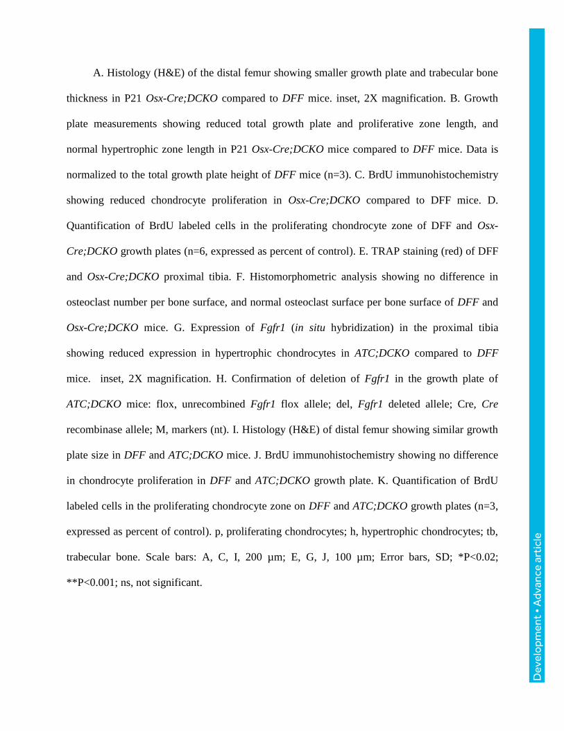
Figure 3. Decreased growth plate size and Decreased chondrocyte proliferation in Osx-Cre;
DCKO mice
Dev
elo
pmen
t • A
dvan
ce a
rtic
le
A. Histology (H&E) of the distal femur showing smaller growth plate and trabecular bone
thickness in P21 Osx-Cre;DCKO compared to DFF mice. inset, 2X magnification. B. Growth
plate measurements showing reduced total growth plate and proliferative zone length, and
normal hypertrophic zone length in P21 Osx-Cre;DCKO mice compared to DFF mice. Data is
normalized to the total growth plate height of DFF mice (n=3). C. BrdU immunohistochemistry
showing reduced chondrocyte proliferation in Osx-Cre;DCKO compared to DFF mice. D.
Quantification of BrdU labeled cells in the proliferating chondrocyte zone of DFF and Osx-
Cre;DCKO growth plates (n=6, expressed as percent of control). E. TRAP staining (red) of DFF
and Osx-Cre;DCKO proximal tibia. F. Histomorphometric analysis showing no difference in
osteoclast number per bone surface, and normal osteoclast surface per bone surface of DFF and
Osx-Cre;DCKO mice. G. Expression of Fgfr1 (in situ hybridization) in the proximal tibia
showing reduced expression in hypertrophic chondrocytes in ATC;DCKO compared to DFF
mice. inset, 2X magnification. H. Confirmation of deletion of Fgfr1 in the growth plate of
ATC;DCKO mice: flox, unrecombined Fgfr1 flox allele; del, Fgfr1 deleted allele; Cre, Cre
recombinase allele; M, markers (nt). I. Histology (H&E) of distal femur showing similar growth
plate size in DFF and ATC;DCKO mice. J. BrdU immunohistochemistry showing no difference
in chondrocyte proliferation in DFF and ATC;DCKO growth plate. K. Quantification of BrdU
labeled cells in the proliferating chondrocyte zone on DFF and ATC;DCKO growth plates (n=3,
expressed as percent of control). p, proliferating chondrocytes; h, hypertrophic chondrocytes; tb,
trabecular bone. Scale bars: A, C, I, 200 µm; E, G, J, 100 µm; Error bars, SD; *P<0.02;
**P<0.001; ns, not significant.

Dev
elo
pmen
t • A
dvan
ce a
rtic
le
Figure 4. Increased expression of Fgf9 and Fgf18 in Osx-Cre;DCKO mice
A. Expression of Fgf9 (in situ hybridization) in DFF and Osx-Cre;DCKO proximal tibia of
P21 mice showing increased expression in the perichondrium, reserve, proliferating, and
prehypertrophic chondrocytes of Osx-Cre;DCKO mice. B. qRT-PCR analysis of Fgf9 expression
in DFF and Osx-Cre;DCKO proximal tibia metaphysis (n=3). C. Expression of Fgf18 (in situ
hybridization) in P21 DFF and Osx-Cre;DCKO proximal tibia showing increased expression in
articular cartilage, proliferating, and prehypertrophic chondrocytes in the growth plate and in
trabecular bone of Osx-Cre;DCKO mice. D. qRT-PCR analysis of Fgf18 expression in DFF and
Osx-Cre;DCKO proximal tibia metaphysis (n=3). rc, reserve chondrocytes; p, proliferating
chondrocytes; h, hypertrophic chondrocytes; ac, articular chondrocytes; tb, trabecular bone; pc,
perichondrium. Scale bars: A, D, 100 µm; Error bars, SD; *P<0.05; **P<0.002.
Dev
elo
pmen
t • A
dvan
ce a
rtic
le
Figure 5. Increased Fgfr3 expression and signaling in Osx-Cre;DCKO mice
A,C. Expression (in situ hybridization) of Fgfr3 (A) and Snail1 (C) in DFF and Osx-
Cre;DCKO distal femur. B. qRT-PCR analysis of Fgfr3 expression in DFF and Osx-Cre;DCKO
proximal tibia and distal femur metaphysis (n=3). D. Immunohistochemistry of DFF and Osx-
Cre;DCKO proximal tibia showing mildly increased SOX9 in Osx-Cre;DCKO compared to DFF
mice. p, proliferating chondrocytes; h, hypertrophic chondrocytes; tb, trabecular bone. Scale
bars: A, C, D, 100 µm; Error bars, SD; *P<0.05.
Dev
elo
pmen
t • A
dvan
ce a
rtic
le
Figure 6. Activation of Fgf9 in the perichondrium suppresses chondrocyte proliferation.

Dev
elo
pmen
t • A
dvan
ce a
rtic
le
A. Fluorescence imaging of induced GFP expression in trabecular bone (tb), cortical bone
(e, endosteum; po, periosteum) and perichondrium (pc) of RunxTFG mice. GFP was not
observed in hypertrophic chondrocytes (h). B. Decreased body weight of P21 RunxTFG mice
(n=3) compared to Runx2-rtTA single transgenic control (n=4). C. Histology (H&E) of the
proximal tibia showing a smaller growth plate in P21 RunxTFG compared to Runx2-rtTA single
transgenic control. D. Growth plate measurements showing reduced total growth plate,
proliferative zone, and hypertrophic zone size in P21 RunxTFG mice. E. TRAP staining of P21
control and RunxTFG mice showing normal osteoclast number. F: BrdU immunohistochemistry
showing reduced chondrocyte proliferation in RunxTFG compared to control P21 mice. G.
Quantification of BrdU labeled cells in the proliferating chondrocyte zone of P21 control and
RunxTFG growth plates (n=3). H. Expression of Fgfr3 (in situ hybridization) in P21 control and
RunxTFG distal femur. rc, reserve chondrocytes; p, proliferating chondrocytes; h, hypertrophic
chondrocytes; tb, trabecular bone. Scale bars: A1,A3,C-H 100 µm; A2, 20 µm; Error bars, SD;
*P<0.01; **P<0.001.

Dev
elo
pmen
t • A
dvan
ce a
rtic
le
Figure 7. Rescue of the Osx-Cre;DCKO growth plate phenotype by administration of
PTH(1-34).

Dev
elo
pmen
t • A
dvan
ce a
rtic
le
A. Expression of Ihh (in situ hybridization) in P21 DFF and Osx-Cre;DCKO distal femur
showing decreased expression in the growth plate of Osx-Cre;DCKO mice. B. qRT-PCR
analysis of Ihh expression in DFF and Osx-Cre;DCKO proximal tibia metaphysis (n=3). C.
Expression of Pthlh (in situ hybridization) in P21 DFF and Osx-Cre;DCKO distal femur
showing decreased expression in the peripheral growth plate in Osx-Cre;DCKO mice (inset: 2X
magnification). D. qRT-PCR analysis of Pthlh expression in DFF and Osx-Cre;DCKO proximal
tibia metaphysis (n=3). E. Expression of Pthlh in P21 control and RunxTFG proximal tibia. F.
Histology (H&E) of the proximal tibia showing a larger growth plate and increased trabecular
bone in P21 PTH treated compared to PBS (control) treated Osx-Cre;DCKO mice. G. Growth
plate measurements showing increased total growth plate, proliferative, and hypertrophic zone
size in PTH treated (n=3) compared to PBS treated (n=4) mice. H. Expression of Fgfr3 (in situ
hybridization) in the distal femur of P21 PTH treated compared to PBS (control) treated Osx-
Cre;DCKO mice. I. BrdU immunohistochemistry showing increased chondrocyte proliferation in
P21 PTH treated compared to PBS (control) treated Osx-Cre;DCKO mice. J. Quantification of
BrdU labeled cells in the proliferating chondrocyte zone of PTH treated compared to PBS
(control) treated Osx-Cre;DCKO mice (n=3). rc, reserve chondrocytes; p, proliferating
chondrocytes; ph, prehypertrophic chondrocytes; h, hypertrophic chondrocytes; tb, trabecular
bone. Scale bars: A, C, 50µm; E, F, H, I, 100 µm; Error bars, SD; *P<0.05; **P<0.005;
***P<0.001.
Dev
elo
pmen
t • A
dvan
ce a
rtic
le
Figure 8. Model of FGF regulated interactions between osteoprogenitor lineages and
growth plate chondrocytes in postnatal endochondral bone growth.
(A) FGFR1 and FGFR2 in the osteoprogenitor lineage are regulated by FGF9 expressed in
osteoprogenitors and adjacent connective tissue and periosteum. Inactivation of FGFR1 and
FGFR2 results in compensatory increased expression of Fgf9, which (B) aberrantly activates
FGFR3 and downstream Snail1 to suppress chondrocyte proliferation and hypertrophy. Increased
FGFR3 signaling also promotes Fgf9 and Fgf18 expression in chondrocytes and suppresses
expression of Ihh and Pthlh. PTHLH functions to suppress Fgfr3 expression and reduced Pthlh
contributes to increased Fgfr3 expression. The aberrant activation of FGFR3 (expression and
signaling in chondrocytes) may initiate a feed-forward signaling loop in chondrocytes that
functions to terminate chondrogenesis. HS, heparan sulfate.
![Rizzoli.and.Isles.s01e03.Hdtv.xvid Xii.eng[Ragbear]Fgf](https://static.fdocuments.in/doc/165x107/577d36f21a28ab3a6b9464d0/rizzoliandisless01e03hdtvxvid-xiiengragbearfgf.jpg)