F.E.S.S. - Karl Storz SE...F.E.S.S. “UNCAPPING THE EGG” The Endoscopic Approach to Frontal...
52
A Surgical Technique of the Graz University Medical School by use of the KARL STORZ HOPKINS ® 45° Telescopes Heinz STAMMBERGER F.E.S.S. “UNCAPPING THE EGG” The Endoscopic Approach to Frontal Recess and Sinuses ®
Transcript of F.E.S.S. - Karl Storz SE...F.E.S.S. “UNCAPPING THE EGG” The Endoscopic Approach to Frontal...
A Surgical Technique of the Graz University Medical School by use of the KARL STORZ HOPKINS® 45° Telescopes
Heinz STAMMBERGER
F.E.S.S. “UNCAPPING THE EGG”
The Endoscopic Approach to Frontal Recess and Sinuses
®

F.E.S.S. “UNCAPPING THE EGG”The Endoscopic Approach to
Frontal Recess and SinusesA Surgical Technique of the Graz University Medical School
by use of the KARL STORZ HOPKINS® 45° Telescopes
Prof. Heinz STAMMBERGER, M.D.HonFRCS(Ed), HonFRCS(Engl), HonFACS
Endoscopic Sinus & Skull Base SurgeryConsultant, Interdisciplinary Skull Base Group
Medical University Graz, Austria
®

FESS – “Uncapping the Egg” – The Endoscopic Approach to Frontal Recess and SinusesA Surgical Technique of the Graz University Medical School4
All anatomical sketches and schematic drawings were made byMs. Astrid Hambrosch, Anatomical Institute of theKarl-Franzens-University, Graz, Austria (Head: Prof. F. Anderhuber, M.D.)
ISBN 978-3-89756-041-3
FESS – “Uncapping the Egg” –The Endoscopic Approach to Frontal Recess and Sinuses A Surgical Technique of the Graz University Medical Schoolby use of the KARL STORZ HOPKINS® 45° Telescopes Prof. Heinz Stammberger, M.D.HonFRCS(Ed), HonFRCS(Engl), HonFACSEndoscopic Sinus & Skull Base Surgery,Consultant, Interdisciplinary Skull Base GroupMedical University Graz, Austria
Correspondence address of the author: Prof. Heinz Stammberger, M.D.Sonnleitenweg 51A - 8034 Graz, AustriaE-mail: [email protected]
All rights reserved.1st edition 2005© 2015 ® GmbHP.O. Box, 78503 Tuttlingen, GermanyPhone: +49 (0) 74 61/1 45 90Fax: +49 (0) 74 61/708-529E-mail: [email protected]
No part of this publication may be translated, reprinted or reproduced, trans-mitted in any form or by any means, electronic or mechanical, now known or hereafter invent ed, including photocopying and recording, or utilized in any information storage or retrieval system without the prior written permission of the copyright holder.
Editions in languages other than English and German are in preparation. For up-to-date information, please contact ® GmbH at the address shown above.
Design and Composing:® GmbH, Germany
Printing and Binding:Straub Druck + Medien AGMax-Planck-Straße 17, 78713 Schramberg, Germany
05.15-1
Important notes:
Medical knowledge is ever changing. As new research and clinical experience broaden our knowledge, changes in treat ment and therapymay be required. The authors and editors of the material herein have consulted sources believed to be reliable in their efforts to provide information that is complete and in accord with the standards accept ed at the time of publication. However, in view of the possibili ty of human error by the authors, editors, or publisher, or changes in medical knowledge, neither the authors, editors, publisher, nor any other party who has been involved in the preparation of this booklet, warrants that the information contained herein is in every respect accurate or complete, and they are not responsible for any errors or omissions or for the results obtained from use of such information. The information contained within this booklet is intended for use by doctors and other health care professionals. This material is not intended for use as a basis for treatment decisions, and is not a substitute for professional consultation and/or use of peer-reviewed medical literature.
Some of the product names, patents, and re gistered designs referred to in this booklet are in fact registered trademarks or proprietary names even though specifi c reference to this fact is not always made in the text. Therefore, the appearance of a name without designation as proprietary is not to be construed as a representation by the publisher that it is in the public domain.
The use of this booklet as well as any implementation of the information contained within explicitly takes place at the reader’s own risk. No liability shall be accepted and no guarantee is given for the work neither from the publisher or the editor nor from the author or any other party who has been involved in the preparation of this work. This particularly applies to the content, the timeliness, the correctness, the completeness as well as to the quality. Printing errors and omissions cannot be completely excluded. The publisher as well as the author or other copyright holders of this work disclaim any liability, particularly for any damages arising out of or associated with the use of the medical procedures mentioned within this booklet.
Any legal claims or claims for damages are excluded.
In case any references are made in this booklet to any 3rd party publication(s) or links to any 3rd party websites are mentioned, it is made clear that neither the publisher nor the author or other copyright holders of this booklet endorse in any way the content of said publication(s) and/or web sites referred to or linked from this booklet and do not assume any form of liability for any factual inaccuracies or breaches of law which may occur therein. Thus, no liability shall be accepted for content within the 3rd party publication(s) or 3rd party websites and no guarantee is given for any other work or any other websites at all.

FESS – “Uncapping the Egg” – The Endoscopic Approach to Frontal Recess and SinusesA Surgical Technique of the Graz University Medical School 5
Contents
Introduction .................................................................................................... 7
Anatomy and Pathophysiology of Frontal Recess and Frontal Sinus ............................................................... 9
Variants of the Uncinate Process ................................................................ 10
“Uncapping the Egg” .................................................................................... 14
Postoperative Care ....................................................................................... 17
Telescopes and Instruments ........................................................................ 18
Surgical Steps (Anatomical Specimen) ....................................................... 21
Case 1 ............................................................................................................. 23
Case 2 ............................................................................................................. 25
Case 3 ............................................................................................................. 27
Case 4 ............................................................................................................. 29
The KARL STORZ HOPKINS® 45° TelescopesTelescopes, Instruments, Units, Video Cameras and Accessories ........................................................... 31–49
Extracts from the following Catalogs:ENDOSCOPES AND INSTRUMENTS FOR ENTTELEPRESENCE, IMAGING SYSTEMS, DOCUMENTATION – ILLUMINATION


FESS – “Uncapping the Egg” – The Endoscopic Approach to Frontal Recess and SinusesA Surgical Technique of the Graz University Medical School 7
IntroductionThe endonasal approach to the frontal sinus undoubtedly requires sound ana tomical knowledge, highest surgical skills and dexterity. The frontal recess is an anatomically very complex structure and can be seen as the ethmoidal ”pre-chamber” to the frontal sinus proper. Its configuration depends on a variety of cells and lamellae with a high degree of individual varia tion. Almost always disease in the frontal sinus results from underlying disease processes in the clefts of the frontal recess. Rarely ever will inflammatory processes originate in the frontal sinus cavity itself. These findings consequently resulted in diagnosis, conservative and surgical therapeutic measures to focus on these ”pre-chambers” to the frontal sinus. Especially in surgery, the delicate bony structures and the mucosa in the frontal recess region require a very care ful, minimally traumatic approach: Overly forceful handling of instruments, traumat ising the mucosa or its removal will rapidly lead to granulations, scar and stenosis formation. In these cases postoperative healing will be significantly delayed, local postoperative care must be intensified and patients frequently be seen for follow-up. It is not a rare finding, that following traumatic manipulations in this region patients show frontal sinus problems which were not present before. These frontal sinus problems therefore must in part be seen as iatro genic.
Fig. 1Original drawing from Halle’s publication from 1906: Drilling away the entire superior nasal spine. Provided Halle already had had telescopes of different angulations, he most likely would have chosen the morephysiologic approach dorso-laterally to the nasal spine (dotted line).
Fig. 2Massive opacification of the frontal sinus bilaterally…
Fig. 3…resulting from blockage of the frontal recess.

FESS – “Uncapping the Egg” – The Endoscopic Approach to Frontal Recess and SinusesA Surgical Technique of the Graz University Medical School8
In recent years many surgical schools have recommended to routinely create as large a communication between the frontal recess and the frontal sinus as possible, to avoid the well know tendencies for scarring and stenosis. In our view this has resulted in too frequently used, too radical, too traumatic approaches. These are usually performed with a drill (or curved blades of powered instruments) as so-called “drill-out procedures”. According to our experience, these are required for a very small number of special indications only. In this brochure we would like to illustrate how to succeed in the vast majority of cases with signi ficantly less traumatic procedures, “simply” following the anatomy. This surgical approach has been applied by us for almost 3 decades now and has been constantly improved.
According to our experiences, atraumatic functional surgery in the vicinity of the frontal sinus ostium can only be performed using endoscopes of different angulations. This fact is sort of self-explanatory if one studies the topographical anatomy of the region. There is no need to drill away the “nasal beak” (the inner superior nasal spine) if one uses telescopes of different angulations and follows the anatomical route predesigned by nature. In doing so, only thin and delicate bony lamella have to be removed, though in a “delicate surrounding”. If one wants to engage in this kind of frontal sinus surgery, it is mandatory that the same principles of care (and I dare to say: of dexterity) are applied as in stapes surgery (D.W. Kennedy).
Following our recommendations KARL STORZ Tuttlingen designed two 45° endoscopes, which allow for an excellent view into the frontal recess, especially if the middle turbinate is preserved.
Fig. 4Mucus transport through frontal sinus and recess. There can be an active transport into the frontal sinus from out of the frontal recess medially.

FESS – “Uncapping the Egg” – The Endoscopic Approach to Frontal Recess and SinusesA Surgical Technique of the Graz University Medical School 9
Anatomy and Pathophysiologyof Frontal Recess and Frontal SinusThe frontal sinus develops from the most anterior and superior parts of the anterior ethmoid: The frontal bone is pneumatised from the frontal recess (Killian). We discourage the use of the term “ductus nasofrontalis, nasofrontal duct”, as a true tubular structure connecting frontal sinus and anterior ethmoid does not exist. In a somewhat simplified way, the frontal sinus is nothing else but a cell of the anterior ethmoid, pneumatising the frontal bone. Not surprisingly therefore the frontal sinus depends on its “origin” in the anterior ethmoid with respect to its normal and pathophysiology.
On a sagittal section (Fig. 5) the transition of frontal sinus to frontal recess has an hour-glass shape: The floor of the frontal sinus (sf) narrows downward like a funnel towards the frontal sinus ostium (osf). From here, the frontal recess widenes like an inverted funnel (rf). So both structures together can be seen as an hour-glass, with the “waist” at the level of the frontal sinus ostium.
The frontal recess can be significantly influenced and narrowed by a number of structures (Fig. 6): 1.) the uncinate process, 2.) agger nasi cell(s), 3.) the eth moidal bulla, 4.) other cells of the anterior ethmoid, located in the frontal recess. Frequently, combinations of the variants to be discussed below are encoun tered in patients suffering from acute or recurrent inflammations of the frontal sinus. In a very schematised and simplified way, all these structures narrow the inferior part of the “hour-glass”, i.e. the frontal recess and thus predispose to recurring problems.
Fig. 5Sagittal CT demonstrating the basal lamellae:1 = basal lamella of the uncinate2 = b. l. of the bulla3 = b. l. of the middle turbinate4 = b. l. of the superior turbinateNote the hourglass configuration of thetransition from frontal sinus to frontal recess.
Fig. 6Schematic drawing of narrowing of frontal recess by agger nasi-cells (blue), un cinate process (red) and ethmoidal bulla andother anterior ethmoidal cells (green), respectively.

FESS – “Uncapping the Egg” – The Endoscopic Approach to Frontal Recess and SinusesA Surgical Technique of the Graz University Medical School10
Variants of the Uncinate Process
There are three basic configurations of the uncinate process (Fig. 7):
a) In a coronal cut, the uncinate process can extend superiorly, bend laterally and insert at the lamina papyracea (orbitalis). Thus a superior blind(recessus terminalis) of the ethmoidal infundibulum is formed, separating the latter to some degree from the frontal recess.
b) The uncinate process can extend straight superiorly and reach the skull base.
c) It can curve medially and fuse with the insertion of the middle turbinate.
Transitions between these three extremes occur.
If there is a pronounced terminal recess (Fig. 8b) reaching extremely far supe ri orly, the uncinate process with this “cap” can almost fill the frontal recess, even contact the skull base and medially the lateral lamella of the cribriform plate. The clinically significant effect of such a configuration can be impairment or even blockage of ventilation and drainage of the frontal sinus proper.
If the agger nasi region is pneumatised, i.e. agger nasi cells exist, these can expand towards superiorly and posteriorly and thus more or less fill the entire frontal recess with their thin “cap” of bone in an analogous fashion.
Fig. 7Anatomical variations of the uncinate process.
Fig. 8Narrowing of frontal recess by combinations of variants of un cinate, bulla and agger nasi cells.
a b c a b c

FESS – “Uncapping the Egg” – The Endoscopic Approach to Frontal Recess and SinusesA Surgical Technique of the Graz University Medical School 11
The ethmoidal bulla (or other cells of the anterior ethmoid, for instance so-called infundibular cells) may expand into the frontal recess from posterior- inferiorly, i.e. they pneumatise upward and forward and thus with their cranial “cap” of bone may fill the frontal recess. This too, may lead to impairment/stenosis. Not only may combinations of these three basic variants prevail, but cells may develop into the frontal sinus (bulla frontalis, intrafrontal cells) presenting a true challenge for diagnosis and even more, surgical therapy (Figs. 5–9).
Figs. 8 and 9 demonstrate some of the possibilities in a schematic fashion and in CT: One can clearly see, that in all cases access to the frontal sinus is blocked by a paper-thin cap of bone with its two mucosal layers.
It is of utmost importance to understand, that access to the frontal sinus ostium is not blocked by the allegedly important “nasal beak”. This massive bony structure is only of relevance in patients with extremely narrow dorsum of the nose. Even then, access to the frontal sinus is only impeded medially, next to the nasal septum. Approaching from dorso-laterally, the internal nasal spine very rarely presents an obstacle or a pathophysiologically relevant structure.
Very clearly this situation can be recognised in Fig. 1 taken from Halle’s pub lication from 1906: It must be assumed that Halle would not have chosen this approach, if at that time endoscopes for “looking around the corner” had been available to him. The removal of the very thin bony septations posterolateral to the “nasal beak” would have resulted in a wide and natural passage, via a far less traumatic approach, avoiding unwarranted removal of bone and mucosa.
Fig. 10bFigs. 10 a + b: Schematic drawing of frontal recess narrowed by agger nasi and infundibular cells (a), as well as by a terminal recess and infundibular cells (b).
Fig. 10a
Fig. 9a Figs. 9a + b: Several cells may be superimposed and narrow the frontal recess, sometimes even reach into the sinus lumen itself as “intrafrontal cells”.
Fig. 9b

FESS – “Uncapping the Egg” – The Endoscopic Approach to Frontal Recess and SinusesA Surgical Technique of the Graz University Medical School12
Fig. 11 aFigs. 11 a–c: Remnants of thin “bone caps” of a terminal recess (a), an agger nasi-cell (b) and an ethmoidal bulla (c), all narrowing access to the frontal sinus.
If uncinate, bulla and agger nasi cells were resected during endonasal surgery and the frontal sinus ostium is not visible, this almost always can be traced back to a typical anatomical situation: A thin curved bony “shell” has remained, which may be in contact with the skull base or the lateral lamella of the middle tur binate. This thin bone layer blocks access to the frontal sinus proper, towards its ostium. The technical challenge is to identify and carefully remove this cap of bone. In the vast majority of cases this manoeuvre will expose the level of the frontal sinus ostium. Rarely there is need then to enlarge the latter let alone to drill away the “nasal beak”.
For these manipulations simple but delicate instruments are required (Figs. 12, 13). With specially curved curettes (according to F. Kuhn) or Giraffe forceps these structures can be removed. All these manoeuvres must be performed under good direct vision and optimal hemostasis. Only in this way true atraumatic surgery is possible. The new KARL STORZ 45° endoscopes were especially developed for this purpose. They offer a crucial “extra”of retrograde upward viewing allowing for a complete inspection of the frontal recess.
Fig. 12Removal of an “cap of the egg” with giraffe forceps.
Fig. 13Remnants of an “egg cap” are identified and carefully removed.
Fig. 11 b Fig. 11 c

FESS – “Uncapping the Egg” – The Endoscopic Approach to Frontal Recess and SinusesA Surgical Technique of the Graz University Medical School 13
In the past it was not always possible to achieve this with the well proven 30° telescopes. On the other hand the 45° telescopes are constructed in a way that they still allow for a straight forward view. This is important when inserting the instruments, as at 6.00 o’clock of the visual field the view is almost parallelto the longitudinal axis of the endoscope. This is an advantage over thewell proven 70° endo scopes, which do not offer any optical information in the direction of the shaft itself.
One of our favourite instruments for surgery in the frontal recess, especially when removing thin bone caps, is the upbent circular cutting punch (Fig.14). With this instrument one can well avoid to expose bare bone which in turn will help to avoid scaring and restenosis. If performed well, this approach leaves behind a wide frontal recess and an open frontal sinus ostium. All walls are covered by mucosa with only minimal areas of bone exposed at the cutting edges of the very thin bony lamella. This considerably shortens the postopera tive healing phase.
Fig. 14 bFig. 14 aSchematic drawing demonstrating the use of the upcurved circular cutting punch.

FESS – “Uncapping the Egg” – The Endoscopic Approach to Frontal Recess and SinusesA Surgical Technique of the Graz University Medical School14
Fig. 15“The Egg” in an egg holder.
Fig. 16The egg is uncapped, the contents re moved and the egg holder turned upside down.
Fig. 17Superimposing the “inverted” egg holder over the frontal sinus infun-dibulum and frontal recess, …
Fig. 18…the challenge is to remove the remaining “cap of the egg” from the frontal recess to reach the frontal sinus ostium and, finally, the frontal sinus proper.
Fig. 17Superimposing the “inverted” egg holder over the frontal sinusinfundibulum and frontal recess, …
“Uncapping the Egg”For many years we have been teaching this technique in our endoscopy cours es at Graz and have presented the concept of “uncapping the egg” for the first time internationally during the “3rd International Symposium on Advanced FESS” at Cairns/Australia in 1995. “Uncapping the Egg” represents a mne monic only; the diagrams are very schematic, over-simplified, not to scale – but do point out the main principle.In a sagittal section, the transition from frontal sinus down to the frontal recess has an hour-glass shape (see page 9), one might as well say: The shape of an egg-holder. The waist corresponds to the level of the frontal sinus ostium. If one now puts a breakfast-egg into the egg-holder, opens and empties it, the lower portion of the “cap of the egg” remains in the egg-holder. If this now is turned upside down, the situation is analoguous to the frontal recess: The superior “empty” part of the egg-holder represents the frontal sinus infundibulum, i.e. the floor of the frontal sinus narrowing towards the ostium. The inferior part of the egg-holder with the cap of the egg represents the frontal recess. The task now is, to remove the eggshell sitting tightly in the “frontal recess”. If one succeeds, the walls of the frontal recess are not traumatised, but the passage through frontal recess via frontal sinus ostium into frontal sinus is free (Figs. 15–18).“Uncapping the Egg” has proven an excellent mnemonic to think of and look for the “eggshells” which usually block the frontal recess in difficult situations. Following these principles and using the new KARL STORZ 45° telescopes, drill-out procedures have remained an absolute rarity amongst thousands of cases we have operated over the years.Especially the circular cutting punch in its upbent version (Fig. 18) allows to enlarge the frontal sinus ostium itself – if required – following the same principles of minimal trauma.

FESS – “Uncapping the Egg” – The Endoscopic Approach to Frontal Recess and SinusesA Surgical Technique of the Graz University Medical School 15
In Figs. 19 and 20 postoperative CT-findings demonstrate why these patients suffer from recurring frontal sinus problems: In all cases the surgeons failed to remove (or to identify) the cranial cap of the individual cells or the uncinate process. Looking back at our own learning curve, it is not rare to mistake a pro nounced terminal recess or supraorbital ethmoid cell for the frontal sinus proper. The pictures clearly identify the thin bony caps remaining and the result ing narrowing and stenosis which must be taken care of during revision surgery. These pictures furthermore stress the need for exact endoscopic and radio logic diagnosis (axial CT-scans would not show these changes!) in avoiding un warranted radical surgery using drills.
Fig. 19Typical postoperative finding: On the left side (of the patient) the bony cap of an agger nasi-cell was not removed, resulting in recurrent frontal sinusitis.
Fig. 20As before, agger nasi-cells on both sides were not removed sufficiently, resulting in recurrent frontal sinus problems.

FESS – “Uncapping the Egg” – The Endoscopic Approach to Frontal Recess and SinusesA Surgical Technique of the Graz University Medical School16
The main prerequisite for the success of this surgical approach is an atraumatic technique.
Particular care must be taken to avoid any damage to the parietal mucosa, i.e. ideally the walls of the frontal recess should be completely covered with mucosa or at least the basal layers thereof. Areas of denuded bone must be reduced to a minimum. In no case the frontal sinus ostium should be traumatised circularly, let alone mucosa be removed in this region. Inevitably this will lead to scar and stenosis formation. This problem is illustrated in Figs. 21–24. Fracturing of the middle turbinate should be avoided by all means, to prevent lateralisation, scar ing and stenosis of the frontal recess. The mucosa at the insertion of the middle turbinate should not be traumatised, especially no opposing raw surfaces be created. If a middle turbinate has been destabilised accidentally, the technique illustrated in Fig. 25 can be used to avoid lateralisation. Stents and drains are not helpful if mucosa has been re moved. The process of reepitheliasation and scarring continues for many months!
Fig. 22Ideal postoperative situation: The arrows indicate the resection margins of uncinate process and anterior ethmoidal cells. Only here has bone been minimallyexposed, all other walls of the surgical cavity are covered by mucosa.
Fig. 23A situation like this must be avoided by all means: Significant areas of bone are denuded and no longer covered by mucosa.
Fig. 24Fractures of the middle turbinate should strictly be avoided, to prevent lateralisa tion and ensuing formation of scars andstenosis.
Fig. 21Schematic drawing of a seriously affected frontal recess.

FESS – “Uncapping the Egg” – The Endoscopic Approach to Frontal Recess and SinusesA Surgical Technique of the Graz University Medical School 17
Postoperative Care
In patients who underwent frontal recess/sinus revisions, careful endoscopic postoperative care is of utmost importance. Usually 8–12 days postoperatively we start to aspirate wound secretions and instrumentally remove crust under direct endoscopic vision, using 45° telescopes for these manipulations. No new trauma should be produced during this procedure. The duration of follow-up varies depending on the findings and the underlying disease. Usually this first endoscopic control gives a good impression on whether or not a problem case is at hands requiring frequent follow-ups at short intervals, or whether the patient needs to be followed up at intervals of 4–5 weeks only. Depending on initial underlying pathology, normalisation/stabilisation can be expected after 6 weeks to 6 months.
Fig. 25If a middle turbinate insertion is fractured by accident, it can be stabilised as follows: Small areas of opposing mucosal surfaces are scarified, inducing circumscribed ad hesions. These scars can – but do not have to – be separated again several weeks later (following W. Bolger).

FESS – “Uncapping the Egg” – The Endoscopic Approach to Frontal Recess and SinusesA Surgical Technique of the Graz University Medical School18
Telescopes and Instruments
Fig. 27 demonstrates the features and advantages of the KARL STORZ 45° telescopes: Despite the significant angulation a straight forward view along the shaft axis is yet possible.
This allows to avoid contact with and lesions of mucosa of the septum, the turbinate and other structures and is a definite advantage over the 70° lenses. Compared to the 30° lenses, the new 45° endoscopes significantly enhance the capability for “looking around the corner”. This is a prerequisite for surgical procedures in the difficult topography of the frontal recess and sinus. The surgeon not only has to look “around the corner”, but the instruments have to reach “around the corner” as well. Endoscope and instrument are inserted almost parallel to the nasal dorsum under (i.e. lateral to) the middle turbinate. From here, the working and viewing direction changes towards superiorly and ante riorly, i.e. in a retrograde fashion when approaching the frontal sinus through the frontal recess. In patients with pronounced supraorbital bulging, this angulation can be almost 90°. Instruments developed for endoscopic surgery of the frontal recess and sinus are designed for this respect. Inserting them through the nostrils requires some training and dexterity, as the tips of the instruments must be guided in an arch to be inserted under the middle turbinate and then retro gradely up and anteriorly. It is especially during these moves that the less experi enced (and sometimes: less patient) surgeon finds it difficult to proceed without occasional lesions of the mucosa. Consequent training is mandatory therefore.
Like the 30° telescopes, the 45° telescopes from KARL STORZ are avail able in two versions: A standard version and one with lateral light cable connection (Fig. 27). This latter version offers the following additional advantage: Some times the eyepiece of the endoscope must be significantly lowered to allow view towards the frontal sinus ostium. In these cases, the light cable may interfere with the chin of the patient and/or the intubation gear. This interference is largely avoided when using the endoscope with lateral light cable connection. Thus the eyepiece can be lowered an additional 10 centimetres, resulting in an important gain of view towards far anteriorly located frontal sinus ostia.
Fig. 26Schematic drawing of the endonasalendo scopic route “around the corner” (yellow broken line) to the frontal sinus, modifying Halle’s depiction from 1906.
Fig. 27 The two KARL STORZ HOPKINS® 45° telescopes.

FESS – “Uncapping the Egg” – The Endoscopic Approach to Frontal Recess and SinusesA Surgical Technique of the Graz University Medical School 19
For the same reason upbiting Blakesley forceps have been designed with a horizontal handle (Fig. 28). This results in a better angulation during the initial work in the frontal recess.
The delicate “giraffe” forceps are used to remove the delicate bony lamellae (“cap of the egg”) from the frontal recess: Frontally oriented segments are grasp ed with the forward-backward opening forceps, longitudinally oriented bone segments with the laterally (left-right) opening one. If there is need to remove thicker bony segments, through-cutting forceps (Figs. 29) are used. Of all the instruments mentioned, we only use the smaller size (size 1) to avoid trauma to the mucosa by trying to insert large instrument tips.
Several years ago we developed the circular cutting punch to resect horizon tally oriented thin bony lamellae if required. If we have to enlarge the frontal sinus ostium proper, we almost exlusively do this with circular cutting forceps. The advantage is evident: Cutting and thus removal of bone is possible in all directions (360°), without any need to rotate the shaft or handle of the instrument. Interference with the endoscope thus is minimised.
Fig. 2845° upturned BLAKESLEY-STAMMBERGER Nasal Forceps with handle in right horizon tal position.
Fig. 30Detail of upturned Circular Cutting Punches.
Fig. 29 a“Giraffe Neck” Forceps.
Fig. 29 bThrough-cutting BLAKESLEY Nasal Forceps.

FESS – “Uncapping the Egg” – The Endoscopic Approach to Frontal Recess and SinusesA Surgical Technique of the Graz University Medical School20
Fig. 4 Fig. 5 Fig. 6
Fig. 1 Fig. 2 Fig. 3
Fig. 7 Fig. 8 Fig. 9
FESS – “Uncapping the Egg” – The Endoscopic Approach to Frontal Recess and SinusesA Surgical Technique of the Graz University Medical School 21
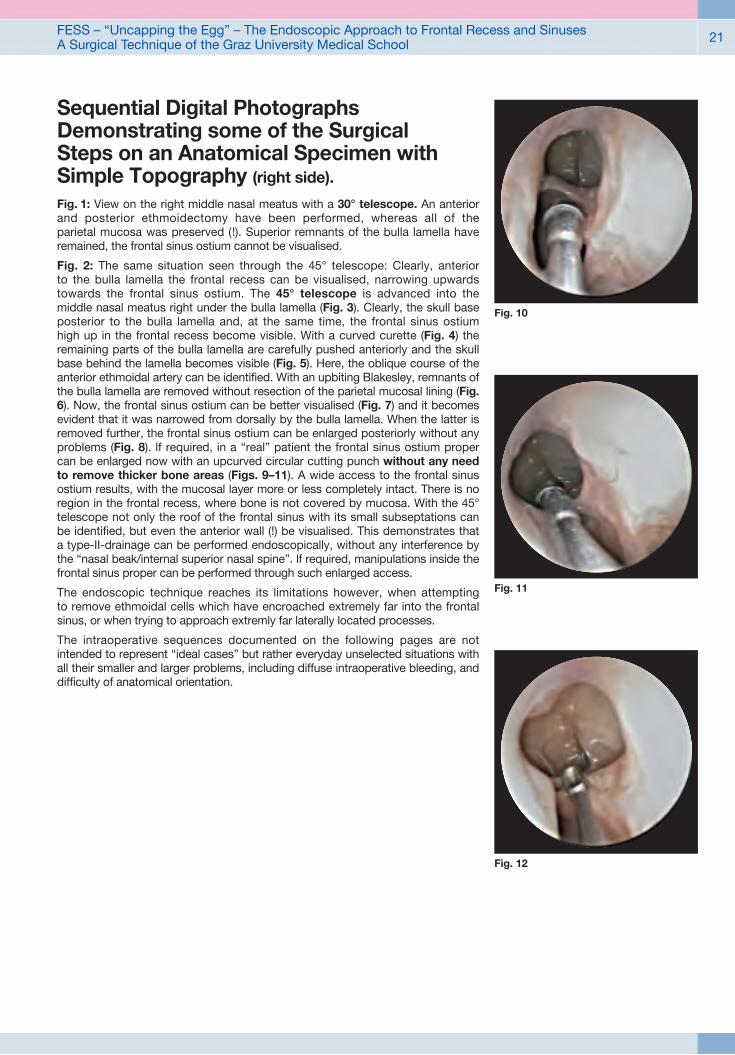
Sequential Digital Photo graphs Demonstrating some of the Surgical Steps on an Anatomical Specimen with Simple Topography (right side).
Fig. 1: View on the right middle nasal meatus with a 30° telescope. An anterior and posterior ethmoidectomy have been performed, whereas all of the parietal mucosa was preserved (!). Superior remnants of the bulla lamella have re mained, the frontal sinus ostium cannot be visualised.
Fig. 2: The same situation seen through the 45° telescope: Clearly, anterior to the bulla lamella the frontal recess can be visualised, narrowing upwards towards the frontal sinus ostium. The 45° telescope is advanced into the middle nasal meatus right under the bulla lamella (Fig. 3). Clearly, the skull base posterior to the bulla lamella and, at the same time, the frontal sinus ostium high up in the frontal recess become visible. With a curved curette (Fig. 4) the remain ing parts of the bulla lamella are carefully pushed anteriorly and the skull base behind the lamella becomes visible (Fig. 5). Here, the oblique course of the an terior ethmoidal artery can be identified. With an upbiting Blakesley, remnants of the bulla lamella are removed without resection of the parietal mucosal lining (Fig. 6). Now, the frontal sinus ostium can be better visualised (Fig. 7) and it becomes evident that it was narrowed from dorsally by the bulla lamella. When the latter is removed further, the frontal sinus ostium can be enlarged posteriorly without any problems (Fig. 8). lf required, in a “real” patient the frontal sinus ostium proper can be enlarged now with an upcurved circular cutting punch without any need to remove thicker bone areas (Figs. 9–11). A wide access to the frontal sinus ostium results, with the mucosal layer more or less completely intact. There is no region in the frontal recess, where bone is not covered by mucosa. With the 45° telescope not only the roof of the frontal sinus with its small subseptations can be identified, but even the anterior wall (!) be visualised. This demonstrates that a type-II-drainage can be performed endoscopically, without any interference by the “nasal beak/internal superior nasal spine”. lf required, manipulations inside the frontal sinus proper can be performed through such enlarged access.
The endoscopic technique reaches its limitations however, when attempting to remove ethmoidal cells which have encroached extremely far into the frontal sinus, or when trying to approach extremly far laterally located processes.
The intraoperative sequences documented on the following pages are not intended to represent “ideal cases” but rather everyday unselected situations with all their smaller and larger problems, including diffuse intraoperative bleed ing, and difficulty of anatomical orientation.
Fig. 10
Fig. 11
Fig. 12

FESS – “Uncapping the Egg” – The Endoscopic Approach to Frontal Recess and SinusesA Surgical Technique of the Graz University Medical School22
Fig. 2 Fig. 3 Fig. 4
Fig. 5 Fig. 6 Fig. 7
Fig. 1

FESS – “Uncapping the Egg” – The Endoscopic Approach to Frontal Recess and SinusesA Surgical Technique of the Graz University Medical School 23
Case 1 Fig. 1: Typical intraoperative situation: Only the surgeon and the scrub nurse are required for endonasal endoscopic procedures (including tumors and other operations at the anterior skull base). No assistant is necessary. The surgeon can control the position of his endoscopic picture via the smaller monitor, the larger monitor providing information for the scrub nurse, the anesthetists and residents/registrars and/or visitors.
Fig. 2: Intraoperative situation on the left side: The 45° telescope has been intro duced into the middle meatus lateral to the middle turbinate. View is up into the frontal recess. The uncinate process and bulla have been resected. The patient is suffering from massive chronic rhinosinusitis with polyposis (type IV). The frontal sinus ostium cannot be visualised yet. Looking for the “cap of the egg”, a curved curette is introduced and the medial free margin of the bony cap care fully palpated. After identification, the thin bony shell is carefully elevated off the middle turbinate (Figs. 3, 4). Clearly one can see now that all which needs to be removed is an eggshell thin bone cap with its bilateral mucosal layer, indeed. This “cap of the egg” is carefully pushed laterally now (Fig. 5) and removed with a thin giraffe forceps. This results in a first free view of the frontal sinus ostium (Fig. 6), and after aspiration of retained mucus a more or less normal mucosa inside the frontal sinus can be visualised.

FESS – “Uncapping the Egg” – The Endoscopic Approach to Frontal Recess and SinusesA Surgical Technique of the Graz University Medical School24
Fig. 1 Fig. 3
Fig. 4 Fig. 5 Fig. 6
Fig. 2

FESS – “Uncapping the Egg” – The Endoscopic Approach to Frontal Recess and SinusesA Surgical Technique of the Graz University Medical School 25
Case 2Right side of a patient: The 45° telescope has been introduced under the middle turbinate into the middle meatus behind the suspected “cap of the egg”, blocking access to the frontal sinus ostium. The posterior portion of this bone cap has been identified and is gently elevated towards anteriorly (Fig. 2). This results in identification of the frontal sinus ostium proper. The latter now is enlarged with the upturned circular cutting forceps (Fig. 3), removing further fragments of the “eggshell” of this high reaching agger nasi cell. In Fig. 4 the preci sion can be seen resulting from the use of the upcurved circular cutting punch. Now view into the frontal sinus proper is possible (Fig. 5). Some minor bony edges are carefully removed out of the frontal recess (Fig. 6), leaving the parietal mucosa to cover all of the bony walls. No packing is used in this region nor are stents applied. Fig. 7 demonstrates the situation encountered at the first endo scopic control 10 days postoperatively, after removal of minor crusts and aspira tion of wound secretions. lt is time and again surprising to see the rapid recovery and healing following these atraumatic approaches to the frontal sinus. We believe, that preservation of the parietal mucosa and the avoidance of bone exposure contribute significantly to this phenomenon.
Fig. 7

FESS – “Uncapping the Egg” – The Endoscopic Approach to Frontal Recess and SinusesA Surgical Technique of the Graz University Medical School26
Fig. 1 Fig. 2
Fig. 4 Fig. 6
Fig. 3
Fig. 5
FESS – “Uncapping the Egg” – The Endoscopic Approach to Frontal Recess and SinusesA Surgical Technique of the Graz University Medical School 27
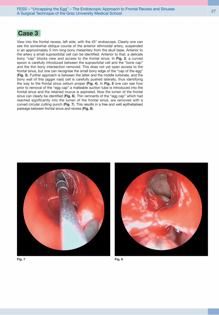
Case 3View into the frontal recess, left side, with the 45° endoscope. Clearly one can see the somewhat oblique course of the anterior ethmoidal artery, suspended in an approximately 5 mm long bony mesentery from the skull base. Anterior to the artery a small supraorbital cell can be identified. Anterior to that, a delicate bony “cap” blocks view and access to the frontal sinus. In Fig. 2, a curved spoon is carefully introduced between the supraorbital cell and the “bone cap” and the thin bony intersection removed. This does not yet open access to the frontal sinus, but one can recognise the small bony edge of the “cap of the egg” (Fig. 3). Further approach is between the latter and the middle turbinate, and the bony wall of this (agger nasi) cell is carefully pushed laterally, thus identifying the way to the frontal sinus ostium proper (Fig. 4). In Fig. 5 one can see how prior to removal of the “egg cap” a malleable suction tube is introduced into the frontal sinus and the retained mucus is aspirated. Now the lumen of the frontal sinus can clearly be identified (Fig. 6). Thin remnants of the “egg cap” which had reached significantly into the lumen of the frontal sinus, are removed with a curved circular cutting punch (Fig. 7). This results in a free and well epithelialised passage between frontal sinus and recess (Fig. 8).
Fig. 7 Fig. 8

FESS – “Uncapping the Egg” – The Endoscopic Approach to Frontal Recess and SinusesA Surgical Technique of the Graz University Medical School28
Fig. 1 Fig. 2 Fig. 3
Fig. 4 Fig. 5 Fig. 6
Fig. 7 Fig. 8 Fig. 9

FESS – “Uncapping the Egg” – The Endoscopic Approach to Frontal Recess and SinusesA Surgical Technique of the Graz University Medical School 29
Case 4Same patient, right side: The anterior ethmoid has been cleared already, the middle turbinate is in situ and unharmed (Fig.1). When a 45° telescope is in serted lateral to the middle turbinate, view is possible into the frontal recess (Fig. 2) and onto the anterior skull base. The frontal sinus ostium itself cannot be identified however. At 12.00 o’clock clearly the dome-like roof of an anterior ethmoidal cell can be recognised, apparently reaching into the frontal sinus proper (Figs. 2 and 3). With a malleable suction tube the posterior circumfe rence of this cell cap is further delineated (Figs. 4 and 5). In Fig. 6 the curved curette points towards the cell cap which needs to be removed. Clearly, the posterior and medial margin of the latter can be identified. The cell cap together with its mucosa is carefully removed progressing from the back to the front (Figs. 7 and 8), and the frontal sinus ostium proper can now clearly be identified (Figs. 9 and 10). Remnants of the “cap of the egg” are carefully removed with the upcurved circular cutting punch (Fig. 11), resulting in a wide access to the frontal sinus proper (Fig.12).
It should be noted that the total opacification of this frontal sinus in the CT-scans was due to the retained mucus only, whereas the mucosa itself does not present with significant pathology. Findings like these further support our use of this surgical technique with its relative atraumatic approach.
Fig. 10
Fig. 11
Fig. 12

FESS – “Uncapping the Egg” – The Endoscopic Approach to Frontal Recess and SinusesA Surgical Technique of the Graz University Medical School30
ConclusionThe endoscopic techniques illustrated in this brochure have been used by us since many years. With these, we have managed to avoid radical endonasal surgery(as well as routine external procedures) by and large. With an average of 400 – 600 patients operated on per year at our department we had to use drill-out techniques in less than 2 per cent. This figure has remained constant over many years and includes revision cases as well. With this, we do not want to dispute the value of “drill-out” procedures. We strongly feel however, that they are required for special, well selected indications only. In the majority of cases one can (re-)establish the natural ventilation and drainage with significantly less radical approaches, following the anatomical “pathways”.
If surgery is performed using the microscope or 0° endoscopes only however, “view around the corner” and exposure of the frontal sinus ostium in many cases is not possible at all: Then, the “nasal beak” has to be drilled away – the natural anatomical access simply is not a straight one!
We feel that the KARL STORZ 45° telescopes are a significant development to further improve and enhance our possibilities of less traumatic, func tional surgery of frontal sinus diseases, following the natural anatomical routes.

FESS – “Uncapping the Egg” – The Endoscopic Approach to Frontal Recess and SinusesA Surgical Technique of the Graz University Medical School 31
The KARL STORZ HOPKINS® 45° Telescopes– Telescopes, Instruments, Units, Video Cameras and Accessories –
Extracts from the following Catalogs:ENDOSCOPES AND INSTRUMENTS FOR ENT
TELEPRESENCE, IMAGING SYSTEMS,DOCUMENTATION – ILLUMINATION

FESS – “Uncapping the Egg” – The Endoscopic Approach to Frontal Recess and SinusesA Surgical Technique of the Graz University Medical School32
HOPKINS® Telescopes, 45° – autoclavablediameter 4 mm and 2.7 mm
7230 FA HOPKINS® Forward-Oblique Telescope 45°, enlarged view, diameter 4 mm, length 18 cm, autoclavable, fiber optic light transmission incorporated, color code: black
7230 FLA HOPKINS® Forward-Oblique Telescope 45°, enlarged view, diameter 4 mm, length 18 cm, autoclavable, fiber optic light transmission incorporated, connection for fiber optic light cable on left side, color code: black
7229 FA HOPKINS® Forward-Oblique Telescope 45°, enlarged view, diameter 2.7 mm, length 18 cm, autoclavable, fiber optic light transmission incorporated, color code: black
7230 FA/7229 FA
7230 FLA
It is recommended to check the suitability of the product for the intended procedure prior to use.

FESS – “Uncapping the Egg” – The Endoscopic Approach to Frontal Recess and SinusesA Surgical Technique of the Graz University Medical School 33
Accessoriesfor use with HOPKINS® Telescopes
39501 A1
39501 A2 Wire Tray for Cleaning, Sterilization and Storage of two rigid endoscopes and one light cable, including holder for adaptors, silicone telescope holders and lid, external dimensions (w x d x h): 352 x 125 x 54 mm, for rigid endoscopes with up to diameter 10 mm and working length 20 cm
39501 A1 Wire Tray for Cleaning, Sterilization and Storage of one rigid endoscope, including holder for light post adaptors, silicone telescope holders and lid, external dimensions (w x d x h): 290 x 80 x 52 mm, for rigid endoscopes with up to 10 mm diameter and 20 cm working length
723772 STAMMBERGER Telescope Handle, round, standard model, length 11 cm, for use with HOPKINS® Telescopes 30° – 120° with diameter 4 mm and length 18 cm
723750 B Protection Tube, working length 19.7 cm, for use with HOPKINS® Telescopes with length 18 cm
39501 A2

FESS – “Uncapping the Egg” – The Endoscopic Approach to Frontal Recess and SinusesA Surgical Technique of the Graz University Medical School34
Sickle Knife, Frontal Sinus Curettes and Antrum Cannulas
628001 Sickle Knife, pointed, length 19 cm
628712 KUHN-BOLGER Frontal Sinus Curette, 55° curved, oval, forward cutting, length 19 cm
628714 KUHN-BOLGER Frontal Sinus Curette, 90° curved, oval, forward cutting, length 19 cm
586125 v. EICKEN Antrum Cannula, LUER-Lock, long curved, malleable, serrated grip plate, outer diameter 2.5 mm, length 12.5 cm
586130 v. EICKEN Antrum Cannula, LUER-Lock, long curved, malleable, serrated grip plate, outer diameter 3.0 mm, length 12.5 cm
628001
628001
586125586130
628712
628712
628714
1/1
1/11/1
651050 STAMMBERGER Punch, circular cutting, for sphenoid, ethmoid and choanal atresia, diameter 4.5 mm, with cleaning connector, working length 18 cm
651055 Same, diameter 3.5 mm
651060 STAMMBERGER Punch, circular cutting, 65° upturned, for frontal sinus recess, diameter 3.5 mm, with cleaning connector, working length 17 cm
651065 Same, diameter 4.5 mm
Circular Cutting STAMMBERGER Punches
651055/651060
651050/651065 651065
651055
651060

FESS – “Uncapping the Egg” – The Endoscopic Approach to Frontal Recess and SinusesA Surgical Technique of the Graz University Medical School 35
Circular Cutting STAMMBERGER Punches
651066 Same, tip diameter 4.5 mm
651061 STAMMBERGER Punch, egg-shaped tip, circular cut, 90° cutting direction, tip diameter 3.5 mm, sheath 65° upturned, for frontal sinus recess, with cleaning connector, working length 17 cm
651058 Same, circular cut 120°
651053 Same, circular cut, 120° cutting direction from distal below to proximal above, tip diameter 4.5 mm
651057 STAMMBERGER Punch, egg-shaped tip, circular cut, 60° cutting direction from distal above to proximal below, tip diameter 3.5 mm, straight sheath, for sphenoid, ethmoid and choanal atresia, with cleaning connector, working length 18 cm
651052 STAMMBERGER Punch, egg-shaped tip, circular cut, 60° cutting direction from distal above to proximal below, tip diameter 4.5 mm, straight sheath, for sphenoid, ethmoid and choanal atresia, with cleaning connector, working length 18 cm
651057
651050 R
651050 R Cleaning Tool, for circular cutting punches type 651050 / 651055 / 60 / 65, double-ended, length 14 cm

FESS – “Uncapping the Egg” – The Endoscopic Approach to Frontal Recess and SinusesA Surgical Technique of the Graz University Medical School36
651010 STAMMBERGER RHINOFORCE® II Forceps, cupped jaws, vertical opening, 65° upturned, cupped jaws diameter 3 mm, with cleaning connector, working length 12 cm
651020 Same, horizontal opening
651010
651020
STAMMBERGER RHINOFORCE® II “Giraffe Neck” Forceps
Through-cutting GRÜNWALD-HENKE RHINOFORCE® II Nasal Forceps
451000 B
451500 B
451000 B GRÜNWALD-HENKE RHINOFORCE® II Nasal Forceps, straight, through-cutting, tissue-sparing, BLAKESLEY shape, size 0, width 3 mm, with cleaning connector, working length 13 cm
451500 B Same, 45° upturned
45° upturned BLAKESLEY-WILDE RHINOFORCE® II Nasal Forceps
456601 B
456601 B BLAKESLEY-WILDE RHINOFORCE® II Nasal Forceps, 45° curved to right, size 1, with cleaning connector, working length 13 cm

FESS – “Uncapping the Egg” – The Endoscopic Approach to Frontal Recess and SinusesA Surgical Technique of the Graz University Medical School 37
STAMMBERGER Bipolar Suction Forceps
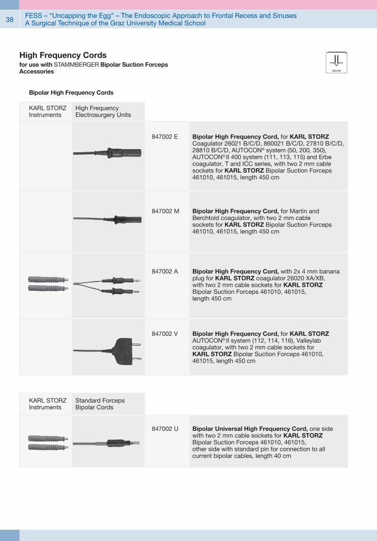
461010 STAMMBERGER Bipolar Suction Forceps, 15° upturned, with suction channel, for bipolar coagulation in paranasal areas, working length 12.5 cm, for use with Bipolar High Frequency Cord 847002 E or 847002 A/M/V/U
Indications/Applications:## Arterial bleeding (ethmoidal, sphenopalatine and maxillary arteries, turbinate and septum vessels)
## Skull base surgery## Oozing hemorrhage from the mucosa edges## Pituitary gland surgery## Vascular processes, i.e. nasopharyngeal fibroma## Epistaxis
## Rendu-Osler-Weber disease## Secondary hemorrhage, e.g., from the nasopharynx following adenotomy
## Edema prevention, shrinkage of mucosa (for example, posterior end of the turbinate)
## Turbinate cauterization
461010
461015 STAMMBERGER Bipolar Suction Forceps, 45° upturned, with suction channel, for bipolar coagulation in paranasal areas, working length 12.5 cm, for use with Bipolar High Frequency Cord 847002 E or 847002 A/M/V/U
461015
bipolar
FESS – “Uncapping the Egg” – The Endoscopic Approach to Frontal Recess and SinusesA Surgical Technique of the Graz University Medical School38
847002 U Bipolar Universal High Frequency Cord, one side with two 2 mm cable sockets for KARL STORZ Bipolar Suction Forceps 461010, 461015, other side with standard pin for connection to all current bipolar cables, length 40 cm
Standard Forceps Bipolar Cords
KARL STORZ Instruments
High Frequency Cordsfor use with STAMMBERGER Bipolar Suction ForcepsAccessories
847002 A Bipolar High Frequency Cord, with 2x 4 mm banana plug for KARL STORZ coagulator 26020 XA/XB, with two 2 mm cable sockets for KARL STORZ Bipolar Suction Forceps 461010, 461015, length 450 cm
847002 V Bipolar High Frequency Cord, for KARL STORZ AUTOCON® II system (112, 114, 116), Valleylab coagulator, with two 2 mm cable sockets for KARL STORZ Bipolar Suction Forceps 461010, 461015, length 450 cm
847002 E Bipolar High Frequency Cord, for KARL STORZ Coagulator 26021 B/C/D, 860021 B/C/D, 27810 B/C/D, 28810 B/C/D, AUTOCON® system (50, 200, 350), AUTOCON® II 400 system (111, 113, 115) and Erbe coagulator, T and ICC series, with two 2 mm cable sockets for KARL STORZ Bipolar Suction Forceps 461010, 461015, length 450 cm
847002 M Bipolar High Frequency Cord, for Martin and Berchtold coagulator, with two 2 mm cable sockets for KARL STORZ Bipolar Suction Forceps 461010, 461015, length 450 cm
Bipolar High Frequency Cords
KARL STORZ Instruments
High Frequency Electrosurgery Units
bipolar

FESS – “Uncapping the Egg” – The Endoscopic Approach to Frontal Recess and SinusesA Surgical Technique of the Graz University Medical School 39
Innovative Design## Dashboard: Complete overview with intuitive menu guidance
## Live menu: User-friendly and customizable## Intelligent icons: Graphic representation changes when settings of connected devices or the entire system are adjusted
## Automatic light source control## Side-by-side view: Parallel display of standard image and the Visualization mode
## Multiple source control: IMAGE1 S allows the simultaneous display, processing and documentation of image information from two connected image sources, e.g., for hybrid operations
Dashboard Live menu
Side-by-side view: Parallel display of standard image and Visualization mode
Intelligent icons
Economical and future-proof## Modular concept for flexible, rigid and 3D endoscopy as well as new technologies
## Forward and backward compatibility with video endoscopes and FULL HD camera heads
## Sustainable investment## Compatible with all light sources
IMAGE1 S Camera System n

FESS – “Uncapping the Egg” – The Endoscopic Approach to Frontal Recess and SinusesA Surgical Technique of the Graz University Medical School40
Brillant Imaging## Clear and razor-sharp endoscopic images in FULL HD
## Natural color rendition
## Reflection is minimized## Multiple IMAGE1 S technologies for homogeneous illumination, contrast enhancement and color shifting
FULL HD image CHROMA
FULL HD image SPECTRA A *
FULL HD image
FULL HD image CLARA
SPECTRA B **
* SPECTRA A : Not for sale in the U.S.** SPECTRA B : Not for sale in the U.S.
IMAGE1 S Camera System n

FESS – “Uncapping the Egg” – The Endoscopic Approach to Frontal Recess and SinusesA Surgical Technique of the Graz University Medical School 41
TC 200EN* IMAGE1 S CONNECT, connect module, for use with up to 3 link modules, resolution 1920 x 1080 pixels, with integrated KARL STORZ-SCB and digital Image Processing Module, power supply 100 – 120 VAC/200 – 240 VAC, 50/60 Hz
including: Mains Cord, length 300 cm DVI-D Connecting Cable, length 300 cm SCB Connecting Cable, length 100 cm USB Flash Drive, 32 GB, USB silicone keyboard, with touchpad, US
* Available in the following languages: DE, ES, FR, IT, PT, RU
Specifications:
HD video outputs
Format signal outputs
LINK video inputs
USB interface SCB interface
- 2x DVI-D - 1x 3G-SDI
1920 x 1080p, 50/60 Hz
3x
4x USB, (2x front, 2x rear) 2x 6-pin mini-DIN
100 – 120 VAC/200 – 240 VAC
50/60 Hz
I, CF-Defib
305 x 54 x 320 mm
2.1 kg
Power supply
Power frequency
Protection class
Dimensions w x h x d
Weight
TC 300 IMAGE1 S H3-LINK, link module, for use with IMAGE1 FULL HD three-chip camera heads, power supply 100 – 120 VAC/200 – 240 VAC, 50/60 Hz, for use with IMAGE1 S CONNECT TC 200ENincluding:Mains Cord, length 300 cm
Link Cable, length 20 cm
For use with IMAGE1 S IMAGE1 S CONNECT Module TC 200EN
IMAGE1 S Camera System n
TC 300 (H3-Link)
TH 100, TH 101, TH 102, TH 103, TH 104, TH 106 (fully compatible with IMAGE1 S) 22 2200 55-3, 22 2200 56-3, 22 2200 53-3, 22 2200 60-3, 22 2200 61-3, 22 2200 54-3, 22 2200 85-3 (compatible without IMAGE1 S technologies CLARA, CHROMA, SPECTRA*)
1x
100 – 120 VAC/200 – 240 VAC
50/60 Hz
I, CF-Defib
305 x 54 x 320 mm
1.86 kg
Camera System
Supported camera heads/video endoscopes
LINK video outputs
Power supply
Power frequency
Protection class
Dimensions w x h x d
Weight
Specifications:
TC 200EN
TC 300
* SPECTRA A : Not for sale in the U.S.** SPECTRA B : Not for sale in the U.S.

FESS – “Uncapping the Egg” – The Endoscopic Approach to Frontal Recess and SinusesA Surgical Technique of the Graz University Medical School42
For use with IMAGE1 S Camera System IMAGE1 S CONNECT Module TC 200EN, IMAGE1 S H3-LINK Module TC 300 and with all IMAGE 1 HUB™ HD Camera Control Units
TH 100 IMAGE1 S H3-Z Three-Chip FULL HD Camera Head, 50/60 Hz, IMAGE1 S compatible, progressive scan, soakable, gas- and plasma-sterilizable, with integrated Parfocal Zoom Lens, focal length f = 15 – 31 mm (2x), 2 freely programmable camera head buttons, for use with IMAGE1 S and IMAGE 1 HUB™ HD/HD
IMAGE1 FULL HD Camera Heads
Product no.
Image sensor
Dimensions w x h x d
Weight
Optical interface
Min. sensitivity
Grip mechanism
Cable
Cable length
IMAGE1 S H3-Z
TH 100
3x 1/3" CCD chip
39 x 49 x 114 mm
270 g
integrated Parfocal Zoom Lens, f = 15 – 31 mm (2x)
F 1.4/1.17 Lux
standard eyepiece adaptor
non-detachable
300 cm
Specifications:
TH 104
TH 104 IMAGE1 S H3-ZA Three-Chip FULL HD Camera Head, 50/60 Hz, IMAGE1 S compatible, autoclavable, progressive scan, soakable, gas- and plasma-sterilizable, with integrated Parfocal Zoom Lens, focal length f = 15 – 31 mm (2x), 2 freely programmable camera head buttons, for use with IMAGE1 S and IMAGE 1 HUB™ HD/HD
IMAGE1 FULL HD Camera Heads
Product no.
Image sensor
Dimensions w x h x d
Weight
Optical interface
Min. sensitivity
Grip mechanism
Cable
Cable length
IMAGE1 S H3-ZA
TH 104
3x 1/3" CCD chip
39 x 49 x 100 mm
299 g
integrated Parfocal Zoom Lens, f = 15 – 31 mm (2x)
F 1.4/1.17 Lux
standard eyepiece adaptor
non-detachable
300 cm
Specifications:
IMAGE1 S Camera Heads n
TH 100

FESS – “Uncapping the Egg” – The Endoscopic Approach to Frontal Recess and SinusesA Surgical Technique of the Graz University Medical School 43
9826 NB
9826 NB 26" FULL HD Monitor, wall-mounted with VESA 100 adaption, color systems PAL/NTSC, max. screen resolution 1920 x 1080, image fomat 16:9, power supply 100 – 240 VAC, 50/60 Hzincluding:External 24 VDC Power SupplyMains Cord
9619 NB
9619 NB 19" HD Monitor, color systems PAL/NTSC, max. screen resolution 1280 x 1024, image format 4:3, power supply 100 – 240 VAC, 50/60 Hz, wall-mounted with VESA 100 adaption,including:
External 24 VDC Power SupplyMains Cord
Monitors

FESS – “Uncapping the Egg” – The Endoscopic Approach to Frontal Recess and SinusesA Surgical Technique of the Graz University Medical School44
Monitors
Optional accessories:9826 SF Pedestal, for monitor 9826 NB9626 SF Pedestal, for monitor 9619 NB
26"
9826 NB
l
–
l
l
l
l
l
–
l
–
l
l
l
l
l
l
19"
9619 NB
l
–
–
l
l
l
l
l
l
l
–
l
l
l
l
l
KARL STORZ HD and FULL HD Monitors
Wall-mounted with VESA 100 adaption
Inputs:
DVI-D
Fibre Optic
3G-SDI
RGBS (VGA)
S-Video
Composite/FBAS
Outputs:
DVI-D
S-Video
Composite/FBAS
RGBS (VGA)
3G-SDI
Signal Format Display:
4:3
5:4
16:9
Picture-in-Picture
PAL/NTSC compatible
19"
optional
9619 NB
200 cd/m2 (typ)
178° vertical
0.29 mm
5 ms
700:1
100 mm VESA
7.6 kg
28 W
0 – 40°C
-20 – 60°C
max. 85%
469.5 x 416 x 75.5 mm
100 – 240 VAC
EN 60601-1, protection class IPX0
Specifications:
KARL STORZ HD and FULL HD Monitors
Desktop with pedestal
Product no.
Brightness
Max. viewing angle
Pixel distance
Reaction time
Contrast ratio
Mount
Weight
Rated power
Operating conditions
Storage
Rel. humidity
Dimensions w x h x d
Power supply
Certified to
26"
optional
9826 NB
500 cd/m2 (typ)
178° vertical
0.3 mm
8 ms
1400:1
100 mm VESA
7.7 kg
72 W
5 – 35°C
-20 – 60°C
max. 85%
643 x 396 x 87 mm
100 – 240 VAC
EN 60601-1, UL 60601-1, MDD93/42/EEC, protection class IPX2
FESS – “Uncapping the Egg” – The Endoscopic Approach to Frontal Recess and SinusesA Surgical Technique of the Graz University Medical School 45
20200032 KARL STORZ Special Beamsplitter, for use with Endovision TRICAM® and TELECAM®, for simultaneous viewing by endoscope and monitor screen
The camera head connector is 120° deflected and can instantly be swiveled to the desired position.
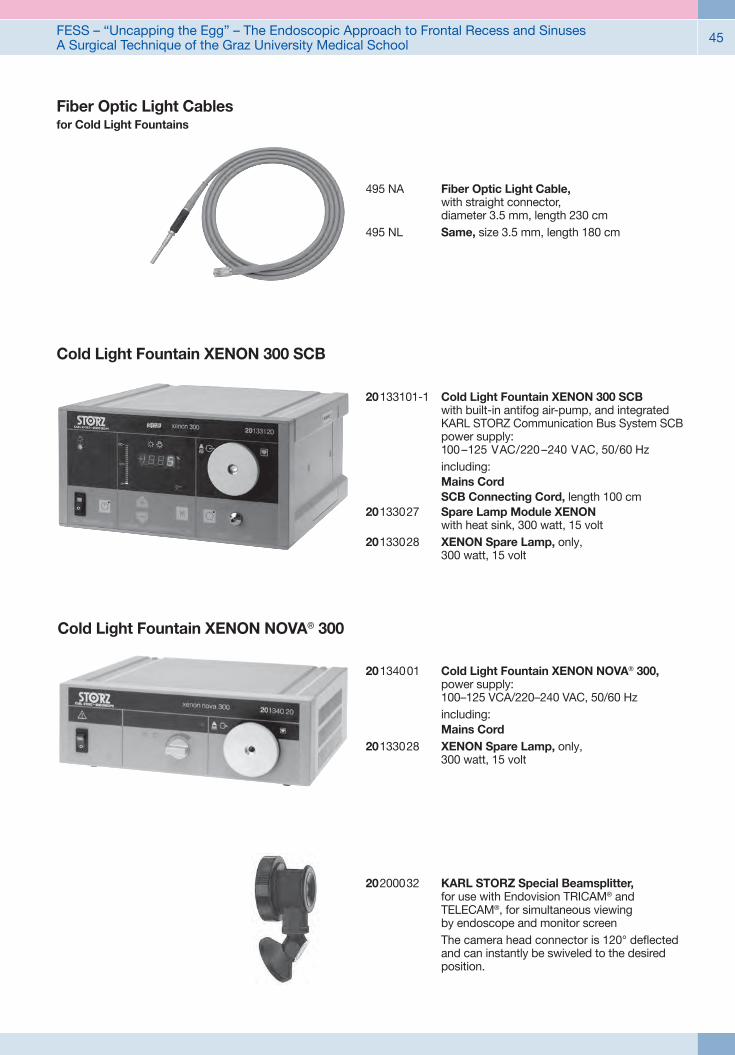
Cold Light Fountain XENON 300 SCB
20 133101-1 Cold Light Fountain XENON 300 SCB
with built-in antifog air-pump, and integrated KARL STORZ Communication Bus System SCB power supply: 100 –125 VAC/220 –240 VAC, 50/60 Hz
including: Mains Cord SCB Connecting Cord, length 100 cm20133027 Spare Lamp Module XENON
with heat sink, 300 watt, 15 volt20133028 XENON Spare Lamp, only,
300 watt, 15 volt
20 1340 01 Cold Light Fountain XENON NOVA® 300, power supply: 100–125 VCA/220–240 VAC, 50/60 Hz
including: Mains Cord20133028 XENON Spare Lamp, only,
300 watt, 15 volt
Cold Light Fountain XENON NOVA® 300
495 NA Fiber Optic Light Cable, with straight connector, diameter 3.5 mm, length 230 cm
495 NL Same, size 3.5 mm, length 180 cm
Fiber Optic Light Cablesfor Cold Light Fountains
FESS – “Uncapping the Egg” – The Endoscopic Approach to Frontal Recess and SinusesA Surgical Technique of the Graz University Medical School46
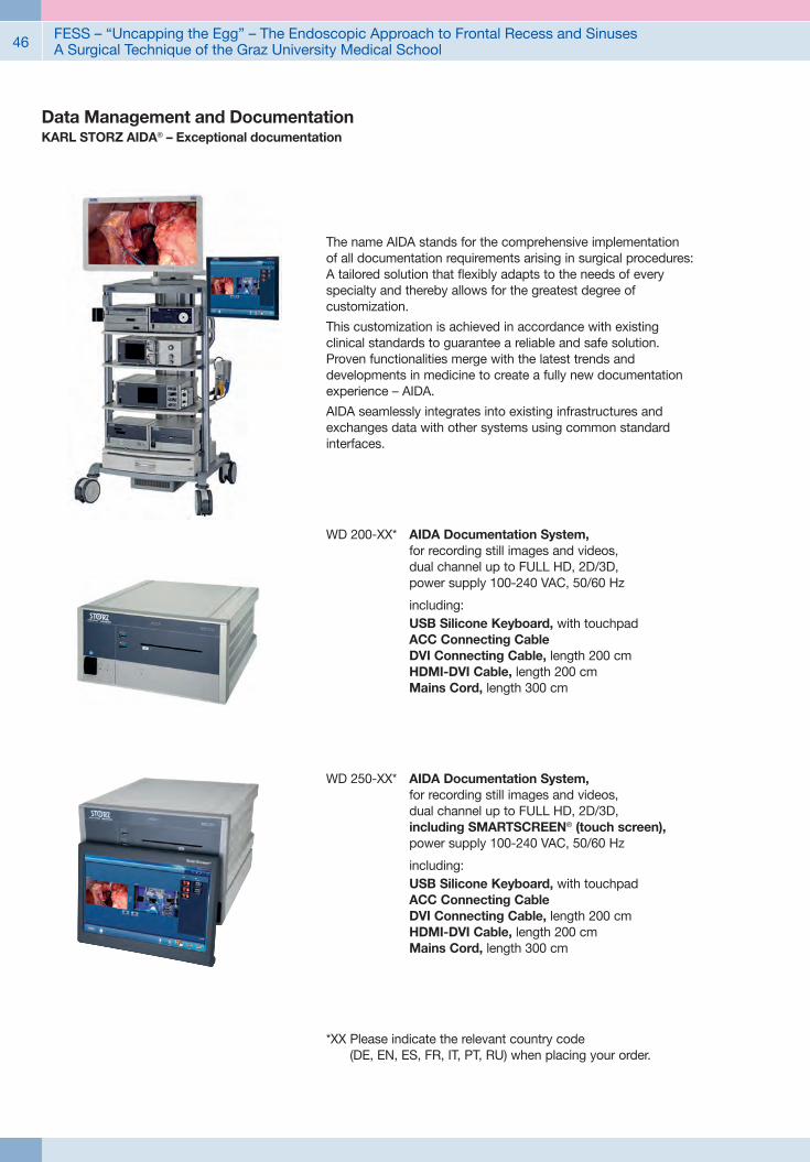
Data Management and DocumentationKARL STORZ AIDA® – Exceptional documentation
The name AIDA stands for the comprehensive implementation of all documentation requirements arising in surgical procedures: A tailored solution that flexibly adapts to the needs of every specialty and thereby allows for the greatest degree of customization.
This customization is achieved in accordance with existing clinical standards to guarantee a reliable and safe solution. Proven functionalities merge with the latest trends and developments in medicine to create a fully new documentation experience – AIDA.
AIDA seamlessly integrates into existing infrastructures and exchanges data with other systems using common standard interfaces.
WD 200-XX* AIDA Documentation System, for recording still images and videos, dual channel up to FULL HD, 2D/3D, power supply 100-240 VAC, 50/60 Hz
including: USB Silicone Keyboard, with touchpad ACC Connecting Cable DVI Connecting Cable, length 200 cm HDMI-DVI Cable, length 200 cm Mains Cord, length 300 cm
WD 250-XX* AIDA Documentation System, for recording still images and videos, dual channel up to FULL HD, 2D/3D, including SMARTSCREEN® (touch screen), power supply 100-240 VAC, 50/60 Hz
including: USB Silicone Keyboard, with touchpad ACC Connecting Cable DVI Connecting Cable, length 200 cm HDMI-DVI Cable, length 200 cm Mains Cord, length 300 cm
*XX Please indicate the relevant country code (DE, EN, ES, FR, IT, PT, RU) when placing your order.

FESS – “Uncapping the Egg” – The Endoscopic Approach to Frontal Recess and SinusesA Surgical Technique of the Graz University Medical School 47
Workflow-oriented use
Patient
Entering patient data has never been this easy. AIDA seamlessly integrates into the existing infrastructure such as HIS and PACS. Data can be entered manually or via a DICOM worklist. ll important patient information is just a click away.
Checklist
Central administration and documentation of time-out. The checklist simplifies the documentation of all critical steps in accordance with clinical standards. All checklists can be adapted to individual needs for sustainably increasing patient safety.
Record
High-quality documentation, with still images and videos being recorded in FULL HD and 3D. The Dual Capture function allows for the parallel (synchronous or independent) recording of two sources. All recorded media can be marked for further processing with just one click.
Edit
With the Edit module, simple adjustments to recorded still images and videos can be very rapidly completed. Recordings can be quickly optimized and then directly placed in the report. In addition, freeze frames can be cut out of videos and edited and saved. Existing markings from the Record module can be used for quick selection.
Complete
Completing a procedure has never been easier. AIDA offers a large selection of storage locations. The data exported to each storage location can be defined. The Intelligent Export Manager (IEM) then carries out the export in the background. To prevent data loss, the system keeps the data until they have been successfully exported.
Reference
All important patient information is always available and easy to access. Completed procedures including all information, still images, videos, and the checklist report can be easily retrieved from the Reference module.

FESS – “Uncapping the Egg” – The Endoscopic Approach to Frontal Recess and SinusesA Surgical Technique of the Graz University Medical School48
UG 540 Monitor Swifel Arm, height and side adjustable, can be turned to the left or the right side, swivel range 180°, overhang 780 mm, overhang from centre 1170 mm, load capacity max. 15 kg, with monitor fixation VESA 5/100, for usage with equipment carts UG xxx
UG 540
Equipment Cart
UG 220
UG 220 Equipment Cart wide, high, rides on 4 antistatic dual wheels equipped with locking brakes 3 shelves, mains switch on top cover, central beam with integrated electrical subdistributors with 12 sockets, holder for power supplies, potential earth connectors and cable winding on the outside,
Dimensions: Equipment cart: 830 x 1474 x 730 mm (w x h x d), shelf: 630 x 510 mm (w x d), caster diameter: 150 mm
inluding: Base module equipment cart, wide Cover equipment, equipment cart wide Beam package equipment, equipment cart high 3x Shelf, wide Drawer unit with lock, wide 2x Equipment rail, long Camera holder

FESS – “Uncapping the Egg” – The Endoscopic Approach to Frontal Recess and SinusesA Surgical Technique of the Graz University Medical School 49
Recommended Accessories for Equipment Cart
UG 310 Isolation Transformer, 200 V – 240 V; 2000 VA with 3 special mains socket, expulsion fuses, 3 grounding plugs, dimensions: 330 x 90 x 495 mm (w x h x d), for usage with equipment carts UG xxx
UG 310
UG 410 Earth Leakage Monitor, 200 V – 240 V, for mounting at equipment cart, control panel dimensions: 44 x 80 x 29 mm (w x h x d), for usage with isolation transformer UG 310
UG 410
UG 510 Monitor Holding Arm, height adjustable, inclinable, mountable on left or right, turning radius approx. 320°, overhang 530 mm, load capacity max. 15 kg, monitor fixation VESA 75/100, for usage with equipment carts UG xxx
UG 510

FESS – “Uncapping the Egg” – The Endoscopic Approach to Frontal Recess and SinusesA Surgical Technique of the Graz University Medical School50
Notes:


with the compliments of
KARL STORZ — ENDOSKOPE