FERNANDA MENDES DE REZENDE - Biblioteca Digital de ...Rezende, Fernanda Mendes Análises químicas...
74
FERNANDA MENDES DE REZENDE Chemical analysis of color change and transcriptional profile in flavonoids biosynthesis during flower development of Tibouchina pulchra (Cham.) Cogn. Análises químicas da mudança de cor e perfil transcricional da biossíntese de flavonoides durante o desenvolvimento floral de Tibouchina pulchra (Cham.) Cogn. São Paulo 2018
Transcript of FERNANDA MENDES DE REZENDE - Biblioteca Digital de ...Rezende, Fernanda Mendes Análises químicas...

FERNANDA MENDES DE REZENDE
Chemical analysis of color change and transcriptional profile in
flavonoids biosynthesis during flower development of Tibouchina
pulchra (Cham.) Cogn.
Análises químicas da mudança de cor e perfil transcricional da
biossíntese de flavonoides durante o desenvolvimento floral de
Tibouchina pulchra (Cham.) Cogn.
São Paulo
2018


FERNANDA MENDES DE REZENDE
Chemical analysis of color change and transcriptional profile in
flavonoids biosynthesis during flower development of Tibouchina
pulchra (Cham.) Cogn.
Análises químicas da mudança de cor e perfil transcricional da
biossíntese de flavonoides durante o desenvolvimento floral de
Tibouchina pulchra (Cham.) Cogn.
Tese apresentada ao Instituto de Biociências da
Universidade de São Paulo, para a obtenção de
Título de Doutor em Botânica, na Área de Recursos
Econômicos vegetais.
Orientadora: Dra. Cláudia Maria Furlan
Co-orientadora: Dra. Maria Magdalena Rossi
São Paulo
2018

Banca Examinadora
________________________________________ ________________________________________
Prof(a). Dr(a) Prof(a). Dr(a)
________________________________________ ________________________________________
Prof(a). Dr(a) Prof(a). Dr(a)
________________________________________
Profa. Dra. Claudia M. Furlan
Rezende, Fernanda Mendes
Análises químicas da mudança de cor e perfil transcricional da
biossíntese de flavonoides durante o desenvolvimento floral de
Tibouchina pulchra (Cham.) Cogn.
165 páginas.
Tese (Doutorado) – Instituto de Biociências da Universidade de
São Paulo. Departamento de Botânica.
Palavra-Chave: 1. antocianinas; 2. Ácidos fenólicos; 3. CHS, 4. FLS,
5. ANS.
Universidade de São Paulo. Instituto de Biociências.
Departamento de Botânica.

Dedico à minha família e a todos
que de alguma forma contribuíram
para a realização deste trabalho.


Agradecimentos
Ao fim de quatro anos tenho muito o que agradecer. Foram anos de aprendizado, dedicação e
mudanças. Muitas pessoas participaram deste processo.
Primeiramente gostaria de agradecer à Claudia, minha orientadora, por toda paciência, incentivo
e suporte em minha formação. Desde a iniciação científica até aqui passaram-se dez anos, meu
amadurecimento científico deve-se em grande parte ao seu apoio. Obrigada.
À Magda, minha co-orientadora, agradeço por me inserir no mundo da biologia molecular e
por todo aprendizado acadêmico compartilhado. Também sou muito grata pelo grande apoio à ida ao
exterior. Não poderia deixar de mencionar que as pausas para um cigarro foram sempre produtivas, seja
para falar da vida ou para ter insights sobre o trabalho.
Aos professores da Fitoquímica por todos os anos em que aprendi muito com vocês, tanto numa
conversa informal, quanto em nossos seminários. Ao Marcelo, agradeço por toda a ajuda nas
identificações químicas e paciência diante dos meus questionamentos.
Ao Eduardo Leal da Sistemática Vegetal que me auxiliou com os nomes de espécies da grande
tabela do capítulo 1. Ao Erismaldo, jardineiro do IB, que muito me ajudou para que as mudas de
Tibouchina ficassem bem.
Ao Mads que me recebeu em seu laboratório na Dinamarca e ao Thomas, além de todos os
técnicos dos laboratórios e amigos que fiz por lá. Em especial Irene, Christou e Jorge.
Aos meus colegas da Fitoquímica, agradeço pelos anos em que discutíamos resultados entre
pausas de almoço e café. Aqui vai um agradecimento especial à Priscila, Alice, Fernanda, Dalila e Kátia
que me ajudaram nas coletas ou regando plantas quando eu estava ausente. Falando em almoço, não
posso deixar de agradecer às amigas que almoçam comigo, quase que diariamente, nos últimos anos:
Aline, Kátia e Mourisa. Apesar de hoje em dia estar ausente, agradeço à Carol, pois muito trocamos nos
anos de convivência.
Às técnicas do laboratório, agradeço por tantas vezes que passamos almoços falando sobre
cromatografias e afins. Obrigada por serem sempre prestativas e por todas as risadas compartilhadas.
Ao pessoal do lab de Biologia Celular e Biologia Molecular de plantas, agradeço pelas trocas de
experiências e bons momentos. Agradecimento especial à Dani, Paula e Bruno que me ajudaram muito
nos procedimentos no laboratório.
À minha família agradeço por serem os pilares que sustentam meus sonhos. Obrigada mãe, pai,
amo vocês! Não posso deixar de fora aquela família que a gente escolhe, nossos amigos. Agradeço aos
meus por me levarem para me descontrair, afinal não é só de um doutorado que se vive.
Por fim, aos órgãos financiadores: CAPES, FAPESP (2013/10413-0) e Instituto de Biociências.


Gosto das cores, das flores, das estrelas, do verde
das árvores, gosto de observar. A beleza da vida se
esconde por ali, e por mais uma infinidade de
lugares, basta saber, e principalmente, basta querer
enxergar.
Clarice Lispector


Rezende, F.M. Análises químicas da mudança de cor e perfil transcricional da biossíntese de flavonoides
durante o desenvolvimento floral de Tibouchina pulchra (Cham.) Cogn. [tese]. São Paulo: Instituto de
Biociências, Universidade de São Paulo; 2018.
Resumo
A mudança de cor em flores é um fenômeno generalizado entre as angiospermas, mas pouco
compreendido do ponto de vista genético e químico. Este estudo teve como objetivo investigar
esse fenômeno em Tibouchina pulcha, uma espécie nativa brasileira, cujas flores mudam do
branco para rosa escuro. Para alcançar o objetivo, os perfis dos compostos e de expressão de
genes da via de biossíntese de flavonoides foram estudados. Utilizando técnicas hifenadas
(UPLC-HRMS and NMR), trinta compostos foram quantificados e identificados, sendo:
dezesseis descritos pela promeira vez em T. pulchra. Além idsso, um composto inédito foi
identificado. Cinco genes que codificam para enzimas chaves da via foram parcialmente
clonados, sequenciados, e os níveis de mRNA foram analisados (RT-qPCR) durante o
desenvolvimento das flores. Coletivamente, os dados obtidos demonstram que a mudança de
cor em flores de T. pulchra é regulada transcricionalmente. Além disso, quantificações de
metais sugerem que o ion Fe3+ esteja relacionado a saturação da cor no estágio rosa escuro.
Palavras-chave: antocianina, ácidos fenólicos, CHS, FLS, ANS

Rezende, F.M. Chemical analysis of color change and transcriptional profile in flavonoids biosynthesis
during flower development of Tibouchina pulchra (Cham.) Cogn. [thesis]. São Paulo: Instituto de
Biociências, Universidade de São Paulo; 2018.
Abstract
Floral color change is a widespread phenomenon in angiosperms, but poorly understood from
the genetic and chemical point of view. This thesis aimed to investigate this phenomenon in
Tibouchina pulcha, a native species whose flowers change from white to dark pink. To reach
this goal, flavonoid biosynthetic gene expression and compounds were profiled. By using
hyphenated techiniques (UPLC-HRMS and NMR), thirty phenolic compounds were quantified
and identified, being sixteen described for the first time in T. pulchra. Moreover, an inedite
compound was also identified. Five key genes of the flavonoid biosynthetic pathway were
partially cloned, sequenced, and the mRNA levels were analyzed (RT-qPCR) along flower
development. Collectively, the obtained results demonstrated that the flower color change in
T. pulchra is regulated at transcriptional level. Additionally, a metal quantification suggests that
Fe3+ ion might influence the saturation of the color at dark pink stage.
Key words: anthocyanin, phenolic acids, CHS, FLS, ANS


List of Abreviations
[M+H]: molecular ion
2-ODD: 2-oxoglutarate/Fe(Ii)-dependent
dioxygenase
4CL: 4-coumarate:CoA-ligase
aa: amino acids
AACT: anthocyanidin acyl transferase
ACT: acyl transferase
amu: atomic mass unit
ANS: anthocyanidin synthase
AT: Arabdopsis thaliana
B5GT: betanidin-5-O-glucosyltransferase
bbCID: broadband collision Induced
dissociation
bHLH: basic helix–loop–helix
BLAST: basic local alignment search tool
Bp: base pair
Brara: Brassica rapa
C3H: coumarate 3-hydroxylase
C4H: cinnamate 4-Hydroxylase
CCD: carotenoid cleavage dioxygenase
CD: carotene desaturase
CD3OD: deuterated methanol
cD5G2’GlcUT: cyclo-DOPA-5-glucose-2’-O-
glucuronyl- transferase
cD5GT: cyclo-DOPA-5-O-glucosyltransferase
CDNA: complementary DNA
cDOPA: cyclo: DOPA
CHI: chalcone isomerase
CHS: chalcone synthase
CI: carotene isomerase
Co-A: Coenzyma A
Ct: treshold cycle
CTAB: cetyl trimethylammonium bromide
d: dublet
DAD: diode array detector
dd: double dublet
DEPC: dietilpirocarbonato
DFR: dihydroflavanol 4-reductase
DMAPP: dimethylallyl diphosphate
DMSO-d6: deuterated dimethyl sulfoxide
DNA: desoxyribonucleic acid
DNAse: desoxyribonuclease
Dntp: riphosphate desoribonucleotides
DO: diphenol oxidase (DO)
DOD: DOPA dioxygenase
DOPA: dihydrophenylalanine
DW: dried weight
DXP: 1-deoxy-D-xylulose-5-phosphate
DXR: DXP reductoisomerase
DXS: deoxy-D-xylulose synthase
DXS: DXP synthase
E4P: erythrose 4-phosphate
EDTA: ethylenediaminetetraacetic acid
ESI: electrospray Ionization
Eucgr: Eucalyptus grandis
F3’5’: flavonoid 3’5’-hydroxylase
F3’H: flavonoid 3’-hydroxylase
F3H: flavonoid 3-hydroxylase
FDP: farnesyl diphosphate synthase
FGT flavonoid glucosyltransferase
FLS: flavonol synthase
FPP: farnesyl diphosphate
FS: flavone synthase
GA3P: glyceraldehyde 3-P
GDS: GPP synthase reaction
GGDS: geranylgeranyl diphosphate synthase
GGPP: geranylgeranyl diphosphate
GPP: geranyl diphosphate
HCl: hydrocloric acid
HMBC: heteronuclear multiple bond correlation
HPLC: high performance liquid
chromatography
HRMS: high-resolution mass spectrometry
HSQC: heteronuclear single quantum
coherence
IFS: isoflavone synthase
IPP: isopentenyl diphosphate
IPPI: isopentenyl diphosphate isomerase
IPTG: isopropil β-D-1-tiogalactopiranosida
IspH: 4-hydroxy-3-methylbut-2-enyl
diphosphate reductase
J: coupling constant

LB: Luria Bertani growth medium
LCYΒ: Lycopene Β-Cyclase
LCYΕ: Lycopene Β-Cyclase
m: multiplet
MAL: malonate pathway
Me: methyl group
Medtr: Mendicago trunculata
MeOH: methanol
MEP: 2-C-methyl-D-erythritol 4- phosphate
MEP: methylerythritol phosphate pathway
MEV: mevalonate pathway
miRNA: micro RNA
mRNA: messenger RNA
MS: mass spectrometry
MYB: myeloblastosis
n: nucleotide
NMR: nuclear magnetic resonance
NS: neoxanthin synthase
OMT: O-methyl transferase
P450: Cytochrome P450 Monooxygenase
PAD: photodiode array detector
PAL: phenylalanine ammonia lyase
PCR: polymerase chain reaction
PD: phytoene desaturase
PEP: phosphoenolpyruvate
ppm: parts per million
PS: phytoene synthase
QTOF: quadrupole time-of-flight
RNA: ribonucleic acid
RNAi: RNA interference
RT: retention time
RT-Qpcr: reverse transcriptase quantitative
polymerase chain reaction
s: singlet
S1: stage one of T. pulchra flower
S2: stage two of T. pulchra flower
S3: stage three of T. pulchra flower
S4: stage four of T. pulchra flower
SKM: shikimate pathway
sl: large singlet
SOC: super optimal broth
Soly: Solanum lycopersicum
tasiRNA: trans-acting short-interfering RNA
TOH: tyrosine hydroxylase
Tp: Trifolium pratense
UPLC: ultra performance liquid
chromatography
UV/Vis: ultraviolet/visible
WDRs: tryptophan-aspartic acid dipeptide
repeat
X-Gal: 5-bromo-4-chloro-3-indoxyl-β-D-
galactopyranoside
ZE: zeaxanthin epoxidase
β-CO: β-carotene oxygenase
Β-RH: Β-Ring Hydroxylase
δ: chemical shift
Ε-RH: Ε-ring hydroxylase

List of Figures
Chapter 1
Figure 1.1. Simplified schematic representation of plant pigment biosynthetic pathways………………. 05
Figure 1.2. Angiosperm orders with color change during flower development according to APG
IV classification………………………………………………………………………………………………………………………………. 15
Chapter 2
Figure 2.1. Tibouchina pulchra tree and its flowers………………………………………………………………………….. 39
Figure 2.2. Chromatogram obtained by UPLC-ESI-QTOF-HRMS from T. pulchra petals extracted
with acidified methanol…………………………………………………………………………………………………………………. 43
Figure 2.3. Mass spectra (MS+) of compounds 7, 8, 9 and 10. Structures represent the proposed
compound and its main fragmentation…………………………………………………………………………………………. 46
Figure 2.4. Mass spectra (MS+) of compounds 11, 12, 13 and 14. Structures represent the
proposed compound and its main fragmentation…………………………………………………………………………. 47
Figure 2.5. Mass spectra (MS+) of compounds 15, 16, 17 and 18. Structures represent the
proposed compound and its main fragmentation…………………………………………………………………………. 48
Figure 2.6. Mass spectra (MS+) of compounds 19, 20, 21 and 22. Structures represent the
proposed compound and its main fragmentation…………………………………………………………………………. 49
Figure 2.7. Mass spectra (MS+) of compounds 23, 24, 25 and 26. Structures represent the
proposed compound and its main fragmentation.……………………………………………………………………...…. 50
Figure 2.8. Mass spectra (MS+) of compounds 27, 28, 29 and 30. Structures represent the
proposed compound and its main fragmentation.………………………………………………………………...………. 51
Figure 2.9. Compounds of T. pulchra flowers identified by UV-visible absorption and mass spectra.. 52
Figure 2.10. Schematic Retro Dies-Alder positions of cleavage.……………………………………………………… 53
Figure 2.11. Compounds of T. pulchra flowers identified by UV-visible absorption, mass and NMR
spectra.……………………………………………………………………….………………………………………………………………… 68
Supplemental Figure 2.1. MS+ spectra of compounds 1, 2, 3, and 4.…………………………………………… 76
Supplemental Figure 2.2. MS+ spectra of compounds 5 and 6.…………………………………………………… 77
Supplemental Figure 2.3. 1H spectra (DMSO-d6) of kaempferol 3-O-(2’’-galloyl)-β-D-
glucopyranoside (13, 16 or 19)..…………………………………………………………………………………………………….. 78
Supplemental Figure 2.4. Zoom from 5.2 to 8.5 ppm, 1H spectra (DMSO-d6) of kaempferol 3-O-
(2’’-galloyl)-β-D-glucopyranoside (13, 16 or 19).…………………………………………………………………………... 79
Supplemental Figure 2.5. HSQC spectra (DMSO-d6) of kaempferol 3-O-(2’’-galloyl)-β-D-
glucopyranoside (13, 16 or 19).……………………………………………………………………………………………………... 80
Supplemental Figure 2.6. Zoom from 2.7 to 3.8 ppm, HSQC spectra (DMSO-d6) of kaempferol 3-
O-(2’’-galloyl)-β-D-glucopyranoside (13, 16 or 19).………………………………………………………………………. 81
Supplemental Figure 2.7. HMBC spectra (DMSO-d6) of kaempferol 3-O-(2’’-galloyl)-β-D-
glucopyranoside (13, 16 or 19).……………………………………………………………………………………………………... 82
Supplemental Figure 2.8. Zoom from 2.7 to 3.9 ppm, HMBC spectra (DMSO-d6) of kaempferol
3-O-(2’’-galloyl)-β-D-glucopyranoside (13, 16 or 19).………………………………………………………………….. 83
Supplemental Figure 2.9. Zoom from 5.2 to 8.6 ppm, HMBC spectra (DMSO-d6) of kaempferol
3-O-(2’’-galloyl)-β-D-glucopyranoside (13, 16 or 19).…………………………………………………………………… 84
Supplemental Figure 2.10. 1H spectra (DMSO-d6) of kaempferol 3-O-(6’’-galloyl)-β-D-
glucopyranoside (13, 16 or 19).…………………………………………………………………………..…………………………. 85
Supplemental Figure 2.11. Zoom from 5.1 to 8.1 ppm, 1H spectra (DMSO-d6) of kaempferol 3-
O-(6’’-galloyl)-β-D-glucopyranoside (13, 16 or 19).……………………………………………………….……………… 86
Supplemental Figure 2.12. HSQC spectra (DMSO-d6) of kaempferol 3-O-(6’’-galloyl)-β-D-
glucopyranoside (13, 16 or 19).……………………………………………………………………………………………………... 87
Supplemental Figure 2.13. Zoom from 3.0 to 4.6 ppm, HSQC spectra (DMSO-d6) of kaempferol
3-O-(6’’-galloyl)-β-D-glucopyranoside (13, 16 or 19)……………………………………………………………………. 88
Supplemental Figure 2.14. HMBC spectra (DMSO-d6) of kaempferol 3-O-(6’’-galloyl)-β-D-
glucopyranoside (13, 16 or 19).……………………………………………………………………………………………………... 89

Supplemental Figure 2.15. Zoom from 2.8 to 4.9 ppm, HMBC spectra (DMSO-d6) of kaempferol
3-O-(6’’-galloyl)-β-D-glucopyranoside (13, 16 or 19)……………………………………………………………………. 90
Supplemental Figure 2.16. Zoom from 5.2 to 8.4 ppm, HMBC spectra (DMSO-d6) of kaempferol
3-O-(6’’-galloyl)-β-D-glucopyranoside (13, 16 or 19).…………………………………………………………………. 91
Supplemental Figure 2.17. 1H spectra (DMSO-d6) of mixture (17).………………………………………………… 92
Supplemental Figure 2.18. Zoom from 5.2 to 8.2 ppm, 1H spectra (DMSO-d6) of mixture (17)………. 93
Supplemental Figure 2.19. 13C spectra (DMSO-d6) of mixture (17)………………………………………………. 94
Supplemental Figure 2.20. HMBC spectra (DMSO-d6) of mixture (17)…………………………………………… 95
Supplemental Figure 2.21. Zoom from 2.4 to 4.5 ppm, HMBC spectra (DMSO-d6) of mixture (17)… 96
Supplemental Figure 2.22. Zoom from 4.8 to 8.6 ppm, HMBC spectra (DMSO-d6) of mixture (17)… 97
Supplemental Figure 2.23. 1H spectra (DMSO-d6) of kaempferol 3-O-glucoronide-6’’-O-
methylester (23).…………………………………………………………………….……………………………………………………… 98
Supplemental Figure 2.24. Zoom from 5.1 to 8.1 ppm, 1H spectra (DMSO-d6) of kaempferol 3-
O-glucoronide-6’’-O-methylester (23).…………………………………………………………………………………………. 99
Supplemental Figure 2.25. HSQC spectra (DMSO-d6) of kaempferol 3-O-glucoronide-6’’-O-
methylester (23).………………………………………………………………………………………………………………………….… 100
Supplemental Figure 2.26. Zoom from 2.9 to 4.5 ppm, HSQC spectra (DMSO-d6) of kaempferol
3-O-glucoronide-6’’-O-methylester (23).,……………………………………………………………………………………… 101
Supplemental Figure 2.27. HMBC spectra (DMSO-d6) of kaempferol 3-O-glucoronide-6’’-O-
methylester (23).………………………………………………………………………………………………………………………….… 102
Supplemental Figure 2.28. Zoom from 2.7 to 4.0 ppm, HMBC spectra (DMSO-d6) of kaempferol
3-O-glucoronide-6’’-O-methylester (23)………………………..……………………………………………………………… 103
Supplemental Figure 2.29. Zoom from 5.0 to 8.6 ppm, HMBC spectra (DMSO-d6) of kaempferol
3-O-glucoronide-6’’-O-methylester (23)………………………..……………………………………………………………… 104
Supplemental Figure 2.30. 1H spectra (DMSO-d6) of Quercetin 3-O-(6’’-p-coumaroyl)-β-D-
glucopyranoside (25).…………………………………………………………….……………………………………………………… 105
Supplemental Figure 2.31. Zoom from 5.1 to 8.1 ppm, 1H spectra (DMSO-d6) of Quercetin 3-O-
(6’’-p-coumaroyl)-β-D-glucopyranoside (25).…………………………………….…………………………………….…… 106
Supplemental Figure 2.32. HSQC spectra (DMSO-d6) of Quercetin 3-O-(6’’-p-coumaroyl)-β-D-
glucopyranoside (25)………………………………………………………………………….…………………………………….…… 107
Supplemental Figure 2.33. Zoom from 2.7 to 4.7 ppm, HSQC spectra (DMSO-d6) of Quercetin 3-
O-(6’’-p-coumaroyl)-β-D-glucopyranoside (25)...……………………………….…………………………………….…… 108
upplemental Figure 2.34. Zoom from 5.0 to 8.8 ppm, HSQC spectra (DMSO-d6) of Quercetin 3-
O-(6’’-p-coumaroyl)-β-D-glucopyranoside (25)...……………………………….…………………………………….…… 109
Supplemental Figure 2.35. 1H spectra (DMSO-d6) of Kaempferol 3-O-(6’’-p-coumaroyl)-β-D-
glucopyranoside (27). 110
Supplemental Figure 2.36. Zoom from 5.1 to 8.1 ppm, 1H spectra (DMSO-d6) of Kaempferol 3-
O-(6’’-p-coumaroyl)-β-D-glucopyranoside (27)...……………………………….…………………………………….…… 111
Supplemental Figure 2.37. HSQC spectra (DMSO-d6) of Kaempferol 3-O-(6’’-p-coumaroyl)-β-D-
glucopyranoside (27)………………………………………………………………………….…………………………………….…… 112
Supplemental Figure 2.38. Zoom from 3.0 to 4.6 ppm, HSQC spectra (DMSO-d6) of Kaempferol
3-O-(6’’-p-coumaroyl)-β-D-glucopyranoside (27)..…………………………….…………………………………….…… 113
Supplemental Figure 2.39. HMBC spectra (DMSO-d6) of Kaempferol 3-O-(6’’-p-coumaroyl)-β-D-
glucopyranoside (27).…………………………………….…………………………………….………………………………………... 114
Supplemental Figure 2.40. Zoom from 2.9 to 4.8 ppm, HMBC spectra (DMSO-d6) of Kaempferol
3-O-(6’’-p-coumaroyl)-β-D-glucopyranoside (27)..…………………………….…………………………………….…… 115
Supplemental Figure 2.41. Zoom from 5.2 to 8.4 ppm, HMBC spectra (DMSO-d6) of Kaempferol
3-O-(6’’-p-coumaroyl)-β-D-glucopyranoside (27). …………………………………….…………………………………. 116
Supplemental Figure 2.42. 1H spectra (DMSO-d6) of kaempferol (29)…………………………………………. 117
Supplemental Figure 2.43. Zoom from 6.1 to 8.2 ppm, 1H spectra (DMSO-d6) of kaempferol (29)… 118
Supplemental Figure 2.44. 13C spectra (DMSO-d6) of kaempferol (29)………………………………………….. 119
Supplemental Figure 2.45. HSQC spectra (DMSO-d6) of quercetin………………………………………………. 120

Supplemental Figure 2.46. Zoom from 3.2 to 4.0 ppm, HSQC spectra (DMSO-d6) of quercetin…….. 121
Supplemental Figure 2.47. Zoom from 5.4 to 8.8 ppm, HSQC spectra (DMSO-d6) of quercetin…… 122
Chapter 3
Figure 3.1. Stages of floral color change of T. pulchra……………………………………………………………………… 123
Figure 3.2. Simplified schematic representation of flavonoid biosynthetic pathway……………………….. 127
Figure 3.3. Phenograms for PHENYLALANINE AMMONIUM LYASE (PAL) (A), CINAMMATE 4-
HYDROXYLASE (C4H) (B), CHALCONE SYNTHASE (CHS) (C), FLAVONOL SYNTHASE (FLS) (D), and
ANTHOCYANIDIN SYNTHASE (ANS) (E) genes……………………………………………………………………………….. 139
Figure 3.4. Expression profile of PHENYLALANINE AMMONIUM LYASE (PAL), CINAMMATE 4-
HYDROXYLASE (C4H), CHALCONE SYNTHASE (CHS), FLAVONOL SYNTHASE (FLS), and
ANTHOCYANIDIN SYNTHASE (ANS) genes along flower development…………………………………………. 140
Figure 3.5. Metal quantification along T. pulchra flower development…………………………………………..... 141
Figure 3.6. Comprehensive schematic representation of the obtained results………………………………… 145
Supplemental Figure 3.1. Alignments used for primer design to clone the gene partial sequences
of PHENYLALANINE AMMONIUM LYASE (PAL) (A), CINAMMATE 4-HYDROXYLASE (C4H) (B),
CHALCONE SYNTHASE (CHS) (C), FLAVONOL SYNTHASE (FLS) (D) and ANTHOCYANIDIN
SYNTHASE (ANS) genes. Boxes indicates the primer region…………………………………………………………… 153
Supplemental Figure 3.2. RNA quantification (A) and integrity (B). Agarose gel (1%) for analysis
of RNA integrity, approximately 500 µg of each sample was deposited. The numbers indicate the
biological replicates for each stage (S1 to S4). S1- buds (day 0), S2- white flowers (day 1), S3-
light pink (day 2), S4- dark pink (day 3)…………………………………………………………………………………………... 158
Supplemental Figure 3.3. Identity of obtained with E. grandis sequences (A). Agarose gel (0.8%)
of the cloning sequences (B). The abbreviations indicate: PHENYLALANINE AMMONIUM LYASE
(PAL), CINAMMATE 4-HYDROXYLASE (C4H), CHALCONE SYNTHASE (CHS), FLAVONOL SYNTHASE
(FLS) and ANTHOCYANIDIN SYNTHASE (ANS), ELONGATION FACTOR 1-α (EF1), and
RIBOSSOMOAL PROTEIN S13 (RPS) genes. C- indicates the negative control (the observed band
corresponds to the primers)…………………………………………………………………………………………………………… 159
Supplemental Figure 3.4. Amino acid (aa) and nucleotide (n) partial sequences of genes cloned
from Tibouchina pulchra. PHENYLALANINE AMMONIUM LYASE (PAL), CINAMMATE 4-
HYDROXYLASE (C4H), CHALCONE SYNTHASE (CHS), FLAVONOL SYNTHASE (FLS),
ANTHOCYANIDIN SYNTHASE (ANS), ELONGATION FACTOR 1-α (EF1) and RIBOSSOMOAL
PROTEIN S13 (RPS) genes, S1- buds (day 0), S2- white flowers (day 1), S3- light pink (day 2), S4-
dark pink (day 3)…………………………………………………………………………………………………………………………….. 160
Supplemental Figure 3.5. Tibouchina pulchra flowers need light to turn from white to pink color.
Plants were maintained indoor under low light irradiance. After 24 h (A to B) white flowers did
not homogenously turn to pink (D) and fell down to following day (B to C). In E a flower properly
changed the color……………………………………………………………………………………………………………………….…. 163

List of Tables
Chapter 1
Table 1.1. Angiosperms that display color change along floral development………………………………. 16
Chapter 2
Table 2.1- Natural products reported for Tibouchina species………………………………………………………. 37
Table 2.2. Phenolic constituents from T. pulchra petals extracted with acidified methanol and
analyzed by UPLC-DAD-ESI-QTOF-HRMS…………………………………………………………………………………….. 44
Table 2.3. NMR data of 1H, 13C and HMBC for Kaempferol 3-O-(6’’-O-galloyl)-β-D-
glucopyranoside (13, 16 or 19) comparing to the literature………………………………………………………… 58
Table 2.4. NMR data of 1H, 13C and HMBC for Kaempferol 3-O-(2’’-O-galloyl)-β-D-
glucopyranoside (13, 16 or 19) comparing to the literature………………………………………………………….. 59
Table 2.5. NMR data of 1H, 13C and HMBC for a mixture of both compounds: kaempferol 3-O-
β-D-glucopyranoside and kaempferol-(2''-O-methyl)-4'-O-α-D-glucopyranoside (17)
comparing to the literature…………………………………………………………………………………………………………… 60
Table 2.6. NMR data of 1H, 13C and HMBC for Kaempferol 3-O-glucuronide-6''-O-methylester
(23) comparing to the literature……………………………………………………………………………………………………. 62
Table 2.7. NMR data of 1H, 13C and HMBC for Quercetin 3-O-(6’’-O-p-coumaroyl)-β-D-
glucopyranoside (25) comparing to literature………………………………………………………………………………. 63
Table 2.8. NMR data of 1H, 13C and HMBC for Kaempferol 3-O-(6’’-O-p-coumaroyl)-β-D-
glucopyranoside (27) comparing to literature………………………………………………………………………………. 65
Table 2.9. NMR data of 1H, 13C e HMBC for Kaempferol (29) comparing to literature…………………… 66
Table 2.10. NMR data of 1H, 13C e HMBC for Quercetin comparing to literature…………………………… 67
Chapter 3
Table 3.1- Pigment profile in each developmental stage of T. pulchra flowers……………………………... 136
Supplemental Table 3.1. Standard curves parameters………………………………………………………………... 164
Supplemental Table 3.2. Primers used for gene cloning and RT-qPCR……………………………………….. 164
Sumary
Chapter 1- Plant pigments involved in floral color. .................................................................... 2
1. INTRODUCTION ..................................................................................................................... 2
1.1. Carotenoids ........................................................................................................................ 3
1.2. Betalains ............................................................................................................................. 7
1.3. Anthocyanins ..................................................................................................................... 9
1.4. Spatial distribution of flower pigments ........................... Erro! Indicador não definido.
1.5. Temporal distribution of pigments in flowers ................. Erro! Indicador não definido.
2. OBJECTIVES ........................................................................................................................... 12
3. REFERENCES .......................................................................................................................... 14
Chapter 2 – Chemical characterization of Tibouchina pulchra (Cham.) Cogn. flowers. 23
1. INTRODUCTION ................................................................................................................... 23
2. MATERIAL AND METHODS ................................................................................................. 28
2.1. Plant Material................................................................................................................... 28
2.2. Extraction and analysis by UPLC-MS ............................................................................ 28
2.3. Isolation by preparative HPLC and identification by Nuclear Magnetic Resonance (NMR) 29
3. RESULTS AND DISCUSSION .................................................... Erro! Indicador não definido.
3.1. Analysis by UPLC-MS .......................................................... Erro! Indicador não definido.
3.2. Isolation by preparative HPLC and identification by Nuclear Magnetic Resonance (NMR)
Erro! Indicador não definido.
4. CONCLUSION ....................................................................................................................... 30
5. REFERENCES .......................................................................................................................... 31
6. SUPPLEMENTAL MATERIAL .................................................... Erro! Indicador não definido.
Chapter 3 – The regulation of floral color change in Tibouchina pulchra. ..................... 34
1. INTRODUCTION ................................................................................................................... 34
2. MATERIAL AND METHODS ................................................................................................. 42
2.1. Plant material ................................................................................................................... 42
2.2. Pigment profile ................................................................................................................ 42
2.3. Gene cloning and expression ........................................................................................ 42
2.4. Metal content .................................................................................................................. 45
2.5. Data analyses ................................................................................................................... 45
3. RESULTS ..................................................................................... Erro! Indicador não definido.
3.1. Pigment profile .................................................................... Erro! Indicador não definido.
3.2. Gene cloning and expression analysis ............................. Erro! Indicador não definido.
3.3. Metal content ...................................................................... Erro! Indicador não definido.
4. DISCUSSION.............................................................................. Erro! Indicador não definido.
5. CONCLUSION ....................................................................................................................... 46
6. REFERENCES .......................................................................................................................... 47
7. SUPPLEMENTAL MATERIAL .................................................... Erro! Indicador não definido.
Final Considerations ...................................................................... Erro! Indicador não definido.


Chapter 1 – Plant pigments involved in floral clor
3
Chapter 1- Plant pigments involved in floral color.
1. INTRODUCTION
Flower colors are important in the interaction between plants and their pollinators. Since
Darwin evolutionary theory, biologists have studied the role of floral variation in plant
reproductive fitness and its influence in pollination. Flower colors define pollination syndromes,
which are associated with pollinator groups such as bees, moths, butterflies, bats, birds, beetles,
flies, and others (Fenster et al. 2004; Yan et al. 2016). The coevolution of pollinators and
angiosperm drives the floral diversity (e.g. flower morphology, color, scent, nectar quantity and
nectar quality) (Van der Niet et al. 2014), and this resulted in reproductive isolation and plant
speciation (Kay & Sargent 2009).
Interestingly, pollinators have different color vision spectra and, due to the diverse
chemical nature of molecules, the differential accumulation of specific metabolites leads to
changes in pollinator preferences, which may not appear evident to the human eye (Grotewold
2006). For example, bee-pollinated flowers are often blue or yellow but rarely red, which may
appear black to bees (Grotewold 2016; Papiorek et al. 2016).
Furthermore, flower coloring is an important aesthetic factor for the ornamental plant
market since it directly influences the commercial value of flowers (Zhao & Tao 2015). Gregor
Mendel, who established the fundamental laws of genetics, was interested in plant breeding
and succeeded in obtaining a new variety of Fuchsia (Onagraceae, Myrtales) with a novel color,
which rendered him a prize for his agronomic research (Freire-Maia 1995). The advent of
genetic engineering tools brings new insights to basic research and breeding, aiming to obtain
different coloring patterns (i.e. more lasting or vibrant), or completely different colors from
those of the wild type genotype. This is the case of the first genetically modified commercial
flower species, Dianthus (Caryophyllaceae, Caryophyllales), which display a wide variation of
colors ranging from white to red (Tanaka et al. 2005).
According to the European Commission, in 2012, the European Union assumed 42.6% of
the total world flower and ornamental plant production, followed by China, USA and Japan
with 15.5%, 11.1% and 9.5%, respectively. Brazil is the ninth in the ranking with 1.71% of the
world market (Neves & Pinto 2015). In Brazil, there is increasing investment and attention to
flower and ornamental plant production market. Since 1970, the floriculture has a commercial

4
and professional character, promoted mainly by Japanese and Dutch immigrants. The creation
of the Brazilian Institute of Floriculture (IBRAFLOR) in 1994, was an important enterprise to
integrate and politically represent the Brazilian`s interests at national and international levels.
In 2010, a strategic agenda was created focused on encouraging and supporting the
development and growth of the flower and ornamental plant domestic market (AEFP 2011).
Since 2012, the increasing of this sector, with 6.17% per year, exposes the success of these
strategies (Neves & Pinto 2015). Brazilian flora provides an invaluable diversity of native
ornamental species to be exploited highlighting the economic potential of this market for the
country's development.
The color of the flowers is one of the best-studied floral traits in terms of its genetic and
chemical bases, as well as its ecological significance. Regardless the chlorophyll, there are three
types of pigments present in vegetal tissues and all confers color to flowers: carotenoids,
betalains and flavonoids (specially anthocyanins). These secondary metabolites are originated
from products of the primary metabolism by the plastidial shikimate (SKM) and
methylerythritol phosphate (MEP) pathways, and the cytosolic malonate (MAL) pathway.
Carotenoids are produced by MEP, SKM originates betalains, while intermediates of SKM and
MAL condensate to produce flavonoids. In this chapter, we review the biosynthesis of these
compounds and the current knowledge about spatial and temporal flower color change, an
unusual, but widespread phenomenon in angiosperms.
1.1. Carotenoids
Carotenoids, also known as tetraterpenoids (C40), belong to terpenoid class of secondary
metabolites. They are lipid soluble substances produced by all photosynthetic organisms
(plants, algae and bacteria), likewise some non-photosynthetic bacteria and fungi (Hirschberg
et al. 1997; Li et al. 2001). In green tissues, carotenoids act as components of the light
harvesting machinery and photo-protectants from oxidative damage, being essential players
for photosynthetic activity (Bartley & Scolnik 1995; Ronen et al. 1999; Davies 2004). Yet, in many
flowers and fruits, after chloroplasts to chromoplast transition, carotenoids act as pollinator
and seed dispersal attractants (Ronen et al. 1999; Niyogi et al. 2001; Lao et al. 2011).
Since 19th century, carotenoids, together with other natural pigments, attracted the
attention of organic chemists. β-carotene was the first carotenoid isolated in 1817 and, in 1937,
xanthophylls were identified as the pigments involved in autumnal leaf color (Isler 1971).

Chapter 1 – Plant pigments involved in floral clor
5
Furthermore, carotenoids are essential components of human diets, providing precursors for
the biosynthesis of vitamin A (Krinsky & Johnson 2005). They also serve as antioxidants,
reducing age related degeneration of the eye and lowering the risk of cancer and
cardiovascular diseases (Sandmann et al. 2006; Johnson & Krinsky 2009; Ford & Erdman 2012).
In general, yellow and orange tones shown by tissue/organ plants are conferred by
carotenoids (Tanaka 2010), especially in flowers. Carotenoids can also coexist with
anthocyanins resulting in brown and bronze tones despite of their contribution for red colors
(Forkmann 1991; Nisar et al. 2015).
The building blocks of all terpenoids are isopentenyl diphosphate (IPP) and its isomer
dimethylallyl diphosphate (DMAPP) (Chappell et al. 1995), being synthetized by two distinct
pathways: mevalonate (MEV) and MEP, cytosolic and plastidial localized, respectively
(Lichtenthaler 1999; Eisenreich et al. 2001; Rodriguez-Concepcion & Boronat 2002). However,
the exchange of intermediates between the pathways is well documented (Dudavera et al.
2006).
MEV pathway supplies most of the C5 units for the biosynthesis of C15 (sesquiterpenes),
C30 (triterpenes and sterols), and larger compounds (dolichols), substances whose later
biosynthetic steps take place in cytosol. However, plastidial localized terpenoids, such as C5
(isoprene), C10 (monoterpenes), C20 (diterpenes), C30 (triterpenes) and C40 carotenoids are
synthesized via MEP (Lichtenthaler et al. 1997; Milborrow & Lee 1998; Matusova et al. 2005;
Rodriguez-Concepcion 2010; Buchanan et al. 2015). The MEP pathway (Figure 1.1) initiates with
glyceraldehyde 3-phosphate (GA3P) and pyruvic acid that by the action of the DEOXY-D-
XYLULOSE SYNTHASE (DXS) form deoxy-D-xylulose 5-phosphate (DXP). Subsequently
intramolecular rearrangement and reduction of DXP by the DXP REDUCTOISOMERASE (DXR)
leads MEP formation. After some steps IPP and DMAPP are formed, and undergo sequential
condensation reactions to form C20 geranylgeranyl diphosphate (GGPP), precursor of
carotenoid biosynthesis. The condensation of two molecules of GGPP, tail-to-tail, results in
phytoene, which is further converted in lycopene by a series of desaturations and
isomerizations by the action of PHYTOENE DESATURASE (PD), CAROTENE DESATURASE (CD)
and CAROTENE ISOMERASE (CI). Lycopene represents a branch point in carotenoid pathway
that is enzyme-mediated cyclized by LYCOPENE CYCLASES (LCYε, LCYβ), to produce α-carotene
or β-carotene (Nisar et al. 2015). These two compounds are precursors of the oxygenated
carotenoids, xanthophylls (e.g. lutein, violaxanthin and zeaxanthin), which are produced by the
6
action of β-RING HYDROXYLASE (β-RH) and ε-RING HYDROXYLASE (ε-RH) (Finkelstein 2013;
Nisar et al. 2015).
The enzyme encoding genes involved in carotenogenesis have been identified in many
plant species, while its regulation has been addressed by several studies (Fraser & Bramley
2004; Tanaka et al. 2008; Li & Yuan, 2013; Liu et al. 2015a). Some steps are regulated by light
and chloroplast redox status (Jahns & Holzwarth 2012; Llorente et al. 2016). In Solanaceae crop
species (e.g. tomato fruit) and Arabidopsis thaliana (L.) Heynh model plant, transcription factors
of carotenoid biosynthetic genes were well characterized (e.g. PHYTOCHROME INTERACTING
FACTOR 1a, RIPENING INHIBITOR, etc) (Toledo-Ortiz et al. 2010; Martel et al. 2011),
demonstrating that the accumulation of carotenoids is transcriptionally regulated. Red-colored
flowers due to the accumulation of carotenoids are rare among angiosperms and even scarcer
are the studies about their biosynthetic regulation (Ohmiya 2011).

Chapter 1 – Plant pigments involved in floral clor
7
Figure 1.1. Simplified schematic representation of plant pigment biosynthetic pathways. Primary metabolism,
carotenoid, flavonoid and betalain pathways are highlighted in green, orange, red, and purple, respectively. In
blue boxes are the names of the main intermediate for each pathway: SKM (shikimate), MEP (methyl-D-erythritol 4-
P), and MAL (malonate). Abbreviations indicate the following metabolites and enzymes: 1-deoxy-D-xylulose-5-P
(DXP), 4-HYDROXY-3-METHYLBUT-2-ENYL DIPHOSPHATE REDUCTASE (IspH), ACYL TRANSFERASE (ACT),
ANTHOCYANIDIN ACYL TRANSFERASE (AACT), ANTHOCYANIDIN SYNTHASE (ANS), AROGENATE DEHYDRATASE
(AD), AROGENATE DEHYDROGENASE (ADH), CAFFEIC ACID O-METHYLTRANSFERASE (COMT), CAROTENE
ISOMERASE (CI), CHALCONE SYNTHASE (CHS), CHALCONE ISOMERASE (CHI), CHALCONE REDUCTASE (CHR),
CHORISMATE MUTASE (CM), CINNAMATE 4-HYDROXYLASE (C4H), Coenzyma A (CoA), COUMARATE 3-
HYDROXYLASE (C3H), cyclo-DOPA (cDOPA), DIHYDROFLAVANOL 4-REDUCTASE (DFR), dihydrophenylalanine
(DOPA), dimethylallyl diphosphate (DMAPP), DIPHENOL OXIDASE (DO), DOPA DIOXYGENASE (DOD), DXP
REDUCTOISOMERASE (DXR), DXP SYNTHASE (DXS), erythrose 4-phosphate (E4P), farnesyl diphosphate (FPP),
FARNESYL DIPHOSPHATE SYNTHASE (FDS), FLAVANONE 3’5’-HYDROXYLASE (F3’5’H), FLAVANONE 3’-
HYDROXYLASE (F3’H), FLAVANONE 3-HYDROXYLASE (F3H), FLAVONE SYNTHASE (FS), FLAVONOID GLYCOSYL
TRANSFERASE (FGT), FLAVONOL SYNTHASE (FLS), geranyl diphosphate (GPP), geranylgeranyl diphosphate (GGPP),
GERANYLGERANYL DIPHOSPHATE SYNTHASE (GGDS), glyceraldehyde 3- phosphate (GA3P), GPP SYNTHASE (GDS),
Coenzyme A (HSCoA) ISOFLAVONE SYNTHASE (IFS), ISOPENTENYL DIPHOSPHATE ISOMERASE (IPPI), isopentenyl
8
diphosphate (IPP), LYCOPENE -CYCLASE (LCYβ), LYCOPENE -CYCLASE (LCYε), NEOXANTHIN SYNTHASE (NS), O-
METHYLTRANSFERASE (OMT), PHENYLALANINE AMMONIA LYASE (PAL), PHENYLPYRUVATE AMINOTRANSFERASE
(PPAT) phosphoenolpyruvate (PEP), PHYTOENE DESATURASE (PD), PHYTOENE SYNTHASE (PS), PREPHENATE
AMINOTRANSFERASE (PAT) PREPHENATE DEHYDROGENASE (PDH), SHIKIMATE DEHYDROGENASE (SDH),
TYROSINE AMMONIA LYASE (TAL), TYROSINE OXYDASE HYDROXYLASE (TOH), UDP-GLUCOSYLTRANSFERASE
(UGT), ZEAXANTHIN EPOXIDASE (ZE), Β-CAROTENE OXYGENASE (β-CO), -RING HYDROXYLASE (β-RH), -RING
HYDROXYLASE (ε-RH). Adapted from Andersen & Markham (2005), Dewick (2009), Khan & Giridhar (2015), and
Nisar et al (2015).
1.2. Betalains
Betalains are pigments possessing a nitrogenous core structure. They confer colors from
red to violet (betacyanins) or yellow to orange (betaxanthins) and are exclusively present in
members of some families of Caryophyllales order. This well-defined clade comprises 38
families being 17 betalain accumulators (Strack et al. 2003; Grotewold 2006; APG 2016).
Betalains are considered chemosystematics markers of the core Caryophyllales (Slimen et al.
2017), where approximately 75 distinct compounds were identified (Khan & Giridhar 2015).
Interestingly, within core Caryophyllales, only two families (i.e. Caryophyllaceae and
Molluginaceae) are known to produce anthocyanin as the noncore Caryophyllales (Brockington
et al. 2011).
As well as many other naturally occurring compounds, betalains present chemical
instability related to several factors, such as light, temperature, oxygen, pH value, water
content, and storage conditions (Gonçalves et al. 2012; Fernández-Lopez et al. 2013; Molina et
al. 2014; Otálora et al. 2016).
Betalains are derived from the amino acid L-tyrosine, an aromatic amino acid synthetized
by the SKM plastidial pathway (Figure 1.1). The primary substrates for this pathway are the
phosphoenolpyruvate (PEP- a glycolytic intermediate) and the erythrose 4-phosphate (E4P-
from the pentose phosphate cycle). Chorismic and prephenic acids are key intermediates that
lead to the synthesis of the aromatic amino acids: phenylalanine, tryptophan, and tyrosine.
These amino acids are involved in aromatic alkaloids and phenolic compounds pathways. In
monocots, tyrosine is the precursor for phenolic compounds synthesis, while in eudicots, the
universal precursor of numerous phenolic compounds (e.g. phenylpropanoids, lignans, lignins,
stilbenes and flavonoids) is the phenylalanine (Maeda & Dudavera 2012).
Betalain synthesis starts with dihydrophenylalanine (DOPA) formation catalyzed by the
TYROSINE OXYDASE HYDROXYLASE (TOH). DOPA is an important precursor not only of
betalains but also of other secondary metabolites in plants (Tanaka 2008). The cleavage of

Chapter 1 – Plant pigments involved in floral clor
9
DOPA mediated by DOPA 4,5-DIOXYGENASE (DOD) followed by non-enzymatical
rearrangements result in the formation of betalamic acid, the core structure of betalains (Khan
& Giridhar 2015). Betalamic acid is the chromophore molecule of both betacyanins (violet) and
betaxanthins (yellow) (Tanaka 2008). Its condensation with cyclo-DOPA (cDOPA) and glucosyl
derivatives, amino acids, and amine groups leads to the formation of these two categories of
betalains: betacyanins and betaxanthins (Khan & Giridhar 2015) (Figure 1.1).
Betaxanthins were structurally identified in Beta vulgaris L. (Amaranthaceae), Portulaca
grandiflora Hook. (Portulacaceae), Mirabilis jalapa L. (Nyctaginaceae), Glottiphyllum longum
(Haw.) N.E.Br. (Aizoaceae), Rivina humilis L. (Phytolacaceae), Amaranthus tricolor L.
(Amaranthaceae), among other species. Moreover, betacyanins were found in Celosia argentea
L. (Amaranthaceae), Iresine herbstii Hook. (Amaranthaceae), Bougainvilea glabra Choisy
(Nyctaginaceae), and Gomphrena globose L. (Amaranthaceae) (Khan & Giridhar 2015).
Betalains and anthocyanins seem to be mutually exclusive pigments because there are
no evidences of the occurrence of these two pigments in the same plant. This two classes of
compounds play equal biological function, since both confer similar color to flowers and fruits,
and accumulate in response to stress signals. The functional and biochemical similarities
among betalains and anthocyanins suggest similar biosynthetic regulatory mechanisms
(Stafford 1994; Brockington et al. 2011). Studies with Phytolacca americana Z. (Phytolacaceae)
(Takahashi et al. 2009) demonstrated that DOD encoding gene is transcriptional regulated. The
identification of MYELOBLASTOSIS (MYB) and BASIC HELIX–LOOP–HELIX (bHLH) transcription
factor responsive elements in P. americana DOD promoter region suggest that these proteins
regulate betalain biosynthesis (Takahashi et al. 2009). Interestingly, MYB and bHLH, together
with TRYPTOPHAN-ASPARTIC ACID DIPEPTIDE REPEAT proteins (WDRs) are transcription
factors that interact among each other to form the MBW complex, which has been extensively
demonstrated to regulate flavonoid biosynthesis (Hichri et al. 2011). Recently, the first MYB-
like regulatory gene for betalain pathway, BvMYB1, has been identified in Beta vulgaris,
however, unlike anthocyanin-related MYBs, the product of this gene does not interact with
bHLH members of the MBW complex. Phylogenetic analyses indicated that BvMYB1 and
anthocyanin MYBs of Antirrhinum majus L. (Plantaginaceae, Lamilaes) (AmROSEA) and A.
thaliana (AtPAP1) (Solanaceae, Solanales) probably derive from a common ancestor gene
(Hatlestad et al. 2014).

10
Recent studies on comparative genetics of betalain producing taxa revealed another
possible explanation for the mutual exclusion between anthocyanins and betalains. The
promoter region of DIHYDROFLAVONOL 4-REDUCTASE (DFR) and ANTHOCYANIDIN
SYNTHASE (ANS) are different in anthocyanin and betalain producing species. Both genes are
expressed in betalain producing taxa, but they do not lead to anthocyanin production (Shimada
et al. 2005, 2007). It has been proposed that, even when the putative MYB and bHLH
transcriptional factors for anthocyanin biosynthesis are present and bind to the DFR and ANS
promoters of the Caryophyllales, other factors involved in the activation of these genes might
be missing in anthocyanin non-producing plants (Broun 2005; Ramsay et al. 2005; Shimada et
al. 2007). The other possible explanation is the presence of a repressor-binding site in
Caryophyllales promoters (Shimada et al. 2007). Despite of advances in this understanding, the
evolution of this pathway and the mechanism behind the mutual exclusivity of betalains and
anthocyanins has not been resolved (Sakuta 2014).
1.3. Anthocyanins
Among angiosperms, flavonoids, specially anthocyanins, are the predominant floral
pigments (Winkel-Shirley 2001; Tanaka et al. 2010). These pigments are responsible for the
red/orange to violet/blue coloration of fruits and flowers according to the vacuole pH
(Katsumoto et al. 2007). Similar to carotenoids, anthocyanins act as photoprotectants, due to
their UV absorption spectra and high antioxidant activity. In general, in non-reproductive
tissues, anthocyanins are accumulated in response to biotic and abiotic stresses, such as
pathogens, high/low temperatures, high light, UV-B radiation, drought, nutrient deficiencies,
high ozone concentration, etc. (Chalker‐Scott 1999; Feild et al. 2001; Neill et al. 2002;
Oberbaueri & Starr 2002; Steyn et al. 2002; Takos et al. 2006; Gould et al. 2008; Rezende &
Furlan 2009).
Anthocyanins are synthesized by the combination of precursors from the SKM and MAL
pathways, being the terminal branch of the highly conserved flavonoid pathway (Holton &
Cornish 1995). Flavonoids constitute a relatively diverse family of aromatic molecules derived
from L-phenylalanine (Winkel-Shirley 2001). Flavonoid synthesis is initiated by the action of the
CHALCONE SYNTHASE (CHS) that catalyzes the condensation of p-coumaroyl-CoA (SKM) with
three molecules of malonyl-CoA (MAL- fatty acid pathway), resulting in the formation of a
tetrahydroxylalcohol. This compound is cyclized producing chalcone, precursor of all classes of

Chapter 1 – Plant pigments involved in floral clor
11
flavonoids including flavonols, flavones, flavandiols, dihydroflavonols, proanthocyanidins,
isoflavonoids and anthocyanins (Dewick 2009).
Flavones are formed from the C-ring dehydrogenation of flavanones, a class of flavonoids
formed by chalcone isomerization mediated by the enzyme CHALCONE ISOMERASE (CHI),
which forms a third ring, characteristic in the C6C3C6 flavonoid basic structure (Dewick 2009).
Dihydroflavonol is the branching point for the synthesis of flavonols and anthocyanins
by FLAVONOL SYNTHASE (FLS) and DFR, respectively (Cooper-Driver 2001; Saito & Yamazaki
2002; Dewick 2009; Buchanan et al. 2015). Anthocyanin synthesis further requires the activity
of ANS, FLAVONOID GLYCOSYL TRANSFERASE (FGT), ANTHOCYANIDIN ACYL TRANSFERASE
(ACT) and O-METHYL TRANSFERASE (OMT). Anthocyanidins are the aglycones and
chromophores of anthocyanins (glycosylated compounds), probably the best-known and most
studied group of flavonoids.
In nature, anthocyanins provide a wide range of colors. Out of the twenty-three described
anthocyanidins, six are particularly abundant: cyanidin, delphinidin, malvidin, pelargonidin,
peonidin, and petunidin. The difference in color tones is determined by the substitution
patterns in the core structure of the B-ring (e.g. hydroxyl and methoxyl) and in the A and C-
ring (e.g. glycosylations and acylations) (Castañeda-Ovando et al. 2009, Trouwillas et al. 2016).
When considering their structures, studies on anthocyanins have demonstrated that they
can exist in various colored and colorless forms; the predominant form is largely determined
by pH. At acidic pH (< pH 2.5), the dominant form is the colored flavylium cation form, however,
about pH 3 most anthocyanins undergo nucleophilic attack by water with subsequent loss of
a proton resulting in the colorless or pale-yellow hemiacetal (or hemiketal) form. The
hemiacetal form can then undergo ring-opening tautomerization to the cis-hydroxychalcone
or (Z)-hydroxychalcone, which can isomerize thermally or photochemically to the trans-
chalcone or (E)-chalcone, both of which are also colorless or pale-yellow (Silva et al. 2016).
Additionally, anthocyanins with two or more ortho-hydroxyl groups in the B-ring form
complexes with divalent and trivalent metal cations, such as Al3+, Fe2+/Fe3+, and Mg2+, which
results in color changes (Buchweitz et al. 2001; Yoshida et al. 2009; Soumille et al., 2013;
Sigurdson et al. 2014).
Two categories of genes are involved in the biosynthesis of anthocyanins (Martin et al.
1991; Jackson et al. 1992): structural enzyme encoding genes, such as PHENILALANINE
AMONIA LIASE (PAL), CINNAMATE 4-HYDROXYLASE (C4H), CHS, FLAVANONE 3-HYDROXYLASE

12
(F3H), DFR, ANS, and FGT; and regulatory transcription factor encoding genes, basically MYB,
bHLH, and WDR proteins, which coregulate the expression of the structural genes (Uematsu et
al. 2014). Maize (Zea mays L., Poaceae, Poales), snapdragon (A. majus) and petunia (Petunia
hybrid Vilm., Solanaceae, Solanales) are the major model species for the study of flavonoid
biosynthesis, from which several structural and regulatory genes have been characterized
(Holton & Cornish 1995; Mol et al. 1998; Whinkel-Shirley 2001; Dhar et al. 2014). Moreover,
anthocyanin biosynthesis and regulation have also been studied in other plants, such as
Solanum tuberosum L. (potato) and S. melongena L. (eggplant), Solanaceae (Solanales), and A.
thaliana, etc. (Jeon et al. 1996; Holton & Cornish 1997; Quattrocchio et al. 1999; Noda et al.
2000; Jung et al. 2005; Katsumoto et al. 2007; Chaudhary & Mukhopadhyay 2012).
Over the past 150 years, flavonoids have featured in major scientific breakthroughs, from
Mendel’s elucidation of genetic bases, where seed coat color was one of the major characters
followed in his experiments with peas (Pisum sativum L., Fabaceae, Fabales), to McClintock’s
discovery of transposable elements, which move in and out of flavonoid biosynthetic genes
expressed in colored maize kernels (Whinkel-Shirley 2001). Flavonoid pathway also helped to
clarify the phenomenon of transgene co-suppression. Aiming to intensify the violet
pigmentation, an extra copy of CHS was introduced in P. hybrida resulting in totally white
flowers. Expression profile analysis showed that in these transgenic plants neither the
transgene nor the endogenous copy of the gene were expressed (Napoli et al. 1990).
Few studies regarding RNAi-based regulation of flavonoid biosynthetic pathway can be
found, but certainly it will be a research focus of interest in the future. In A. thaliana, it was
reported the regulation of MYB anthocyanin regulators by miRNAs and tasiRNA (Rajagopalan
et al. 2006; Hsieh et al. 2009; Gou et al. 2011).
Recently, additional levels of regulation have been reported to modulate the production
of anthocyanins. These include the post-translational modifications of anthocyanin
transcription factors (Xie et al. 2012; Ye et al. 2012; Maier et al. 2013; Patra et al. 2013),
chromatin remodeling (Hernandez et al. 2007), and the identification of repressor proteins that
limit expression of the anthocyanin biosynthetic genes (Aharoni et al. 2001; Dubos et al. 2008;
Matsui et al. 2008; Yuan et al. 2013).
Despite vacuolar pH, metal complexation and biosynthetic regulations, copigmentation
influence the color of plant organs (Tanaka 2010). Copigmentation can be defined as the
formation of noncovalent complexes involving an anthocyanin or anthocyanin derived pigment

Chapter 1 – Plant pigments involved in floral clor
13
and a copigment (in the presence or absence of metal ions), and the subsequent changes in
optical properties of the pigment. There are over ten-thousand compounds that could
potentially serve as copigments but, in any case, they should have (i) sufficiently extended π-
conjugated systems, which are supposed to favor π−π stacking interactions and (ii) hydrogen
bond donor/acceptor groups such as OH and C=O groups (Trouwillas et al. 2016). The major
natural copigments are hydrolyzable tannins, flavonoids, and phenolic acids. Some flavonoid
classes are copigments (flavonols, flavones, flavanols, and even dihydroflavonols), which
appear to be the most efficient due to extension of their π-conjugation over their entire tricyclic
core structure (rings A, B, and C), being the flavones and flavonols (e.g. quercetin, kaempferol,
isoquercitrin, and rutin) the most efficient among them. Hydroxycinnamic acids and derivatives
such as caffeic, p-coumaric, ferulic, sinapic, chlorogenic, and caftaric acids are commonly
described as relatively efficient copigments as well (Trouwillas et al. 2016). Some non-phenolic
copigments have also been described, including alkaloids, amino acids, organic acids,
nucleotides, and polysaccharides (He et al. 2012), but their efficiency is usually lower than
polyphenols. It remains unclear whether these compounds can significantly contribute to color
expression provided by anthocyanins (Trouwillas et al. 2016).
2. OBJECTIVES
The overall aim of this thesis was to explore the phenomenon of flower color change in
Tibouchina pulchra by the chemical characterization of flower extracts and the study of the
regulatory mechanisms involved in this temporal phenotypic change. Results were organized
in the following to chapters:
Chapter 2: Chemical characterization of Tibouchina Pulchra (Cham.) Cogn. flowers.
Chapter two describes the chemical characterization of petal pigments by isolation
procedures and spectroscopic analysis (UV-Vis absorption spectra, mass spectra and nuclear
magnetic resonance).
Chapter 3: The Regulation of color change in Tibouchina pulchra flowers.

14
In this chapter, the goal was to investigate whether the color change is determined
either by the differential accumulation of flavonoid derivatives and/or metals along the
different stages of floral development. In order to investigate the putative transcriptional
regulation of the biosynthetic enzyme encoding genes, key genes of the flavonoid biosynthesis
were cloned and the transcript accumulation patterns profiled along floral development.

Chapter 1 – Plant pigments involved in floral clor
15
3. REFERENCES
AEFP-Agenda estratégica de Flores e Plantas Ornamentais. Ministério da Agricultura, Pecuária e
Abastecimento Secretaria Executiva, Brasília. 2011.
Albert, N.W., Lewis, D.H., Zhang, H., Schwinn, K.E., Jameson, P.E. & Davies K.M. Members of an R2R3-MYB
transcription factor family in Petunia are developmentally and environmentally regulated to control
complex floral and vegetative pigmentation patterning. The Plant Journal. 2011, 65: 771–784.
An, T.Q. The mystery of flower color. Beijing: China Forestry Publishing House. 1989.
Andersen, O.M., & Markham, K.R. Flavonoids: chemistry, biochemistry and applications. CRC press. 2005.
APG IV - The Angiosperm Phylogeny Group. An update of the Angiosperm Phylogeny Group
classification for the orders and families of flowering plants: APG IV. Botanical Journal of the Linnean
Society. 2016, 181: 1-20.
Averett, J.E., & Raven, P.H. Flavonoids of Onagraceae. Annals of the Missouri Botanical Garden, 1984: 30-
34. DOI: 10.2307/2399055.
Bailey, L.H. Manual of cultivated plants. Revised Edition. The MacMillan Company. New York, NY. 1949.
Bar-Akiva, A.; Ovadia, R.; Rogachev, I.; Bar-Or, C.; Bar, E.; Freiman, Z.; Nissim-Levi, A.; Gollop, N.;
Lewinsohn, E.; Aharoni, A.; Weiss, D.; Koltai, H. & Oren-Shamir, M. Metabolic networking in Brunfelsia
calycina petals after flower opening. Journal of Experimental Botany. 2010, 61 (5): 1393–1403. DOI:
org/10.1093/jxb/erq008
Barrows, E.M. Floral maturation and insect visitors of Pachyptera hymenaea (Bignoniaceae). Biotropica.
1977, 9: 133-134 p.
Bartley, G.E. & Scolnik, P.A. Plant carotenoids: pigments for photoprotection, attraction, and human
health. Plant Cell. 1995; 7: 1027–1038. DOI: 10.2307/3870055
Blundell, M. Collins guide to the wild flowers of East Africa. Collins and Gafton. London. 1987.
Bramwell D. & Bramwell, Z.I. Wild Flowers of the Canary Islands. Cabildo Insular de Tenerife, Santa Cruz
de Tenerife, Spain. 1974, p. 190
Brandi, F.; Bar, E.; Mourgues, F.; Horváth, G.; Turcsi, E.; Giuliano, G.; Liverani, A.; Tartarini, S.; Lewinsohn, E.
& Rosati, C. Study of 'Redhaven' peach and its white-fleshed mutant suggests a key role of CCD4
carotenoid dioxygenase in carotenoid and norisoprenoid volatile metabolism. BMC Plant Biology,
2011, 11(1), 24.
Brito, V.L.; Weynans, K.; Sazima, M. & Lunau, K. Trees as huge flowers and flowers as oversized floral
guides: the role of floral color change and retention of old flowers in Tibouchina pulchra. Frontiers in
plant science. 2015, 6: 1-6.
Brock, J. Top End native plants. John Brock, Darwin. Australia. 1988
Brockington, S.F.; Walker, R.H.; Glover, B.J.; Soltis, P.S. & Soltis, D.E. Complex pigment evolution in the
Caryophyllales. New Phytologist. 2011; 190: 854–864.
Broun P. Transcriptional control of flavonoid biosynthesis: a complex network of conserved regulators
involved in multiple aspects of differentiation in Arabidopsis. Current Opinion in Plant Biology. 2005,
8: 272–279.
Buchanan, B.B.; Gruissem, W. & Jones, R.L. Biochemistry and molecular biology of plants. John Wiley &
Sons. 2ªedition, 1283p. 2015.
Buchweitz, M., Gudi, G., Carle, R., Kammerer, D. R., & Schulz, H. Systematic investigations of anthocyanin–
metal interactions by Raman spectroscopy. Journal of Raman Spectroscopy. 2012, 43(12): 2001-2007.
DOI: 10.1002/jrs.4123.
Burg, S.D. & Dijkman, M.J. Ethylene and auxin participation in pollen induced fading of Vanda orchid
blossoms. Plant Physiology. 1967, 42(11): 1648-1650 p. DOI: 10.1104/pp.42.11.1648
Cai, Y.Z., Xing, J., Sun, M., Zhan, Z.Q., & Corke, H. Phenolic antioxidants (hydrolyzable tannins, flavonols,
and anthocyanins) identified by LC-ESI-MS and MALDI-QIT-TOF MS from Rosa chinensis flowers.
Journal of agricultural and food chemistry. 2005, 53(26): 9940-9948. DOI: 10.1021/jf052137k.
Campbell, R., Ducreux, L.J., Morris, W.L., Morris, J.A., Suttle, J.C., Ramsay, G., Bryan, G.J.; Hedley, P.E. &
Taylor, M.A. The metabolic and developmental roles of carotenoid cleavage dioxygenase4 from
potato. Plant Physiology. 2010, 154(2): 656-664. DOI: 10.1104/pp.110.158733
16
Castañeda-Ovando, A., Pacheco-Hernández, M.D.L., Páez-Hernández, M.E., Rodríguez, J.A., & Galán-
Vidal, C.A. Chemical studies of anthocyanins: A review. Food Chemistry. 2009, 113(4): 859–871. DOI:
10.1016/j.foodchem.2008.09.001
Chalker-Scott, L. Environmental significance of anthocyanins in plant stress responses. Photochemistry
and photobiology. 1999; 70: 1–9.
Chappell, J., Wolf, F., Proulx, J., Cuellar, R. & Saunders, C. Is the reaction catalyzed by 3-hydroxy-3-
methylglutaryl coenzyme A reductase a rate-limiting step for isoprenoid biosynthesis in plants? Plant
Physiol. 1995; 109: 1337–1343.
Chaudhary B. & Mukhopadhyay K. Induction of anthocyanin pigment in callus cultures of Solanum
melongena L. in response to plant growth regulators and light. IOSR Journal of Pharmacy. 2012; 2:76–
80.
Chin, H.F. Malaysian flowers in color. Tropical Press Sdn.Bhd. Kuala Lumpur, Malaysia. 1977, ISBN 967-
73-0018-0.
Chung, H.H., Schwinn, K.E., Ngo, H.M., Lewis, D.H., Massey, B., Calcott, K. E., Joyce D.C.; Gould, KS., Davies,
K.M. & Harrison, D.K. Characterization of betalain biosynthesis in Parakeelya flowers identifies the key
biosynthetic gene DOD as belonging to an expanded LigB gene family that is conserved in betalain-
producing species. Frontiers in Plant Science. 2015, 6: 499. DOI: 10.3389/fpls.2015.00499.
Collenette, S. An Illustrated Guide to the Flowers of Saudi Arabia. Flora Publication 1. Meteorological
and Environmental Protection Administration, Jeddah, K. S. A.; Scorpion Publishing Ltd. London. 1985,
514 p.
Cooper-Driver, G.A. Contributions of Jeffrey Harborne and co-workers to the study of anthocyanins.
Phytochemistry. 2001, 56(3): 229-236.
Costa, N.; Costa, C.B. & Ramalho, M. Biologia reprodutiva de espécies simpátricas de Malpighiaceae em
dunas costeiras da Bahia, Brasil. Revista Brasileira de Botânica. São Paulo. 2006, 29 (1): 103-114, p.
DOI: 10.1590/S0100-84042006000100010
Costermans, L.F. Native trees and shrubs of south-eastern Australia. Libraries Australia. Adelaide: Rigby.
1981, ISBN: 072701403X
Davies, K.M., Albert, N.W. & Schwinn, K.E. From landing lights to mimicry: The molecular regulation of
flower colouration and mechanisms for pigmentation patterning. Functional and Plant Biology. 2012,
39:619-38. DOI: 10.1071/FP12195.
Delph, L.F. & Lively, C.M. The evolution of floral color change: pollinator attraction versus physiological
constraints in Fuchsia excorticata. Evolution. 1989, 43(6): 1252-1 262. DOI: 10.1111/j.1558-
5646.1989.tb02572.x
Dewick, P.M. Medicinal Natural Products – A biosynthetic approach. Third edition. John Wiley and Sons.
West Sussex. UK. 2009, 546 p.
Dhar, M.K., Sharma, R., Koul, A., & Kaul, S. Development of fruit color in Solanaceae: a story of two
biosynthetic pathways. Briefings in functional genomics. 2014, 14(3): 199-212.
Domonkos, I., Kis, M., Gombos, Z., & Ughy, B. Carotenoids, versatile components of oxygenic
photosynthesis. Progress in lipid research. 2013, 52(4): 539-561.
Dudavera, N., Negre, F., Nagegowda, D.A. & Orlova, I. Plant Volaties: Recent Advances and Future
Perspectives. Critical Reviews in Plant Science, 25:417-440.
Duncan, W.H., & Foote, L.E. Wildflowers of the Southeastern United States. Athens: University of Georgia
Press. 1975, 296 p.
Eisenreich, W., Rohdich, F. & Bacher, A. Deoxyxylulose phosphate pathway to terpenoids. Trends in Plant
Science. 2001, 6:78–84.
Erickson, R.A.S; George, N.; Marchant, G. & Morcombe, M.K. Flowers and plants of western Australia.
Reed Books, Chatswood, NSW. Sydney. 1973.
Farzad, M., Griesbach, R. & Weiss, M.R. Floral color change in Viola cornuta L. (Violaceae): a model system
to study regulation of anthocyanin production. Plant Science. 2002, 162: 225–231.
Farzad, M., Griesbach, R., Hammond, J., Weiss, M.R., & Elmendorf, H.G. Differential expression of three
key anthocyanin biosynthetic genes in a color-changing flower, Viola cornuta cv. Yesterday, Today
and Tomorrow. Plant Science. 2003, 165(6): 1333–1342. DOI: 10.1016 DOI:/j.plantsci.2003.08.001

Chapter 1 – Plant pigments involved in floral clor
17
Feild, T.S., Lee, D.W. & Holbrook, N.M. Why leaves turn red in autumn. the role of anthocyanins in
senescing leaves of red-osier dog- wood. Plant Physiology. 2001, 127: 566–574. DOI:
10.1104/pp.010063.
Fenster, C.B.; Armbruster, W.S.; Wilson, P.; Dudash, M.R. & James, D. Pollination syndromes and floral
specialization. Annual Review of Ecology, Evolution, and Systematics. 2004, 35: 375–403. DOI:
10.1146/annurev.ecolsys.34.011802.132347.
Fernández-López, J.A.; Angosto, J.M.; Giménez, P.J. & León, G. Thermal stability of selected natural red
extracts used as food colorants. Plant Foods for Human Nutrition. 2013, 68,11–17. DOI:
10.1007/s11130-013-0337-1.
Finkelstein, R. Abscisic acid synthesis and response. The Arabidopsis Book. Abscisic acid synthesis and
response. 2013, 11: e0166. DOI: 10.1199/tab.0166.
Ford, N.A., & Erdman Jr, J.W. Are lycopene metabolites metabolically active. Acta Biochimica Polonica.
2012, 59(1): 1-4.
Forkmann, G. Flavonoids as flower pigments: the formation of the natural spectrum and its extension by
genetic engineering. Plant Breeding. 1991, 106: 1-26. DOI: 10.1111/j.1439-0523.1991.tb00474.x
Fraser, P.D. & Bramley, P.M. The biosynthesis and nutritional uses of carotenoids. Progress in Lipid
Research, 2004, 43: 228–26. DOI 10.1016/j.plipres.2003.10.002
Freeman, C.C., & Schofield, E.K. Roadside wildflowers of the southern Great Plains. University Press of
Kansas. Lawrence, KS. 1991, 280 p.
Freire-Maia, N. Gregor Mendel: Vida e Obra. T. A. Queiroz, São Paulo. 1995, 97 p.
Friedmann, F. Fleurs et arbres des Seychelles. Office de la Recherche Scientifique et Technique Outre-
Mer, Paris. 1986.
Gao, L., Yang, H., Liu, H., Yang, J., & Hu, Y. Extensive Transcriptome Changes Underlying the Flower Color
Intensity Variation in Paeonia ostia. Frontiers in Plant Science. 2016, 6: 1–16. DOI:
10.3389/fpls.2015.01205
Gill, F.B., & Wolf, L.L. Foraging strategies and energetics of east African sunbirds at mistletoe flowers.
American Naturalist. 1975, 109: 491-510. DOI: 10.1086/283022
Gonçalves, L.C.P., Trassi, M.A.D.S., Lopes, N.B., Dorr, F.A., Dos Santos, M.T., Baader, W.J., Oliveira, V.X. &
Bastos, E.L. A comparative study of the purification of betanin. Food Chemistry. 2012, 131: 231–238.
DOI: 10.1016/j.foodchem.2011.08.067.
Gottsberger, G. Colour change of petals in Malvaviscus arboreus flowers. Acta Botanica Neerlandica.
1971, 20: 38 1-388. DOI: 10.1111/j.1438-8677.1971.tb00723.x
Gould, K., Davies, K. M. & Winefield, C. Anthocyanins: Biosynthesis, Functions, and Applications. Springer
New York. 2008, 356 p.
Gracie, C. Pollination of Cyphomandra endopogon var endopogon (Solanaceae) by Eufriesea spp.
(Euglossini) in French Guiana. Brittonia. 1993, 45:39-46. DOI: 10.2307/2806859
Grotewold, E. Flavonols drive plant microevolution. Nature Genetics. 2016, 48(2): 112–113. DOI:
10.1038/ng.3490.
Grotewold, E. The genetics and biochemistry of floral pigments. Annual Review of Plant Biology. 2006,
57: 761–80. DOI: 10.1146/annurev.arplant.57.032905.105248
Hatlestad, G.J., Akhavan, N.A., Sunnadeniya, R.M., Elam, L., Cargile, S., Hembd, A., Gonzalez, A., McGrath,
J.M. & Lloyd, A. M. The beet Y locus encodes an anthocyanin MYB-like protein that activates the
betalain red pigment pathway. Nature Genetics. 2014, 47(1): 92–96. DOI: 10.1038/ng.3163.
Harsha, P.S., Khan, M.I., Prabhakar, P., & Giridhar, P. Cyanidin-3-glucoside, nutritionally important
constituents and in vitro antioxidant activities of Santalum album L. berries. Food research
international. 2013, 50 (1): 275-281. DOI: 10.1016/j.foodres.2012.10.024.
Hichri, I., Barrieu, F., Bogs, J., Kappel, C., Delrot, S. & Lauvergeat, V. Recent advances in the transcriptional
regulation of the flavonoid biosynthetic pathway. Journal of Experimental Botany. 2011, 62(8): 2465–
2483. DOI: 10.1093/jxb/erq442.
Hirschberg, J., Cohen, M., Harker, M., Lotan, T., Mann, V. & Pecker, I. Molecular genetics of the carotenoid
biosynthesis pathway in plants and algae. Pure and Applied Chemistry. 1997; 69: 2152–8. DOI:
10.1351/pac199769102151.

18
Holton, T.A. & Cornish E.C. Genetics and Biochemistry of Anthocyanin Biosynthesis. The Plant Cell Online.
1995; 7:1071–83. DOI: 10.1105/tpc.7.7.1071.
Ibdah, M., Azulay, Y., Portnoy V., Wasserman, B., Bar, E., Meir, A., Burger, Y., Hirschberg, J., Schaffer, A. A.,
Katzir, N., Tadmor, Y. & Lewinsohn, E.. Functional characterization of CmCCD1, a carotenoid cleavage
dioxygenase from melon. Phytochemistry. 2006, 67: 1579–1589. DOI:
10.1016/j.phytochem.2006.02.009.
Ida, T.Y. & Kudo, G. Floral color change in Weigela middendorffiana (Caprifoliaceae): reduction of
geitonogamous pollination by bumble bees. American Journal of Botany. 2003, 90: 1751–1757. DOI:
10.3732/ajb.90.12.1751.
Ida, T.Y. & Kudo, G. Modification of bumblebee behavior by floral color change and implications for
pollen transfer in Weigela middendorffiana. Evolutionary Ecology. 2010, 24: 671–684. DOI:
10.1007/s10682-009-9324-2.
Isler, O. Carotenoids. 1971, 850p. DOI: 10.1007/978-3-0348-5831-1
Jackson, D., Roberts, K. & Martin, C. Temporal and spatial control of expression of anthocyanin
biosynthetic genes in developing flowers of Antirrhinum majus. The Plant Journal. 1992, 2: 425–434.
DOI: 10.1111/j.1365-313X.1992.00425.x.
Jahns, P. & Holzwarth, A.R. The role of the xanthophyll cycle and of lutein in photoprotection of
photosystem II. Biochimica et Biophysica Acta. 2012, 1817(1):182–193. DOI:
10.1016/j.bbabio.2011.04.012.
Jeon, J.H., Joung, H. & Byun, S.M. Characterization of two members of the chalcone synthase gene family
from Solanum tuberosum L. Plant Physiology. 1996; 111:348.
Johnson, E.J. & Krinsky, N.I. Carotenoids and coronary heart disease. Carotenoids. 2009, 287-300.
Jones, C.E., & Cruzan, M. Floral color changes in deenveed (Lotus scoparius): possible function.
Crossosoma. 1982, 8: 1-6.
Jung, C.S., Griffiths, H.M., DeJong, D.M., Cheng, S., Bodis, M. & De Jong, W.S. The potato P locus codes
for flavonoid 3’,5’-hydroxylase. Theoretical and Applied Genetics. 2005;190: 269–75. DOI:
10.1007/s00122-004-1829-z.
Katsumoto, Y., Fukuchi-Mizutani, M., Fukui, Y., Brugliera, F., Holton, T.A., Karan, M., Nakamura, N.,
Yonekura-Sakakibara, K., Togami, J., Pigeaire, A., Tao, G.Q., Nehra, N.S., Lu, C.Y., Dyson, B.K., Tsuda, S.,
Ashikari, T., Kusumi, T., Mason, J.G. & Tanaka, Y. Engineering of the rose flavonoid biosynthetic
pathway successfully generated blue-hued flowers accumulating delphinidin. Plant and Cell
Physiology. 2007, 48: 1589–600. DOI: 10.1093/pcp/pcm131.
Kay, K.M., & Sargent, R.D. The role of animal pollination in plant speciation: integrating ecology,
geography, and genetics. Annual Review of Ecology, Evolution, and Systematics. 2009, 40: 637-656.
DOI: 10.1146/annurev.ecolsys.110308.120310.
Khan, M I., & Giridhar, P. Plant betalains: Chemistry and biochemistry. Phytochemistry. 2015, 117: 267–
295. DOI: 10.1016/j.phytochem.2015.06.008
Kim, S., Hwang, G., Lee, S., Zhu, J. Y., Paik, I., Nguyen, T. T., & Oh, E. High ambient temperature represses
anthocyanin biosynthesis through degradation of HY5. Frontiers in plant science. 2017, 8: 1787. DOI:
10.3389/fpls.2017.01787.
Kirilovsky, D., & Kerfeld, C. A. The orange carotenoid protein: a blue-green light photoactive protein.
Photochemical & Photobiological Sciences. 2013, 12(7): 1135-1143. DOI: 10.1039/c3pp25406b.
Krinsky, N.I., & Johnson, E.J. Carotenoid actions and their relation to health and disease. Molecular
aspects of medicine. 2005, 26(6), 459-516. DOI: 10.1016/j.mam.2005.10.001.
Lacke, P.M., Thomas, C.D., Brisco, M.J. & Hepper, D.N. On the pollination ecology of Hamelia patens
(Rubiaceae) at Monteverde, Costa Rica. Brenesia. 1986, 25-26: 203-213.
Lamont, B.B. & Collins, B.G. 1988. Flower colour change in Banksia ilicifolia: a signal for pollinators.
Australian Journal of Ecology
Lamont, B.B. The significance of flower colour change in eight co-occurring shrub species. Botanical
Journal of the Linnean Society. 1985, 90: 145-1 55.

Chapter 1 – Plant pigments involved in floral clor
19
Lao, Y.M., Xiao, L., Jiang, J.G. & Zhou, S.S. In silico analysis of phytoene synthase and its promoter reveals
hints for regulation mechanisms of carotenogenesis in Duanliella bardawil. Bioinformatics. 2011, 27:
2201–8. DOI: 10.1093/bioinformatics/btr371.
Larsen, K., Larsen, S.S. & Vidal, J.E. Flora of Thailand, Leguminosae-Caesalpinoideae. The Forest
Herbarium, Royal Forest Department, Bangkok. 1984, 4(1).
Li, J.J., Zhang, X.F. & Zhao, X.Q. Tree Peony of China. Beijing: Encyclopedia of China Publishing House.
2011: 10-11.
Li, L., & Yuan, H. Chromoplast biogenesis and carotenoid accumulation. Archives of Biochemistry and
Biophysics. 2013, 539: 102–109. DOI: 10.1016/j.abb.2013.07.002.
Li, L., Paolillo, D.J., Parthasarathy, M.V., Dimuzio, E.M. & Garvin, D.F. A novel gene mutation that confers
abnormal patterns of beta-carotene accumulation in cauliflower (Brassica oleracea var botrytis). The
Plant Journal. 2001, 26: 59–67. DOI: 10.1046/j.1365-313x.2001.01008.x.
Lichtenthaler, H.K. The 1-deoxy-D-xylulose-5-phosphate pathway of isoprenoid biosynthesis in plants.
Annual Review of Plant Biology. 1999, 50: 47–65. DOI: 10.1146/annurev.arplant.50.1.47
Lichtenthaler, H.K., Schwender, J., Disch, A., and Rohmer, M. (1997). Biosynthesis of isoprenoids in higher
plant chloroplasts proceeds via a mevalonate-independent pathway. FEBS Lett. 400:271–274
Liu, L., Shao, Z., Zhang, M., & Wang, Q. Regulation of carotenoid metabolism in tomato. Molecular Plant.
2015a, 8(1), 28–39. DOI: 10.1016/j.molp.2014.11.006.
Liu, Z., Zhang, Y., Wang, J., Li, P., Zhao, C., Chen, Y., et al. Phytochrome-interacting factors PIF4 and PIF5
negatively regulate anthocyanin biosynthesis under red light in Arabidopsis seedlings. Plant Sci.
2015b, 238, 64–72. DOI: 10.1016/j.plantsci.2015.06.001
Llorente, B., D’Andrea, L., Ruiz-Sola, M. A., Botterweg, E., Pulido, P., Andilla, J., Loza-Alvarez, P. &
Rodriguez-Concepcion, M. Tomato fruit carotenoid biosynthesis is adjusted to actual ripening
progression by a light-dependent mechanism. The Plant Journal. 2016, 85: 107–119. doi:
10.1111/tpj.13094
Ludwig, F. Einige neue Falle von Farbenwechsel in verbliihenden Bliitenstanden. Biologisches
Centralblatt.1886, 6: 1-3.
Lunau, K. Unidirectionality of floral colour changes. Plant Systematics and Evolution. 1996, 200: 125–140.
DOI: 10.1007/BF00984753.
Mabberley, D.J. The plant-book. Cambridge University Press, Cambridge. 1987
Macaboy, S. Trees for Flower and fragrance, Lansdowne Press, Sydney.1982.
Macnish, A.J., Jiang, C.Z., Zakharov-Negre, F. & Reid, M. Physiological and molecular changes during
opening and senescence of Nicotiana mutabilis flowers. Plant Science. 2010, 179: 267-272. DOI:
10.1016/j.plantsci.2010.05.011.
Macoboy, S. What tree is that? Tiger Books, London. 1979.
Macoboy, Stirling. Flowering shrubs. Lansdowne Press, Sydney. 1984.
Maeda, H & Dudavera, N. The Shikimate pathway and aromatic amino acid biosynthesis in plants. Annual
reviews of plant biology. 2012, 63: 73-105. DOI: 10.1146/annurev-arplant-042811-105439.
Martel, C., Vrebalov, J., Tafelmeyer, P. & Giovannoni, J.J. The tomato MADS-box transcription factor
RIPENING INHIBITOR interacts with promoters involved in numerous ripening processes in a
COLORLESS NONRIPENING-dependent manner. Plant Physiology. 2011, 157: 1568–1579. DOI:
10.1104/pp.111.181107.
Martins, T.R., Berg, J.J., Blinka, S., Rausher, M.D. & Baum, D.A. Precise spatio-temporal regulation of the
anthocyanin biosynthetic pathway leads to petal spot formation in Clarkia gracilis (Onagraceae). New
Phytologist. 2013, 197: 958–969. DOI: 10.1111/nph.12062.
Mathur, G. & Mohanram, H. Y. Significance of petal colour in thrips-pollinated Lantana camara L. Annals
of Botany. 1978, 42: 1473-1476.
Matusova, R., Rani, K., Verstappen, F.W.A., Franssen, M.C.R., Beale, M.H. & Bouwmeester, H.J. The
strigolactone germination stimulants of the plant-parasitic Striga and Orobanche spp. are derived
from the carotenoid pathway. Plant Physiol. 2005, 139:920–934. DOI: 10.1104/pp.105.061382.

20
Milborrow, B.V. & Lee, H.S. Endogenous biosynthetic precursors of (+)-abscisic acid. VI. Carotenoids and
ABA are formed by the ‘non-mevalonate’ triose-pyruvate pathway in chloroplasts. Australian Journal
of Plant Physiology. 1998, 25: 507–512. DOI: 10.1071/PP98006.
Moehs, C.P., Tian, L., Osteryoung, K.W. & Dellapenna, D. Analysis of carotenoid biosynthetic gene
expression during marigold petal development. Plant Molecular Biology. 2001, 45: 281–293. DOI:
10.1023/A:1006417009203.
Mol, J., Grotewold, E., & Koes, R. How genes paint flowers and seeds. Trends in Plant Science. 1998, 3(6):
212-217. DOI: 10.1007/978-94-011-4661-6_134.
Molina, G.A., Hernández-Martínez, A.R., Cortez- Valadez, M., García-Hernández, F. & Estevez, M. Effects
of tetraethyl orthosilicate (TEOS) on the light and temperature stability of a pigment from Beta
vulgaris and its potential food industry applications. Molecules. 2014, 19: 17985–18002. DOI:
10.3390/molecules191117985.
Müller, H. The effect of the change of colour in the flowers of Pulmonaria oficinalis upon its pollinators.
Nature. 1883, 28: 8 1.
Napoli, C., Lemieux, C. & Jorgensen, R. lntroduction of a Chimeric Chalcone Synthase Gene into Petunia
Results in Reversible Co-Suppression of Homologous Genes Ín trans, 1990, 2: 279–289. DOI:
10.1105/tpc.2.4.279.
Neill, S.O., Gould, K.S., Kilmartin, P.A., Mitchell, K.A. & Markham, K.R. Antioxidant activities of red versus
green leaves in Elatostema rugosum. Plant, Cell and Environment. 2002, 25: 539–547. DOI:
10.1046/j.1365-3040.2002.00837.x.
Nielsen, K. M., Lewis, D.H., & Morgan, E.R. Characterization of carotenoid pigments and their biosynthesis
in two yellow flowered lines of Sandersonia aurantiaca (Hook). Euphytica, 2003, 130(1): 25-34. DOI:
10.1023/A:1022328828688.
Nielsen, K.M., Lewis, D.H., & Morgan, E.R. Characterization of carotenoid pigments and their biosynthesis
in two yellow flowered lines of Sandersonia aurantiaca (Hook). Euphytica. 2003, 130(1), 25-34 p. DOI:
10.1023/A:1022328828688.
Niering, W.A. & Olmstead, N.C. The Audubon society field guide to North American wildflowers-eastern.
Alfred A. Knopf, New York, NY. 1979.
Niesenbaum, R.A. The effects of pollen load size and donor diversity on pollen performance, selective
abortion, and progeny vigor in Mirabilis jalapa (Nyctaginaceae). American Journal of Botany, 86(2):
261-268.
Nisar, N., Li, L., Lu, S., Khin, N.C. & Pogson, B.J. Carotenoid metabolism in plants. Molecular Plant, 2015,
8(1), 68–82. DOI: 10.1016/j.molp.2014.12.007.
Niyogi, K.K., Shih, C., Chow, W.S., Pogson, B.J., DellaPenna, D. & Björkman, O. Photoprotection in a
zeaxanthin- and lutein-deficient double mutant of Arabidopsis. Photosynthesis Research. 2001,
67:139–45. DOI: 10.1023/A:101066110.
Noda, Y., Kneyuki, T., Igarashi, K., Morri, A. & Packer, L. Antioxidant activity of nasunin, an anthocyanin in
eggplant peels. Toxicology 2000, 148: 119–23. DOI: 10.1016/S0300-483X(00)00202-X.
Oberbaueri, S.F. & Starr, G. The role of anthocyanins for photo- synthesis of Alaskan arctic evergreens
during snowmelt. Advances in Botanical Research. 2002, 37: 129–145. DOI: 10.1016/S0065-
2296(02)37047-2.
Oberrath, R., & Böhning-Gaese, K. Floral color change and the attraction of insect pollinators in lungwort
(Pulmonaria collina). Oecologia, 1999, 121(3): 383-391. DOI: 10.1007/s004420050943.
Ohmiya A. Diversity of carotenoid composition in flower petals. Japan International Research Center for
Agricultural Sciences. 2011, 45: 163–171. DOI: 10.6090/jarq.45.163.
Ohmiya, A., Kishimoto, S., Aida, R., Yoshioka, S. & Sumitomo, K. Carotenoid cleavage dioxygenase
(CmCCD4a) contributes to white color formation in chrysanthemum petals. Plant Physiology. 2006,
142(3): 1193-1201.
Otálora, M.C., Carriazo, J.G., Iturriaga, L., Osorio, C. & Nazareno, M.A. Encapsulating betalains from
Opuntia ficus-indica fruits by ionic gelation: Pigment chemical stability during storage of beads. Food
chemistry. 2016, 202: 373-382. DOI: 10.1016/j.foodchem.2016.01.115.

Chapter 1 – Plant pigments involved in floral clor
21
Papiorek, S., Junker, R.R., Alves-Dos-Santos, I., Melo, G.A., Amaral-Neto, L.P., Sazima, M., Wolowski, M.,
Freitas, L. & Lunau, K. Bees, birds and yellow flowers: pollinator-dependent convergent evolution of
UV patterns. Plant Biology. 2016, 18: 46–55. DOI: 10.1111/plb.12322.
Polunin, I. Plants and flowers of Singapore. Times editions, Singapore. 1987.
Polunin, O. & Stainton, A. Flowers of the Himalaya. Oxford University Press, Delhi. 1984
Proctor, J. & S. Proctor. Nature's use of colour in plants and their flowers. Peter Lowe, Eurobook Ltd.,
London. 1978.
Qi, Y., Lou, Q., Li, H., Yue, J., Liu, Y. & Wang, Y. Anatomic al and biochemical studies of bicolored flower
development in Muscari latifolium. Protoplasma. 2013, 250: 1273–1281. DOI: 10.1007/s00709-013-
0509-8.
Quattrocchio, F., Wing, J., Van der Woude, K., Souer, E., de Vetten, N., Mol, J. & Koes, R. Molecular analysis
of the anthocyanin2 gene of Petunia and its role in the evolution of flower color. Plant Cell. 1999,11:
1433–44. DOI: 10.1105/tpc.11.8.1433.
Ramsay, N.A. & Glover, B.J. MYB–bHLH–WD40 protein complex and the evolution of cellular diversity.
Trends in Plant Science. 2005, 10: 63–70. DOI: 10.1016/j.tplants.2004.12.011.
Rezende, F.M., & Furlan, C.M. Anthocyanins and tannins in ozone-fumigated guava trees. Chemosphere,
2009, 76 (10): 1445-1450. DOI: 10.1016/j.chemosphere.2009.05.028.
Riveros, M., Arroyo, M.T. K. & Humaña, A.M. An unusual kind of distyly in Quinchamalium chilense
(Santalaceae) on Volcan Casablanca, Southern Chile. American Journal of Botany. 1987: 313-320.
Rodriguez-Concepcion, M. & Boronat, A. Elucidation of the methylerythritol phosphate pathway for
isoprenoid biosynthesis in bacteria and plastids. A metabolic milestone achieved through genomics.
Plant Physiology. 2002, 130: 1079–1089. DOI: 10.1104/pp.007138.
Rodriguez-Concepcion, M. Supply of precursors for carotenoid biosynthesis in plants. Archives of
Biochemistry and Biophysics. 2010, 504: 118–122. DOI: 10.1016/j.abb.2010.06.016.
Ronen, G., Cohen, M., Zamir, D. & Hirschberg, J. Regulation of carotenoid biosynthesis during tomato
fruit development: ex- pression of the gene for lycopene epsilon-cyclase is down- regulated during
ripening and is elevated in the mutant delta. Plant Journal. 1999, 17: 341–351.
Rubio, A., Rambla, J.L., Santaella, M., Gomez, M.D., Orzaez, D., Granell, A. & Gomez-Gomez, L. Cytosolic
and plastoglobule-targeted carotenoid dioxygenases from Crocus sativus are both involved in beta-
ionone release. The Journal of Biological Chemistry. 2008, 283: 24816–24825. DOI:
10.1074/jbc.M804000200.
Ruxton, G.D., & Schaefer, H.M. (2016). Floral colour change as a potential signal to pollinators. Current
Opinion in Plant Biology, 32, 96–100. DOI: 10.1016/j.pbi.2016.06.021
Saito, K. & Yamazaki, M. Biochemistry and molecular biology of the late-stage of biosynthesis of
anthocyanin: lessons from Perilla frutescens as a model plant. New Phytologist. 2002, 155: 9–23. DOI:
10.1046/j.1469-8137.2002.00440.x.
Saito, R., Fukuta, N., Ohmiya, A., Itoh, Y., Ozeki, Y., Kuchitsu, K. & Nakayama, M. Regulation of anthocyanin
biosynthesis involved in the formation of marginal picotee petals in Petunia. Plant Science. 2006, 170:
828–834. DOI: 10.1016/j.plantsci.2005.12.003.
Sakuta, M. Diversity in plant red pigments: Anthocyanins and betacyanins. Plant Biotechnology Reports.
2014, 8(1), 37–48. DOI: 10.1007/s11816-013-0294-z.
Salmon, J. Collins guide to the alpine plants of New Zealand. Collins, Auckland. 1968
Sazima, M., Vogel, S., Cocucci, A. & Hausner, G.. The perfume flowers of Cyphomandra (Solanaceae):
pollination by euglossine bees, bellows mechanism, osmophores, and volatiles. Plant Systematics and
Evolution. 1993, 187: 51-88.
Schemske, D.W. Floral Ecology and Hummingbird Pollination of Combretum farinosum in Costa Rica.
Biotropica. 1980, 12(3): 169-81.
Schoen,D.J. Floral biology of Diervilla lonicera (Caprifoliaceae). Bulletin of the Torrey Botanical Club.
1977, 104: 234-240.
Schwinn, K., Venail, J., Shang, Y., Mackay, S., Alm, V., Butelli, E., Oyama, R., Bailey, P., Davies, K. & Martin,
C.A small family of MYB-regulatory genes controls floral pigmentation intensity and patterning in the
genus Antirrhinum. The Plant Cell. 2006, 18: 831–851.

22
Shanker, R. & Vankar, P.S. Dyeing cotton, wool and silk with Hibiscus mutabilis (Gulzuba). Dyes and
pigments. 2007, 74(2): 464-469. DOI: 10.1016/j.dyepig.2006.03.007.
Shang, Y., Venail, J., Mackay, S., Bailey, P.C., Schwinn, K.E., Jameson, P.E., Martin, C.R. & Davies K.M. The
molecular basis for venation patterning of pigmentation and its effect on pollinator attraction in
flowers of Antirrhinum. New Phytologist. 2011, 189:602-15. DOI: 10.1111/j.1469-8137.2010.03498.x.
Shimada, S., Inoue, Y.T. & Sakuta, M. Anthocyanidin synthase in non- anthocyanin-producing
Caryophyllales species. The Plant Journal. 2005, 44: 950–959. DOI: 10.1111/j.1365-313X.2005.02574.x.
Shimada, S., Otsuki, H., & Sakuta, M. Transcriptional control of anthocyanin biosynthetic genes in the
Caryophyllales. Journal of Experimental Botany. 2007, 58(5): 957–967. DOI: 10.1093/jxb/erl256.
Shin, D.H., Choi, M.G., Kang, C.S., Park, C.S., Choi, S.B., & Park, Y.I. Overexpressing the wheat
dihydroflavonol 4-reductase gene TaDFR increases anthocyanin accumulation in an Arabidopsis dfr
mutant. Genes & Genomics, 2016, 38(4), 333-340. DOI: 10.1007/s13258-015-0373-3
Shoji, K., Miki, N., Nakajima, N., Momonoi, K., Kato, C., & Yoshida, K. Perianth bottom-specific blue color
development in Tulip cv. murasak- izuisho requires ferric ions. Plant Cell Physiol. 2007, 48: 243–251.
DOI: 10.1093/pcp/ pcl060
Silva, V.O., Freitas, A.A., Maçanita, A.L., & Quina, F.H. Chemistry and photochemistry of natural plant
pigments: the anthocyanins. Journal of Physical Organic Chemistry. 2016, 29(11), 594–599. DOI:
10.1002/poc.3534.
Simirgiotis, M.J., Silva, M., Becerra, J., & Schmeda-Hirschmann, G. Direct characterisation of phenolic
antioxidants in infusions from four Mapuche medicinal plants by liquid chromatography with diode
array detection (HPLC-DAD) and electrospray ionisation tandem mass spectrometry (HPLC-ESI–MS).
Food chemistry. 2012, 131(1): 318-327. DOI: 10.1016/j.foodchem.2011.07.118.
Slimen, I.B., Najar, T. & Abderrabba, M. Chemical and antioxidant properties of betalains. Journal of
Agricultural and Food Chemistry. 2017,65(4), 675–689. DOI: 10.1021/acs.jafc.6b04208.
Sohmer, S.H. & Gustafson, R. Plants and flowers of Hawai'i. University of Hawaii Press. 1987,159 p.
Stafford, H.A. Anthocyanins and betalains: evolution of the mutually exclusive pathways. Plant Science.
1994, 101: 91–98. DOI: 10.1016/0168-9452(94)90244-5.
Stehmann, J.R., Semir, J. & Ippolito, A. Nicotiana mutabilis (Solanaceae), a new species from southern
Brazil, Kew Bull. 57 (2002) 639–646.
Steyn, W.J., Wand, S.J.E., Holcroft, D. M. & Jacobs, G. Anthocyanins in vegetative tissues: a pryamaoposed
unified function in photoprotection. New Phytologist. 2002. 155: 349–361. DOI: 10.1046/j.1469-
8137.2002.00482.x.
Sun, S.G., Liao, K., Xia, J. & Guo, Y.H. Floral colour change in Pedicularis monbeigiana (Orobanchaceae).
Plant Systematics and Evolution. 2005, 255: 77–85. DOI: 10.1007/s00606-005-0348-y.
Takahashi, K., Takamura, E. & Sakuta, M. Isolation and expression analysis of two DOPA dioxygenases in
Phytolacca americana. Zeitschrift Fur Naturforschung - Section C Journal of Biosciences. 2009, 64(7-
8): 564–573. DOI: 0939 – 5075/2009/0700 – 0564.
Takos, A.M., Jaffé, F., Jacob, S.R., Bogs, J., Robinson, S.P. & Walker, A.R. Light-induced expression of a
MYB gene regulates anthocyanin biosynthesis in red apples. Plant Physiology. 2006, 142: 1216–1232.
DOI: 10.1104/pp.106.088104.
Tan, J., Wang, M., Tu, L., Nie, Y., Lin, Y., & Zhang, X. The Flavonoid Pathway Regulates the Petal Colors of
Cotton Flower. Plos One. 2013, 8(8). DOI: 10.1371/journal.pone.0072364.
Tanaka, Y., Brugliera, F., Kalc, G., Senior, M., Dyson, B., Nakamura, N., Katsumoto, Y. & Chandler, S. Flower
color modification by engineering of the flavonoid biosynthetic pathway: practical perspectives.
Bioscience, Biotechnology, and Biochemistry. 2010, 74: 1760–1769.
Tanaka, Y., Katsumoto, Y., Brugliera, F. & Mason, J. Genetic engineering in floriculture. Plant Cell, Tissue
and Organ Culture. 2005, 80: 1-24. DOI: 10.1007/s11240-004-0739-8.
Tanaka, Y., Sasaki, N., & Ohmiya, A. Biosynthesis of plant pigments: anthocyanins, betalains and
carotenoids. The Plant Journal : For Cell and Molecular Biology. 2008, 54(4): 733–49. DOI:
10.1111/j.1365-313X.2008.03447.x
Timme, S.L. Wildflowers of Mississippi. University Press of Mississippi, Jackson, MS. 1989.

Chapter 1 – Plant pigments involved in floral clor
23
Toledo-Ortiz, G., Huq, E. & Rodríguez-Concepción, M. Direct regulation of phytoene synthase gene
expression and carotenoid biosynthesis by phytochrome-interacting factors. Proceedings of the
National Academy of Sciences. 2010, 107: 11626–31. DOI: 10.1073/pnas.0914428107.
Trouillas, P., Sancho-garc, J.C., Freitas, V. De, Gierschner, J., Otyepka, M. & Dangles, O. Stabilizing and
Modulating Color by Copigmentation : Insights from Theory and Experiment. 2016, 116(9): 4937-82.
DOI: 10.1021/acs.chemrev.5b00507.
Uematsu, C., Katayama, H., Makino, I., Inagaki, A., Arakawa, O., & Martin, C. Peace, a MYB-like
transcription factor, regulates petal pigmentation in flowering peach “Genpei” bearing variegated
and fully pigmented flowers. Journal of Experimental Botany, 2014, 65(4): 1081–94. DOI:
10.1093/jxb/ert456.
Vaknin, H., Bar-Akiva, A., Ovadia, R., Nissim-Levi, A., Forer, I., Weiss, D., & Oren-Shamir, M. (2005). Active
anthocyanin degradation in Brunfelsia calycina (yesterday-today- tomorrow) flowers. Planta, 222(1),
19–26. https://doi.org/10.1007/s00425-005-1509-5.
Van der Niet, T., Pirie, M.D., Shuttleworth, A., Johnson, S. D., & Midgely, J.J. Do pollinator distributions
underlie the evolution of three pollination ecotypes in the Cape shrub Erica plukenetii? Annals of
Botany. 2014, 113 (2): 301-316.
Van Der Spuy. South African trees and shurbs for the garden. Hugh Keartland, Johannesberg. 1971.
Van, W.Y.K. & Malan, B. S. Field guide to the wild flowers of the Witwatersrand and Pretoria region. Struik,
Cape Town. 1988.
Webby, R., & Bloor, S. Pigments in the blue pollen and bee pollen of Fuchsia excorticata. Zeitschrift für
Naturforschung C. 2000, 55(7-8): 503-505. DOI: 10.1515/znc-2000-7-803.
Weiss, M.R. & Lamont, B. Floral color change and insect pollination: a dynamic relationship. Israel Journal
of Plant Sciences. 1997, 45: 185–199. DOI: 10.1080/07929978.1997.10676683.
Weiss, M.R. Floral color change: A widespread functional convergence. American Journal of Botany. 1995,
82(2), 167–185. DOI: 10.2307/2445525.
Weiss, M.R. Floral colour changes as cues for pollinators. Nature. 1991, 35: 227-229. DOI:
10.1038/354227a0.
Welsh, S.L., & Ratcliffe, B. Flowers of the Canyon Country. Canyonlands Natural History Association,
Moab, UT. 1986.
Williams, K. A.W. Native plants-Queensland. Keith A. W. Williams, North Ipswich. 1979.
Winkel-shirley, B. Flavonoid Biosynthesis. A Colorful Model for Genetics, Biochemistry, Cell Biology, and
Biotechnology. Plant Physiology. 2001, 126(2): 485-493. DOI: 10.1104/pp.126.2.485.
Yan, J., Wang, G., Sui, Y., Wang, M., & Zhang, L. Pollinator responses to floral colour change, nectar, and
scent promote reproductive fitness in Quisqualis indica (Combretaceae). Scientific Reports. 2016, 6(1):
24408. DOI: 10.1038/srep24408.
Yoshida, K., Miki, N., Momonoi, K., Kawachi, M., Katou, K., Okazaki, Y., Uozumi, N., Meshima, M. & Kondo,
T. Synchrony between flower opening and petal-color change from red to blue in morning glory
Ipomoea tricolor cv. heavenly blue. Proceedings of the Japan Academy, Ser. B. 2009, 85: 187–197. DOI:
10.2183/pjab.85.187.
Zhang, Y. W., Zhao, X. N., Huang, S. J., Zhang, L. H. & Zhao, J. M. Temporal pattern of floral color change
and time retention of post‐change flowers in Weigela japonica var. sinica (Caprifoliaceae). Journal of
Systematics and Evolution. 2012, 50: 519–526. DOI: 10.1111/j.1759-6831.2012.00218.x.
Zhao, D., & Tao, J. Recent advances on the development and regulation of flower color in ornamental
plants. Frontiers in Plant Science. 2015, 6: 1–13, DOI: 10.3389/fpls.2015.00261.
Zipor, G., Shahar, L., Ovadia, R., Teper-bamnolker, P., Levin, Y., Doron-faigenboim, A., & Oren-shamir, M.
In planta anthocyanin degradation by a vacuolar class III peroxidase in Brunfelsia calycina flowers.
New Phytologist. 2014, 205: 653–665. DOI: 10.1111/nph.13038.
Chapter 2 – Chemical characterization of Tibouchina pulchra
(Cham.) Cogn. flowers.
1. INTRODUCTION
Tibouchina Aubl. is one of the most representative genera within Melastomataceae A.
Juss., the fifth largest angiosperm family in Brazil (BFG 2015). This family contains approximately
170 genera and 4,500 species with a pantropical distribution, but particularly rich and dominant
in the Neotropics with about 3,000 species, being 1,367 Brazilian natives. Melastomataceae
species can be easily recognized among eudicots by their leaves with characteristic
acrodromous venation. It contains thirteen well differentiated tribes (APG 2016).
Recently, a study that addressed the reproductive strategies within three tribes (i.e.
Miconieae, Microlicieae and Melastomeae) found that Miconieae encompasses pollinator
independent and fleshy fruit bearing species, mainly bird dispersed. On the other hand, in
Microlicieae and Melastomeae, although the dispersion is abiotic (dry fruit), the breeding
strategy depends on pollinators, mainly bees (Brito et al. 2017). By means nuclear and plastid
markers, a molecular phylogenetic analysis demonstrated that Tibouchina, the largest genus in
Melastomeae, is polyphyletic once some species grouped together with other genera, such as
Brachyotum, Bucquetia, Castratella, Centradenia, Chaetolepis, Heterocentron, Itatiaia, Microlepis,
Monochaetum, Pilocosta, Svitramia, and Tibouchinopsis, which are all monophyletic
(Michelangeli et al. 2013). Moreover, Tibouchina has several plesiomorphic characters as the
core Melastomeae: tetramerous or pentamerous flowers that show anthers with developed
pedoconnectives (i.e. the connective tissue below the anther locules) and short bilobed ventral
anther appendages. These data clearly expose that Tibouchina phylogeny is unsolved and it
has been proposed that either the genus is recognized as much-expanded, although no
morphological synapomorphies for this clade were identified, or all the other genera could be
maintained, while Tibouchina should be divided into smaller and diagnosable units
(Michelangeli et al. 2013).
Tibouchina has approximately 460 species (Clausing & Renner 2001, Tropicos 2017,
Guimarães 2014). High level of endemism was verified for Brazilian species, being 145 out of
the 166 reported considered restricted to the country, occurring in Atlantic rainforest and
Cerrado (BFG 2015, Guimarães 2016). Studies aiming to describe the biological activities of

Chapter 1 – Plant pigments involved in floral clor
25
Tibouchina species have shown promising results. Jiménez et al. (2015) reported the use of
Tibuchina kingii Wurdack in traditional medicine due to its anti-inflammatory properties and
demonstrated high antioxidant potential of the aqueous leaf extract. T. pereirae Brade &
Markgr also exhibited antioxidant activity by free radical scavenging assay and antinociceptive
effect of n-hexanic extract (Dias et al. 2016). Low antimicrobial activity was found in extracts of
Tibouchina candolleana Cong. (Santos et al. 2012). Additionally, isolated compounds (i.e. 2,8-
dihydroxy-7H-furo[2,3-f] chromen-7-one and isoquercitrin) from Tibouchina paratropica
(Grised.) Cong. showed antibacterial and antifungal activity. Interestingly, 2,8-dihydroxy-7H-
furo[2,3-f] chromen-7-one also displayed potent antiparasitic activity against Leishmania
donovani (Tracanna 2015). Antifungal (Niño et al. 2003, Kuster et al. 2009), anticancer (Jones et
al. 1981) and antiparasitic activities (Singha et al. 1992, Mpalantinos et al. 1998, Cunha et al.
2009) have also been reported for the genus.
Phytochemical studies in eleven species of Tibouchina (T. candolleana, T. ciliares (Vent.)
Cong., T. grandiflora Cong., T. lepidota (Bonpl.) Baill., T. multiflora Cong., T. paratropica, T.
pereirae, T. pulchra, T. semidecantra (Mart & Schrank ex DC.) Cong., T. urvilleana (DC.) Cong.,
T. granulosa (Desr.) Cong.) reported the presence of several natural products, such as
flavonoids (flavonol glycosides and anthocyanins), isoflavonoids, phenolic derivatives, tannins
and triterpenes (Table 2.1). Structural elucidation by Nuclear Magnetic Resonance (NMR) was
performed for some triterpenoids, tannins, flavonols, and anthocyanins in T. urvilleana and T.
lepidota (Terahara et al. 1993, Pérez-Castorena 2014, Hendra & Keller 2016). As reviewed above,
few studies have addressed the chemical profile of Tibouchina species, which remains largely
elusive.

26
Table 2.1- Natural products reported for Tibouchina species.
Species Class of metabolite Compound Organ Reference
T. candolleana triterpene/ flavone/
isoflavoid
α- and β-amyrin, β- sitosterol, ursolic and oleanolic acids/ luteolin/
genistein
aerial parts Dos Santos et al.
2012
T. ciliaris flavonol kaempferol 7-O-p-coumaroyl, quercetin 3-O-rhamnopyranoside,
quercetin 3-O-arabnoside
leaves Colorado et al.
2007
T. grandiflora flavonol/ anthocyanin quercetin 3-O-β-D-glucuronide, quercetin 3-O-β-D-glucopyranoside,
quercetin 3-O-β-D-galactopyranoside, quercetin 3-O-α-L-
rhamnopyranoside, quercetin 3-O-β-L-arabinopyranoside, quercetin 3-
O-β-D-(6″-p-coumaroyl)-glucopyranoside/ peonidin 3-sophoroside,
peonidin 3-sambubioside, malvidin 3,5-diglucose, malvidin 3-(p-
coumaroyl)-sambubioside-5-glucoside
leaves/ flowers Kuster et al. 2009,
Bobbio et al. 1985
T. granulosa flavonol/ flavone/
proanthocyanidin/
anthocyanin
Isorhamnetin 3-O-glucuronide, isorhamnetin 3-O-di-glucoside,
isorhamnetin 3-O-rutinoside, quercetin 3-(O-galloyl)-hexoside/
hispidulin 7-O-glucoside/ B-type procyanidin monomer, dimer, trimer
and pentamer/ petunidin, pelargonidin
flowers/ leaves Okumura et al.
2002, Sobrinho et
al. 2017
T. lepidota tannin/ flavonol/
anthocyanin
gallic acid, 2,3,5-trihydroxybenzoic acid/ quercetin 3-O-arabnoside,
quercetin, quercetin 3-glucoside, isorhamnetin 3-rutinoside/ malvidin 3-
(p-coumaryl-glucoside)-5-(acetyl-xyloside)
flowers Hendra & Keller
2016
T. multiflora tannin nobotanins O and P leaves Yoshida et al.1999,
T. paratropica phenolic derivative/
flavonoL
2,8-dihydroxy-7H-furo (2,3-f)-chromen-7-one/ isoquercitrin aerial parts Tracanna et al.
2015
T. pereirae flavonol Isorhamnetin 3-O-galloyl-glucoside, isorhamnetin-3-O-glucoside,
kaempferol 3-O-rutinoside, quercetin 3-O-(galloyl)-glucoside, rutin
aerial parts Dias 2013
T. pulchra flavonol/ tannin kaempferol 3-O-galactoside, kaempferol 3-O-glucoside, myricetin 3-O-
galactoside, myricetin 3-O-glucoside, quercetin, myricetin, kaempferol,
luteolin/ gallic acid
leaves Furlan 2004, Motta
et al. 2005
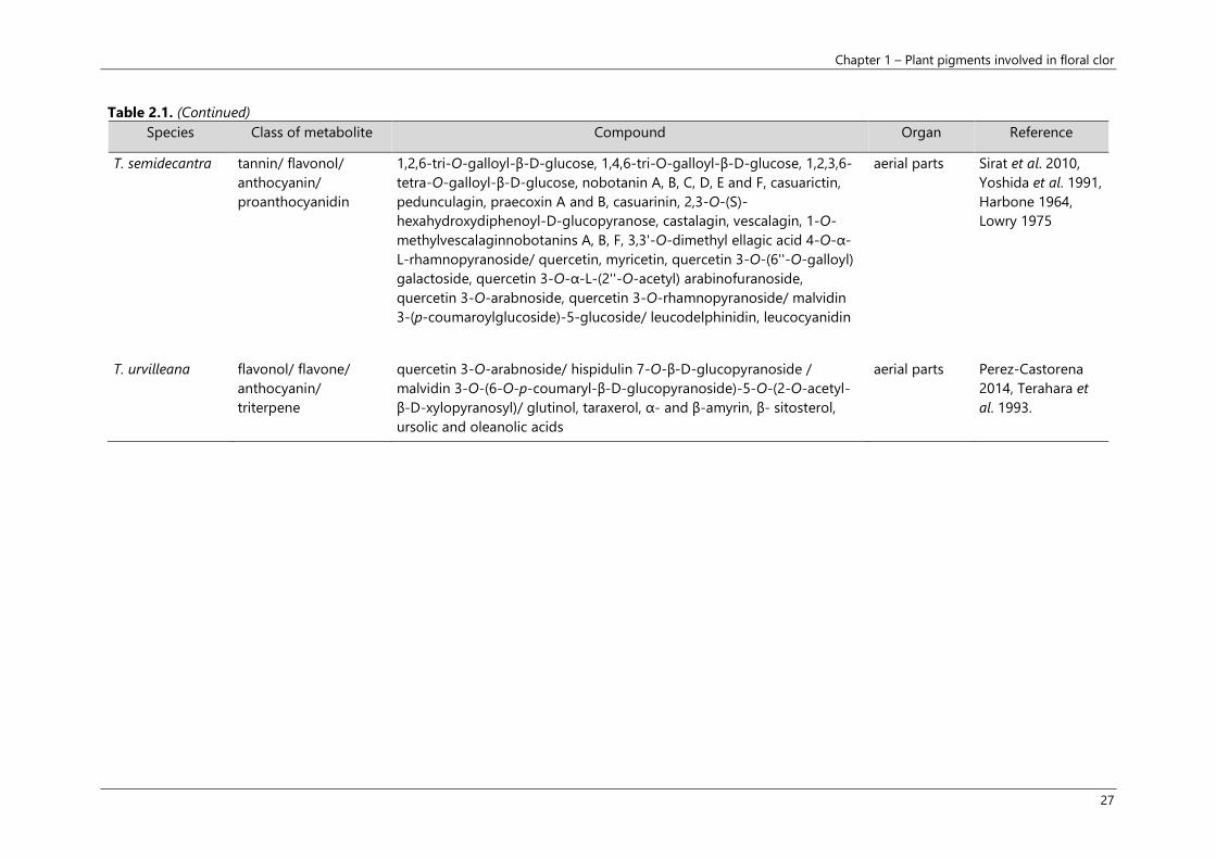
Chapter 1 – Plant pigments involved in floral clor
27
Table 2.1. (Continued)
Species Class of metabolite Compound Organ Reference
T. semidecantra tannin/ flavonol/
anthocyanin/
proanthocyanidin
1,2,6-tri-O-galloyl-β-D-glucose, 1,4,6-tri-O-galloyl-β-D-glucose, 1,2,3,6-
tetra-O-galloyl-β-D-glucose, nobotanin A, B, C, D, E and F, casuarictin,
pedunculagin, praecoxin A and B, casuarinin, 2,3-O-(S)-
hexahydroxydiphenoyl-D-glucopyranose, castalagin, vescalagin, 1-O-
methylvescalaginnobotanins A, B, F, 3,3'-O-dimethyl ellagic acid 4-O-α-
L-rhamnopyranoside/ quercetin, myricetin, quercetin 3-O-(6''-O-galloyl)
galactoside, quercetin 3-O-α-L-(2''-O-acetyl) arabinofuranoside,
quercetin 3-O-arabnoside, quercetin 3-O-rhamnopyranoside/ malvidin
3-(p-coumaroylglucoside)-5-glucoside/ leucodelphinidin, leucocyanidin
aerial parts Sirat et al. 2010,
Yoshida et al. 1991,
Harbone 1964,
Lowry 1975
T. urvilleana flavonol/ flavone/
anthocyanin/
triterpene
quercetin 3-O-arabnoside/ hispidulin 7-O-β-D-glucopyranoside /
malvidin 3-O-(6-O-p-coumaryl-β-D-glucopyranoside)-5-O-(2-O-acetyl-
β-D-xylopyranosyl)/ glutinol, taraxerol, α- and β-amyrin, β- sitosterol,
ursolic and oleanolic acids
aerial parts Perez-Castorena
2014, Terahara et
al. 1993.

28
T. granulosa and T. pulchra have been characterized as possible biomonitors of air
pollution, i.e. particulate matter and ozone (Klumpp et al. 2001, Furlan et al. 2004, Furlan et al.
2008, Santos & Furlan 2013, Zampieri et al. 2013, Espósito & Domingos 2014, Espósito &
Domingos 2016, Pedroso et al. 2016). Moreover, as they occur mainly in open areas, such as
forest edges and clearings, Tibouchina species are considered important for
restoration/reforestation purposes (Ellison et al. 1993).
Tibouchina pulchra (Figure 2.1), popularly known as “manacá-da-serra”, has been
increasingly used for urban ornamentation and, nowadays several cultivars are available in the
flower market. The beauty and fascinating feature of these flowers is the development-
associated color change: the buds open as white flowers that the next day become pink, further,
from the third day towards senescent, the flowers maintain a deep pink color. The well-
determined stages of flower coloring in T. pulchra raise interesting questions about the genetic
and chemical mechanisms involved in the regulation of this phenomenon, which has up to now
been unexplored (see Chapter 3).
In this context, this chapter presents results of isolation and identification of the petal
tissue pigments of Tibouchina pulchra.
Figure 2.1. Tibouchina pulchra tree and its flowers.

Chapter 2 – Chemical characterization of Tibouchina pulchra (Cham.) Cogn. flowers.
29
2. MATERIAL AND METHODS
2.1. Plant Material
Pink petals of Tibouchina pulchra (cv. “manacá-anão”) flowers were sampled at Praça
Carlos José Gíglio (Latitude: -23.57998, Longitude: -46.73403) in the most vigorous flowering
period (May and June/2016) between 8 and 9 a.m. Petals were immediately frozen in liquid
nitrogen and stored at -80°C until processing. Samples were freeze dried in a lyophilizer (K202,
Liobras) and crushed in a ball mill (TissueLyser, Qiagen) for further analyses. An exsiccate was
deposited on herbarium of Institute of Bioscience (SPF) of University of São Paulo (ID: Furlan73).
2.2. Extraction and analysis by UPLC-MS
Phenolic compounds were extracted from 100 mg of petal powder with two times of
1.5 mL of 0.2 % HCl in methanol (MeOH). The samples were sonicated for 10 min and
centrifugated at 10,000 rpm for 10 min. The extract was filtered (PTFE 0.45 μm) and analyzed
by Ultra Performance Liquid Chromatography (UPLC) system with Diode Array Detector (DAD)
(Dionex Ultimate 3000) and Electrospray Ionization Quadrupole Time-of-Flight High-
Resolution Mass Spectrometry (ESI-QTOF-HRMS) detector (Bruker Maxis), MS/MS analysis was
performed with a Broadband Collision Induced Dissociation (bbCID) detector. Separation was
achieved by using a C18 column (Waters Acquity UPLC 100 x 2.1 mm - 1.7 µm) at a flow rate
of 0.3 mL min-1, and 4 µL of injection volume, the column temperature was 45°C, and the
solvent system composed of 1% formic acid in water (A) and 1% formic acid in acetonitrile (B).
Gradient elution: 5 to 25% of B (0- 40 min), 25 to 100% of B (40- 42 min), 100% of B (42- 42.5
min), 100 to 5% of B (42.5- 43 min), 5% of B (43- 46 min). Separated compounds were first
monitored using DAD (200 to 600 nm) and then MS scans were performed in positive ion mode
(MS+), in the range m/z 75−1250 m/z, in the following conditions: capillary voltage set to 4,500
V, end plate offset at -500 V, nebulizer at 2 Bar, dry gas 12 L min-1 and dry gas temperature at
200°C MS was calibrated using sodium formate. All data were processed using Data analysis
software 4.2 (Bruker). This analysis was performed in the Department of Chemistry at Denmark
Technical University under the supervision of Ph.D. Mads H. Clausen.

30
2.3. Isolation by preparative HPLC and identification by Nuclear Magnetic
Resonance (NMR)
Also in partnership with Ph.D. Mads H. Clausen, flavonoids were isolated from 10 g of
petal powder by extracting four times in 200 mL of hydrochloric acid 0.2% in MeOH. The
samples were sonicated for 15 min, pillowed for 10 min, vacuum filtered and concentrated with
a rotary evaporator. The crude extract was diluted to, approximately, 250 mg/mL and injected
in a preparative High Performance Liquid Chromatography (HPLC- Waters) with Photodiode
Array Detector (PAD). Separation was achieved on a C18 column (Waters X Bridge BEH ODB 30
x 150 mm, 5µm) at a flow rate of 20 mL min-1, using 1 mL of injection volume, and a solvent
system composed of 1% formic acid in water (A) and 1% formic acid in acetonitrile (B). Gradient
elution: 10% of B (0- 3 min), 10 to 15% of B (3- 30 min), 15% of B (30- 50 min), 15 to 20% of B
(50- 60 min), 20 to 25% of B (60- 80 min), 25 to 35% of B (80- 90 min), 35 to 45% of B (90- 95
min), 45 to 100% of B (95- 96 min), 100% of B (96- 98 min), 100 to 10% of B (98- 98.5 min),
10% of B (98.5- 102 min), and monitored using DAD (200 to 600 nm). Fractions were collected
by time. All the fractions were concentrated by rotary evaporator. An aliquot was resuspended
in 0.2% HCl in MeOH to check the purity by UPLC-MS. For the isolated compounds the dried
sample was dissolved in deuterated dimethylsulfoxide (DMSO-d6) for NMR analysis. 1H and 13C
NMR spectra were obtained by using a AVANCE III HD spectrometer (Bruker) operating at
frequency of 800 MHz and equipped with a 5 mm TCI CryoProbe (Bruker). Two-dimensional
analysis (HSQC and HMBC) were also performed. All data were processed using MestreNova
software 11.0 (Mestrelab Research). The identification of compounds was performed in
collaboration with Ph.D. Marcelo José Pena Ferreira from Department of Botany, University of
São Paulo.

Chapter 2 – Chemical characterization of Tibouchina pulchra (Cham.) Cogn. flowers.
31
3. CONCLUSION
Melastomataceae and, in particular, Tibouchina are chemically poorly characterized
taxons. Here, out of the sixteen compounds described for the first time in T. pulchra, five of
them described for the first time in Melastomataceae, moreover an unpublished compound
was identified as kaempferol 4’-O-(2’’-methyl)-α-D-glucopyranoside. The advances in
spectrometric techniques offer a unique opportunity to improve our knowledge about the
chemical structure of natural products. Studies about flower anthocyanins are scarce and the
understanding of their structure, biosynthesis and the regulatory mechanisms involved in their
accumulation pattern can improve our knowledge about plant secondary metabolism, the
relationship between flower and pollinators, and bring new insights for future biotechnological
applications.

32
4. REFERENCES
Atoui, A.K., Mansouri, A., Boskou, G. & Kefalas, P. Tea and herbal infusions: Their antioxidant activity and
phenolic profile. Food Chemistry. 2005, 89, p. 27–36. DOI:10.1016/j.foodchem.2004.01.075.
BFG [The Brazilian Flora Group]. Growing knowledge: an overview of Seed Plant diversity in Brazil.
Rodriguésia. 2015, 66(4): 1085–1113.
Bobbio, F.O., Bobbio, P.A. & Degáspari, C.H. Anthocyanins from Tibouchina grandiflora. Food Chem.
1985, 18, 153-159. DOI: 10.1016/0308-8146(85)90138-4.
Braca, A., Politi, M., Sanogo, R., Sanou, H., Morelli, I., Pizza, C. & De Tommasi, N. Chemical composition
and antioxidant activity of phenolic compounds from wild and cultivated Sclerocarya birrea
(Anacardiaceae) leaves. Journal of agricultural and food chemistry. 2003, 51(23), 6689-6695 p. DOI:
10.1021/jf030374m.
Brito, V.L.G., Maia, F.R., Silveira, F.A.O. & Fracasso, C.M. Reproductive phenology of Melastomataceae
species with contrasting reproductive systems : contemporary and historical drivers. 2017, 19:806–
817. DOI: 10.1111/plb.12591,
Clausing G, Renner S.S. Molecular phylogenetics of Melastomataceae and Memecylaceae: implications
for character evolution. American Journal of Botany. 2001, 88(3):486–498. DOI: 10.2307/2657114.
Colorado, A., Maya, D.C., Díaz, G.S.J., Isaza, M.J.H., Tapias, I.L.J., Veloza, L. A., Ramírez, A. L. S. Flavonoides
del extracto isopropanol- agua de Tibouchina ciliaris. Scientia et Técnica. Universidad Tecnológica de
Pereira, Pereira, Colombia, 2007, 3(33): 355-357.
Cunha W.R., dos Santos, F.M., Peixoto, J., Veneziani, E.C.S., Crotti, A.E. M., Silva, M.L.A., da Silva Filho, A.A.,
Albuquerque, S., Turatti, I.C.C. & Nastos, J.K. 2009. Screening of plant extracts from the Brazilian
Cerrado for their in vitro trypanocidal activity. Pharmaceutical Biology. 2009, 47(8): 744–749 p. DOI:
10.1080/13880200902951361.
Cuyckens, F. & Claeys, M. Mass spectrometry in the structural analysis of flavonoids. Journal of Mass
Spectrometry. 2004, 39(1), 1–15 p. DOI:10.1002/jms.585
Dos Santos, A.C., & Furlan, C.M. Levels of phenolic compounds in Tibouchina pulchra after fumigation
with ozone. Atmospheric Pollution Research. 2013, 4:250–256 p. DOI: 10.5094/APR.2013.027
Dias, Ê.R., Dias, T. de L.M.F., Alexandre-Moreira, M.S., & Branco, A. Antinociceptive activity of Tibouchina
pereirae, an endemic plant from the Brazilian semiarid region. Zeitschrift Für Naturforschung C. 2016,
71(7-8):261–265 p. DOI: 10.1515/znc-2015-0155.
Dias, E.R. Estudo fitoquímico e avaliação da atividade biológica de Tibouchina pereirae Aubl.
(Melastomataceae). Master Dissertation, Universidade Estadual de Feira de Santana. 2013, 80p.
dos Santos, F.M., de Souza, M.G., Miller Crotti, A.E., Martins, C.H. G., Ambrósio, S.R., Veneziani, R.C.S.,
Andrade e Silva, M. L. & Cunha, W. R. Evaluation of antimicrobial activity of extracts of Tibouchina
candolleana (melastomataceae), isolated compounds and semi-synthetic derivatives against
endodontic bacteria. Brazilian Journal of Microbiology. 2012, 43(2), 793–799 p. DOI: 10.1590/S1517-
83822012000200045.
Ducrey, B., Wolfender, J.L., Marston, A.& Hostettmann, K. Analysis of flavonol glycosides of thirteen
Epilobium species (Onagraceae) by LC–UV and thermospray LC-MS. Phytochemistry. 1995, 38(1): 129-
137 p. DOI: 10.1016/0031-9422(94)00629-8.
Ellison, A.M., Denslow, J.S., Loiselle B.A. & Brénes D.M. Seed and seedling ecology of Neotropical
Melastomataceae. Ecology. 1993, 6: 1733–1749 p. DOI: 10.2307/1939932.
Esposito, M.P., Pedroso, A.N.V. & Domingos, M. Assessing redox potential of a native tree from the
Brazilian Atlantic Rainforest: A successful evaluation of oxidative stress associated to a new power
generation source of an oil refinery. Science of the Total Environment. 2016, 550: 861–870 p. DOI:
10.1016/j.scitotenv.2016.01.196.
Esposito, M.P. & Domingos, M. Establishing the redox potential of Tibouchina pulchra (Cham.) Cogn., a
native tree species from the Atlantic Rainforest in the vicinity of an oil refinery in SE Brazil.
Environmental Science and Pollution Research. 2014, 21: 5484–5495 p. DOI: 10.1007/s11356-013-
2453-8.
Chapter 2 – Chemical characterization of Tibouchina pulchra (Cham.) Cogn. flowers.
33
Furlan, C.M., Moraes, R.M., Bulbovas, P., Sanz, M.J., Domingos, M. & Salatino, A. Tibouchina pulchra
(Cham.) Cogn., a native Atlantic Forest species, as a bioindicator of ozone: visible injury.
Environmental Pollution. 2008, 152: 361–365 p. DOI: 10.1016/j.envpol.2007.06.042.
Furlan, C.M., Salatino, A. & Domingos, M. Influence of air pollution on leaf chemistry, herbivore feeding
and gall frequency on Tibouchina pulchra leaves in Cubatão (Brazil). Biochemical Systematics and
Ecology. 2004, 32(3): 253–263 p. DOI: 10.1016/S0305-1978(03)00176-5.
Furlan, C.M. Efeito de poluentes atmosféricos na composição química de indivíduos jovens de Tibouchina
pulchra (Cham.) Cong. e Psidium guajava L. PhD thesis, Universidade de São Paulo, 2004, 117 p.
Guimarães, P.J.F. Two New Species of Tibouchina (Melastomataceae) from Brazil. Novon: A Journal for
Botanical Nomenclature. 2014, 23(1): 42–46 p. DOI: 10.3417/2012029.
Guimarães, P.J.F. Tibouchina. Lista de Espécies da Flora do Brasil. Jardim Botânico do Rio de Janeiro.
2016. Available from: http:// floradobrasil.jbrj.gov.br/jabot/floradobrasil/FB19731 (accessed: October
29th 2016).
Guo, D., Xue, W. J., Zou, G.A. & Aisa, H.A. Chemical Composition of Alhagi sparsifolia Flowers. Chemistry
of Natural Compounds. 2016, 52(6): 1095-1097 p. DOI: 10.1007/s10600-016-1871-5.
Harbone, J.B. Plant Polyphenols- XI. The structure of acilated anthocyanins. Phytochemistry. 1964,
3:151–160 p. DOI: 10.1016/S0031-9422(00)88035-8.
Haslam, E. Vegetable tannins - lessons of a phytochemical lifetime. Phytochemistry. 2007, 68(22-24),
2713–21 p. DOI: 10.1016/j.phytochem.2007.09.009.
Hendra, R. & Keller, P.A. Flowers in Australia: Phytochemical Studies on the Illawarra Flame Tree and
Alstonville. Australian Journal of Chemistry. 2016, 69(8): 925–927 p. DOI: 10.1071/CH16058.
Isobe, T., Ito, N. & Noda, Y. Minor flavonoids of Polygonum nodosum. Phytochemistry. 1980, 19(8):1877.
Iwashina T. The structure and distribution of the flavonoids in plants. Journal of Plant Research. 2000,
113(3): 287-299 p. DOI: 10.1007/PL00013940.
Janna, O., Khairul, A. & Maziah, M. Anthocyanin stability studies in Tibouchina semidecandra L. Food
Chemistry. 2007, 101(4): 1640–1646. DOI: 10.1016/j.foodchem.2006.04.034.
Jiménez, N., Carrillo-Hormaza, L., Pujol, A., Álzate, F., Osorio, E., & Lara-Guzman, O. Antioxidant capacity
and phenolic content of commonly used anti-inflammatory medicinal plants in Colombia. Industrial
Crops and Products. 2015, 70, 272–279 p. DOI: 10.1016/j.indcrop.2015.03.050.
Jones, E., Ekundayo, O. & Kingston, D.G.I. Plant anticancer agents. XI. 2, 6-Dimethoxybenzoquinone as a
cytotoxic constituent of Tibouchina pulchra. Journal of Natural Products. 1981, 44: 493–494 p. DOI:
10.1021/np50016a019.
Joshi, H., Joshi, A.B., Gururaj, M.P. & Sati, H. (2009). Flavonoids of Memecylon umbellatum (Burm.). Asian
Journal of Chemistry. 2009, 21(2): 1639.
Jung, H.A., Kim, J.E., Chung, H.Y. & Choi, J.S. Antioxidant principles of Nelumbo nucifera stamens. Archives
of pharmacal research. 2003, 26(4): 279-285. DOI: 10.1007/BF02976956.
Klumpp, G., Furlan, C.M., Domingos, M. & Klumpp, A. Response of stress indicators and growth
parameters of Tibouchina pulchra Cogn. exposed to air and soil pollution near the industrial complex
of Cubatão, Brazil. Science of The Total Environment. 2000, 246: 79–91 p. DOI: 10.1016/S0048-
9697(99)00453-2.
Kuster, R.M., Arnold, N. & Wessjohann, L. Anti-fungal flavonoids from Tibouchina grandifolia.
Biochemical Systematics and Ecology. 2009, 37(1): 63–65 p. DOI: 10.1016/j.bse.2009.01.005.
Kyekyeku, J., Adosraku, R. & Asare–Nkansah, S. LC–HRESI–MS/MS Profiling of Flavonoids from
Chlorophora regia (Moraceae). British Journal of Pharmaceutical Research. 2017, 16(1):1–9 p. DOI:
10.9734/BJPR/2017/32893.
Lavault, M. & Richomme, P. Constituents of Helichrysum stoechas variety olonnense. Chemistry of natural
compounds. 2004, 40(2): 118-121 p.DOI: 10.1023/B:CONC.0000033925.00693.7b.
Ma, Y.L., Li, Q.M., Van den Heuvel, H.& Claeys, M. Characterization of flavone and flavonol aglycones by
collision-induced dissociation tandem mass spectrometry. Rapid Communications Mass
Spectrometry. 1997, 11(12): 1357-1364 p. DOI: 10.1002/(SICI)1097-0231(199708)11:12<1357:AID-
RCM983>3.0.CO,2-9.

34
Mahmoud, I.I., Marzouk, M.S.A., Moharram, F.A., El-Gindi, M.R. & Hassan, A.M.K. Acylated flavonol
glycosides from Eugenia jambolana leaves. Phytochemistry. 2001, 58(8): 1239-1244. DOI:
10.1016/S0031-9422(01)00365-X.
Markham, K.R. Techniques of Flavonoid Identification. Academic Press: London, 1982.
Michelangeli, F.A., Guimaraes, P.J.F., Penneys, D.S., Almeda, F., Kriebel, R. Phylogenetic relationships and
distribution of New World Melastomeae (Melastomataceae ). Botanical Journal of the Linnean
Society. 2013, 171(1): 38–60. DOI: 10.1111/j.1095-8339.2012.01295.x.
Motta, L.B., Kraus, J.E., Salatino, A. & Salatino, M.L.F. (2005). Distribution of metabolites in galled and
non-galled foliar tissues of Tibouchina pulchra. Biochemical Systematics and Ecology. 2012, 33(10):
971–981. DOI: 10.1016/j.bse.2005.02.004.
Mpalantinos, M.A., Pereira, N.A., Parente, J.P. & Kuste, R.M. Hispidulina-7-O-B-glucoside, the main
constituent of the aqueous extract of Tibouchina granulosa. Abstract 8th Brazilian Meeting of Organic
Synthesis, Sao Pedro, Brazil. 1988.
Nikaido, T., Ohmoto, T. & Sankawa, U. Inhibitors of adenosine 3', 5'-cyclic monophosphate
phosphodiesterase in Daphne genkwa Sieb. et Zucc. Chemical and pharmaceutical bulletin. 1987,
35(2), 675-681. DOI: 10.1248/cpb.35.675.
Niño, J., Espinal, C.M., Mosquera, O.M. & Correa, Y.M. Antimycotic activity of 20 plants from Colombian
flora. Pharmaceutical Biology. 2003, 41(7): 491–496. DOI: 10.1080/13880200308951341.
Okumura, F., Soares, M., Barbosa H.F., Cavalheiro & Gomes, E.T. Identificação de pigmentos naturais de
espécies vegetais utilizando-se cromatografia em papel. Química Nova. 2002, 25(4): 680-683. DOI:
10.1590/S0100-40422002000400025.
Pedroso, A.N.V., Bussotti, F., Papini, A., Tani, C. & Domingos, M. Pollution emissions from a petrochemical
complex and other environmental stressors induce structural and ultrastructural damage in leaves of
a biosensor tree species from the Atlantic Rain Forest. Ecological Indicators. 2016, 67: 215–226. DOI:
10.1016/j.ecolind.2016.02.054.
Pérez-Castorena, A.L. Triterpenes and other metabolites from Tibouchina urvilleana. Journal of the
Mexican Chemical Society. 2014, 58(2), 218–222.
Renner, S. S. Phylogeny and classification of the Melastomataceae and memecylaceae. Nordic Journal
of Botany. 1993, 13(5): 519–593. DOI: 10.1111/j.1756-1051.1993.tb00096.x.
Rodrigues, J., Rinaldo, D., dos Santos, L.C. & Vilegas, W. An unusual linked flavonoid from Miconia
cabucu (Melastomataceae). Phytochemistry. 2007, 68: 1781–1784. DOI:
10.1016/j.phytochem.2007.04.020.
Scheer, T. & Wichtl, M. Zum Vorkommen von Kämpferol-4′-O-β-D-glucopyranosid in Filipendula ulmaria
und Allium cepa1. Planta medica. 1987, 53(06):573-574. DOI: 10.1055/s-2006-962817.
Serna, D.M. O. & Martínez, J.H.I. Phenolics and polyphenolics from melastomataceae species. Molecules.
2015, 20(10): 17818–17847. DOI: 10.3390/molecules201017818.
Singha, U.K., Guru, P.Y., Sen, A.B.& Tandon J. S. Antileishmanial activity of traditional plants against
Leishmania donovani in Golden hamsters. International Journal of Pharmacognosy. 1992, 30: 289–
295. DOI: 10.3109/13880209209054015.
Sirat, H.M., Rezali, M.F. & Ujang, Z. Isolation and identification of radical scavenging and tyrosinase
inhibition of polyphenols from Tibouchina semidecandra L. Journal of Agricultural and Food
Chemistry. 2010, 58(19): 10404–10409. DOI: 10.1021/jf102231h.
Sobrinho, A.P., Minho, A. S., Ferreira, L.L., Martins, G.R., Boylan, F. & Fernandes, P.D. Characterization of
anti-inflammatory effect and possible mechanism of action of Tibouchina granulosa. Journal of
Pharmacy and Pharmacology. 2017, 69(6): 706-713. DOI: 10.1111/jphp.12712.
Tamura, H., Hayashi, Y., Sugisawa, H. & Kondo, T. Structure determination of acylated anthocyanins in
muscat baily A grapes by homonuclear Hartmann–Hahn (HOHAHA) spectroscopy and liquid
chromatography–mass spectrometry. Phytochemical Analysis. 1994, 5: 190-196. DOI:
10.1002/pca.2800050404.
Terahara, N., Suzuki, H., Toki, K., Kuwano, H., Saito, N., Honda, T. J. A diacylated anthocyanin from
Tibouchina urvilleana flowers. Journal of Natural Products. 1993, 56: 335-340.DOI:
10.1021/np50093a004.

Chapter 2 – Chemical characterization of Tibouchina pulchra (Cham.) Cogn. flowers.
35
Tracanna, M.I., Fortuna, A.M., Contreras Cárdenas, A.V., Marr, A.K., McMaster, W.R., Gõmez-Velasco, A.,
Sánchez Arreola, E., Hernández, L.R. & Bach, H. Anti-Leishmanial, anti-inflammatory and antimicrobial
activities of phenolic derivatives from tibouchina paratropica. Phytotherapy Research. 2015, 29(3),
393–397. DOI: 10.1002/ptr.5263.
Tropicos.org. Missouri Botanical Garden, St. Louis, Missouri. 2013. Available from:
http://www.tropicos.org. Accessed in: August 27th 2017.
Wang, H., Race, E. J. & Shrikhande, J. Characterization of anthocyanins in grape juices by ion trap liquid
chromatography–Mass Spectrometry. Journal of Agricultural and Food Chemistry. 2003, 51(7):1839-
1844. DOI: 10.1021/jf0260747.
Wahab, A., Begum, S., Ayub, A., Mahmood, I., Mahmood, T., Ahmad, A. & Fayyaz, N. Luteolin and
kaempferol from Cassia alata, antimicrobial and antioxidant activity of its methanolic extracts.
FUUAST Journal of Biology. 2014, 4(1):1.
Wei, Y., Xie, Q., Fisher, D. & Sutherland, I.A. Separation of patuletin-3-O-glucoside, astragalin, quercetin,
kaempferol and isorhamnetin from Flaveria bidentis (L.) Kuntze by elution-pump-out high-
performance counter-current chromatography. Journal of Chromatography A. 2011, 1218(36): 6206-
6211. DOI: 10.1016/j.chroma.2011.01.058.
Yoshida, T., et al. Tannins and related polyphenols of Melastomataceous plants I. Hydrolyzable tannins
from Tibouchina semidecandra Cogn. Chemical and Pharm aceutical Bulletin 1991, 39: 2233–2240.
DOI: 10.1248/cpb.39.2233.
Yoshida, T., Amakura, Y., Yokura, N., Ito, H., Isaza, J. H., Ramírez, S., Pelaez, D. P. & Renner, S. S. Oligomeric
hydrolyzable tannins from Tibouchina multiflora. Phytochemistry. 1999, 52: 1661–1666. DOI:
10.1016/S0031-9422(99)00329-5.
Zampieri, M.C.T., Sarkis, J.E.S., Pestana, R.C.B., Tavares, A.R. & Melo-de-Pinna, G.F.A. Characterization of
Tibouchina granulosa (Desr.) Cong. (Melastomataceae) as a biomonitor of air pollution and
quantification of particulate matter adsorbed by leaves. Ecological Engineering. 2013, 61: 316–327.
DOI: 10.1016/j.ecoleng.2013.09.050.
Chapter 3 – The regulation of floral color change in Tibouchina
pulchra.
1. INTRODUCTION
Tibouchina pulchra (Cham.) Cogn. (Melastomataceae, Myrtales), named as “manacá da
serra”, is a native species from the Atlantic Forest. Its use for urban ornamentation is increasing
due to its beautiful flowers. In this sense, the ornamental dwarf cultivar of T. pulchra (“manacá-
anão”) blooms with the same temporal pattern of floral color as the wild parental: buds, first
stage (S1), open as white flowers (S2) that, one day after, become light pink (S3), further, flowers
are deep pink (S4), from the third day towards senescence (Figure 3.1). As reviewed in chapter
1, the temporal alteration of flower color can be regulated by pollination, gene expression
regulation, the presence of copigments and flavonoid-metal complexation, resulting in the
differential accumulation of phenolic acids, flavonols, and anthocyanins extensively
characterized in chapter 2.

36
Figure 3.1. Stages of floral color change of T. pulchra. S1- buds (day 0), S2- white flowers (day 1), S3-
light pink (day 2), S4- dark pink (day 3).
T. pulchra flowers are nectarless, with poricidal anthers, hermaphrodite, heterantherous,
herkogamous and self-compatible (Renner 1989, Brito & Sazima 2012). Concerning pollinators,
Pereira et al. (2011) observed fifteen species of floral visitors in T. pulchra, being only four
described as effective pollinators (Bombus morio, B. pauloensis, Xylocopa frontalis and X.
brasilianorum). These large bees interact with poricidal anthers transferring the pollen to the
stigmas by vibration. Xylocopa species collected pollen exclusively from white flowers, while
Bombus visited pink flowers only at the end of the flowering season, when white flowers are
scarce. Moreover, by performing controlled pollination experiments, it was demonstrated that
the pollen viability, stigma receptivity and fruit setting were similar in white and pink flowers.
This data together with the bee color preference described above suggest that flower color
change is not associated to pollination but, according to Da Silva (2006), may enlarge the visitor
diversity.
As aforementioned, the pigments responsible for the color of T. pulchra flowers are
phenolic acids and flavonoids (flavonols and anthocyanins). These phenolic compounds are
produced by the combination of products from shikimic (SKM) and malonate (MAL) pathways,
whose building blocks are derived from primary metabolism (Figure 3.2). Many simple benzoic
acid derivatives (e.g. gallic acid, C6C1) and phenylpropanoids (e.g. p-coumaric, C6C3) are
produced by SKM pathway, while flavonoids (C6C3C6) are biosynthesized by intermediates from
both, SKM and MAL pathways (Davies et al. 2009).
PHENYLALANINE AMMONIA LYASE (PAL) is the first enzyme in the phenylpropanoid
and flavonoid pathway, it catalyzes the nonoxidative deamination of phenylalanine yielding
trans-cinnamic acid. CINNAMATE 4-HYDROXYLASE (C4H) is a CYTOCHROME P450
MONOOXYGENASE (P450) enzyme involved in the hydroxylation of trans-cinnamic acid at C4
position to produce p-coumaric acid. This is a branching point that can channel the metabolic
flux towards phenylpropanoids or flavonoids. By the action of COUMARATE 3-HYDROXYLASE

Chapter 3 – The regulation of floral color change in Tibouchina pulchra.
37
(C3H), cinnamic acid derivatives as caffeic, ferulic, and sinapic acids are synthetized. On the
other hand, p-coumaric acid is activated to the corresponding Co-A thioester facilitating further
conversion to flavonoids, this reaction is catalyzed by 4-COUMARATE:CO-A LIGASE (4CL)
(Dewick 2009, Saito et al. 2013).
The first produced flavonoid class comprises the chalcones, which are formed by the
action of CHALCONE SYNTHASE (CHS). This enzyme belongs to a family of type III polyketide
synthases and is the first committed enzyme in biosynthesis of flavonoids. Three sequential
additions of the extender molecule malonyl-CoA in a p-coumaroyl-CoA is the reaction
catalyzed by CHS (Dewick 2009, Saito et al. 2013). Chalcones are the first colored flavonoid,
and provide yellow coloration to petals of a few plant species (e.g. Dianthus caryophyllus L.,
Caryophyllaceae, Caryophyllales) (Ono et al. 2006a, 2006b). CHALCONE ISOMERASE (CHI) acts
in the stereospecific cyclization of naringenin chalcone to flavanone. FLAVANONE 3-
HYDROXYLASE (F3H) is responsible for the oxygenation at C3 position of flavanone to form
dihydroflavonol (dihydrokaempferol). FLAVONOID 3′-HYDROXYLASE (F3′H) and FLAVONOID
3’-5’-HYDROXYLASE (F3’5’H) are P450 that catalyze the hydroxylation at C3′and C3’-C5’-
positions of the B-ring of flavonoid, respectively, rendering dihydroflavonols.
FLAVONOL SYNTHASE (FLS) and DIHYDROFLAVONOL REDUCTASE (DFR) are
competitors for the same substrate, dihydroflavonol, a branching point for flavonols and
anthocyanins formation (Pelletier et al. 1997). FLS catalyzes the double bond formation
between C2 and C3, while DFR reduces the C4-keto group of dihydroflavonol to the
corresponding leucoanthocyanidin. FLS, F3H and ANTHOCYANIDIN SYNTHASE (ANS) are the
three 2-OXOGLUTARATE/FE(II)-DEPENDENT DIOXYGENASE (2-ODD) enzymes in the flavonoid
biosynthesis pathway. The enzyme ANS catalyzes the formation of anthocyanidin from
leucoanthocyanidin. Anthocyanidins exhibit a conjugated double bond system encompassing
A, B and C rings, the later with two double bonds carrying a positive charge. Anthocyanidins
are the chromophore moiety of anthocyanins, which are formed after glycosylation, acylation
and/or methylation by the action of specific enzymes as FLAVONOID GLUCOSYLTRANSFERASE
(FGT), ANTHOCYANIDIN ACYLTRANSFERASE (ACT) and/or O-METHYLTRANSFERASE (OMT)
(Figure 3.2). In Arabdopsis thaliana (L.) Heynh. (Brassicaceae, Brassicales), FGT can transfer
glucose to the C3 position of both, flavonol and anthocyanin aglycones (Yonekura et al. 2012).
Due to anthocyanins appeal as natural food colorant, nutraceutic and bioactive
compounds (Norberto et al. 2013, De Ferrars et al. 2014, Xiaonan et al. 2016, Gowd et al. 2017),
38
the regulation of anthocyanin biosynthetic pathway has been extensively explored. Most of the
structural genes have been isolated (Winkel-Shirley 2001) and several regulatory genes have
been characterized. Petunia (Petunia hybrid Vilm., Solanaceae, Solanales) and snapdragon
(Antirrhinum majus L., Plantaginaceae, Lamiales) are the major model species for the study of
anthocyanin biosynthesis, especially in floral tissues, however, maize (Zea mays L., Poaceae,
Poales) and Arabdopsis thaliana have been extensively studied as well (Cone et al. 1986,
Goodrich et al. 1992, Quattrocchio et al. 1998, 1999, Broun 2005, Dixon et al. 2005, Koes et al.
2005, Grotewold 2006).
Regarding the position in the pathway, structural genes can be grouped into two
classes: early biosynthetic genes (e.g. PAL, C4H, CHS, and FLS) and late biosynthetic genes (e.g.
DFR and ANS) (Tornielli et al. 2009). The early genes of anthocyanin pathway were functionally
characterized in many plant species such as A. thaliana, Solanum lycopersicum L. (Solanaceae,
Solanales), Vitis vinifera L. (Vitaceae, Vitales), P. hybrida, and A. majus (Sparvolli et al. 1994,
Brugliera et al. 1999, Mol et al. 1983, Guo et al. 2009, Huang et al. 2010), while late genes (DFR
and ANS) have been characterized mostly in P. hybrid and A. thaliana. (Pelletier et al. 1997,
Devic etal. 1999). However, much less is known about the subsequent modifications
(substituents), such as glycosylation, acylation, and methylation (Grotewold 2006). In fact, there
are several types of substituents that guarantee the chemical diversity in flower color. For
example, P. hybrida synthesizes methylated anthocyanins with an acylated rutinose moiety,
while A. thaliana accumulates more complex anthocyanins with seven sugar and acyl moieties
but no methyl groups (Brouillard et al. 2003, Saito et al. 2013).

Chapter 3 – The regulation of floral color change in Tibouchina pulchra.
39
Figure 3.2. Simplified schematic representation of flavonoid biosynthetic pathway. Primary metabolism is
highlighted in green. Abbreviations indicate the following enzymes: 4-COUMARATE:CO-A LIGASE (4CL),
ANTHOCYANIDIN ACYL TRANSFERASE (AACT), ANTHOCYANIDIN SYNTHASE (ANS), CAFFEIC ACID O-
METHYLTRANSFERASE (COMT), CINNAMATE 4-HYDROXYLASE (C4H), CHALCONE ISOMERASE (CHI),
CHALCONE REDUCTASE (CHR), CHALCONE SYNTHASE (CHS), Coenzyma A (CoA), COUMARATE 3-
HYDROXYLASE (C3H), DIHYDROFLAVONOL 4-REDUCTASE (DFR), erythrose 4-phosphate (E4P), FLAVANONE
3-HYDROXYLASE (F3H), FLAVONE SYNTHASE (FS), FLAVONOID 3'-HYDROXYLASE (F3'H), FLAVONOID 3'5'-
HYDROXYLASE (F3'5'H), FLAVONOID GLYCOSYL TRANSFERASE (FGT), FLAVONOL SYNTHASE (FLS), HSCoA:
coenzyme A, ISOFLAVONE SYNTHASE (IFS), malonate (MAL), O-METHYLTRANSFERASE (OMT),
PHENYLALANINE AMMONIA LYASE (PAL), phosphoenolpyruvate (PEP), SHIKIMATE DEHYDROGENASE
(SDH), and shikimate (SKM). The enzymes studied in the present work are highlighted in bold. Dotted lines
represent more than one enzymatic reaction. Adapted from Dewick et al. (2009).
40
Numerous studies have demonstrated the transcriptional regulation of anthocyanin
biosynthesis. A transcription factor complex (MBW complex) composed by a MYELOBLASTOSIS
(MYB), a BASIC HELIX-LOOP-HELIX (bHLH), and a BETA-TRANSDUCIN REPEAT (WD40) protein,
has shown to regulate the expression of the structural genes (Koes et al. 2005, Hichri et al.
2011, Xu et al. 2014, 2015). In vegetative tissues of A. thaliana, an eudicot, anthocyanin early
biosynthetic genes are co-activated by bHLH and WD40 transcription factors in a MYB-
independent manner, while the late biosynthetic genes require all three proteins to form the
active MBW complex. Interestingly, in maize, a monocot, early and late biosynthetic genes are
transcriptionally activated exclusively by MYB and bHLH proteins (Petroni & Tonelli 2011). By
the characterization of mutant genotypes and transgenic lines, the transcriptional induction of
anthocyanin biosynthesis in flowers has been addressed in several species, where distinct
combinations of the aforementioned transcriptional factors have shown to be needed to
determine floral pigmentation. MYB transcription factors induce early and late biosynthetic
genes in Nicotiana tabacum L. (Solanaceae, Solanales), Brassica oleracea L. (Brassicaceae,
Brassicales), Anthurium andraeanum Linden ex André (Araceae, Alismatales), Mimulus lewisii
Pursh (Scrophulariaceae, Lamiales), and M. cardinalis Douglas ex Benth. flowers (Chiu et al.
2010, Pattanaik et al. 2010, Li et al. 2016, Yuan et al. 2016). In Ipomea purpurea (L.) Roth
(Convolvulaceae, Solanales) transposon-insertion double mutants for bHLH and MYB genes
resulted in snow-white flowers where the expression of the late biosynthetic genes was
completely abolished (Park et al. 2007).
Additionally, the existence of post-transcriptional regulatory mechanisms has also been
verified to control anthocyanin biosynthesis. In A. thaliana, it has been reported that the
expression of the anthocyanin-associated MYB transcription factors is regulated by miRNAs
and tasiRNAs-mediated hetero-silencing (Rajagopalan et al. 2006, Hsieh et al. 2009). In
particular, the miR156 targets SQUAMOSA PROMOTER BINDING PROTEIN-LIKE (SPL) mRNA,
whose product destabilizes the MBW transcriptional activation complex. Thus, increased
miR156 activity promotes the accumulation of anthocyanins by the MBW complex-mediated
transcriptional induction of the late biosynthetic genes (Gou et al. 2011). Moreover, in Petunia,
CHS has also shown to be post-transcriptionally regulated by siRNA (Koseki et al. 2005).
Interestingly, anthocyanin accumulation in maize kernels is controlled by the regulation of the
splicing patter of Bz2 gene, a GLUTATHIONE S-TRANSFERASE (GST) that mediates transfer of
cytoplasmic anthocyanin into the vacuole (Pairoba & Walbot 2003).

Chapter 3 – The regulation of floral color change in Tibouchina pulchra.
41
Finally, post-translational regulation has also been reported for anthocyanin
biosynthesis. In A. thaliana, KELCH REPEAT F-BOX proteins have shown to physically interact
with four PAL isozymes inducing their 26S proteasome- mediated ubiquitination, resulting in
the downregulation of phenylpropanoid biosynthesis (Zhang et al. 2013).
Although for some biological systems the regulation of anthocyanin biosynthesis has
been elucidated, including the identification of master transcription factors (Butelli et al. 2008),
many gaps remain unsolved for the complete understanding of the mechanisms underneath
the accumulation of these pigments. Improving the knowledge about the multilevel regulation
of gene expression will favor the development of new biotechnological tools for the generation
of new varieties with enhanced health-promoting properties and visual appeal (Hichri 2011).
Some studies showed that metals can influence the color by establishing a flavonoid-
metal complex. In cornflowers (Centaurea sp., Asteraceae, Asterales), the bright blue color is
the result of a supramolecular structure with stacking between copigments, pigments and
metals: six molecules of apigenin, six molecules of cyanidin and four metal ions (Fe3+, Mg2+ and
two Ca2+) (Shiono et al. 2005). Hortensia flowers (Hydrangea macrophylla (Thumb.) Ser.,
Hydrangeaceae, Cornales) are the best-known example of flavonoid-metal complexation
influencing the corolla color. Under high aluminum content and acidity of the soil, the flowers
are blue due to a nonstoichiometric complex with Al3+, delphinidin 3-glucosides, quinic acid
derivatives, while upon low aluminum concentrations, the same metal-pigment-copigment
complex became light pink, as the result of soil pH change (Yoshida et al. 2003, Kondo et al.
2005, Oyama et al. 2015, Trouillas et al. 2016).
The pH affects the final color of flower petals by determining the conformation and
absorption spectrum of anthocyanins. The light blue petals of morning glory (Ipomoea tricolor
Cav., Convolvulaceae, Solanales) owe their color to the effect of the alkaline pH on the
accumulated anthocyanins. The closed buds of these flowers are purplish red and their cells
have a pH of 6.6. However, when the flowers open, the petal cell pH increases up to 7.7, and
the pigment changes color to sky blue (Yoshida et al. 1995). Recently, studying the genetic
determinants of P. hybrida flower pigmentation, Verweij et al. (2016) have identified PH3
protein, a WRKY-type transcriptional factor that regulates the expression of the proteins
responsible for the vacuolar acidification.
The well-determined stages of flower coloring in T. pulchra, raise interesting questions
about the genetic and chemical mechanisms involved in the regulation of this phenomenon,

42
which has been unexplored up to now. Here, we addressed this issue by cloning key flavonoid
biosynthetic genes and profiling mRNA levels, pigment composition and metal content along
flower development.

Chapter 3 – The regulation of floral color change in Tibouchina pulchra.
43
2. MATERIAL AND METHODS
2.1. Plant material
Petals at S1 to S4 stages (Figure 3.1) were sampled from five Tibouchina pulchra (cv.
“manacá-anão”) plants located in Praça Carlos José Gíglio (Latitude: -23.57998, Longitude: -
46.73403) in the most vigorous flowering period (May and June/2016) between 8 and 9 a.m.
For each developmental stage a pool of petals (~ 5 g) from each plant was considered a
biological replicate (n = 5). Samples were collected with gloves, in a sterile tube and
immediately frozen in liquid nitrogen. Petals were stored at -80°C for mRNA extraction, or
freeze dried in a lyophilizer (K202, Liobras) for chemical analysis. All material was crushed in a
ball mill (TissueLyser, Qiagen) for further analyses. For gene cloning a pool of all stages
collected in May and June/2014 was used. An exsiccate was deposited on herbarium of Institute
of Bioscience (SPF) of University of São Paulo (ID: Furlan73).
2.2. Pigment profile
Phenolic profile was assessed analyzing petal extracts by Ultra Performance Liquid
Chromatography (UPLC) system with Diode Array Detector (DAD) (Dionex Ultimate 3000) and
Electrospray Ionization Quadrupole Time-of-Flight High-Resolution Mass Spectrometry (ESI-
QTOF-HRMS) detector (Bruker Maxis). The extraction was performed as described in chapter 2
(item 2.2). To quantify the compounds, the areas of MS+ chromatograms were compared with
standard curves of p-coumaric acid, kaempferol and cyanidin. This analysis was performed in
the Department of Chemistry at Denmark Technical University under the supervision of Ph.D.
Mads H. Clausen.
2.3. Gene cloning and expression
2.3.1. Sequence analysis for primers design
The sequences of the functionally characterized genes PAL (AT2G37040.1), C4H
(AT2G30490.1), CHS (AT5G13930.1), FLS (AT5G08640.1) and ANS (AT4G22880.1) from
Arabdopsis thaliana (Wanner et al. 1995, Bell-Lelong et al. 1997, Pelletier et al. 1997, Feinbaum
& Ausubel 1998) were used as query to identified, by tBLASTx program (Altschul et al. 1997),
the orthologous sequences from Vitis vinifera, Eucalyptus grandis W.Hill (Myrtaceae, Myrtales),
and Solanum lycopersicum fully sequenced genomes available in the Phytozome v 9.0 database
44
(http://www.phytozome.net/). The amino acid coding sequences were aligned using the
MUSCLE program following the standard configurations of the MEGA 6 package (Tamura et al.
2011). After the identification of conserved domains, the alignments were converted to
nucleotides for degenerated primer design based on Eucalyptus grandis sequences, which is
the T. pulchra closest related species with completely sequenced genome (Supplemental Figure
3.1 and Supplemental Table 3.2). The quality of the designed primers was verified using the
software Oligo Analyzer 3.1 (http://www.idtdna.com).
Additionally, using the same strategy described above, primers were designed for the
cloning of two reference genes for Reverse Transcriptase Quantitative Polymerase Chain
Reaction (RT-qPCR) normalization. The chosen genes were those previously described for gene
expression analyses in Petunia hybrida flowers, ELONGATION FACTOR 1α (EF1) and
RIBOSOMAL PROTEIN S13 (RPS) (Mallona et al. 2010).
2.3.2. RNA extraction and cDNA synthesis
Total RNA from 3 g of petal samples was extracted with CTAB (Cetyl
trimethylammonium bromide- Chang et al. 1993) (Supplemental Figure 3.2). DNA was removed
with 100 U of amplification-grade DNase (Invitrogen) following the recommended protocol.
cDNA was synthesized from 1 µg of RNA using oligo dT (for gene cloning) or random primers
(for RT-qPCR) and the SuperScript III kit (Invitrogen). cDNA quality was confirmed by PCR using
RPS constitutive gene primers.
2.3.3. Cloning
Gene fragments were amplified by PCR in a total volume of 50 μL containing 0.2 mM
each dNTPs, 0.2 µM each primer (Supplemental Table 3.2), 1X reaction buffer, 1.5 mM MgCl2,
50 ng of cDNA, and 2 U of Taq DNA polymerase (Invitrogen). The amplification conditions
were: 94°C for 3 min, 35 cycles of 94°C for 30 s, 50°C for 30 s, and 72°C for 1 min, and then
72°C for 10 min. Amplification products were purified (Kit GFX Amersham Biosciences) and
cloned into a TOPO-TA vector (Dual promoter Kit Invitrogen) following the recommended
protocols. Transformation was carried by using 50 μL of Escherichia coli DH10B competent cells
with 2 μL of the ligation product. The mixture was kept on ice for 30 min, and subjected to a
thermal shock of 45 sec at 65°C and 2 min on ice. 500 μL of SOC (Super Optimal Broth) medium
(Hanahan 1983) was added for a shaking incubation (45 min at 37°C). Samples were centrifuged
(3 min, 3,000 rpm at room temperature) and cells resuspended and plated on Luria-Bertani (LB)

Chapter 3 – The regulation of floral color change in Tibouchina pulchra.
45
agar (Bertani et al. 1951) with kanamycin, X-Gal (5-bromo-4-chloro-3-indoxyl-β-D-
galactopyranoside) and IPTG (isopropil β-D-1-tiogalactopiranosida). Five positive colonies for
each gene were grown in liquid LB and plastidial DNA was purified and sequenced with vector
universal primers.
2.3.4. Sequence analyses
To verify the identity of the cloned T. pulchra gene fragments, phenetic analyses were
performed. Orthologous sequences from Arabdopsis thaliana (AT) and Brassica rapa L. (Brara)-
Brassicaceae, Brassicales, Eucalyptus grandis (Eucgr)- Myrtaceae, Myrtales, Mendicago
trunculata (Medtr) and Trifolium pratense L. (Tp) – Fabaceae, Fabales, and Solanum
lycopersicum (Soly) – Solanaceae, Solanales, were retrieved from Phytozome v 12.1 database
(http://www.phytozome.net /) by tBLASTx program (Altschul et al. 1997). The species were
selected according to the following criteria: i) fully sequenced genome, ii) genes belonging to
gene families whose functional annotation was associated to phenolic compound biosynthesis
(gene family 94476114 for PAL, 94469571 for C4H, 94475332 for CHS, 94475392 for FLS,
94470119 for ANS, 94475526 for RPS and 94476231 for EF1), and iii) species placed in Rosids
clade in APG IV classification (AT, Brara, Eucgr, Medtr and Tp, and Soly as outgroup). The coding
sequences were aligned using the Clustal program following the standard configurations of
the MEGA 6 package (Tamura et al. 2011), with manual verification of each codon according to
the amino acid sequence. The phenograms were constructed with the following parameters:
Neighbor-joining, Bootstrap of 1,000 replicates and the best model test for each analysis.
2.3.5. RT-qPCR
Based on T. pulchra sequences, gene specific primers for expression analysis were
designed to amplify fragments of approximately 160 bp (Supplemental Table 3.2). To quantify
mRNA levels by RT-qPCR, reactions were carried out in a QuantStudio 6 Flex Real-Time PCR
system (Applied Biosystems) using 2X Power SYBR Green Master Mix reagent (Life
Technologies) in 14 µL final volume. Absolute fluorescence data were analyzed using the
LinRegPCR software package (Ruijter et al. 2009) to obtain quantitation cycle (Cq) values and
calculate primer efficiencies (the primer efficiency values ranged from 1.85 to 1.98). Expression
values were normalized against the geometric mean of the two reference genes (EF1 and RPS),
according to Quadrana et al. (2013). A permutation test lacking sample distribution assumption
46
(Pfaffl et al. 2002) was applied (formula below) to detect statistical differences (P<0.05) in
expression ratios using the algorithms in the fgStatistics software package (Di Rienzo 2009).
RE: relative expression, Pe: primer efficiency, goi: gene of interest, Ct: treshold cycle, control Ct: Ct mean of
control biological replicates, sample Ct: Ct mean of sample biological replicates, ref: reference gene.
2.4. Metal content
To analyze the content of metals 500 mg of freeze-dried powder petals were sent to
Laboratory of Vegetal Tissue (ESALQ, USP). After a nitro-perchloric digestion, contents of Mg,
K, Na, Ca, Cu, Mn, Zn and Fe3+ were determined by Atomic Absorption Spectroscopy (AAS).
2.5. Data analyses
Statistical analysis for pigment and metal profiles were performed using Infostat
package (Di Rienzo et al. 2011). When the data set showed normality, ANOVA followed by
Tukey test (P<0.05) were performed to compare differences between groups. In the absence
of normality, a non-parametric comparison was performed by applying the Kruskal-Wallis test
(P<0.05). All values represent the mean of five biological replicates.

Chapter 3 – The regulation of floral color change in Tibouchina pulchra.
47
3. CONCLUSION
In this work, by applying a combined approach of detailed biochemical and molecular
genetic techniques, we characterized the pigment profile and investigated the mechanism that
determines T. pulchra flower color change. The thirty phenolic compounds identified in chapter
2 were quantified showing variations in pigment and copigment contents along flower
development. The partial cloning of five key genes of flavonoid biosynthetic pathway allowed
to profile mRNA levels that explained, at least in part, the pigment accumulation pattern.
Collectively these data indicate that the flower color change in T. pulchra is regulated by the
transcriptional control of the structural genes of the flavonoid biosynthetic pathway that
determine the accumulation of petunidin and malvidin, as well as, the copigment kaempferol
p-coumaroylhexoside. Additionally, the quantification of metal contents suggested that Fe3+
might influence the saturation of the color at dark pink stage.

48
4. REFERENCES
Abeynayake, S.W., Panter, S., Chapman, R., Webster, T., Rochfort, S., Mouradov, A., & Spangenberg, G.
Biosynthesis of proanthocyanidins in white clover flowers: cross talk within the flavonoid pathway.
Plant Physiology. 2012, 158(2): 666–78. DOI: 10.1104/pp.111.189258
Aharoni, A., De Vos, C.H., Wein, M., Sun, Z., Greco, R., Kroon, A., Mol, J.N., O’Connell, A.P. The strawberry
FaMYB1 transcription factor suppresses anthocyanin and flavonol accumulation in transgenic
tobacco. Plant Journal. 2001, 28: 319–332.
Altschul, S.F., Madden, T.L., Schaffer, A.A., Zhang, J., Zhang, Z., Miller, W., Lipman, D. Gapped & J. BLAST
and PSLBLAST: a new generation of protein database search programs. Nucleic acids research. 1997,
25: 3389-3402. DOI: 10.1093/nar/25.17.3389.
Bell-Lelong, D.A., Cusumano, J.C., Meyer, K., & Chapple, C. Cinnamate-4-hydroxylase expression in
Arabidopsis (regulation in response to development and the environment). Plant Physiology. 1997,
113(3): 729-738.
Bertani, G. Studies on lysogenesis: The Mode of Phage Liberation by Lysogenic Escherichia coli1. Journal
of bacteriology. 1951, 62(3): 293. DOI: 10.1016/j.tifs.2017.07.015.
Bobbio, F.O., Bobbio, P.A. & Degáspari, C.H. Anthocyanins from Tibouchina grandiflora Food Chem. 1985,
18: 153-159.
Brito, L.G., & Sazima, M. Tibouchina pulchra (Melastomataceae): reproductive biology of a tree species
at two sites of an elevational gradient in the Atlantic rainforest in Brazil. Plant Systematics and
Evolution. 2012, 7: 1271–1279. DOI: 10.1007/s00606-012-0633-5.
Brouillard, R. & Dangles, O. Flavonoids and flower colour. In: Harborne, J.B. (ed.) The Flavonoids:
Advances in Research Since 1986. Chapman & Hall. 1993: 565–587.
Broun P. Transcriptional control of flavonoid biosynthesis: a complex network of conserved regulators
involved in multiple aspects of differentiation in Arabidopsis. Current Opinion Plant Biology. 2005, 8:
272–279.
Brugliera, F., Barri‐Rewell, G., Holton, T.A., & Mason, J.G. Isolation and characterization of a flavonoid 3′‐
hydroxylase cDNA clone corresponding to the Ht1 locus of Petunia hybrida. The Plant Journal. 1999,
19(4): 441-451. DOI: 10.1046/j.1365-313X.1999.00539.x.
Carocho, M., Barros, L., Bento, A., Santos-Buelga, C., Morales, P., & Ferreira, I.C. Castanea sativa Mill.
flowers amongst the most powerful antioxidant matrices: A phytochemical approach in decoctions
and infusions. BioMededical research international. 2014. DOI: 10.1155/2014/232956.
Chang, S., Puryear, J. & Cairney, J.A simple and Efficient Method for Isolating RNA from Pine Trees. Plant
Molecular Biology Reporter, 1993, 11(2): 113-116.
Chiu, L.W., Zhou, X., Burke, S., Wu, X., Prior, R.L., & Li, L. The purple cauliflower arises from activation of a
MYB transcription factor. Plant Physiology. 2010, 154(3): 1470-1480.
Cone, K.C., Cocciolone, S.M., Burr, F.A. and Burr, B. (Maize anthocyanin regulatory gene pl is a duplicate
of c1 that functions in the plant. Plant Cell. 1993, 5: 1795–1805.
Czemmel, S., Heppel, S.C., & Bogs, J. R2R3 MYB transcription factors: key regulators of the flavonoid
biosynthetic pathway in grapevine. Protoplasma, 2012, 249: 109–118. DOI: 10.1007/s00709-012-
0380-z.
Da silva, J. Biologia das interações entre os visitantes florais (Hymenoptera, Apidae) em Tibouchina
pulchra cogn. (Melastomataceae). Master dissertation, Universidade Federal do Paraná. 200661p.
De Ferrars, R.M., Czank, C., Zhang, Q., Botting, N.P., Kroon, P.A., Cassidy, A. & Kay, C.D. The
pharmacokinetics of anthocyanins and their metabolites in humans. British Journal of Pharmacology.
2014, 171: 3268–3282. doi:10.1111/bph.12676.
Devic, M., Guilleminot, J., Debeaujon, I., Bechtold, N., Bensaude, E., Koornneef, M., ... & Delseny, M. The
BANYULS gene encodes a DFR‐like protein and is a marker of early seed coat development. The Plant
Journal. 1999, 19(4): 387-398. DOI:10.1046/j.1365-313X.1999.00529.x.
Dewick, P.M. Medicinal Natural Products – A biosynthetic approach. Third edition. John Wiley and Sons.
West Sussex. UK. 2009, 546 p.

Chapter 3 – The regulation of floral color change in Tibouchina pulchra.
49
Di Rienzo, J.A. (2009). InfoStat versión 2009. Grupo InfoStat, FCA, Universidad Nacional de Córdoba,
Argentina. http://www. infostat. com. ar.
Di Rienzo, J.A., Casanoves, F., Balzarini, M. G., Gonzalez, L., Tablada, M., & Robledo, Y. C. InfoStat versión
2011. Grupo InfoStat, FCA, Universidad Nacional de Córdoba, Argentina. URL http://www. infostat.
com. Ar. 2011, 8: 195-199.
Dixon R.A., Xie, D.Y., Sharma, S.B. Proanthocyanidins— a final frontier in flavonoid research? New
Phytologist. 2005,165: 9–28.
Du, H., Lai, L., Wang, F., Sun, W., Zhang, L., Li, X., … Zheng, Y. Characterisation of flower colouration in 30
Rhododendron species via anthocyanin and flavonol identification and quantitative traits. Plant
Biology. 2018, 20(1): 121–129. DOI: 10.1111/plb.12649.
Farzad, M., Griesbach, R., Hammond, J., Weiss, M.R., & Elmendorf, H.G. Differential expression of three
key anthocyanin biosynthetic genes in a color-changing flower, Viola cornuta cv. Yesterday, Today
and Tomorrow. Plant Science. 2003, 165(6): 1333–1342. DOI: 10.1016 DOI:/j.plantsci.2003.08.001
Feinbaum, R.L. & Ausubel, F.M. Transcriptional Regulation of the Arabidopsis thaliana Chalcone Synthase
Gene. Molecular and cellular biology., 1988, 8(5): 1985-1992.
Gao, L., Yang, H., Liu, H., Yang, J., & Hu, Y. Extensive Transcriptome Changes Underlying the Flower Color
Intensity Variation in Paeonia ostii. Frontiers in Plant Science. 2016, 6: 1–16. DOI:
10.3389/fpls.2015.01205.
Gou, J.-Y., Felippes, F.F., Liu, C.-J., Weigel, D., & Wang, J.-W. (2011). Negative Regulation of Anthocyanin
Biosynthesis in Arabidopsis by a miR156-Targeted SPL Transcription Factor. The Plant Cell. 2011,
23(4): 1512–1522. DOI: 10.1105/tpc.111.084525.
Gowd, Vemana, Jia, Zhenquan, Chen, Wei (2017). Anthocyanins as promising molecules and dietary
bioactive components against diabetes – A review of recent advances. Trends in Food Science &
Technology. 2017, 68: 1-13 2017.
Grotewold, E. The genetics and biochemistry of floral pigments. Annu Rev Plant Biol. 2006, 57: 761–780.
Guo, J. & Wang, M. H. Characterization of the phenylalanine ammonia-lyase gene (SlPAL5) from tomato
(Solanum lycopersicum L.). Molecular biology reports. 2009, 36(6): 1579. DOI: 10.1007/s11033-008-
9354-9
Guo, J., Han, W. & Wang, M. H. Ultraviolet and environmental stresses involved in the induction and
regulation of anthocyanin biosynthesis: a review. African Journal of Biotechnology, 2008, 7(25).
Hanahan, D. Studies on transformation of Escherichia coli with plasmids. Journal of Molecular Biology.
1983, 166: 557 – 580.
Hendra, R., & Keller, P.A. Flowers in Australia: Phytochemical Studies on the Illawarra Flame Tree and
Alstonville. Australian Journal of Chemistry. 2016, 69(8), 925–927. DOI: 10.1071/CH16058.
Hichri, I., Barrieu, F., Bogs, J., Kappel, C., Delrot, S. & Lauvergeat, V. Recent advances in the transcriptional
regulation of the flavonoid biosynthetic pathway. Journal of Experimental Botany. 2011, 62(8): 2465–
2483. DOI: 10.1093/jxb/erq442.
Hsieh, L.C., Lin, S.I., Shih, A.C.C., Chen, J.W., Lin, W.Y., Tseng, C.Y., Li, W.H. & Chiou, T.J. Uncovering small
RNA-mediated responses to phosphate deficiency in Arabidopsis by deep sequencing. Plant
physiology. 2009, 151(4): 2120-2132. DOI: 10.1104/pp.109.147280.
Huang, J., Gu, M., Lai, Z., Fan, B., Shi, K., Zhou, Y.-H., Yu, J. Q. & Chen, Z. Functional Analysis of the
Arabidopsis PAL Gene Family in Plant Growth, Development, and Response to Environmental Stress.
Plant Physiology. 2010, 153(4): 1526–1538. DOI: 10.1104/pp.110.157370.
Kim, S., Hwang, G., Lee, S., Zhu, J. Y., Paik, I., Nguyen, T. T., & Oh, E. High ambient temperature represses
anthocyanin biosynthesis through degradation of HY5. Frontiers in plant science. 2017, 8: 1787. DOI:
10.3389/fpls.2017.01787.
Koes, R., Verweij, W. & Quattrocchio, F. Flavonoids: a colorful model for the regulation and evolution of
biochemical pathways. Trends in Plant Science. 2005, 10(5): 236–242. DOI:
10.1016/j.tplants.2005.03.002.
Kondo, T., Toyama-Kato, Y. & Yoshida, K. Essential structure of co-pigment for blue sepal-color
development of hydrangea. Tetrahedron letters. 2005, 46(39), 6645-6649.
50
Koseki, M., Goto, K., Masuta, C. & Kanazawa, A. The star-type color pattern in Petunia hybrida “Red Star”
flowers is induced by sequence-specific degradation of chalcone synthase RNA. Plant and Cell
Physiology. 2005, 46(11): 1879–1883. DOI: 10.1093/pcp/pci192.
Li, S., Gao, J., Yin, K., Wang, R., Wang, C., Petersen, M., Mundy, J. & Qiu, J.L. MYB75 Phosphorylation by
MPK4 Is Required for Light-Induced Anthocyanin Accumulation in Arabidopsis. The Plant Cell. 2016,
tpc-00130. DOI: 10.1105/tpc.16.00130.
Liu, Z., Zhang, Y., Wang, J., Li, P., Zhao, C., Chen, Y. & Bi, Y. Phytochrome-interacting factors PIF4 and PIF5
negatively regulate anthocyanin biosynthesis under red light in Arabidopsis seedlings. Plant Science.
2015, 238: 64–72. DOI: 10.1016/j.plantsci.2015.06.001.
Luo, P., Ning, G., Wang, Z., Shen, Y., Jin, H., Li, P., Huang, S., Zhao, J. & Bao, M. Disequilibrium of Flavonol
Synthase and Dihydroflavonol-4-Reductase Expression Associated Tightly to White vs. Red Color
Flower Formation in Plants. Frontiers in Plant Science. 2016, 6:1257. DOI: 10.3389/fpls.2015.01257.
Macnish, A.J., Jiang, C.Z., Zakharov-Negre, F. & Reid, M. Physiological and molecular changes during
opening and senescence of Nicotiana mutabilis flowers. Plant Science. 2010, 179: 267-272. DOI:
10.1016/j.plantsci.2010.05.011.
Maier, A., & Hoecker, U. COP1/SPA ubiquitin ligase complexes repress anthocyanin accumulation under
low light and high light conditions. Plant signaling & behavior. 2015, 10(1), e970440. DOI:
10.4161/15592316.2014.970440.
Maier, A., Schrader, A., Kokkelink, L., Falke, C., Welter, B., Iniesto, E., Rubio, V., Uhrig, J.F., Hülskamp &
Hoecker, U. Light and the E3 ubiquitin ligase COP1/SPA control the protein stability of the MYB
transcription factors PAP1 and PAP2 involved in anthocyanin accumulation in Arabidopsis. The Plant
Journal. 2013, 74(4): 638-651. DOI: 10.1111/tpj.12153.
Mallona, I., Lischewski, S., Weiss, J., Hause, B. & Egea-Cortines, M. Validation of reference genes for
quantitative real-time PCR during leaf and flower development in Petunia hybrida. BMC Plant Biology,
2010, 10(4): 1-11. DOI: 10.1186/1471-2229-10-4.
Mol, J.N.M., Schram, A.W., De Vlaming, P., Gerats, A.G.M., Kreuzaler, F., Hahlbrock, K., ... & Veltkamp, E.
Regulation of flavonoid gene expression in Petunia hybrida: description and partial characterization
of a conditional mutant in chalcone synthase gene expression. Molecular and General Genetics MGG.
1983, 192(3): 424-429. DOI: 10.1007/BF00392185.
Mulinacci, N., Romani, A., Pinelli, P., Vincieri, F.F., & Prucher, D. Characterization of Matricaria recutita L.
Flower extracts by HPLC-MS and HPLC-DAD analysis. Chromatographia. 2000, 51(5-6): 301–307. DOI:
10.1007/BF02490607.
Norberto, S., Silva, S., Meireles, M., Faria, A., Pintado, M. & Calhau, C. Blueberry anthocyanins in health
promotion: A metabolic overview. Journal of Functional Foods. 2013, 5:1518–1528. DOI:
10.1016/j.jff.2013.08.015.
Ono, E., Fukuchi-Mizutani, M., Nakamura, N., Fukui, Y., Yonekura-Sakakibara, K., Yamaguchi, M.,
Nakayama, T., Tanaka, T., Kusumi, T. & Tanaka, Y. Yellow flowers generated by expression of the
aurone biosynthetic pathway. Proceedings of the National Academy of Sciences. 2006A, 103: 11075–
11080. DOI: 10.1073/pnas.0604246103.
Ono, E., Hatayama, M., Isono, Y., Sato, T., Watanabe, R., Yonekura-Sakakibara, K., Fukuchi- Mizutani, F.,
Tanaka, Y., Kusumi, T., Nishino T. & Nakayama, T. Localization of a flavonoid biosynthetic polyphenol
oxidase in vacuoles. Plant Journal. 2006b, 45: 133–143. DOI: 10.1111/j.1365-313X.2005.02625.x.
Oyama, K.I., Yamada, T., Ito, D., Kondo, T., & Yoshida, K. Metal Complex Pigment Involved in the Blue
Sepal Color Development of Hydrangea. Journal of Agricultural and Food Chemistry. 2015, 63(35):
7630–7635. DOI: 10.1021/acs.jafc.5b02368.
Pairoba, C.F. & Walbot, V. Post-transcriptional regulation of expression of the Bronze2 gene of Zea mays
L. Plant Molecular Biology. 2003, 53: 75. DOI: 10.1023/B:PLAN.0000009267.76482.ce.
Park, K.I., Hoshino, A., Saito, N. & Tatsuzawa, F. Anthocyanins in the flowers of Ipomoea tricolor
Cav.(Convolvulaceae). Biochemical Systematics and Ecology. 2014, 54: 15-18. DOI:
10.1016/j.bse.2013.12.034
Park, K I., Ishikawa, N., Morita, Y., Choi, J.D., Hoshino, A. & Iida, S. A bHLH regulatory gene in the common
morning glory, Ipomoea purpurea, controls anthocyanin biosynthesis in flowers, proanthocyanidin

Chapter 3 – The regulation of floral color change in Tibouchina pulchra.
51
and phytomelanin pigmentation in seeds, and seed trichome formation. The Plant Journal. 2007,49(4):
641-654. DOI: 10.1111/j.1365-313X.2006.02988.x.
Pattanaik, S., Kong, Q., Zaitlin, D., Werkman, J. R., Xie, C.H., Patra, B. & Yuan, L. Isolation and functional
characterization of a floral tissue-specific R2R3 MYB regulator from tobacco. Planta. 2010, 231(5):
1061-1076. DOI: 10.1007/s00425-010-1108-y.
Pelletier, M.K., Murrell, J.R. & Shirley, B. W. Characterization of flavonol synthase and leucoanthocyanidin
dioxygenase genes in Arabidopsis (Further evidence for differential regulation of "early" and "late"
genes). Plant physiology. 1997, 113(4): 1437-1445. PMC158268.
Pereira, A.C., da Silva, J.B., Goldenberg, R., Melo, G. A. R. & Varassin, I.G. Flower color change accelerated
by bee pollination in Tibouchina (Melastomataceae). Flora - Morphology, Distribution, Functional
Ecology of Plants. 2011, 206(5): 491–497. DOI: 10.1016/j.flora.2011.01.004.
Petroni, K. & Tonelli, C. Recent advances on the regulation of anthocyanin synthesis in reproductive
organs. Plant Science. 2011, 181(3): 219-229. DOI: 10.1016/j.plantsci.2011.05.009
Pfaffl, M.W., Horgan, G.W. & Dempfle, L. Relative expression software tool (REST©) for group-wise
comparison and statistical analysis of relative expression results in real-time PCR. Nucleic Acids
Research. 2002, 30(9): e36. DOI: 10.1093/nar/30.9.e36.
Quattrocchio, F., Wing, J.F., Va, K., Mol, J.N. & Koes, R. Analysis of bHLH and MYB domain proteins:
species‐specific regulatory differences are caused by divergent evolution of target anthocyanin
genes. The Plant Journal. 1998, 13(4): 475-488. DOI: 10.1046/j.1365-313X.1998.00046.x.
Quattrocchio, F., Wing, J., van der Woude, K., Souer, E., de Vetten, N., Mol, J. & Koes, R. Molecular analysis
of the anthocyanin2 gene of petunia and its role in the evolution of flower color. The Plant Cell. 1999,
11(8), 1433-1444. DOI: 10.1105/tpc.11.8.1433.
Rajagopalan, R., Vaucheret, H., Trejo, J. & Bartel, D.P. A diverse and evolutionarily fluid set of microRNAs
in Arabidopsis thaliana. Genes and Development. 2006, 20(24): 3407–3425. DOI:
10.1101/gad.1476406.
Renner, S.S. A survey of reproductive biology in neotropical Melastomataceae and Memecylaceae. Ann.
Mo. Bot. Gard. 1989, 76: 496–518.
Ruijter, J.M., Ramakers, C., Hoogaars, W.M., Karlen, Y., Bakker, O., van den Hoff, M.J. & Moorman, A.F.
Amplification efficiency: linking baseline and bias in the analysis of quantitative PCR data. Nucleic
Acids Research. 2009, 37: e45. DOI: 10.1093/nar/gkp045.
Saito, K., Yonekura-Sakakibara, K., Nakabayashi, R., Higashi, Y., Yamazaki, M., Tohge, T. & Fernie, A. R.
The flavonoid biosynthetic pathway in Arabidopsis: Structural and genetic diversity. Plant Physiology
and Biochemistry. 2013, 72: 21–34. DOI: 10.1016/j.plaphy.2013.02.001
Saito, R., Fukuta, N., Ohmiya, A., Itoh, Y., Ozeki, Y., Kuchitsu, K. & Nakayama, M. Regulation of anthocyanin
biosynthesis involved in the formation of marginal picotee petals in Petunia. Plant Science. 2006, 170:
828–834. DOI: 10.1016/j.plantsci.2005.12.003.
Schmitzer, V., Veberic, R., Osterc, G., & Stampar, F. Color and phenolic content changes during flower
development in groundcover rose. Journal of the American Society for Horticultural Science. 2010,
135(3): 195–202. ISSN: 2327-9788.
Shin, D.H., Choi, M.G., Kang, C.S., Park, C.S., Choi, S.B., & Park, Y.I. Overexpressing the wheat
dihydroflavonol 4-reductase gene TaDFR increases anthocyanin accumulation in an Arabidopsis dfr
mutant. Genes & Genomics. 2016, 38(4): 333-340.
Shiono, M., Matsugaki, N., & Takeda, K. Phytochemistry: structure of the blue cornflower pigment.
Nature. 2005, 436(7052): 791-791. DOI: 10.1038/436791ª.
Snoussi, M., Trabelsi, N., Dehmeni, A., Benzekri, R., Bouslama, L., Hajlaoui, B. ... & Papetti, A. Phytochemical
analysis, antimicrobial and antioxidant activities of Allium roseum var. odoratissimum (Desf.) Coss
extracts. Industrial Crops and Products. 2016, 89: 533-542. DOI: 10.1016/j.indcrop.2016.05.048.
Sparvoli, F., Martin, C., Scienza, A., Gavazzi, G., & Tonelli, C. Cloning and molecular analysis of structural
genes involved in flavonoid and stilbene biosynthesis in grape (Vitis vinifera L.). Plant molecular
biology. 1994, 24(5), 743-755.

52
Tamura, K., Peterson, D., Peterson, N., Stecher, G., Nei, M. & Kumar, S. MEGA5: molecular evolutionary
genetics analysis using maximum likelihood, evolutionary distance, and maximum parsimony
methods. Molecular Biology and Evolution, 2011, 28: 2731-9. DOI: 10.1093/molbev/msr121.
Tan, J., Wang, M., Tu, L., Nie, Y., Lin, Y. & Zhang, X. The Flavonoid Pathway Regulates the Petal Colors of
Cotton Flower. Plos One. 2013, 8(8). DOI: 10.1371/journal.pone.0072364.
Terahara, N., Suzuki, H., Toki, K., Kuwano, H., Saito, N. &Honda, T. J. Anthocyanin, A. D. A diacylated
anthocyanin from. Journal of Natural Products 1993, 56(3): 3–8. DOI: 10.1271/bbb.63.1420.
Tornielli, G., Koes, R. & Quattrocchio, F. The genetics of flower color In: T. Gerats and J. Strommer (eds.).
Petunia. Springer. 2009, 269–299.
Trouillas, P., Sancho-garc, J.C., Freitas, V. De, Gierschner, J., Otyepka, M. & Dangles, O. Stabilizing and
Modulating Color by Copigmentation: Insights from Theory and Experiment. 2016, 116(9): 4937-82.
DOI: 10.1021/acs.chemrev.5b00507.
Verweij, W., Spelt, C.E., Bliek, M., de Vries, M., Wit, N., Faraco, M., et al. Functionally similar WRKY proteins
regulate vacuolar acidification in Petunia and hair development in Arabidopsis. Plant Cell. 2016, 28:
786– 803. DOI: 10.1105/tpc.15.00608.
Wang, L., Albert, N.W., Zhang, H., Arathoon, S., Boase, M.R., Ngo, H., … & Lewis, D.H. Temporal and spatial
regulation of anthocyanin biosynthesis provide diverse flower colour intensities and patterning in
Cymbidium orchid. Planta. 2014, 240(5): 983–1002. DOI: 10.1007/s00425-014-2152-9.
Wanner, L.A., Li, G., Ware, D., Somssich, I.E. & Davis, K.R. The phenylalanine ammonia-lyase gene family
in Arabidopsis thaliana. Plant Molecular Biology. 1995, 27: 327-338.
Williams, C. A. & Grayer, R.J. Anthocyanins and other flavonoids. Natural product reports. 2004, 21(4):
539-573. DO: 10.1039/B311404J.
Williams, C.A., Greenham, J., Harborne, J. B. & Kong, J. (2002). Acylated anthocyanins and flavonols from
purple flowers of Dendrobium cv. “Pompadour”. 2002, 30: 667–675.
Winkel-shirley, B. Flavonoid Biosynthesis. A Colorful Model for Genetics, Biochemistry, Cell Biology, and
Biotechnology. Plant Physiology. 2001, 126(2): 485-493. DOI: 10.1104/pp.126.2.485.
Xiaonan, S., Zhang, Y. & Weibiao, Z. Bread fortified with anthocyanin-rich extract from black rice as
nutraceutical sources: Its quality attributes and in vitro digestibility. Food Chemistry. 2016, 196: 910
– 916. DOI: 10.1016/j.foodchem.2015.09.113.
Xu, W., Dubos, C., & Lepiniec, L. Transcriptional control of flavonoid biosynthesis by MYB–bHLH–WDR
complexes. Trends in Plant Science. 2015, 20(3): 176-185. DOI: 10.1016/j.tplants.2014.12.001.
Xu, W., Grain, D., Bobet, S., Le Gourrierec, J., Thévenin, J., Kelemen, Z., … & Dubos, C. Complexity and
robustness of the flavonoid transcriptional regulatory network revealed by comprehensive analyses
of MYB-bHLH-WDR complexes and their targets in Arabidopsis seed. New Phytologist. 2014, 202 (1):
132–144. DOI: 10.1111/nph.12620.
Yonekura‐Sakakibara, K., Fukushima, A., Nakabayashi, R., Hanada, K., Matsuda, F., Sugawara, S., ... &
Wangwattana, B. Two glycosyltransferases involved in anthocyanin modification delineated by
transcriptome independent component analysis in Arabidopsis thaliana. The Plant Journal. 2012,
69(1): 154-167. DOI: 10.1111/j.1365-313X.2011.04779.x.
Yoshida, K., Toyama-Kato, Y., Kameda, K., & Kondo, T. Sepal color variation of Hydrangea macrophylla
and vacuolar pH measured with a proton-selective microelectrode. Plant and Cell Physiology. 2003,
44(3), 262–268. DOI: 10.1093/pcp/pcg033.
Yuan, Y.W., Rebocho, A.B., Sagawa, J.M., Stanley, L.E., & Bradshaw, H.D. Competition between
anthocyanin and flavonol biosynthesis produces spatial pattern variation of floral pigments between
Mimulus species. Proceedings of the National Academy of Sciences. 2016, 113(9), 2448-2453. DOI:
10.1073/pnas.1515294113
Zhang, H.N., Li, W.C., Wang, H.C., Shi, S.Y., Shu, B., Liu, L.Q., et al. Transcriptome profiling of light-
regulated anthocyanin biosynthesis in the pericarp of Litchi. Frontiers in Plant Science. 2016, 7:963.
DOI: 10.3389/fpls.2016.00963.
Zhang, X., Gou, M., & Liu, C.-J. Arabidopsis Kelch Repeat F-Box Proteins Regulate Phenylpropanoid
Biosynthesis via Controlling the Turnover of Phenylalanine Ammonia-Lyase. The Plant Cell, 2013,
25(12): 4994–5010. DOI: 10.1105/tpc.113.119644.

Chapter 3 – The regulation of floral color change in Tibouchina pulchra.
53
Zong, Y., Xi, X., Li, S., Chen, W., Zhang, B., Liu, D., … & Zhang, H. Allelic Variation and Transcriptional
Isoforms of Wheat TaMYC1 Gene Regulating Anthocyanin Synthesis in Pericarp. Frontiers in Plant
Science. 2017, 8: 1645. DOI: 10.3389/fpls.2017.01645.

_____________________________________________________________________________
54