FERA - Accumast Plus Booklet
59
Transcript of FERA - Accumast Plus Booklet


ABOUT US:
FERA Diagnostics and Biologicals combines
innovation and science to bring practical
products to the animal health field that
will enhance the health, productivity and
welfare of pets and production animals.
We are a young and highly energetic
research-driven company. Our
growing, scientifically-proven product
portfolio is designed to improve animal
health, create value and contribute to
the success of our customers.

FERA Animal Health, LLC was founded in 2014 by Dr. Rodrigo Bicalho, and named
in honor of his two sons. The company’s first product, an early version of Accu-
Mast®, was developed in response to a large demand from Central New York dairy
farms for a new mastitis testing methodology.
In February 2018, FERA Animal Health, LLC was restructured and rename FERA
Diagnostics and Biologicals to reflect our growth and future plans.
Accumast plus is NEW SYSTEM for diagnosis of all types of Mastitis with the abili-
ty to di�erentiate Streptococcus Agalactiae.
The same precision and convenience of Accumast®, plus the capability of distin-
guishing S. Agalactiae from other Streptococcus.

LABORATORY SET UP
On-Farm Laboratory Area.
A counter or horizontal work surface that can be easily disinfected and kept free
of clutter is needed to set up your incubator and culture plating space in a dedi-
cated area for your on-farm laboratory. Avoid places that exhibit large fluctua-
tions in room temperature and are not free of drafts. This area should always be
kept clean in order to minimize the risk of contamination of the culture plates,
which may result in false positive or influence the accuracy of your results.
Workers should always wear new, clean disposable gloves when working with mastitis
samples or culture plates and wash their hands when the work is completed.

Incubator Setup and Operation.
The environment inside an incubator is controlled for warmth and moisture.
The goal is to provide a controlled environment that allows bacteria to grow and
multiply, therefore your incubator must maintain its temperature at 37ºC
(98.6ºF). Please keep an easy-to-read thermometer inside your incubator to allow
you to monitor that your incubator is working properly. It is critical to verify if
the door of the incubator is well closed and latched at all times.
The humidity inside the incubator should be about 50%. As a rule of thumb, keep
1 ⁄2 cup of water in the middle reservoir in the bottom of the incubator. Add water
as needed to keep level 1 ⁄2 full. Add warm tap water. Without the proper tempera-
ture and humidity in your incubator, disease causing bacteria may fail to grow in
your samples, leading to a false negative result of “No growth”. Temperature and
water level in the incubator should be checked daily.

Note: if you are not culturing plates everyday do not turn the incubator o�.
Leave the incubator on at all times so the temperature inside the incubator
maintains fairly constant.

RECORD KEEPING:
Complete and accurate records are an essential component for any laboratory. We
recommend a written log book be kept at the incubator. At a minimum you should
record the Sample Date, Cow ID, A�ected Quarter, and the final Culture Result. You
may wish to capture additional information that will help you with quality control
in your on-farm laboratory.
Culture results may also be entered in your computer records system (such as
DairyComp 305) to help you monitor what organisms are causing mastitis on your
dairy, and analyze treatment success or failure. Your veterinarian can help you
design a consistent scheme for recording culture results.

SUPPLIES:
Disposable gloves. Sterile 2-ounces leakproof tube or vial for general collection
and transportation of samples. Milk sample vials are sterile until opened, and
should remain closed until the milk sample is collected.
Alcohol pads or cotton ball soaked in 70% alcohol. Racks for holding sample vials.
Waterproof marking pen.
Disinfectant for cleaning teats. Paper or clean cloth towels. Surface disinfectant.
Sterile disposable cotton tipped swabs. Sterile disposable cotton tipped swabs
should be in a clean dry place. Their packing should not be opened until you are
ready to plate a sample. Open the packaging at the wooden stick end so that when
you remove a sab from the packing you do not touch the cotton-tipped end.
Unused swabs should be kept no the original packaging.
Unused media plates should be stored upside down (lid side down) in the refrigera-
tor. Media should not be used if the expiration date has passed. Do not freeze the
media plates.

Used plates, swabs, milk samples and vials should be disposed of properly. Please
watch our Demo video (Diagnosis of bovine mastitis- from laboratory to farm) at
feraah.com for more details.

INSTRUCTIONS:
The unique and proprietary media formulated and manufacturing process are one
of a kind. The media is formulated for rapid growth (fastest in the market) and also
for high accuracy and easy interpretability.
Our proprietary chromogenic system is linked to both biochemical tests and to
markers specific to the footprint of each organism to provide a multi-layered, high
accuracy system. Accumast plus takes the subjectivity as well as the time out of
interpreting microbiology, making it possible for the first time to bring these diag-
nostics back in-house without sacrificing accuracy or simplicity.

ACCUMAST PLUS MEDIA
The Accumast plus tests use a four-section plate system, enabling veterinarians,
farmers and herd managers to accurately diagnose almost all commonly encounte-
red infections faster.
Gram-negative:
E.coli; Klebsiella; Pseudomonas
Streptococcus spp:
Streptococcus spp. Enterococcus spp.
Lactococcus spp.
Staphylococcus spp:
Coagualse negative Staphylococcus;
Staphylococcus aureus
Streptococcus plus:
Streptococcus Agalactiae
Staphylococcus spp.( I I )
Gram-negat ive( I I I I )
Streptococcus spp.( I I I )
Strep. agalact iae( I )

STORAGE:
Accumast plus may be shipped and stored for brief periods at room temperature.
Upon receipt store at 2-8ºC. Away from direct light. Media should not be used if
there are any signs of deterioration (shrinking, cracking, or discoloration), conta-
mination, or if the expiration date has passed. Product is light and temperature
sensitive; protect from light, excessive heat, moisture and freezing.
It is recommended Accumast plus to be refrigerated once the pack is open. Expira-
tion is 6 months from the date of manufacturer. It can last up to 8 months if kept
protected from light, contamination and dehydration

MAKING THE CULTURE:
Dip a swab cotton tipped swab or loop into your milk sample. If needed, tap excess
moisture/debris o� on the side of your vial. Use a side to side motion to lightly
streak onto the surface of first section of the plate, covering the entire media.
Re-dip the swab into the sample and repeat for each of the next sections, dipping
into your sample between each. Label the plates with the cow info on the bottom
and place upside down (on lid) into incubator at 37ºC.
All bacterial organisms will grow within 16 hours, although in many cases the cau-
sative organism can be identified in as little as 8-10 hours.

When reading the plate, always place the white media at the top and
the yellow media at the bottom. Hold the plate facing the surface
of the agar – lid side up.
IMPORTANT:
If you cannot distinguish the solid medium by its color, remember
that the Gram-negative medium is in the compartment IIII
(writen on the back of the plate), and the Staphylococcus
medium is in compartment II.
HOW TO READ

INTERPRETINGTHE RESULTS

GRAM-NEGATIVE
IIIII

E.COLIPink
IIIII

PSEUDOMONASAEROGINOSA
White colonies with yellow media
IIIII

KLEBSIELLA SPP.Blue
IIIII

STREPTOCOCCUS SPP.
I I I

Light blue
STREPTOCOCCUS AGALACTIAE & DYSGALACTIAE
I I I

I I I
Dark blue
STREPTOCOCCUS UBERIS

Large purple colonies
ENTEROCOCCUS SPP.
I I I

LACTOCOCCUS SPP.Small lavender or White colonies
I I I

STAPHYLOCOCCUS SPP.
II II

Pink
STAPHYLOCOCCUSAUREUS
II

White
STAPHYLOCOCCUSCHROMOGENES
II

Blue or green colonies
STAPHYLOCOCCUSHAEMOLYTICUS
II

STREPTOCOCCUS PLUS
IVI
Blue
STREPTOCOCCUS AGALACTIAE
IVI

STREPTOCOCCUS AGALACTIAE
STREP. DYSGALACTIAE & UBERIS
ENTEROCOCCUS & LACTOCOCCUS SPP.
Purple
No growth


CULTURENEGATIVE

Culture negative is characterized by the absence of bacterial growth in the selecti-
ve plates. A No Growth result may indicate that the bacterial is no longer present,
most cases are due to spontaneous cure of the infection and antibiotic therapy is
not likely to be beneficial.
Some plates may result in no bacterial growth, even if a cow has obvious signs of
mastitis. This occurs for several reasons. We recommend to consult your veterina-
rian and investigate for the presence of Anaerobic bacteria, Mycoplasma bovis, and
other unknown pathogens. Low bacteria load at the time that the sample is taken
is also an obstacle to overcome, therefore we recommend to continuous sampling
to catch the pathogen when it is in a shedding state.

Keep in mind that the clots, flakes, and abnormal milk secretions are a result of
udder damage from the bacterial assault, not from the bacteria themselves. The
artifact caused by blurring and milk clots are not bacterial colonies and are consi-
dered a negative result (i.e. plate showing growth of Enterobacter colonies on
Gram negative media; and presence of milk clots on Streptococcus spp.. media)

In general, growth in any section
of the plate indicates bacteria are
present in the milk and antibiotic
therapy may be indicated.
This may not be true if more
than two di�erent pathogens
are present in a sample.
CONTAMINATED SAMPLE

TREATMENTACCORDING TO
EACH DIAGNOSTIC

HOW TO TREAT MASTITISCAUSED BY E. COLI?
Severe cases must be treated!
Systemic antibiotics treatment is
recommended to reduce the risk of bacteremia
Supportive therapy
• 2 L of hypertonic saline (7.2%)
• 500 mL of Calcium borogluconate
• 500 mL of dextrose 50%
• Anti-inflammatory

Severe cases must be treated with
systemic broad-spectrum antibiotics
and supportive therapy
• 2 L of hypertonic saline (7.2%)
• 500 mL of calcium borogluconate
• 500 mL of dextrose 50%
• Anti-inflammatory
Extended IM therapy with broad-spectrum anitbiotics
Probability of cure of ~60-70%
Milk production is normally impaired throughout the productive life
HOW TO TREAT MASTITISCAUSED BY KLEBSIELLA?

• Sporadic occurance
• It can occur in cases of outbreaks where a single source of contamination is detected
Cloth towels used in the milking routine
• Infections become chronic and do NOT respond to antimicrobials
• Permanent dry o� the chronic mastitic quarters or culling
• Severe cases must be treated with systemic broad-spectrum antibiotics and
fluid therapy
• 2 L of hypertonic saline (7.2%)
• 500 mL of calcium borogluconate
• 500 mL of dextrose 50%
• Anti-inflammatory
PSEUDOMONAS SPP.

Pasteurella, Pseudomonas, Serratia and Enterobacter
• Supportive therapy
• Do not use intramammary antibiotics
Severe cases must be treated with systemic broad-spectrum antibiotics and
fluid therapy
• 2 L of hypertonic saline (7.2%)
• 500 mL of calcium borogluconate
• 500 mL of dextrose 50%
• Anti-inflammatory
OTHER GRAM-NEGATIVESPATHOGENS

Severe cases must be treated with systemic broad-spectrum antibiotics and
fluid therapy
• 2 L of hypertonic saline (7.2%)
• 500 mL of calcium borogluconate
• 500 mL of dextrose 50%
• Anti-inflammatory
Extended intra-mammary antibiotic therapy (5-8 days)
Systemic Antibiotics are NOT necessary
HOW TO TREAT MASTITISCAUSED BY STREP. UBERIS?

USE OF INTRA-MAMMARY ANTIBIOTIC
PROTOCOLS OF SHORT DURATION
5% of the cows are chronic cases → CULL
HOW TO TREAT MASTITISCAUSED BY STREP. AGALACTIAE?

USE OF INTRA-MAMMARY ANTIBIOTIC
PROTOCOLS OF SHORT DURATION
HOW TO TREAT MASTITIS CAUSEDBY OTHER STREPTOCOCCUS SPP.?

USE OF INTRA-MAMMARY ANTIBIOTIC
PROTOCOLS OF SHORT DURATION
HOW TO TREAT MASTITISCAUSED BY ENTEROCOCCUS?

Extended intra-mammary antibiotic (5-8 days)
Systemic antibiotics are NOT necessary
HOW TO TREAT MASTITISCAUSED BY LACTOCOCCUS SPP.?

USE OF INTRA-MAMMARY ANTIBIOTIC
PROTOCOLS OF SHORT DURATION
HOW TO TREAT MASTITIS CAUSEDBY STAPHYLOCOCCUS NON-AUREUS?

HOW TO TREAT MASTITIS CAUSEDBY STAPHYLOCOCCUS AUREUS?
Primiparous cows → USE OF INTRA-MAMMARY ANTIBIOTIC (5 – 8 days)
Multiparous cows → CULL

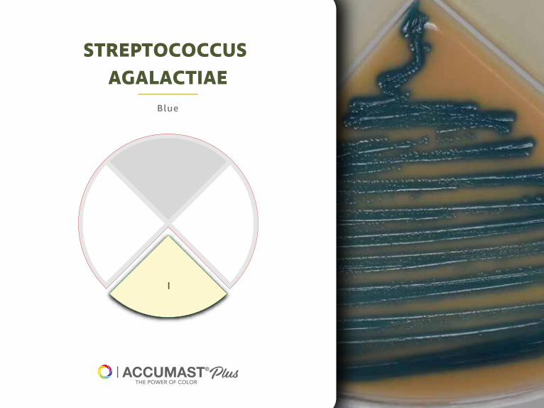
DISCOVER OUROTHER PRODUCTS
Ent ire herd
management for ident ificat ion
of Gram-pos it ive pathogens
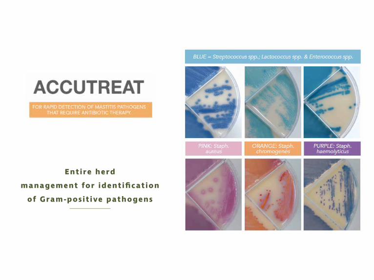
Ent ire herd
control management for
Streptococcus agalact iae

Ent ire herd
control management for
Staphylococcus aureus
YELLOW: Staphylococcuswarneri
BLUE: Staphylococcusequorum
WHITE: Staphylococcuschromogenes
GREEN: Staphylococcushaemolyticus

RESOURCES
Cebra C.K., Garry F., Dinsmore R. Naturally occurring acute coliform mastitis in Holstein
cattle J. Vet. Intern. Med., 10 (1996), pp. 252-257
Erskine RJ. Antibacterial therapy of clinical mastitis - part I. Drug selection. Part II Adminis-
tration. North Am Vet Conf, Proc. 2003. pp. 13–16.
Ferreira JC, Gomes MS, Bonsaglia ECR, Canisso IF, Garrett EF, Stewart JL, et al. (2018) Compa-
rative analysis of four commercial on-farm culture methods to identify bacteria associated
with clinical mastitis in dairy cattle. PLoS ONE 13(3): e0194211. https://doi.org/10.1371/jour-
nal. pone.0194211
Fuenzalida MJ, Ruegg PL. Negatively controlled, randomized clinical trial to evaluate intra-
mammary treatment of nonsevere, gram-negative clinical mastitis. J Dairy Sci.
2019;102(6):5438-5457. doi:10.3168/jds.2018-16156

Ganda EK, Bisinotto RS, Decter DH, Bicalho RC. Evaluation of an On-Farm Culture System
(Accumast) for Fast Identification of Milk Pathogens Associated with Clinical Mastitis in
Dairy Cows. PLoS One. 2016;11(5): e0155314. Published 2016 May 13. doi: 10.1371/journal.po-
ne.0155314
Ganda, E. K. et al. Longitudinal metagenomic profiling of bovine milk to assess the impact
of intramammary treatment using a third-generation cephalosporin. Sci. Rep. 6, 37565; doi:
10.1038/ srep37565 (2016).
Gröhn Y.T., Wilson D.J., González R.N., Hertl J.A., Bennett G., Schukken Y.H. E�ect of patho-
gen-specific clinical mastitis on milk yield in dairy cows. J. Dairy Sci. 2004; 87 (15377615):
3358-3374. 10.3168/jds. S0022-0302(04)73472-4

Lago A., Godden S.M., Bey R., Ruegg P.L., Leslie K. The selective treatment of clinical mastitis
based on on-farm culture results: I. E�ects on antibiotic use, milk withholding time, and
short-term clinical and bacteriological outcomes. J. Dairy Sci. 2011; 94 (21854917): 4441-
-445610.3168/jds.2010-4046
Lago A., Godden S.M., Bey R., Ruegg P.L., Leslie K. The selective treatment of clinical mastitis
based on on-farm culture results: II. E�ects on lactation performance, including clinical mas-
titis recurrence, somatic cell count, milk production, and cow survival. J. Dairy Sci. 2011; 94
(21854918): 4457-4467. 10.3168/jds.2010-4047
Lago A, Godden SM. Use of Rapid Culture Systems to Guide Clinical Mastitis Treatment Deci-
sions. Vet Clin North Am Food Anim Pract. 2018;34(3):389-412. doi: 10.1016/j.cvfa.2018.06.001

Lima SF, Bicalho MLS, Bicalho RC (2018) Evaluation of milk sample fractions for
characterization of milk microbiota from healthy and clinical mastitis cows. PLoS ONE 13(3):
e0193671.https://doi.org/10.1371/journal.pone.0193671
Oikonomou G., Bicalho M.L., Meira E., Rossi R.E., Foditsch C., Machado V.S. Teixeira A.G.,
Santisteban C., Schukken Y.H., Bicalho R.C. Microbiota of cow's milk; distinguishing healthy,
sub-clinically and clinically diseased quarters. PLoS One. 2014; 9 (24465777): e85904.
10.1371/journal.pone.0085904 Roberson J.R.
Oliver SP, Almeida RA, Gillespie BE. et al. Extended ceftiofur therapy for treatment of
experimentally-induced Streptococcus uberis mastitis in lactating dairy cattle. J Dairy Sci.
2004; 87:3322–3329. doi: 10.3168/jds. S0022-0302(04)73468-2.
Pyörälä S, Pyörälä E. E�cacy of parenteral administration of three antimicrobial agents in
treatment of clinical mastitis in lactating cows: 487 cases: (1989-1995) J Am Vet Med Assoc.
1998;212:407–412.

Rodrigues M.X., Lima S.F., Higgins C.H., Canniatti-Brazaca S.G., Bicalho R.C. The Lactococcus
genus as a potential emerging mastitis pathogen group: a report on an outbreak investiga-
tion. J. Dairy Sci., 99 (2016), pp. 9864-9874, 10.3168/jds.2016-11143
Tomazi T, Freu G, Alves BG, de Souza Filho AF, Heinemann MB, Veiga dos Santos M (2019)
Genotyping and antimicrobial resistance of Streptococcus uberis isolated from bovine clini-
cal mastitis. PLoS ONE 14(10): e0223719.
Vasquez A.K., Nydam D.V., Capel M.B., Eicker S., Virkler P.D. Clinical outcome comparison of
immediate blanket treatment versus a delayed pathogen-based treatment protocol for clini-
cal mastitis in a New York dairy herd. J. Dairy Sci. 2017; 100 (28161180): 2992-3003. 10.3168/-
jds.2016-11614
Vasquez A.K., Ganda E.K., Capel M.B., Virkler P.D., Bicalho R.C., Nydam D.V. The microbiome
of Escherichia coli and culture-negative non-severe clinical mastitis: Characterization and
associations with linear score and milk production. J. Dairy Sci. 2018; 102:578-594

Warnick L.D., Moore G. Mild to moderate clinical mastitis: E�cacy of intramammary amoxi-
cillin, frequent milk-out, a combined intramammary amoxicillin, and frequent milk-out
treatment versus no treatment. J. Dairy Sci. 2004; 87 (15202642): 583-592
Wenz J.R., Barrington G., Garry F., McSweeney K., Dinsmore R., Goodell G., Callan R. Bactere-
mia associated with naturally occurring acute coliform mastitis in dairy cows. J. Am. Vet.
Med. Assoc., 219 (2001), pp. 976-981


![FERA Final[1]](https://static.fdocuments.in/doc/165x107/577d22871a28ab4e1e979ccd/fera-final1.jpg)


![FERA Final[1] (1)](https://static.fdocuments.in/doc/165x107/577cde941a28ab9e78af690b/fera-final1-1.jpg)