FePt nanoparticle adsorption on a chemically patterned silicon–gold substrate
6
FePt nanoparticle adsorption on a chemically patterned silicon–gold substrate Nisha Shukla ⁎ , Erik B. Svedberg, John Ell Seagate Research, Pittsburgh, Pennsylvania 15222, United States Received 16 September 2005; accepted in revised form 21 January 2006 Available online 9 March 2006 Abstract Chemically modified patterns were used to achieve selective adsorption of magnetic FePt nanoparticles on substrates. In this work, thiol chemistry was used to create chemical patterns on silicon surfaces and gold patterned silicon surfaces. The adsorption of FePt nanoparticles on the silicon and gold patterned surface was studied using FTIR and HRSEM (high-resolution scanning electron microscopy). We have demonstrated in this work that chemical patterning can be used to achieve selective adsorption of magnetic nanoparticles on surfaces. Our results indicate that FePt nanoparticles coated with a 2 nm thick surfactant layer will adsorb when spin-coated onto gold surfaces modified with 11-mercapto-1-undecanol {HS(CH 2 ) 11 OH, MDOL}. The adsorption of nanoparticles was confirmed by FTIR spectra and HRSEM. FTIR spectra revealed the presence of peaks at 3004 cm - 1 and 1709 cm - 1 on MDOL modified gold surfaces, which are indicative of the presence of the FePt nanoparticles on the surface. Adsorption of FePt nanoparticles was not observed on 1-octadecanethiol {CH 3 (CH 2 ) 17 SH, ODT} modified gold surfaces. © 2006 Elsevier B.V. All rights reserved. Keywords: FePt nanoparticle; Chemical pattering; FTIR 1. Introduction The high magnetic anisotropy of FePt nanoparticles makes them a potential candidate for use as future magnetic recording media with data recording densities of 1 Tbit/in. 2 and beyond. One of the key challenges in the development of high-density recording media is the need to control the monodispersity and the size of the magnetic grains in the media. Development of high-density magnetic recording media will require the use of monodisperse magnetic grains with diameters less than 10 nm. FePt nanoparticles deposited on substrates can serve as magnetic recording media in which each particle is a single grain of the media. Chemically synthesized FePt nanoparticles have advantages over conventional sputtered FePt films and current magnetic media because the use of FePt nanoparticles allows control of the size and the monodispersity of the magnetic grains. One of the impediments to the development of this technology is the fact that it is very difficult to self-assemble the FePt nanoparticles into monolayer films that are uniform over a length scale of inches. Several studies have reported [1,2] that one can achieve self-assembly of nanoparticles using polymer-mediated or Langmuir Blodgett techniques. Recently, Kodama et al. [3] described a technique for deposition of FePt nanoparticles on disk substrates that were then used as recording media. Their method was based on spin-coating but with the disk immersed in the vapor of the solvent during spin- coating. They were able to achieve local, short-range self- assembly of the nanoparticles on the substrate surface. The short range self-assembly introduces domain boundaries, which cause jitter noise in the magnetic media. Dyker et al. [4] have also suggested formation of extended monolayers of FePt nanoparticles on silicon wafers. However, Dyker's work does not show formation of well-defined edges. Viable media based on the use of FePt nanoparticles requires the formation of bits with well-defined edges. Production of magnetic bits with sharp edges requires patterning of the grains on the substrate. Patterns can be produced by lithographic patterning, nano-imprinting, chemical patterning, etc. The problem with lithographic etching of a pattern into a substrate is that nanoparticles assemble on both the etched and non-etched portions of the surface. Chemical Surface & Coatings Technology 201 (2006) 1256 – 1261 www.elsevier.com/locate/surfcoat ⁎ Corresponding author. E-mail address: [email protected] (N. Shukla). 0257-8972/$ - see front matter © 2006 Elsevier B.V. All rights reserved. doi:10.1016/j.surfcoat.2006.01.043
-
Upload
nisha-shukla -
Category
Documents
-
view
214 -
download
2
Transcript of FePt nanoparticle adsorption on a chemically patterned silicon–gold substrate

201 (2006) 1256–1261www.elsevier.com/locate/surfcoat
Surface & Coatings Technology
FePt nanoparticle adsorption on a chemicallypatterned silicon–gold substrate
Nisha Shukla ⁎, Erik B. Svedberg, John Ell
Seagate Research, Pittsburgh, Pennsylvania 15222, United States
Received 16 September 2005; accepted in revised form 21 January 2006Available online 9 March 2006
Abstract
Chemically modified patterns were used to achieve selective adsorption of magnetic FePt nanoparticles on substrates. In this work, thiolchemistry was used to create chemical patterns on silicon surfaces and gold patterned silicon surfaces. The adsorption of FePt nanoparticles on thesilicon and gold patterned surface was studied using FTIR and HRSEM (high-resolution scanning electron microscopy). We have demonstrated inthis work that chemical patterning can be used to achieve selective adsorption of magnetic nanoparticles on surfaces. Our results indicate that FePtnanoparticles coated with a 2 nm thick surfactant layer will adsorb when spin-coated onto gold surfaces modified with 11-mercapto-1-undecanol{HS(CH2)11OH, MDOL}. The adsorption of nanoparticles was confirmed by FTIR spectra and HRSEM. FTIR spectra revealed the presence ofpeaks at 3004 cm−1 and 1709 cm−1 on MDOL modified gold surfaces, which are indicative of the presence of the FePt nanoparticles on thesurface. Adsorption of FePt nanoparticles was not observed on 1-octadecanethiol {CH3(CH2)17SH, ODT} modified gold surfaces.© 2006 Elsevier B.V. All rights reserved.
Keywords: FePt nanoparticle; Chemical pattering; FTIR
1. Introduction
The high magnetic anisotropy of FePt nanoparticles makesthem a potential candidate for use as future magnetic recordingmedia with data recording densities of 1 Tbit/in.2 and beyond.One of the key challenges in the development of high-densityrecording media is the need to control the monodispersity andthe size of the magnetic grains in the media. Development ofhigh-density magnetic recording media will require the use ofmonodisperse magnetic grains with diameters less than 10 nm.FePt nanoparticles deposited on substrates can serve asmagnetic recording media in which each particle is a singlegrain of the media. Chemically synthesized FePt nanoparticleshave advantages over conventional sputtered FePt films andcurrent magnetic media because the use of FePt nanoparticlesallows control of the size and the monodispersity of themagnetic grains. One of the impediments to the development ofthis technology is the fact that it is very difficult to self-assemblethe FePt nanoparticles into monolayer films that are uniform
⁎ Corresponding author.E-mail address: [email protected] (N. Shukla).
0257-8972/$ - see front matter © 2006 Elsevier B.V. All rights reserved.doi:10.1016/j.surfcoat.2006.01.043
over a length scale of inches. Several studies have reported [1,2]that one can achieve self-assembly of nanoparticles usingpolymer-mediated or Langmuir Blodgett techniques. Recently,Kodama et al. [3] described a technique for deposition of FePtnanoparticles on disk substrates that were then used asrecording media. Their method was based on spin-coating butwith the disk immersed in the vapor of the solvent during spin-coating. They were able to achieve local, short-range self-assembly of the nanoparticles on the substrate surface. The shortrange self-assembly introduces domain boundaries, whichcause jitter noise in the magnetic media. Dyker et al. [4] havealso suggested formation of extended monolayers of FePtnanoparticles on silicon wafers. However, Dyker's work doesnot show formation of well-defined edges. Viable media basedon the use of FePt nanoparticles requires the formation of bitswith well-defined edges.
Production of magnetic bits with sharp edges requirespatterning of the grains on the substrate. Patterns can beproduced by lithographic patterning, nano-imprinting, chemicalpatterning, etc. The problem with lithographic etching of apattern into a substrate is that nanoparticles assemble on boththe etched and non-etched portions of the surface. Chemical

1257N. Shukla et al. / Surface & Coatings Technology 201 (2006) 1256–1261
patterning generates alternating regions of hydrophobic andhydrophilic surfaces on the substrate. These regions willselectively adsorb nanoparticles to render a patterned overlayer.In this work, we have demonstrated that chemical patterns canbe used for selective deposition of nanoparticles ontopredetermined areas of a substrate by tailoring the chemicalproperties of those regions to match those of the nanoparticles.Chemical patterns on silicon substrates patterned with goldwere created using thiol chemistry. There are many studies,which have demonstrated chemical patterning of surfaces usingthiol chemistry [5–7]; however, none have reported adsorptionof nanoparticles on these chemical patterns.
FePt nanoparticles can be prepared using a synthesisdescribed by Sun et al. [8], which involves thermal decompo-sition of Fe(CO)5 and reduction of Pt(acac)2 in the presence of aequimolar mixture of oleic acid (cis-9-octadecenoic acid) andoleylamine (cis-9-octadecenoic amine) surfactants. The finalproduct is a solution of nanoparticles formed of an FePt coresurrounded by a layer of surfactant. These particles aresuspended in a solution, which contains excess surfactant.The surfactant coating on the nanoparticles controls theirstability in solution and the dynamics of their self-assembly onsurfaces.
One of the important challenges in the synthesis of FePtnanoparticles is the preparation of highly monodispersenanoparticles. Once monodispersity has been achieved, thenanoparticles must be self-assembled uniformly on a surface inorder to make viable media. The uniformity of self-assemblednanoparticle films is influenced by the surfactant coating on thenanoparticles and the concentration of excess surfactant in thesolution [9]. The self-assembly of the nanoparticles is alsoinfluenced by the type of surfactant and the surfactant chainlength.
In this work, we have used thiol chemistry to create chemicalpatterns on surfaces and demonstrated that these chemicalpatterns can be utilized to selectively adsorb FePt nanoparticlescoated with organic surfactant. The selectivity of FePtnanoparticle adsorption depends on the nature of the thiolsused. We have investigated several different thiols in this workand selected the most hydrophobic (1-octadecanethiol, ODT)and the most hydrophilic (11-mercapto-1-undecanol, MDOL)for patterning of substrates. These were deposited separately onpatterned substrates exposing regions of gold and silicon andthen coated with FePt nanoparticles deposited by spin-coating.This technique of coating FePt nanoparticles on surfaceschemically modified with ODT resulted in selective adsorptionof the FePt nanoparticles on specifically modified regions of thesurface.
2. Experimental
FePt nanoparticles were prepared using the synthesisdescribed by Sun et al. [8]. A typical synthesis involvedthermal decomposition of iron pentacarbonyl, Fe(CO)5, andreduction of platinum acetylacetonate, Pt(acac)2, in the presenceof oleic acid and oleylamine surfactants dissolved in diocty-lether. The synthesis was conducted under an inert atmosphere.
Following synthesis, the nanoparticles were subjected towashing cycles to remove excess surfactant from the synthesissolution. The ‘excess surfactant’ is the fraction of surfactant insolution not bonded to the FePt nanoparticles. The originalmixture of surfactant-coated nanoparticles and excess surfactantin dioctylether solvent was first mixed with 10 parts of ethanolcausing the larger nanoparticles to precipitate. The mixture wascentrifuged to separate the nanoparticles from the solution. Thesupernatant solvent consisting of dioctylether and smallernanoparticles was then discarded and the nanoparticles wereredispersed in non-polar hexane. This sequence of precipitationin ethanol, centrifuging to separate the solvent and redispersionin hexane was then repeated three times to remove excesssurfactant and small particles. Finally, the nanoparticles weredispersed in hexane. This procedure was sufficient to produce adispersion of 3–4 nm diameter nanoparticles suspended inhexane with no surfactant in the solution.
Si(111) wafers were used as substrates for deposition of goldpatterns. The substrates were obtained from Si-Tech Corp. andwere coated with a native oxide film. The Si substrates wereused as obtained without further chemical etching. A hexagonalcopper TEM grid (SPI Supplies Co.) was used as a mask duringsputter deposition of 100 Å thick Ta and 1000 Å thick gold filmsonto the Si surface. This mask generated hexagonally shapedregions of gold separated by regions of bare Si.
The formation of SAMS from thiols with phenyl groups suchas 4-mercaptophenol, 4-methylbenzenethiol and 4-aminothio-phenol (MPOL, MBT and ATPOL) was performed using aprocedure described by Johnson et al. [10]. SAMS of thiols withalkyl groups such as 1-dodecanethiol, 1-hexadecanethiol and 1-octadecanethiol (DDT, HDT and ODT), hydroxyl group (11-mercapto-1-decanol, MDOL), acidic groups such as 11-mercaptoundecanoic acid and 16-mercaptohexadecanoic acid(MUDAc, MHDAc) were performed using the proceduredescribed by Malinsky et al. [11]. The thiols terminated withphenyl groups (MPOL, MBT and ATPOL) were dissolved inethanol to a concentration of 0.095 M. The thiols terminatedwith alkyl groups (DDT, HDT and ODT), carboxylic groups(MUDAc and MHDAc) or hydroxyl groups (MDOL) weredissolved in methanol to a concentration of 0.01 M. Self-assembled monolayers of thiols on gold and silicon surfaceswere prepared under a nitrogen atmosphere in a glove box. Goldsubstrates and silicon substrates were immersed in thiol solutionfor 12–24 h. After immersion, excess thiol was washed from thesurface in a stream of methanol or ethanol and the surfaces werethen dried with nitrogen.
The FePt nanoparticles were deposited on the gold patternedsilicon surfaces or modified gold patterned silicon surfaces byspin-coating. A colloidal solution of FePt nanoparticles in a50:50 mixture of octane and hexane was spin-coated on gold ormodified gold patterned silicon surfaces.
The nanoparticle films self-assembled on thiol coatedsubstrates were characterized using FTIR and contact anglemeasurements. All FTIR spectra were taken using a NicoletNexus 670 spectrometer with a deuterated triglyceride sulphate(DTGS) detector. Spectra were obtained using 4 cm− 1
resolution and 32 scans were taken for statistical averaging. In

1000 1250 1500 1750 2750 3000 3250 3500
3350
3425
2920
2850
1058
Inte
nsi
ty (
a.u
.)
Wavenumber (cm-1)
Fig. 2. FTIR spectrum of MDOL (11-mercapto-1-undecanol) coated on goldsurface in 800–1800 cm−1 and 2700–3550 cm−1 region.
1258 N. Shukla et al. / Surface & Coatings Technology 201 (2006) 1256–1261
order to obtain FTIR spectra of films on silicon or gold coatedsilicon surfaces, a Smart Apertured Grazing Angle (SAGA)accessory for FTIR was used. The accessory samples at anaverage angle of 80° from the normal. Analysis of films as thinas 10 Å is possible with this accessory. A permanently mountedpolarizer minimizes ‘S’ polarized light and increases sensitivity.The films were analyzed by placing them on a horizontalsampling surface and they are masked using a unique aperturesliding assembly.
The contact angle measurements were obtained using aVCA-Optima system, which is equipped with a precise waterdrop dispenser. The contact angles of the self-assembledmonolayers with water were measured using a VCA OptimaSystem. A 10 μl of water drop was placed precisely on the thiolcoated surface using a precision dispenser and the advancingcontact angle was measured immediately. The contact angle wasmeasured at four different places on the sample and an averagecontact angle was reported with its standard deviation.
The self-assembly of FePt nanoparticles was analyzed usinghigh-resolution scanning microscope (HRSEM). The scanningelectron beam microscope (Leo 982 Gemini) is a high-resolution and low-acceleration voltage SEM equipped withthe GEMINI field emission column. The SEM can be used withacceleration voltages from 0.2 kV to 30 kV. The GEMINIcolumn provides typical resolutions of 4 nm at 1 kVand 1 nm at30 kV.
The oleic acid, oleylamine, ethanol and hexane (anhydrous)were bought from Aldrich Chemical Co. and were used withoutfurther purification. The ethanol was obtained in Sure Sealbottles with very low water content (<0.002%). Pt(acac)2, Fe(CO)5, 1,2-hexadecanediol, 11-mercapto-1-undecanol {MDOL,HS(CH2)11OH}, 16-mercaptohexadecanoic acid {MHDAc, HS(CH2)15CO2H}, 11-mercaptoundecanoic acid {MUDAc, HS(CH2)10CO2H}, 1-dodecanethiol {DDT, CH3(CH2)11SH}, 1-hexadecanethiol {HDT, CH3(CH2)15SH}, 1-octadecanethiol
30
35
40
45
50
55
60
80
90
100
110
120
MHDAc
MDOLODT
HDTDDT
MUDAcMBT
MPOL
ATPOL
An
gle
(O)
Fig. 1. Contact angle measurements of nine different types of thiols coated ongold surface.
{ODT, CH3(CH2)17SH}, 4-methylbenzenethiol {MBT, CH3C6-
H4SH}, 4-mercaptophenol {MPOL, HSC6H4OH} and 4-aminothiophenol {ATPOL, NH2C6H4SH} were bought fromAldrich Chemical Co. and used as obtained.
3. Results and discussion
Before preparing patterned surfaces, a set of thiols wereexamined to identify those that produce the most hydrophobicand most hydrophilic surfaces. The thiols used were 11-mercapto-1-undecanol (MDOL), 16-mercaptohexadecanoicacid (MHDAc), 11-mercaptoundecanoic acid (MUDAc), 1-dodecanethiol (DDT), 1-hexadecanethiol (HDT), 1-octadeca-nethiol (ODT), 4-methylbenzenethiol (MBT), 4-mercaptophe-nol (MPOL) and 4-aminothiophenol (ATPOL). The intent wasto find the two that would give the greatest difference inselectivity for adsorption of FePt nanoparticles. Fig. 1 shows
Table 1Infrared vibrational assignments
Vibrational modes Frequency (cm−1) References
ν(NH) 3300 [23]trans ν(–CHf) 3066 [16]trans ν(–CHf) 3035 [16]trans ν(–CHf) 3020 [16]cis ν(–CHf) 3006 [16,18,19]νa(–CH2) 2922 [16,18,19]νs(–CH2) 2854 [16,18,19]ν(OH) adjacent to –CfO 2670 [20,18]ν(–CfO) 1709 [15,16,20,18]ν(–CfC) 1647 [20,18]ν(NH2) 1593 [23]ν(–COO) 1512 [21,20,18,22]trans ν(–CHf) 970 [16,19]

1000 1250 1500 1750 2750 3000 3250 3500
3004
1570
1710
1630
Inte
nsi
ty (
a.u
.)
Wavenumber (cm-1)
Fig. 3. FTIR spectrum of FePt nanoparticles spin-coated on MDOL (11-mercapto-1-undecanol) coated on gold surface in 800–1800 cm−1 and 2700–3550 cm−1 region.
1000 1250 1500 1750 2750 3000 3250 3500
2918
2852
2878
1215
Inte
nsi
ty (
a.u
.)
Wavenumber (cm-1)
Fig. 5. FTIR spectrum of ODT (1-octadecanethiol) coated on gold surface in800–1800 cm−1 and 2700–3550 cm−1 region.
2880
1415
1530
2850
2919
2962
1259N. Shukla et al. / Surface & Coatings Technology 201 (2006) 1256–1261
the contact angle measurements obtained using the ninedifferent thiols. The contact angle of water with the monolayerof 1-octadecanethiol (ODT) is 112° suggesting that it forms avery hydrophobic surface. 11-Mercaptoundecanoic acid(MHDAc) exhibited the lowest contact angle (37°) indicatingthat it forms the most hydrophilic surface. Although MHDAchas the lowest contact angle with water, we chose to use 11-mercapto-1-undecanol (MDOL) with a contact angle of 52° asour hydrophilic surface, since it was very difficult to obtain asmooth coating with (MHDAc). Thus, ODT and MDOL wereidentified for use as hydrophobic and hydrophilic thiols forpatterning surfaces.
The self-assembled thiol films were first prepared on goldand silicon surfaces separately. These films were characterizedusing FTIR as described in Section 2. Fig. 2 shows the FTIR
Fig. 4. High resolution SEM image of FePt nanoparticles spin-coated on MDOLcoated gold surface.
spectrum of MDOL on gold in the regions 2700–3550 cm−1
and 800–1800 cm−1. The peak at 2850 cm−1 is assigned to theνs(CH2) mode and the peak at 2920 cm−1 is due to the νas(CH2) mode [12] (Table 1). The vibrational peaks character-istic of the ν(SH) stretch in free thiol were not observed in theregion of 2560–2600 cm−1. This is consistent with studies byPham et al. [12] and Bryant et al. [13], which reported theabsence of the ν(S–H) mode in thiols chemisorbed on goldsurfaces. The broad peak in the region of 3350–3425 cm−1 isdue to the ν(OH) stretch of MDOL.
1000 1250 1500 1750 2750 3000 3250 3500
Wavenumber (cm-1)
Inte
nsi
ty (
a.u
.)
Fig. 6. FTIR spectrum of FePt nanoparticles spin-coated on ODT (1-octadecanethiol) coated gold surface in 800–1800 cm−1 and 2700–3550 cm−1
region.
100 nm100 µm
Gold
Silicon
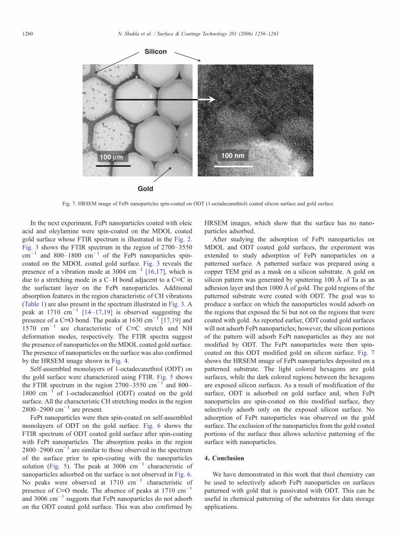
Fig. 7. HRSEM image of FePt nanoparticles spin-coated on ODT (1-octadecanethiol) coated silicon surface and gold surface.
1260 N. Shukla et al. / Surface & Coatings Technology 201 (2006) 1256–1261
In the next experiment, FePt nanoparticles coated with oleicacid and oleylamine were spin-coated on the MDOL coatedgold surface whose FTIR spectrum is illustrated in the Fig. 2.Fig. 3 shows the FTIR spectrum in the region of 2700–3550cm−1 and 800–1800 cm−1 of the FePt nanoparticles spin-coated on the MDOL coated gold surface. Fig. 3 reveals thepresence of a vibration mode at 3004 cm−1 [16,17], which isdue to a stretching mode in a C–H bond adjacent to a CfC inthe surfactant layer on the FePt nanoparticles. Additionalabsorption features in the region characteristic of CH vibrations(Table 1) are also present in the spectrum illustrated in Fig. 3. Apeak at 1710 cm−1 [14–17,19] is observed suggesting thepresence of a CfO bond. The peaks at 1630 cm−1 [17,19] and1570 cm− 1 are characteristic of CfC stretch and NHdeformation modes, respectively. The FTIR spectra suggestthe presence of nanoparticles on the MDOL coated gold surface.The presence of nanoparticles on the surface was also confirmedby the HRSEM image shown in Fig. 4.
Self-assembled monolayers of 1-octadecanethiol (ODT) onthe gold surface were characterized using FTIR. Fig. 5 showsthe FTIR spectrum in the region 2700–3550 cm−1 and 800–1800 cm−1 of 1-octadecanethiol (ODT) coated on the goldsurface. All the characteristic CH stretching modes in the region2800–2900 cm−1 are present.
FePt nanoparticles were then spin-coated on self-assembledmonolayers of ODT on the gold surface. Fig. 6 shows theFTIR spectrum of ODT coated gold surface after spin-coatingwith FePt nanoparticles. The absorption peaks in the region2800–2900 cm−1 are similar to those observed in the spectrumof the surface prior to spin-coating with the nanoparticlessolution (Fig. 5). The peak at 3006 cm−1 characteristic ofnanoparticles adsorbed on the surface is not observed in Fig. 6.No peaks were observed at 1710 cm−1 characteristic ofpresence of CfO mode. The absence of peaks at 1710 cm−1
and 3006 cm−1 suggests that FePt nanoparticles do not adsorbon the ODT coated gold surface. This was also confirmed by
HRSEM images, which show that the surface has no nano-particles adsorbed.
After studying the adsorption of FePt nanoparticles onMDOL and ODT coated gold surfaces, the experiment wasextended to study adsorption of FePt nanoparticles on apatterned surface. A patterned surface was prepared using acopper TEM grid as a mask on a silicon substrate. A gold onsilicon pattern was generated by sputtering 100 Å of Ta as anadhesion layer and then 1000 Å of gold. The gold regions of thepatterned substrate were coated with ODT. The goal was toproduce a surface on which the nanoparticles would adsorb onthe regions that exposed the Si but not on the regions that werecoated with gold. As reported earlier, ODT coated gold surfaceswill not adsorb FePt nanoparticles; however, the silicon portionsof the pattern will adsorb FePt nanoparticles as they are notmodified by ODT. The FePt nanoparticles were then spin-coated on this ODT modified gold on silicon surface. Fig. 7shows the HRSEM image of FePt nanoparticles deposited on apatterned substrate. The light colored hexagons are goldsurfaces, while the dark colored regions between the hexagonsare exposed silicon surfaces. As a result of modification of thesurface, ODT is adsorbed on gold surface and, when FePtnanoparticles are spin-coated on this modified surface, theyselectively adsorb only on the exposed silicon surface. Noadsorption of FePt nanoparticles was observed on the goldsurface. The exclusion of the nanoparticles from the gold coatedportions of the surface thus allows selective patterning of thesurface with nanoparticles.
4. Conclusion
We have demonstrated in this work that thiol chemistry canbe used to selectively adsorb FePt nanoparticles on surfacespatterned with gold that is passivated with ODT. This can beuseful in chemical patterning of the substrates for data storageapplications.

1261N. Shukla et al. / Surface & Coatings Technology 201 (2006) 1256–1261
Acknowledgement
Authors would like to thank Chao Liu for providingnanoparticles.
References
[1] Q. Guo, X. Teng, S. Rahman, H. Yang, J. Am. Chem. Soc. 125 (2003) 630.[2] S. Sun, A. Ansers, H.F. Hamann, J.U. Thiele, J.E.E. Baglin, T. Thomson,
E.E. Fullerton, C.B. Murray, B.D. Terris, J. Am. Chem. Soc. 124 (2002)2884.
[3] H. Kodama, S. Momose, N. Ihara, T. Uzumaki, A. Tanaka, Appl. Phys.Lett. 83 (2003) 5253.
[4] M. Acet, C. Mayer, O. Muth, A. Terheiden, G. Dyker, J. Cryst. Growth 285(2005) 365.
[5] H.B. Pyo, Y.B. Shin, M.G. Kim, H.C. Yoon, Langmuir 21 (2005) 166.[6] S. Sun, K.S.L. Chong, G.J. Leggett, J. Am. Chem. Soc. 124 (2002) 2414.[7] A. Camposeo, A. Fioretti, F. Tantussi, S. Gozzini, E. Arimondo, C.
Gabbanini, Appl. Phys., B 79 (2004) 539.[8] S. Sun, C.B. Murray, D. Weller, Liesl Folks, A. Moser, Science 287 (2000)
1989.
[9] N. Shukla, J. Ahner, D. Weller, JMMM 272–276 (Supplement 1) (2004)e1349.
[10] S.R. Johnson, S.D. Evans, R. Brydson, Langmuir 14 (1998) 6639.[11] M.D. Malinsky, K.L. Kelly, G.C. Schatz, R.P. Van Duyne, J. Am. Chem.
Soc. 123 (2001) 1471.[12] T. Pham, J.B. Jackson, N.J. Halas, T.R. Lee, Langmuir 18 (2002) 4915.[13] M.A. Bryant, J.E. Pemberton, J. Am. Chem. Soc. 113 (1991) 8284.[14] N.J. Turro, P.H. Lakshminarasimhan, S. Jockusch, S.P. O'Brien, S.G.
Grancharov, F.X. Redl, Nano Lett. 2 (4) (2002) 325.[15] L.J. Kirwan, P.D. Fawell, W. van Bronswijk, Langmuir 19 (2003) 5802.[16] R.G. Sinclair, A.F. Mckay, G.S. Myers, R.N. Jones, J. Am. Chem. Soc. 74
(1952) 2578.[17] N. Shukla, C. Liu, P.M. Jones, D. Weller, JMMM 266 (2003) 178.[18] D.H. Lee, R.A. Condrate SR, W.C. Lacourse, J. Mater. Sci. 35 (2000)
4961.[19] C. Vogel-Weill, A. Gruger, Spectrochim. Acta, Part A 52 (1996) 1737.[20] D.H. Lee, R.A. Condrate SR., J. Mater. Sci. 34 (1999) 139.[21] Q. Liu, Z. Xu, Langmuir 11 (1995) 4617.[22] P.J. Thistlethwaite, M.S. Hook, Langmuir 16 (2000) 4993.[23] W. Erley, J.C. Hemminger, Surf. Sci. 316 (1994) (L1025-L1030).