Female Patellofemoral Biomechanical Functionpatellofemoral.org/pdf/FemalePatelloFemoral.pdfFemale...
31
Karen M. Sutton, M.D. Department of Orthopaedic Surgery Yale University Female Patellofemoral Biomechanical Function
Transcript of Female Patellofemoral Biomechanical Functionpatellofemoral.org/pdf/FemalePatelloFemoral.pdfFemale...
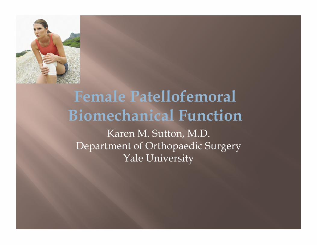
Karen M. Sutton, M.D. Department of Orthopaedic Surgery
Yale University
Female Patellofemoral Biomechanical Function
Patellofemoral Pain in Women
More common in young female athletes Most common orthopaedic concern among
active young women Symptoms include peripatellar pain during
repetitive knee flexion associated with weightbearing activities Running, jumping, and climbing stairs
Factors Influencing Patellofemoral Pain in Women
High pelvis width (distance between anterior superior iliac spines) to femoral length ratio
Imbalance of vastus medialis and vastus lateralis Tibiofemoral abduction angular impulse Studies suggest that individuals with malalignment
related anterior knee pain may be more likely to develop patellofemoral osteoarthritis later in life
Abnormally large “Q”uadriceps angle was initially thought to cause patellofemoral pain; however is currently debated
Factors Influencing Patellofemoral Pain in Women
Difference in trochlear groove orientation and tibia rotation
Hip and trunk muscle weakness may also increase retropatellar stress
Decreased hip abductor, hip external rotator, and trunk lateral flexor strength causes hip adduction or internal rotation during weightbearing Such rotations in cadaveric studies have been linked to
increased retropatellar stress Femoral internal rotation has been shown to be
associated with decreased patellofemoral joint contact area in individuals with malalignment related anterior knee pain
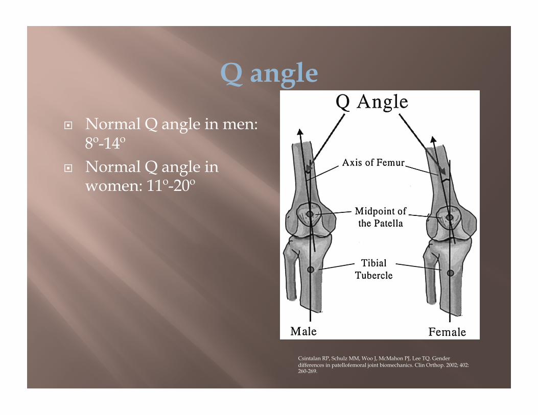
Q angle
Normal Q angle in men: 8º-14º
Normal Q angle in women: 11º-20º
Csintalan RP, Schulz MM, Woo J, McMahon PJ, Lee TQ. Gender differences in patellofemoral joint biomechanics. Clin Orthop. 2002; 402: 260-269.
Q angle
Huberti and Hayes: increases and decreases in the Q angle were associated with increased peak patellofemoral pressures and unpredictable patterns of cartilage unloading
A 10-degree increase in the Q-angle resulted in increased peak pressures (up to 45% at 20 degrees of flexion)
Q angle
Increased external rotation increases the Q angle of the knee Places tibial tubercle in a more lateral position Both men and women have increased pressures on
the lateral patellar facet with external rotation of the tibia Especially at lower flexion angles
Q Angle Debate
The Q angle was assumed to predispose an individual to patellofemoral pain due to increased lateral patellar position
Q angle was associated with a more medially and inferiorly located patella in a study conducted by Sheehan et al comparing dynamic MRI findings of symptomatic and asymptomatic subjects
The data supported a correlation between the Q-angle and medial, not lateral, patellar displacement
Quadriceps Atrophy and Weakness
Leads to imbalance and maltracking of the patella with resultant pain, instability or both
Line of pull of the quadriceps muscle parallels the shaft of the femur
Quadriceps muscle is in relative valgus with respect to the axis of the tibia Favors lateral deviation of the patella
Vastus medialis has an important role as a medial stabilizer of the patella Aids in normal tracking of the patella
Vastus Medialis
Typically the weakest of the quadriceps group First muscle to atrophy and last to rehabilitate Short arc quadriceps strengthening exercises,
emphasizing the vastus medialis are part of the initial treatment of patellofemoral disorders
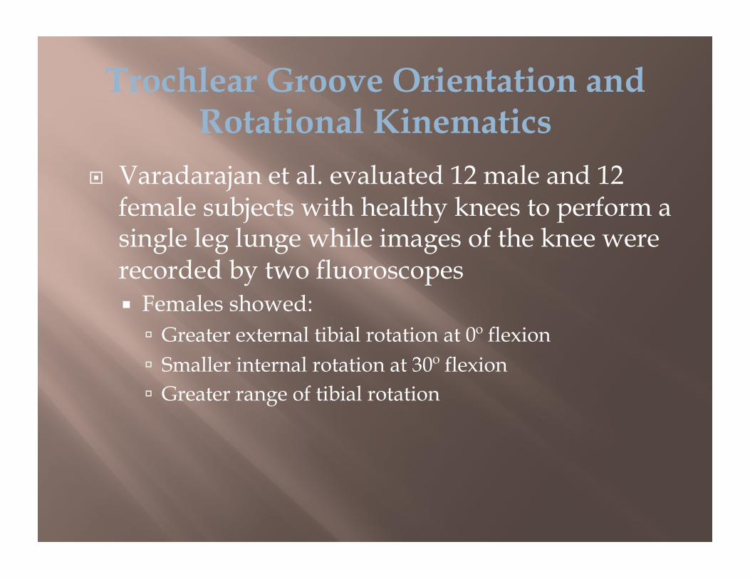
Trochlear Groove Orientation and Rotational Kinematics
Varadarajan et al. evaluated 12 male and 12 female subjects with healthy knees to perform a single leg lunge while images of the knee were recorded by two fluoroscopes Females showed:
Greater external tibial rotation at 0º flexion Smaller internal rotation at 30º flexion Greater range of tibial rotation
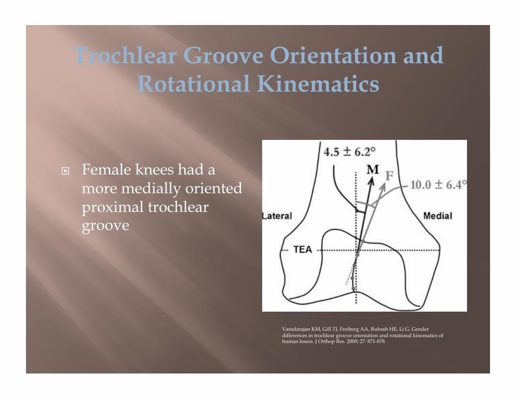
Trochlear Groove Orientation and Rotational Kinematics
Female knees had a more medially oriented proximal trochlear groove
Varadarajan KM, Gill TJ, Freiberg AA, Rubash HE, Li G. Gender differences in trochlear groove orientation and rotational kinematics of human knees. J Orthop Res. 2009; 27: 871-878.

Csintalan et al.: Evaluation of Gender Differences in Patellofemoral Joint Biomechanics Twelve fresh-frozen knees from cadavers were tested Anatomic multiplane loading of the extensor
mechanism was used with varying vastus medialis loads
Study measured patellofemoral contact area and pressure
Comparison was made between female and male specimens
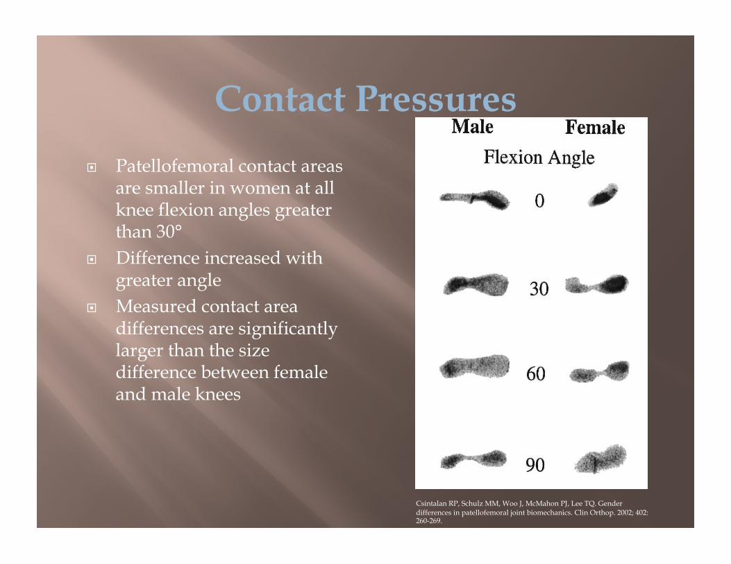
Contact Pressures Patellofemoral contact areas
are smaller in women at all knee flexion angles greater than 30°
Difference increased with greater angle
Measured contact area differences are significantly larger than the size difference between female and male knees
Csintalan RP, Schulz MM, Woo J, McMahon PJ, Lee TQ. Gender differences in patellofemoral joint biomechanics. Clin Orthop. 2002; 402: 260-269.
Effects of Tibial Rotation
Csintalan et al concluded that patellofemoral contact areas were larger in men in neutral, internal rotation and external rotation positions
No significant contact difference was seen within a gender with respect to tibial rotation
Csintalan Study
Csintalan noted a statistically significant difference in contact area when varying vastus medialis loads between 0% and 50%, and 50% and 150% at the 0 ° angle in both genders
Contact area increased with increasing vastus medialis load
No significant difference between genders when varying vastus medialis load
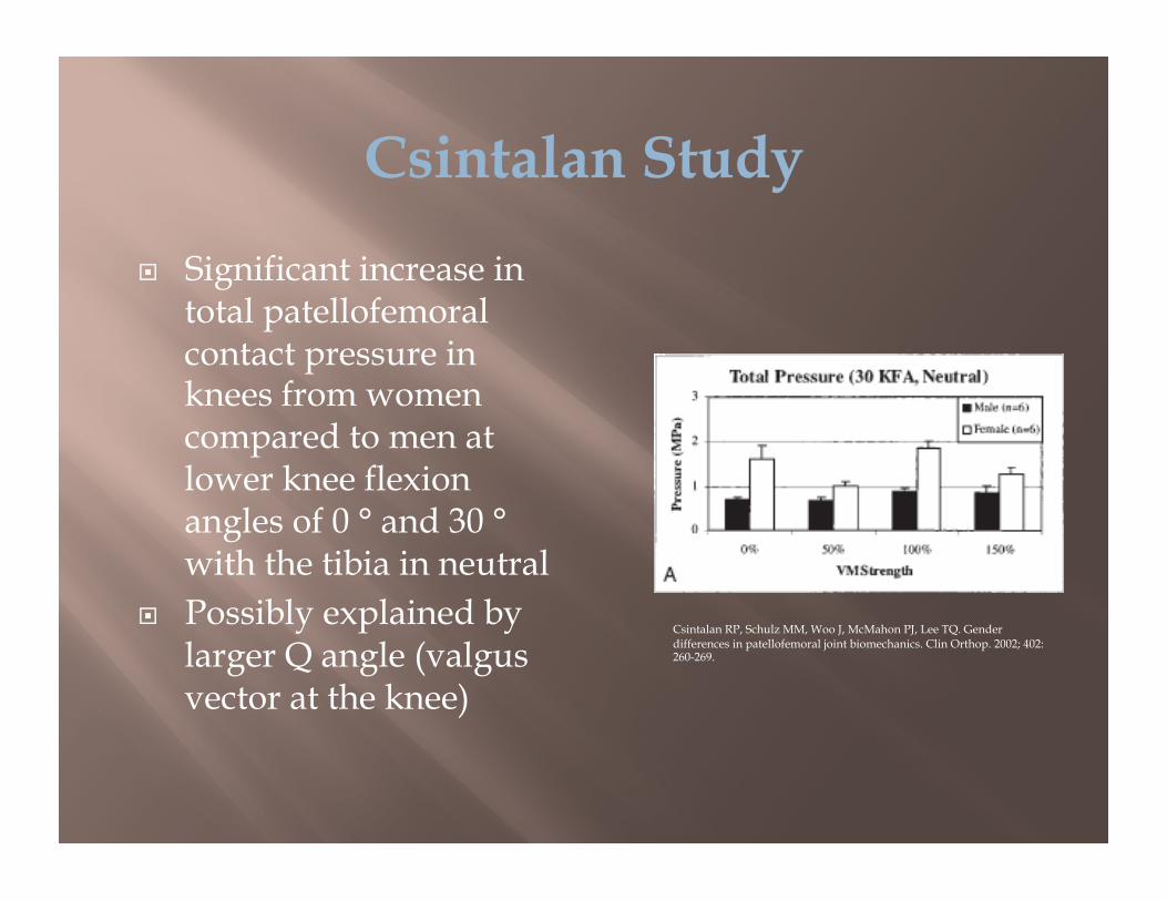
Csintalan Study
Significant increase in total patellofemoral contact pressure in knees from women compared to men at lower knee flexion angles of 0 ° and 30 ° with the tibia in neutral
Possibly explained by larger Q angle (valgus vector at the knee)
Csintalan RP, Schulz MM, Woo J, McMahon PJ, Lee TQ. Gender differences in patellofemoral joint biomechanics. Clin Orthop. 2002; 402: 260-269.
Csintalan Study
Women had significant increase in total pressures when compared to men at 30º knee flexion with the tibia in internal and external rotation
Both genders had higher pressures with external rotation of the tibia at lower knee flexion angles
Csintalan Study
Vastus medialis load Women showed a greater change in patellofemoral
contact pressures to varying vastus medialis loads from 0-50% and 50-150% Pressure changed inversely to vastus medialis load at
0º, 30º, and 60º
Peak Pressure Women had statistically significantly higher peak
values at 0º, 30º, and 60º of knee flexion with the tibial in neutral
Hip Muscle Function in Relation to Females with Malalignment Related Anterior Knee Pain
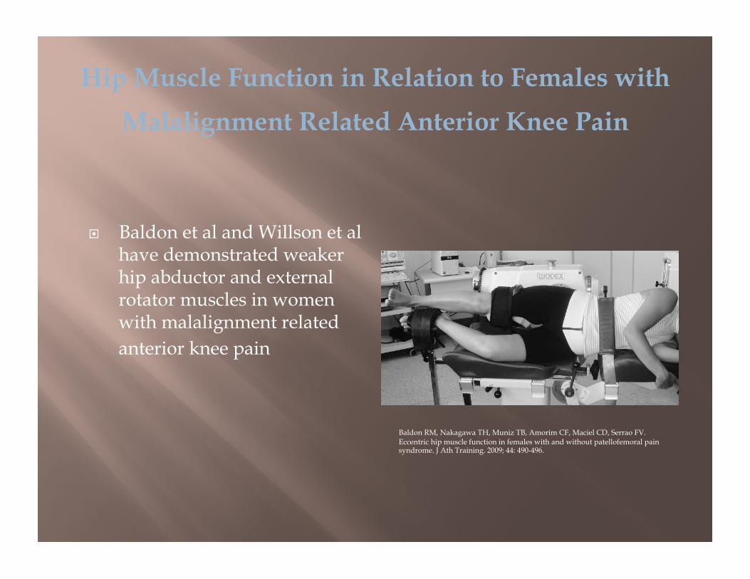
Baldon et al and Willson et al have demonstrated weaker hip abductor and external rotator muscles in women with malalignment related anterior knee pain
Baldon RM, Nakagawa TH, Muniz TB, Amorim CF, Maciel CD, Serrao FV. Eccentric hip muscle function in females with and without patellofemoral pain syndrome. J Ath Training. 2009; 44: 490-496.
Hip Muscle Function in Relation to Females with Malalignment Related Anterior Knee Pain
Baldon et al evaluated females with and without malalignment related anterior knee pain Females with malalignment related anterior knee pain
demonstrated: 28% lower eccentric hip abduction mean peak torque 15% lower eccentric hip adduction mean peak torque
No difference in the normalized eccentric hip external or internal rotation peak torque
Hip Muscle Function in Relation to Females with Malalignment Related Anterior Knee Pain
Malalignment related anterior knee pain has been related to poor hip adduction and internal rotation control during weight-bearing activities
Excessive femoral adduction and internal rotation may increase the dynamic quadriceps angle and lead to greater lateral patellar contact pressure
During weight-bearing, females with malalignment related anterior knee pain demonstrate internal rotation of the femur underneath the patella
Greater hip external rotation during walking, squatting, running and jumping has been reported to compensate for a larger Q angle
Jumping Mechanics of Female Athletes
Willson et al evaluated 20 women with patellofemoral pain and compared them to 20 healthy female controls
Women participated in an exertion protocol of repetitive single-legged jumps
Examined the effect of exertion on variables thought to increase retropatellar stress in women with and without patellofemoral pain during repetitive single-legged jumping
Jumping Mechanics of Female Athletes
Women in Willson’s jumping mechanics study with patellofemoral pain when compared to controls had: 24% less lateral trunk flexion strength 13% less hip abduction strength 14% less hip external rotation strength
Jumping Mechanics of Female Athletes
When performing single-leg jumps, women with patellofemoral pain when compared to controls demonstrated: Increased hip adduction Increased hip flexion Decreased hip internal rotation Greater hip abduction angular impulse
Risk Factors in Developing Patellofemoral Pain
The Joint Undertaking to Monitor and Prevent ACL Injury (JUMP-ACL) prospectively investigated risk factors for patellofemoral pain 1597 participants enrolled from the United States Naval
Academy Followed for 2.5 years
Risk Factors Decreased quadriceps and hamstring strength Increased hip external rotator strength Increased navicular drop
Difference in navicular tuberosity height in a nonweightbearing subtalar joint neutral position and a weightbearing position
During jump-landing task Decreased knee flexion angle Decreased vertical ground-reaction force Increased hip internal rotation angle
NSAIDS (however not significant inflammation involved)
Simple bracing with elastic patellar cut-out brace
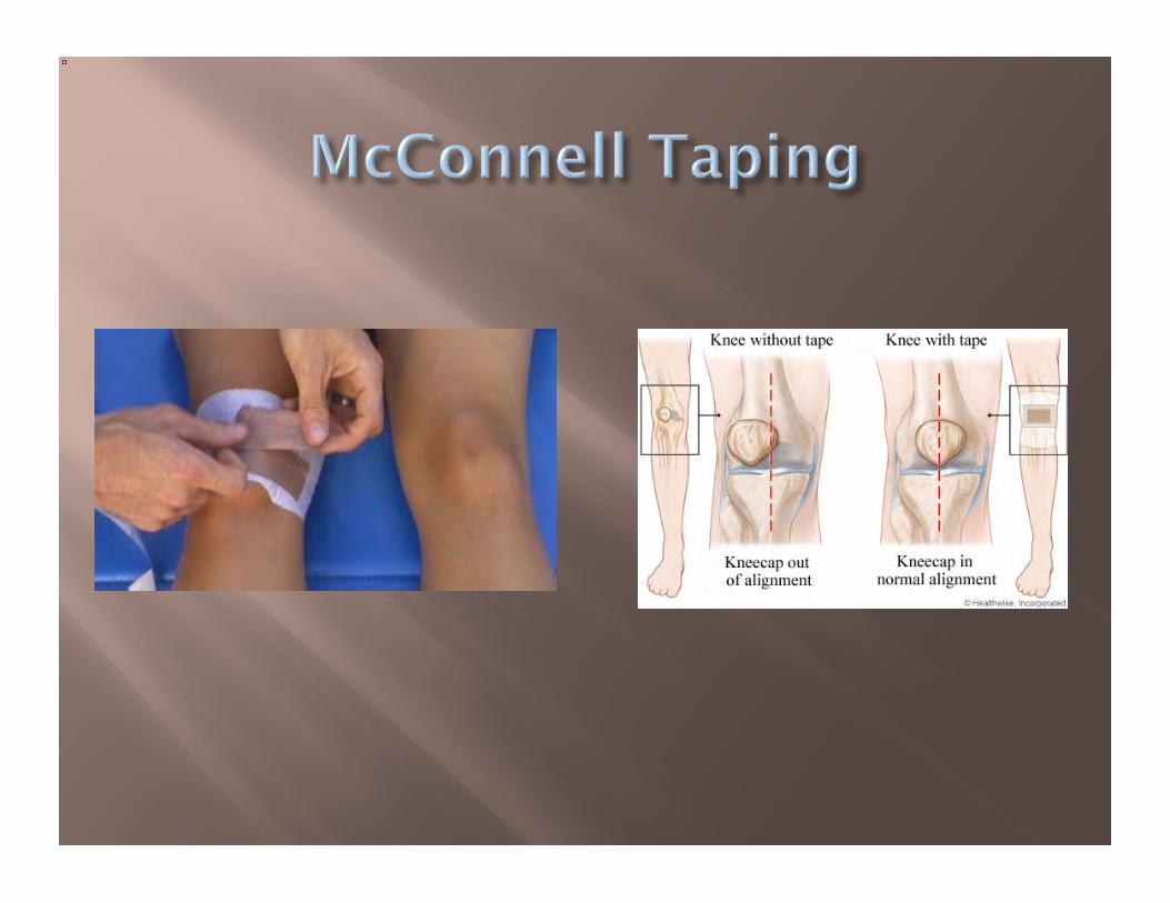
McConnell taping technique Stretching quadriceps mechanism Straight-leg exercises with weights on the ankle Exercises within a pain-free arc Vocational Rehabilitation Orthotics
Rehabilitation Recommendations Based on Willson’s Study
Kinematic retraining of landing from jumping Eccentric Hip Strengthening Trunk Strengthening
Recommendations of the Joint Undertaking to Monitor and Prevent ACL Injury (JUMP-ACL)
Regarding Patellofemoral Pain Strengthening of the quadriceps and hamstring
musculature Teaching proper technique for performing
dynamic tasks Decrease hip internal rotation angle Increase knee flexion
References
1. Baldon RM, Nakagawa TH, Muniz TB, Amorim CF, Maciel CD, Serrao FV. Eccentric hip muscle function in females with and without patellofemoral pain syndrome. J Ath Training. 2009; 44: 490-496.
2. Boling MC, Padua DA, Marshall SW, Guskiewicz K, Pyne S, Beutler A. A prospective investigation of biomechanical risk factors for patellofemoral pain syndrome: The joint undertaking to monitor and prevent ACL injury (JUMP-ACL) cohort. Am J Sports Med. 2009; 37: 2108-2116.
3. Conley S, Rosenberg A, Crowninshield R. The female knee: anatomic variations. J Am Acad Orthop Surg. 2007; 15: S31-S36.
4. Csintalan RP, Schulz MM, Woo J, McMahon PJ, Lee TQ. Gender differences in patellofemoral joint biomechanics. Clin Orthop. 2002; 402: 260-269.
5. Draper CE, Besier TF, Santos JM, Jennings F, Fredericson M, Gold GE, Beaupre GS, Delp SL. Using real-time MRI to quantify altered joint kinematics in subjects with patellofemoral pain and to evaluate the effects of a patellar brace or sleeve on joint motion. J Orthop Res. 2008: 571-577.
6. Escamilla RF, Zheng N, Macleod TD, Edwards WB, Imamura R, Hreljac A, Fleisig GS, Wilk KE, Moorman CT, Andrews JR. Patellofemoral joint force and stress during the wall squat and one-leg squat. Med Sci Sports Exerc. 2009: 879-888.
7. Huberti HH, Hayes WC. Patellofemoral contact pressures. J Bone Joint Surg 1984; 66A: 715-723. 8. Horton MG, Hall TL. Quadriceps femoris muscle angle: normal values and relationships with gender and
selected skeletal measures. Phys Ther 1989; 69: 897-901. 9. Macintyre NJ, Hill NA, Fellows RA, Ellis RE, Wilson DR. Patellofemoral joint kinematics in individuals with
and without patellofemoral pain syndrome. J Bone Joint Surg 2006; 88A: 2596-2605. 10. Sheehan FT, Derasari A, Fine KM, Brindle TJ, Alter KE. Q-angle and J-sign: indicative of maltracking
subgroups in patellofemoral pain. Clin Orthop Relat Res. 2010; 468: 266-275. 11. Varadarajan KM, Gill TJ, Freiberg AA, Rubash HE, Li G. Gender differences in trochlear groove orientation
and rotational kinematics of human knees. J Orthop Res. 2009; 27: 871-878. 12. Willson JD, Binder-Macleod S, Davis IS. Lower extremity jumping mechanics of female athletes with and
without patellofemoral pain before and after exertion. Am J Sports Med. 2008; 36: 1587-1596.