Female genital tract-Part2. Pathology of the uterine corpus
83
2nd Department of Pathology Lilla Madaras MD PhD Female genital tract-Part2. Pathology of the uterine corpus 26 th April 2021
Transcript of Female genital tract-Part2. Pathology of the uterine corpus
2nd Department of Pathology
Lilla Madaras MD PhD
Female genital tract-Part2.Pathology of the uterine corpus
26th April 2021

2
Anatomy, development
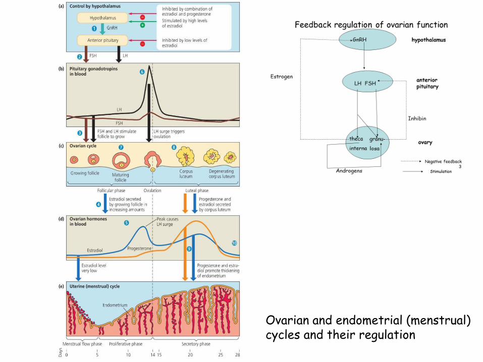
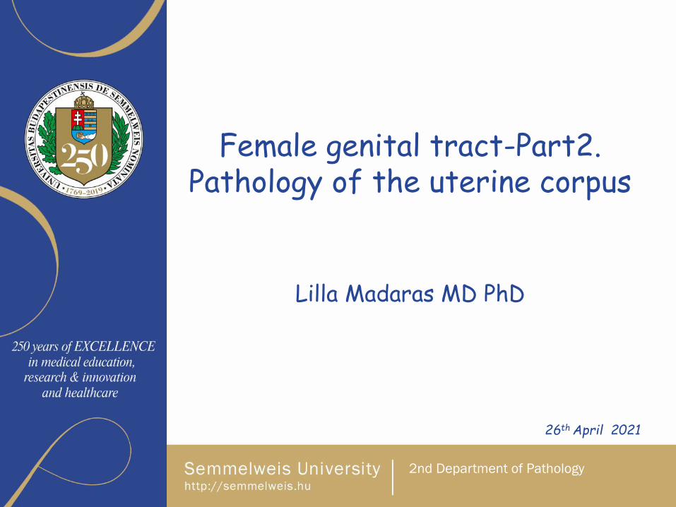
Ovarian and endometrial (menstrual) cycles and their regulation

„Dating” the endometrium
4
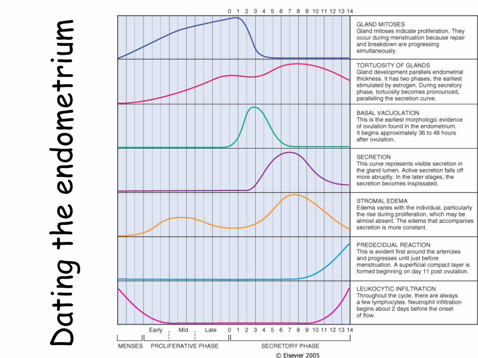
5Dat
ing
the e
ndom
etr
ium

Early proliferative phase-(„blue doughnuts” glands)

7Day 17-subnuclear vacuoles (piano key morphology) within endometrial glands
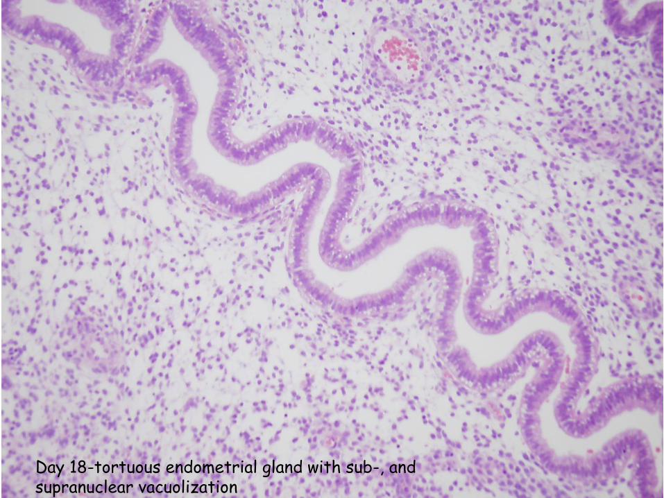
8Day 18-tortuous endometrial gland with sub-, and supranuclear vacuolization

9Exhausted secretory glands, predecidual changes around spiral arterioles

Exhausted secretory glands, predecidual changes within the stroma- Day 26
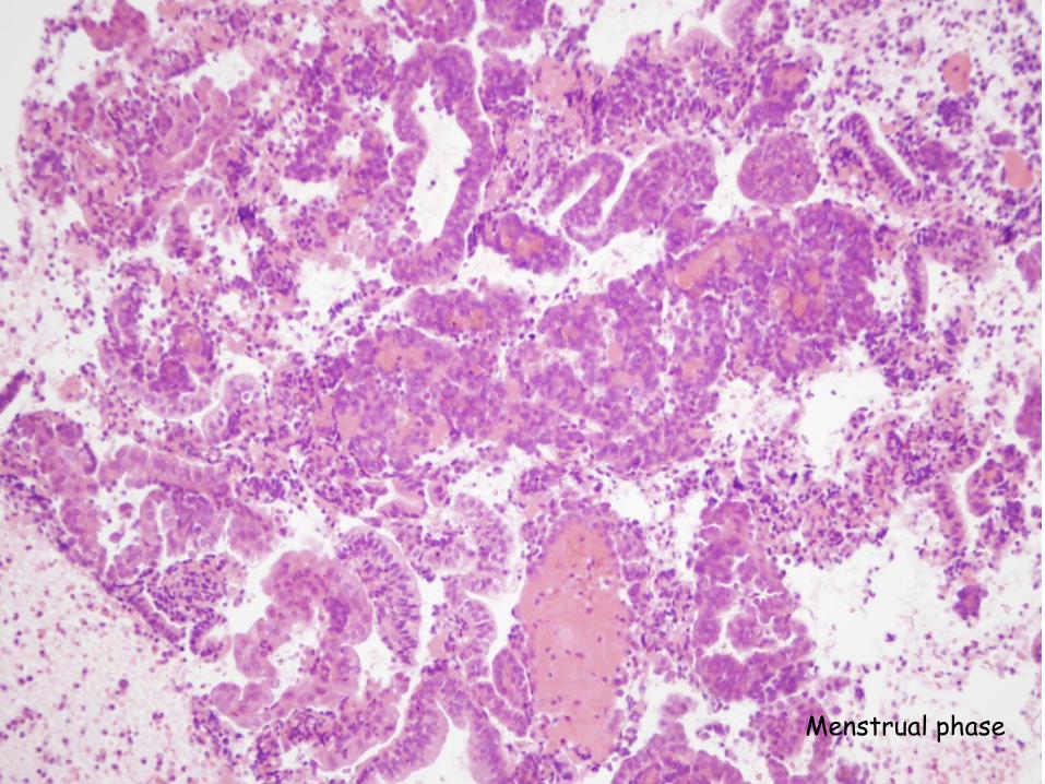
Menstrual phase
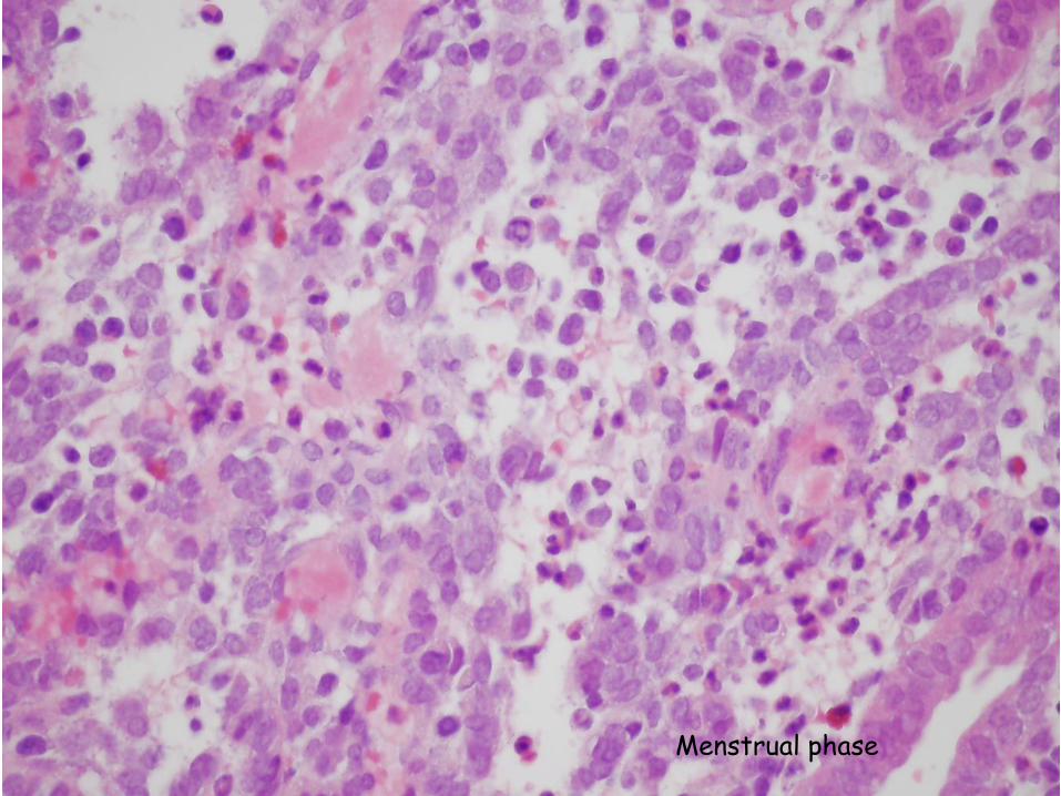
Menstrual phase
How do we assess the endometrium?
1. By sampling procedures
2. In hysterectomy specimens (from TAH with or without BSO, laparoscopic hysterectomy, Chrobak surgery, Wertheim surgery…)
13

Endometrium Sampling Techniques
1. Dilatation and Curettage(D&C)
2. Fractional curettage (separatesampling of the endometrium and endocervix)
3. Hysteroscopy+ polyp/endometrium ablation
4. Endometrial biopsy (Pipelle)
14
www.fmhs.auckland.ac.nz

Endometrial sampling-when?
• AUB (abnormal uterine bleeding)
• Abortion
• „Dating” the endometrium in cases of infertility
• Hormone replacement therapy, Tamoxifen
15
Pathology of the endometrium
16
• Inflammation
• Clinical terms
• AUB
• Endometrial hyperplasia
• Adenomyosis end endometriosis
• Tumors of the endometrium
17
18
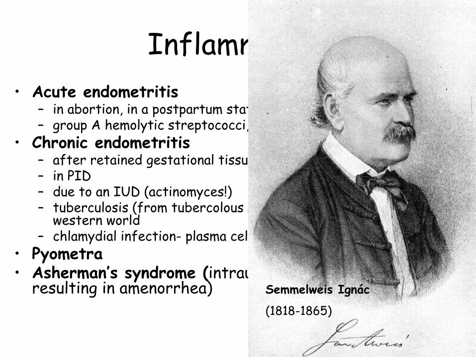
Inflammation
• Acute endometritis– in abortion, in a postpartum state– group A hemolytic streptococci, staphylococci
• Chronic endometritis– after retained gestational tissue– in PID– due to an IUD (actinomyces!)– tuberculosis (from tubercolous salpingitis)- rare in the
western world– chlamydial infection- plasma cells!
• Pyometra• Asherman’s syndrome (intrauterine adhesion
resulting in amenorrhea) Semmelweis Ignác
(1818-1865)
19
Clinical terms (still in your book and in use)
• amenorrhea- primary or secondary
• Oligomenorrhea ↔ polymenorrhea
• hypomenorrhea↔ menorrhagia
• metrorrhagia
• dysmenorrhea

20
Clinical terms• Dysfunctional uterine bleeding
• Oligomenorrhea, polymenorrhea
• hypomenorrhea↔ menorrhagia
• Metrorrhagia, metropathia haemorrhagica
• Dysmenorrhea
• Abnormal Uterine Bleeding (AUB)
AUB facts
• Premenop AUB- 1/3 of gynecological consultations
• Peri- and postmenop AUB- 70% of gynecological consultations
• FIGO Menstrual Disorders Working Group-2011
• American College of Obstetricians and Gynecologists (ACOG)-2013
21
AUB definitions
• AUB: bleeding from the uterine corpus that is abnormal in volume, regularity, and/or timing
– Heavy menstrual bleeding (HMB)
– Intermenstrual bleeding (IMB)
22

AUB definitions (cont.)
• Chronic AUB: bleeding from the uterine corpus that is abnormal in volume, regularity, and/or timing, and has been present for 6 months
• Acute AUB: was defined as an episode of heavy bleeding that, in the opinion of the clinician, is of sufficient quantity to require immediate intervention to prevent further blood loss
• Intermenstrual bleeding (IMB): occurs between clearly defined cyclic and predictable menses
23

AUB in reproductive agesPALM/COEIN
24
PALM
• Polyp-AUB-P
• Adenomyosis AUB-A
• Leiomyoma AUB-L
• Malignancy and hyperplasia AUB-M
• These lesions are detected by imaging and assessed by histology
25

26
Causes of Abnormal Uterine Bleeding by Age Group
Prepuberty Precocious puberty (hypothalamic, pituitary or ovarian origin)
Adolescence Anovulatory cycle
Reproductive age
Complication of pregnancy (abortion, trophobl.disease, ectopic pregnancy)
Organic lesions (leiomyoma, polyp, adenomyosis, endometrial hyperplasia, carcinoma)
Anovulatory cycle
Perimenopausal Anovulatory cycle
Organic lesions (carcinoma, hyperplasia, polyp)
Postmenopausal Organic lesions (carcinoma, hyperplasia, polyp)
Endometrial atrophy

Dysfunctional endometrial bleeding (now AUB-COEIN group)
• Inadequate proliferative phase– discrepancy between the
observed and the expected endometrial pattern in the proliferative phase
• Inadequate luteal phase– low progesteron level– infertility– amenorrhea or abnormal
bleeding– sampling 2 days before
expected menstruation!• Irregular shedding of the
endometrium– menstruation lasts longer than 7
days without prolongation of the cycle
– sampling on the 5th day of the menstruation demonstrates menstruation type and late secretory type endometrium and early proliferative endometrium
27

28
Dysfunctional endometrial bleeding
• Anovulatory cycle (AUB-O)– in adolescence and premenopausa most commonly due to slight hormonal imbalances and no apparent
causes– Less commonly:
• endocrine causes: thyroid, adrenal or pituitary disease• ovarian causes: PCO, granulosa-theca cell tumor• systemic metabolic causes: obesity, malnutrition (anorexia
nervosa!), chronic systemic diseases etc.
– no ovulation→ prolonged unopposed estrogenic stimulation →persistant proliferative endometrium → endometrial hyperplasia or unsheduled breakdown of the stroma →abnormal bleeding

29
Endometrial hyperplasia
• result of unopposed, prolonged estrogenic stimulation (due to anovulation or increased estrogen production- PCO, Stein-Leventhal sy, cortical stromal hppl, estrogen replacement therapy, functioning granulosa cell tumor)
• (Simple or Complex) Hyperplasia without atypia
• (Simple or Complex) Hyperplasia with atypia
30
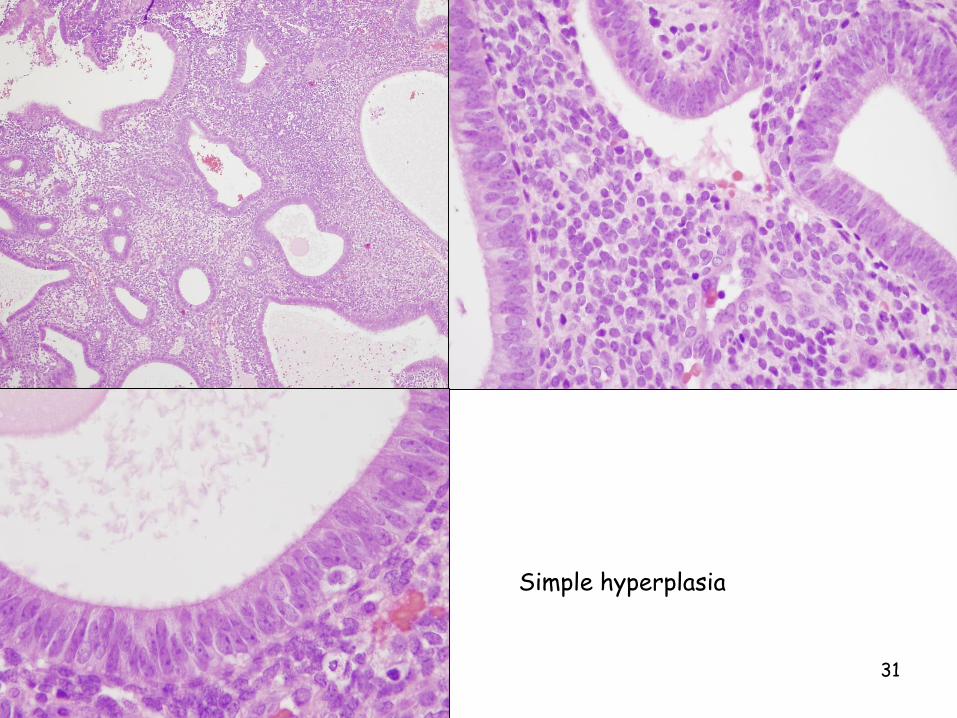
Hyperplasia (formerly called simple hyperplasia)
• Without atypia – diffuse alteration
– increased amount of glands and stroma– (Mildly increased) almost normal ratio of glands
and stroma– differences in glandular size and shape– cystically dilated glands – glandular epithelium: proliferative– Usually no progression to adenocarcinoma (1%)
• With atypia– the previous features + cytological atypia– Uncommon– Progression to adenocarcinoma 8%
31
Simple hyperplasia

32
Simple hyperplasia- endometrium

33
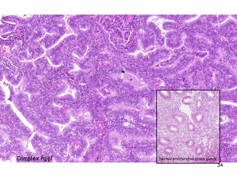
Formerly called complex hyperplasia
• Without atypia– focal alteration
– irregular glands
– increased ratio of glands to stroma (less intervening stroma, back-to-back placed glands)
– Progression to adenocarcinoma 3%
• With atypia (Endometrioid Intraepithelial Neoplasia)– cytological atypia
– Usually hysterectomy is done
– Progression to adenocarcinoma 25-30%
34
Complex hppl Normal proliferative phase glands

35
Metaplasias
• different forms: squamous,tubal, eosinophilic, mucinous, etc.
• frequently associated with hyperplasia
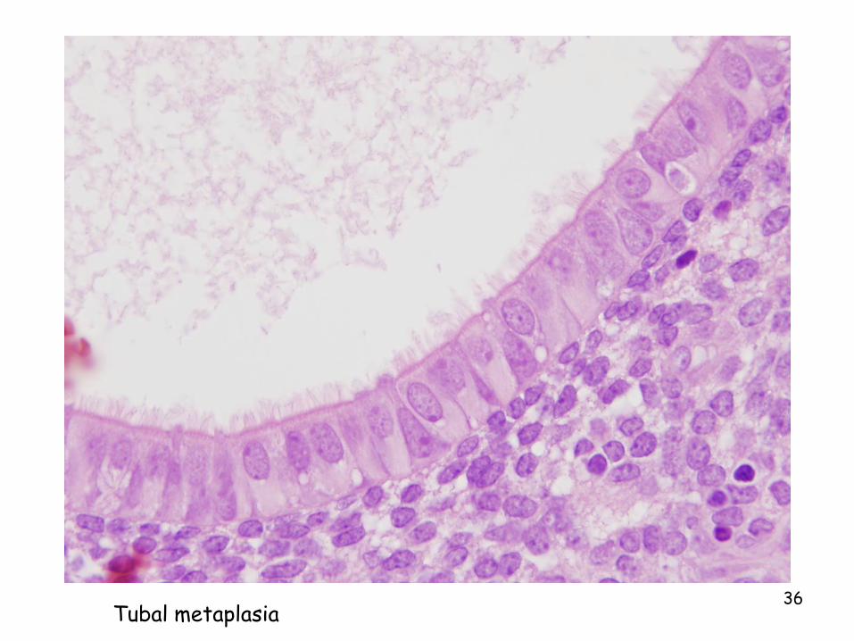
36Tubal metaplasia

37
Adenomyosis and endometriosis
• Adenomyosis: endometrial glands and stroma deep within the myometrium (by at least 2-3 mm from endometrium)
• Endometriosis: endometrial tissue outside the uterus– made of functional endometrium undergoing cyclic
changes– origin from müllerian rests?
implantation?lymphatic or hematogenous spread?– most commonly within the ovaries, uterine
ligaments, on the pelvic peritoneum, bowel, appendix, cervix, fallopian tube, laparotomy scars
– pelvic pain, dysmenorrhea, infertility– Deep infiltrating endometrosis (DIE)

38Adenomyosis and endometrial polyp
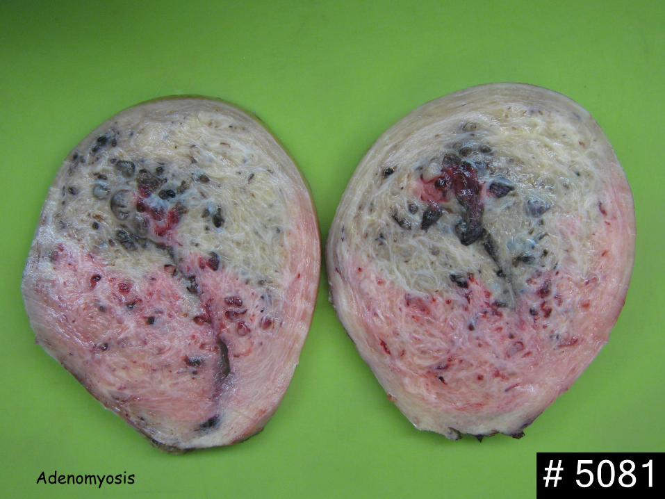
39Adenomyosis
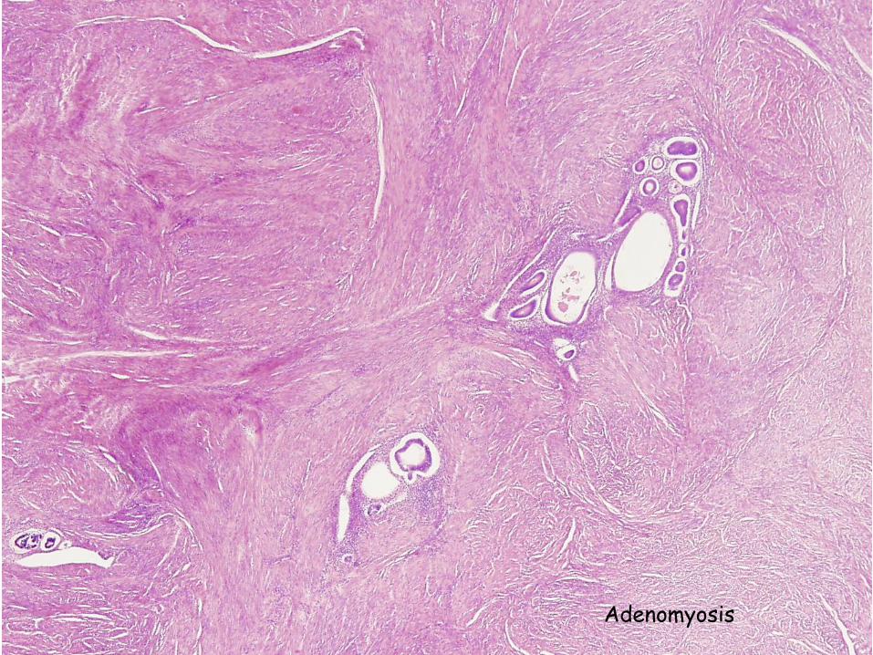
40Adenomyosis

41
Endometriosis of ovary- cyst formation (chocolate cyst)

42Peritoneal endometriosis

43
Endometrial polyps
• not true neoplasms, exophytic mass• may occur after Tamoxifen (SERM)
administration• asymptomatic or may produce abnormal
bleeding (AUB-P) • MA: projects into the body cavity• MI: cystically dilated glands, fibrous
stroma and thick-walled vessels• adenocarcinoma arising in ~ is possible
UTERINE NEOPLASIAS
44

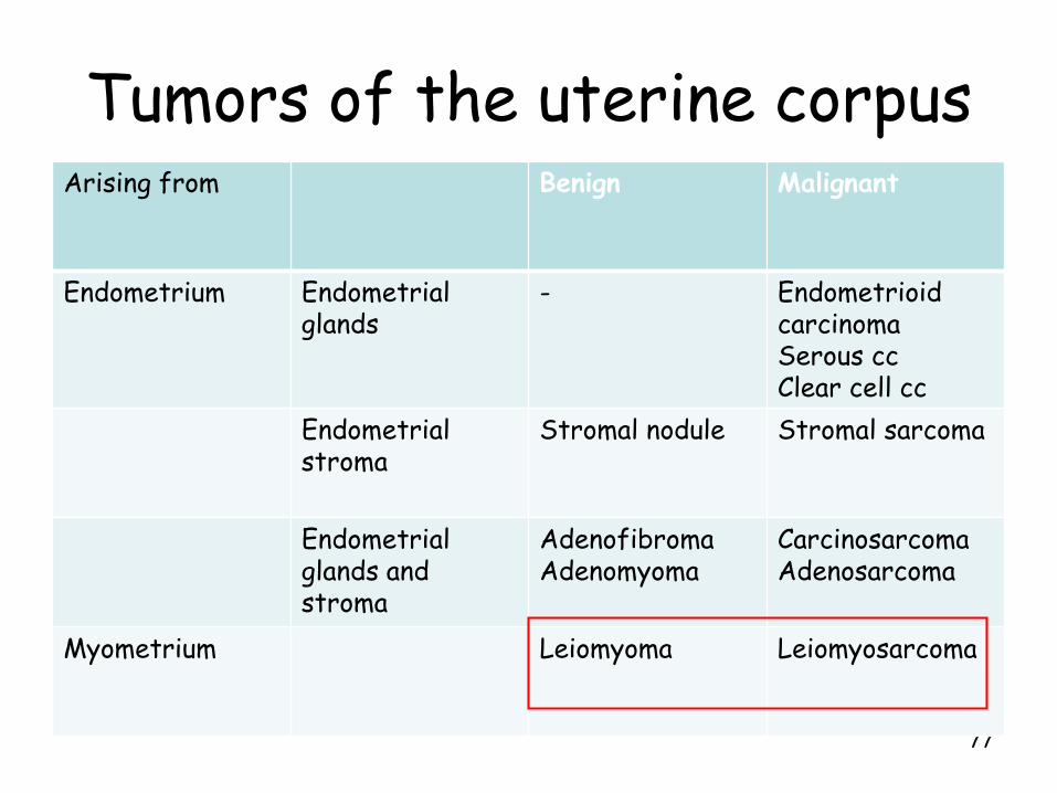
Tumors of the uterine corpus
45
Arising from Benign Malignant
Endometrium Endometrialglands
- EndometrioidcarcinomaSerous ccClear cell cc
Endometrialstroma
Stromal nodule Stromal sarcoma
Endometrialglands and stroma
AdenofibromaAdenomyoma
CarcinosarcomaAdenosarcoma
Myometrium Leiomyoma Leiomyosarcoma

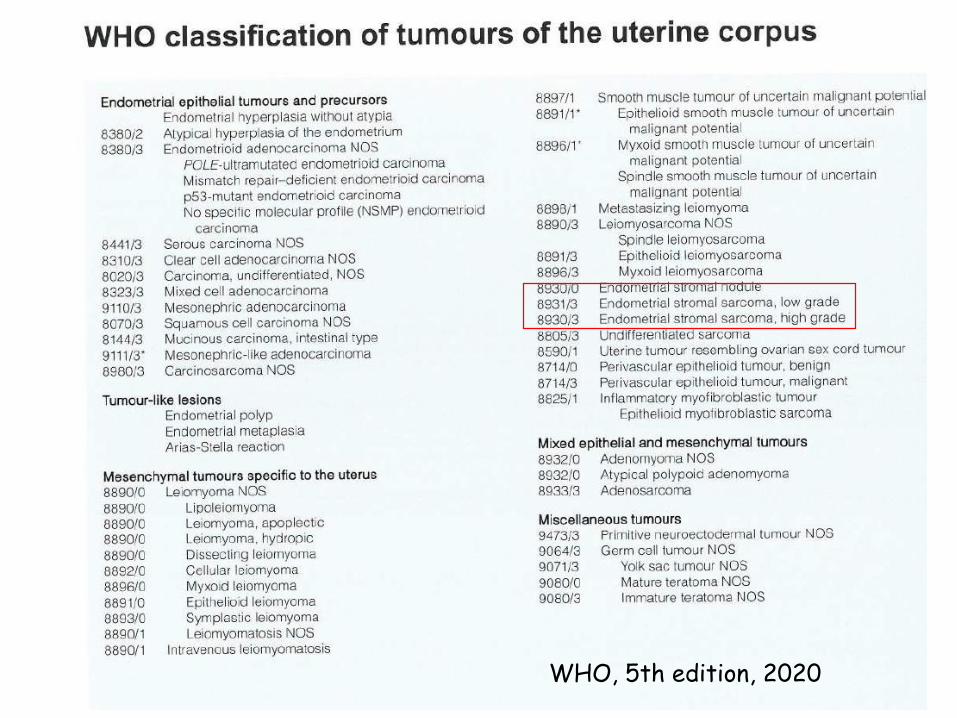
46WHO, 5th edition, 2020

47

48

49
Endometrial carcinoma
• 7% of all (non-skin)cancers in women
• mainly in postmenopausal woman (55-65 y)
• If it affects women ≤ 40 y Lynch syndrome should be excluded
• SY: usually postmenopausal bleeding
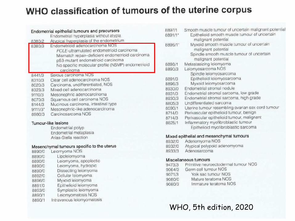
50WHO, 5th edition, 2020

51
Az endometrialis endometrioid carcinoma molekuláris klasszifikációjaWHO 5.kiadás, 2020 (ProMisE (Proactive Molecular Risk Classifier for Endometrial Cancer)POLEmut: hotspot mutációk a DNS polimeráz epsilon (POLE) exonukleáz doménjénMMR: mismatch repair

52
Endometrial carcinoma
– Endometrioid carcinoma• on a background of endometrial hyperplasia• Obesity, diabetes, hypertension, infertility,
Stein- Leventhal sy, longstanding estrogenusers, breast cancer patients treated withTamoxifen
• more favorable prognosis• usually well differentiated

Endometrial carcinoma
• Endometrioid carcinoma (cont)– Grading
• Grade 1: if<5% solid growth,
• Grade 2:if < 50% solid growth
• Grade 3: if > 50% solid growth
53

54WHO, 5th edition, 2020
55
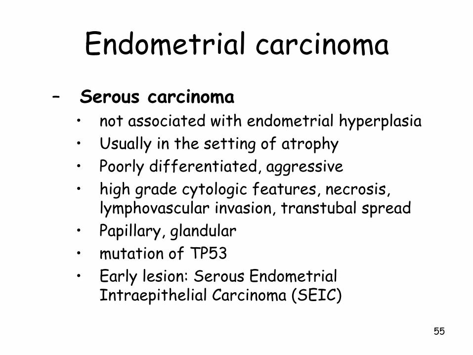
Endometrial carcinoma
– Serous carcinoma• not associated with endometrial hyperplasia
• Usually in the setting of atrophy
• Poorly differentiated, aggressive
• high grade cytologic features, necrosis, lymphovascular invasion, transtubal spread
• Papillary, glandular
• mutation of TP53
• Early lesion: Serous Endometrial Intraepithelial Carcinoma (SEIC)

Endometrial carcinoma
• Spread and metastases
– local spread: myometrium and cervix
– extrauterine spread: pelvic and paraaortic lymph nodes, ovaries
– serous carcinoma: early spread to the peritoneum, transtubal spread
56
TNM, 8th edition

57Endometrioid carcinoma
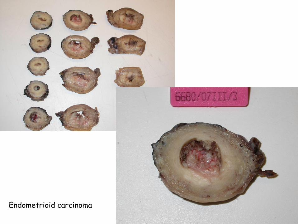
58Endometrioid carcinoma

59Endometrioid carcinoma
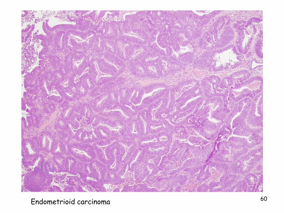
60Endometrioid carcinoma

61Serous carcinoma

62Papillary structures-high grade cytomorphology- Serous carcinoma

63
Clear cell carcinoma
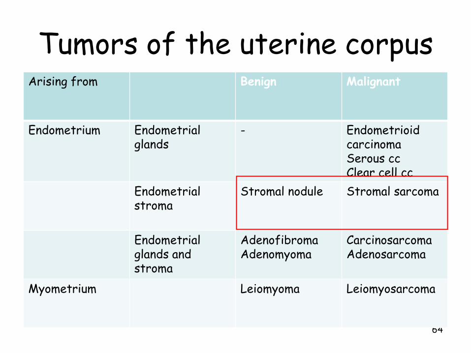
Tumors of the uterine corpus
64
Arising from Benign Malignant
Endometrium Endometrialglands
- EndometrioidcarcinomaSerous ccClear cell cc
Endometrialstroma
Stromal nodule Stromal sarcoma
Endometrialglands and stroma
AdenofibromaAdenomyoma
CarcinosarcomaAdenosarcoma
Myometrium Leiomyoma Leiomyosarcoma
65WHO, 5th edition, 2020

66
Endometrial stromal tumors
• middle- aged (average 45 y) women
• tumor cells mimicking endometrial stromal cells
• Endometrial stromal nodule
• Endometrial stromal sarcomas– low and high grade group

67
Endometrial stromal sarcoma

68Endometrial stromal sarcoma

69

Tumors of the uterine corpus
70
Arising from Benign Malignant
Endometrium Endometrialglands
- EndometrioidcarcinomaSerous ccClear cell cc
Endometrialstroma
Stromal nodule Stromal sarcoma
Endometrialglands and stroma
AdenofibromaAdenomyoma
CarcinosarcomaAdenosarcoma
Myometrium Leiomyoma Leiomyosarcoma
71WHO, 5th edition, 2020

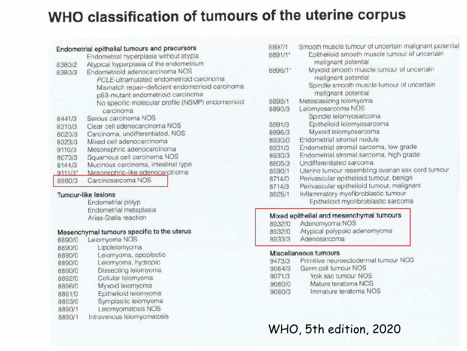
Mixed epithelial and mesenchymal tumors
• Adenomyoma
• Atypical polypoid adenomyoma
• Adenofibroma
• Adenosarcoma
• Carcinosarcoma (formerly malignant mixed müllerian tumor, now it is considered epithelial in origin with EMT)
72
73
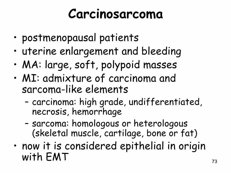
Carcinosarcoma
• postmenopausal patients• uterine enlargement and bleeding• MA: large, soft, polypoid masses• MI: admixture of carcinoma and
sarcoma-like elements– carcinoma: high grade, undifferentiated,
necrosis, hemorrhage– sarcoma: homologous or heterologous
(skeletal muscle, cartilage, bone or fat)
• now it is considered epithelial in origin with EMT

74
Carcinosarcoma-uterus
75
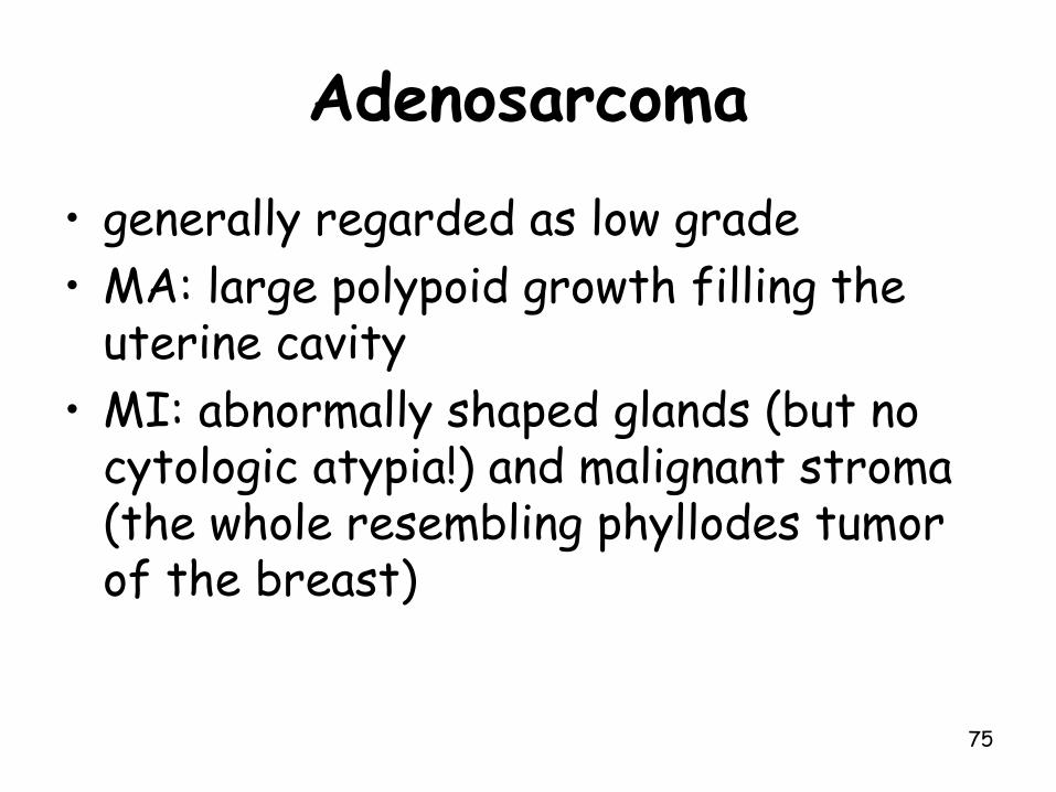
Adenosarcoma
• generally regarded as low grade
• MA: large polypoid growth filling the uterine cavity
• MI: abnormally shaped glands (but no cytologic atypia!) and malignant stroma (the whole resembling phyllodes tumor of the breast)

Myometrium
76
Tumors of the uterine corpus
77
Arising from Benign Malignant
Endometrium Endometrialglands
- EndometrioidcarcinomaSerous ccClear cell cc
Endometrialstroma
Stromal nodule Stromal sarcoma
Endometrialglands and stroma
AdenofibromaAdenomyoma
CarcinosarcomaAdenosarcoma
Myometrium Leiomyoma Leiomyosarcoma
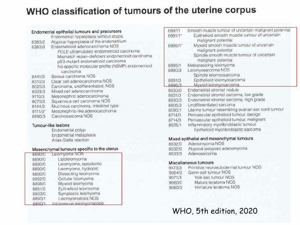
78WHO, 5th edition, 2020

79
Tumors of the Myometrium
• Leiomyoma– in 40% of women over the age of 50 years
– location: submucosal, intramural, subserosal
– symptoms: abnormal bleeding, pain, spontaneous abortion, impaired fertility, compression of the urinary bladder ( frequency)
– MA: well circumscribed, round, grayish-whitish nodule(s) with a whorling pattern
– MA: uniform spindle shaped cells, no atypia, scanty mitoses

80

81
Tumors of the Myometrium
• Leiomyosarcoma– in older patients (average 55 years)
– MA: fleshy with necrosis and hemorrhage
– MI: hypercellular, nuclear atypia, pleomorphism, increased mitotic index, atypical mitoses, necrosis
– Metastases: pelvis, lung, bone, brain but lymph node metastases exceptional!

82Leiomyosarcoma-uterus

83Leiomyosarcoma