
Feasibility study for minor enhancements of TG 414 ... · Review: Feasibility study for minor...
38
Review: Feasibility study for minor enhancements of TG 414 (Prenatal Developmental Toxicity Study) with ED-relevant endpoints DTU FOOD, October, 2017 Draft (for 1st WNT comments) Feasibility study for minor enhancements of TG 414 (Prenatal Developmental Toxicity Study) with ED- relevant endpoints October, 2017 Draft (for 1 st WNT comments) Sofie Christiansen & Ulla Hass Division of Diet, Disease prevention and Toxicology, National Food Institute, Technical University of Denmark
Transcript of Feasibility study for minor enhancements of TG 414 ... · Review: Feasibility study for minor...

Review: Feasibility study for minor enhancements of TG 414 (Prenatal Developmental Toxicity Study) with ED-relevant endpoints DTU FOOD, October, 2017 Draft (for 1st WNT comments)
Feasibility study for minor enhancements of TG 414 (Prenatal Developmental Toxicity Study) with ED-
relevant endpoints October, 2017 Draft (for 1st WNT comments)
Sofie Christiansen & Ulla Hass Division of Diet, Disease prevention and Toxicology, National Food Institute,
Technical University of Denmark
Review: Feasibility study for minor enhancements of TG 414 (Prenatal Developmental Toxicity Study) with ED-relevant endpoints DTU FOOD, October 2017 Draft (for 1st WNT comments)
2
Table of contents Terms of reference ............................................................................................................................................ 4
Aim ..................................................................................................................................................................... 4
Background and expected regulatory need/data requirement that will be met by the proposed outcome of the project ......................................................................................................................................................... 4
Anogenital distance (AGD) ................................................................................................................................ 7
Method .......................................................................................................................................................... 7
Data analysis, sensitivity and power ............................................................................................................. 7
Data collection ........................................................................................................................................... 7
Results ........................................................................................................................................................... 8
Human relevance ......................................................................................................................................... 10
Animal welfare............................................................................................................................................. 10
Inclusion of AGD in TG 414 .......................................................................................................................... 11
Testosterone levels in male foetuses .............................................................................................................. 12
Method ........................................................................................................................................................ 12
Data analysis, sensitivity/power .................................................................................................................. 13
Human relevance ......................................................................................................................................... 14
Animal welfare............................................................................................................................................. 15
Inclusion of Testosterone in TG 414 ............................................................................................................ 15
Thyroid hormones ........................................................................................................................................... 15
Method ........................................................................................................................................................ 15
Data analysis, sensitivity/power .................................................................................................................. 16
Human relevance ......................................................................................................................................... 16
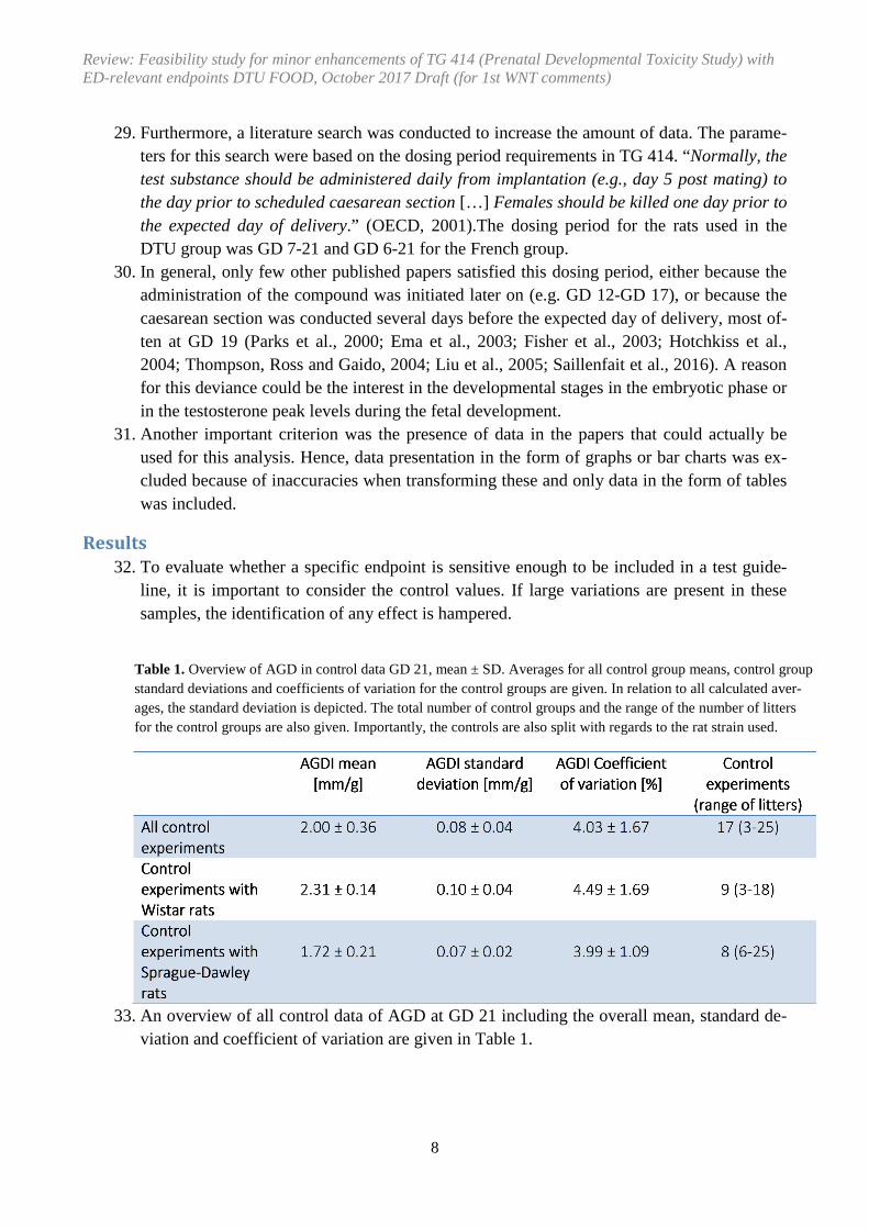
Animal welfare............................................................................................................................................. 16
Inclusion of thyroid hormones in TG 414 .................................................................................................... 16
Abnormalities of external genital organs ........................................................................................................ 17
Method ........................................................................................................................................................ 17
Data analysis, sensitivity/power .................................................................................................................. 17
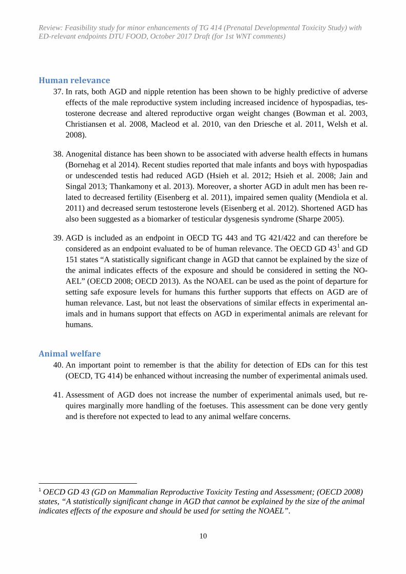
Human relevance ......................................................................................................................................... 17
Animal welfare............................................................................................................................................. 17
Inclusion abnormalities of external genital organs in TG 414 ..................................................................... 18

Review: Feasibility study for minor enhancements of TG 414 (Prenatal Developmental Toxicity Study) with ED-relevant endpoints DTU FOOD, October 2017 Draft (for 1st WNT comments)
3
Overall discussion and conclusions ................................................................................................................. 19
References ....................................................................................................................................................... 20
Appendix 1 Text changes suggestions for TG 414 shown with track changes ................................................ 25

Review: Feasibility study for minor enhancements of TG 414 (Prenatal Developmental Toxicity Study) with ED-relevant endpoints DTU FOOD, October 2017 Draft (for 1st WNT comments)
4
Terms of reference
1. This feasibility report has initially been prepared by the National Food Institute, Technical University of Denmark, DK that is leading the project in OECD. The report gives input for discussions in the OECD expert group on reproductive toxicity involved in the project Fea-sibility study for minor enhancements of TG 414 (Prenatal Developmental Toxicity Study) with ED-relevant endpoints. Subsequently, the report will be revised based on input after EDTA meetings, commenting rounds in OECD and discussions in the OECD expert group on reproductive toxicity.
Aim
2. The aim of this project is to do a Feasibility study for minor enhancements of TG 414 (Pre-natal Developmental Toxicity Study) with ED-relevant endpoints. This review addresses scientific and technical concerns regarding inclusion of additional ED related endpoints in TG 414. The endpoints considered include anogenital distance (AGD), Testosterone and thyroid hormones and guidance for genital malformations in male fetuses. For these end-points, the scientific and technical questions considered include: • Are standardized methods available? • Is the sensitivity sufficient with the number of litters per group? • Are the endpoints of relevance for humans? • Are there animal welfare concerns? • Is the enhancement possible without changes or with only minor changes in study de-
sign?
Background and expected regulatory need/data requirement that will be met by the proposed outcome of the project
3. A scientific approach will be used to give input to the existing TG 414 (Prenatal Develop-mental Toxicity Study) in relation to the feasibility of inclusion of sensitive endpoints for detection of chemicals with endocrine disrupting properties.
4. The specific purpose of this project is to consider the relevance and feasibility of enhance-ment of the OECD 414 (OECD, 2001). The TG 414 provides information on adverse ef-fects on prenatal development and is used in various regulatory frameworks (such as REACH and several pesticide regulations) to generate information for risk assessment of chemicals.
5. OECD TG 414 is included in Level 4 (OECD conceptual framework) as the TG involves repeated dosing of pregnant females and therefore potential exposure of the developing foe-

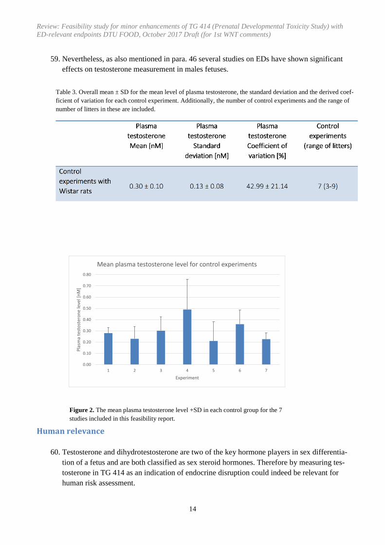
Review: Feasibility study for minor enhancements of TG 414 (Prenatal Developmental Toxicity Study) with ED-relevant endpoints DTU FOOD, October 2017 Draft (for 1st WNT comments)
5
tus. The assay includes some endpoints that may detect endocrine disruption (e.g. abnor-malities of male and female genitalia) (OECD GD 150, 2012a, currently under revision).
6. However, an update in relation to inclusion of more endpoints relevant for endocrine dis-ruption would increase the possibility for detecting effects of endocrine disrupting sub-stances. Assessment of testosterone levels in serum and anogenital distance (AGD) in male foetuses has been used in several published studies and appears as sensitive endpoints for detecting effects of endocrine disrupters with anti-androgenic properties. Thus, inclusion of these endpoints and possibly also other relevant endpoints in TG 414 in dams or GD 21 foe-tuses at the time of caesarean section could be a significant enhancement with regards to detection of effects of endocrine disrupting substances. Also, an enhancement with some additional text giving guidance on evaluation of abnormalities of male and female genitalia would be relevant to include.
7. After the inclusion on the workplan in OECD (as project number 4.100) also possibility to include thyroid hormones in the dams and/or fetuses has been mentioned in the OECD ex-pert group on reproductive toxicity as well as at international meetings in 2017 (Thyroid workshop report, 2017; Priority setting workshop report, 2017) and EDTA meetings in 2017.
8. The expert group on reproductive toxicity (EG) from the update on TG 421/422 (Project 4.71 on OECD Workplan) have been convened as the scientific discussion is similar. Addi-tional experts have been invited to the EG via the WNT NCs. This EG and the EDTA meet-ings in 2017 (May and October) have provided guidance on which endpoints to be consid-ered and how this can be done (e.g. timing and logistics) based on a proposal from the lead.
9. DK has undertaken the examination of available existing data. Data have been received from OECD countries and also peer reviewed scientific relevant papers have been included to make a proposal to the EG on whether or not it is relevant to include the ED related end-points in a proposal for revision of OECD TG 414.
10. It will also be considered whether certain slight adaptions of the test design of the test guideline may be warranted to include for consideration other ED related endpoints if such are being suggested by the EG for this project. However, the termination of the OECD TG 414 cannot be changed as was the case in TGs 421/422 screening studies (which was termi-nated later due to assessment of Nipple retention).
11. The results of this project may contribute to an improved sensitivity for identification of developmental toxicants in mammalian species at an early stage in the regulatory testing schemes for industrial chemicals (e.g. REACH) as information from TG 414 is already re-quired in such regulatory testing schemes.
12. If these endpoints are implemented in TG 414 it will enhance the international harmoniza-tion of hazard assessment with regard to developmental toxicity effects.
13. An important point is that the ability for detection of EDs can be enhanced without increas-ing the number of experimental animals used.

Review: Feasibility study for minor enhancements of TG 414 (Prenatal Developmental Toxicity Study) with ED-relevant endpoints DTU FOOD, October 2017 Draft (for 1st WNT comments)
6
14. TG 414 is designed to provide general information concerning the effects of prenatal expo-sure on the pregnant test animal and on the developing organism; this may include assess-ment of maternal effects as well as death, structural abnormalities, or altered growth in the fetus. The proposed update of TG 414 may not impair the ability to fulfill the purpose of TG 414.
15. Assessment of AGD is mandatory in TG 443 and TGs 421/422 and could probably easily be included in the TG 414. See below for more information.
16. TG 414 was revised in 2001 but not with regard to inclusion of ED relevant endpoints. It seems relevant to include some ED relevant endpoints in TG 414 as the exposure periods cover some of the sensitive periods during development (prenatal period). Proposed end-points are described below.
17. The OECD TG 407 (Repeated dose 28- day oral toxicity study in rodents) has been updated in 2008. The assay has been validated for some endocrine endpoints but the sensitivity of the assay is not sufficient to identify all EATS-mediated EDs. The validation of the assay (OECD, 2006) showed that it identified strong and moderate EDs acting through the ER and AR; and EDs weakly and strongly affecting thyroid function. It was relatively insensi-tive to weak EDs acting through the ER and AR. This assay also has some optional end-points such as uterine and ovary weight, changes in vaginal smears, histopathologic chang-es in mammary gland histopathology as well as serum T3, T4, TSH as well as thyroid weight which can be examined if there is additional concern.
18. The extended one-generation reproductive toxicity study (EOGRTS) (OECD, 2012b) in-cludes more endpoints sensitive to endocrine disruption than OECD TG 416 and, as it also uses reduced animal numbers if conducted without F2. It is expected that it will often re-place OECD TG 416 for mammalian reproductive toxicity testing (OECD GD 150, 2012a). Endpoints sensitive to endocrine disruption, not specified in OECD TG 416, include ano-genital distance at birth, areola/nipple retention, measurement of thyroid hormones and TSH levels. Effects on the developing nervous and immune systems are also assessed by the DNT and DIT cohorts. These systems may also be sensitive to endocrine influences. This test is also expected to have greater sensitivity than OECD TG 416 as it requires an in-creased number of pups to be examined. In summary, the new EOGRT study (OECD TG 443) is preferable for detecting endocrine disruption because it provides an evaluation of a number of endocrine endpoints in the juvenile and adult F1, which are not included in the 2-generation study (OECD TG 416) adopted in 2001.
19. In 2015 and 2016, the OECD 421 (Reproduction/Developmental Toxicity Screening Test) and OECD 422 (Combined Repeated Dose Toxicity Study with the Reproduc-tion/Developmental Toxicity Screening Test) guidelines were revised to include estrogen, androgen, thyroid, and steroidogenesis (EATS) relevant endpoints (OECD, 2016 a; OECD 2016b; OECD, 2015).
20. In April 2015, OECD launched this feasibility study for the enhancement of OECD 414 (Prenatal Developmental Toxicity Study) (project 4.100 on the OECD workplan) with se-

Review: Feasibility study for minor enhancements of TG 414 (Prenatal Developmental Toxicity Study) with ED-relevant endpoints DTU FOOD, October 2017 Draft (for 1st WNT comments)
7
lected parameters intended to increase the detection of EATS disrupting potential. In the au-tumn 2015 the lead and OECD secretariat requested data from OECD member states to en-hance TG 414 with endocrine disrupter relevant endpoints.
Anogenital distance (AGD)
Method 21. New-born male rats have no scrotum, and the external genitalia are undeveloped, and only a
genital tubercle is apparent for both sexes. The AGD is the distance from the anus to the in-sertion of this tubercle, the developing genital bud. The AGD is androgen dependent, and studies show that the AGD is normally about twice as long in male as in female rats. Simi-larly, in new-born humans the AGD measure was about two-fold greater in males than in females (Salazar-Martinez et al. 2004).
22. In TG 414 the AGD will be measured one day prior to the expected day of delivery.
Data analysis, sensitivity and power
23. Important parameters when evaluating the sensitivity and power of the data are the standard deviation, SD (σ) and coefficient of variation
24. The standard deviation is an expression of how much the value disperses from the popula-tion mean (µ). Coefficient of variation is expressed as σ/µ and is often evaluated as a per-centage and therefore expresses the standard deviation as the percentage of the population mean, µ.
25. In order to take into account the size of the rat when evaluating the AGD, was divided by the cubic root of the body weight, i.e. (AGD [mm])/∛(body weight [g]) resulted in the ano-genital distance index (AGDI).
26. Body weight is a three-dimensional endpoint and AGD is one-dimensional, the cubic root is used because this gives the best comparison between the two endpoints (OECD, 2015).
Data collection 27. The evaluation of both AGD and plasma testosterone levels as endpoints is based on data
from studies at DTU National food institute, external (non-published data from the expert group) and published studies primarily from Saillenfait et al. from The French Research and Safety Institute for the prevention of occupational accidents and diseases (INRS) (Saillen-fait, et al. 2009; Saillenfait et al, 2011a and 2011b; Saillenfait, et al. 2013).
28. Unfortunately the data sent from the expert group (unpublished) could not be included in the analysis. One dataset missed information about litter affiliation and thereby group means based on litter means could not be obtained. The other dataset did not give any de-tails on the strain of rodent.
Review: Feasibility study for minor enhancements of TG 414 (Prenatal Developmental Toxicity Study) with ED-relevant endpoints DTU FOOD, October 2017 Draft (for 1st WNT comments)
8
Table 1. Overview of AGD in control data GD 21, mean ± SD. Averages for all control group means, control group standard deviations and coefficients of variation for the control groups are given. In relation to all calculated aver-ages, the standard deviation is depicted. The total number of control groups and the range of the number of litters for the control groups are also given. Importantly, the controls are also split with regards to the rat strain used.
29. Furthermore, a literature search was conducted to increase the amount of data. The parame-ters for this search were based on the dosing period requirements in TG 414. “Normally, the test substance should be administered daily from implantation (e.g., day 5 post mating) to the day prior to scheduled caesarean section […] Females should be killed one day prior to the expected day of delivery.” (OECD, 2001).The dosing period for the rats used in the DTU group was GD 7-21 and GD 6-21 for the French group.
30. In general, only few other published papers satisfied this dosing period, either because the administration of the compound was initiated later on (e.g. GD 12-GD 17), or because the caesarean section was conducted several days before the expected day of delivery, most of-ten at GD 19 (Parks et al., 2000; Ema et al., 2003; Fisher et al., 2003; Hotchkiss et al., 2004; Thompson, Ross and Gaido, 2004; Liu et al., 2005; Saillenfait et al., 2016). A reason for this deviance could be the interest in the developmental stages in the embryotic phase or in the testosterone peak levels during the fetal development.
31. Another important criterion was the presence of data in the papers that could actually be used for this analysis. Hence, data presentation in the form of graphs or bar charts was ex-cluded because of inaccuracies when transforming these and only data in the form of tables was included.
Results 32. To evaluate whether a specific endpoint is sensitive enough to be included in a test guide-
line, it is important to consider the control values. If large variations are present in these samples, the identification of any effect is hampered.
33. An overview of all control data of AGD at GD 21 including the overall mean, standard de-viation and coefficient of variation are given in Table 1.

Review: Feasibility study for minor enhancements of TG 414 (Prenatal Developmental Toxicity Study) with ED-relevant endpoints DTU FOOD, October 2017 Draft (for 1st WNT comments)
9
34. As seen in table 1 the Coefficient of Variation in male control foetuses is low (overall 4.03±1.67). However, it is also observed that Sprague-Dawley rats in these studies have a lower mean AGDI, standard deviation and coefficient of variation compared to Wistar rats.
35. To evaluate whether the prenatal AGD measurement is sensitive enough it is also relevant
to compare to postnatal data. This endpoint is not feasible to include if the prenatal meas-urements has a much higher variation than postnatal measurement. The measurements must be consistent with standard deviations and coefficients of variation not greatly influenced by the gestation day compared to postnatal measurements. Table 2 and figure 1 outlines such a comparison between coefficients of variation for AGDI measured pre- and postnatal-ly.
36. As seen in table 2 the CoV for prenatal AGDI is 4.49 (9 studies) whereas in new born males it is 4.00 (23 studies). This indicates that the sensitivity/power for detecting effect on AGD is rather similar in GD 21 foetuses and new born male pups. Thus the power analysis per-formed in relation to inclusion of AGD in new born males in TG 421/422 is also useful for this TG.
Figure 1. Mean coefficient of variation for prenatal studies: 4.49 ±1.69 % and postnatal studies: 4.00 ±1.50 % (23 stud-ies)
Table 2. Comparison of coefficients of variation for AGDI measured prenatally and postnatally. The given coefficients of variation are mean of all control group coefficients, mean ± SD. The total number of control groups and the range of number of litters for the control groups are given. Only data on Wistar rats (from table 1) is included from DTU, DK.
Review: Feasibility study for minor enhancements of TG 414 (Prenatal Developmental Toxicity Study) with ED-relevant endpoints DTU FOOD, October 2017 Draft (for 1st WNT comments)
10
Human relevance 37. In rats, both AGD and nipple retention has been shown to be highly predictive of adverse
effects of the male reproductive system including increased incidence of hypospadias, tes-tosterone decrease and altered reproductive organ weight changes (Bowman et al. 2003, Christiansen et al. 2008, Macleod et al. 2010, van den Driesche et al. 2011, Welsh et al. 2008).
38. Anogenital distance has been shown to be associated with adverse health effects in humans (Bornehag et al 2014). Recent studies reported that male infants and boys with hypospadias or undescended testis had reduced AGD (Hsieh et al. 2012; Hsieh et al. 2008; Jain and Singal 2013; Thankamony et al. 2013). Moreover, a shorter AGD in adult men has been re-lated to decreased fertility (Eisenberg et al. 2011), impaired semen quality (Mendiola et al. 2011) and decreased serum testosterone levels (Eisenberg et al. 2012). Shortened AGD has also been suggested as a biomarker of testicular dysgenesis syndrome (Sharpe 2005).
39. AGD is included as an endpoint in OECD TG 443 and TG 421/422 and can therefore be considered as an endpoint evaluated to be of human relevance. The OECD GD 431 and GD 151 states “A statistically significant change in AGD that cannot be explained by the size of the animal indicates effects of the exposure and should be considered in setting the NO-AEL” (OECD 2008; OECD 2013). As the NOAEL can be used as the point of departure for setting safe exposure levels for humans this further supports that effects on AGD are of human relevance. Last, but not least the observations of similar effects in experimental an-imals and in humans support that effects on AGD in experimental animals are relevant for humans.
Animal welfare 40. An important point to remember is that the ability for detection of EDs can for this test
(OECD, TG 414) be enhanced without increasing the number of experimental animals used.
41. Assessment of AGD does not increase the number of experimental animals used, but re-quires marginally more handling of the foetuses. This assessment can be done very gently and is therefore not expected to lead to any animal welfare concerns.
1 OECD GD 43 (GD on Mammalian Reproductive Toxicity Testing and Assessment; (OECD 2008) states, “A statistically significant change in AGD that cannot be explained by the size of the animal indicates effects of the exposure and should be used for setting the NOAEL”.

Review: Feasibility study for minor enhancements of TG 414 (Prenatal Developmental Toxicity Study) with ED-relevant endpoints DTU FOOD, October 2017 Draft (for 1st WNT comments)
11
Inclusion of AGD in TG 414 42. There are standardized OECD test methods for assessing AGD and AGD measured at birth
(e.g. PD 1-4) have been included in several OECD TGs.
43. The current report have shown that the power for assessment of AGD is almost equal in GD 21 fetuses versus in new-borns as the analysis showed similar CoV in these two time points. Therefore this endpoint can be included in TG 414 at GD 21 without any modifica-tion of the overall test design.
44. AGD is an endpoint of high human relevance and there are no concerns for animal welfare related to the assessment of this endpoint.
45. This all supports that assessment of AGD in all fetuses can be included in TG 414 (Appen-dix 1).
Review: Feasibility study for minor enhancements of TG 414 (Prenatal Developmental Toxicity Study) with ED-relevant endpoints DTU FOOD, October 2017 Draft (for 1st WNT comments)
12
Testosterone levels in male foetuses
Method
46. Changes in testosterone levels in sensitive time windows can cause permanent/long lasting reproductive changes in laboratory rats. Studies have shown that exposure during gestation to e.g. some phthalates, can show effects on testosterone synthesis in males foetuses leading to effects observed postnatally e.g. anogenital distance and reproductive organ weight changes (Borch et al. 2004, Saillenfait et al. 2013, Borch et al 2006).
47. In general, only few other published papers satisfied this dosing period, either because the administration of the compound was initiated later on (e.g. GD 12), or because the caesare-an section was conducted several days before the expected day of delivery, most often at GD 19 (Parks et al., 2000; Ema et al., 2003; Fisher et al., 2003; Hotchkiss et al., 2004; Lehmann et al., 2004; Thompson, Ross and Gaido, 2004; Liu et al., 2005; Saillenfait et al., 2016). A reason for this deviance could be the interest in the developmental stages in the embryotic phase or in the testosterone peak levels during the foetal development.
48. In relation to the feasibility of inclusion of testosterone hormone measurements the lead and expert group have discussed whether serum testosterone levels, testosterone synthesis (tes-ticular) or ex vivo testosterone production in foetal testes would be the most suitable meth-od.
49. Only measurement of hormones in the serum would be feasible as other methods might im-pair the ability to fulfil the purpose of TG 414.
50. Blood sampling in TG 414 (also other hormones) can be done by:
1. Cardiac puncture, can only be done for fetuses selected for skeletal assessment
2. Decapitation, will impact all fetal assessments
3. The umbilical cord, will not impact fetal assessments but only gives a very small amount of blood (so blood of most fetuses/litter should be pooled)
i. This latter method is not relevant for testosterone as this should be only pooled by sex and the amount of blood might be too small.
51. Fetal blood for hormone measurements: T4, T3 and TSH is doable to pool, however testos-terone should not be pooled blood from both sexes.

Review: Feasibility study for minor enhancements of TG 414 (Prenatal Developmental Toxicity Study) with ED-relevant endpoints DTU FOOD, October 2017 Draft (for 1st WNT comments)
13
52. In general, only few other published papers satisfied this dosing period, either because the administration of the compound was initiated later on (e.g. GD 12), or because the caesare-an section was conducted several days before the expected day of delivery, most often at GD 19 (Parks et al., 2000; Ema et al., 2003; Fisher et al., 2003; Hotchkiss et al., 2004; Lehmann et al., 2004; Thompson, Ross and Gaido, 2004; Liu et al., 2005; Saillenfait et al., 2016). A reason for this deviance could be the interest in the developmental stages in the embryotic phase or in the testosterone peak levels during the foetal development.
Data analysis, sensitivity/power
53. Weisz and Ward (1980) reported significantly higher serum testosterone levels in male rat fetuses with a peak at GD 18 and this finding was confirmed by Lichtensteiger and Schlumpf (1981, 1985). Due to this testosterone peak at GD18, this stage have been exam-ined when the focus have been on sexual dimorphism of sex steroid regulation. As already stated this TG 414 performs caesarean section the day before expected delivery and there-fore it is not feasible to assess testosterone level in serum from other time-windows.
54. As stated above (para. 46), plasma testosterone levels have also been found to be affected by chemicals at subsequent fetal and neonatal stages, and have been related to developmen-tal disturbances. This is why this project suggests determining plasma testosterone at around GD 21 in TG414.
55. In many studies testosterone production or content in testis rather than plasma levels were reported (Parks et al., 2000; Fisher et al., 2003; Hotchkiss et al., 2004; Lehmann et al., 2004; Thompson, Ross and Gaido, 2004). This endpoint is however not feasible to include in TG 414 due to the purpose of the TG 414.
56. In Table 3, the overall mean and standard deviation for the individually measured mean plasma testosterone levels, the standard deviations and the coefficient of variation for each control experiment are presented. Furthermore, the number of control experiments included in the calculations and the range of the numbers of litters in these are stated.
57. It is seen at table 3 that a large overall coefficient of variation of around 43% in controls is obtained indicating that the possibility for detecting effects is low. However, it is also seen that the number of litters included is relatively low compared the number of litters in TG 414, i.e. 3-9 litters per group compared to 20 litters per group in TG 414. Therefore the power for detecting effects is expected to be clearly higher in the TG 414 than in those studies.
58. Moreover, in two of the 7 studies (see fig. 2), the coefficient of variation is actually around 20% in spite of the low number of litters per group. This indicates that the power for detect-ing effect with around 20 litters per group may be sufficient.
Review: Feasibility study for minor enhancements of TG 414 (Prenatal Developmental Toxicity Study) with ED-relevant endpoints DTU FOOD, October 2017 Draft (for 1st WNT comments)
14
Figure 2. The mean plasma testosterone level +SD in each control group for the 7 studies included in this feasibility report.
0.00
0.10
0.20
0.30
0.40
0.50
0.60
0.70
0.80
1 2 3 4 5 6 7
Plas
ma
test
oste
rone
leve
l [nM
]
Experiment
Mean plasma testosterone level for control experiments
59. Nevertheless, as also mentioned in para. 46 several studies on EDs have shown significant effects on testosterone measurement in males fetuses.
Human relevance
60. Testosterone and dihydrotestosterone are two of the key hormone players in sex differentia-tion of a fetus and are both classified as sex steroid hormones. Therefore by measuring tes-tosterone in TG 414 as an indication of endocrine disruption could indeed be relevant for human risk assessment.
Table 3. Overall mean ± SD for the mean level of plasma testosterone, the standard deviation and the derived coef-ficient of variation for each control experiment. Additionally, the number of control experiments and the range of number of litters in these are included.

Review: Feasibility study for minor enhancements of TG 414 (Prenatal Developmental Toxicity Study) with ED-relevant endpoints DTU FOOD, October 2017 Draft (for 1st WNT comments)
15
Animal welfare
61. Blood samples for assessment of testosterone or other steroid hormones in fetus are taken at caesarian section by termination of the study. This leads to no concern for animal welfare, as the blood samples will be collected at the time of sacrifice.
Inclusion of Testosterone in TG 414
62. There are standardized test methods for assessing testosterone hormones in serum. The per-formed power analysis shoved coefficient of variation of around 43% in controls indicating that the possibility for detecting effects is low.
63. Nevertheless, several studies on EDs have shown effects on testosterone measurement in male fetuses and this endpoint will be suggested included as optional in fetuses in TG 414.
Thyroid hormones
Method
64. Thyroid hormones was included in the TGs 421/422 as blood samples from the day 13 pups and the adult males was required assessed for serum levels for thyroid hormones (T4) whereas further assessment of T4 in blood samples from the dams and day 4 pups is to be done if relevant. Moreover the TGs also include an option for other hormones.
65. Due to the circadian rhythm of thyroid hormones, sample collection should occur at approx-imately the same time of day and be randomized across dosage groups, preferably in the morning hours at which time basal values should be present (Döhler et al., 1979).
66. Blood samples for evaluation of triiodothyronine (T3), thyroxine (T4), and TSH should be collected immediately following sacrifice.
67. Hormonal analyses should be conducted on GD 20 fetuses and dams in this TG 414. 68. If there are inadequate fetuses in a litter to obtain sufficient blood for the hormonal
measures, the measurements of T4 and TSH would be a priority, with less emphasis placed on T3 measures (ref. EPA Dev Thyroid Protocol).
69. Prior to sacrifice, every effort should be made to avoid inducing stress that could affect hormone concentrations (Döhler et al., 1979)

Review: Feasibility study for minor enhancements of TG 414 (Prenatal Developmental Toxicity Study) with ED-relevant endpoints DTU FOOD, October 2017 Draft (for 1st WNT comments)
16
Data analysis, sensitivity/power
70. The number of animals included in TG 414 is similar to the number of animals in TG 443, where assessment of thyroid hormones is included. Thus, specific data analysis of power re-lated to number of animals is not needed here.
71. Moreover, the feasibility study from TGs 421/422 (OECD, 2015) included intensive power simulations for TH measurements with fewer animals.
72. However, blood samples will be taken in both dams and fetuses (optional) in TG 414 com-pared to non-pregnant adult animals in TG 443 and TG 421/422 (males). This may affect the sensitivity and therefore needs to be addressed.
Human relevance 73. Thyroid hormones (TH) are needed for proper nerve cell differentiation and proliferation,
and normal status of these hormones during early development is therefore crucial. In hu-mans even moderate and transient reductions in maternal T4 levels during pregnancy, may adversely affect the child’s neurological development.
74. This indicates that by measuring thyroid hormones (T4, T3 and TSH) in dams and fetuses in TG 414 as an indication of thyroid disruption could indeed be relevant for human risk as-sessment as also described in the feasibility study for TGs 421/422 (OECD, 2015).
Animal welfare
75. Blood samples for assessment of testosterone or other steroid hormones in fetus are taken at caesarian section by termination of the study. This leads to no concern for animal welfare, as the blood samples will be collected at the time of sacrifice.
Inclusion of thyroid hormones in TG 414
76. There are standardized OECD test methods for assessing thyroid hormones. The performed power analysis in TGs 421/422 made in the feasibility study supported that assessment of thyroid hormones could be included in these TGs.
77. Thyroid measurements (T3, T4 and TSH) will be included as mandatory endpoints in the updated TG 408 (90 days study).
78. Therefore the assessment of Thyroid hormones in TG 414 dams (mandatory) and fetuses (optional) is sufficiently sensitive and can provide relevant data.
79. Due to the adverse effects seen in humans after developmental hypothyroidism, this end-point is of high human relevance and there are no concerns for animal welfare related to the assessment of this endpoint as long as blood sampling is done in animals that are being sac-rificed anyway.
80. This all supports that assessment of thyroid hormones can be included in TG 414.
Review: Feasibility study for minor enhancements of TG 414 (Prenatal Developmental Toxicity Study) with ED-relevant endpoints DTU FOOD, October 2017 Draft (for 1st WNT comments)
17
Abnormalities of external genital organs
Method
81. In TG 414 the reproductive tract is examined for signs of altered development. This project will result in more guidance on evaluation of abnormalities of external genitalia in foetuses such as hypospadias (Hsieh et al. 2007).
82. In the current TG 414 it is mentioned (para. 29) that each foetus should be examined for ex-ternal alterations the text have been modified to take also abnormalities of external genital organs into account.
Data analysis, sensitivity/power
83. The feasibility study for TG 421/422 (OECD, 2015) included a calculation of the effect size needed for finding significant effect for abnormalities/malformations.
84. The data in this feasibility study strongly supported that all male pups in TG 421/422 needs to be evaluated, similarly as in OECD TG 414.
85. This limited sensitivity for detecting significant effects on rare adverse outcomes is general-ly recognized for malformations. Thus, the occurrence of a few similar rare malformations such as hypospadias may generally be considered toxicologically relevant although the finding is not statistically significant.
Human relevance 86. Hypospadias in humans is one of the most common urogenital congenital anomalies affect-
ing boys (Harris 1990). Prevalence estimates in Europe range from 4 to 24 per 10,000 births, depending on definition (Dolk et al. 2004) with higher rates of about 5% reported in a Danish study (Boisen et al. 2005). Little is known about the aetiology of hypospadias, but a role for EDCs has been proposed, and especially the anti-androgenic EDCs (Baskin et al. 2001).
87. Exposure during critical developmental phases such as in utero and in the early postnatal period may lead to adverse effects on both reproductive development and neurodevelop-ment. The fact that many of the basic mechanisms underlying this developmental process are similar in all mammals indicates that chemicals that have adverse effects on reproduc-tive development in rodents should be considered as potential human reproductive toxicants as well (Gray 1992).
Animal welfare
88. Assessment of abnormalities of external genital organs requires slightly more handling of fetuses. This assessment can be done very gently and is therefore not expected to lead to

Review: Feasibility study for minor enhancements of TG 414 (Prenatal Developmental Toxicity Study) with ED-relevant endpoints DTU FOOD, October 2017 Draft (for 1st WNT comments)
18
any animal welfare concerns. However, as the assessment of abnormalities of external geni-tal organs is done after termination of the fetuses in TG 414, there will obviously be no concern for animal welfare.
Inclusion abnormalities of external genital organs in TG 414 89. Assessment of abnormalities is already included in TG 414. However, no details with re-
gard to assessment of abnormalities of external genitals organs are included. The text pro-posed to be added in the revised TG 414 in relation to abnormalities is modified from para 29 in OECD TG 414.

Review: Feasibility study for minor enhancements of TG 414 (Prenatal Developmental Toxicity Study) with ED-relevant endpoints DTU FOOD, October 2017 Draft (for 1st WNT comments)
19
Overall discussion and conclusions
90. The aim of this project was to do a feasibility study for minor enhancements of TG 414 with ED-relevant endpoints. The endpoints considered for inclusion were AGD, Testos-terone levels in fetuses, thyroid hormones (and other hormones) in the dams and fetuses.
91. For all endpoints, OECD test methods are available for assessing these. Power analyses have been done showing sufficient sensitivity to get relevant data with the number of litters per group in the TGs 421/422. All four endpoints are of relevance for humans as described in this review. All four of them are mandatory to assess in some OECD Test guidelines used for human risk assessment of chemicals. The overall animal welfare considerations will not increase by the assessments of the 4 endpoints. Inclusion of all four endpoints in TG 421/422 does not trigger any animal welfare concerns.
92. The Test Guideline has been updated with specific text proposals. No changes in study de-sign and only few text changes are necessary to include the assessment of anogenital dis-tance (AGD), hormone measurements (e.g. thyroid hormones and sex steroids) and inclu-sion of guidance on abnormalities of external genital organs.
93. In conclusion, it is feasible to make the proposed minor enhancements of TG 414 with ED-relevant endpoints: anogenital distance (AGD), testosterone and thyroid hormones (in dams and fetuses) and inclusion of guidance on abnormalities of external genital organs in male fetuses.
94. This report conclude that the proposals for the update of TG 414 are:
1. AGD mandatory – all fetuses
2. Testosterone – optional in male fetuses
3. T4, T3 and TSH dams – mandatory
4. T4, T3 and TSH in fetuses optional (T4 priority, then TSH and T3)

Review: Feasibility study for minor enhancements of TG 414 (Prenatal Developmental Toxicity Study) with ED-relevant endpoints DTU FOOD, October 2017 Draft (for 1st WNT comments)
20
References Baskin LS, Himes K, Colborn T. 2001. Hypospadias and endocrine disruption: is there a connection? Environmental Health Perspectives 109:1175-1183. Boisen KA, Chellakooty M, Schmidt IM, Kai CM, Damgaard IN, Suomi AM, Toppari J, Skakkebaek NE, Main KM. 2005. Hypospadias in a cohort of 1072 Danish newborn boys: prevalence and relationship to placental weight, anthropometrical measurements at birth, and reproductive hormone levels at three months of age. The Journal of Clinical Endocrinology & Metabolism 90:4041-4046. Borch J, Ladefoged O, Hass U, Vinggaard AM. Steroidogenesis in fetal male rats is reduced by DEHP and DINP, but endocrine effects of DEHP are not modulated by DEHA in fetal, prepubertal and adult male rats. Reprod. Toxicol. 2004;18(1):53-61 Borch J, Axelstad M, Vinggaard AM, Dalgaard M. Diisobutyl phthalate has comparable anti-androgenic effects to di-n-butyl phthalate in fetal rat testis. Toxicol. Let. 2006, 163,183-90. Bornehag CG1, Carlstedt F, Jönsson BA, Lindh CH, Jensen TK, Bodin A, Jonsson C, Janson S, Swan SH. 2014 Prenatal Phthalate Exposures and Anogenital Distance in Swedish Boys. Environ Health Perspect. 2014 Oct 29. Bowman CJ, Barlow NJ, Turner KJ, Wallace DG, Foster PMD. 2003. Effects of in utero exposure to finasteride on androgen-dependent reproductive development in the male rat. Toxicological Sci-ences 74:393-406. Christiansen S, Scholze M, Axelstad M, Boberg J, Kortenkamp A, Hass U. Combined exposure to anti-androgens causes markedly increased frequencies of hypospadias in the rat. International Jour-nal of Andrology 2008;31:241–8. Dolk H, Vrijheid M, Scott JE, Addor MC, Botting B, de Vigan C, de Walle H, Garne E, Loane M, Pierini A, Garcia-Minaur S, Physick N, Tenconi R, Wiesel A, Calzolari E, Stone D. 2004. Toward the effective surveillance of hypospadias. Environmental Health Perspectives 112:398-402. Döhler, K. D., Wong, C. C., & von zur Mühlen, A. (1979). The rat as model for the study of drug effects on thyroid function: consideration of methodological problems. Pharmacology & Therapeu-tics. Part B: General & Systematic Pharmacology, 5(1-3), 305–18. Retrieved from http://www.ncbi.nlm.nih.gov/pubmed/386373

Review: Feasibility study for minor enhancements of TG 414 (Prenatal Developmental Toxicity Study) with ED-relevant endpoints DTU FOOD, October 2017 Draft (for 1st WNT comments)
21
Eisenberg ML, Hsieh MH, Walters RC, Krasnow R, Lipshultz LI. 2011. The relationship between anogenital distance, fatherhood, and fertility in adult men. PLoS One 6(5):e18973. Eisenberg ML, Jensen TK, Walters RC, Skakkebaek NE, Lipshultz LI. 2012. The relationship between anogenital distance and reproductive hormone levels in adult men. J Urol 187(2):594-598. Ema, M., Miyawaki, E., Hirose, A. and Kamata, E. (2003) ‘Decreased anogenital distance and in-creased incidence of undescended testes in fetuses of rats given monobenzyl phthalate, a major me-tabolite of butyl benzyl phthalate’, Reproductive Toxicology, 17, pp. 407–412 Fisher, J. S., Macpherson, S., Marchetti, N. and Sharpe, R. M. (2003) ‘Human “testicular dysgen-esis syndrome”: a possible model using in-utero exposure of the rat to dibutyl phthalate’, Human Reproduction, 18(7), pp. 1383–1394. Gray LE. 1992. Chemical-induced alterations of sexual differentiation: A review of effects in humans and rodents. In: Chemically induced alterations in sexual and functional development: the wildlife/human connection (Colborn T, Clement C, eds). Princeton, NJ:Princeton Scientific Publishing,203-230. Harris EL. 1990. Genetic epidemiology of hypospadias. Epidemiologic Reviews 12:29-40. Hotchkiss, A. K., Parks-Saldutti, L. G., Ostby, J. S., Lambright, C., Furr, J., Vandenbergh, J. G. and Gray, L. E. (2004) ‘A mixture of the “antiandrogens” linuron and butyl benzyl phthalate alters sex-ual differentiation of the male rat in a cumulative fashion’, Biology of Reproduction, 71, pp. 1852–1861. Hsieh,M.H., Grantham,E.C., Liu,B., Macapagal,R., Willingham,E., and Baskin,L.S., 2007. In utero exposure to benzophenone-2 causes hypospadias through an estrogen receptor dependent mecha-nism. Journal of Urology 178, 1637-1642. Hsieh MH, Breyer BN, Eisenberg ML & Baskin LS 2008 Associations among hypospadias, cryp-torchidism, anogenital distance, and endocrine disruption. Current Urology Reports 9 132–142. (doi:10.1007/s11934- 008-0025-0) Hsieh MH, Eisenberg ML, Hittelman AB, Wilson JM, Tasian GE, Baskin LS. 2012. Caucasian male infants and boys with hypospadias exhibit reduced anogenital distance. Human reproduction 27(6):1577-1580. Jain VG. Singal AK. 2013. Shorter anogenital distance correlates with undescended testis: a de-tailed genital anthropometric analysis in human newborns. Human Reproduction 28(9): 2343–2349. Lehmann, K. P., Phillips, S., Sar, M., Foster, P. M. D. and Gaido, K. W. (2004) ‘Dose-dependent alterations in gene expression and testosterone synthesis in the fetal testes of male rats exposed to di(n-butyl) phthalate’, Toxicological Sciences, 81, pp. 60–68.

Review: Feasibility study for minor enhancements of TG 414 (Prenatal Developmental Toxicity Study) with ED-relevant endpoints DTU FOOD, October 2017 Draft (for 1st WNT comments)
22
Lichtensteiger W, Schlumpf M (1981). Steroids and neurotransmitter mechanisms in the prenatal period. In: Steroid Hormone Regulation of the Brain, Wenner Gren Symposium Series Vol. 34 (K. Fuxe, J. A. Gustafsson and L. Wetterberg, Eds.), Pergamon Press, Oxford, pp. 161-172. Lichtensteiger W, Schlumpf M (1985). Prenatal nicotine affects fetal testosterone and sexual di-morphism of saccharin preference. Pharmacol. Biochem. Behav. 23: 439-444. Liu, K., Lehmann, K. P., Sar, M., Young, S. S. and Gaido, K. W. (2005) ‘Gene expression profiling following in utero exposure to phthalate esters reveals new gene targets in the Etiology of testicular dysgenesis’, Biology of Reproduction, 73, pp. 180–192. Macleod DJ, Sharpe RM, Welsh M, Fisken M, Scott HM, Hutchison GR, Drake AJ, van den Driesche S. 2010.Androgen action in the masculinization programming window and development of male reproductive organs. Int J Androl. 2010 Apr;33(2):279-87. doi: 10.1111/j.1365-2605.2009.01005.x. Epub 2009 Nov 30 Mendiola J, Stahlhut RW, Jørgensen N, Liu F, Swan SH. 2011. Shorter anogenital distance predicts poorer semen quality in young men in rochester, new york. Environ Health Perspect 119(7):958. OECD (2001) Prenatal Developmental Toxicity Study, Guideline for the Testing of Chemicals (no.414). Organisation for Economic Cooperation and Development, Paris. OECD (2006). Report of the Validation of the Updated Test Guideline 407: Repeat Dose 28-Day Oral Toxicity Study in Laboratory Rats. No.59. OECD (2008). Guidance document on mammalian reproductive toxicity testing and assessment. OECD Series on Testing and Assessment no. 43. Organisation for Economic Cooperation and De-velopment, Paris. 88 pp OECD (2012a). Guidance Document on Standardised Test Guidelines for Evaluating Chemicals for Endocrine Disruption. Series on Testing and Assessment No. 150, ENV/JM/MONO(2012)22 (UP-DATED 2018!) OECD (2012b) ‘Extended One-Generation Reproductive Toxicity Study’, Guideline for the Testing of Chemicals (no.443). Organisation for Economic Cooperation and Development, Paris. OECD (2013). Guidance document in support of the test guideline on the extended one generation reproductive toxicity study No. 151. Organisation for Economic Cooperation and Development, Paris.
Review: Feasibility study for minor enhancements of TG 414 (Prenatal Developmental Toxicity Study) with ED-relevant endpoints DTU FOOD, October 2017 Draft (for 1st WNT comments)
23
OECD (2015). Feasibility Study for Minor Enhancements of TG 421/422 with ED Relevant End-points. Environment, Health and Safety Publications, Series on Testing and Assessment (No. 217), Organisation for Economic Cooperation and Development, Paris. OECD (2016a). OECD guideline for testing of chemicals No. 421. Reproduction/developmental toxicity screening test. 27-July-1995, 28-July-2015 (former) and 29-July-2016 (updated) http://www.oecd-ilibrary.org/environment/oecd-guidelines-for-the-testing-of-chemicals-section-4-health-effects_20745788 OECD (2016b). OECD guideline for testing of chemicals No. 422. Combined repeated dose toxicity study with the reproduction/developmental toxicity screening test. 22-March-1996, 28- July-2015 (former) and 29-July-2016 (updated) http://www.oecd-ilibrary.org/environment/oecd-guidelines-for-the-testing-of-chemicals-section-4-health-effects_20745788 Parks, L. G., Ostby, J. S., Lambright, C. R., Abbott, B. D., Klinefelter, G. R., Barlow, N. J. and Gray, L. E. (2000) ‘The plasticizer diethylhexyl phthalate induces malformations by decreasing fetal testosterone synthesis during sexual differentiation in the male rat’, Toxicological Sciences, 58, pp. 339–349. Priority setting workshop report, 2017 Saillenfait, A., Gallissot, F. and Sabaté, J. (2009) ‘Diff erential developmental toxicities of di-n-hexyl phthalate and dicyclohexyl phthalate administered orally to rats’, Journal of Applied Toxicol-ogy, 29, pp. 510–521. Saillenfait, A., Roudot, A. and Gallissot, F. (2011a) ‘Developmental toxic potential of di-n-propyl phthalate administered orally to rats’, 31, pp. 36–44. Saillenfait, A., Roudot, A., Gallissot, F. and Sabaté, J. (2011b) ‘Prenatal developmental toxicity studies on di-n-heptyl and di-n-octyl phthalates in Sprague-Dawley rats’, Reproductive Toxicology, 32, pp. 268–276. Saillenfait, A.-M., Sabaté, J.-P., Robert, A., Rouiller-Fabre, V., Roudot, A.-C., Moison, D. and Denis, F. (2013).Dose-dependent alterations in gene expression and testosterone production in fetal rat testis after exposure to di-n-hexyl phthalate. J. Appl. Toxicol., 33: 1027–1035. Saillenfait, A.-M., Ndiaye, D., Sabaté, J.-P., Denis, F., Antoine, G., Robert, A., Rouiller-Fabre, V. and Moison, D. (2016) Evaluation of the effects of deltamethrin on the fetal rat testis, Journal of Applied Toxicology, 36, pp. 1505–1515.

Review: Feasibility study for minor enhancements of TG 414 (Prenatal Developmental Toxicity Study) with ED-relevant endpoints DTU FOOD, October 2017 Draft (for 1st WNT comments)
24
Salazar-Martinez E, Romano-Riquer P, Yanez-Marquez E, Longnecker M, Hernandez-Avila M. 2004. Anogenital distance in human male and female newborns: a descriptive, cross-sectional study. Environmental Health: A Global Access Science Source 3:8. Sharpe RM. 2005. Phthalate exposure during pregnancy and lower anogenital index in boys: Wider implications for the general population? Environ Health Perspect 113(8):A504-5 Thankamony A, Lek N, Carroll D, Williams M, Dunger DB, Acerini CL et al. 2013. Anogenital distance and penile length in infants with hypospadias or cryptorchidism: Comparison with norma-tive data. Environ Health Perspect http://dx.doi.org/10.1289/ehp.1307178. Thyroid workshop report, 2017 Thompson, C. J., Ross, S. M. and Gaido, K. W. (2004) ‘Di(n-butyl) phthalate impairs cholesterol transport and steroidogenesis in the fetal rat testis through a rapid and reversible mechanism’, En-docrinology, 145(3), pp. 1227–1237. van den Driesche S, Scott HM, MacLeod DJ, Fisken M, Walker M, Sharpe RM. 2011 Relative im-portance of prenatal and postnatal androgen action in determining growth of the penis and anogeni-tal distance in the rat before, during and after puberty. Int J Androl. Dec;34(6 Pt 2):e578-86. doi: 10.1111/j.1365-2605.2011.01175.x. Epub 2011 Jun 2. Weisz J, Ward IL (1980). Plasma testosterone and progesterone titers of pregnant rats, their male and female fetuses, and neonatal offspring. Endocrinology 106: 306-316. Welsh M, Saunders P, Fisken M, Scott HM, Hutchison GR, Smith L & Sharpe RM 2008 Identifica-tion in rats of a programming window for reproductive tract masculinization, disruption of which leads to hypospadias and cryptorchidism. J.Clin.Invest. 118 1479-1490.

Review: Feasibility study for minor enhancements of TG 414 (Prenatal Developmental Toxicity Study) with ED-relevant endpoints DTU FOOD, October 2017 Draft (for 1st WNT comments)
25
Appendix 1 Text changes suggestions for TG 414 shown with track changes
The TG 414 shown with proposed text suggestions as track changes are on the next pages

OECD/OCDE 414 Adopted:
22nd January 2001TB changed
1/11
OECD GUIDELINE FOR THE TESTING OF CHEMICALS
PROPOSAL FOR UPDATING GUIDELINE 414
Prenatal Developmental Toxicity Study
INTRODUCTION
1. OECD Guidelines for the Testing of Chemicals are periodically reviewed in the light of scientific progress. In Copenhagen in June 1995, an OECD Working Group on Reproduction and Developmental Toxicity discussed the need to update existing OECD Test Guidelines for reproduction and developmental toxicity and the development of new Guidelines for endpoints not yet covered. The Working Group recommended that the Guideline for Developmental Toxicity should be revised, based on a proposal received from the US (1). The Working Group reached agreement on all major elements of the revised version of this Guideline. This update was adopted in 2001.
1a. This TG has been updated with endocrine disruptor relevant endpoints, as a follow up to the high-priority activity initiated at OECD in 1998 to revise existing Test Guidelines and to develop new Test Guidelines for the screening and testing of potential endocrine disruptors (ref1). In this context TG 407 (Repeated Dose 28-Day Oral Toxicity Study in Rodents) was enhanced in 2008 by parameters suitable to detect endocrine activity of test chemicals. In 2015 and 2016 TGs 421 and 422 (Reproduction/Developmental Toxicity Screening Test and Combined Repeated Dose Toxicity Study with the Reproduction/Developmental Toxicity Screening Test) were updated to include some endocrine disruptor relevant endpoints in screening TGs where the exposure periods cover some of the sensitive periods during development (pre- or early postnatal periods) (ref. to feasibility report TG 422/421).
1b. The selected additional endocrine disrupter relevant endpoints, were included in TG 414 based on a feasibility study addressing scientific and technical questions related to their inclusion, as well as possible changes in the methods needed for their inclusion (ref. to feasibility report TG 414).
INITIAL CONSIDERATIONS 2. This guideline for developmental toxicity testing is designed to provide general information concerning the effects of prenatal exposure on the pregnant test animal and on the developing organism; this may include assessment of maternal effects as well as death, structural abnormalities, or altered growth in the foetus. Functional deficits, although an important part of development, are not a part of this Guideline. They
1 OECD (1998). Report of the First Meeting of the OECD Endocrine Disrupter Testing and Assessment (EDTA) Task Force, 10th-11th March 1998, Available upon request at Organisation for Economic Cooperation and Development, Paris

414 OECD/OCDE
2/13
may be tested for in a separate study or as an adjunct to this study using the Guideline for developmental neurotoxicity. For information on testing for functional deficiencies and other postnatal effects the Guidelines 443, 416, 421/422 and 426for the two-generation reproductive toxicity study and the developmental neurotoxicity study should be consulted. 3. This guideline may require specific adaptation in individual cases on the basis of specific knowledge on e.g. physicochemical or toxicological properties of the test substance. Such adaptation is acceptable, when convincing scientific evidence suggests that the adaptation will lead to a more informative test. In such a case, this scientific evidence should be carefully documented in the study report.
4. Definitions used are given in the Annex.
PRINCIPLE OF THE TEST 5. Normally, the test substance is administered to pregnant animals at least from implantation to one day prior to the day of scheduled kill, which should be as close as possible to the normal day of delivery without risking loss of data resulting from early delivery. The guideline is not intended to examine solely the period of organogenesis, (e.g. days 5-15 in the rodent, and days 6-18 in the rabbit) but also effects from preimplantation, when appropriate, through the entire period of gestation to the day before caesarean section. Shortly before caesarean section, the females are killed, the uterine contents are examined, and the foetuses are evaluated for soft tissue and skeletal changes. PREPARATION FOR THE TEST Selection of animal species 6. It is recommended that testing be performed in the most relevant species, and that laboratory species and strains which are commonly used in prenatal developmental toxicity testing be employed. The preferred rodent species is the rat and the preferred non-rodent species is the rabbit. Justification should be provided if another species is used.
Housing and feeding conditions 7. The temperature in the experimental animal room should be (22 ± 3)°C for rodents and (18 ± 3)°C rabbits. Although the relative humidity should be at least 30% and preferably not exceed 70% other than during room cleaning, the aim should be 50-60%. Lighting should be artificial, the sequence being 12 hours light, 12 hours dark. For feeding, conventional laboratory diets may be used with an unlimited supply of drinking water.
8. Mating procedures should be carried out in cages suitable for the purpose. While individual housing of mated animals is preferred, group housing in small numbers is also acceptable. Preparation of the animals
9. Healthy animals, which have been acclimated to laboratory conditions for at least 5 days and have not been subjected to previous experimental procedures, should be used. The test animals should be
OECD/OCDE 414
3/13
characterised as to species, strain, source, sex, weight and/or age. The animals of all test groups should, as nearly as practicable, be of uniform weight and age. Young adult nulliparous female animals should be used at each dose level. The females should be mated with males of the same species and strain, and the mating of siblings should be avoided. For rodents day 0 of gestation is the day on which a vaginal plug and/or sperm are observed; for rabbits day 0 is usually the day of coitus or of artificial insemination, if this technique is used. Mated females should be assigned in an unbiased manner to the control and treatment groups. Cages should be arranged in such a way that possible effects due to cage placement are minimised. Each animal should be assigned a unique identification number. Mated females should be assigned in an unbiased manner to the control and treatment groups, and if the females are mated in batches, the animals in each batch should be evenly distributed across the groups. Similarly, females inseminated by the same male should be evenly P
PROCEDURE Number and sex of animals 10. Each test and control group should contain a sufficient number of females to result in approximately 20 female animals with implantation sites at necropsy. Groups with fewer than 16 animals with implantation sites may be inappropriate. Maternal mortality does not necessarily invalidate the study providing it does not exceed approximately 10 percent.
Preparation of doses 11. If a vehicle or other additive is used to facilitate dosing, consideration should be given to the following characteristics: effects on the absorption, distribution, metabolism, and retention or excretion of the test substance; effects on the chemical properties of the test substance which may alter its toxic characteristics; and effects on the food or water consumption or the nutritional status of the animals. The vehicle should neither be developmentally toxic nor have effects on reproduction. Dosage 12. Normally, the test substance should be administered daily from implantation (e.g., day 5 post mating) to the day prior to scheduled caesarean section. If preliminary studies, when available, do not indicate a high potential for preimplantation loss, treatment may be extended to include the entire period of gestation, from mating to the day prior to scheduled kill. It is well known that inappropriate handling or stress during pregnancy can result in prenatal loss. To guard against foetal loss from factors which are not treatment-related, unnecessary handling of pregnant animals as well as stress from outside factors such as noise should be avoided.
13. At least three dose levels and a concurrent control should be used. Healthy animals should be assigned in an unbiased manner to the control and treatment groups. The dose levels should be spaced to produce a gradation of toxic effects. Unless limited by the physical/chemical nature or biological properties of the test substance, the highest dose should be chosen with the aim to induce some developmental and/or maternal toxicity (clinical signs or a decrease in body weight) but not death or severe suffering. At least one intermediate dose level should produce minimal observable toxic effects. The lowest dose level should not produce any evidence of either maternal or developmental toxicity. A descending sequence of dose levels should be selected with a view to demonstrating any dosage-related response and no-observed-adverse-effect level (NOAEL) or doses near the limit of detection that would allow the determination of a benchmark dose.

414 OECD/OCDE
4/13
Two- to four-fold intervals are frequently optimal for setting the descending dose levels, and the addition of a fourth test group is often preferable to using very large intervals (e.g. more than a factor of 10) between dosages. Although establishment of a maternal NOAEL is the goal, studies which do not establish such a level may also be acceptable (2).
14. Dose levels should be selected taking into account any existing toxicity data as well as additional information on metabolism and toxicokinetics of the test substance or related materials. This information will also assist in demonstrating the adequacy of the dosing regimen.
15. A concurrent control group should be used. This group should be a sham-treated control group or a vehicle-control group if a vehicle is used in administering the test substance. All groups should be administered the same volume of either test substance or vehicle. Animals in the control group(s) should be handled in an identical manner to test group animals. Vehicle control groups should receive the vehicle in the highest amount used (as in the lowest treatment group).
Limit test
16. If a test at one dose level of at least 1000 mg/kg body weight/day by oral administration, using the procedures described for this study, produces no observable toxicity and if an effect would not be expected based upon existing data (e.g., from structurally and/or metabolically related compounds), then a full study using three dose levels may not be considered necessary. Expected human exposure may indicate the need for a higher oral dose level to be used in the limit test. For other types of administration, such as inhalation or dermal application, the physical chemical properties of the test substance often may indicate the maximum attainable level of exposure (for example, dermal application should not cause severe localised toxicity).
Administration of doses
17. The test substance or vehicle is usually administered orally by intubation. If another route of administration is used, the tester should provide justification and reasoning for its selection, and appropriate modifications may be necessary (3)(4)(5). The test substance should be administered at approximately the same time each day.
18. The dose to each animal should normally be based on the most recent individual body weight determination. However, caution should be exercised when adjusting the dose during the last trimester of pregnancy. Existing data should be used for dose selection to prevent excess maternal toxicity. However, if excess toxicity is noted in the treated dams, those animals should be humanely killed. If several pregnant animals show signs of excess toxicity, consideration should be given to terminating that dose group. When the substance is administered by gavage, this should preferably be given as a single dose to the animals using a stomach tube or a suitable intubation canula. The maximum volume of liquid that can be administered at one time depends on the size of the test animal. The volume should not exceed 1 ml/100 g body weight, except in the case of aqueous solutions where 2 ml/100 g body weight may be used. When corn oil is used as a vehicle, the volume should not exceed 0.4 ml/100 g body weight. Variability in test volume should be minimised by adjusting the concentrations to ensure a constant volume across all dose levels.
Observations of the dams
19. Clinical observations should be made and recorded at least once a day, preferably at the same time(s) each day taking into consideration the peak period of anticipated effects after dosing. The condition of the

OECD/OCDE 414
5/13
animals should be recorded including mortality, moribundity, pertinent behavioural changes, and all signs of overt toxicity.
Body weight and food consumption.
20. Animals should be weighed on day 0 or no later than day 3 if time-mated animals are supplied by an outside breeder, on the first day of dosing, at least every 3 days during the dosing period and on the day of scheduled kill.
21. Food consumption should be recorded at three-day intervals and should coincide with days of body weight determination.
Post-mortem examination
22. Females should be killed one day prior to the expected day of delivery. Females showing signs of abortion or premature delivery prior to scheduled kill should be killed and subjected to a thorough macroscopic examination.
23. At the time of termination or death during the study, the dam should be examined macroscopically for any structural abnormalities or pathological changes. Evaluation of the dams during caesarean section and subsequent foetal analyses should be conducted preferably without knowledge of treatment group in order to minimise bias.
Examination of uterine contents 24. Immediately after termination or as soon as possible after death, the uteri should be removed and the pregnancy status of the animals ascertained. Uteri that appear non-gravid should be further examined (e.g. by ammonium sulphide staining for rodents and Salewski staining or a suitable alternative method for rabbits) to confirm the non-pregnant status (6). 25. Gravid uteri including the cervix should be weighed. Gravid uterine weights should not be obtained from animals found dead during the study.
26. The number of corpora lutea should be determined for pregnant animals.
27. The uterine contents should be examined for numbers of embryonic or foetal deaths and viable foetuses. The degree of resorption should be described (early, late) in order to estimate the relative time of death of the conceptus (see Annex for definitions).
Examination of foetuses 28. The sex and body weight of each foetus should be determined. The anogenital distance (AGD) should be measured in all live fetuses using a dissecting microscope with a micrometer eyepiece.
414 OECD/OCDE
6/13
29. Each foetus should be examined for external alterations including those of the oral cavity (7). Particular attention should be paid to the external reproductive genitals which should be examined for signs of altered development.
30. Foetuses should be examined for skeletal and soft tissue alterations (e.g. variations and malformations or anomalies) (8)(9)(10)(11)(12)(13)(14)(15)(16)(17)(18)(19)(20)(21)(22)(23)(24)(25). Categorisation of foetal alterations is preferable but not required. When categorisation is done, the criteria for defining each category should be clearly stated. Particular attention should be paid to the reproductive tract which should be examined for signs of altered development.
31. For rodents, approximately one-half of each litter should be prepared and examined for skeletal alterations. The remainder should be prepared and examined for soft tissue alterations, using accepted or appropriate serial sectioning methods or careful gross dissection techniques.
32. For non-rodents, e.g. rabbits, all foetuses should be examined for both soft tissue and skeletal alterations. The bodies of these foetuses are evaluated by careful dissection for soft tissue alterations, which may include procedures to further evaluate internal cardiac structure (26). The heads of one-half of the foetuses examined in this manner should be removed and processed for evaluation of soft tissue alterations (including eyes, brain, nasal passages and tongue), using standard serial sectioning methods (27) or an equally sensitive method. The bodies of these foetuses and the remaining intact foetuses should be processed and examined for skeletal alterations, utilising the same methods as described for rodents.
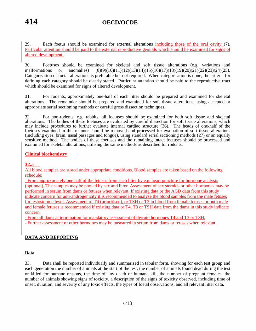
Clinical biochemistry
32.a All blood samples are stored under appropriate conditions. Blood samples are taken based on the following schedule: - From approximately one half of the fetuses from each litter by e.g. heart puncture for hormone analysis (optional). The samples may be pooled by sex and litter. Assessment of sex steroids or other hormones may be performed in serum from dams or fetuses when relevant. If existing data or the AGD data from this study indicate concern for anti-androgenicity it is recommended to analyse the blood samples from the male fetuses for testosterone level. Assessment of T4 (prioritised), or TSH or T3 in blood from female fetuses or both male and female fetuses is recommended if existing data or T4, T3 or TSH data from the dams in this study indicate concern. - From all dams at termination for mandatory assessment of thyroid hormones T4 and T3 or TSH. - Further assessment of other hormones may be measured in serum from dams or fetuses when relevant.
DATA AND REPORTING
Data 33. Data shall be reported individually and summarised in tabular form, showing for each test group and each generation the number of animals at the start of the test, the number of animals found dead during the test or killed for humane reasons, the time of any death or humane kill, the number of pregnant females, the number of animals showing signs of toxicity, a description of the signs of toxicity observed, including time of onset, duration, and severity of any toxic effects, the types of foetal observations, and all relevant litter data.

OECD/OCDE 414
7/13
34. Numerical results should be evaluated by an appropriate statistical method using the litter as the unit for data analysis. A generally accepted statistical method should be used; the statistical methods should be selected as part of the design of the study. Data from animals that do not survive to the scheduled kill should also be reported. These data may be included in group means where relevant. Relevance of the data from such an animal, and therefore inclusion or exclusion from any group mean(s), should be judged on an individual basis.
Evaluation of Results 35. The findings of the Prenatal Developmental Toxicity Study should be evaluated in terms of the observed effects. The evaluation will include the following information:
• maternal and foetal test results, including an evaluation of the relationship, or lack thereof, between the exposure of the animals to the test substance and the incidence and severity of all findings;
• criteria used for categorising foetal external, soft tissue, and skeletal alterations if categorisation has been done;
• when appropriate, historical control data to enhance interpretation of study results; • the numbers used in calculating all percentages or indices; • adequate statistical analysis of the study findings, when appropriate, which should include sufficient
information on the method of analysis, so that an independent reviewer/statistician can re-evaluate and reconstruct the analysis.
• The statistical analysis should take litter effect into account. 36. In any study which demonstrates an absence of toxic effects, further investigation to establish absorption and bioavailability of the test substance should be considered.
Test report 37. The test report must include the following specific information:
Test chemicalsubstance: -
- source, lot number, limit date for use, if available - stability of the test chemical, if known. physical nature and, where relevant, physiochemical properties;
- identification including CAS number if known/established; - purity. Mono-constituent substance:
- physical appearance, water solubility, and additional relevant physicochemical properties;
- chemical identification, such as IUPAC or CAS name, CAS number, SMILES or InChI code, structural formula, purity, chemical identity of impurities as appropriate and practically feasible, etc.
Multi-constituent substance, UVBCs and mixtures: - characterised as far as possible by chemical identity (see above), quantitative
occurrence and relevant physicochemical properties of the constituents.

414 OECD/OCDE
8/13
Vehicle (if appropriate): - justification for choice of vehicle, if other than water. Test animals: - species and strain used; - number and age of animals; - source, housing conditions, diet, etc.; - individual weights of animals at the start of the test. Test conditions: - rationale for dose level selection; - details of test substance formulation/diet preparation, achieved concentration, stability and
homogeneity of the preparation; - details of the administration of the test substance; - conversion from diet/drinking water test substance concentration (ppm) to the actual dose (mg/kg
body weight/day), if applicable; - environmental conditions; - details of food and water quality. Results: - Maternal toxic response data by dose, including but not limited to: - the number of animals at the start of the test, the number of animals surviving, the number
pregnant, and the number aborting, number of animals delivering early; - day of death during the study or whether animals survived to termination; - data from animals that do not survive to the scheduled kill should be reported but not included in
the inter-group statistical comparisons; - day of observation of each abnormal clinical sign and its subsequent course; - body weight, body weight change and gravid uterine weight, including, optionally, body weight
change corrected for gravid uterine weight; - food consumption and, if measured, water consumption;
- thyroid hormone and other hormone levels, - necropsy findings, including uterine weight; - NOAEL values for maternal and developmental effects should be reported. Developmental endpoints by dose for litters with implants, including: - number of corpora lutea; - number of implantations, number and percent of live and dead foetuses and resorptions; - number and percent of pre- and post-implantation losses. Developmental endpoints by dose for litters with live foetuses, including:

OECD/OCDE 414
9/13
- number and percent of live offspring; - sex ratio; - foetal body weight, preferably by sex and with sexes combined;
- Anogenital distance of all foetuses - Thyroid hormone and other hormone levels (if measured)
- external, soft tissue, and skeletal malformations and other relevant alterations; - criteria for categorisation if appropriate; - total number and percent of foetuses and litters with any external, soft tissue, or skeletal alteration,
as well as the types and incidences of individual anomalies and other relevant alterations (including in external reproductive genitals).

414 OECD/OCDE
10/13
Discussion of results. Conclusions. Interpretation of Results 38. A prenatal developmental toxicity study will provide information on the effects of repeated oral exposure to a substance during pregnancy. The results of the study should be interpreted in conjunction with the findings of subchronic, reproduction, toxicokinetic and other studies. Since emphasis is placed on both general toxicity and developmental toxicity endpoints, the results of the study will allow for the discrimination between developmental effects occurring in the absence of general toxicity and those which are only expressed at levels that are also toxic to the maternal animal (28).
LITERATURE (Should be updated!) (1) Organisation for Economic Co-operation and Development (1995). Report of the OECD Ad Hoc
Working Group on Reproduction and Developmental Toxicity. Copenhagen, Denmark, (13th-14th June 1995).
(2) Kavlock R.J. et al. (1996). A Simulation Study of the Influence of Study Design on the Estimation of
Benchmark Doses for Developmental Toxicity. Risk Anal. 16, 399-410. (3) Kimmel, C.A. and Francis, E.Z. (1990). Proceedings of the Workshop on the Acceptability and
Interpretation of Dermal Developmental Toxicity Studies. Fundam. Appl. Toxicol. 14, 386-398. (4) Wong, B.A., et al. (1997). Developing Specialized Inhalation Exposure Systems to Address
Toxicological Problems. CIIT Activities 17, 1-8. (5) US Environmental Protection Agency (1985). Subpart E - Specific Organ/Tissue Toxicity, 40 CFR
798.4350: Inhalation Developmental Toxicity Study. (6) Salewski, E. (1964). Faerbermethode zum Makroskopischen Nachweis von Implantations Stellen am
Uterus der Ratte. Naunyn-Schmeidebergs Arch. Pharmakol. Exp. Pathol. 247, 367. (7) Edwards, J.A. (1968). The External Development of the Rabbit and Rat Embryo. In: Adv. Teratol.
D.H.M. Woolam (ed.), Vol. 3. Academic Press, NY. (8) Inouye, M. (1976). Differential Staining of Cartilage and Bone in Fetal Mouse Skeleton by Alcian
Blue and Alizarin Red S. Congen. Anom. 16, 171-173. (9) Igarashi, E. et al. (1992). Frequency of Spontaneous Axial Skeletal Variations Detected By the
Double Staining Technique for Ossified and Cartilaginous Skeleton in Rat Foetuses. Congen. Anom. 32, 381-391.

OECD/OCDE 414
11/13
(10) Kimmel, C.A. et al. (1993). Skeletal Development Following Heat Exposure in the Rat. Teratology 47, 229-242.
(11) Marr, M.C. et al. (1988). Comparison of Single and Double Staining for Evaluation of Skeletal
Development: The Effects of Ethylene Glycol (EG) in CD Rats. Teratology 37, 476. (12) Barrow, M.V. and Taylor, W.J. (1969). A Rapid Method For Detecting Malformations in Rat
Foetuses. J. Morphol. 127, 291-306. (13) Fritz, H. (1974). Prenatal Ossification in Rabbits as Indicative of Foetal Maturity. Teratology 11,
313-320. (14) Gibson, J.P. et al. (1966). Use of the Rabbit in Teratogenicity Studies. Toxicol. Appl. Pharmacol. 9,
398-408. (15) Kimmel, C.A. and Wilson, J.G. (1973). Skeletal Deviation in Rats: Malformations or Variations?
Teratology 8, 309-316. (16) Marr, M.C. et al. (1992). Developmental Stages of the CD (Sprague-Dawley) Rat Skeleton after
Maternal Exposure to Ethylene Glycol. Teratology 46, 169-181. (17) Monie, I.W. et al. (1965). Dissection Procedures for Rat Foetuses Permitting Alizarin Red Staining of
Skeleton and Histological Study of Viscera. Supplement to Teratology Workshop Manual, pp. 163-173.
(18) Spark, C. and Dawson, A.B. (1928). The Order and Time of Appearance of Centers of Ossification in
the Fore and Hind Limbs of the Albino Rat, With Special Reference to the Possible Influence of the Sex Factor. Am. J. Anat. 41, 411-445.
(19) Staples, R.E. and Schnell, V.L. (1964). Refinements in Rapid Clearing Technique in the KOH-
Alizarin Red S Method for Fetal Bone. Stain Technol. 39, 61-63. (20) Strong, R.M. (1928). The Order, Time and Rate of Ossification of the Albino Rat (Mus norvegicus
albinus) Skeleton. Am. J. Anat. 36, 313-355. (21) Stuckhardt, J.L. and Poppe, S.M. (1984). Fresh Visceral Examination of Rat and Rabbit Foetuses
Used in Teratogenicity Testing. Teratogen. Carcinogen. Mutagen. 4, 181-188. (22) Walker, D.G. and Wirtschafter, Z.T. (1957). The Genesis of the Rat Skeleton. Thomas, Springfield,
IL. (23) Wilson, J.G. (1965) Embryological Considerations in Teratology. In: Teratology: Principles and
Techniques, J.G. Wilson and J. Warkany (eds.), University of Chicago, Chicago, IL, pp 251-277. (24) Wilson, J.G. and Fraser, F.C. (eds.) (1977). Handbook of Teratology, Vol. 4. Plenum, NY.

414 OECD/OCDE
12/13
(25) Varnagy, L. (1980). Use of Recent Fetal Bone Staining Techniques in the Evaluation of Pesticide Teratogenicity. Acta Vet. Acad. Sci. Hung. 28, 233-239.
(26) Staples, R.E. (1974). Detection of Visceral Alterations in Mammalian Foetuses. Teratology 9, 37-38. (27) Van Julsingha, E.B. and C.G. Bennett (1977). A Dissecting Procedure For the Detection of
Anomalies in the Rabbit Foetal Head. In: Methods in Prenatal Toxicology (eds. D. Neubert, H.J. Merker and T.E. Kwasigroch). University of Chicago, Chicago, IL, pp. 126-144.
(28) US Environmental Protection Agency (1991). Guidelines for Developmental Toxicity Risk
Assessment. Fed. Register 56, 63798-63826. (29) Wise, D.L. et al. (1997). Terminology of Developmental Abnormalities in Common Laboratory
Mammals (Version 1). Teratology 55, 249-292.

OECD/OCDE 414
13/13
ANNEX
Developmental toxicology: the study of adverse effects on the developing organism that may result from exposure prior to conception, during prenatal development, or postnatally to the time of sexual maturation. The major manifestations of developmental toxicity include 1) death of the organism, 2) structural abnormality, 3) altered growth, and 4) functional deficiency. Developmental toxicology was formerly often referred to as teratology. Adverse effect: any treatment-related alteration from baseline that diminishes an organism’s ability to survive, reproduce or adapt to the environment. Concerning developmental toxicology, taken in its widest sense it includes any effect which interferes with normal development of the conceptus, both before and after birth. Altered growth: an alteration in offspring organ or body weight or size. Alterations (anomalies): structural alterations in development that include both malformations and variations (29): Malformation/Major Abnormality: Structural change considered detrimental to the animal (may also
be lethal) and is usually rare. Variation/Minor Abnormality Structural change considered to have little or no detrimental effect on
the animal; may be transient and may occur relatively frequently in the control population. Conceptus: the sum of derivatives of a fertilised ovum at any stage of development from fertilisation until birth including the extra-embryonic membranes as well as the embryo or foetus. Implantation (nidation): attachment of the blastocyst to the epithelial lining of the uterus, including its penetration through the uterine epithelium, and its embedding in the endometrium. Embryo: the early or developing stage of any organism, especially the developing product of fertilisation of an egg after the long axis appears and until all major structures are present. Embryotoxicity: detrimental to the normal structure, development, growth, and/or viability of an embryo. Foetus: the unborn offspring in the post-embryonic period. Foetotoxicity: detrimental to the normal structure, development, growth, and/or viability of a foetus. Abortion: the premature expulsion from the uterus of the products of conception: of the embryo or of a nonviable foetus. Resorption: a conceptus which, having implanted in the uterus, subsequently died and is being, or has been resorbed: Early resorption: evidence of implantation without recognisable embryo/foetus. Late resorption: dead embryo or foetus with external degenerative changes. NOAEL: abbreviation for no-observed-adverse-effect level and is the highest dose level where no adverse treatment-related findings are observed.