FDA STAMP Conference on CNS Shunts Agenda January 1999
39
SHUNT TECHNOLOGY: CHALLENGES AND EMERGING DIRECTIONS Sponsored by United States Food and Drug Administration Center for Devices and Radiological Health at National Naval Medical Center Bethesda, Maryland Friday, January 8, 1999 CONTACT: Janine M. Morris Office of Device Evaluation Center for Devices and Radiological Health 9200 Corporate Blvd, HFZ-404 Rockville, Maryland 20850 Tel. (301) 594-1190 ext. 139 Fax. (301) 594-2977 Email. [email protected] INTRODUCTION FDA's first STAMP (Systematic Technology Assessment of Medical Products) conference was held at the National Naval Medical Center (NNMC), Bethesda, Maryland, January 8, 1999. The one-day international conference brought together over 140 attendees from various groups, including neurosurgical, nursing, patient advocacy, government and industry. The meeting addressed -- Cerebrospinal Shunt Technology: Challenges and Emerging Directions -- and was introduced by Dr. Bruce Burlington, Director, Center for Devices and Radiological Health (CDRH). Dr. Burlington explained the STAMP effort as similar to one in 1997, evolving from the concerns of another medical device -- the pulmonary artery catheter (PA catheter). This early effort involved both FDA and the National Heart Lung and Blood Institute and focused on the utilization, technology, and training associated with the use of the pulmonary artery catheter (PA catheter). Similarly, this STAMP effort deals with cerebrospinal fluid shunt systems and meets the STAMP mission criteria. The STAMP mission reviews families of closely related medical devices having broad use and most often, several years of marketing experience, as well as, a significant potential for adverse events. In the process of device selection for STAMP, certain device aspects are raised including, product characteristics, evidence of performance and effectiveness, adverse event experience, and manufacturing and supply issues. The expected outcome is
-
Upload
stephen-dolle -
Category
Government & Nonprofit
-
view
49 -
download
0
Transcript of FDA STAMP Conference on CNS Shunts Agenda January 1999

SHUNT TECHNOLOGY:CHALLENGES AND EMERGING DIRECTIONS
Sponsored byUnited States Food and Drug AdministrationCenter for Devices and Radiological Health
atNational Naval Medical Center
Bethesda, MarylandFriday, January 8, 1999
CONTACT:Janine M. MorrisOffice of Device EvaluationCenter for Devices and Radiological Health9200 Corporate Blvd, HFZ-404Rockville, Maryland 20850
Tel. (301) 594-1190 ext. 139Fax. (301) 594-2977Email. [email protected]
INTRODUCTIONFDA's first STAMP (Systematic Technology Assessment of Medical Products) conferencewas held at the National Naval Medical Center (NNMC), Bethesda, Maryland, January 8,1999. The one-day international conference brought together over 140 attendees fromvarious groups, including neurosurgical, nursing, patient advocacy, government andindustry. The meeting addressed -- Cerebrospinal Shunt Technology: Challenges andEmerging Directions -- and was introduced by Dr. Bruce Burlington, Director, Center forDevices and Radiological Health (CDRH). Dr. Burlington explained the STAMP effort assimilar to one in 1997, evolving from the concerns of another medical device -- thepulmonary artery catheter (PA catheter). This early effort involved both FDA and theNational Heart Lung and Blood Institute and focused on the utilization, technology, andtraining associated with the use of the pulmonary artery catheter (PA catheter). Similarly,this STAMP effort deals with cerebrospinal fluid shunt systems and meets the STAMPmission criteria.
The STAMP mission reviews families of closely related medical devices having broad useand most often, several years of marketing experience, as well as, a significant potentialfor adverse events. In the process of device selection for STAMP, certain device aspectsare raised including, product characteristics, evidence of performance and effectiveness,adverse event experience, and manufacturing and supply issues. The expected outcome is

to interact with stakeholders in a public forum (medical community, patient groups,others) as well as to develop recommendations on steps to improve patient outcome.
In 1976 FDA's Neurological Devices Classification Panel recommended Regulatory ClassII for CSF shunts, which require premarket clearance for marketing. Classification wasbased upon the belief that standards could be written to assure the safety andeffectiveness of marketed shunts, and that clinical experience had proven shunts to bereasonably safe and effective. Today, marketing requirements for shunts with significantdesign change require bench testing (ASTM Standard F4794) as well as clinical data toshow that the modification does not impact the safety and effectiveness of the device.Furthermore, biocompatibility data and labeling are carefully reviewed.
Over the past five years, between 114 and 160 adverse event reports for CSF valves weresubmitted by manufacturers through the FDA Medical Device Reporting System. Thereports involved the following valve technologies: proximal slit, distal slit, diaphragm,ball-in-cone, gravitational (hydrostatic) ball-in-cone, siphon-preventing diaphragm, andauto-regulated (flow-limitation) diaphragm and needle.
While CSF shunts have been used to manage and treat hydrocephalus for over 40-years,clinical experience and adverse event reporting indicate continuing problems, despiteadvances in technology. Over 60% of shunt patients manifest some type of shuntcomplication over their lifetime such as shunt obstruction, over-drainage, infection, devicemigration, disconnection and fracture. This conference aims to examine the issues andexplore different approaches to improving patient outcome.
Agenda and list of presentations

SESSION I
Title: Shunt Technology Perspectives
Objective: To recognize alternative perspectives of shunt technology from selectedstakeholders.
Moderator:
JANINE M. MORRIS, Mechanical EngineerCenter for Devices and Radiological HealthFood and Drug Administration
Speakers:
EMILY S. FUDGEExecutive DirectorHydrocephalus Association“Patient and Faimily Perspectives and Needs”
SUSAN MCGEE, CNPPediatric Special Focus Group FacilitatorAmerican Association of Neuroscience Nurses“Shunt Technology – A Nursing Perspective”
MARVIN L. SUSSMAN, Ph.D.Industry Consultant“Shunt Technology: Challenges and Emerging Directions – A ManufacturingPerspective”
HAROLD REKATE, MDNeurosurgeonBarrows Neurological Institute“BioPhysics of the CSF Pathways: What Can and What Should a Shunt Do?”
SESSION II
Title: Hydrocephalus and Assessment of Shunt Function
Objective: To develop an increased understanding of the relationship between thepathophysiology of hydrocephalus and shunt performance.

Moderator:
JEAN RINALDI, Biomedical EngineerCenter for Devices and Radiological HealthFood and Drug Administration
SESSION IIA
Title: Pathophysiology of Hydrocephalus
Speakers:ANTHONY MARMAROU, Ph.D.Professor and Vice ChairmanDivision of NeurosurgeryMedical College of Virginia"Fundamentals of Intracranial Pressure"
CONRAD E. JOHANSON, Ph.D.Professor and Director of Cerebrospinal Fluid LaboratoryDepartment of Clinical NeurosciencesBrown University Medical School"Growth Factor Induction of Normal Pressure Hydrocephalus"
MARK G. LUCIANO, MD, Ph.D.Head, Section of Pediatric NeurosurgeryDepartment of NeurosurgeryThe Cleveland Clinic Foundation"Animal & Clinical Testing of Systems for Adult Onset Chronic Hydrocephalus"
SESSION IIB
Title: Assessment of Shunt Function
Speakers:
MICHAEL A. WILLIAMS, MDAssistant Professor and Medical DirectorClinical Neurocirculatory Laboratory Johns Hopkins Medical Institutions"In Vivo Assessment of Shunt Function and Failure"
AZAR P. DAGHER, MDDiagnostic Radiology DepartmentNational Institutes of Health
"Radiological Tools Used in the Evaluation of Shunt Malfunction"
PROFESSOR JOHN PICKARD, MChir, FRCS, FmedSciMAREK CZOSNYKA, PhD, DSC, and ZOFIA CZOSNYKAUniversity of CambridgeCambridge England"UK Shunt Evaluation Laboratory"
SESSION III
Title: Challenges of Infection and New Perspectives
Objective: To identify potential approaches to mitigate or prevent shunt-centeredinfections
Moderator:
ROGER BAYSTON, MMedSci, FRCPathBiomaterials-Related Infection GroupUniversity Division of MicrobiologyUniversity of Nottingham England
Speakers:
ROGER BAYSTON, MMedSci, FRCPathBiomaterials-Related Infection GroupUniversity Division of MicrobiologyUniversity of Nottingham England"Incidence and Aetiology of Shunt-Associated Infections"
WILLIAM R. JARVIS, MDChief, Investigation and Prevention BranchNational Center for Infectious DiseasesCenters for Disease Control and Prevention"Vancomycin Use in Pediatric Neurosurgery Patients--Can We ImproveCompliance with Vancomycin Recommendations?"
ROBERT J. SHERERTZ, MDChief, Section on Infectious DiseasesDepartment of MedicineWake Forest University School of Medicine"Pathogenesis and Prevention of Foreign Body Infections"
SESSION IV
Title: Clinical Outcomes and Methods of Surveillance
Objective: To describe selected strategies to improve clinical outcomes and theperformance of CSF shunts.
Moderator:
JANINE M. MORRIS, Mechanical EngineerCenter for Devices and Radiological HealthFood and Drug Administration
Speakers:
PROFESSOR. JOHN PICKARD, Mchir, FRCS, FMedSciHUGH RICHARDS, PhD, HELEN SEELEY and MERYL MADAKBASUniversity of CambridgeCambridge England"Experience with Setting up and Maintaining the UK Shunt Registry"
ROBERT E. HARBAUGH, MD, FACSChairman, AANS/CNS Outcomes CommitteeAmerican Association of Neurological Surgeons and Congress of Neurological SurgeonsDartmouth-Hitchcock Medical Center"On-Line Neurosurgical Outcomes Studies".
JOHN R. W. KESTLE, MD, M.Sc., FRCSCDivision of Pediatric NeurosurgeryUniversity of UtahPrimary Children's Medical Center"Issues in Data Collection and Outcome Analysis"
PANEL DISCUSSION
Title: Future Directions – Prioritizing Our Efforts
Objective: To achieve a shared vision on how to advance shunt technology in themanagement and treatment of hydrocephalus and establish priorities forfuture action.

Moderator:
LARRY KESSLER, Sc.D.Director, Office for Surveillance and BiometricsCenter for Devices and Radiological HealthFood and Drug Administration
Panel:
HAROLD REKATE, MDNeurosurgeonBarrows Neurological Institute
JOHN R. W. KESTLE, MDDivision of Pediatric NeurosurgeryUniversity of UtahPrimary Children's Medical Center
ROGER BAYSTON, MMedSciBiomaterials-Related Infection GroupUniversity Division of MicrobiologyUniversity of Nottingham England
GREG A. TOCCOHydrocephalus Foundation (HyFl)Boston, Massachusetts
KIMBER RICHTER, MDDeputy DirectorOffice of Device EvaluationCenter for Devices and RadiologicalHealthFood and Drug Administration
MICHAEL D. WALKER, MDDirectorDivision of Stroke, Trauma, andNeurological Degenerative DisordersNational Institute of NeurologicalDisorders and StrokeNational Institutes of Health
Summary:The discussion began with suggestions on improving patient protection. One idea was toissue patient cards containing basic information on the type of shunt implanted as well asthe size of patient’s ventricles. These cards could be used in case of emergency.Additional comments included the potential benefit of on-line data collection in reducingthe burden on neurosurgeons, providing framework for longitudinal follow-up, andreducing chances of missed forms. Standard definitions would increase theunderstanding between individual neurosurgeons as well as sequential enrollment of allcases by participating neurosurgeons and hospitals would be needed in order torigorously assess the effectiveness of care.
Return to top of agenda

SESSION IShunt Technology Perspectives
Patient and Family Perspectives and Needs
Emily Fudge, Executive DirectorThe Hydrocephalus AssociationSan Francisco, California
The first generation of infants and children treated for hydrocephalus is nowreaching adulthood. While many are living normal and productive lives, theyhave been beset with a myriad of problems and complications – learningdisabilities, developmental and social skill delays, neurological impairment,visual problems, and all too frequently, shunt malfunction.
The Hydrocephalus Association (HA) founded in 1983, provides support,education, and advocacy resources to individuals with hydrocephalus and theirfamilies. Also it functions as a source of educational materials for distributionby health care providers. There were 900 dues-paying members in 1998. Inaddition to individuals and parents of infants and children with hydrocephalus,health professionals and organizations with complementary goals maintainmembership.
In preparation for the conference a twelve-item survey of 675 HA members wasinitiated. The sample represented members with a diagnosis of hydrocephalus.Completed surveys were returned by 239 males and 175 females, i.e., 414respondents (61%).
DIAGNOSIS / CAUSE OF HYDROCEPHALUS
40.3% CONGENITAL (AQUEDUCTAL STENOSIS, SPINA BIFIDA, ARACHNOID CYSTS,DANDY-WALKER, CHIARI MALFORMATIONS…)
27.3% Acquired ( Intraventricular Hemorrhage, Meningitis, Trauma, Tumor…)11.6% Normal Pressure Hydrocephalus17.6% Unknown3.1% Other
CURRENT AGE of RESPONDENTS with SHUNTS
3.6% LESS THAN YEAR OLD31.4% 1 to 5yrs25.6% 6 to 12yrs12.6% 13 to 20yrs

7.2% 21 to 35yrs 6.0% 36 to 50yrs13.5% 51yrs or older with 85yrs being the oldest
AGE at which FIRST SHUNTED
65.5% LESS THAN YEAR OLD5.1% 1 to 2 yrs6.8% 2 to 5yrs2.7% 6 to 12yrs1.4% 13 to 20yrs2.7% 21 to 35yrs4.8% 36 to 50yrs11.1% 51yrs or older
KNOWLEDGE of SHUNT TYPE
233 YES169 No
IDENTIFICATION CARD on PERSON
97 YES303 No
NUMBER of REVISIONS
32.6% NONE49.6% 1 to 59.7% 6 to 103.5% 11 to 152.2% 6 to 202.7% > 20
REASONS for REVISIONS
154 OBSTRUCTION (DISTAL AND/OR PROXIMAL)71 Malfunction (Mechanical or Unknown)63 Infection61 Pressure related47 Broken or disconnected catheter29 Lengthen the catheter24 Place a 2nd shunt14 Hemorrhage; hematoma7 Distal catheter migration39 Others

RECD NEUROPSYCHOLOGICAL/DEVELOPMENTAL TESTING
238 YES239 No
SIDE EFFECTS EXPERIENCED
198 YES190 No
EXAMPLES
131 HEADACHE81 Balance and Coordination problems38 General Pain24 Dizziness8 Visual disturbance8 Learning6 Seizures4 Confusion2 Short term Memory problems29 Other
PERCEIVED DISADVANTAGESWith SHUNT TECHNOLOGY
137 MALFUNCTION, REVISION, OBSTRUCTION, BREAKS 51 Technology, No interest/money, Moving too slowly 35 Difficulty Assessing shunt function 33 Infection 27 Need for Specialized Care and Knowledge/Interested Doctors 26 Invasive techniques needed to diagnose malfunction and replace shunt 22 Lifestyle Restrictions 19 Pressure related problems 13 Brain Injury 54 Other
PERCEIVED ADVANTAGES With SHUNT TECHNOLOGY
133 ALLOWS A NORMAL LIFE115 Saved Life 63 Treats Hydrocephalus 53 Good Technology; New Technology possible 37 Design is simple; Shunt is invisible and reliable

16 Surgery is a simple operation 21 Other
In summary it was found that individuals with hydrocephalus and their familiesare grateful for the extension of life that shunt technology has provided.However, they believe additional measures need to be explored to improvequality of life. These include attention to the high revision rate resulting fromthe complications of infection, and mechanical failure. Increased support fordevelopment of new technologies is eagerly awaited.
Return back to agenda and list of presentations

Shunt Technology – A Nursing Perspective
Susan McGee, RN, MSN, CNPPediatric Special Focus Group FacilitatorAmerican Association of Neuroscience Nurses (AANN)
The AANN is composed of 4000 members with chapters in the United States andCanada. Activities include an annual scientific meeting, journal, newsletter, andclinical guidelines publications. Drawing on her experience as an AANNPediatric Focus Group Facilitator, Ms. McGee discussed ongoing problems forcaregivers related to discrepancies between patients’ subjective symptom reportsand objective diagnostic tool findings as well as inability to identify implantedhardware. Patients’ problems include headache, pain control, and intervention-associated infections. Furthermore, the potential for shunt malfunction and thetransition to adult care, involving new health care providers, are frequentsources of patient anxiety.
As a pediatric nurse practitioner, Ms. McGee is responsible for the nursingmanagement of inpatients and outpatients with hydrocephalus treated withventricular shunting devices. This includes: teaching patients and families abouthydrocephalus and treatment options, recognizing and reporting symptoms ofshunt malfunction, caring for patients during acute malfunction episodes, andassisting with patient and family lifestyle management issues. At the CincinnatiChildren’s Hospital Medical Center, over the first year of initial shuntimplantation, 14% malfunction, and 3% infection rates were reported. Of thirdventriculostomies 80% were an initial procedure with a 67% success rate. A 75%success rate was reported in 20% of previously shunted patients converted to athird ventriculostomy. Success is defined as a minimum of one year post-surgery without recurrent symptoms of hydrocephalus.
From a nursing perspective it was proposed that the following shunt-relatedissues continue to be addressed:
• Outcomes improvement for premature infants• Malfunction assessment• Reality of “stiff ventricles,” overshunting, and post-shunting synostosis• Reliability of intracranial pressure (ICP) monitoring• Headache/pain reduction• Safety of external ventricular drains• Criteria for selection of third ventriculostomy candidates• Transition to adult care• Real life/time testing of hardware

Shunt Technology: Challenges and Emerging Directions – AManufacturing Perspective
Marvin L. Sussman, PhDIndustry ConsultantMiami, Florida
Dr. Sussman spent 20 years in the corporate neurological shunt industry. Heserved as co-chair of the American Society for Testing and Materials (ASTM)Shunt Standards Committee that developed the Standard Practice for Evaluatingand Specifying Implantable Assemblies for Neurosurgical Application (F647-94)and worked on the development of the International Organization forStandardization (ISO) Shunt Standard (ISO/TC 150/SC-7197).
According to Dr. Sussman valve designs consist of five types: silicone slit, hybridsilicone, ball-in-cone, hybrid silicone/ruby and programmable ball–in-cone.Self-adjusting flow control, programmable differential pressure valves, andcoating for friction-reducing lubricity are examples of recent advances inneurological shunt technology that may not be addressed by current standards.Biomaterials currently used include:
• Silicone elastomer – catheters, valve housings/mechanisms, suture clamps,guides, siphon devices, etc.
• Polypropylene/Polysufone/Nylon/Polyethersulfone – valve housings/seats,needle stops, connectors, reservoirs, etc.
• Ruby/Sapphire – valve pins, balls, seats• Titanium/Stainless Steel – valve housings, needle stops• Tantalum – radiopaque markers• Barium – radiopaciofier (homogenous or stripe)
Self-healing properties and the ability to elongate with patient growth, may becharacteristic of future biomaterials. Silicone elastomers are the primarymaterials used for neurological shunts at present. In 1998, the American MedicalAssociation published a position paper in response to concerns raised by issuesidentified with silicone breast implants in which it was stated that, “data do notsuggest a specific immune response to silicone elastomer in shunts. It isrecommended that research and development continue on materials, as well asdesigns and procedures for shunts. Also that monitoring of adverse reactions todevices take place so that appropriate research can be conducted.
Methodologies to assess shunt component radiopacity; flexibility; security ofassembly; tensile properties; unidirectional flow; and leakage are manufacturer-

specific and therefore not readily comparable between manufacturers. In-vitroassessment of factors that may affect the performance of certain shunts or shuntcomponents, such as tissue encapsulation of siphon devices, and effects ofsleeping on the valve/siphon device and position of the device relative to theForamen of Monro, are not addressed in ASTM standard F647-94.
As illustrated in the following 1990 table, another issue not resolved inindustry’s approach to the specification of shunt operating characteristics, underdynamic and steady-state conditions, is agreement regarding standardizeddefinitions of the various differential pressure ranges.
DP Shunt Specifications (Ca. 1990)Cordis J&J/Codman
Accu-FloDenver Codman
HolterP -S
Flow-ControlRadionics
NominalRange
mm H 2OOperatingPressure5 ml/hr
mm H 2 OClosing
Pressure5/50 ml/hr
Flow Rate(cc/hr) @100 mm
H 2O
mm H 2OOperatingPressure8.6 ml/hr
mm H 2OOperatingPressure
5/50 ml/hr
mm H 2OOperatingPressure
5/40 ml/hrVery/Extra/
Low Low15-40
Blue
0-10 5/40
Low 40-80White
2/75 6-17 11-40 10/70 35
Medium 80-120Yellow
45/140 18-59 41-75 60/130 75/100
High 120-170Brown
100/220 60-120 76-110 120/200 175/175
Very High 170-230Green
NominalRanges
5 3 3 5 3 3
1998Manufacturer
NMT J&JCodman
J&JCodman
J&JCodman
MedtronicP-S
Radionics
R e f e r e n c e : T h e S h u n t B o o k ( D r a k e a n d Sa in te-R o s e; 1995 p p. 75-92)
In addition, there is little consensus regarding the definition of terms:differential pressure valve, flow-control valve, and physiological valve. Indeed,the neurosurgeon is often the “final manufacturer-assembler” of a neurologicalshunt using components from a variety of manufacturers. This leads to attemptsto “test” shunt assemblies in the operating room and complicates labelingefforts. No standardized tests or outcomes have been approved for this practicewhich also invites increased risk of infection.
In many instances, shunt components are not identifiable once implanted. Thereis a need to give patients generic device identification cards on which all the
shunt assembly components are specified. Improvement in physician andpatient instruction materials using a variety of approaches, such as, handouts,manufacturer and FDA web sites, and support groups, are suggested by Dr.Sussman.
It was also noted that the approximately 100 annual neurological shunt adverseevent reports may reflect under-reporting. A need for guidance on what shunt-related events should be reported was proposed, as was repetition of theCDRH/WEAC 1990 study, Conformance Assessment to Voluntary Standards, usingthe 1994 ASTM standard F647, and extension of the Drake & Kestle study (1998 –2 year results), Pediatric Cerebrospinal Fluid Shunt Design.
In summary, it was recommended that from an industry perspective, thefollowing actions were proposed:
• Information dissemination• Shunt comparison efforts• Development of new technologies and biomaterials• Protection of biomaterials supply access
Return back to agenda and list of presentations

BioPhysics of the CSF Pathways: What Can and What Should aShunt Do?
Harold L. RekateChief, Pediatric NeurosurgeryBarrow Neurological InstitutePhoenix, Arizona
Dr. Rekate has been an attending neurosurgeon for twenty years. He isprimarily involved in the clinical care of pediatric patients with hydrocephalus,but also treats adults with complex sequelae of hydrocephalus. His extensiveresearch experience in CSF physiology includes a comparison of computersimulation of a mathematical model of cerebrospinal fluid (CSF) flow tophysiology experiments in animal models of hydrocephalus. In addition, Dr.Rekate is the author of sixty publications, and holds several administrationpositions in professional neurosurgical groups.
In his presentation, Dr.Rekate stated that from 50% to 90% of the CSF isproduced within the cerebral ventricles by both choroid plexuses. Theremainder is produced as a by-product of cerebral metabolism in the cerebraland spinal cord whitematter. CSF flows from the ventricles through a series ofconduits to the subarachnoid space and eventually is absorbed into the duralvenous sinuses – primarily the superior sagittal sinus (SSS).
Intracranial pressure (ICP) in the normal, non-hydrocephalic condition isprimarily a function of cerebral vein pressure.
• Sagittal sinus pressure (SSP) is 3-5mm Hg above right atrial pressure inrecumbency
• Cortical veins have valve mechanisms that create about a 5 mm Hgdifferential between ICP and SSP. This pressure is transmitted to the brainparenchyma and is the primary determinant of brain turgor.
• Jugular veins are collapsible and close when their pressure falls belowatmospheric pressure.
A CSF shunt is composed of a ventricular catheter, a reservoir that allowspressure measurement, a valve, and a distal catheter. Maintenance of normalintracranial pressure is relatively simple in the recumbent position, but that doesnot mean intracranial dynamics are normal.
• CSF flow in the normal situation is pulsatile with large volumes of CSFflowing back and forth across the aqueduct of Sylvius.
• Net flow of 0.3cc/min = 20cc/hour – a pint a day.
• With each pulsation, the CSF that flows through the shunt is irrevocably lost.• Relatively large volumes of CSF are lost with cough and straining and
essentially all available CSF is lost at the time of assuming the erect position.• For much of the day there is often no flow through the device.
In the erect position, the height of the inflow (ventricular catheter) is muchhigher than the height of the outflow (peritoneal catheter). In a normallyfunctioning differential pressure valve with an opening or closing pressure of90-120 mm H2O (7-9 mm Hg) recumbent ICP at steady state also would be about90-120 mm H2O. In the erect position, ICP is predicted to be, and has beenmeasured to be, minus 20 to minus 30 mm Hg. Chronically shunted patientshave significantly lower overall volumes of intracranial CSF, both in theventricles and in the cortical subarachnoid spaces, resulting in markedlythickened skulls in patients who have been shunted in infancy, and enlargedparanasal sinuses that develop earlier than in age-matched controls. This leadsto chronically decreased CNS buffering capacity for changes in ICP and leads tothe question of whether there is a role for, or a possibility of, producing a shuntwhich incorporates a component which stores and can reinfuse stored CSF.
A natural shunt consists of a ventriculo-sagittal sinus where the arachnoid villifunctions as a medium differential pressure valve that handles 20cc of CSF perhour without a siphon effect. There are four good re-creations of nature,however. A ventriculo-sagittal sinus shunt with a high differential pressurevalve, the distance between the inlet and outflow does not change with changesin position and no siphoning occurs, but there is danger of occlusion and/orinfection of the superior sagittal sinus, of which there is only one. In contrast,there are two transverse sinuses into which ventricular CSF can be shunted.Another option is a ventriculo-jugular shunt that functions against the directionof flow, and finally, ventricular-atrial or ventricular-peritoneal shunts thatrequire a siphon-controlling mechanism if the patient is more than 2 ½ feet tall.There are several devices that have been developed to retard the overdrainagecaused by siphoning.
• Anti-siphon device• Siphon control device• Siphon limiting device• Delta valve• Orbis sigma valve• Horizontal-vertical valve• Programmable valve – although even the highest valve pressure will not
prevent very low ICP

Most patients do quite well regardless of the shunt selected, but some requirespecific types of shunts. The most important factor seems to be the concept ofbrain turgor. Specific condition/treatment pairs that have been identifiedinclude:
• Posthemorrhagic hydrocephalus of the premature newborn/Pressuredifferential valve of ball-in-cup or diaphragm design, because of theforgiveness of high protein and cellular debris.
• High brain turgor patients (achondroplasia, Crouzons, others)/Highestpressure valve tolerated. May even need valves in series.
• Low brain turgor patients (Normo Pressure Hydrocephalus)/Low pressurevalve with a mechanism that prevents or retards siphoning.
Reflecting the perspective of a neurosurgeon, Dr. Rekate outlined the followingproblem areas as needing continued attention:
• Prevention of infection• Maintenance of CSF pressure buffering capacity• Development of valve programmability with a broader pressure range• Development of more effective and reliable siphon prevention/retardation,
and• Exploration of alternatives to silastic
Return back to agenda and list of presentations

SESSION IIHydrocephalus and Assessment of Shunt Function
Fundamentals of Intracranial Pressure
Anthony Marmarou, Ph.D.Professor and Vice ChairmanDivision of NeurosurgeryMedical College of Virginia
Dr. Anthony Marmarou from the Medical College of Virginia began this sessionwith an overview of the basics of intracranial pressure (ICP). An understandingof the fundamentals of ICP and its role in hydrocephalus and subsequentneurologic effects are important in understanding the use of shunts and othertechnologies in the treatment of ICP abnormalities. It is well-known thatcerebrospinal fluid (CSF) production is based upon weight and that the bulk ofCSF absorption takes place at the arachnoid villi. The arachnoid villi are theprimary components to the resistance of flow of CSF.
The fundamental basis of intracranial pressure volume dynamics is affected bycerebrospinal fluid (CSF) diversion. Intracranial pressure (ICP) is defined as thepressure, which must be exerted on a needle placed in the cerebrospinal fluidspace, sufficient to prevent the escape of fluid. According to Dr. Marmarou, adirect ventriculostomy gives the most accurate measure of ICP. The pressure isabove atmospheric and contains both respiratory and cardiac pulsatilecomponents. These components can be observed using different time scaleswhen recording the ICP. As fluid enters the intracranial cavity, pressure willrise according to the pressure volume relationship, which defines the"compliance" of the intracranial contents. At equilibrium, the combination of allthe compartmental volumes, blood, brain, and CSF, are constant. An increase inone compartment must be compensated by a decrease by another compartmentcapable of volume change. This occurs at the expense of pressure rise, which inpart is governed by the transient pressure volume curve and by the resistance tooutflow in the steady state. The pressure-to-volume relationship is not linear butexponential, and thus, derivation of the general equation describing intracranialpressure dynamics results in a first-order non-linear differential equation. Thisequation can be solved and a general solution obtained allowing the predictionof ICP change for any volume input. The relationship between ICP and volumecan be described by the pressure-volume index (PVI). Infants have a lower PVIthan adults, indicating a higher cranial compliance and subsequently greater ICPchange resulting from a smaller volume change. In addition, in injury, a longerduration of elevated ICP results in a lower survival rate.
Growth Factor Induction of Normal Pressure Hydrocephalus
Conrad E. Johanson, Ph.D.Professor and Director of Cerebrospinal Fluid LaboratoryDepartment of Clinical NeurosciencesBrown University Medical School
Dr. Conrad Johanson from Brown University in Rhode Island, presented hisresearch involving growth factor effects on the choroid plexus and how thesegrowth factors as (peptides) may influence hydrocephalus. It is important tounderstand how these biochemicals interact in the central nervous system tofurther our knowledge of the etiologies of hydrocephalus.
Growth factor upregulation in CNS can alter brain fluid dynamics.Hydrocephalus was induced in adult Sprague-Dawley rats by infusing basicfibroblast growth factor (FGF-2) at 1ug/d into a lateral ventricle for 2, 3, 5 or 10-12 d. Ventricular enlargement, without elevated cerebrospinal fluid (CSF)pressure, progressively increased from 2 to 10 d. At 10-12 d there was a 29%reduced CSF formation rate from 2.51 to 1.78 ul/min (P<0.01), and a concurrent101% increased resistance to CSF absorption, i.e., 0.84 to 1.69 mmHg.min-u1-1.Choroid plexus, the main site of CSF formation, had an increased number ofdark epithelial cells and a decrease in CSF formation, while the arachnoid villiregion, a major absorption site for CSF, displayed enhanced fibrosis and collagendeposition thus inhibiting CSF absorption. A long-term pharmacologic goal hasbeen to regulate CSF secretion. Diuretics have achieved a limited success. Ifcontrol of drainage at the arachnoid villi can be accomplished using growthfactors or other agents, perhaps an increase in outflow will reduce the need forCSF shunts. This is important since shunting may remove beneficial metabolitesfrom the CSF that are crucial for brain function.
Return back to agenda and list of presentations

Animal and Clinical Studies in Adult-Onset Chronic Hydrocephalus
Mark G. Luciano, MD, PhDHead, Section of Pediatric NeurosurgeryDepartment of NeurosurgeryThe Cleveland Clinic Foundation
Dr. Mark Luciano from the Cleveland Clinic Foundation in Ohio brought hisperspective on the usefulness of various animal models in hydrocephalusresearch. The advantages and disadvantages of various in vivo models werediscussed. It is important to consider the three interrelated aspects of the brainand central nervous system when developing research methodologies. First, thebrain can be treated as a purely mechanical system with associated pressures,flows, compliance, and resistance. Next, the brain can be affected by itssurrounding biochemistry, expanded in the previous presentation by Dr.Johanson. Lastly, the overall neurological function of the brain can beconsidered.
Although hydrocephalus continues to take a significant neurological toll on awide spectrum of neurosurgical patients, it remains incompletely understoodand inadequately treated. Several different animal models have been used toexplore a variety of basic pathophysiology questions. The variety of animalmodels reflect the heterogeneity of the disease and each animal model haslimitations in application. One animal model has fetal hydrocephalus inducedby ligature of the placental vessels or catheterization of the Aqueduct of Sylvius.This can cause undesired confounding effects on the development of the brain.Another type consists of genetic models occurring congenitally in dogs, pigs,horses, and oxen. Unfortunately, the basic pathophysiology underlying geneticmodels is poorly understood. Lastly, surgical induction of hydrocephalus hasbeen tried using balloons, silicone oil, and kaolin. Hydrocephalus induced inthis manner is affected by the amount of inflammation from the inducing agent.An animal model is described that is appropriate for the study of onset chronichydrocephalus. This model uses cyanoacrylate to obstruct the fourth ventricleallowing the severity and time course of the disease to be repeatable with noventricular deformation or tissue inflammation. This model can be used tostudy both basic pathophysiology and new clinically relevant hardware andprocedures.
Return back to agenda and list of presentations

In Vivo Assessment of Shunt Function and Failure
Michael A. Williams, M.D.Assistant Professor and Medical DirectorJohns Hopkins Medical InstitutionsBaltimore, Maryland
Dr. Michael Williams from the Johns Hopkins Medical Institutions in Marylandopened this session with his work involving the assessment of shunt problemswhile in clinical use. According to Dr. Williams, shunt malfunctions in adultsare common but severely under-recognized. This is critical because theassessment of shunt function is the determining factor for shunt revision, ahighly invasive procedure with potential for complications. If shunts could bepaired with intracranial pressure monitoring technology, an objective evaluationof shunt performance could be used clinical.
The treatment of hydrocephalus is predicated on a functioning shunt. Shuntscan malfunction in at least 3 ways: 1) complete obstruction of the valve orcatheters that results in symptomatic hydrocephalus, 2) valve mismatch,malfunction, or partial catheter obstruction that leads to under-drainage andhigher than desired intracranial pressure (ICP) associated with symptomatichydrocephalus, and 3) valve mismatch or malfunction that leads to over-drainage (siphoning) and ICP that is lower than desired and associated withpostural headaches.
In vivo assessment of shunt function was reported for 52 adult patients whosefirst shunt for normal pressure hydrocephalus (NPH) was between July 1, 1989and October 1, 1995 and were followed for a minimum of 1 year. Two methodsof shunt assessment were used: 1) radionuclide shunt patency study, and 2)computer recorded overnight continuous ICP monitoring via a needle inserted inthe shunt reservoir. Poor clinical outcome occurred in 2/3 of patients after shuntsurgery for NPH. Up to 1/3 of adults shunt malfunctions were caused byobstruction of the peritoneal catheter, identified by radionuclide shunt patencystudy. A smaller proportion were caused by a mismatch between the valvefunction and the desired ICP, identified by ICP monitoring. In vivo ICPmonitoring via the shunt also demonstrates that siphoning occurs, that it can becorrected by using a shunt system with an anti-siphon device, and thatsiphoning can also occur when an anti-siphon device is in place. Clinicalrecovery occurred in 75% of patients who had shunt revision surgery.
In vivo ICP monitoring is an invasive procedure requiring hospitalization andaccess to ICP monitoring technology. There is a need to monitor shuntregulation of ICP in most patients. JHU is currently evaluating a telemetered

ICP (TICP) sensor in a Phase I human study in 5 patients. Drift is a majorproblem.
Return back to agenda and list of presentations
Radiological Tools Used in the Evaluation of Shunt Malfunction
Azar P. Dagher, MDDiagnostic Radiology DepartmentNational Institutes of Health
Dr. Azar Dagher from the National Institutes of Health in Maryland brought acompletely different aspect to the conference: radiologic imaging techniques.Dr. Dagher gave an overview of the types of imaging techniques used clinically,including standard cross-sectional techniques, scintography, and magneticresonance imaging (MRI). Any of these imaging modalities can be used to assessvolume changes in the ventricular brain spaces. Imaging can be used to describeparticular feature changes in hydrocephalus. These features are: commensuratetemporal horn dilatation, dilatation of the anterior and posterior recesses of thethird ventricle, widening of the ventricular angle, a narrowing of themammillopontine distance, widening of the frontal horns and effaced sulci.There are additional changes evident with a malfunctioning shunt: increasedventricular size, abnormal MRI cerebrospinal fluid (CSF) flow analysis, evidenceof infection, subdural hematomas, calvarial thickening, prematurecraniosynostosis, and interstitial leakage.
Regional normalized volumetrics, used in the Diagnostic Radiology Departmentin the NIH, Clinical Center is presented as a semi-automated, sensitive andaccurate method for the detection of subtle changes in ventricular size. Imagesobtained by either MRI or computed tomography can be processed using theregional volumetric method. An example is described showing how the imageprocessing tool preserved CSF volume as intensity-based maps. Also, the systemprovides a semi-automated method to detect regional change in CSF volumeover time, making it possible to quantify types of hydrocephalus.
Dr. Dagher examined the day-to-day variations in the ventricular size for normalpatients. This could provide a baseline for hydrocephalic patients’ ventricularsize and thus, the quantitation of shunt malfunction. In addition, Dr. Dagherperformed a ventricular CSF study of aging. There are significant ventricularchanges and atrophy associated in populations greater than 70 years of age.
Return back to agenda and list of presentations
Shunt Evaluation Laboratory
Professor John Pickard, MChir, FRCS, FMedSci.Marek Czosnyka, PhD, DSC, and Zofia CzosnykaUniversity of CambridgeCambridge, United Kingdom
Dr. John Pickard from the University of Cambridge in the United Kingdomexplained the need to evaluate various types of shunts using in vitro testingtechniques in order to understand the relationship between the patient, theshunt, the surgeon, and the management team. This is particularly importantconsidering shunts are used from youth (infancy) all the way through old age.Dr. Pickard also noted that testing involved the use of current shunt standards,and warned that these standards are the lowest common denominator. That is,the standards are primarily designed for industry, not for the clinic, and that thestandards are generally governed by liability concerns rather than overallusefulness.
A computer-supported shunt testing laboratory, based on the new ISO standard,was described. Various aspects of pressure-flow shunt performance werecharacterized, such as: ability to drain CSF and its variability with time,susceptibility to reflux, hystersis, temperature, external pressure, presence ofpulsation in differential pressure, particles in the perfusion fluid, siphoning, etc.
Sixteen different valve models used in the U.K, have been tested:• Medtronic PS Medical: Delta Valve, Flow Control and Lumbo-Peritoneal
Shunt• Heyer-Schulte Neuro-Care: In-line, Low Profile and Pudenz Flushing Valve• Codman: Codman-Medos Programmable, Hakim Precision Valve, Accu-Flo,
Holter Valve and Uni-Shunt• Sophysa: Sophy Programmable Valve• Radionics: Contour-Flex Valve
The majority of tested valves had a non-physiologically low hydrodynamicresistance (with the exception of Orbis-Sigma, PS Lumbo-Peritoneal and Heyer-Schulte In-Line). This may result in overdrainage related to both posture andduring nocturnal cerebral vasogenic waves.
A long distal catheter increases the resistance of these valves by 100-200%. Afew shunts (Delta, LowProfile and Pudenz-Flushing with Anti-Siphon Devices)offer a reasonable resistance to negative outlet pressure, and hence potentiallyprevent complications related to overdrainage.
Drainage through valves without siphon-preventing mechanism is very sensitiveto body posture. This may result in grossly negative intracranial pressure afterimplantation. In most of the silicone-diaphragm valves, closing pressure variedand reached pressures lower than that specified by the manufacturer (exception:Heyer-Schulte Pudenz Flushing Valve). Pulsatile flow will also shift openingand closing pressures, creating pressure waves. The OSV is an example. Valveswith siphon-preventing device may be blocked by raised subcutaneous pressure.All programmable valves are susceptible to overdrainage in the upright bodyposition. Programmed settings may be changed by external magnetic fields.Most shunts are very sensitive to the presence of small particles in the drainedfluid. The behavior of a valve revealed during such testing is of immediaterelevance to the surgeon and may not be adequately described in themanufacturer’s product information.
Return back to agenda and list of presentations
SESSION IIIChallenges of Infection and New Perspectives
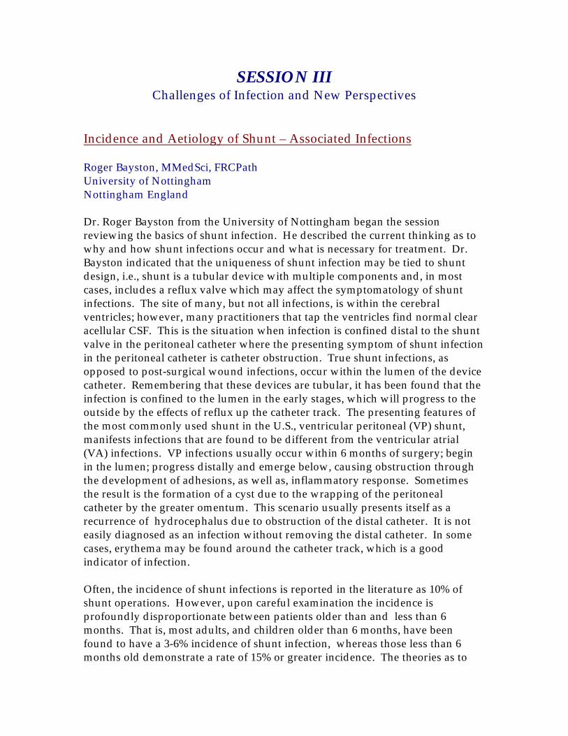
Incidence and Aetiology of Shunt – Associated Infections
Roger Bayston, MMedSci, FRCPathUniversity of NottinghamNottingham England
Dr. Roger Bayston from the University of Nottingham began the sessionreviewing the basics of shunt infection. He described the current thinking as towhy and how shunt infections occur and what is necessary for treatment. Dr.Bayston indicated that the uniqueness of shunt infection may be tied to shuntdesign, i.e., shunt is a tubular device with multiple components and, in mostcases, includes a reflux valve which may affect the symptomatology of shuntinfections. The site of many, but not all infections, is within the cerebralventricles; however, many practitioners that tap the ventricles find normal clearacellular CSF. This is the situation when infection is confined distal to the shuntvalve in the peritoneal catheter where the presenting symptom of shunt infectionin the peritoneal catheter is catheter obstruction. True shunt infections, asopposed to post-surgical wound infections, occur within the lumen of the devicecatheter. Remembering that these devices are tubular, it has been found that theinfection is confined to the lumen in the early stages, which will progress to theoutside by the effects of reflux up the catheter track. The presenting features ofthe most commonly used shunt in the U.S., ventricular peritoneal (VP) shunt,manifests infections that are found to be different from the ventricular atrial(VA) infections. VP infections usually occur within 6 months of surgery; beginin the lumen; progress distally and emerge below, causing obstruction throughthe development of adhesions, as well as, inflammatory response. Sometimesthe result is the formation of a cyst due to the wrapping of the peritonealcatheter by the greater omentum. This scenario usually presents itself as arecurrence of hydrocephalus due to obstruction of the distal catheter. It is noteasily diagnosed as an infection without removing the distal catheter. In somecases, erythema may be found around the catheter track, which is a goodindicator of infection.
Often, the incidence of shunt infections is reported in the literature as 10% ofshunt operations. However, upon careful examination the incidence isprofoundly disproportionate between patients older than and less than 6months. That is, most adults, and children older than 6 months, have beenfound to have a 3-6% incidence of shunt infection, whereas those less than 6months old demonstrate a rate of 15% or greater incidence. The theories as to
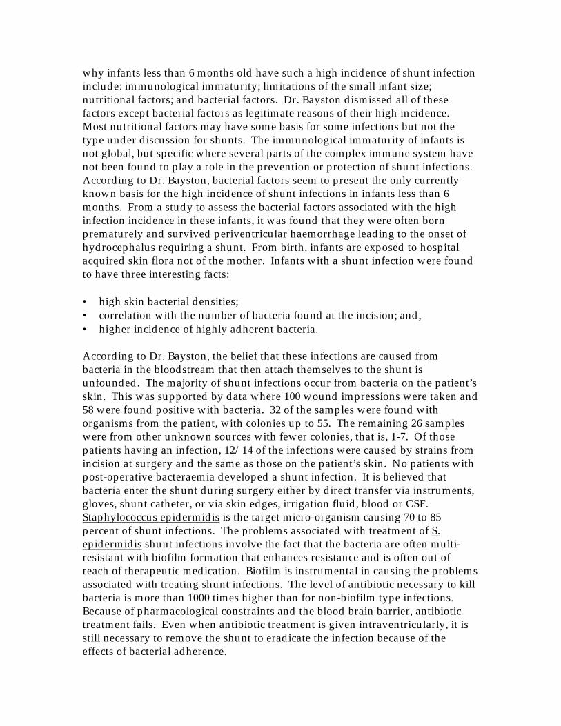
why infants less than 6 months old have such a high incidence of shunt infectioninclude: immunological immaturity; limitations of the small infant size;nutritional factors; and bacterial factors. Dr. Bayston dismissed all of thesefactors except bacterial factors as legitimate reasons of their high incidence.Most nutritional factors may have some basis for some infections but not thetype under discussion for shunts. The immunological immaturity of infants isnot global, but specific where several parts of the complex immune system havenot been found to play a role in the prevention or protection of shunt infections.According to Dr. Bayston, bacterial factors seem to present the only currentlyknown basis for the high incidence of shunt infections in infants less than 6months. From a study to assess the bacterial factors associated with the highinfection incidence in these infants, it was found that they were often bornprematurely and survived periventricular haemorrhage leading to the onset ofhydrocephalus requiring a shunt. From birth, infants are exposed to hospitalacquired skin flora not of the mother. Infants with a shunt infection were foundto have three interesting facts:
• high skin bacterial densities;• correlation with the number of bacteria found at the incision; and,• higher incidence of highly adherent bacteria.
According to Dr. Bayston, the belief that these infections are caused frombacteria in the bloodstream that then attach themselves to the shunt isunfounded. The majority of shunt infections occur from bacteria on the patient’sskin. This was supported by data where 100 wound impressions were taken and58 were found positive with bacteria. 32 of the samples were found withorganisms from the patient, with colonies up to 55. The remaining 26 sampleswere from other unknown sources with fewer colonies, that is, 1-7. Of thosepatients having an infection, 12/14 of the infections were caused by strains fromincision at surgery and the same as those on the patient’s skin. No patients withpost-operative bacteraemia developed a shunt infection. It is believed thatbacteria enter the shunt during surgery either by direct transfer via instruments,gloves, shunt catheter, or via skin edges, irrigation fluid, blood or CSF.Staphylococcus epidermidis is the target micro-organism causing 70 to 85percent of shunt infections. The problems associated with treatment of S.epidermidis shunt infections involve the fact that the bacteria are often multi-resistant with biofilm formation that enhances resistance and is often out ofreach of therapeutic medication. Biofilm is instrumental in causing the problemsassociated with treating shunt infections. The level of antibiotic necessary to killbacteria is more than 1000 times higher than for non-biofilm type infections.Because of pharmacological constraints and the blood brain barrier, antibiotictreatment fails. Even when antibiotic treatment is given intraventricularly, it isstill necessary to remove the shunt to eradicate the infection because of theeffects of bacterial adherence.

In concluding, Dr. Bayston emphasized that in the development of any strategyfor prevention of shunt infections, it is necessary to have knowledge of thefollowing factors:
• Source of bacteria• Time of entry• Nature of infection and pathogenesis• Other risk factors
Return back to agenda and list of presentations

The Role of Standard Medical Treatment for Infection in theEmergence of Antimicrobial Resistance
William R. Jarvis, M.D.Hospital Infections ProgramCenters for Disease Control and Prevention (CDC)
One of the strategies in the prevention of infection is the use of antimicrobials.The wide spread use of antimicrobials has led to antimicrobial resistance. Tobring attention to this problem, Dr. Jarvis described the following:
• epidemiology of antimicrobial resistance;• Vancomycin use in adult and pediatric patients;• Vancomycin use in pediatric neurosurgery patients; and,• CDC recommendations for appropriate use of antimicrobials.
Vancomycin is critical in the armamentarium for treatment of patients withinfection; frequently neurosurgeons are using it prophylactically. Multidrugresistance has become an increasing worldwide problem; the annual cost in theUnited State exceeds $30 billion. Nearly half this cost is attributable to thetreatment and care of inpatient nosocomial infections. The major risk factorsassociated with antimicrobial resistance are the misuse of antimicrobial agentsand the non-compliance with infection control practices. From the NationalNosocomial Infections Surveillance System (NNIS), Dr. Jarvis showed that from1978 through 1997, there was a dramatic increase in resistant organisms,particularly to the primary drug used to treat these pathogens. When examiningantibiotic use in hospitals, Dr. Jarvis described three categories of antibiotictreatment:
• Empiric therapy, where there is an unidentified infection;• Definitive therapy, where there is a confirmed infection by an identified
organism; and,• Prophylaxis
Up to 41% of all antibiotic use has been found to be inappropriate. There is awide variety of approaches for optimizing antibiotic use, for which Dr. Jarvis hasprovided a reference (Avorn et al., Rev Infect Dis, 1987, 9:S285-S296).
Dr. Jarvis presented NNIS data on the percentage of Methicillin-resistantStaphylococcus aureus (MRSA) infections in hospitals from 1975 through 1996.The data show that there was not a particular problem in the late 70s or early80s, but by the mid-80s there was a dramatic increase in the prevalence of MRSAinfections. There have been no data on the proportion of patients with

methicillin resistant S. aureus colonization; failure to recgonize this populationand to use appropriate infection control measures, especially handwashingbefore and after contact with such patients, has contributed to the establishmentof MRSA endemnicity in the United States. This increase in MRSA lead to thewidespread use of Vancomycin. Vancomycin, a glycopeptide, is one of ourmedicine miracles and the only agent uniformly effective against MRSA. Out ofconcern for the increased use of Vancomycin and knowledge that resistance wasto emerge, in 1995 the Hospital Infections Program, CDC with it’s HospitalInfection Control Practices Advisory Committee (HICPAC) issued Vancomycin-use recommendations. From 1996 through 1997 it was shown that as much as 50to 60% of Vancomycin use was not consistent with the CDC/HICPACrecommendations. A number of hospital based studies in 1998 have shown thatVancomycin use can be improved.
A concern of widespread Vancomycin use is the emergence of Vancomycin-resistant enterococcus (VRE). An organism not identified in this country until1989; however, over the past 5 to 10 years there has been a dramatic increase.Today, VRE account for 15-16% of the enterococcal infections reported at NNIShospitals.
Dr. Jarvis described a retrospective epidemiological pharmacy review ofVancomycin usage data collected from 1981-91 at a university hospital. Itreviewed all patients receiving intravenous Vancomycin. The most significantfinding was a 200 fold increase in Vancomycin use, primarily in oncologyservice. Upon review, the overall use indicated approximately 1/3 was forempiric therapy, 1/3 was for definitive therapy and 1/3 for prophylaxis. Uponcloser examination over 60% of Vancomycin use was found to be inappropriateprimarily due to inappropriate monitoring. Risk factor review for receipt ofVancomycin revealed that patients with a medical device had a 14 times greaterrisk of receiving Vancomycin than patients without a medical device. Amultivariate analysis controlling risk factors indicated a significant risk ofreceiving Vancomycin with intravenous and Hickman catheters, as well asdevices such as CSF shunts.
Until recently, S. aureus was always susceptible to Vancomycin. In July, 1997the first documented case of Vancomycin (or glycopeptide) IntermediateResistant S. aureus was reported in Japan. Dr. Jarvis described this case, as wellas two other case studies documented in the U.S. shortly after the Japaneseoccurrence.
In a 1993-97 study from a children’s hospital surgical services, it was found that75% of patients receiving Vancomycin were from either Cardiovascular orNeurosurgery. Since nearly 18% of all Vancomycin use at the hospital were inpediatric neurosurgery patients a closer examination of these patients was

performed to determine why Vancomycin was used and if it was usedappropriately:
Age: 0.2 – 17.8 years median 8 yearsGender: 56.7% maleHospitalization: 1 – 12 days median 2 daysProcedures: cranial 13.3% 4
Spinal 6.7% 2Shunt 76.7% 23
Primary 30.4% 7Revision 69.6% 16
Duration: < 3 hrs 82.8%3-6 hrs 13.8%> 6 hrs 3.4%
Surgical prophylaxis use was found in all patients except one where the numberof doses ranged from 1-12, with a median of 2. It is alarming that in six cases,treatment was administered post-incision. Noting that organisms are presumedat the site of incision, to be effective it is necessary to have peak antibioticprophylactic levels at the time of incision. Another fact is, a large proportion ofthe procedures (82.8%) were less than 3 hours in duration. It is recognized thatantibiotic prophylaxis for a procedure of this duration would require only onedose.
In comparison with CDC/HICPAC recommendations it was found:
• Vancomycin use was inconsistent with CDC/HICPAC recommendationsbecause of surgical prophylaxis in 28 patients;
• in six patients (21.4%), the initial dose was administered post-incision; and,• prophylaxis was continued inappropriately in 26 patients (92.9%) where a
total of 50 extra doses were administered in 28 patients.
The current CDC/HICPAC recommendations for appropriate Vancomycin useincludes situations for:
• Treatment of serious infections due to beta-lactam resistant gram-positivemicroorganisms;
• Treatment of infections due to gram-positive microorganisms with seriousallergy to beta-lactam antimicrobials;
• Treatment of serious or potentially life-threatening antibiotic-associatedcolitis (AAC) or AAC which fails to respond to metronidazole;

• Prophylaxis as recommended by the American Heart Association forendocarditis following certain procedures in patients at high risk forendocarditis; and,
• Prophylaxis for major surgical procedures involving implantation ofprosthetic materials or devices, e.g., shunts, at institutions with a high rate ofinfection due to Methicillin-resistant S. aureus or S. epidermidis (maximum 2doses).
Situations in which the use of Vancomycin should be discouraged include:
• Routine surgical prophylaxis;• Empiric antimicrobial therapy for febrile neutropenia;• Treatment for a single blood culture positive for coagulase-negative
Staphylococcus; and,• Continued empiric use for presumed infection in patients whose cultures are
negative.
In summary, Dr. Jarvis restated the highlights of his talk:
• Over- and inappropriate antimicrobial-use is associated with the emergenceof antimicrobial-resistant pathogens;
• Patients with “plastics,” including CSF shunts, are at risk of receivingVancomycin;
• Pediatric neurosurgery patients were one of the top three groups to receiveVancomycin at a children’s hospital;
• At one children’s hospital, Vancomycin was inappropriately used as surgicalprophylaxis or empiric therapy in all patients evaluated; and,
• Improved practice of Vancomycin use and other antimicrobials are needed inneurosurgery patients.
In conclusion, Dr. Jarvis stated that there is currently no published study in theEnglish language that shows prophylactic use of antimicrobials is efficacious,nor is the use of Vancomycin more effective than other agents.
Return back to agenda and list of presentations

Pathogenesis and Prevention of Catheter-Related Infection
Robert J. Sherertz, MDChief, Section on Infectious DiseasesDepartment of MedicineWake Forest University School of Medicine
Dr. Sherertz, an infectious disease physician with expertise in the pathogenesisand prevention of infection associated with vascular catheters, described some ofthe current work being done to prevent catheter-related infections.
Dr. Sherertz described skin as the major source of early infections for catheter-related infections. Whereas breaks in the line, resulting in contamination of thelumen, is the most common source of infections for long term cahteters. Dr.Sherertz showed data on vascular catheters demonstrating the relationship of thenumber of organisms (S. aureus) and the likelihood of purulence associated withinfection of a foreign body. In a rabbit model, it was shown that likelihood ofassociated purulence with inoculation up to 100,000 organisms occurs about 50%of the time when there is no foreign body. However, when a foreign body, e.g.,catheter, is introduced, the likelihood of associated purulence is 100% at thesame level of organisms.
Dr. Sherertz discussed the difference in the risk of infection between differentbiomaterials. According to Dr. Sherertz, silicone has a higher likelihood ofpurulent infection than materials polyurethane (PU), polyvinyl chloride (PVC),and Teflon. One theory is that there is less killing of bacteria by neutrophils onthe surface of silicone as opposed to PU. Looking at chemotaxis,data Dr.Sherertz showed that neutrophils migrate differently, i.e., the data showed whenusing serum as a stimulus neutrophils move faster on silicone when compared toother materials, e.g., glass, polystyrene, and PU. In a rabbit model, catheterscomposed of different materials, e.g., silicone, PU, PVC, and Teflon, wereimplanted in the subcutaneous space and evaluated by an inflammatory index.The inflammatory index associated with the silicone catheter was greater than inthe other materials. This was shown whether the specimens were inoculatedwith bacteria or not. According to Dr. Sherertz a possible mechanism for thisdifference is complement activation. Complement activation associated withsilicone is 10 times greater than with other materials. Excessive complementactivation during the time of insertion may produce a unique microenvironmentnext to the catheter that may led to an increased likelihood of infection.
Dr. Sherertz presented an overview of some current methods being investigatedin the prevention of infections on the outer surface of catheters. He restatedwhat was previously stated by other speakers that there is no good data tosupport the use of prophylaxic antibiotics based on 3 randomized studies of

vascular catheters that demonstrate prophylaxis does not work. Anotherprevention method is that of anti-infective coatings, where at least 3 clinical trialshave shown a definitive reduction in risk of bloodstream infection. It is unclearwhether such anti-infective coatings will be appropriate for CSF shunts becauseof the unique environment of the brain resulting in unwanted effects, e.g.,toxicity, inflammation, neural damage, etc.
The concept of anti-infective coatings has been investigated on animals, where ithas been shown that the size of the zone of bacterial inhibition of implantedspecimens in animals correlates with the size of the zone of inhibition on thesurface of agar plates.
The use of intermittent or continuous flow of anti-infective solutions within thelumen of catheters has been investigated to try to minimize infections within thevascular catheter lumen. Such solutions include: Vancomycin, EDTA,Gentamycin/Chymotrypsin, chlorine dioxide, and Minocycline/EDTA.
In an in vitro study, S. epidermidis survival on various catheter surfaces wasevaluated. Dr. Sherertz showed that a combination of Minocycline and EDTAdemonstrated the greatest effect, where no organisms could be detected on thesurface. The results presented were as follows:
Control (broth) – 10,000 organisms controlUrokinase no effectHeparin no effectVancomycin/Heparin decreased by 1 to 2 log scaleVancomycin decreased by 1 to 2 log scaleEDTA some effectMinocycline some effectMEDTA large effect; equivalent to a sterilespecimen
Although the data show some promise in the prevention of catheter-relatedinfections there is only minimal clinical data available for such preventivemeasures. Further work needs to be done to understand how best to use thesenew technologies to decrease the risk of catheter-related infections.
Return back to agenda and list of presentations
SESSION IVClinical Outcomes And Methods Of Surveillance
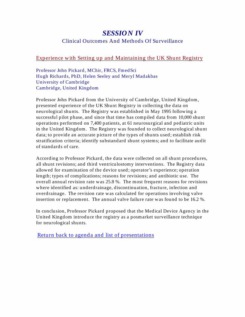
Experience with Setting up and Maintaining the UK Shunt Registry
Professor John Pickard, MChir, FRCS, FmedSciHugh Richards, PhD, Helen Seeley and Meryl MadakbasUniversity of CambridgeCambridge, United Kingdom
Professor John Pickard from the University of Cambridge, United Kingdom,presented experience of the UK Shunt Registry in collecting the data onneurological shunts. The Registry was established in May 1995 following asuccessful pilot phase, and since that time has compiled data from 10,000 shuntoperations performed on 7,400 patients, at 61 neurosurgical and pediatric unitsin the United Kingdom. The Registry was founded to collect neurological shuntdata; to provide an accurate picture of the types of shunts used; establish riskstratification criteria; identify substandard shunt systems; and to facilitate auditof standards of care.
According to Professor Pickard, the data were collected on all shunt procedures,all shunt revisions; and third ventriculostomy interventions. The Registry dataallowed for examination of the device used; operator’s experience; operationlength; types of complications; reasons for revisions; and antibiotic use. Theoverall annual revision rate was 25.8 %. The most frequent reasons for revisionswhere identified as: underdrainage, discontinuation, fracture, infection andoverdrainage. The revision rate was calculated for operations involving valveinsertion or replacement. The annual valve failure rate was found to be 16.2 %.
In conclusion, Professor Pickard proposed that the Medical Device Agency in theUnited Kingdom introduce the registry as a posmarket surveillance techniquefor neurological shunts.
Return back to agenda and list of presentations
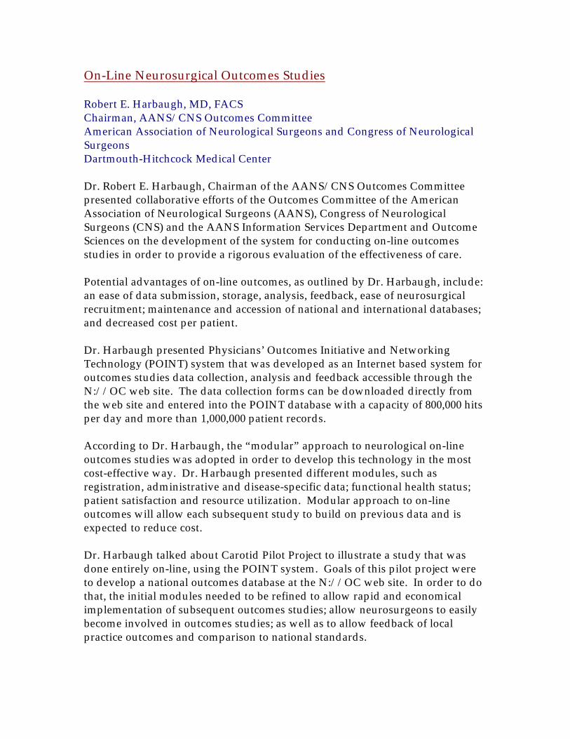
On-Line Neurosurgical Outcomes Studies
Robert E. Harbaugh, MD, FACSChairman, AANS/CNS Outcomes CommitteeAmerican Association of Neurological Surgeons and Congress of NeurologicalSurgeonsDartmouth-Hitchcock Medical Center
Dr. Robert E. Harbaugh, Chairman of the AANS/CNS Outcomes Committeepresented collaborative efforts of the Outcomes Committee of the AmericanAssociation of Neurological Surgeons (AANS), Congress of NeurologicalSurgeons (CNS) and the AANS Information Services Department and OutcomeSciences on the development of the system for conducting on-line outcomesstudies in order to provide a rigorous evaluation of the effectiveness of care.
Potential advantages of on-line outcomes, as outlined by Dr. Harbaugh, include:an ease of data submission, storage, analysis, feedback, ease of neurosurgicalrecruitment; maintenance and accession of national and international databases;and decreased cost per patient.
Dr. Harbaugh presented Physicians’ Outcomes Initiative and NetworkingTechnology (POINT) system that was developed as an Internet based system foroutcomes studies data collection, analysis and feedback accessible through theN://OC web site. The data collection forms can be downloaded directly fromthe web site and entered into the POINT database with a capacity of 800,000 hitsper day and more than 1,000,000 patient records.
According to Dr. Harbaugh, the “modular” approach to neurological on-lineoutcomes studies was adopted in order to develop this technology in the mostcost-effective way. Dr. Harbaugh presented different modules, such asregistration, administrative and disease-specific data; functional health status;patient satisfaction and resource utilization. Modular approach to on-lineoutcomes will allow each subsequent study to build on previous data and isexpected to reduce cost.
Dr. Harbaugh talked about Carotid Pilot Project to illustrate a study that wasdone entirely on-line, using the POINT system. Goals of this pilot project wereto develop a national outcomes database at the N://OC web site. In order to dothat, the initial modules needed to be refined to allow rapid and economicalimplementation of subsequent outcomes studies; allow neurosurgeons to easilybecome involved in outcomes studies; as well as to allow feedback of localpractice outcomes and comparison to national standards.

According to Dr. Harbaugh, the modular approach to on-line outcomes studieswill allow subsequent studies to be built on previous work. That would allowsubsequent studies to be conducted on-line at low cost. Dr. Harbaugh informedthe audience that a “Neurosurgical Report Card” is now available and a spinestudy should be available on-line soon.
Dr. Harbaugh concluded his presentation by pointing out that the availability ofreliable outcomes data will be essential for documenting the effectiveness ofsurgical intervention and that outcomes data may best be done using an internetbased approach. Therefore, Dr. Harbaugh suggested that this approach could beused to collect and evaluate data on shunt technology.
Return back to agenda and list of presentations

Issues in data collection and outcome analysis
John R.W. Kestle, M.D., MSC, FRCSCDivision of Pediatric NeurosurgeryUniversity of UtahPrimary Children’ Medical CenterSalt Lake City, Utah
Dr. John Kestle, from the University of Utah, Salt Lake City, presented examplesfrom the two multicenter trials in pediatric hydrocephalus in order to illustratedata issues, outcome definitions, and analyses.
The importance of readable, understandable, and easy-to-use forms washighlighted. Additionally, using separate forms for each event and use of suchforms in a pretest phase, clearly enhanced the quality of data collection.Methods of data collection were outlined as well as data accuracy assessment.According to Dr. Kestle, data accuracy depends on prospective acquisition;prospective review at data center; internal consistency and source verification.Data monitoring components were presented with emphasis on accrual;database safety (infection rate, surgical complications, and deaths) andrechecking sample size.
Dr. Kestle highlighted the importance of clear definition of primary outcome andapplying the definition in a consistent unbiased way. Using neurological shuntsas an example, Dr. Kestle presented a shunt failure as a primary outcomeincluding categories of shunt obstruction, overdrainage, loculated compartmentsand shunt infection.
In conclusion, Dr. Kestle discussed methods of assessing the observer bias;presented examples of blinding; and analyzed the effects of learning curve andthe center volume on shunt survival.
Return back to agenda and list of presentations