FATIGUE LIFE PREDICTION IN A CANCELLOUS … Comparison of cancellous bone morphological data 27 4.2...
22
FATIGUE LIFE PREDICTION IN A CANCELLOUS BONE STRUCTURE NUR DIANAH BT ABD LATIFF A thesis submitted in fulfillment of the requirements for the award of the degree of Master of Mechanical Engineering Faculty of Mechanical Engineering Universiti Teknologi Malaysia JAN 2013
Transcript of FATIGUE LIFE PREDICTION IN A CANCELLOUS … Comparison of cancellous bone morphological data 27 4.2...

FATIGUE LIFE PREDICTION IN A CANCELLOUS BONE STRUCTURE
NUR DIANAH BT ABD LATIFF
A thesis submitted in fulfillment of the
requirements for the award of the degree of
Master of Mechanical Engineering
Faculty of Mechanical Engineering
Universiti Teknologi Malaysia
JAN 2013
iii
ACKNOWLEDGEMENT
In the name of Allah, the Most Gracious and the Most Merciful.
Alhamdullilah, all praises to Allah for the strengths and His blessing in
completing this thesis. Firstly, I would like to express my deepest gratitude to my
supervisor, Dr. Ardiyansyah Syahrom for his guidance, patience and invaluable help
of constructive comments and suggestion throughout the experimental and thesis
work. Not forgotten, my co-supervisor, Dr. Muhamad Noor Harun for his support
and knowledge regarding this study.
I would like to express my appreciation to my fellow colleague, Mohd Al-
fatihhi for his endless support and help. Without his guidance and assistance this
research would not have been possible. Thank you to my former colleagues, Zainal,
Amir Putra, Fakhrizal and others for their kindness and support. My
acknowledgment also goes to all the technicians and office staff from Faculty of
Mechanical Engineering and SITC for their co-operations.
Sincere thanks to all my friends for their kindness and moral support during
my study. Last but not least, my utmost gratitude goes to my beloved parents and
my siblings for their endless love, prayers and encouragement. To those who
indirectly contributed in this research, your kindness means a lot to me. Thank you
very much.

iv
ABSTRACT
Repetitive cyclic loading of bone during daily course activities is one of the
primary causes of bone fracture in humans. Stress fractures and fragility fractures in
elderly generation due to osteoporosis have been associated with the reduction of
bone strength of the cancellous bone. The aim of this study is to predict the failure of
cancellous bone as a function of density and porosity. In this present study, two of
cancellous specimens were extracted from bovine medial-condyle bone and were
loaded in cyclic compression. Monotonic compressions were first tested to determine
the boundary conditions prior to the fatigue testing. The loading transferred to the
cancellous bone are chosen between 16%-55% of the ultimate stress. The result
showed different hysteresis loop with large variation in strain between both medial-
condyle of cancellous bone. They both adapt different physiological apparent load
until failure. From the obtained result, we can conclude that the same anatomic site
with different value of bone density and porosity imply a large effect of the fatigue
behavior in related to modulus degradation and strain changes.

v
ABSTRAK
Tahap aplikasi kitaran beban yang berulang-ulang pada tulang ketika aktiviti
harian adalah salah satu penyebab utama keretakan atau fraktur tulang manusia.
Tekanan retakan dan kerapuhan tulang terhadap golongan warga emas di sebabkan
oleh penyakit osteoporosis sering dikaitkan dengan pengurangan daya kekuatan pada
tulang kanselus. Tujuan utama pengajian ini adalah untuk menjangka tahap
kegagalan tulang kanselus sebagai fungsi kepada ketumpatan dan poros. Dalam
ujikaji ini, dua sampel dari tulang tengah dari bovin telah diekstrak dan diuji pada
tahap kitaran beban yang berulang-ulang. Bebanan termampat secara monotonic
telah diuji terlebih dahulu untuk menentukan garisan panduan untuk uji kaji
seterusnya, iaitu bebanan termampat yang terjurus kepada kegagalan terhadap
sampel tulang kanselus. Aplikasi galas beban terhadap tulang kanselus telah
ditentukan daripada 16% sehingga 55% daripada maksimum stres daripada bebanan
termampat secara monotonic . Keputusan menunjukkan ketidaksamaan pada
hysteresis loop dengan perubahan besar pada ketegangan di antara dua tulang tengah
kanselus. Daya ketahanan pada kedua-dua tulang ini adalah berbeza sebelum fraktur.
Daripada keputusan ini juga, kami mendapati walaupun lokasi anatomik yang sama
tetapi berbeza dari segi ketumpatan dan poros, mempengaruhi besar terhadap tingkah
laku daya ketahanan tulang kanselus tersebut. Ini adalah berkaitan dengan modulus
dan ketengangan daripada ujikaji yang telah dijalankan.

vi
TABLE OF CONTENT
CHAPTER TITLE PAGE
DECLARATION ii
ACKNOWLEDGEMENTS iii
ABSTRACT iv
ABSTRAK v
TABLE OF CONTENTS vi
LIST OF TABLES viii
LIST OF FIGURES ix
LIST OF ABBREVIATION AND SYMBOL xii
1 INTRODUCTION 1
1.1 Objective 3
1.2 Scope 4
1.3 Problem statement 4
2 LITERATURE REVIEW 7

vii
2.1 Human Knee Joint 7
2.2 Physiological Activity 9
2.3 Cancellous Bone 11
2.3.1 Microstructure of cancellous bone 13
2.3.2 Mechanical properties of cancellous bone 14
3 RESEARCH METHODOLOGY 18
3.1 Specimen Preparation 18
3.2 Morphological Data 21
3.3 Compressive Testing 22
4 RESULT 26
4.1 Morphological Parameters 27
4.2 Fatigue Testing Result 27
5 DISCUSSION 32
6 CONCLUSION AND RECOMMENDATION 34
6.1 Conclusion 34
6.2 Recommendation 35
REFERENCES 36

viii
LIST OF TABLES
TABLE NO. TITLE PAGE
2.1 Relevant angles of the tibiofemoral joint 9
4.1 Comparison of cancellous bone morphological data 27
4.2 Comparison of mechanical parameters of the
tested specimen 30
ix
LIST OF FIGURES
FIGURE NO. TITLE PAGE
2.1 The knees comprises of two joints made up by the 7
femur, tibia and patella. The four major ligaments of
the knee are the medial, lateral, anterior, and posterior
collateral ligaments. The tibia is partially covered by
menisci
2.2 Distal femoral topography 8
2.3 (a) Mechanical medial proximal tibial angle; (b) 9
Mechanical medial distal femoral angle; (c) Anatomic
lateral distal femoral angle; (d) Joint line congruency
angle (e) Anatomic posterior proximal tibial angle
2.4 Range of motion of the tibiofemoral joint in the sagittal 10
plane during level walking in one gait cycle
2.5 Knee wear simulator inputs: flexion angle, axial force, 11
AP force and IE torque
2.6 The differences in the hierarchical structure between 12
cortical bone (left) and cancellous bone (right)

x
2.7 Reconstruction of human trabecular bone from the 13
(A) vertebra, (B) proximal tibia, (C) femoral greater
trochanter, and (D) femoral neck
2.8 Dependence of ultimate stress on age for cancellous bone 15
from the human vertebra and femur. For both anatomic
site, strength decreases approximately 10 percent
per decade
2.9 (A) Elastic modulus as a function of apparent density 16
for cancellous bone specimens from a wide variety of
species and anatomic site. (B) Compressive yield stress as
a function of apparent density for human trabecular bone
specimens from multiple anatomic sites
3.0 The relationships between ultimate compressive stress, 17
porosity and apparent density for fresh human bone
3.1 (a) The femur bone with marked medial condyle site 18
(b) The endplates of the medial condyle of the femur were
chosen as references, the 0° direction in the alignment
with the physiological axis
3.2 Femur bone were cut by sections through Bosch circular saw 19
3.3 (a) The femoral ball is cut by a precision cutter with 19
wafering blade under water constant irrigation

xi
(b) The specimen is being cut into a cubic shape.
3.4 Specimen is soak in the Crest ultrasonic with a 20
chemical detergent Pumicized citrius, Gent-l-kleen.
3.5 Specimen before and after cleaning and vacuum suction 20
3.6 The specimens were then aligned in a custom jig for 21
better vertical oriented
3.7 (a) Universal Testing Machine Instron 880-100kN 23
Hydraulic
(b) The close-up of the specimen
3.8 Illustration of fatigue of stress-strain curve showing the 25
first and a later cycle with the defined parameters
4.1 Typical stress-strain curves of a fatigue test for specimen 28
#1. Using the criteria D=0.4, failure was found at = 26.
4.2 Typical stress-strain curves of a fatigue test for specimen 29
#2. Using the criteria D=0.4, failure was found at = 22.
4.3 Relationship between the maximum strain and the number 30
of cycles to failure. The dotted line corresponds to the cycle
of failure with D=0.4.
4.4 Relationship between the modulus and the number of cycles 31
to failure.

xii
LIST OF ABBREVIATION AND SYMBOL
- Volume geometric
- Volume hydrated
ℎ: - Weight in air
: - Weight in submerged
- Density
- Volume fraction
- Material Density
- Modulus Young
- Normal stress
- Strain
- Number of cycle to failure

CHAPTER 1
INTRODUCTION
More than 250,000 hip fractures were reported in 1996; approximately 10%
are thought to be spontaneous fractures associated with cyclic loading during the
daily activities. Military recruits, athletes and ballet dancers are among those affected
[1]. Under physiologic conditions, micro-damage events created from both static and
cyclic load are subsequently repaired through the coordinated process of bone
remodeling [2]. Micro-damage accumulation leads to diminished bone quality and
together with loss of bone quantity, results in weakened bones which may break
following minor falls [3].
There are two types of bone tissue in the skeletal system; cortical (or
compact) and cancellous (or trabecular) bone. Adult human skeletal mass consists of
80% cortical bone (porosity 5-30%) and 20% (porosity 20-90%) cancellous bone [4].
The micro-damage accumulation incidence is greatest at sites where cancellous bone
is the dominant form and has increased over the past 30 years [3]. Thus, changes in
cancellous bone stiffness in even a small region can cause large differences in whole
bone strength [5]. Repetitive cyclic loading of bone during the daily course of
activities is one of the primary causes of bone fractures in humans [6]. A typical
loading for bone is cyclic loading that is variable in time; behaviors under such
loading can termed ‘fatigue behaviors’ [7]. Excessive fatigue loading of bones in

2
vivo can lead to micro crack accumulation and coalescence, reducing stiffness and
strength and increasing the risk of fracture [8].
The fatigue behavior of cancellous bone has been characterized in a number
of studies [1,6,8,13,39,40,42]. In these experiments, the fatigue life of these
cancellous bones was observed and characterized by the number of cycles to failure,
, increases as the cyclic decreases. The mechanical response of the cancellous
bone under fatigue was characterized by a decrease in the elastic modulus throughout
the test, with rapid modulus loss near failure, and increasing plastic, or permanent
strain [8, 9]. They also found increasing residual strain and modulus reduction with
increasing strain amplitude [2].
A study showed that bone fatigue can occur at strain magnitudes comparable
to those measured on living bones in the physiological loading environment during
vigorous activity in animals and humans. From this study, the fatigue life to failure is
predicted in the order of 107 load cycles, which is approximately 5-10 years of use in
life [9]. Significance amounts of fatigue damage occur throughout the loading
history; damage which must be repaired in order not to lead to fatigue failure of
skeletal elements [9].
Fatigue fractures are usually sustained during continuous strenuous physical
activity which causes the muscles to become fatigued and reduces their ability to
contract. Fatigue fractures on the compressive side appear to be produced more
slowly because the remodeling is less easily outpaced by the fatigue process, thus the
bone may not proceed to complete fracture [10]. The ability of the skeleton to resist
fracture under applied loading varies primarily through changes in these constituents
of bone failure load and bone strength [11]. Most the site that is prone to fractures
due to this disease is at the hip, vertebrae and the distal radius [12]. This is because
of their high prevalence and their frequent asymptomatic characteristics which are
associated with low bone mass and micro architectural deterioration [13].
If the applied load exceeds the failure load of the bone of interest, then the
factor of risk is greater than one and fracture will occur. Thus, to predict fracture

3
accurately, characteristics of the applied load such as the manner and location of its
applications must be considered [11]. Fatigue behaviors on cancellous bone induced
by many case of physiological human activities which can contribute to stress
fracture from various activity such for athletes and fragility fracture in aging. Studies
have shown that the volume fraction of cancellous bone strongly influences the
mechanical properties specifically the compressive strength, stiffness and elastic
modulus [14]. Hence, understanding the damage properties of cancellous bone is
important to understand bone fractures [15]. Past study by Bowman et al. (1994)
showed that modulus of the cancellous bone can decrease with fatigue as the strain
accumulation increases due to creep [16].
In the course of everyday activities human bone is submitted to a great
variety of loading patterns. The loading varies in direction, magnitude, frequency and
mode (tension, compression and shear) and also in combinations of the previous
factors [17]. This repetitive physiological loading pattern is referring to human gait
cycle. This fundamental task has been the subject of study by scientists for several
centuries, both with respect to description of typical body movements and of
pathological conditions and therapeutic interventions [10].
1.1 Objective
The objectives of the research project are to:
1. To predict of fatigue life of cancellous bone structure.
2. To analyze the fatigue behavior of cancellous bone respect to physiological
axis
4
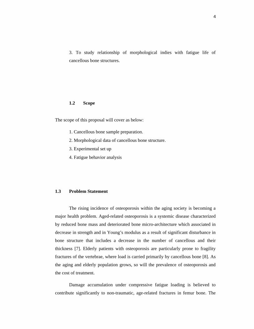
3. To study relationship of morphological indies with fatigue life of
cancellous bone structures.
1.2 Scope
The scope of this proposal will cover as below:
1. Cancellous bone sample preparation.
2. Morphological data of cancellous bone structure.
3. Experimental set up
4. Fatigue behavior analysis
1.3 Problem Statement
The rising incidence of osteoporosis within the aging society is becoming a
major health problem. Aged-related osteoporosis is a systemic disease characterized
by reduced bone mass and deteriorated bone micro-architecture which associated in
decrease in strength and in Young’s modulus as a result of significant disturbance in
bone structure that includes a decrease in the number of cancellous and their
thickness [7]. Elderly patients with osteoporosis are particularly prone to fragility
fractures of the vertebrae, where load is carried primarily by cancellous bone [8]. As
the aging and elderly population grows, so will the prevalence of osteoporosis and
the cost of treatment.
Damage accumulation under compressive fatigue loading is believed to
contribute significantly to non-traumatic, age-related fractures in femur bone. The

5
advantage of using the compressive fatigue tests is the ability to conduct variable test
with the use of small numbers of samples [7]. Even if these failure types are of
known, data for cancellous bone exposed to cyclic loading are still insufficient [18].
Studies of the fatigue of bone have dealt most extensively with cortical bone since it
is consequently plays a dominant role in determining the overall strength of a given
bone of the skeleton [10].
A great number of investigations have probed the mechanical properties of
both cortical and cancellous bone. Studies have investigated the Young’s modulus,
yield strain, creep behavior and fatigue behavior of both cancellous and cortical bone
[19]. The proximal femoral head exhibited of hip contact forces [20] has been
studied for average patient. This has been developed the maximum peak forces
during human activities and it has contributed such a loading method to be apply on
fatigue analysis in cancellous bone. In revision of total knee arthoplasty, the
epicondyles often provide the only available clues for rotational and proximal/distal
positioning of the femoral component. Thus, a relevant study of the anatomic
relationship based on the epicondyles of the distal femur will somehow help
orthopedists position the femoral components appropriately in primary and revision
total knee arthoplasty [21]. It is also significance for this study in order to obtain the
main physiological axis for the load to transmit to the epicondyle femur.
From previous study, it is suggested that more than 75% of the load adjacent
to endplates is carried by cancellous bone [22]. The relationship between
morphology of cancellous bone to the mechanical properties and failure mechanism
can be accessed through experimental and computational means [23]. Computer
simulation (microCT) has become more accessible in the past years [13], but these
data are still connected to many problems such as the high costs of the microCT
scans and the rare availability of the high end scanning facilities.
The underlying deformation and damage mechanism within cancellous bone
with respect to physiological activities are not yet sufficiently investigated. Thus, it is
necessary to evaluate bone quality parameters such as the morphological index of the

6
cancellous bone structure. In order to obtain the loading conditions, the monotonic
test were first tested and performed into the fatigue testing. This paper will determine
the prediction of the compressive fatigue behavior on several sample of cancellous
bone as a function of density and porosity.

36
REFERENCES
1. Markus C. Michel, Xiang-Dong E.Guo, Lorna J,Gibson (1993). Compressive
Fatigue Behavior of Bovine Trabecular Bone. Journal Biomechanics, 26, 453-
463.
2. Srinidhi Nagaraja, Mario D. Ball, Robert E. Guldberg (2007) .Time-dependent
Damage Accumulation Under Stress Relaxation Testing of Bovine Trabecular
Bone. Journal of Fatigue, 29, 1034-1038.
3. T. Clive Lee, Fergal J. O’Brien, David Taylor (2000). The Nature of Fatigue
Damage in Bone. Journal of fatigue, 22, 847-853
4. Solomon Praveen Samuel, George R. Baran, Yen Wei, Brian L. Davis. (2009)
Chapter 4: Biomechanics-Part 2, Bone Pathology, Springer Science & Business
Media.
5. Xiang Wang, Glen L. Niebur (2006) Microdamage Propagation in Trabecular
Bone due to Changes in Loading Mode, Journal of biomechanics,39, 781-790
6. P. Ganguly, T.L.A Moore, L.J.Gibson. (2004). A Phenomenological Model for
Predicting Fatigue Life in Bovine Trabecular Bone. ASME, 126.
7. Tomasz Topolinski, Artur Cichanski, Adam Mazurkiewiz, Krysztof Nowicki,
(2011). Study of the Behavior of the Trabecular Bone under Cyclic Compression
with Stepwise Increasing Amplitude. Journal of the mechanical behavior of
biomedical materials, 4, 1755-1763
8. Tara L. Moore, Lorna J. Gibson (2003). Fatigue of Bovine Trabecular Bone.
Journal of Biomechanical, 125, 761-768
9. Mitchell B. Schaffler, Karl J. Jepsen (2000). Fatigue and Repair in Bone. Journal
of fatigue, 22,839-846
10.Margareta Nordin, Victor H.Frankel. (2001). Basic Biomechanics of the
Musculoskeletal System. 3rd edition, copyright Lippincott Williams & Wilkins

37
11. Jacqueline H. Cole, Marjolein C.H. Van der Meulen. (2010). Biomechanics of
Bone, Osteoporosis: Path physiology and Clinical Management, Springer
Science & Business Media.
12. J.Homminga, B.R. Mccreadie, T.E. Ciarelli, H.Weinans, S.A.Goldstein, R.
Huiskes (2002). Cancellous Bone Mechanical Properties from Normal and
Patients with Hip Fractures Differ on the Structural Level, not on the Bone Hard
Tissue Level. Journal of Bone, 30 no.5, 759-764
13. Laurent Rapillard, Mathieu Charlebois, Philippe K. Zysset (2006). Compressive
Fatigue Behavior of Human Vertebral Trabecular Bone. Journal of
Biomechanics, 39, 2133-2139
14. L.Zou, R.D. Bloebaum, K.N. Bachus. (1996). Reproducibility of techniques
using Archimedes’ principle in measuring cancellous bone volume.S1350-4533
15. Uwe Wolfram, Hans-Joachim Wilke, Philippe K. Zysset. (2011). Damage
Accumulation in Vertebral Trabecular Bone depends on Loading Mode and
Direction. Journal of biomechanics, 44, 1164-1169
16. Shaun Kevin Stinton. (2011). Development, validation and application of a
noninvasive spinal motion measurement system. University of Kentucky,
Doctoral Dissertation. Paper 169.
17. Peter Zioupos, Adria Casinos. (1998). Cumulative damage and the response of
human bone in two-step loading fatigue. Journal of Biomechanics, 31, 825-833
18. S. Dendofer, H.J Maier, J. Hammer. (2009). Fatigue Damage in Cancellous
Bone: An Experimental Approach from Continuum to Micro scale. Journal of
mechanical behavior of biomedical materials, 2, 113-119
19. Abel Z Hastings. (1995). An investigation of the high cycle fatigue behavior of
bovine trabecular bone. Massachusetts Institute of Technology.

38
20. Tomas A. Correa, Kay.M.Crossley, Hyung J.Kim, Marcus G. Pandy. (2010).
Contributions of Individual Muscles to Hip Joint contact Force in Normal
Walking. Journal of Biomechanics, 43, 1618-1622
21. Frankie M. Griffin, Kevin Math, Giles R. Scuderi (2000). Anatomy of
Epicondyles of the Distal Femur: MRI Analysis of Normal Knees. Journal of
Arthoplasty, 15, 354-359
22. Liu, X.S. (2009). Micromechanical analyses of vertebral trabecular bone based
on individual trabeculae segmentation of plates and rods. Journal of
biomechanics, 42, 249-256
23. Ardiyansyah Syahrom, Mohammed Rafiq Abdul Kadir, Jaafar Abdullah,
Andreas Ochsner. (2011). Mechanical and Microarchitectural Analyses of
Cancellous Bone through Experiment and Computer Simulation”, Med Biol Eng
Comput, 49, 1393-1403
24. Robert C. Schenck, Jr, MD. (1999). Athletic Training and Sport Medicine. Knee
Injuries. American Academy of Orthopaedic Surgeons.
25. Turner A.Blackburn, Emily Craig. (1980). Knee Anatomy: A Brief Review.
Journal of American Physical Therapy Association, 60, 1556-1560.
26. Knee Anatomy Multimedia Health Education
http://www.hipsknees.info/flash/HTML-KNEES/knee-anatomy-ypo.pdf
27. D.E. Bonasia, P. Rossi, R. Rossi. (2011). Anatomy and Biomechanics of Knee.
Journal Orthopedic Sports Medicine, 301-318
28. V. Racic, A.Pavic, J.M.W. Brownjohn. (2009). Experimental Identification and
Analytical Modeling of Human Walking Forces: Literature Review. Journal of
sound and vibration, 326, 1-49
29. Lamoreaux,L. (1971). Kinematic measurements in the study of human walking.
Biomechanics Lab, University of California, San Francisco. Bull Prosthetic Res.

39
30. Murray, M., Drought, A.B., Kory,R.C. (1964). Walking pattern of normal men.
Journal of Bone Joint Surgery. 335.
31. Lucy A.Knight, Saikat Pal, John C.Coleman, Fred Bronson, Hani Haider.
(2007). Comparison of long-term numerical and experimental total knee
replacement wear during simulated gait loading. Journal of Biomechanics, 40,
1550-1558
32. Elise F.Morgan. (2008). Biomechanics of bone and age-related fractures.
Orthopedic and development biomechanics library, principles of bone biology
3rd edition.
33. Tony M. Keaveny, Elise F.Morgan, Oscar C. Yeh. (2004). Chapter 8:Bone
Mechanics. Standard handbook of biomedical engineering and design.
34. Elise F. Morgan, Tony M.Keaveny. (2001). Dependence of yield strain of
human trabecular bone on anatomic site. Journal of Biomechanics. 34, 569-577
35. Burr,D.B., Schaffler, M.B., and Frederickson,R.G. (1988). Composition of the
cement line and its possible mechanical role as a local interface in human
compact bone. Journal of Biomechanical. 21. 939-945
36. G.A.P. Renders, L.Mulder, L.J.Van Rujiven, (2007), Porosity of human
mandibular condylar bone. Journal of Anatomy, 210, 239-248.
37. S.A. Goldstein (1987). The Mechanical Properties of Trabecular Bone
Dependence on Anatomic Location and Function. Journal of Biomechanics, 20,
1055-1061.
38. John R. Cotton, Keith Winwood, Peter Zioupos, Mark Taylor. (2005). Damage
Rate is a Predictor of Fatigue Life and Creep Stain Rate in Tensile Fatigue of
Human Cortical Bone Samples. Journal of Biomechanical Engineering, 127,
213-219
39. S.Dendorfer, H.J Maier, D.Taylor. (2008). Anistropy of the fatigue behavior of
cancellous bone. Journal of biomechanics, 41, pp 636-641

40
40. Travis A. Burgers, Jim Mason, Glen Niebur, Heidi L.Ploeg. (2008). Compressive
properties of trabecular bone in the distal femur. Journal of Biomechanics.1-8
41. Peter Zioupos, Richard B. Cook, John R. Hutchinson. (2008). Some basic
relationships between density values in cancellous and cortical bone. Journal of
Biomechanics , 41, 1961-1968
42. Tara L.A. Moore, Lorna J.Gibson. (2003). Fatigue Microdamage in Bovine
Trabecular Bone. Journal of Biomechanical Engineering. 125, 769-776