Fate mapping analysis of lymphoid cells expressing the NKp46 cell
6
Fate mapping analysis of lymphoid cells expressing the NKp46 cell surface receptor Emilie Narni-Mancinelli a,b,c,1 , Julie Chaix a,b,c,1,2 , Aurore Fenis a,b,c , Yann M. Kerdiles a,b,c , Nadia Yessaad a,b,c , Ana Reynders a,b,c , Claude Gregoire a,b,c , Herve Luche a,b,c , Sophie Ugolini a,b,c , Elena Tomasello a,b,c , Thierry Walzer a,b,c,3 , and Eric Vivier a,b,c,d,4 a Centre d’Immunologie de Marseille-Luminy, Université de la Méditerranée UM 631, Campus de Luminy Case 906, 13288 Marseille, France; b Institut National de la Santé et de la Recherche Médicale, Unité Mixte de Recherche S 631, 13009 Marseille, France; c Centre National de la Recherche Scientifique, Unité Mixte de Recherche 6102, 13288 Marseille, France; and d Assistance Publique, Hôpitaux de Marseille, Hôpital de la Conception, 13385 Marseille, France Edited* by Christophe Benoist, Harvard Medical School, Boston, MA, and approved September 19, 2011 (received for review July 26, 2011) NKp46 is a cell surface receptor expressed on natural killer (NK) cells, on a minute subset of T cells, and on a population of innate lymphoid cells that produce IL-22 and express the transcription factor retinoid-related orphan receptor (ROR)-γt, referred to as NK cell receptor (NKR) + ROR-γt + cells. Here we describe Nkp46 iCre knock- in mice in which the gene encoding the improved Cre (iCre) recom- binase was inserted into the Nkp46 locus. This mouse was used to noninvasively trace cells expressing NKp46 in vivo. Fate mapping experiments demonstrated the stable expression of NKp46 on NK cells and allowed a reappraisal of the sequential steps of NK cell maturation. NKp46 genetic tracing also showed that gut NKR + ROR-γt + and NK cells represent two distinct lineages. In addi- tion, the genetic heterogeneity of liver NK cells was evidenced. Finally, Nkp46 iCre mice also represent a unique mouse model of conditional mutagenesis specifically in NKp46 + cells, paving the way for further developments in the biology of NKp46 + NK, T, and NKR + ROR-γt + cells. natural killer cell differentiation | NKp46 knock-in N atural killer (NK) cells are effector and regulatory lym- phocytes of the innate immune system that contribute to tumor surveillance, hematopoietic allograft rejection, control of microbial infections, and pregnancy (1). NK cells can be cyto- toxic and secrete an array of cytokines and chemokines, such as IFN-γ and β-chemokines. NKp46 (NCR1, CD335) is a marker of NK cells in all mam- malian species tested so far, including human, nonhuman pri- mates, mouse, rat, and cow (2–8). NKp46 is an Ig-like superfamily cell surface receptor, which is a member of a group of natural cytotoxicity receptors (NCRs) with NKp44 (NCR2, CD336) and NKp30 (NCR3, CD337) (9). NKp46 is associated with immunor- eceptor tyrosine-based activation motif-bearing polypeptides, such as CD3-ζ and FcR-γ, which transduce potent activating signals upon triggering (4, 10). NKp46 is involved in tumor cell recogni- tion via still-unidentified ligands (11, 12) and has also been de- scribed as binding viral hemagglutinins (13, 14). Finally, it has been reported that NK cells contribute via NKp46 to type I diabetes through the destruction of pancreatic β-islets (15). NKp46 is found on all mature NK cells regardless of their anatomic localization in both human and mouse. The selective expression of NKp46 on NK cells has two reported exceptions: rare T-cell subsets (6, 16, 17) and a mucosal population of NKp46 + innate lymphoid cells (ILCs) that express the transcription factor retinoid-related or- phan receptor (ROR)-γt and produce IL-22, a key cytokine for the activation and defense of epithelial cells (18–28). The NKp46 amino acid sequence is highly conserved in all mammals. The striking homologies in the regulatory regions of the NKP46 gene from opossum to human prompted us to generate a transgenic mouse line, called NDE, in which the diphtheria toxin receptor (DTR) and the enhanced green fluorescent protein (eGFP) expression were driven by a 450-bp conserved promoter region (P1) upstream of Nkp46 (Ncr1) start codon (6). Although the selectivity of expression of eGFP in NDE mice mirrored that of endogenous NKp46 in a fraction of transgenic mice, variegation at the transgenic locus led to unpredictable variation in the pene- trance of the transgene expression in mouse littermates. Another transgenic mouse expressing the improved Cre (iCre) recombinase under the control of the Nkp46 promoter was recently reported and crossed to eGFP reporter mice (29). However, on average only 80% of NKp46 + NK cells expressed eGFP, and no eGFP expression was detected in T cells, suggesting that the regulation of the iCre transgene expression in this model did not match with the endogenous expression of NKp46, hence hampering the use of these mice for selective gene targeting in NKp46 + cells. To circumvent these caveats of expression pattern that are classically observed in transgenic mice (30), we generated a knock-in mouse line in which the gene encoded the improved recombinase (icre) was inserted by homologous recombination at the 3′ end of Nkp46, in an attempt to drive its expression by all endogenous Nkp46 regulatory elements. Here we report that iCre expression faithfully corresponds to the endogenous expression of NKp46, on bona fide NK cells, on a subset of gut ILCs, as well as on very discrete sub- populations of T cells, allowing us to trace the fate of the het- erogeneous NKp46 + populations of cells. Results Characterization of Nkp46 iCre Knock-in Mice. We generated a knock- in mouse line in which icre was inserted by homologous re- combination at the 3′ end of the Nkp46 gene (Fig. 1A and Fig. S1). Nkp46 iCre/wt mice were obtained at Mendelian frequencies, de- veloped normally, were fertile, and showed no significant varia- tions in the numbers of lymphoid and myeloid subsets compared with wild-type (wt) littermates. NK cell counts, phenotype, and in vitro effector function were not affected in Nkp46 iCre/wt mice, despite a down-regulation in the density of cell surface NKp46 that did not alter the percentage of NKp46 + NK cells (Fig. S2). Rosa26eYFP reporter mice (R26R eYFP/wt ) carry under the Rosa26 promoter a loxP-flanked STOP sequence that prevents the expression of the downstream eYFP gene. The STOP sequence is removed and eYFP is expressed in cells where iCre is expressed Author contributions: E.N.-M., T.W., and E.V. designed research; E.N.-M., J.C., A.F., Y.M.K., N.Y., A.R., E.T., and T.W. performed research; J.C., A.F., C.G., H.L., and T.W. contributed new reagents/analytic tools; E.N.-M., S.U., and E.V. analyzed data; and E.N.-M. and E.V. wrote the paper. Conflict of interest statement: E.V. is a co-founder and shareholder of Innate-Pharma. *This Direct Submission article had a prearranged editor. 1 E.N.-M. and J.C. contributed equally to this work. 2 Present address: Abramson Family Cancer Research Institute, Department of Medicine, University of Pennsylvania, Philadelphia, PA 19104. 3 Present address: Université de Lyon, Institut National de la Santé et de la Recherche Médicale U851, F-69007 Lyon, France. 4 To whom correspondence should be addressed. E-mail: [email protected]. This article contains supporting information online at www.pnas.org/lookup/suppl/doi:10. 1073/pnas.1112064108/-/DCSupplemental. 18324–18329 | PNAS | November 8, 2011 | vol. 108 | no. 45 www.pnas.org/cgi/doi/10.1073/pnas.1112064108
Transcript of Fate mapping analysis of lymphoid cells expressing the NKp46 cell

Fate mapping analysis of lymphoid cells expressingthe NKp46 cell surface receptorEmilie Narni-Mancinellia,b,c,1, Julie Chaixa,b,c,1,2, Aurore Fenisa,b,c, Yann M. Kerdilesa,b,c, Nadia Yessaada,b,c,Ana Reyndersa,b,c, Claude Gregoirea,b,c, Herve Luchea,b,c, Sophie Ugolinia,b,c, Elena Tomaselloa,b,c,Thierry Walzera,b,c,3, and Eric Viviera,b,c,d,4
aCentre d’Immunologie de Marseille-Luminy, Université de la Méditerranée UM 631, Campus de Luminy Case 906, 13288 Marseille, France; bInstitut Nationalde la Santé et de la Recherche Médicale, Unité Mixte de Recherche S 631, 13009 Marseille, France; cCentre National de la Recherche Scientifique, Unité Mixtede Recherche 6102, 13288 Marseille, France; and dAssistance Publique, Hôpitaux de Marseille, Hôpital de la Conception, 13385 Marseille, France
Edited* by Christophe Benoist, Harvard Medical School, Boston, MA, and approved September 19, 2011 (received for review July 26, 2011)
NKp46 is a cell surface receptor expressed on natural killer (NK)cells, on a minute subset of T cells, and on a population of innatelymphoid cells that produce IL-22 and express the transcriptionfactor retinoid-related orphan receptor (ROR)-γt, referred to as NKcell receptor (NKR)+ROR-γt+ cells. Here we describe Nkp46iCre knock-in mice in which the gene encoding the improved Cre (iCre) recom-binase was inserted into the Nkp46 locus. This mouse was used tononinvasively trace cells expressing NKp46 in vivo. Fate mappingexperiments demonstrated the stable expression of NKp46 onNK cells and allowed a reappraisal of the sequential steps of NKcell maturation. NKp46 genetic tracing also showed that gutNKR+ROR-γt+ and NK cells represent two distinct lineages. In addi-tion, the genetic heterogeneity of liver NK cells was evidenced.Finally, Nkp46iCre mice also represent a unique mouse model ofconditional mutagenesis specifically in NKp46+ cells, paving theway for further developments in the biology of NKp46+ NK, T, andNKR+ROR-γt+ cells.
natural killer cell differentiation | NKp46 knock-in
Natural killer (NK) cells are effector and regulatory lym-phocytes of the innate immune system that contribute to
tumor surveillance, hematopoietic allograft rejection, control ofmicrobial infections, and pregnancy (1). NK cells can be cyto-toxic and secrete an array of cytokines and chemokines, such asIFN-γ and β-chemokines.NKp46 (NCR1, CD335) is a marker of NK cells in all mam-
malian species tested so far, including human, nonhuman pri-mates, mouse, rat, and cow (2–8). NKp46 is an Ig-like superfamilycell surface receptor, which is a member of a group of naturalcytotoxicity receptors (NCRs) with NKp44 (NCR2, CD336) andNKp30 (NCR3, CD337) (9). NKp46 is associated with immunor-eceptor tyrosine-based activation motif-bearing polypeptides, suchas CD3-ζ and FcR-γ, which transduce potent activating signalsupon triggering (4, 10). NKp46 is involved in tumor cell recogni-tion via still-unidentified ligands (11, 12) and has also been de-scribed as binding viral hemagglutinins (13, 14). Finally, it has beenreported that NK cells contribute via NKp46 to type I diabetesthrough the destruction of pancreatic β-islets (15). NKp46 is foundon all mature NK cells regardless of their anatomic localizationin both human and mouse. The selective expression of NKp46 onNK cells has two reported exceptions: rare T-cell subsets (6, 16,17) and a mucosal population of NKp46+ innate lymphoid cells(ILCs) that express the transcription factor retinoid-related or-phan receptor (ROR)-γt and produce IL-22, a key cytokine for theactivation and defense of epithelial cells (18–28).The NKp46 amino acid sequence is highly conserved in all
mammals. The striking homologies in the regulatory regions of theNKP46 gene from opossum to human prompted us to generatea transgenic mouse line, called NDE, in which the diphtheria toxinreceptor (DTR) and the enhanced green fluorescent protein(eGFP) expression were driven by a 450-bp conserved promoterregion (P1) upstream of Nkp46 (Ncr1) start codon (6). Although
the selectivity of expression of eGFP inNDEmicemirrored that ofendogenous NKp46 in a fraction of transgenic mice, variegation atthe transgenic locus led to unpredictable variation in the pene-trance of the transgene expression in mouse littermates. Anothertransgenic mouse expressing the improved Cre (iCre) recombinaseunder the control of the Nkp46 promoter was recently reportedand crossed to eGFP reporter mice (29). However, on averageonly 80% of NKp46+ NK cells expressed eGFP, and no eGFPexpression was detected in T cells, suggesting that the regulationof the iCre transgene expression in this model did not match withthe endogenous expression of NKp46, hence hampering the useof these mice for selective gene targeting in NKp46+ cells. Tocircumvent these caveats of expression pattern that are classicallyobserved in transgenic mice (30), we generated a knock-in mouseline in which the gene encoded the improved recombinase (icre)was inserted by homologous recombination at the 3′ end ofNkp46,in an attempt to drive its expression by all endogenous Nkp46regulatory elements. Here we report that iCre expression faithfullycorresponds to the endogenous expression of NKp46, on bona fideNK cells, on a subset of gut ILCs, as well as on very discrete sub-populations of T cells, allowing us to trace the fate of the het-erogeneous NKp46+ populations of cells.
ResultsCharacterization of Nkp46iCre Knock-in Mice. We generated a knock-in mouse line in which icre was inserted by homologous re-combination at the 3′ end of the Nkp46 gene (Fig. 1A and Fig. S1).Nkp46iCre/wt mice were obtained at Mendelian frequencies, de-veloped normally, were fertile, and showed no significant varia-tions in the numbers of lymphoid and myeloid subsets comparedwith wild-type (wt) littermates. NK cell counts, phenotype, andin vitro effector function were not affected in Nkp46iCre/wt mice,despite a down-regulation in the density of cell surface NKp46 thatdid not alter the percentage of NKp46+ NK cells (Fig. S2).Rosa26eYFP reporter mice (R26ReYFP/wt) carry under the
Rosa26 promoter a loxP-flanked STOP sequence that prevents theexpression of the downstream eYFP gene. The STOP sequence isremoved and eYFP is expressed in cells where iCre is expressed
Author contributions: E.N.-M., T.W., and E.V. designed research; E.N.-M., J.C., A.F., Y.M.K.,N.Y., A.R., E.T., and T.W. performed research; J.C., A.F., C.G., H.L., and T.W. contributednew reagents/analytic tools; E.N.-M., S.U., and E.V. analyzed data; and E.N.-M. andE.V. wrote the paper.
Conflict of interest statement: E.V. is a co-founder and shareholder of Innate-Pharma.
*This Direct Submission article had a prearranged editor.1E.N.-M. and J.C. contributed equally to this work.2Present address: Abramson Family Cancer Research Institute, Department of Medicine,University of Pennsylvania, Philadelphia, PA 19104.
3Present address: Université de Lyon, Institut National de la Santé et de la RechercheMédicale U851, F-69007 Lyon, France.
4To whom correspondence should be addressed. E-mail: [email protected].
This article contains supporting information online at www.pnas.org/lookup/suppl/doi:10.1073/pnas.1112064108/-/DCSupplemental.
18324–18329 | PNAS | November 8, 2011 | vol. 108 | no. 45 www.pnas.org/cgi/doi/10.1073/pnas.1112064108

(31). Nkp46iCre/wt and R26ReYFP/wt mice were crossed to obtainNkp46iCre/wtR26ReYFP/wt mice heterozygous at both loci and re-ferred as to Nkp46iCreR26ReYFP thereafter. Nkp46iCreR26ReYFP
were generated to ensure the irreversible expression of the eYFPreporter gene in NKp46+ cells and their progeny, irrelevant of thepossible arrest in Nkp46 transcription.We monitored the expression of eYFP to analyze the distribu-
tion of iCre in Nkp46iCreR26ReYFP mice. The cell surface expres-sion of NKp46 on NK1.1+CD3− splenic cells matched with theexpression of YFP expression (Fig. 1B). NK cells isolated frombone marrow (BM), peripheral blood, spleen, parietal lymphnodes (LNs), mesenteric LNs, lungs, and thymus uniformlyexpressed eYFP (Fig. 1C). Besides NK cells, discrete subsets ofCD4−CD8−NK1.1+ T cells also express NKp46 (6, 16, 17). Con-sistent with these data, minute percentages of eYPF+αβT and γδTcells were detected in the spleen (Fig. S3). Reciprocally, splenicNKp46− cells, such as B cells, CD4 and CD8 αβT cells, CD1d-restricted NKT cells, dendritic cells, neutrophils, andmacrophagesdid not express eYFP (Fig. 1D). Thus, the expression of eYFP inNkp46iCreR26ReYFP mice corresponded to the endogenous cellsurface expression of NKp46, showing the faithful expression ofiCre driven by Nkp46 regulatory regions.We also generated NKp46iCreR26RDTR mice by crossing
Nkp46iCre to Rosa26lsl-DTR/lsl-DTR mice, which carry under theRosa26 promoter a loxP-flanked STOP sequence that prevents theexpression of the downstream diphtheria toxin receptor (DTR) gene(32). Diphtheria toxin (DT) treatment inNKp46iCreR26RDTRmice
induced a depletion of NK cells in blood, spleen, BM, and LN (Fig.2A and Fig. S4). The depletion induced by DT treatment inNKp46iCreR26RDTR mice was restricted to NK cells (Fig. 2B),
Gated on NK1.1+/NKp46+/CD3- NK cells
D
NKp46iCre
BM BLOOD SPLEEN LNs
LUNG THYMUS MLNs
eYFP
eYFP
NK
1.1
CD3eY
FPNKp46
Gated on splenic lymphocytesB
IRES CreNKp46/Ncr1
STOPA
NKp46iCre
R26ReYFP
B cells CD4+ αβT cells CD8+ αβT cells NKT
DCs neutrophils monocytes macrophages
C
Fig. 1. Faithful expression of eYFP according to NKp46 expression inNKp46iCreR26ReYFP mice. (A) Schematic representation of the strategy usedto generate NKp46iCre mice. (B) Flow cytometric measurement of eYFP andNKp46 expressions on NK cells from spleen of NKp46iCreR26ReYFP. (C and D)Flow cytometric measurement of eYFP expression on gated NK cells (C) orindicated cell population (D) from indicated organs of NKp46iCreR26ReYFP
(open histograms) and control NKp46iCre mice (gray histograms). Results arerepresentative of three experiments.
***
A
B
DT—
0
1
2
3
4
NK
cells
(x10
6 )
Spleen
0
1
2
3
NK
cells
(x10
5 )
Liver
0
1
2
NK
cells
(x10
5 )
BM
0
2
4
6
NK
cells
(x1 0
4 )
LN
***
******
Spleen Liver BM LN
% of non-treated0 50 100 150
NK
NKT
T
B
Fig. 2. Specific depletion of NK cells in DT-treated NKp46iCreR26RDTR mice.Groups of mice were treated i.v. with DT, and cell recovery was analyzed 2 dlater. (A) NK cell proportions among live leukocytes of nontreated (−) orDT-treated mice. (B) Lymphoid cell populations recovery expressed as per-cent of cell numbers in nontreated mice (mean ± SEM). n ≥ 12 per group andper organ. ***P < 0.001.
Gated on CD122+lin-c-Kit-CD11b- BM cells
0%
100%
88%
12%
88%
10%
66%
0%
100%
100%
B
77%
23%
NK1.1
NKp46
eYFP
CD
27
eYFP
eYFP
CD11b
C Gated on CD122+NK1.1+lin- BM cells
eYFP
A
NK1.1
Gated on CD122+lin-
BM cells
98%
Fig. 3. Early expression of NKp46 during NK cell maturation. (A) Flowcytometric measurement of NK1.1 and eYFP on gated CD122+lin− NK cells inBM of NKp46iCreR26ReYFP mice. (B) Flow cytometric measurement of eYFPand NKp46 expression on gated CD122+lin−c-Kit−CD11b−NK1.1− NK pre-cursors and NK1.1+ immature NK cells in BM of NKp46iCreR26ReYFP mice. (C)eYFP expression is shown on CD122+lin−NK1.1+ BM NK cells according totheir expression of CD27 and CD11b markers.
Narni-Mancinelli et al. PNAS | November 8, 2011 | vol. 108 | no. 45 | 18325
IMMUNOLO
GY
reinforcing the demonstration of the selectivity of iCre expressionassessed in NKp46iCreR26ReYFP mice.
Expression of NKp46 During NK Cell Differentiation. It has beenproposed that NK cell differentiation is initiated at a precursorstage (stage I) defined by the cell surface expression of CD122, theβ-chain of IL-2/IL-15 receptors, and the lack of the lineage (lin)markers (33). The acquisition of surface NK1.1 was used to definestage II of NK cell differentiation. Stage III NK cells was definedby the expression of c-kit (the stem cell factor receptor CD117),stage IV by the CD49 β1 integrin DX5, and stage V by the CD11bβ2 integrin (34). On fully mature NK cells (stage V), c-kit andCD27 expression are lost, whereas KLRG-1 and CD43 are ex-pressed (35). One of the caveats of this classification residesin the use of mouse markers that are not conserved in human(e.g., DX5). In contrast to these molecules, NKp46 is conservedacross species (7). We took advantage of the Nkp46iCreR26ReYFP
mice to revisit the stages of NK cell differentiation. Stage I(CD122+NK1.1−) BMNK cells were negative for bothNKp46 andeYFP, whereas NKp46 and eYFP were expressed on a majority ofNK1.1+ BMNK cells (Fig. 3 A and B). Furthermore, after in vitroculture in IL-15 of sorted CD122+CD3−CD19−NKp46− BM cells,we observed the induction of NKp46 selectively on NK1.1+CD3−
NK cells (Fig. S5). Thus, the cell surface expression of NKp46 wasacquired on BM NK cells after NK1.1. The monitoring of NK cellmaturation using CD27 and CD11b markers further supportedthis kinetics of differentiation (36, 37), because the acquisition ofNKp46 occurred at the CD27+CD11b−/low stage and remainedstable (Fig. 3C and Fig. S6). Similar data were obtained when NKcells were isolated from spleen, LNs, and lung.
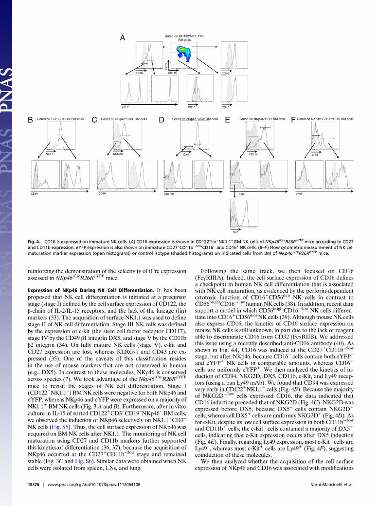
Following the same track, we then focused on CD16(FcγRIIIA). Indeed, the cell surface expression of CD16 definesa checkpoint in human NK cell differentiation that is associatedwith NK cell maturation, as evidenced by the perforin-dependentcytotoxic function of CD16+CD56dim NK cells in contrast toCD56brightCD16−/low humanNK cells (38). In addition, recent datasupport a model in which CD56brightCD16−/low NK cells differen-tiate into CD16+CD56dim NK cells (39). AlthoughmouseNK cellsalso express CD16, the kinetics of CD16 surface expression onmouse NK cells is still unknown, in part due to the lack of reagentable to discriminate CD16 from CD32 (FcγRIIB). We addressedthis issue using a recently described anti-CD16 antibody (40). Asshown in Fig. 4A, CD16 was induced at the CD27+CD11b−/low
stage, but after NKp46, because CD16− cells contain both eYFP−
and eYFP+ NK cells in comparable amounts, whereas CD16+
cells are uniformly eYFP+. We then analyzed the kinetics of in-duction of CD94, NKG2D, DX5, CD11b, c-Kit, and Ly49 recep-tors (using a pan Ly49 mAb). We found that CD94 was expressedvery early in CD122+NK1.1− cells (Fig. 4B). Because the majorityof NKG2D−/low cells expressed CD16, the data indicated thatCD16 induction preceded that of NKG2D (Fig. 4C). NKG2D wasexpressed before DX5, because DX5− cells contain NKG2D+
cells, whereas all DX5+ cells are uniformlyNKG2D+ (Fig. 4D). Asfor c-Kit, despite its low cell surface expression in both CD11b−/low
and CD11b+ cells, the c-Kit− cells contained a majority of DX5+
cells, indicating that c-Kit expression occurs after DX5 induction(Fig. 4E). Finally, regarding Ly49 expression, most c-Kit− cells areLy49−, whereas most c-Kit+ cells are Ly49+ (Fig. 4F), suggestingcoinduction of these molecules.We then analyzed whether the acquisition of the cell surface
expression of NKp46 and CD16 was associated with modifications
C
CD16
NKG2D
Gated on NKp46+CD3- BM cells
NKG2D
DX5
c-Kit
CD11b
DX5
Ly49
c-Kit
Gated on NKp46+CD3- BM cells Gated on NKp46+CD3- BM cells Gated on NKp46+CD11b-CD3- BM cells
CD
27
CD11b
CD16
CD16
eYFP
A Gated on CD122+NK1.1+lin-
BM cells
CD16
CD16
CD94
NK1.1
Gated on CD122+CD3- BM cellsB ED F
Fig. 4. CD16 is expressed on immature NK cells. (A) CD16 expression is shown in CD122+lin−NK1.1+ BM NK cells of NKp46iCreR26ReYFP mice according to CD27and CD11b expression. eYFP expression is also shown on immature CD27+CD11b−/lowCD16− and CD16+ NK cells. (B–F) Flow cytometric measurement of NK cellmaturation marker expression (open histograms) or control isotype (shaded histograms) on indicated cells from BM of NKp46iCreR26ReYFP mice.
18326 | www.pnas.org/cgi/doi/10.1073/pnas.1112064108 Narni-Mancinelli et al.

in NK cell function. The induction of NKp46 expression was as-sociated with a higher ability to secrete IFN-γ upon stimulationwith IL-12 and -18, although the response of NKp46−NK1.1+ NKcells and NKp46+NK1.1+ NK cells to phorbol 12-myristate 13-acetate (PMA) and ionomycin was comparable (Fig. S7A). Thishigher reactivity of NKp46+NK1.1+ NK cells to IL-12 and -18stimulation was associated with the induction of cell surface IL-12and -18 receptors (Fig. S8). In contrast, the response ofCD16−NK1.1+ NK cells and CD16+NK1.1+ NK cells to IL-12and -18 or PMA and ionomycin was similar (Fig. S7B). Un-expectedly, the IFN-γ production of CD11b−/lowNK1.1+ andCD11b+NK1.1+ NK cells was also comparable (Fig. S7C). Thus,despite the reported acquisition of NK cell functional response tovarious stimulations at the CD11b+ stage (34), our data define the
induction of NKp46 as a key step in the acquisition of NK cellreactivity to IL-12 and -18.Together these data converged to propose the acquisition of
NKp46 expression as a useful and stable tool to mark a previouslyundescribed stage 2 in NK cell differentiation. These findings alsolead us to propose a revisedmodel of NK cell differentiation basedon the sequential and stable acquisition of cell surface moleculesas follows: CD122 (stage 1), NK1.1 (stage 2), NKp46 (stage 3),CD16 (stage 4), and CD11b (stage 5) (Table 1). Stage 6 of NK cellmaturation is still defined by the disappearance of CD27 inCD11b+ mature NK cells, awaiting the identification of a cellsurface molecule that would be selectively and stably induced atthis stage. Indeed, although KLRG-1 and CD43 are preferentiallyexpressed in stage 6 NK cells (35), their cell surface expression isinitiated at stage 5. This model is fully compatible with the pre-vious classification (34) but refines the early stages of NK celldevelopment, i.e., between stage II, as defined earlier by the in-duction of NK1.1 and CD94, and stage III, as defined by the in-duction of Ly49 and c-kit.
Gut NKp46+ Cells. NKp46+ cells comprise not only bona fide NKcells and a minute subset of T cells, but also a population of mu-cosal cells. We thus took advantage of the Nkp46iCreR26ReYFP
mice to revisit the heterogeneity of NKp46+ cells in various organsby comparing for the expression of eYFP and the cell surface ofNKp46. In BM, spleen, LNs, and lungs, NKp46 and eYFP werecoexpressed (Fig. S9A Upper). Less than 10% of splenic and BMeYFP+ lymphocytes expressed low surface density of NKp46, butthey did not correspond to a population of eYFP+NKp46− cellsbecause they appeared as a continuum with NKp46+ cells (Fig.S9A Upper) and were uniformly NK1.1+ (Fig. S9A Lower). Con-firmation of this interpretation was obtained by using a brighterrevelation of the NKp46 fluorescence using an anti-NKp46 goatantiserum and Alexa 647-conjugated secondary reagent instead ofan anti-NKp46 mAb (Fig. S10).
Sytox
ROR t YFP Merge
eYFP
NK1.1
eYFPhiNK1.1-
eYFPhiNK1.1dim
eYFP+NK1.1+
NKp46 CD127 CD45 c-Kit
Gated on CD3-CD19- gut cells
783 1525 9448 370
1721 1579 10412 286
2632 432 17318 57
A
B
Fig. 5. Characterization of gut NKp46+eYFP+ and NKp46dim/loweYFPhigh cells. (A) Flow cytometric characterization of CD3-CD19-eYFPhiNK1.1−, eYFP-hiNK1.1dim, eYFP+NK1.1+ cells from small intestine of NKp46iCreR26ReYFP mice for indicated makers (open histograms) or control isotype (gray histograms).Results are representative of two experiments. (B) Immunofluorescence histology of small intestine frozen sections from NKp46CreR26ReYFP mice stained withanti-eYFP (green) and anti-RORt (red). Nuclei were counterstained with Sytox (grey). Scale bars, 10 mm. Data are representative of at least three experiments.
Table 1. NK cell maturation
Stage 1 Stage 2 Stage 3 Stage 4 Stage 5 Stage 6
CD122 + + + + + +NK1.1 − + + + + +NKp46 − − + + + +CD16 Low Low Low + + +CD11b Low Low Low Low + +CD27 − Low + + +/− −NKG2D Low Low Low + + +DX5 − − − + + +c-Kit − − − + +/− −Ly49 − − − + + +CD94 Low + + + + +CD43 − − − − Low +KLRG1 − − − − Low +
A revised model of NK cell maturation is proposed based on the sequen-tial expression of CD122, NK1.1, NKp46, CD16, and CD11b at NK cell surface.
Narni-Mancinelli et al. PNAS | November 8, 2011 | vol. 108 | no. 45 | 18327
IMMUNOLO
GY

A distinct picture emerged from the analysis of gut lymphocytesbecause two populations of cells expressing eYFP were detected inthe small intestine (Fig. 5A Left). Indeed, besides gut eYFP+ cellsthat uniformly express NK1.1 and the same level of eYFP com-pared with NKp46+ BM, splenic, LN, and lung cells, a subset ofgut eYFPhi cells with lower cell surface expression of NK1.1 wasdetected. Further analysis revealed that eYFP+NK1.1+ cells wereCD3−NKp46+CD127dimCD45hic-Kit−/dim (Fig. 5A Right) andcorresponded to bona fide small intestine NK cells (28). In con-trast, eYFPhiNK1.1− and eYFPhiNK1.1dim cells corresponded to acontinuum of NKp46dimCD127+c-Kit+ expressing intermediatelevels of CD45 (Fig. 5A Right). These cells corresponded to theNKp46+ROR-γt+ subset that produces IL-22 (22, 23, 26, 28, 41),consistent with the coexpression of eYFP and ROR-γt in gut cellsfrom isolated lymphoid follicules (Fig. 5B) and their ability toproduce IL-22 at steady state and upon IL-23 stimulation (Fig.S11). In contrast, only the eYFP+NK1.1+ cells produced IFN-γupon IL-12 and -18 stimulation, consistent with their bona fidesmall intestine NK cell identity (Fig. S11).Because the expression of iCre induced the irreversible ex-
pression of eYFP in Nkp46iCreR26ReYFP mice, the distinct levelsof eYFP observed at a single cell level could not result froma distinct regulation at the Nkp46 locus but, rather, indicateda distinct regulation of the Rosa26 locus in eYFPhi vs. eYFP+
cells, as shown in other reporter mouse models (42). Thus, thegenetic tracing in Nkp46iCreR26ReYFP mice indicated that theRosa26 locus was differently regulated in bona fide NK cells andin NKp46+ROR-γt+ cells, supporting by another approach thatNK cell receptor (NKR)+ROR-γt+ and NK cells represent twodistinct lineages (19, 24, 28).
Liver NKp46+ Cells. The analysis of liver lymphocytes inNkp46iCreR26ReYFP mice also revealed an unexpected complex-ity within eYFP+ cells. As in the gut, two populations of eYFPhi
and eYFP+ lymphocytes were detected. eYFPhi cells wereNK1.1+ and comprised a continuum of NKp46dim and NKp46+
cells (Fig. 6), whereas eYFP+ cells consisted of a more homo-geneous NK1.1+NKp46+ cell population. Besides the cell sur-face expression of NKp46, eYFPhi and eYFP+ cells expresseda panel of cell surface receptors present on NK cells, such asCD122, DX5, 2B4, NKG2D, and CD16, indicating that theycorresponded to two distinct subsets of liver NK cells (Fig. 6 andFig. S12A). Of note, it has recently been shown that liver NKcells comprised a population of CXCR6+ NK cells capable ofmediating hapten- and virus-specific memory responses (43, 44).However, the cell surface expression of CXCR6 did not correlatewith eYFP levels in liver NK cells from Nkp46iCreR26ReYFP. Anoticeable difference between liver eYFPhi and eYFP+ lympho-cytes resided in the high percentage of CD11b−/dim cells withinthe eYFPhi NK cell subset. This sign of immaturity in a subsetof liver NK cells has been reported and associated with the lowsurface expression of DX5 and the constitutive expression of tu-
mor necrosis factor-related apoptosis-inducing ligand (TRAIL)(45). A similar phenotype was observed for the eYFPhi liverNK cells (Fig. 6), indicating that they corresponded to theCD11b−/lowDX5dimTRAIL+NK cell subset previously described inthe same organ. The cell surface expression of Ly49 molecules wasmostly restricted to the eYFP+ liver NK cells as in the spleen (Fig.S12B), which is consistent with the bona fide NK cell phenotypeof eYFP+ liver NK cells and the more immature phenotype ofeYFPhi liver NK cells. Genetic tracing revealed that these two liversubsets are quite divergent, because the level of eYFP expression inCD11b−/low and CD11b+ BM NK cells was similar, in contrast tothe distinct eYFP levels in CD11b−/low and CD11b+ liver NK cells(Fig. S13). Thus, the levels of YFP are not merely associated withvarious stages of NK cell maturation, but with a more profoundlineage commitment of NK cell subsets, highlighting the use ofNkp46iCreR26ReYFP mice for in vivo fate-mapping experiments.
ConclusionsWe report here the fate mapping of NKp46+ cells in vivo throughthe generation and characterization of an unprecedented model ofNkp46iCre knock-in mice. Earlier attempts to create a mousemodel selectively targeting NKp46+ cells by using nontargetedtransgenesis have failed due to a complex and still poorly un-derstood regulation of the NKp46 locus (6, 29). To visualize iCreactivity in Nkp46iCre mice, we crossed them with R26ReYFP re-porter mice. In Nkp46iCreR26ReYFP mice, the fluorescent reporterpermanently labeled cells that had switched on the expression ofthe NKp46 gene. Using these mice, we have shown that the ex-pression of iCre faithfully corresponded to the endogenous ex-pression of NKp46. The genetic tracing of NKp46+ cells in vivoallowed us to reveal the stability of NKp46 cell surface expression.In addition, the acquisition of NKp46 marked a checkpoint of NKcell maturation. Based on these data, we propose a unique modelof NK cell differentiation, which also includes CD16 as a marker ofNK cell maturation. One advantage of this unique model resides inthe use of cell surface molecules that are conserved in both mouseand human, with the exception of mouse NK1.1. Along this line,preliminary data obtained on CD34+ hematopoietic cell progeni-tors from human cord blood indicate that the induction of surfaceCD56 precedes that of NKp46 in an in vitro NK cell differentiationassay, supporting the hypothesis that CD56 could be positioned inthe human NK cell differentiation pathway as NK1.1 in the mouse.Furthermore, the differential expression of YFP in Nkp46iCre
R26ReYFP mice showed that gut NKR+ROR-γt+ and NK cellsrepresent two distinct lineages. In addition, fate mapping experi-ments revealed the genetic heterogeneity of the two subsetsof CD11b−/lowDX5dimTRAIL+ and CD11b+DX5+TRAIL− liverNK cells. It is puzzling that besides bona fide eYFP+
CD3−NKp46+NK1.1+ NK cells, subsets of NKp46dim/loweYFPhigh
cells were present in gut and liver. Besides their commonNKp46dim/loweYFPhigh phenotype, gut and liver eYFPhi cells didnot appear to be directly related, because gut eYFPhi cells were
eYFP
NK1.1
Gated on CD3- liver cells
NKp46 CD11b TRAIL DX5
eYFPhi
eYFP+
357
801
134
923
294
44
361
1861
Fig. 6. Characterization of liver NKp46+eYFP+ and NKp46dim/loweYFPhigh cells. Flow cytometric characterization of NK1.1+CD3−eYFPhi and NK1.1+CD3−eYFP+
liver cells from NKp46iCreR26ReYFP mice for indicated makers (open histograms) or control isotype (gray histograms). Results are representative of two to fourexperiments.
18328 | www.pnas.org/cgi/doi/10.1073/pnas.1112064108 Narni-Mancinelli et al.

NK1.1−/dim and corresponded to NKR+ROR-γt+ cells, whereasliver eYFPhi cells were NK1.1+ and expressed TRAIL. Together,these results define Nkp46iCre mice as a unique mouse model ofspecific targeting in NKp46+ cells, allowing the generation ofunique mouse strains based on the crossing of Nkp46iCre mice to avariety to floxed mice to dissect the biology of NKp46+ NK, T, andgut NKR+ROR-γt+ cells.
Material and MethodsMice and Crosses. We have generated a Nkp46iCre knock-in mouse line inwhich the gene encoded the improved recombinase (icre) was inserted byhomologous recombination at the 3’ end of the Nkp46 gene (see SIMaterials and Methods). Recombinant offspring of chimeric mice wascrossed with transgenic flipase (FLP)-expressing mice (46). Nkp46iCre micewere crossed to R26ReYFP (31) (CDTA, Orleans, France). All mice used inthese studies were Nkp46iCreR26ReYFP, Nkp46iCre, or WT control litter-mates. All mice were bred in pathogen-free breeding facilities at Centred’Immunologie de Marseille-Luminy and used between 6 and 10 weeksof age. Experiments were conducted in accordance with institutionalguidelines for animal care and use. Nkp46iCre mice were crossed toRosa26lsl-DTR/lsl-DTR mice (32) to obtain NKp46iCre/wt; Rosa26lsl-DTR/wt micereferred as to Nkp46iCreR26RDTR. Six- to eight-week-old offspring mice were
used for experiments. Unnicked Diphtheria Toxin from Corynebacteriumdiphtheria (Calbiochem, 500 ng/mouse) diluted in PBS was injected i.p.
Cell Preparation. Cells were prepared as described (22). For details, see SIMaterials and Methods.
Flow Cytometry. Flow cytometric analysis was done on a FACS Canto (BectonDickinson, SanDiego, CA). For details on reagents, see SIMaterials andMethods.
Tissue Immunofluorescence. Portions of small and large intestine were iso-lated from mice and treated as previously described (22). For staining details,see SI Materials and Methods.
ACKNOWLEDGMENTS. We thank C. Cognet for help and advice and theCentre d’Immunologie de Marseille-Luminy (CIML) mouse house, cytome-try, and knockout/knock-in facilities. E.V. and S.U. are supported by Euro-pean Research Council Advanced Grants, grants from Agence Nationale dela Recherche (ANR) and Ligue Nationale Contre le Cancer (Equipe Labellisée“La Ligue”), and institutional grants from Institut National de la Santé etde la Recherche Médicale, Centre National de la Recherche Scientifique,and Université de la Méditerranée (to CIML). E.V., E.T., and N.Y. are sup-ported by ANR Grant N°R08063AS. E.V. is a scholar from Institut Universi-taire de France. E.N.-M. is a recipient of Agence pour la Recherche Contrele Cancer. J.C. was supported by Région Provence Alpes Côte d’Azur andInnate-Pharma.
1. Vivier E, et al. (2011) Innate or adaptive immunity? The example of natural killer cells.Science 331:44–49.
2. Biassoni R, et al. (1999) The murine homologue of the human NKp46, a triggeringreceptor involved in the induction of natural cytotoxicity. Eur J Immunol 29:1014–1020.
3. Boysen P, Storset AK (2009) Bovine natural killer cells. Vet Immunol Immunopathol130:163–177.
4. Pessino A, et al. (1998) Molecular cloning of NKp46: A novel member of the immu-noglobulin superfamily involved in triggering of natural cytotoxicity. J Exp Med 188:953–960.
5. Storset AK, et al. (2004) NKp46 defines a subset of bovine leukocytes with naturalkiller cell characteristics. Eur J Immunol 34:669–676.
6. Walzer T, et al. (2007) Identification, activation, and selective in vivo ablation ofmouse NK cells via NKp46. Proc Natl Acad Sci USA 104:3384–3389.
7. Walzer T, Jaeger S, Chaix J, Vivier E (2007) Natural killer cells: From CD3(-)NKp46(+) topost-genomics meta-analyses. Curr Opin Immunol 19:365–372.
8. Westgaard IH, et al. (2004) Rat NKp46 activates natural killer cell cytotoxicity and isassociated with FcepsilonRIgamma and CD3zeta. J Leukoc Biol 76:1200–1206.
9. Bottino C, Moretta L, Moretta A (2006) NK cell activating receptors and tumor rec-ognition in humans. Curr Top Microbiol Immunol 298:175–182.
10. Vivier E, Nunès JA, Vély F (2004) Natural killer cell signaling pathways. Science 306:1517–1519.
11. Halfteck GG, et al. (2009) Enhanced in vivo growth of lymphoma tumors in the ab-sence of the NK-activating receptor NKp46/NCR1. J Immunol 182:2221–2230.
12. Sivori S, et al. (1997) p46, a novel natural killer cell-specific surface molecule thatmediates cell activation. J Exp Med 186:1129–1136.
13. Gazit R, et al. (2006) Lethal influenza infection in the absence of the natural killer cellreceptor gene Ncr1. Nat Immunol 7:517–523.
14. Mandelboim O, et al. (2001) Recognition of haemagglutinins on virus-infected cells byNKp46 activates lysis by human NK cells. Nature 409:1055–1060.
15. Gur C, et al. (2010) The activating receptor NKp46 is essential for the development oftype 1 diabetes. Nat Immunol 11:121–128.
16. Stewart CA, et al. (2007) Germ-line and rearranged Tcrd transcription distinguishbona fide NK cells and NK-like gammadelta T cells. Eur J Immunol 37:1442–1452.
17. Yu J, et al. (2011) NKp46 identifies an NKT cell subset susceptible to leukemic trans-formation in mouse and human. J Clin Invest 121:1456–1470.
18. Cella M, et al. (2009) A human natural killer cell subset provides an innate source of IL-22 for mucosal immunity. Nature 457:722–725.
19. Crellin NK, Trifari S, Kaplan CD, Cupedo T, Spits H (2010) Human NKp44+IL-22+ cellsand LTi-like cells constitute a stable RORC+ lineage distinct from conventional naturalkiller cells. J Exp Med 207:281–290.
20. Cupedo T, et al. (2009) Human fetal lymphoid tissue-inducer cells are interleukin 17-producing precursors to RORC+ CD127+ natural killer-like cells. Nat Immunol 10:66–74.
21. Hughes T, et al. (2009) Stage 3 immature human natural killer cells found in secondarylymphoid tissue constitutively and selectively express the TH 17 cytokine interleukin-22. Blood 113:4008–4010.
22. Luci C, et al. (2009) Influence of the transcription factor RORgammat on the de-velopment of NKp46+ cell populations in gut and skin. Nat Immunol 10:75–82.
23. Sanos SL, et al. (2009) RORgammat and commensal microflora are required for the dif-ferentiation of mucosal interleukin 22-producing NKp46+ cells. Nat Immunol 10:83–91.
24. Satoh-Takayama N, et al. (2010) IL-7 and IL-15 independently program the differen-tiation of intestinal CD3-NKp46+ cell subsets from Id2-dependent precursors. J ExpMed 207:273–280.
25. Satoh-Takayama N, et al. (2008) Microbial flora drives interleukin 22 production inintestinal NKp46+ cells that provide innate mucosal immune defense. Immunity 29:958–970.
26. Spits H, Di Santo JP (2011) The expanding family of innate lymphoid cells: regulatorsand effectors of immunity and tissue remodeling. Nat Immunol 12:21–27.
27. Vivier E, Spits H, Cupedo T (2009) Interleukin-22-producing innate immune cells: Newplayers in mucosal immunity and tissue repair? Nat Rev Immunol 9:229–234.
28. Vonarbourg C, et al. (2010) Regulated expression of nuclear receptor RORγt confersdistinct functional fates to NK cell receptor-expressing RORγt(+) innate lymphocytes.Immunity 33:736–751.
29. Eckelhart E, et al. (2011) A novel Ncr1-Cre mouse reveals the essential role of STAT5for NK-cell survival and development. Blood 117:1565–1573.
30. Kioussis D, Festenstein R (1997) Locus control regions: Overcoming heterochromatin-induced gene inactivation in mammals. Curr Opin Genet Dev 7:614–619.
31. Srinivas S, et al. (2001) Cre reporter strains produced by targeted insertion of EYFPand ECFP into the ROSA26 locus. BMC Dev Biol 1:4.
32. Buch T, et al. (2005) A Cre-inducible diphtheria toxin receptor mediates cell lineageablation after toxin administration. Nat Methods 2:419–426.
33. Rosmaraki EE, et al. (2001) Identification of committed NK cell progenitors in adultmurine bone marrow. Eur J Immunol 31:1900–1909.
34. Kim S, et al. (2002) In vivo developmental stages in murine natural killer cell matu-ration. Nat Immunol 3:523–528.
35. Yokoyama WM, Kim S, French AR (2004) The dynamic life of natural killer cells. AnnuRev Immunol 22:405–429.
36. Chiossone L, et al. (2009) Maturation of mouse NK cells is a 4-stage developmentalprogram. Blood 113:5488–5496.
37. Hayakawa Y, Smyth MJ (2006) CD27 dissects mature NK cells into two subsets withdistinct responsiveness and migratory capacity. J Immunol 176:1517–1524.
38. Caligiuri MA (2008) Human natural killer cells. Blood 112:461–469.39. Romagnani C, et al. (2007) CD56brightCD16- killer Ig-like receptor- NK cells display
longer telomeres and acquire features of CD56dim NK cells upon activation. J Im-munol 178:4947–4955.
40. Braun A, et al. (2009) STIM1 is essential for Fcgamma receptor activation and auto-immune inflammation. Blood 113:1097–1104.
41. Sawa S, et al. (2010) Lineage relationship analysis of RORgammat+ innate lymphoidcells. Science 330:665–669.
42. Zhang DJ, et al. (2005) Selective expression of the Cre recombinase in late-stagethymocytes using the distal promoter of the Lck gene. J Immunol 174:6725–6731.
43. O’Leary JG, Goodarzi M, Drayton DL, von Andrian UH (2006) T cell- and B cell-in-dependent adaptive immunity mediated by natural killer cells. Nat Immunol 7:507–516.
44. Paust S, et al. (2010) Critical role for the chemokine receptor CXCR6 in NK cell-me-diated antigen-specific memory of haptens and viruses. Nat Immunol 11:1127–1135.
45. Takeda K, et al. (2005) TRAIL identifies immature natural killer cells in newborn miceand adult mouse liver. Blood 105:2082–2089.
46. Dymecki SM, Tomasiewicz H (1998) Using Flp-recombinase to characterize expansionof Wnt1-expressing neural progenitors in the mouse. Dev Biol 201:57–65.
Narni-Mancinelli et al. PNAS | November 8, 2011 | vol. 108 | no. 45 | 18329
IMMUNOLO
GY