![Influence of Organic Derivatives on Direct Regeneration of ... · using wide- or cross-hybridization among the closely related species [6]. Advances in regeneration and genetic transformation](https://static.fdocuments.in/doc/165x107/5f7bcaed21e8833a92237b0d/influence-of-organic-derivatives-on-direct-regeneration-of-using-wide-or-cross-hybridization.jpg)
FACTORS THAT INFLUENCE REGENERATION OF THE NEUROMUSCULAR...
18
J. exp. Biol. (1980), 89, 3 i-4a With 9 figures in Great Britain FACTORS THAT INFLUENCE REGENERATION OF THE NEUROMUSCULAR JUNCTION BY U. J. MCMAHAN, D. R. EDGINGTON AND D. P. KUFFLER From the Department of Neurobiology, Stanford University School of Medicine, Stanford, California 94305, U.S.A. SUMMARY Regeneration of neuromuscular junctions after trauma occurs in an orderly way and relies on communication between nerve and muscle. This paper summarizes evidence that factors which direct the growth and differentiation of both pre- and postsynaptic components of regenerating neuromuscular junctions are associated with the extracellular matrix of muscles. INTRODUCTION Motor axons and skeletal myofibres regenerate after injury and they form neuro- muscular junctions similar to normal ones. The way in which regeneration proceeds indicates that the axons and myofibres receive cues from tissue components that persist after degeneration of original pre- and postsynaptic cells. Accordingly, re- generating axons grow through original pathways of Schwann and perineurial cells, regenerating myofibres develop within the basal lamina sheaths of original myofibres, and neuromuscular junctions are formed at the original synaptic sites of the muscle. This paper summarizes findings from a series of experiments performed in this laboratory that were aimed at determining what structures within muscle influence regeneration of the neuromuscular junction. The findings demonstrate that factors which play a role in re-establishing the junction after damage to nerve and muscle are associated with the extracellular components of the muscle and that they are stably maintained in the absence of cells. The preparation The experiments we describe were done on the thin, paired cutaneous pectoris muscles of the frog (Rana pipiens). Thus, in this section we outline salient features of these muscles and of the frog's neuromuscular junctions. The neuromuscular junction of the frog is the most well understood of all synapses, with regard to structure and function, and nowhere is the arrangement of synaptic components more simple and orderly. The layout of the neuromuscular junctions in the cutaneous pectoris muscles and the methodology available for their study has made these muscles well suited for experiments on synapse regeneration. The cutaneous pectoris muscles (Fig. 1) lie just beneath the skin of the thorax. A nerve enters each muscle from the lateral edge and courses across it. Each muscle gists of about 500 myofibres and the entering nerve trunk contains about 25
Transcript of FACTORS THAT INFLUENCE REGENERATION OF THE NEUROMUSCULAR...

J. exp. Biol. (1980), 89, 3i-4aWith 9 figures
in Great Britain
FACTORS THAT INFLUENCE REGENERATIONOF THE NEUROMUSCULAR JUNCTION
BY U. J. M C M A H A N , D. R. EDGINGTON AND D. P. KUFFLER
From the Department of Neurobiology, Stanford University School ofMedicine, Stanford, California 94305, U.S.A.
SUMMARY
Regeneration of neuromuscular junctions after trauma occurs in an orderlyway and relies on communication between nerve and muscle. This papersummarizes evidence that factors which direct the growth and differentiationof both pre- and postsynaptic components of regenerating neuromuscularjunctions are associated with the extracellular matrix of muscles.
INTRODUCTION
Motor axons and skeletal myofibres regenerate after injury and they form neuro-muscular junctions similar to normal ones. The way in which regeneration proceedsindicates that the axons and myofibres receive cues from tissue components thatpersist after degeneration of original pre- and postsynaptic cells. Accordingly, re-generating axons grow through original pathways of Schwann and perineurial cells,regenerating myofibres develop within the basal lamina sheaths of original myofibres,and neuromuscular junctions are formed at the original synaptic sites of the muscle.This paper summarizes findings from a series of experiments performed in thislaboratory that were aimed at determining what structures within muscle influenceregeneration of the neuromuscular junction. The findings demonstrate that factorswhich play a role in re-establishing the junction after damage to nerve and muscle areassociated with the extracellular components of the muscle and that they are stablymaintained in the absence of cells.
The preparationThe experiments we describe were done on the thin, paired cutaneous pectoris
muscles of the frog (Rana pipiens). Thus, in this section we outline salient features ofthese muscles and of the frog's neuromuscular junctions. The neuromuscular junctionof the frog is the most well understood of all synapses, with regard to structure andfunction, and nowhere is the arrangement of synaptic components more simple andorderly. The layout of the neuromuscular junctions in the cutaneous pectoris musclesand the methodology available for their study has made these muscles well suited forexperiments on synapse regeneration.
The cutaneous pectoris muscles (Fig. 1) lie just beneath the skin of the thorax.A nerve enters each muscle from the lateral edge and courses across it. Each muscle
gists of about 500 myofibres and the entering nerve trunk contains about 25

32 U. J. MCMAHAN, D. R. EDGINGTON AND D. P. KUFFLER
Fig. i. The cutaneous pectoris muscles. Right muscle: slabs have been cut out leaving a narrowbridge of damaged muscle fibre segments in region of innervation. Left muscle: shaded areais the portion of the muscle that was frozen in order to kill all cells at the neuromuscularjunctions. Points of nerve damage are marked by X 's. Bar, 3 mm.
myelinated axons, most of which are motor (Rotshenker & McMahan, 1976). Theneuromuscular junctions are situated near the nerve trunk and its large branches,and thus they are confined to a band across the central region of the muscle (Fig. 1).
Our understanding of the structure of the frog's neuromuscular junction is based onseveral studies including those of Couteaux (1947), Birks, Huxley & Katz (1960a),Couteaux & Pe"cot-Dechavassine (1970), McMahan, Spitzer & Peper (1972), Dreyer etal. (1973), and Heuser, Reese & Landis (1974). Examples of terminals as seen in theelectron microscope are shown in Figs. 2, 3 and 4. For each muscle fibre, a myelinatedmotor axon gives rise to several unmyelinated terminal branches (1-3 /im in diameter)that run longitudinally along the fibre surface for up to 300 (im. Each terminal branchlies in its own shallow gutter several micrometres away from its nearest neighbour.The presynaptic membrane is separated from the post-synaptic membrane at pointsof closest apposition by a 50 nm synaptic cleft. As in other vertebrates, the musclefibre surface is depressed at intervals along the gutter to form junctional folds.Schwann cell processes cap the nerve terminal; in general their cell bodies are situatedalongside the longer terminal branches.
Synaptic vesicles, the most conspicuous of the axon terminal's organelles, aremainly situated in the half of the terminal that faces the muscle fibre (Figs. 2-4).Clusters of vesicles are focused on bands of osmiophilic material, which lie adjacentto the presynaptic membrane, just opposite the mouths of junctional folds in themyofibre surface (e.g. Couteaux & Pe'cot-Dechav&ssine, 1970). Accordingly, thesebands are distributed at regular intervals along the terminal. Combined anatomical,physiological and biochemical evidence from studies on the neuromuscular junctionand other synapses leads to the conclusion that each synaptic vesicle containsacetylcholine and releases its content into the synaptic cleft by exocytosis (See Katz,1969; Nickel & Potter, 1970; Heuser & Reese, 1973; Heuser et al. 1979). Whennerves are stimulated, exocytosis occurs at or near the bands of dense material onjfcg

Regeneration of neuromuscular junctions 33
esynaptic membrane (Couteaux & Pe'cot-Dechavassine, 1970; Heuser et al. 1974;fc et al. 1979) and thus these regions of the terminal are called 'active zones'
(Couteaux & Pe'cot-Dechavassine, 1970).Each skeletal muscle fibre in frogs, as in mammals, is ensheathed by several con-
centric layers (Uehara, Campbell & Burnstock, 1976; Sanes, Marshall & McMahan,1978). The myofibre surface, a typical lipid-rich osmiophilic plasma membrane, iscoated by a thin carbohydrate-rich glycocalyx. Separated from the glycocalyx by anarrow gap is the basal lamina (Figs. 2, 3 and 4; also called external lamina). Thisparticulate layer is 10-15 nm thick and like the basal lamina of other tissues (e.g.Carlson et al. 1978) probably contains collagen-like proteins. A reticular lamina ofindeterminant width and consisting of collagen fibrils lies beyond the basal lamina.The basal lamina and reticular lamina together form the myofibre's basementmembrane. The term basement membrane is also used by some investigators to referto the basal lamina alone.
The myofibre basal lamina lies in the synaptic cleft roughly equidistant from thepre- and postsynaptic membrane and sends projections into the junctional folds(Birks et al. 1960 a)'. It is closely associated with the surface of both the nerve terminaland myofibre by thin strands of material (Figs. 3 and 4; see also Heuser & Salpeter,1979). The basal lamina of the synaptic cleft is chemically distinctive from that cover-ing extrasynaptic portions of the myofibre. For example, the synaptic basal laminaappears to have different antigenic characteristics (Sanes & Hall, 1979). Other dif-ferences will be described below. The Schwann cell also has a thin basal lamina sheathwhich apparently fuses with that of the myofibre at the edges of the junction(Birks et al. 1960a). The reticular lamina of the myofibre does not enter the cleft but iscontinuous with the reticular lamina of the Schwann cell. We found additional extra-cellular material at the neuromuscular junction when we prolonged aldehyde fixationfrom 30 min to 2 h and then stained whole muscles with ruthenium red, which binds toglycosaminoglycans and other acidic carbohydrates (Luft, 1966; Luft, 1971; Kanwar &Farquhar, 1979), or stained thin sections of muscles with uranyl acetate in methanol. Anetwork of fine irregular filaments occupies the portion of the synaptic gutter not filledby the terminal and covers the Schwann cell cap (Fig. 2). This filamentous material iscontinuous with the basal laminae of the myofibre and Schwann cell. It varies inthickness from one portion of a terminal to the next, reaching as much as 1 fim.; andsome of the collagen fibrils of the reticular lamina are embedded in it. The fila-mentous material is also present around the short (1-5 ftm) unmyelinated preterminalsegments of motor axons but it is absent from the myelinated portion of motor axonsand elsewhere on the myofibre surface; thus it is a special feature of the neuromuscularjunction.
Acetylcholine receptors (AChRs) on skeletal muscle fibres are selectively con-centrated at the neuromuscular junction (Peper & McMahan, 1972; Hartzell &Fambrough, 1972; Kuffler & Yoshikami, 1975). The density of AChRs beneath thenerve terminal is at least 500 times greater than in extrajunctional regions (Fambrough& Hartzell, 1972; Kuffler & Yoshikami, 1975; Burden, 1977; Matthews-Bellinger &Salpeter, 1978). Cholinesterase (ChE) of skeletal muscle is also concentrated at theneuromuscular junction (e.g. Couteaux, 1955; Katz & Miledi, 1973). At least some of
is associated with the basal lamina of the synaptic cleft and when all cellular

34 U. J. MCMAHAN, D. R. EDGINGTON AND D. P. KUFFLER
components (nerve terminal, Schwann cell, and myofibres) have been removed frothe synaptic site (Fig. 56) ChE remains attached to the basal lamina for weeks& Kelly, 1971; Betz & Sakmann, 1973; McMahan, Sanes & Marshall, 1978).
Upon cutting or crushing motor nerves, the nerve endings degenerate and then arephagocytized by adjacent Schwann cells (Birks, Katz & Miledi, 19606). In the frog,both spontaneous and nerve-evoked transmitter release cease within a few days. Aftera ' silent' period of a few more days, miniature potentials can again be recorded fromdenervated muscle fibres. It is likely that they arise from ACh released by Schwanncells which, having phagocytized the terminals, are separated from the musclesurface by only the 50 nm cleft. Schwann cells gradually retract from the postsynapticmembrane over the ensuing weeks (Letinsky, Fischbeck & McMahan, 1976) but evenafter several months, aberrant miniature potentials can be detected (Birks et al.19606). In mammals, Schwann cells retract within a few days after phagocytosis of theterminal and thus Schwann potentials are seldom recorded (Miledi & Slater, 1968).
Many properties of muscle fibres in general and their plasma membrane in particularare altered by denervation and they are restored to their original state when the muscleis re-innervated (e.g. Harris, 1974). For example, the density of AChRs in extra-junctional areas is markedly increased within a few days after denervation. At the siteof the junction in mammals, there is little change in the density of AChRs over thefirst 1-2 weeks following denervation but then it gradually declines (Frank, Gautvik &Sommerschild, 1976; Loring & Salpeter, 1980). The turnover rate of AChRs at thejunction, with a half-time of about 10 days normally, decreases after denervation toabout 3 days, approaching the turnover rate of extrajunctional receptors, about 1 day(Loring & Salpeter, 1980). Junctional folds become broader and shallower and ChEdecreases (Guth, Albers & Brown, 1964; Hall, 1973). In the frog, even though manyproperties of the muscle cell are altered after denervation, specializations at thesynaptic site (a relatively high concentration of AChRs and ChE and the presence ofjunctional folds) persist for at least two months (Miledi, 1960a; Letinsky et al. 1976).
Muscle fibres undergo marked degenerative changes when they are damaged in anyof a number of ways, including mechanical (cutting or crushing), chemical, thermal, orischemic injuries (Price, Howes & Blumbert, 1964; Benoit & Belt, 1970; Carlson,1972; Vracko & Benditt, 1972; Hudgson & Field, 1973; Duchen et al. 1974; Snow,1977). Depending upon the extent of injury, portions of or entire muscle fibres aredisrupted. The disrupted cytoplasm and plasma membranes degenerate and arephagocytized by invading macrophages, but the basal lamina sheaths survive (Figs.5, bd and e, 7). Thus injury and its sequelae cleave the surface complex of the myo-fibre between the plasma membrane and basal lamina; the fate of the glycocalyx isunknown.
The role of extracellular structures in directing growth of axons to original synapticsites on myofibres
At the site of nerve damage, the portion of the axons that remains connected to theircell bodies sprouts processes which grow into the denervated muscle. Signs of re-formation of the neuromuscular junction in the frog can be detected both physio-logically and microscopically 7-10 days after cutting or crushing a nerve within1-2 mm of its muscle, and regeneration is complete by 30 days (Dennis & Miledi,

Regeneration of neuromuscular junctions 3 5Letinsky et al. 1976). The re-innervation of original synaptic sites on myonbres isBecise; by one month nearly all of the original subsynaptic membrane of the myonbres18 covered by nerve terminals and few, if any, contacts occur elsewhere on the myo-fibre surface (Letinsky et al. 1976; Sanes et al. 1978). Re-innervation in mammals canalso take place within a few days after nerve damage (e.g. Hines, Thomson & Lazere,1942; Guth, 1956; Dennis & Miledi, 1974) and the axons return to original endplateregions on myonbres (e.g. Tello, 1907; Gutmann & Young, 1944); however, newsynaptic sites can form in certain cases (e.g. Saito & Zacks, 1969; Frank et al. 1975).
Perineurial cells, Schwann cells and connective tissue elements, that survive in themotor nerve long after damaged axons have degenerated, may provide cues that directthe axons, for regenerating axons often grow through these tubes (Ram6n y Cajal,1928; Holmes & Young, 1942; Gutmann & Young, 1944; Haftek & Thomas, 1968;Letinsky et al. 1976). However, regenerating axons can cover long stretches ofsynaptic surface after they leave the perineurium, lifting off the muscle fibre at theend of the synaptic sites (Letinsky et al. 1976). Also axons growing beyond or outsideperineurial tubes can 'select' and precisely re-innervate original sites (Tello, 1907;Gutmann & Young, 1944; Letinsky et al. 1976). Thus there must be factors at thesynaptic site itself that provide cues to regenerating axons.
One possibility is that the target cells - the myofibres - guide re-innervation oforiginal synaptic sites (Tello, 1907; Ram6n y Cajal, 1928; Fischbach, 1974). We(Marshall, Sanes & McMahan, 1977) examined the role of the myofibre in nerveregeneration by studying the precision of re-innervation of original synaptic sites indamaged cutaneous pectoris muscles.
A rectangular slab was cut from the muscles on each side of the nerve trunk leavingbehind a bridge (3-4 mm long; 1-1-5 m m wide) of muscle fibre segments extendingbetween strips of undamaged fibres at the muscle's medial and lateral edges (Fig. 1).The nerve was then crushed with fine forceps 2-3 mm from the muscle's lateral border.By 4 days after the operation, phagocytosis of myofibres was extensive and profiles ofthe basal lamina sheaths enclosed numerous small cells that included macrophagesand myoblasts. The myoblasts are probably derived from mononucleated satellitecells which are situated at intervals along the surface of myofibres in normal musclesand remain intact in the bridge. By 2 weeks after the operation, new myofibres hadformed within the basal lamina sheaths by fusion of myoblasts, and nerves had grownback to the muscle. Both electron microscopy and electrophysiology demonstratedthat the myofibres became innervated. The most convenient way to identify originalsynaptic sites on the sheaths was to stain for ChE (Figs. 5, 6a; Karnovsky, 1964);over 95 % of the stained patches in these preparations marked the old sites (seeMarshall, Sanes & McMahan, 1977, for experimental details). When we scanned theperimeter of cross-sectioned sheaths, we found that 99 % of the terminals lay within1 /im of a ChE patch and 40 % of the patches were occupied by nerve terminals (Fig.6 b). These results demonstrate that the presence of the original myofibre is notnecessary for the expression of topographic specificity in denervated skeletal muscle;instead, factors that guide re-innervation of synaptic sites are maintained external tothe muscle cell.
We have extended these studies by examining re-innervation of damaged muscleremoval of Schwann cells as well as the original muscle cells, leaving only

36 U. J. M C M A H A N , D. R. EDGINGTON AND D. P. KUFFLER
extracellular components of synaptic sites intact (Edgington, Kuffler & McMaharufull manuscript in preparation). Instead of cutting muscle fibres as described abo\Wwe froze the region of innervation in the cutaneous pectoris muscles (Fig. 1; e.g.Vracko & Benditt, 1972; McMahan et al. 1980). A brass bar cooled in liquid nitrogenwas repeatedly applied to the muscle for 10 min. By 10 days the portions of musclefibres in the frozen region along with nerve terminals and their Schwann cells (as wellas all cellular components of intramuscular nerve bundles) degenerated and werephagocytized but the basal lamina sheaths remained intact. Two weeks after freezing,myofibres had regenerated within the basal lamina sheaths (Fig. 5 c) and were con-tacted by regenerating axon terminals that evoked muscle twitches when the nerveswere stimulated.
We examined the perimeter of cross-sectioned myofibres that had been stained forChE and found that by 3 weeks about 50% of the ChE spots were apposed by nerveterminals (Fig. 6c). Moreover, more than 98% of the terminals situated within 1 fimof the basal lamina were at ChE spots. To determine whether all of the ChE spotsrepresented original synaptic sites or whether any new spots had been formed, wetreated a set of muscles in situ with an irreversible inhibitor of ChE at the time wedamaged them (Marshall, Sanes & McMahan, 1977) which eliminated all detectableChE activity at the old sites. Three weeks later we stained the muscles for ChE,reasoning that any reaction product would represent new ChE produced subsequentto the operation. The inhibitor, diisopropyl fluorophosphate (DFP), dissolved inRinger to a concentration of 10 mM was applied topically by injecting it beneath thefrozen muscle and by placing pieces of gauze soaked in it directly on the muscle for2 h. DFP rapidly hydrolyzes in aqueous solutions; therefore we made changes inDFP soaked gauze and injections of freshly dissolved DFP every 30 min. Electronmicroscopy showed that both nerve and muscle cells degenerated in the DFP treatedmuscles as they did in unpoisoned preparations and by 3 weeks regenerated terminalscontacted myofibre basal lamina. At all sites there was some staining indicating thatnew ChE had been formed at the regenerated synapses. However, measurementsmade on electron micrographs showed that the amount of stain in all of the synapticclefts in DFP treated preparations was less than that in the clefts of preparations runin parallel but not treated with DFP. Thus the cholinesterase spots marked originalsynaptic sites and the axons returned precisely to these sites in the absence of theoriginal Schwann cells and myofibres.
In muscles damaged by freezing, nearly all of the original synaptic sites becamere-innervated by 42 days. On the other hand, in muscles damaged by cuttingmyofibres, only about 50% of the sites were re-innervated. Perhaps sites remaineduninnervated in the bridged preparations because access to them was blocked byconnective tissue that built up after cutting the muscle; a similar explanation has beenadvanced to account for the incomplete re-innervation of atrophied mammalianmuscle (Gutmann & Young, 1944).
The growth of axons and their guidance to the original synaptic sites on denervatedfibres are complex processes and we do not know how many factors are involved.Our observations showing that none of the original cells (myofibres and Schwanncells) of the synaptic sites need be present for precise re-innervation of sites meansthat at least one factor important for guidance is located extracellularly. This

Regeneration of neuromuscular junctions 37
ay have been produced by muscle cells and/or Schwann cells, or its productionhave required their presence, or its expression may require the presence of one
or the other cell type, but clearly it is stably maintained for a week or more in theabsence of cells. Extracellular factors that might play a role in directing topographicallyprecise re-innervation include (a) the reticular lamina which coats extrasynapticportions of the myofibre basal lamina and therefore may act as a mechanical barrierto regenerating axons, (b) the basement membrane of the Schwann cell which is notimpermeable to axons (Gutmann & Young, 1944; Letinsky et al. 1976) but mayprovide mechanical guidance and (c) the synaptic basal lamina of the myofibre or theextracellular material of the Schwann cell which may contain molecules that growingaxons specifically recognize.
The role of the basal lamina in differentiation of motor nerve terminals
Regenerated nerve terminals contain a high density of synaptic vesicles (Saito &Zacks, 1969) and their active zones are situated opposite junctional folds of myofibres(e.g. Rotshenker & McMahan, 1976). Preterminal portions of axons and portions ofterminals that grow beyond the end of the synaptic gutter (to become separated fromthe myofibre plasma membrane by more than ioo nm) have relatively few vesiclesand no active zones (Letinsky et al. 1976; Rotshenker & McMahan, unpublishedobservations). Thus, regenerating axons, like normal axons, have synaptic specializa-tions only at neuromuscular junctions.
We examined the factors that influence differentiation of the nerve terminal byasking whether axon terminals accumulate synaptic vesicles and form active zoneswhen they re-innervate synaptic basal lamina in the absence of myofibres (Sanes,Marshall & McMahan, 1978). For these experiments muscles were damaged bycutting muscle fibres as described above. To inhibit mitosis of myoblasts and thustheir subsequent fusion to form myofibres, the muscles were x-irradiated afterdamage and denervation. As in non-irradiated muscles, myofibres and axonsdegenerated and were phagocytized within 1 week but basal lamina sheaths remaineddevoid of myofibres for more than a month. Axons grew to the 'empty' basal laminasheaths and by 3 weeks occupied more than 40% of spots marked by ChE stain(Fig. 6d). Moreover, more than 95 % of the terminals within o-i /*m of myofibre basallamina were situated at stained patches.
A set of muscles were treated with DFP when they were damaged and denervatedto determine whether all of the ChE spots represented original synaptic sites orwhether some had been formed by regenerating axons that might have contactednon-synaptic areas of basal lamina. At the 25 terminals examined in DFP treatedpreparations at 3 weeks, there was little ChE staining, while in preparations nottreated with DFP, staining was intense (Figs. 6d and e). Thus the ChE stain was areliable marker for original synaptic sites in the myofibre-free preparations.
The portions of axons contacting the 'empty' sheaths differentiated, acquiringconcentrations of synaptic vesicles and active zones (Fig. ja, b). Moreover, the activezones were preferentially situated at or near intersections of synaptic cleft basallamina and basal lamina that projected into the folds of the original myofibre as atnormal neuromuscular junctions; of 32 active zones examined, 90% were within
nm of an intersection, which is a much higher percentage than predicted if
38 U. J. MCMAHAN, D. R. EDGINGTON AND D. P. KUFFLER
the active zones were randomly distributed along the' presynaptic' membrane. Theremay be a number of factors in these preparations that play a role in the differentiationof the nerve terminals, including factors provided by the myoblasts and/or Schwanncells; neurones growing in vitro can be stimulated to differentiate by glial and musclecells (Patterson & Chun, 1974; Giller et al. 1977). Nonetheless, these results indicatethat morphological differentiation of the nerve terminal is directed by moleculesattached to or a part of the synaptic portion of the myofibre basal lamina sheath andthat these molecules are situated at intervals along this region of the sheath.
The role of the basal lamina in organizing the postsynaptic membrane ofregenerating myofibres
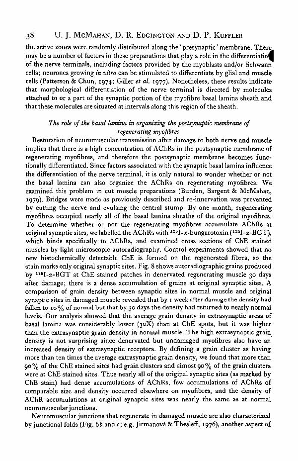
Restoration of neuromuscular transmission after damage to both nerve and muscleimplies that there is a high concentration of AChRs in the postsynaptic membrane ofregenerating myofibres, and therefore the postsynaptic membrane becomes func-tionally differentiated. Since factors associated with the synaptic basal lamina influencethe differentiation of the nerve terminal, it is only natural to wonder whether or notthe basal lamina can also organize the AChRs on regenerating myofibres. Weexamined this problem in cut muscle preparations (Burden, Sargent & McMahan,1979). Bridges were made as previously described and re-innervation was preventedby cutting the nerve and evulsing the central stump. By one month, regeneratingmyofibres occupied nearly all of the basal lamina sheaths of the original myofibres.To determine whether or not the regenerating myofibres accumulate AChRs atoriginal synaptic sites, we labelled the AChRs with 126I-a-bungarotoxin (mI-ct-BGT),which binds specifically to AChRs, and examined cross sections of ChE stainedmuscles by light microscopic autoradiography. Control experiments showed that nonew hi8tochemically detectable ChE is formed on the regenerated fibres, so thestain marks only original synaptic sites. Fig. 8 shows autoradiographic grains producedby 126I-a-BGT at ChE stained patches in denervated regenerating muscle 30 daysafter damage; there is a dense accumulation of grains at original synaptic sites. Acomparison of grain density between synaptic sites in normal muscle and originalsynaptic sites in damaged muscle revealed that by 1 week after damage the density hadfallen to 10% of normal but that by 30 days the density had returned to nearly normallevels. Our analysis showed that the average grain density in extrasynaptic areas ofbasal lamina was considerably lower (30X) than at ChE spots, but it was higherthan the extrasynaptic grain density in normal muscle. The high extrasynaptic graindensity is not surprising since denervated but undamaged myofibres also have anincreased density of extrasynaptic receptors. By defining a grain cluster as havingmore than ten times the average extrasynaptic grain density, we found that more than90% of the ChE stained sites had grain clusters and almost 90% of the grain clusterswere at ChE stained sites. Thus nearly all of the original synaptic sites (as marked byChE stain) had dense accumulations of AChRs, few accumulations of AChRs ofcomparable size and density occurred elsewhere on myofibres, and the density ofAChR accumulations at original synaptic sites was nearly the same as at normalneuromuscular junctions.
Neuromuscular junctions that regenerate in damaged muscle are also characterizedby junctional folds (Fig. 6b and c; e.g. Jirmanova & Thesleff, 1976), another aspect of
Regeneration of neuromuscular junctions 39
100
•3o. 75
50
25
777L6 8
Days after damage30
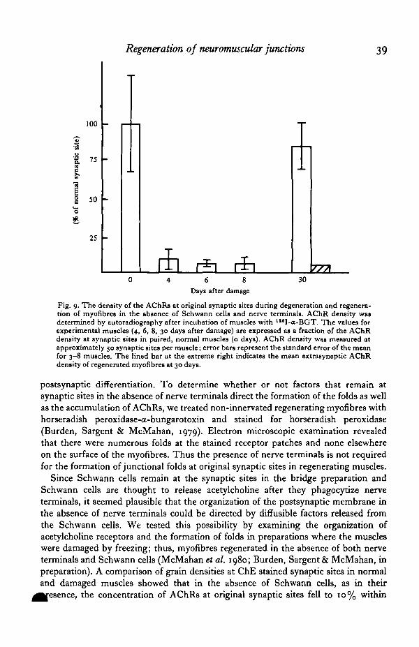
Fig. 9. The density of the AChRs at original synaptic sites during degeneration and regenera-tion of myofibres in the absence of Schwann cells and nerve terminals. AChR density wasdetermined by autoradiography after incubation of muscles with I lsI-a-BGT. The values forexperimental muscles (4, 6, 8, 30 days after damage) are expressed as a fraction of the AChRdensity at synaptic sites in paired, normal muscles (o days). AChR density was measured atapproximately 50 synaptic sites per muscle; error bars represent the standard error of the meanfor 3-8 muscles. The lined bar at the extreme right indicates the mean extrasynaptic AChRdensity of regenerated myofibres at 30 days.
postsynaptic differentiation. To determine whether or not factors that remain atsynaptic sites in the absence of nerve terminals direct the formation of the folds as wellas the accumulation of AChRs, we treated non-innervated regenerating myofibres withhorseradish peroxidase-a-bungarotoxin and stained for horseradish peroxidase(Burden, Sargent & McMahan, 1979). Electron microscopic examination revealedthat there were numerous folds at the stained receptor patches and none elsewhereon the surface of the myofibres. Thus the presence of nerve terminals is not requiredfor the formation of junctional folds at original synaptic sites in regenerating muscles.
Since Schwann cells remain at the synaptic sites in the bridge preparation andSchwann cells are thought to release acetylcholine after they phagocytize nerveterminals, it seemed plausible that the organization of the postsynaptic membrane inthe absence of nerve terminals could be directed by diffusible factors released fromthe Schwann cells. We tested this possibility by examining the organization ofacetylcholine receptors and the formation of folds in preparations where the muscleswere damaged by freezing; thus, myofibres regenerated in the absence of both nerveterminals and Schwann cells (McMahan et al. 1980; Burden, Sargent & McMahan, inpreparation). A comparison of grain densities at ChE stained synaptic sites in normaland damaged muscles showed that in the absence of Schwann cells, as in their
, the concentration of AChRs at original synaptic sites fell to 10% within

40 U. J. MCMAHAN, D. R. EDGINGTON AND D. P. KUFFLER
IO days after freezing and returned to near normal levels (80%) by one month whenmyofibres had regenerated (Fig. 9). Junctional folds were also selectively localized tqthe ChE stained sites. Since the only structure that remained at the synaptic site inthis experiment was the basal lamina of the myofibre, factors that direct the organiza-tion of the subsynaptic apparatus must be associated with the basal lamina.
Concluding remarks
The experiments described in this review demonstrate that factors which influencethe direction of axonal growth and the differentiation of synaptic specializations inregenerating nerve and muscle are associated with the extracellular matrix of themuscle. At least some of these factors are a part of or connected to the synapticportion of the myofibre's basal lamina sheath. Knowledge of where the factors thatinfluence regeneration of neuromuscular junctions are situated is a step towardscharacterizing them and determining how they are regulated.
REFERENCES
BENOIT, P. W. & BELT, P. (1970). Destruction and regeneration of skeletal muscle after treatment with alocal anesthetic, bupivicaine (Marcaine). J. Anat. 230, 331-357.
BETZ, W. & SAKMANN, B. J. (1973). Effects of proteolytic enzymes on function and structure of frogneuromuscular junctions. J. Pkysiol. 230, 673-688.
BIRKS, R., HUXLEY, H. E. & KATZ, B. (1960a). The fine structure of the neuromuscular junction of thefrog. J. Physiol. 150, 134-144.
BIRKS, R., KATZ, B. & MILEDI, R. (19606). Physiological and structural changes at the amphibianmyoneural junction, in the course of nerve degeneration. .J. Physiol. 150, 145—168.
BURDEN, S. (1977). Development of the neuromuscular junction in the chick embryo: the number,distribution, and stability of acetylcholine receptors. Devi Biol. 57, 317-329.
BURDEN, S. J., SARGENT, P. B. & MCMAHAN, U. J. (1979). Acetylcholine receptors in regeneratingmuscle accumulate at original synaptic sites in the absence of the nerve. J. Cell Biol. 82, 412—425.
CARLSON, B. M. (1972). The regeneration of skeletal muscle - a review. Am.J. Anat. 137, 119-149.CARLSON, E. C , BRENDEL, K., HJELLE, J. T. & MEEZAN, E. (1978). Ultrastructural and biochemical
analyses of isolated basement membranes from kidney glomeruli and tubules and brain and retinalmicrovessels. J. Ultrastruct. Res. 6a, 26—53.
COUTKAUX, R. (1947). Contribution a 1'etude de la synapse myoneurale. Revue can. Biol. 6, 563-711.COUTEAUX, R. (1955). Localization of cholinesterase at neuromuscular junctions. Int. Rev. Cytol. 4,
335-375-COUTEAUX, R. & PECOT-DECHAVASSINE, M. (I970). Visicules synaptiques et poches au niveau des ' zones
actives' de la jonction neuromusculaire. Comptes Rendus des siances de V Acadimie des Sciences, Paris D.371, 2346-2349.
DENNIS, M. J. & MILEDI, R. (1974). Non-transmitting neuromuscular junctions during an early stageof end-plate reinnervation. J. Physiol. 239, 553-570.
DRBYER, F., PEPER, K., AKERT, K., SANDRI, C. & MOOR, H. (1973). Ultrastructure of the 'active zone'in the frog neuromuscular junction. Brain Res. 6a, 373-380.
DUCHEN, L. W., EXCELL, B. J., PATEL, R. & SMITH, B. (1974). Changes in motor endplates resultingfrom muscle-fibre necrosis and regeneration. A light and electron microscopic study of the effects ofthe depolarizing fraction (cardiotoxin) of Dendroaspis jamesoni venom. J. neurol. Sci. 21, 391-417.
FAMBROUGH, D. M. & HARTZELL, H. C. (1972). Acetylcholine receptors: number and distribution atneuromuscular junctions in rat diaphragm. Science 176, 189-191.
FISCHBACH, G. D. (1974). Some aspects of neuromuscular junction formation. In Cell Communication(ed. R. P. Cox), pp. 43-66. New York: John Wiley.
FRANK, E., GAUTVIK, K. & SOMMERSCHILD, H. (1976). Persistence of junctional acetylcholine receptorsfollowing denervation. Cold Spring Harb. Symp. quant. Biol. 40, 275-281.
FRANK, E., JANSEN, J. K. S., LOMO, T. & WESTGAARD, R. (1975). The interaction between foreign andoriginal motor nerves innervating the soleus muscle of rats. J. Physiol. 247, 725—743.
GILLER, E. L., NEALE, J. H., BULLOCK, P. N., SCHRIER, B. K. & NELSON, P. G. (1977). Choline acetyl-transferase activity of spinal cord cell cultures increased by co-culture with muscle and by muscle-conditioned medium. J. Cell Biol. 74, 16—29.
GUTH, L. (1956). Regeneration in the mammalian peripheral nervous system. Physiol. Rev. 36, 441-478

Regeneration of newromuscular junctions 41
GUTH, L., ALBERS, R. W. & BROWN, W. C. (1964). Quantitative changes in cholinesterase activity ofdenervated muscle fibres and sole plates. Exp. Neurol. 10, 236-250.
GUTMANN, E. & YOUNG, J. Z. (1944). The re-innervation of muscle after various periods of atrophyJ. Anat. 78, 15-43.
HAFTBK, J. & THOMAS, P. K. (1968). Electron microscope observations on the effects of localized crushinjuries on the connective tissues of peripheral nerve. J. Anat. 103, 233—243.
HALL, Z. W. (1973). Multiple forms of acetylcholinesterase and their distribution in endplate andnonendplate regions of rat diaphragm muscle. J. Neurobiol. 4, 343-361.
HALL, Z. W. & KELLY, R. B. (1971). Enzymatic detachment of endplate acetylcholinesterase frommuscle. Nature New Biol, 33a, 62-63.
HARRIS, A. J. (1974). Inductive functions of the nervous system. Ann. Rev. Pkytiol. 36, 251-305.HARTZELL, H. C. & FAMBROUGH, D. M. (1972). Acetylcholine receptors: distribution and extra-
junctional density in rat diaphragm after denervation correlated with acetylcholine sensitivity.J. gen. Physiol. 60, 248-262.
HEUSBR, J. E. & REESE, T. S. (1973). Evidence for recycling of synaptic vesicle membrane duringtransmitter release at the frog neuromuscular junction. J. Cell Biol. 57, 315-344.
HEUSBR, J. E., REESE, T. S., DENNIS, M. J., JAN, Y., JAN, L. & EVANS, L. (1979). Synaptic vesicleexocytosis captured by quick freezing and correlated with quantal transmitter release. J. Cell Biol. 81,275-300.
HEUSER, J. E., REESE, T. S. & LANDIS, D. M. D. (1974). Functional changes in frog neuromuscularjunctions studied with freeze-fracture. J. Neurocytol. 3, 109-131.
HEUSBR, J. E. & SALPETER, S. R. (1979). Organization of acetylcholine receptors in quick-frozen, deep-etched, and rotary-replicated Torpedo postsynaptic membrane. J. Cell Biol. 8a, 150-173.
HlNES, H. M., THOMSON, J. D. & LAZERE, B. (1942). Quantitative studies on muscle and nerve regenera-tion in the rat. Am.J. Pkytiol. 137, 527-532.
HOLMES, W. & YOUNG, J. Z. (1942). Nerve regeneration after immediate and delayed suture. J. Anat.77,63-96.
HUDCSON, P. & FIELD, E. J. (1973). Regeneration of muscle. In Structure and Function of Muscle,vol. 2 (ed. G. H. Bourne), pp. 312—363. New York: Academic Press.
JIRMANOVA, I. & THBSLEFF, S. (1976). Motor endplates in regenerating rat skeletal muscle exposed tobotulinum toxin. Neuroscience i, 345—347.
KANWAR, Y. S. & FARQUHAK, M. G. (1979). Presence of heparan sulfate in the glomerular basementmembrane. Proc. natn. Acad. Set. U.S.A. 76, 1303—1307.
KARNOVSKY, M. J. (1964). The localization of cholinesterase activity in rat cardiac muscle by electronmicroscopy, jf. Cell Biol. 23, 217-232.
KATZ, B. (1969). The Release of Neuronal Transmitter Substances. Liverpool: Liverpool University Press.KATZ, B. & MILBDI, R. (1973). The binding of acetylcholine to receptors and its removal from the
synaptic cleft. J. Physiol. 231, 549-574.KUFFLER, S. W. & YOSHIKAMI, D. (1975). The distribution of acetylcholine sensitivity at the post-
synaptic membrane of vertebrate skeletal twitch muscles: iontophoretic mapping in the micronrange. J. Physiol. 344, 703-730.
LETINSKY, M. S., FISCHBECK, K. H. & MCMAHAN, U. J. (1976). Precision of reinnervation of originalpostsynaptic sites in frog muscle after a nerve crush. J'. Neurocytol. 5, 691—718.
LORING, R. H. & SALPETER, M. M. (1980). Denervation increases turnover rate of junctional acetyl-choline receptors. Proc. natn. Acad. Sci. U.S.A. 77, 2293-2297.
LUFT, J. H. (1966). Fine structure of a capillary and endocapillary layer as revealed by ruthenium red.Federation Proc. 35, 1773-1783.
LUFT, J. H. (1971). Ruthenium red and violet II. Fine structural localization in animal tissues. Anat.Rec. 171, 369-416.
MARSHALL, L. M., SANES, J. R. & MCMAHAN, U. J. (1977). Reinnervation of original synaptic sites onmuscle fiber basement membrane after disruption of the muscle cells. Proc. natn. Acad. Sci. U.S.A.74,3O73-3O77-
MATTHEWS-BELLINGER, J. & SALPETER, M. M. (1978). Distribution of acetylcholine receptors at frogneuromuscular junctions with a discussion of some physiological implications. J. Physiol. 379,197-213.
MCMAHAN, U. J., SANES, J. R. & MARSHALL, L. M. (1978). Cholinesterase is associated with the basallamina at the neuromuscular junction. Nature, hand. 271, 172-174.
MCMAHAN, U. J., SARGENT, P. B., RUBIN, L. L. & BURDEN, S. J. (1980). Factors that influence theorganization of acetylcholine receptors in regenerating muscle are associated with the basal lamina atthe neuromuscular junction. In Ontogenesis and Functional Mechanisms of Peripheral Synapses (ed.J. Taxi). Amsterdam: Elsevier/North-Holland. (In the Press.)
MCMAHAN, U. J., SPITZER, N. C. & PEPER, K. (1972). Visual identification of nerve terminals in livingisolated skeletal muscle. Proc. R. Soc. B 181, 421-430.
42 U. J. MCMAHAN, D. R. EDGINGTON AND D. P. KUFFLER
MILEDI, R. (1960a). Acetylcholine sensitivity of frog muscle fibres after complete or partial denervation.J.Physiol. 151,1-23.
Mn.imi, R. (19606). Properties of regenerating neuromuscular synapses in the frog. J. Phytiol. 154,190-203.
MILEDI, R. & SLATER, R. C. (1968). Electrophysiology and electron-microscopy of rat neuromuscularjunctions after nerve degeneration. Proc. R. Soc. B 169, 289—306.
NICKEL, E. & POTTER, L. T. (1970). Synaptic vesicles in freeze-etched electric tissue of Torpedo. Brain•R"- »3. 95-100-
PATTERSON, P. H. & CHUN, L. L. Y. (1974). The influence of non-neuronal cells on catecholamine andacetylcholine synthesis and accumulation in cultures of dissociated sympathetic neurons. Proc. natn.Acad. Set. U.S.A. 71, 3607-3610.
PEHER, K. & MCMAHAN, U. J. (1972). Distribution of acetylcholine receptors in the vicinity of nerveterminals on skeletal muscle of the frog. Proc. R. Soc. B 181, 431-440.
PRICE, H. M., HOWES, E. L. & BLUMBERT, J. M. (1964). Ultrastructural alterations in skeletal musclefibers injured by cold. I. Acute degenerative changes. Lab. Invest. 13, 1264-1278.
RAMON V CAJAL, S. Degeneration and Regeneration of the Nervous System. Originally written 1913 inSpanish. (Translated and ed. by R. M. May - English translation published 1928.) Oxford UniversityPress. Reprinted 1959 by Hafner Publishing Co., New York.
ROTSHENKER, S. & MCMAHAN, U. J. (1976). Altered patterns of innervation in frog muscle afterdenervation. J. Neurocytol. 5, 719-730.
SAITO, A. & ZACKS, S. I. (1969). Fine structure of neuromuscular junctions after nerve section andimplantation of nerve in denervated muscle. Exp. Mol. Pathol. 10, 256-273.
SANES, J. R. & HALL, Z. W. (1979). Antibodies that bind specifically to synaptic sites on muscle fibrebasal lamina. J. CellBiol. 83, 357-370.
SANES, J. R., MARSHALL, L. M. & MCMAHAN, U. J. (1978). Reinnervation of muscle fibre basal laminaafter removal of muscle fibres. J. CellBiol. 78, 176-198.
SNOW, M. H. (1977). Myogenic cell formation in regenerating rat skeletal muscle injured by mincing. I.A fine structural study. Anat. Ret. 188, 181-200.
TELLO, F. (1007). Degeneration et regeneration des plaques motrices apres la section des nerfs. Trav.Lab. Rich. Biol. Univ. Madrid5, 117-149.
UEHARA, Y., CAMPBELL, G. R. & BURNSTOCK, G. (1976). Muscle and its innervation. London: Arnold.VRACKO, R. & BENDITT, E. P. (1972). Basal lamina: The scaffold for orderly cell replacement. J. Cell
Biol. 55, 406-419.
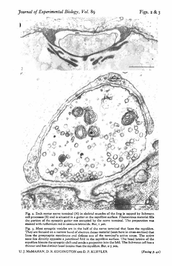
Journal of Experimental Biology, Vol. 89 Figs. 2 & 3
Fig. 2. Each motor nerve terminal (N) in skeletal muscles of the frog is capped by Schwanncell processes (S) and is situated in a gutter in the myofibre surface. Filamentous material fillsthe portion of the synaptic gutter not occupied by the nerve terminal. The preparation wasstained with ruthenium red in osmium tetroxide. Bar, I /im.
Fig. 3. Most synaptic vesicles are in the half of the nerve terminal that faces the myofibre.They are focused on a narrow band of electron dense material (seen here in cross-section) thatlines the presynaptic membrane and defines one of the terminal's active zones. The activezone lies directly opposite a junctional fold in the myofibre surface. The basal lamina of themyofibre bisects the synaptic cleft and sends a projection into the fold. The Schwann cell has athinner and less distinct basal lamina than the myofibre. Bar, 05 fun.
U. J. McMAHAN, D. R. EDGINGTON AND D. P. KUFFLER (Facing p. 42 )

Journal of Experimental Biology, Vol. 89 Fig. 4
»
Fig. 4. The active zone in this nerve terminal profile is sectioned longitudinally. The sectionalso passes longitudinally through a junctional fold and the sheet of basal lamina that projectsinto it. Bar, o- 5 /«n.
U. J. McMAHAN, D. R. EDGINGTON AND D. P. KUFFLER

Journal of Experimental Biology, Vol. 89 F i g -5
Fig. 5. When myofibres (a) are damaged they degenerate and are phagocytized but extracellularsheaths remain intact (6). Regenerating myofibres (c) form within the basal lamina sheaths ofthe original myofibres. Muscles shown in 6 and c had been damaged by freezing. ChE stain(arrows) marks synaptic sites on the myofibre basal lamina sheaths. Bar, 30 fim.
TJ. J. McMAHAN, D . R . EDGINGTQN AND D, P. KUFFLER
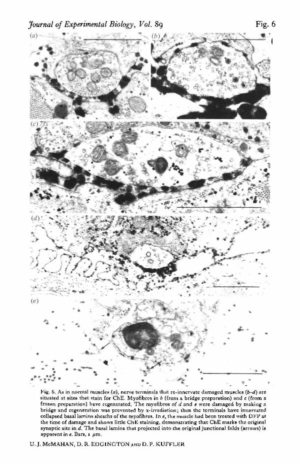
Journal of Experimental Biology, Vol. 89 Fig. 6
* ^ 5 * ^
(e)
Fig. 6. As in normal muscles (a), nerve terminals that re-innervate damaged muscles {b-d) aresituated at sites that stain for ChE. Myofibres in 6 (from a bridge preparation) and c (from afrozen preparation) have regenerated. The myofibres of d and t were damaged by making abridge and regeneration was prevented by x-irradiation; thus the terminals have innervatedcollapsed basal lamina sheaths of the myofibres. In e, the muscle had been treated with DFP atthe time of damage and shows little ChE staining, demonstrating that ChE marks the originalsynapric site in d. The basal lamina that projected into the original junctional folds (arrows) isapparent in e. Bars, I /tm.
U. J. McMAHAN, D. R. EDGINGTON AND D. P. KUFFLER
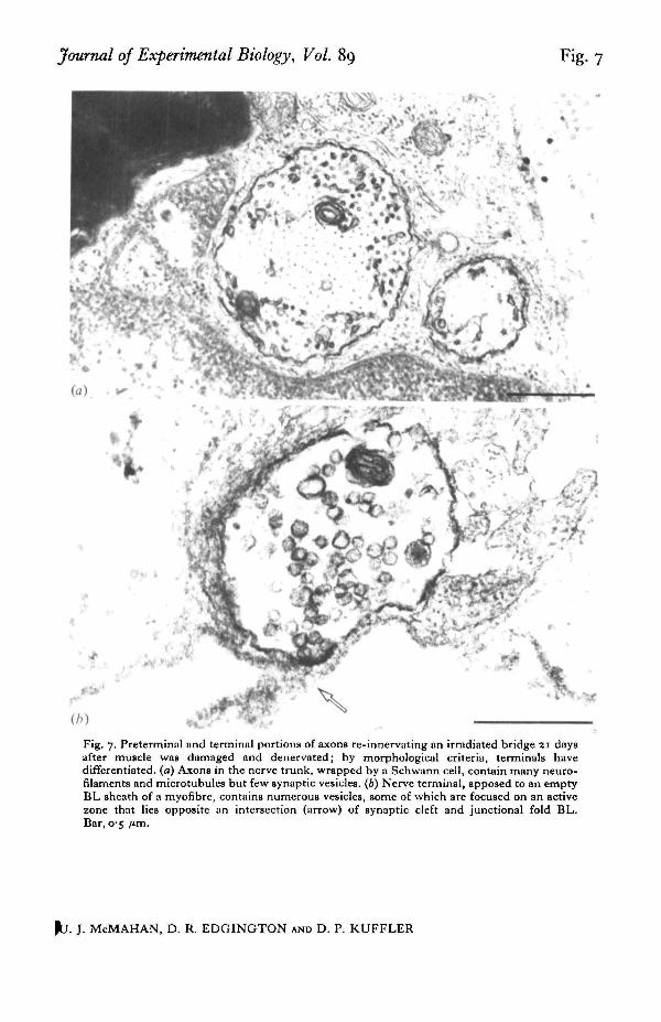
Journal of Experimental Biology, Vol. 89 Fig. 7
Fig. 7. Preterminal and terminal portions of axons re-innervating an irradiated bridge 21 daysafter muscle was damaged and denervated; by morphological criteria, terminals havedifferentiated, (a) Axons in the nerve trunk, wrapped by a Schwann cell, contain many neuro-filaments and microtubules but few synaptic vesicles. (6) Nerve terminal, apposed to an emptyBL sheath of a myofibre, contains numerous vesicles, some of which are focused on an activezone that lies opposite an intersection (arrow) of synaptic cleft and junctional fold BL.Bar, 05 /tm.
|b . J. McMAHAN, D. R. EDGINGTON AND D. P. KUFFLER

Journal of Experimental Biology, Vol. 89 Fig. 8
1 V)
Fig. 8. Regenerating myofibres accumulate AChRs at original synaptic sites in the absenceof nerve. Top: original synaptic sites marked by ChE stain (arrows). Bottom: autoradiographicgrains at same sites produced by "*I-tx-BGT bound to AChRs. 30 days after muscle damage.Bar, 10 /im.
U. J. McMAHAN, D. R. EDGINGTON AND D. P. KUFFLER