Facile synthesis of biodegradable affinity purified lectin ... · PDF fileMamidipudi...
13
Page 1 of 13 ANALYTICAL CHEMISTRY | RESEARCH ARTICLE Facile synthesis of biodegradable affinity-purified lectin nanoparticles Ashapogu Venugopal, Belur Shivappa Gnanesh Kumar, Mahamad Ahamad Mohiddon, Mamidipudi Ghanashyam Krishna and Nadimpalli Siva Kumar Cogent Chemistry (2016), 2: 1230951
Transcript of Facile synthesis of biodegradable affinity purified lectin ... · PDF fileMamidipudi...

Page 1 of 13
ANALYTICAL CHEMISTRY | RESEARCH ARTICLE
Facile synthesis of biodegradable affinity-purified lectin nanoparticlesAshapogu Venugopal, Belur Shivappa Gnanesh Kumar, Mahamad Ahamad Mohiddon, Mamidipudi Ghanashyam Krishna and Nadimpalli Siva Kumar
Cogent Chemistry (2016), 2: 1230951

Venugopal et al., Cogent Chemistry (2016), 2: 1230951http://dx.doi.org/10.1080/23312009.2016.1230951
ANALYTICAL CHEMISTRY | RESEARCH ARTICLE
Facile synthesis of biodegradable affinity-purified lectin nanoparticlesAshapogu Venugopal1, Belur Shivappa Gnanesh Kumar1,2, Mahamad Ahamad Mohiddon1,3, Mamidipudi Ghanashyam Krishna4 and Nadimpalli Siva Kumar1*
Abstract: Lectins are proteins/glycoproteins that exhibit distinct sugar specificities and interact with cell surface molecules and have therefore being used as potential tools for drug delivery systems. In the present study, we purified different sugar-spe-cific lectins by affinity chromatography. A glucose/mannose-specific lectin (DLL-I) as well as galactose-specific lectin (DLL-II) [from the seeds of Dolichos lablab], lactose-specific lectin (LSL) [from the fresh water mussel Lamellidens corrianus] and wheat germ agglutinin (WGA) [from wheat germ seeds] were affinity purified. Purified DLL-I migrated as a pentamer (α-1, α-2, α-3, α-4 in the range of 12–20 kDa and β subunit), DLL-II migrated as a dimer (α and β, 31 kDa and 29 kDa), LSL migrated as three subunit protein in SDS-PAGE (29 kDa, 28 kDa, and 26 kDa), and WGA mi-grated as single subunit (18 kDa). Nanoparticles of these lectins were prepared and characterized for the size and shape by transmission electron microscopy, scanning electron microscopy, and atomic force microscopy. The DLL-I, DLL-II, LSL, and WGA nanoparticles sizes are in the range 90 ± 20 nm, 150 ± 20 nm, 145 ± 10 nm, and 75 ± 15 nm, respectively.
*Corresponding author: Nadimpalli Siva Kumar, Protein biochemistry and Glycobiology laboratory, Department of Biochemistry, University of Hyderabad, Hyderabad-500046, India E-mails: [email protected], [email protected]
Reviewing editor:Youcef Mehellou, University of Birmingham, UK
Additional information is available at the end of the article
ABOUT THE AUTHORNadimpalli Siva Kumar is a professor and the head at the Biochemistry Department, University of Hyderabad. He is the author and co-author of about more than 70 peer reviewed journals. His group is working on purification and characterization of biologically important proteins from plant and animal sources, which includes:• Affinity purification of plant lectins, glycosidases,
and determination of their primary sequences as well as analysis of their glycan structures to understand their structure and function.
• Purification and characterization of the lysosomal enzymes and their sorting receptors to understand the lysosomal biogenesis in the vertebrates and invertebrates. Methods such as biochemical, cell biological, immunological, and molecular cloning techniques are adopted in the lab for the studies.
• Preparation and characterization protein nanoparticles (plant lectins) and polysaccharide nanoparticles in order to explore their utility as potential drug delivery systems.Nadimpalli Siva Kumar has been awarded the
DAAD fellowship and is the coordinator for the IRTG-MCGS exchange program.
PUBLIC INTEREST STATEMENTResearchers have developed biocompatible nanoparticles to achieve the effective targeting of specific drugs into various cell types for treatment of several diseases that affect humans. Our research work on lectins, the carbohydrate-binding proteins, was initiated with a long-term goal to make available different lectins as effective tools for targeted drug delivery. Preparation of biocompatible and biodegradable nanoparticles is very important in reducing the risks of drug retention inside the body, toxicity and immunogenicity associated with the other chemically modified nanoparticles. Drugs coated onto these nanoparticles can be introduced into body through oral delivery, intravenous, and subcutaneous injections. Using an array of lectins with wide sugar specificities should have potential applications for targeted drug delivery and enables us to have larger applications in treatment of several diseases which often are caused due to changes in glycan components in cells.
Received: 02 July 2016Accepted: 25 August 2016First Published: 01 September 2016
© 2016 The Author(s). This open access article is distributed under a Creative Commons Attribution (CC-BY) 4.0 license.
Page 2 of 13
Nadimpalli Siva Kumar
Page 3 of 13
Venugopal et al., Cogent Chemistry (2016), 2: 1230951http://dx.doi.org/10.1080/23312009.2016.1230951
Subjects: Biochemistry; General Science; Medicinal & Pharmaceutical Chemistry
Keywords: Dolichos lablab; lactose-specific lectin; Lamellidens corrianus; lectin; nanoparticle; wheat germ agglutinin
1. IntroductionLectins are sugar-binding proteins of non-immune origin that bind carbohydrate moieties of the glycoconjugates and have been affinity purified from a variety of sources. The interaction of the lectin with particular carbohydrates can be as specific as the interaction between those of the anti-gen and antibody or substrate and enzyme (Minko, 2004). Lectins which are able to bind certain sugars on the cell membrane, can increase bio-adhesion and potentially improve drug delivery via specific interaction and increase the residence time of the dosage form. The surface of the all verte-brate cells contain a carbohydrate coating called glycocalyx composed of glycoproteins, glycolipids, and glycosaminoglycans. In invertebrates, the involvement of these interesting proteins was ob-served in the processes such as differentiation and development of organism as well as in the elimi-nation of foreign substances by binding to their carbohydrate structures (Medeiros et al., 2010).
More recent research focused on using a few lectins as nanoparticles in drug delivery emphasizing their importance and applications in biomedicine. Quantum dots, carbon nanotubes, paramagnetic nanoparticles, liposomes, and gold nanoparticles are among the well-studied nanoparticles (Cai, Gao, Hong, & Sun, 2008). Biocompatibility, in vivo kinetics, tumor-targeting efficacy, acute and chronic toxicity, ability to escape through reticuloendothelial system, and cost-effectiveness are the current barriers in in vivo applications of nanoparticles in preclinical and potentially clinical use (Cai & Chen, 2007, 2008). The focus of recent research with important outcomes in medicine for diagno-sis and therapy is to design functional nanoparticles possessing optical and bio-affinity properties (Hillaireau & Couvreur, 2009; Sinha, Kim, Nie, & Shin, 2006) and much effort is now being devoted to the preparation of functionalized delivery systems, allowing active targeting while sparing healthy cells (Basu et al., 2009).
Among several nanoparticulate systems present currently, lipid-based nanocarriers, such as li-posomes, solid–lipid nanoparticles, and nano-structured lipid carriers and biodegradable polymeric nanoparticles are the most widely adopted nanosystems for drug delivery (Agrawal, Sharma, Gupta, & Vyas, 2014), and poly (lactic-co-glycolic acid) [PLGA] nanoparticles were proved to be useful in drug delivery when considered biocompatibility, biodegradability, and sustained targeted delivery. The overview of hybrid PLGA nanoparticles and their significance is well reviewed (Pandita, Kumar, & Lather, 2015). The selective accumulation of drugs on the targeted site can be achieved through receptor ligand interactions with carbohydrate-conjugated nanoparticles (Niikura et al., 2009), and the existence of endogenous lectins on the epithelial cells is exploited for drug targeting using sug-ars (Kumar Khanna, 2012). It has been shown in a specific study that the efficiency of lectin-conju-gated pac-MNPs was studied in leukemia cell line (K562), enhanced the uptake after conjugating the lectin to nanocarriers through lectin receptors. The use of such specific molecular signatures at the target site helps in delivering the appropriate therapeutic concentrations via receptor-mediated en-docytosis, (Singh, Dilnawaz, & Sahoo, 2011). Mucus glycoproteins which possess oligosaccharide units with L-fucose, D-galactose, N-acetyl-D-glucosamine, N-acetyl-D-galactosamine, and sialic acid, are the major structure-forming component of the mucus gel. Hence, muco-adhesive drug delivery system shows promising future in enhancing the bioavailability in drug delivery and capabil-ity of adhering to certain GI segments would offer various advantages. The development of nano-particles containing carbohydrates aimed at targeting certain lectins and the development of nanoparticles with lectins that bind to cell surface carbohydrates are considered to be the promising ways to detect cancer at an early stage and to improve drug delivery (Abu-Dahab, Schäfer, & Lehr, 2001; Smart, 2004; Yamazaki et al., 2000; Zhu, Yan, Guo, & Marchant, 2005).

Page 4 of 13
Venugopal et al., Cogent Chemistry (2016), 2: 1230951http://dx.doi.org/10.1080/23312009.2016.1230951
Due to their ability to recognize and bind specifically to carbohydrate moieties, lectins are being used in targeted drug delivery. Further, the delivery of lectins in the form of nanoparticles can greatly enhances the specific targeting, taking the advantage of nanoparticles size and surface characteris-tics. Additionally, the nanoparticles can be administered by oral, nasal, parenteral, and intra-ocular routes. Although many experimental studies on preparation of effective (functional) nanoparticles were carried out, preparation of stable, selective, and biodegradable nanoparticles is still at the bay. The main obstacle to oral administration of drug is the physiological barrier the mucus lining in the body. As lectins can recognize specific sugars present in the mucus lining of the GI tract, they can serve as valuable tools as the nanocarriers of therapeutics. Particles in the GI tract can be absorbed through various sites and mechanisms. Thus, the nanoparticles with a diameter of 1 μm may be phagocytosed by intestinal macrophages, whereas small particles (< 10 μm) may be absorbed through the Peyer’s patches in the small intestines and nanoparticles smaller than 200 nm can be transported via endocytosis by enterocytes (Ahmad, Othman, Md Zain, & Chowdhury, 2012; O’Hagan, 1996). Hence, the present study was undertaken with an objective of preparation of biocompatible lectin nanoparticles that can have the ability to cross the gut mucus layer (Kumar Khanna, 2012). As our work is focused on lectins we intended to prepare different sugar-specific biodegradable lectin nanoparticles as efficient tools for drug delivery into specific cell types, the present work was initi-ated. The lectins selected in the study were DLL-I, DLL-II, LSL, and WGA that were first affinity puri-fied in the lab for which nanoparticles were prepared. The present report describes their properties.
2. Materials methods
2.1. MaterialsDolichos lablab seeds (KR-307) were purchased from Wipro seeds (India); L.corrianus whole animal tissue was procured from M/s UV Scientifics, Hyderabad; Wheat germ, DVS, Sephacryl S-200 were from Sigma-Aldrich; Seralose 6B was from SRL, India; carbon-coated grids and uranyl acetate from M/s Icon analytical equipment Pvt LTD, Mumbai. All other chemicals and reagents used in this study were of high purity and procured from standards firms.
2.2. Preparation of sugar-specific affinity matricesCoupling of specific sugars (galactose, lactose, mannose, and N-acetyl glucosamine) to Seralose gel was carried out as described earlier (Latha, Rao, & Nadimpalli, 2006) and the gels were stored in equilibration buffer Tris-buffered saline (TBS) until use.
2.3. Extraction of Dolichos lablab–I (DLL-I), Dolichos lablab-II (DLL-II) from Dolichos lablab seeds and affinity purificationAbout 50 g of the seed powder was homogenized and extracted overnight with 1 L (20 vol) of 25 mM Tris-HCl buffer pH 7.4 (TBS). The suspension was centrifuged at 16260 × g for 20 min at 4°C. The clear supernatant was saturated to 60% with solid ammonium sulfate and stirred for 3 h. The supernatant and pellet were separated by centrifugation and the clear supernatant was subjected to 60–80% saturation with ammonium sulfate, stirred for 2 h, and centrifuged as described above and the pellet obtained was used to purify the DLL-I.
The glucose/mannose-specific lectin (DLL-I) was purified as described earlier (Gnanesh Kumar, Pohlentz, Schulte, Mormann, & Nadimpalli, 2014). Briefly, the pellet obtained after 60% ammonium sulfate saturation was dialyzed extensively against sodium acetate buffer pH 5.0. The precipitate obtained after dialysis was discarded and clear supernatant was dialyzed against TBS and was ap-plied on Seralose–mannose affinity gel equilibrated with TBS. After extensive washing with column buffer, bound protein was eluted with 0.25 M glucose in TBS. The protein-containing fractions were pooled, concentrated, and applied on Sephacryl S-200 gel filtration column to remove any possible aggregates or contaminating proteins.

Page 5 of 13
Venugopal et al., Cogent Chemistry (2016), 2: 1230951http://dx.doi.org/10.1080/23312009.2016.1230951
The DLL-II was purified as follows. The pellet obtained after 80% saturation was dissolved in TBS and applied onto pre-equilibrated Seralose–galactose matrix. After washing, the bound galactose-specific lectin was eluted with 300 mM galactose in TBS, fractions were collected, purified lectin was analyzed by 10% SDS PAGE and this protein sample was used for nanoparticles preparation.
2.4. Extraction of soluble proteins from L.corrianus and affinity purification of the LSLAbout 50 g of L.corrianus whole animal tissue was homogenized with 500 mL (10 vol) of TBS and the proteins were extracted overnight at 4°C. The homogenate was centrifuged at 16260 × g for 20 min. The supernatant collected and brought to 80% saturation by adding solid ammonium sulfate. The suspension was clarified by centrifugation and the pellet that was obtained after centrifugation was suspended in TBS and subjected to affinity chromatography on Sepharose–lactose affinity matrix.
Purification of LSL has been carried as described earlier (Radha, 2002). Briefly, about 20 mL of Seralose–lactose matrix was packed in a glass column and equilibrated with 25 mM TBS. The soluble extract obtained above was applied on to the column and the unbound proteins were removed by washing the gel with same buffer until the A280 reached ≤ 0.05. Elution of the bound lectin was car-ried out with 25 mM TBS containing 0.3 M lactose. The eluted fractions that contain protein were pooled, concentrated, and subjected to gel filtration chromatography on Sephadex G-100 gel. About 100 mL of the swollen Sephadex G-100 gel was packed in to a glass column, and equilibrated with TBS. The affinity-eluted lectin was passed and 3-mL fractions were collected. The eluted fractions were pooled, concentrated, and analyzed by 10% SDS-PAGE.
2.5. Extraction and purification of WGAWheat germ powder was ground to a fine powder in a blender, defatted using acetone, and the pow-der air-dried. Forty-six grams of the defatted powder was extracted with 460 mL (10 vol) of 50 mM sodium acetate buffer pH 4.5, kept under stirring for overnight at 4°C. The homogenate was centri-fuged at 16260 × g for 25 min at 4°C; the clear supernatant obtained was brought to 70% saturation with ammonium sulfate and kept for stirring for 3 h at 4°C. The pellet was collected by centrifugation at 16260 × g for 20 min, dissolved in 50 mM sodium acetate buffer pH 4.5 and dialyzed against the same for overnight. The dialyzed sample was loaded on the Seralose-N-acetyl glucosamine affinity matrix. After washing, the bound lectin was desorbed with 0.5 M N-acetylglucosamine in PBS and 4-mL fractions were collected. The fractions with maximum A280 were pooled and dialyzed extensively against PBS to remove the free sugar, and was used for nanoparticles preparation and SDS-PAGE.
2.6. SDS-PAGEThe purity of each lectin was analyzed by 10 and 12% SDS-PAGE under reducing conditions (Laemmli, 1970).
2.7. Preparation of nanoparticlesThe nanoparticles of DLL-I, DLL-II, LSL, and WGA lectins were prepared separately by oil dispersion-assisted sonication technique. To each purified lectin sample (5 mg/ml), 15 ml of chilled olive oil was added slowly at 4°C with continuous dispersion by gentle vortexing separately. Then the sample was sonicated with probe sonicator for 30 s for 15 times with a gap of 1 min between each successive step. The resulting mixture was then frozen immediately in liquid nitrogen for 10 min to arrest the dispersed protein nanoparticles in oil matrix and incubated on ice for 4 h. The particles formed (sam-ple) were then centrifuged at 4065 × g for 10 min, the resulting pellet was washed extensively with diethyl ether, dried in 4°C overnight, and the particle pellet was dissolved in PBS. The resulting parti-cles size and shape was characterized by transmission electron microscopy, scanning electron mi-croscopy, atomic force microscopy, and scanning near-field optical microscopy.
2.8. Characterization of nanoparticlesThe intrinsic properties of lectin nanoparticles such as particle size, monodispersity, and morphology were characterized by TEM, SEM, AFM, and SNOM. The experimental details of the each characteriza-tion were described as follows.
Page 6 of 13
Venugopal et al., Cogent Chemistry (2016), 2: 1230951http://dx.doi.org/10.1080/23312009.2016.1230951
The bright-field TEM micrographs were obtained from a Technai G2 STwin, FEI electron micro-scope, operated at 200 kV. A 10-nm gold film deposited on the grid was used for the purpose of camera length calibration. Prior to the characterization, samples were prepared for imaging by plac-ing a drop of lectin nanoparticle solution on carbon-coated grid and allowed it to dry in closed con-tainer. The sample on the grid was washed twice with deionized distilled water to dilute nanoparticles that were bound to the mesh of the TEM grid and then allowed to dry. The grid with sample was then stained with 1% uranyl acetate followed by washing with deionized distilled water (negative staining).
The surface morphology of lectin nanoparticles were recorded by a field emission scanning elec-tron microscope (FE-SEM) Model Ultra55 of Carl Zeiss and AFM Model SPA 400 of Seiko Instruments Inc., Japan with SPI-3800 probe station. Before the measurement, samples were fabricated by coat-ing the lectin nanoparticles solution on a thin glass substrate. The glass substrates were extensively cleaned with deionized distilled water and isopropyl alcohol in sonicator for 5 min each, before coat-ing. Few drops of sample solution were placed over the glass substrate and the substrate was placed over the rotor of the spin coater (Euro, Model). The sample was spin coated at 3,000 rpm for 5 min under ambient atmospheric conditions to produce reasonably thin films. The samples were com-pletely dried before taking to AFM and SEM measurements. For SEM measurements the samples were further subjected to thin Au film deposition by DC sputtering. The gold-coated samples were vacuum dried and then examined.
Further, particle size and shape of the lectin nanoparticles were characterized by near-field optical microscopy. The lectin nanoparticles solution is coated on 150-μm-thick glass substrate as described for AFM sample preparation. SNOM imaging of coated sample was carried out using a commercial near-field microscope equipped with a cantilever style SNOM probe (Model alpha 300, WiTec Instrument Corp.), Nd-YAG laser with power 40 mW and 532 nm was used as source for recording the near-field images. The samples were illuminated in the near field through SNOM tip and the in-teracted light with the sample was collected in the far field (Mohiddon, Sangani, & Krishna, 2013).
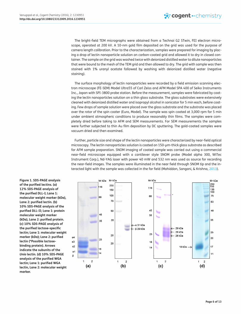
Figure 1. SDS-PAGE analysis of the purified lectins. (a) 12% SDS-PAGE analysis of the purified DLL-I; Lane 1: molecular weight marker (kDa), Lane 2: purified lectin. (b) 10% SDS-PAGE analysis of the purified DLL-II; Lane 1: protein molecular weight marker (kDa), Lane 2: purified protein. (c) 10% SDS-PAGE analysis of the purified lactose-specific lectin; Lane 1: molecular weight marker (kDa); Lane 2: purified lectin (*Possible lactose-binding protein). Arrows indicate the subunits of the Unio lectin. (d) 10% SDS-PAGE analysis of the purified WGA lectin; Lane 1: purified WGA lectin, Lane 2: molecular weight marker.

Page 7 of 13
Venugopal et al., Cogent Chemistry (2016), 2: 1230951http://dx.doi.org/10.1080/23312009.2016.1230951
3. Results and discussion
3.1. Purification of DLL-I, DLL-II, LSL, and WGAThe affinity-purified DLL-I from lablab beans was resolved into five subunits on 12% SDS-PAGE with molecular masses in the range of 12–20 kDa and the subunits were designated as alpha α-1, α-2, α-3, α-4, and β (Figure 1(a)) (Gnanesh Kumar et al., 2014). The affinity-purified galactose-specific lectin was resolved in to two distinct subunits with molecular masses corresponding to 31 kDa (α) and 29 kDa (β) (Figure 1(b)) (Latha et al., 2006), consistent with our earlier results. Lactose-specific lectin was purified by employing the combination of affinity and gel filtration chromatographic methods. The purified lactose-specific lectin migrated as three subunit protein bands in 10% SDS-PAGE under reducing con-ditions with molecular masses of approximately 29 kDa, 28 kDa, and 26 kDa (Figure 1(c)). The affinity-purified WGA migrated as a single subunit in 10% SDS PAGE under reducing conditions (Figure 1(d)).
3.2. Characterization of lectin nanoparticlesThe nanoparticles of each purified lectin were prepared and observed under TEM, SEM, AFM, and SNOM.
3.2.1. Characterization of DLL-I nanoparticlesThe bright-field TEM image and SEM micrographs of DLL-I nanoparticles were presented in Figure 2(a) and (b), respectively. The TEM image has perfect spherical shape with approximate diameter of 90 ± 15 nm. On the other hand, the SEM image shows, slightly irregular spherical shape with approxi-mate diameter of 80 ± 20 nm. Further, the size of the DLL-I nanparticle was extracted in the form of AFM image in Figure 2(c). The spin-coated lectin nanoparticles are in spherical shape with average diameter of 115 ± 25 nm.
Further, the dimensions of the DLL-I nanoparticles were extracted by drawing a line across the AFM image and studying the profile along the selected line. The selected line on AFM image, its line profile and the extracted information of the line profile were presented in Figure 2(d). The peak width of the line profile gives the size of the nanoparticle. The details of each peak width were presented in the table form of the same Figure 2(d). The color on either side of the peak is denoted by the same color in the table. From the dense morphology of AFM images we can draw out the particle size distribution
Figure 2. TEM, SEM, and AFM micrographs of the purified DLL-I nanoparticles. (a) Transmission electron micrograph of the purified DLL-I lectin nanoparticle. (b) Scanning electron micrograph of the purified DLL-I lectin nanoparticle. (c) AFM images of DLL-I nanoparticles. (d) Cross-sectional line profile of the AFM image of DLL-I lectin nanoparticles.

Page 8 of 13
Venugopal et al., Cogent Chemistry (2016), 2: 1230951http://dx.doi.org/10.1080/23312009.2016.1230951
statistics of spin-coated sample. Nearly, 50 nanoparticles were measured as described above and their distribution in the form of histogram was presented in the inset of the AFM image Figure 2(c). The particle size distribution confirms the nearly monodisperse nature of the synthesized nanoparti-cles. The difference in the dimensions and the shapes of the DLL-I nanoparticle in both TEM and SEM is expected to be due to the entirely different way of sample preparations carried for their respective measurements and we observed that the stained surface of nanoparticle in TEM measurement has produced perfect spherical shape compared to that of SEM micrographs. The image observed in the TEM is the resultant of electron scattering by the heavier molecules (uranyl acetate) stained on the surface of the lectin nanoparticle. The AFM images are taken at the center of the spin-coated sample substrate, while for the SEM imaging the edge of the substrate (sample coated) has been selected to see the individual nanoparticles. Due to this reason, inspite of following the same sample preparation, the AFM image shows dense morphology compared to that of SEM micrographs.
3.2.2. Characterization of DLL-II nanoparticlesThe TEM, SEM, and AFM images of DLL-II nanoparticles were presented in Figure 3(a), (b), and (c), respectively. The bright-field TEM micrograph in Figure 3(a) shows stained surface of DLL II nanopar-ticles, with approximate spherical morphology having an average diameter of 150 ± 20 nm. Highly dispersed and clearly isolated DLL-II nanoparticles were observed throughout the TEM grid, which confirms the homogenious sample preparation of DLL-II lectin nanoparticles. The average nanopar-ticle size of DLL-II nanoparticle calculated from SEM and AFM images of Figure 3(b) and (c) are ap-proximately 70 ± 20 nm and 98 ± 15 nm, respectively. The particle size extracted from AFM measurements were presented in Figure 3(d).
Though both DLL-I and DLL-II are isolated from the same seed material both exhibit distinct sugar specificites and differ in their molecular mass and subunit patters. The apparent differences in the size of the nanoparticles is possibly due to the differences in their molecular masses and subnit patterns.
Figure 3. TEM, SEM, and AFM micrographs of the purified DLL-II nanoparticles. (a) Transmission electron micrograph of the purified DLL-II lectin nanoparticles. (b) Scanning electron micrograph of the purified DLL-II lectin nanoparticles. (c) AFM images of DLL-II lectin nanoparticles. (d) Cross-sectional line profile of the AFM image of DLL-II lectin nanoparticle.

Page 9 of 13
Venugopal et al., Cogent Chemistry (2016), 2: 1230951http://dx.doi.org/10.1080/23312009.2016.1230951
3.2.3. Characterization of LSL nanoparticlesThe particle size and shape of LSL nanoparticles characterized by TEM, SEM, and AFM were presented in Figure 4(a)–(d) simultaneously. The TEM micrograph in Figure 4(a) and (b) shows perfectly spheri-cal nanoparticles of the average diameter of 145 ± 10 nm. The average nanoparticle size of lactose lectin nanoparticle calculated from SEM and AFM images of Figure 4(c) and (d) are approximately 200 ± 10 nm and 146 ± 15 nm, respectively. Further nanoparticle characterization has been carried by SNOM. The SNOM image along with its surface morphology of the lactose lectin nanoparticles spin coated on the thin glass substrate is presented in the Figure 4(e) and (f), the diameter of the particles is in the range of 146 ± 15 nm.
In normal confocal optical microscope, the nanoparticle of the dimension less than 300 nm can-not be resolved, due to the Rayleigh diffraction limit. This limit has been solved by introducing a near-field concept in the SNOM measurement. In the SNOM measurement, the sample is illuminated by a near-filed source, i.e. by a SNOM probe having an aperture of the dimension of 50 nm. SNOM
Figure 4. TEM, SEM, AFM, and SNOM micrographs of LSL nanoparticles. (a) & (b) Transmission electron micrographs of the LSL nanoparticles. (c) Scanning electron micrograph of the lactose-specific lectin nanoparticles. (d) AFM image of the lactose-specific lectin nanoparticles. (e) & (f) SNOM images of the lactose-specific lectin nanoparticles.

Page 10 of 13
Venugopal et al., Cogent Chemistry (2016), 2: 1230951http://dx.doi.org/10.1080/23312009.2016.1230951
probe works exactly same as contact AFM tip, in which the tip moves on the surface of the sample. Hence, by scanning the surface with near-field probe, one can obtain the surface features followed by the corresponding optical image. By imposing this technique one can resolve much smaller nano-particles beyond the Rayleigh’s diffraction limit. The surface morphology and its SNOM image are presented in Figure 4(e) and (f) simultaneously. From the surface morphology of the image of Figure 4(e), the calculated average diameter of the lactose lectin nanoparticle is found to be 350 ± 50 nm. The large discripancy in the size of nanoparticle compared with that of the AFM measurement is due to the huge dimension of SNOM probe (250 nm) compared to AFM tip (10 nm). The true optical image carried by SNOM images of the nanoparticle is presented in Figure 4(f) and the diameter of the par-ticles is in the range of 146 ± 15 nm.
3.2.4. Characterization of WGA nanoparticlesThe morphology of WGA nanoparticles that were characterized by TEM, SEM, and AFM were pre-sented in the Figure 5(a)–(c), respectively. The shape of WGA nanoparticle shown in TEM image (Figure 5(a)) is approximately spherical with particle size of 75 ± 15 nm. The SEM image shown in Figure 5(b) has particle size of 105 ± 20 nm and that of AFM image shown in Figure 5(c) has particle size of 82 ± 10 nm. The slight devaiation of the particle in three different techniques is expected to be due to different treatement given to samples. The particle size calculated from AFM image was presented in Figure 5(d).
Among the lectin nanoparticles characterized in this study, WGA nanoparticles appear to be hav-ing smaller size as seen in TEM. The different sizes and shapes of the nanoparticles prepared is pos-sibly due to the different treatments given to the samples. Further it might also depend on the size as well as the glycoprotein nature of the protein.
The role of lectins in various physiological processes and functions is expanding rapidly in recent years. In the vertebrates, different types of lectins are present and are involved in a variety of func-tions such as cell adhesion and differentiation, control of cell growth. By virtue of their recognition specificities, lectins have been used as reliable biochemical, cytochemical, and histochemical probes in the study of cell surface glycoconjugate differences in malignant and non-malignant cells (Mody, Joshi, & Chaney, 1995).
Figure 5. TEM, SEM, and AFM micrographs of the purified WGA lectin nanoparticles. (a) Transmission electron micrograph of the purified WGA lectin nanoparticles. (b) Scanning electron micrograph of the purified WGA lectin nanoparticles. (c) AFM images of WGA lectin nanoparticles. (d) Cross-sectional line profile of the AFM image of WGA lectin nanoparticles.

Page 11 of 13
Venugopal et al., Cogent Chemistry (2016), 2: 1230951http://dx.doi.org/10.1080/23312009.2016.1230951
The nanoparticle size should be so optimized that it should not escape from the kidney filtration. Nanoparticles used in the drug delivery system should be large enough to prevent their speedy out-flow into blood capillaries, but they should be small enough to escape capture by fixed macrophages stuck in the reticuloendothelial system, such as liver and spleen (Kumar Khanna, 2012). Nanoparticles smaller than 10 nm, will be filtered by the kidneys. In addition, they will be captured by the liver if they are larger than 100 nm. Thus, the optimum nanoparticle size for the in vivo applications is 10–100 nm. However, in vivo imaging and diagnostic prefers NPs of larger sizes. A larger particle can be selectively delivered to tumor tissues for therapeutic or diagnostic purpose due to enhanced perme-ability and retention effect (Alexis, Pridgen, Molnar, & Farokhzad, 2008; Caliceti & Veronese, 2003; Gullotti & Yeo, 2009). Similar studies were carried out earlier. Self-assembly of amphiphilic glycocon-jugates that contain lactose and N-acetyl glucosamine into nanoparticles that bind lectins was evaluated successfully (Dal Bó et al., 2012). Preparation of biodegradable nanoparticles is highly desirable to avoid the accumulation of nanoparticles inside the body and the use of biodegradable proteins is tremendously increasing for the preparation of nanoparticles; for example, biodegrada-ble protein silk nanoparticles were used for the delivery of bovine lactoferrin in breast cancer cells (Roy et al., 2016), encapsulated bovine lactoferrin nanocapsules against Toxoplasma gondii (Anand et al., 2015).
As far as the toxicity and immunogenicity concerns associated with most of the plant lectins, WGA the most studied plant lectin, however, has been shown to be toxic at micromolar concentrations, but at nanomolar concentrations, it shows less toxicity and anti-tumor effect on the gastrointestinal epithelium (Dalla Pellegrina et al., 2009). The cytotoxicity effects of the WGA, is depended on the internalization events which may differ among the cell lines used in the study, for example, in K562 cell lines its toxicity was observed to be minimum (Dalla Pellegrina et al., 2004). A few evidences are now available on the use of WGA as anti-tumor drug and also as carrier in the oral drug delivery (Dalla Pellegrina et al., 2005). Recently, involvement of mannose-binding dietary lectins in insulin-like signaling mechanism was reported (Bajaj et al., 2011). Mannose-binding lectin (FRIL) from the Dolichos lab lab was shown contain the properties of stem cell preservation factor by preserving the primitive progenitors in suspension cultures for the longer periods by preventing the proliferation and differentiation (Colucci, Moore, Feldman, & Chrispeels, 1999). The galactose-binding lectin (DLL-II) and lactose-specific lectins (LSL) were first reported by this group, and the study of their toxic and immunogenic effects is the future prospective of the research.
In summary this study provided the basis for the preparation and understanding the nature of the different lectin nanoparticles thus paving way to further examine their potential applications as drug delivering systems which is the current and future plan of our work.
4. ConclusionsPresent study describes the preparation of affinity-purified lectins such as DLL-I, DLL-II, LSL, and WGA. The authenticity of the purified lectins was confirmed by SDS-PAGE analysis and nanoparticles for these were separately prepared for the first time. Extensive characterization of these revealed that the nanoparticles prepared by oil dispersion method produced are in the range of 80–150 nm. The size depended on the native/subunit molecular mass of the lectins. Lectins because of their dif-ferences in molecular masses as well as the concentration of sugars, might behave differently in the formation of nanoparticles. Indeed we found that there appears to be a correlation with respect to the molecular mass of the lectin and the nanoparticle size obtained, and this varied from lectin to lectin as shown in the study. Future work on these biodegradable nanoparticles should conclusively establish their potential applications for drug delivery into cells through specific lectin receptors on cell surfaces.

Page 12 of 13
Venugopal et al., Cogent Chemistry (2016), 2: 1230951http://dx.doi.org/10.1080/23312009.2016.1230951
AcknowledgmentsThe authors thank University with Potential for Excellence-II and University Grants Commission (UGC), India; for a research support to carry out this work. A. Venugopal thanks Dr D. S. Kothari Postdoctoral fellowship UGC & CSIR, India, and B. S. Gnanesh Kumar thanks Council of Scientific and Industrial Research (CSIR), India for a research fellowship.
FundingThis work was supported by University with Potential for Excellence-I I (UPE-II), UGC, India.
Author detailsAshapogu Venugopal1
E-mail: [email protected] Shivappa Gnanesh Kumar1,2
E-mail: [email protected] Ahamad Mohiddon1,3
E-mail: [email protected] Ghanashyam Krishna4
E-mail: [email protected] Siva Kumar1
E-mails: [email protected], [email protected] Protein Biochemistry and Glycobiology Laboratory,
Department of Biochemistry, University of Hyderabad, Hyderabad 500046, Telangana, India.
2 Molecular Biophysics Unit, Indian Institute of Science, Bangalore 560012, Karnataka India.
3 Department of Science and Humanities, National Institute of Technology , Tadepalligudem–534101, Andhra Pradesh, India.
4 School of Physics, Centre for Advanced Studies in Electronics Science and Technology, University of Hyderabad, Hyderabad 500046, Telangana, India.
Citation informationCite this article as: Facile synthesis of biodegradable affinity-purified lectin nanoparticles, Ashapogu Venugopal, Belur Shivappa Gnanesh Kumar, Mahamad Ahamad Mohiddon, Mamidipudi Ghanashyam Krishna & Nadimpalli Siva Kumar, Cogent Chemistry (2016), 2: 1230951.
Cover imageAffinity purified lectins in nanoparticle form can bind cell surface receptors based on their specificity and can be internalized. Drugs coated with lectin nanoparticles can be internalized by receptors and the lectins are biodegradable intracellularly.Source: Authors.
ReferencesAbu-Dahab, R., Schäfer, U. F., & Lehr, C. M. (2001). Lectin-
functionalized liposomes for pulmonary drug delivery: effect of nebulization on stability and bioadhesion. European Journal of Pharmaceutical Sciences, 14, 37–46. Epub 2001/07/18. http://dx.doi.org/10.1016/S0928-0987(01)00147-6
Agrawal, U., Sharma, R., Gupta, M., & Vyas, S. P. (2014). Is nanotechnology a boon for oral drug delivery? Drug Discovery Today, 19, 1530–1546. Epub 2014/05/03. http://dx.doi.org/10.1016/j.drudis.2014.04.011
Ahmad, A, Othman, I., Md Zain, D. A. Z., & ChowdhuryE. H. (2012). Oral nano-insulin therapy: Current progress on nanoparticle-based devices for intestinal epithelium-targeted insulin delivery. Journal of Nanomedicine & Nanotechnology. doi:10.4172/2157-7439.S4-007
Alexis, F., Pridgen, E., Molnar, L. K., & Farokhzad, O. C. (2008). Factors affecting the clearance and biodistribution of polymeric nanoparticles. Molecular Pharmaceutics, 5,
505–515. Epub 2008/08/05. http://dx.doi.org/10.1021/mp800051m
Anand, N., Sehgal, R., Kanwar, R. K., Dubey, M. L., Vasishta, R. K., & Kanwar, J. R. (2015). Oral administration of encapsulated bovine lactoferrin protein nanocapsules against intracellular parasite Toxoplasma gondii. International Journal of Nanomedicine, 10, 6355–6369. Epub 2015/10/28.
Bajaj, M., Hinge, A., Limaye, L. S., Gupta, R. K., Surolia, A., & Kale, V. P. (2011). Mannose-binding dietary lectins induce adipogenic differentiation of the marrow-derived mesenchymal cells via an active insulin-like signaling mechanism. Glycobiology, 21, 521–529. Epub 2010/11/26. http://dx.doi.org/10.1093/glycob/cwq194
Basu, S., Harfouche, R., Soni, S., Chimote, G., Mashelkar, R. A., & Sengupta, S. (2009). Nanoparticle-mediated targeting of MAPK signaling predisposes tumor to chemotherapy. Proceedings of the National Academy of Sciences, 106, 7957–7961. Epub 2009/04/23. http://dx.doi.org/10.1073/pnas.0902857106
Cai, W., & Chen, X. (2007). Nanoplatforms for targeted molecular imaging in living subjects. Small, 3, 1840–1854. Epub 2007/10/19. http://dx.doi.org/10.1002/(ISSN)1613-6829
Cai, W., & Chen, X. (2008). Multimodality molecular imaging of tumor angiogenesis. Journal of Nuclear Medicine, 49, 113S–128SEpub 2008/07/01. http://dx.doi.org/10.2967/jnumed.107.045922
Cai, W., Gao, T., Hong, H., Sun, J. (2008). Applications of gold nanoparticles in cancer nanotechnology. Nanotechnology, Science and Applications, 2008, Epub 2008/09/01. doi:10.2147/NSA.S3788
Caliceti, P., & Veronese, F. M. (2003). Pharmacokinetic and biodistribution properties of poly(ethylene glycol)-protein conjugates. Advanced Drug Delivery Reviews, 55, 1261–1277. Epub 2003/09/23. http://dx.doi.org/10.1016/S0169-409X(03)00108-X
Colucci, G., Moore, J. G., Feldman, M., & Chrispeels, M. J. (1999). cDNA cloning of FRIL, a lectin from Dolichos lablab, that preserves hematopoietic progenitors in suspension culture. Proceedings of the National Academy of Sciences, 96, 646–650. Epub 1999/01/20. http://dx.doi.org/10.1073/pnas.96.2.646
Dal Bó, A. G., Soldi, V., Giacomelli, F. C., Travelet, C., Jean, B., Pignot-Paintrand, I., ... Fort, S. (2012). Self-assembly of amphiphilic Glycoconjugates into lectin-adhesive nanoparticles. Langmuir, 28, 1418–1426. Epub 2011/12/17. http://dx.doi.org/10.1021/la204388h
Dalla Pellegrina, C., Matucci, A., Zoccatelli, G., Rizzi, C., Vincenzi, S., Veneri, G., ... Chignola, R. (2004). Studies on the joint cytotoxicity of wheat germ agglutinin and monensin. Toxicology in Vitro, 18, 821–827. Epub 2004/10/07. http://dx.doi.org/10.1016/j.tiv.2004.04.008
Dalla Pellegrina, C., Rizzi, C., Mosconi, S., Zoccatelli, G., Peruffo, A., & Chignola, R. (2005). Plant lectins as carriers for oral drugs: Is wheat germ agglutinin a suitable candidate? Toxicology and Applied Pharmacology, 207, 170–178. Epub 2005/08/17. http://dx.doi.org/10.1016/j.taap.2005.01.001
Dalla Pellegrina, C., Perbellini, O., Scupoli, M. T., Tomelleri, C., Zanetti, C., Zoccatelli, G., ... Chignola, R. (2009). Effects of wheat germ agglutinin on human gastrointestinal epithelium: Insights from an experimental model of immune/epithelial cell interaction. Toxicology and Applied Pharmacology, 237, 146–153. Epub 2009/04/01. http://dx.doi.org/10.1016/j.taap.2009.03.012
Gnanesh Kumar, B. S., Pohlentz, G., Schulte, M., Mormann, M., & Nadimpalli, S. K. (2014). N-glycan analysis of mannose/glucose specific lectin from Dolichos lablab seeds. International Journal of Biological Macromolecules, 69, 400–407.

Page 13 of 13
Venugopal et al., Cogent Chemistry (2016), 2: 1230951http://dx.doi.org/10.1080/23312009.2016.1230951
Gullotti, E., & Yeo, Y. (2009). Extracellularly activated nanocarriers: A new paradigm of tumor targeted drug delivery. Molecular Pharmaceutics, 6, 1041–1051. Epub 2009/04/16. http://dx.doi.org/10.1021/mp900090z
Hillaireau, H., & Couvreur, P. (2009). Nanocarriers’ entry into the cell: Relevance to drug delivery. Cellular and Molecular Life Sciences, 66, 2873–2896. Epub 2009/06/06. http://dx.doi.org/10.1007/s00018-009-0053-z
Kumar Khanna V. (2012). Targeted delivery of nanomedicines. ISRN pharmacology, 2012. doi:10.5402/2012/571394
Laemmli, U. K. (1970). Cleavage of structural proteins during the assembly of the head of bacteriophage T4. Nature, 227, 680–685. Epub 1970/08/15. http://dx.doi.org/10.1038/227680a0
Latha, V. L., Rao, R. N., & Nadimpalli, S. K. (2006). Affinity purification, physicochemical and immunological characterization of a galactose-specific lectin from the seeds of Dolichos lablab (Indian lablab beans). Protein Expression and Purification, 45, 296–306. Epub 2005/08/30. http://dx.doi.org/10.1016/j.pep.2005.06.010
Medeiros, D. S., Medeiros, T. L., Ribeiro, J. K., Monteiro, N. K., Migliolo, L., Uchoa, A. F., ... Santos, E. A. (2010). A lactose specific lectin from the sponge Cinachyrella apion: Purification, characterization, N-terminal sequences alignment and agglutinating activity on Leishmania promastigotes. Comparative Biochemistry and Physiology Part B: Biochemistry and Molecular Biology, 155, 211–216. Epub 2009/11/12. http://dx.doi.org/10.1016/j.cbpb.2009.10.016
Minko, T. (2004). Drug targeting to the colon with lectins and neoglycoconjugates. Advanced Drug Delivery Reviews, 56, 491–509. Epub 2004/02/19. http://dx.doi.org/10.1016/j.addr.2003.10.017
Mody, R., Joshi, S., & Chaney, W. (1995). Use of lectins as diagnostic and therapeutic tools for cancer. Journal of Pharmacological and Toxicological Methods, 33, 1–10. Epub 1995/02/01. http://dx.doi.org/10.1016/1056-8719(94)00052-6
Mohiddon, M. A., Sangani, L. D. V., & Krishna, M. G. (2013). Scanning near field optical microscopy of gold nano-disc arrays fabricated by electron beam lithography and their application as surface enhanced Raman scattering substrates. Chemical Physics Letters., 588, 160–166. http://dx.doi.org/10.1016/j.cplett.2013.10.008
Niikura, K., Nagakawa, K., Ohtake, N., Suzuki, T., Matsuo, Y., Sawa, H., & Ijiro, K. (2009). Gold nanoparticle arrangement on viral particles through carbohydrate recognition: A non-cross-linking approach to optical virus detection. Bioconjugate Chemistry, 20, 1848–1852. Epub 2009/09/15. http://dx.doi.org/10.1021/bc900255x
O’Hagan, D. T. (1996). The intestinal uptake of particles and the implications for drug and antigen delivery. Journal of Anatomy, 189, 477–482. Epub 1996/12/01.
Pandita, D., Kumar, S., & Lather, V. (2015). Hybrid poly(lactic-co-glycolic acid) nanoparticles: Design and delivery prospectives. Drug Discovery Today, 20, 95–104. Epub 2014/10/04.
Radha, Y. (2002). Studies on some biologically important proteins from the invertebrate Unio. Hyderabad: University of Hyerabad.
Roy, K., Patel, Y. S., Kanwar, R. K., Rajkhowa, R., Wang, X., & Kanwar, J. R. (2016). Biodegradable Eri silk nanoparticles as a delivery vehicle for bovine lactoferrin against MDA-MB-231 and MCF-7 breast cancer cells. International Journal of Nanomedicine, 11, 25–44. Epub 2016/01/06.
Singh, A., Dilnawaz, F., & Sahoo, S. K. (2011). Long circulating lectin conjugated paclitaxel loaded magnetic nanoparticles: A new theranostic avenue for leukemia therapy. PLoS ONE, 6, e26803. Epub 2011/11/24. http://dx.doi.org/10.1371/journal.pone.0026803
Sinha, R., Kim, G. J., Nie, S., & Shin, D. M. (2006). Nanotechnology in cancer therapeutics: Bioconjugated nanoparticles for drug delivery. Molecular Cancer Therapeutics, 5, 1909–1917. Epub 2006/08/25. http://dx.doi.org/10.1158/1535-7163.MCT-06-0141
Smart, J. D. (2004). Lectin-mediated drug delivery in the oral cavity. Advanced Drug Delivery Reviews, 56, 481–489. Epub 2004/02/19. http://dx.doi.org/10.1016/j.addr.2003.10.016
Yamazaki, N., Kojima, S., Bovin, N. V., André, S., Gabius, S., & Gabius, H. J. (2000). Endogenous lectins as targets for drug delivery. Advanced Drug Delivery Reviews, 43, 225–244. Epub 2000/09/01. http://dx.doi.org/10.1016/S0169-409X(00)00071-5
Zhu, J., Yan, F., Guo, Z., & Marchant, R. E. (2005). Surface modification of liposomes by saccharides: Vesicle size and stability of lactosyl liposomes studied by photon correlation spectroscopy. Journal of Colloid and Interface Science, 289, 542–550. Epub 2005/06/01. http://dx.doi.org/10.1016/j.jcis.2005.03.088
© 2016 The Author(s). This open access article is distributed under a Creative Commons Attribution (CC-BY) 4.0 license.You are free to: Share — copy and redistribute the material in any medium or format Adapt — remix, transform, and build upon the material for any purpose, even commercially.The licensor cannot revoke these freedoms as long as you follow the license terms.
Under the following terms:Attribution — You must give appropriate credit, provide a link to the license, and indicate if changes were made. You may do so in any reasonable manner, but not in any way that suggests the licensor endorses you or your use. No additional restrictions You may not apply legal terms or technological measures that legally restrict others from doing anything the license permits.