Facial Palsy
21
1 Facial Palsy BANDAR AL-QAHTANI, M.D. KSMC
-
Upload
zephania-gentry -
Category
Documents
-
view
32 -
download
1
description
BANDAR AL-QAHTANI, M.D. KSMC. Facial Palsy. Etiology. Past theories: vascular vs. viral McCormick (1972) – herpes simplex virus Murakami (1996) 11/14 patients with HSV-1 in neural fluid None in controls or Ramsay-Hunt syndrome Temporal bone section at autopsy - PowerPoint PPT Presentation
Transcript of Facial Palsy

1
Facial Palsy
BANDAR AL-QAHTANI, M.D.KSMC
2
Etiology Past theories: vascular vs. viral McCormick (1972) – herpes simplex
virus Murakami (1996)
11/14 patients with HSV-1 in neural fluid None in controls or Ramsay-Hunt
syndrome Temporal bone section at autopsy Animal model inoculated with HSV-1
3
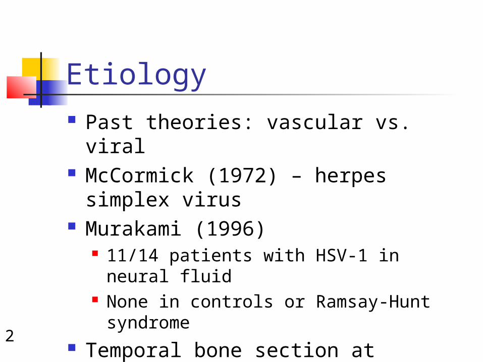
Evaluation Careful history –
timing HX of present illness
Associated symptoms (pain, dysgeusia)
SNHL, vesicles, severe pain
Trauma, acute or chronic OM, recurrent
Exposures Physical exam Audiometry CT/MRI/other Topographic Electrophysiology
4
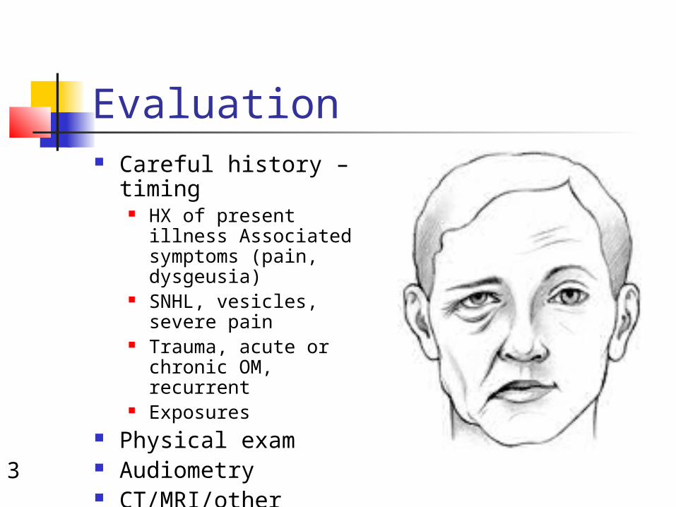
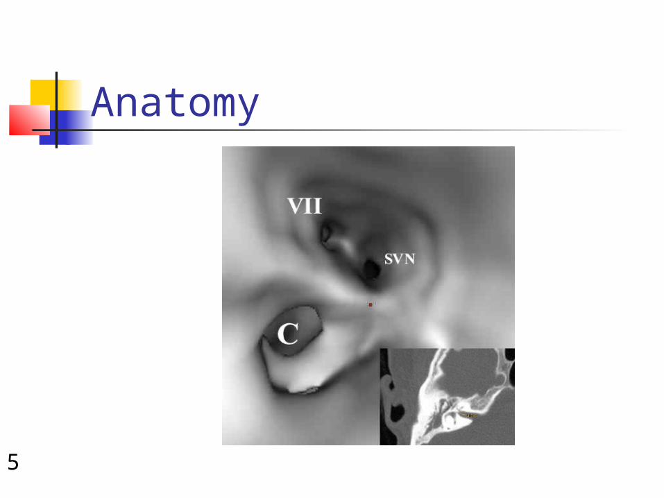
Anatomy Intracranial Meatal Labyrinthine (2-4
mm) Tympanic (11
mm) Mastoid (13 mm) Extracranial
5
Anatomy
6
Traumatic facial nerve palsy/paralysis
Birth trauma Penetrating injury Iatrogenic Temporal bone #
Longitudinal vs transverse or mixed Transection vs edema injury Immediate or delayed

7
Infection
Herpes virus,TB ..etc Otitis
media ,cholesteatoma,mastoiditis
8
Metabolic & systemic DM Guillian barre syndrome Autimmune
9
Bell’s Palsy Facial paralysis
Acute onset, limited duration, minimal symptoms, spontaneous recovery
Idiopathic in past Diagnosis of exclusion Most common diagnosis of acute
facial paralysis
10
Pathophysiology HSV viral reactivation leading to
damage of facial nerve Neuropraxia– no axonal discontinuity Axonotmesis
Wallerian degeneration (distal to lesion) Axoplasmic disruption, endoneural sheaths intact
Neurotmesis Wallerian degeneration (distal to lesion) Axon disrupted, loss of tubules, support cells
destroyed
11
Electrophysiology Treatment plan based on 16% of
patients who do not fully recover Several tests used for prognosis
Measure amounts of neural degeneration occurred distal to injury by measuring muscle response to electrical stimulus
NET, MST, ENoG, EMG Able to differentiate nerve fibers
undergoing Wallerian degeneration
12
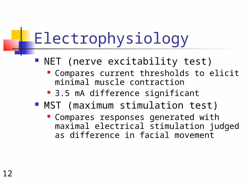
Electrophysiology NET (nerve excitability test)
Compares current thresholds to elicit minimal muscle contraction
3.5 mA difference significant MST (maximum stimulation test)
Compares responses generated with maximal electrical stimulation judged as difference in facial movement
13
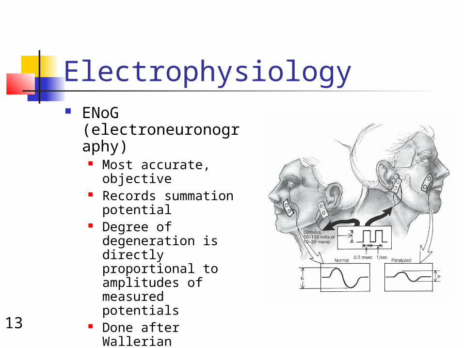
Electrophysiology ENoG
(electroneuronography)
Most accurate, objective
Records summation potential
Degree of degeneration is directly proportional to amplitudes of measured potentials
Done after Wallerian degeneration starts (3-4 days)
Compare each day
14
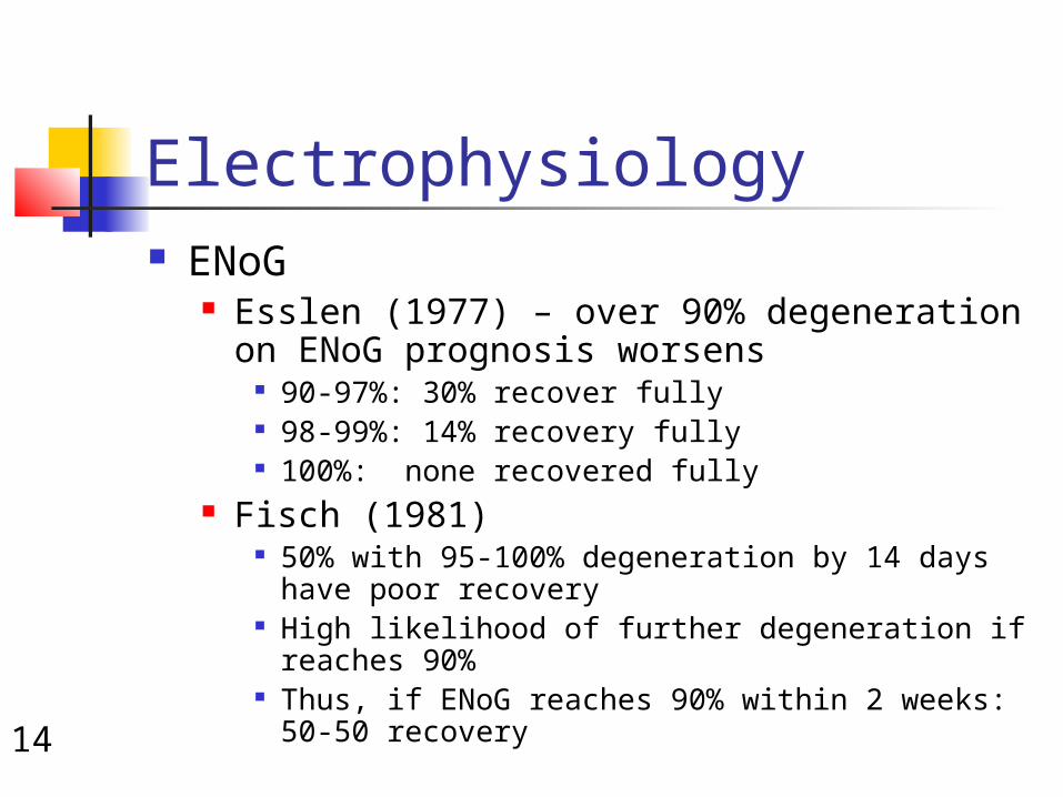
Electrophysiology ENoG
Esslen (1977) – over 90% degeneration on ENoG prognosis worsens
90-97%: 30% recover fully 98-99%: 14% recovery fully 100%: none recovered fully
Fisch (1981) 50% with 95-100% degeneration by 14 days have
poor recovery High likelihood of further degeneration if reaches
90% Thus, if ENoG reaches 90% within 2 weeks: 50-50
recovery
15
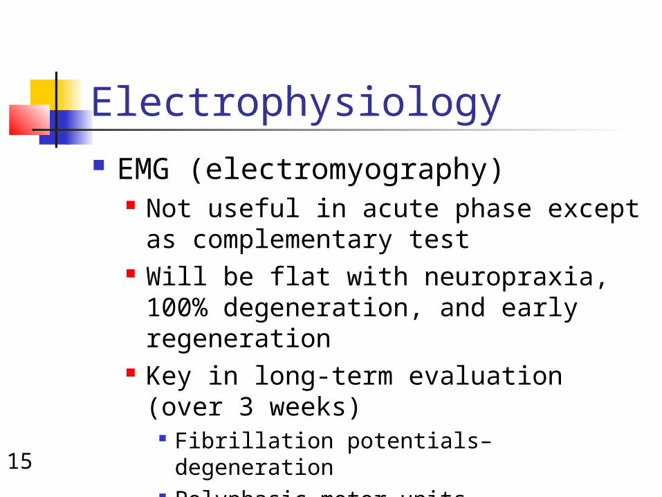
Electrophysiology EMG (electromyography)
Not useful in acute phase except as complementary test
Will be flat with neuropraxia, 100% degeneration, and early regeneration
Key in long-term evaluation (over 3 weeks)
Fibrillation potentials– degeneration Polyphasic motor units– regenerating
nerve
16
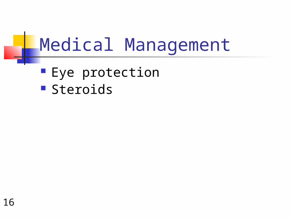
Medical Management Eye protection Steroids
17
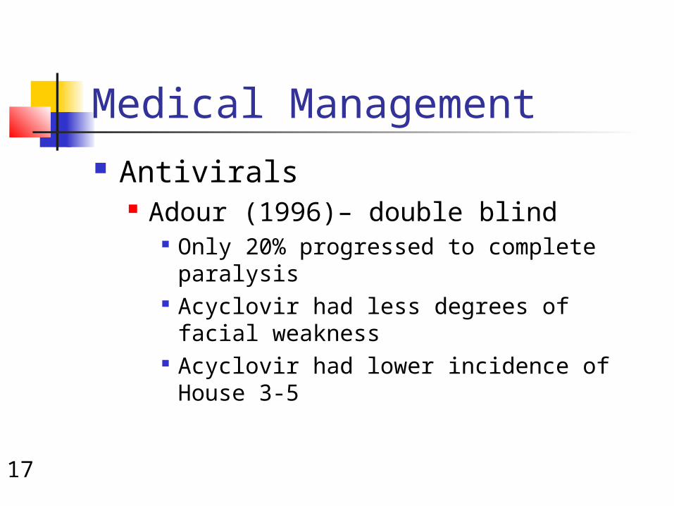
Medical Management Antivirals
Adour (1996)– double blind Only 20% progressed to complete
paralysis Acyclovir had less degrees of facial
weakness Acyclovir had lower incidence of House 3-
5

18
Surgical Management debate over years
No surgery Immediate decompression when
complete

19
Surgical Management Fisch and Esslen (1972)– 12 patients
Total facial nerve decompression via middle cranial fossa and transmastoid
Found conduction block at meatal foramen (94% patients)
Fisch (1981) Decompression within 14 days for 90% degeneration
for maximum benefit May (1979)
Transmastoid decompression beneficial (decreased SF, Schirmer’s, MST reduced)
May (1984) No patients benefited from surgery within 14 days

20
Surgical Management Gantz (1999)– multi-institutional
review Assess if patients with degeneration
over 90% within 14 days would benefit
Middle cranial fossa (meatal foramen to tympanic segment)
If conductive block not identified (6%)– transmastoid added
92% with surgery recovered to House 1-2
45% without surgery to House 1-2

21
ANY QUESTIONS