Fabrication of pomegranate peel extract/honey nanofibers ...
97
American University in Cairo American University in Cairo AUC Knowledge Fountain AUC Knowledge Fountain Theses and Dissertations Student Research 2-1-2019 Fabrication of pomegranate peel extract/honey nanofibers loaded Fabrication of pomegranate peel extract/honey nanofibers loaded with bee venom as effective antibacterial wound dressings with bee venom as effective antibacterial wound dressings Sara Abouzekry Follow this and additional works at: https://fount.aucegypt.edu/etds Recommended Citation Recommended Citation APA Citation Abouzekry, S. (2019).Fabrication of pomegranate peel extract/honey nanofibers loaded with bee venom as effective antibacterial wound dressings [Master's Thesis, the American University in Cairo]. AUC Knowledge Fountain. https://fount.aucegypt.edu/etds/518 MLA Citation Abouzekry, Sara. Fabrication of pomegranate peel extract/honey nanofibers loaded with bee venom as effective antibacterial wound dressings. 2019. American University in Cairo, Master's Thesis. AUC Knowledge Fountain. https://fount.aucegypt.edu/etds/518 This Master's Thesis is brought to you for free and open access by the Student Research at AUC Knowledge Fountain. It has been accepted for inclusion in Theses and Dissertations by an authorized administrator of AUC Knowledge Fountain. For more information, please contact [email protected].
Transcript of Fabrication of pomegranate peel extract/honey nanofibers ...
American University in Cairo American University in Cairo
AUC Knowledge Fountain AUC Knowledge Fountain
Theses and Dissertations Student Research
2-1-2019
Fabrication of pomegranate peel extract/honey nanofibers loaded Fabrication of pomegranate peel extract/honey nanofibers loaded
with bee venom as effective antibacterial wound dressings with bee venom as effective antibacterial wound dressings
Sara Abouzekry
Follow this and additional works at: https://fount.aucegypt.edu/etds
Recommended Citation Recommended Citation
APA Citation Abouzekry, S. (2019).Fabrication of pomegranate peel extract/honey nanofibers loaded with bee venom as effective antibacterial wound dressings [Master's Thesis, the American University in Cairo]. AUC Knowledge Fountain. https://fount.aucegypt.edu/etds/518
MLA Citation Abouzekry, Sara. Fabrication of pomegranate peel extract/honey nanofibers loaded with bee venom as effective antibacterial wound dressings. 2019. American University in Cairo, Master's Thesis. AUC Knowledge Fountain. https://fount.aucegypt.edu/etds/518
This Master's Thesis is brought to you for free and open access by the Student Research at AUC Knowledge Fountain. It has been accepted for inclusion in Theses and Dissertations by an authorized administrator of AUC Knowledge Fountain. For more information, please contact [email protected].

i
School of Sciences and Engineering
Fabrication of Pomegranate Peel Extract/Honey
Nanofibers Loaded with Bee Venom as Effective
Antibacterial Wound Dressings
A Thesis Submitted to
The Biotechnology Graduate program
In partial fulfillment of the requirements for
the degree of Master of Science
By: Sara Samy Abouzekry
Advisors
Dr. Hassan Azzazy
Professor & Chair
Department of Chemistry
Dr. Ahmed Abdellatif
Assistant Professor
Department of Biology
June 2018
The American University in Cairo
School of Sciences and Engineering (SSE)
ii
Fabrication of Pomegranate Peel Extract/Honey Nanofibers Loaded with Bee Venom as
Effective Antibacterial Wound Dressings
A Thesis Submitted by
Sara Samy Abouzekry
Submitted to the Biotechnology Master’s Program
June 2018
In partial fulfilment of the requirements for the degree of
Master of Science in Biotechnology
Has been approved by
Thesis Committee Supervisor/Chair
_______________________________________________ Affiliation
______________________________________
Thesis Committee Co-advisor
_______________________________________________ Affiliation
______________________________________
Thesis Committee Reader/Examiner
______________________________________________ Affiliation
_____________________________________
Thesis Committee Reader/Examiner
______________________________________________ Affiliation
______________________________________
Thesis Committee Reader/External Examiner
______________________________________________
Affiliation _____________________________________
________________ _____________ ______________
Dept. Chair/Director Date Dean Date

iii
DEDEICATION
To the most important people in my life,
Dad, i love you. i wish i can always make you proud.
Mum, you are my true hero. None of this would have been possible without your endless help
and support.
My Grandfather, thank you for your continuous care, compassion and love.
My Husband, thank you for your patience and for being there for me.
My Brothers, thank you for being supportive and caring throughout the way.
My sister, thank you for your comforting words and love in tough times.
My children, i extremely love you. You are my true blessing. Looking into your eyes makes
everything look perfect.

iv
ACKNOWLDGEMENTS
I would like to express my deepest gratitude to all who contributed in my thesis project. First,
I would like to thank my thesis supervisor Dr. Hassan Azzazy for his guidance and support
during thesis work. I would like to thank him for believing in my ideas, supporting my
research and allowing me to learn and grow as a scientist. I am extremely thankful to all the
time and effort he spent on guiding me. Also I would like to thank my co-supervisor Dr.
Ahmad Abdel-Latif for his support, Kindness and patience throughout the project. I couldn’t
have done anything without his continuous support and encouragement.
In addition, I would like to express my thanks and gratitude to Nano-ebers LLC, where I
learned a lot and grew as a researcher. I truly appreciate joining the promising startup
company. I had the opportunity of gaining knowledge not only on developing biocompatible
nano-fibrous wound dressings but also i am gaining an experience on how to deliver the
startup’s products to the market. This has been an exciting experience that I was honored to
be a part of.
I would also like to thank all the professors in the Biotechnology program who have been a
true role model for me, taught me and allowed me to have a strong base knowledge in
Biotechnology.
My deep appreciation goes to my colleagues for always offering advice and help which has
helped me a lot in my thesis work. I’m sincerely grateful to Diana Samy, Razan Msaad,
Nagruess Marei, Myret Ghabriel, Sara Hassan, Salma Elshafei, James Kegere, Nancy
Ahmad, Sara Kamel and Hagar Nofal for their scientific advice and help in the laboratory
work. I would like to thank Amgad Ouaf for helping me while i was still learning the
antibacterial tests. Special thanks goes to Marina Nabil for her help and encouragement while
I was doing the antibacterial tests. I would also like to thank all the lab members and
colleagues for all the interesting discussions we had which resulted in a productive
knowledge transfer between us.
Last but not least, I would to express my extreme gratitude and appreciation to Alalfi
foundation for funding my studies. I am honored to be the recipient of this scholarship and
extremely grateful for all the efforts made by the foundation. In addition, i would like to
express my gratitude to the AUC for providing the research grant that funded this research
project and for partially funding my studies through the laboratory instruction fellowship.

v
List of Abbreviations
3D: Three dimensional
BV: Bee venom
DMSO: Dimethylsulfoxide
E. coli: Escherichia coli
ECM: Extracellular matrix
EGF: Epidermal growth factor
GH: Glutaraldehyde
H&E: Hematoxylin and eosin
IL: Interleukin
LH: Lyophilized multiflora honey
MGO: Methyl glyoxal
MH: Manuka honey
MMP-9: Matrix metalloproteinase-9
MT: Masson Trichrome
MTT: (3-(4, 5-Dimethylthiazol-2-yl)-2, 5-Diphenyltetrazolium Bromide)
MW: Molecular weight
Oprf : Outer membrane protein F
PBS: Phosphate buffer solution
PCL: Poly(ε-caprolactone)
PGE2: Prostagiandin E2
PPP: Pomegranate peel powder extract
PVA: Polyvinyl alcohol
PVAc: Polyvinyl acetate
RPMI: Roswell Park Memorial Institute medium
S. aureus: Staphylococcus aureus
SEM: Scanning electron microscope
TGF-p: Transforming growth factor-ß
TGF-β1: Transforming growth factor β1
TNFα: Tumor necrosis factor-a
VAc: Vinyl acetate
VGEF: Vascular endothelial growth factor

vi
Wt %: weight percent

vii
List of Figures
Figure 1. The global prevelance of wounds by type in 2011 and that expected for 2025. ............ 41
Figure 3. Stages of wound healing: (a) Inflammation step, followed by proliferation step and
finally (c) remodeling .......................................................................................................................... 42
Figure 4. Tissue injury of acute wound vs. chronic wounds ........................................................... 43
Figure 5. Methylglyoxal structure ..................................................................................................... 44
Figure 6. Suggested mechanism by which MH inhibits S. aureus .................................................. 45
Figure 7. A suggested mechanism by which MH inhibits P. aeruginosa........................................ 46
Figure 8. Suggested modulatory effects of honey throughout the stages of wound healing.. ....... 47
Figure 9. A diagram showing the process of polymeric nanofibers formation via electro-
spinning, different techniques for electro-spinning and different types of collectors. .................. 48
Figure 10 . Parameters affecting the electrospinning process.. ....................................................... 49
Figure 11. PVA structure ................................................................................................................... 50
Figure 12. Potential of pomegranate peel extract (PPP) in treating various health conditions. .. 51
Figure 13. Representative SEM images of the corresponding fiber mats, including histogram for
the diameters of fibers (n≈50) ............................................................................................................ 53
Figure 14. Representative SEM images of the crosslinked (MH/PPP) sample .............................. 54
Figure 15. Representative SEM image for MH/PPP/BV sample .................................................... 55
Figure 16. Representative SEM images of the samples: a) (MH 10%/PPP 1%), b) (MH
20%/PPP 2%), and (MH 25%/PPP 2.5 %) ...................................................................................... 56
Figure 17. A bar chart showing a comparison between the swelling behaviors of the electro-spun
scaffolds. ............................................................................................................................................... 57
Figure 18. A bar chart showing a comparison between the water loss behaviors of the
electrospun samples. ........................................................................................................................... 58
Figure 19. Antibacterial activity against S. aureus increases with increasing in the MH/PPP
concentration ....................................................................................................................................... 59
Figure 20. Antibacterial activity against S. aureus .......................................................................... 60
Figure 21. Antibacterial activity against E. coli .............................................................................. 61
Figure 22. Cytotoxicity of electro-spun scaffolds ............................................................................. 62
Figure 23. Representative graphs showing average wound surface area at day 3, 5 and 10 ....... 63
Figure 24. Images showing healing progression ............................................................................... 64
Figure 25. Representative histological assessment for all treatment groups vs. PVA control
group at day 5 and day 10 as shown by H&E staining (magnification 100 x) ............................... 67
Figure 26. Representative histological assessment for all treatment groups vs. PVA control
group at day 5 and day 10 as shown by Masson Trichrome staining (magnification 100 x)........ 71
viii
List of Tables
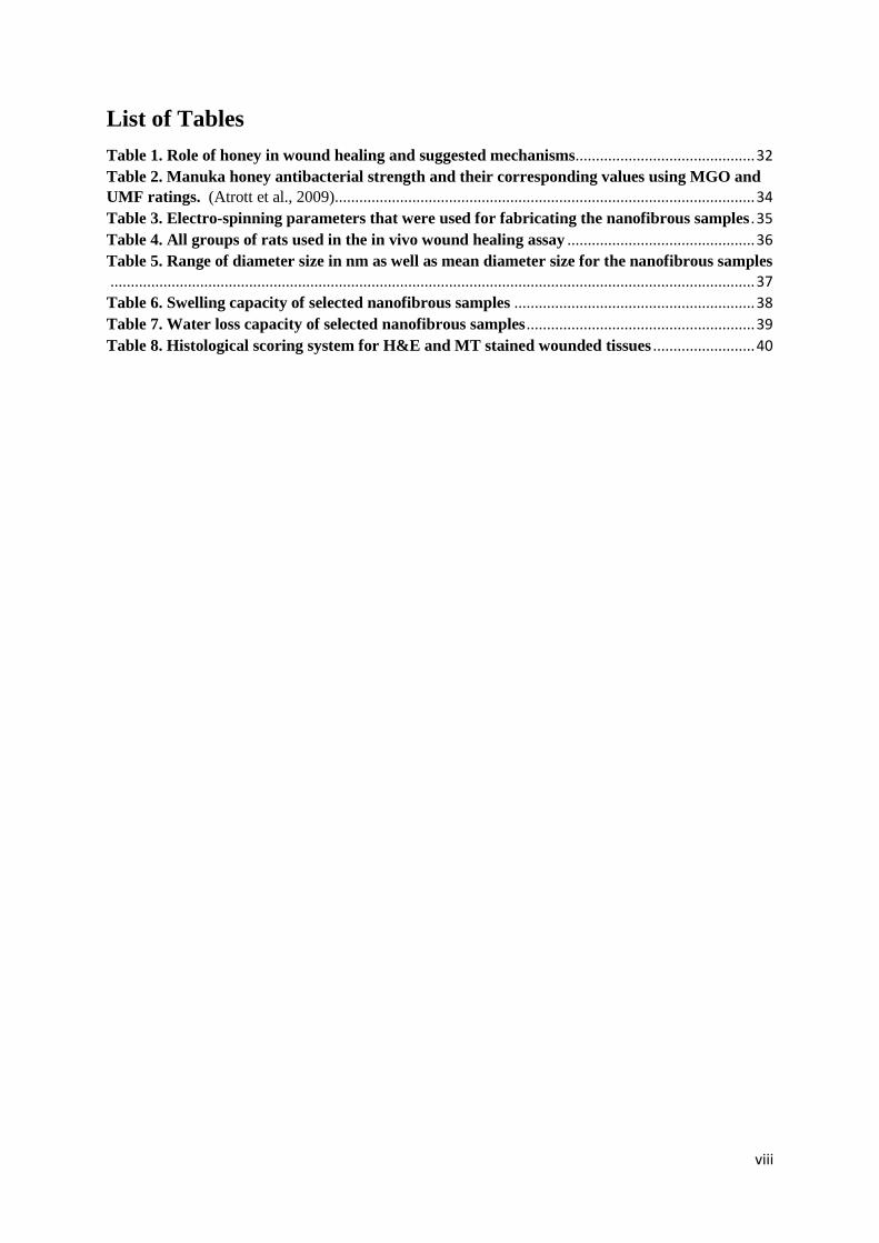
Table 1. Role of honey in wound healing and suggested mechanisms ............................................ 32
Table 2. Manuka honey antibacterial strength and their corresponding values using MGO and
UMF ratings. (Atrott et al., 2009)....................................................................................................... 34
Table 3. Electro-spinning parameters that were used for fabricating the nanofibrous samples . 35
Table 4. All groups of rats used in the in vivo wound healing assay .............................................. 36
Table 5. Range of diameter size in nm as well as mean diameter size for the nanofibrous samples
.............................................................................................................................................................. 37
Table 6. Swelling capacity of selected nanofibrous samples ........................................................... 38
Table 7. Water loss capacity of selected nanofibrous samples ........................................................ 39
Table 8. Histological scoring system for H&E and MT stained wounded tissues ......................... 40

ix
Table of Contents
Abstract .................................................................................................................................................. 1
Chapter 1: Introduction and literature review .................................................................................. 3
1.1. Introduction ........................................................................................................................... 3
1.2. Global impact ............................................................................................................................. 4
1.3. Wounds and their classification ............................................................................................... 4
1.4. Wound healing process .............................................................................................................. 5
1.5. Modern wound healing products classification ....................................................................... 6
1.6. Challenges in wound healing ..................................................................................................... 7
1.7. Honey based dressings and Manuka honey ............................................................................. 8
1.8. Electro-spun nanofibrous scaffolds for wound healing ........................................................ 11
1.9 Poly vinyl alcohol, structure, synthesis and applications ...................................................... 12
1.10. Pomegranate peel powder extract ........................................................................................ 13
1.11. Bee Venom .............................................................................................................................. 14
1.12. Honey powder ......................................................................................................................... 14
Chapter 2: Materials and Methods ................................................................................................... 16
2.1. Materials ................................................................................................................................... 16
2.2. Experimental Procedures ........................................................................................................ 16
2.2.1. Extraction of the pomegranate peel powder ................................................................... 16
2.2.2. Preparation of polymer solutions .................................................................................... 16
2.2.3. Electro-spinning ................................................................................................................ 17
2.2.4. Cross linking of fiber mats ............................................................................................... 17
2.3. Characterization of the prepared electro-spun fibrous meshes ........................................... 18
2.3.1. Scanning electron microscopy .......................................................................................... 18
2.3.2. Swelling and water retention capacity ............................................................................ 18
2.3.3. Water loss capacity ........................................................................................................... 18
2.4. In vitro antibacterial assessment ......................................................................................... 19
2.5. Cytotoxicity assay ..................................................................................................................... 19
2.6. In vivo wound healing assay .................................................................................................... 20
2.7. Statistical analysis .................................................................................................................... 21
Chapter 3: Results ............................................................................................................................... 22
3.1. Morphological characterization .............................................................................................. 22
3.2. Swelling and water retention capacity ................................................................................... 22
3.3. Water loss capacity .................................................................................................................. 22
3.4. In vitro antibacterial assessment ............................................................................................. 23

x
3.5. Cytotoxicity assay: ................................................................................................................... 23
3.6. In vivo wound healing assay .................................................................................................... 24
Chapter 4: Discussion ......................................................................................................................... 25
Future directions ................................................................................................................................. 31
References ............................................................................................................................................ 72
Appendix ............................................................................................................................................... 80
1
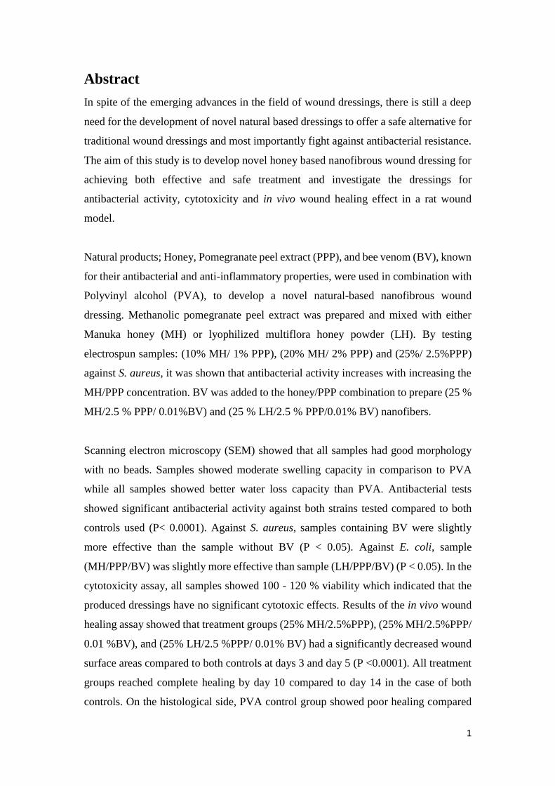
Abstract
In spite of the emerging advances in the field of wound dressings, there is still a deep
need for the development of novel natural based dressings to offer a safe alternative for
traditional wound dressings and most importantly fight against antibacterial resistance.
The aim of this study is to develop novel honey based nanofibrous wound dressing for
achieving both effective and safe treatment and investigate the dressings for
antibacterial activity, cytotoxicity and in vivo wound healing effect in a rat wound
model.
Natural products; Honey, Pomegranate peel extract (PPP), and bee venom (BV), known
for their antibacterial and anti-inflammatory properties, were used in combination with
Polyvinyl alcohol (PVA), to develop a novel natural-based nanofibrous wound
dressing. Methanolic pomegranate peel extract was prepared and mixed with either
Manuka honey (MH) or lyophilized multiflora honey powder (LH). By testing
electrospun samples: (10% MH/ 1% PPP), (20% MH/ 2% PPP) and (25%/ 2.5%PPP)
against S. aureus, it was shown that antibacterial activity increases with increasing the
MH/PPP concentration. BV was added to the honey/PPP combination to prepare (25 %
MH/2.5 % PPP/ 0.01%BV) and (25 % LH/2.5 % PPP/0.01% BV) nanofibers.
Scanning electron microscopy (SEM) showed that all samples had good morphology
with no beads. Samples showed moderate swelling capacity in comparison to PVA
while all samples showed better water loss capacity than PVA. Antibacterial tests
showed significant antibacterial activity against both strains tested compared to both
controls used (P< 0.0001). Against S. aureus, samples containing BV were slightly
more effective than the sample without BV (P < 0.05). Against E. coli, sample
(MH/PPP/BV) was slightly more effective than sample (LH/PPP/BV) (P < 0.05). In the
cytotoxicity assay, all samples showed 100 - 120 % viability which indicated that the
produced dressings have no significant cytotoxic effects. Results of the in vivo wound
healing assay showed that treatment groups (25% MH/2.5%PPP), (25% MH/2.5%PPP/
0.01 %BV), and (25% LH/2.5 %PPP/ 0.01% BV) had a significantly decreased wound
surface areas compared to both controls at days 3 and day 5 (P <0.0001). All treatment
groups reached complete healing by day 10 compared to day 14 in the case of both
controls. On the histological side, PVA control group showed poor healing compared
2
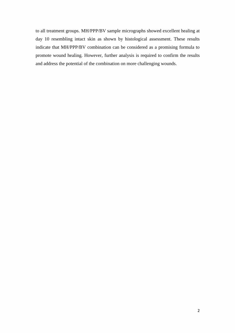
to all treatment groups. MH/PPP/BV sample micrographs showed excellent healing at
day 10 resembling intact skin as shown by histological assessment. These results
indicate that MH/PPP/BV combination can be considered as a promising formula to
promote wound healing. However, further analysis is required to confirm the results
and address the potential of the combination on more challenging wounds.

3
Chapter 1: Introduction and literature review
1.1. Introduction
Wounds are considered one of the critical public health problems in the world. In the
United States alone, undertreated wounds and chronic wounds affected 6.5 million
patients in 2009, with an annual cost in excess of 25 billion USD (Sen et al., 2009). In
2011, the global market of wound-care products produced 6,500 million USD with
7.5% annual growth rate. In 2023, the global advanced wound-care products market
share is expected to reach nearly 16,300 million USD. Therefore, new technologies are
essential to face this problem ((Sharma & Paul, 2015).
Nano material scaffolds are among the promising wound dressings to improve wound
healing. These scaffolds have better properties compared to conventional dressings
such as having high porosity, very small pore size and large surface area to volume
ratio. These properties lead to higher exudate absorption, better wound permeation and
prevention from further infection (Huang et al., 2003).
For decades, honey has been used as wound healing remedy in various forms, owing to
its remarkable antimicrobial properties. Honey consists of water (20%), fructose (40%),
glucose (30%), sucrose (5%), and other substances such as, minerals, vitamins, amino
acids, and enzymes (Bagde et al., 2013). It was demonstrated from previous studies that
honey has antibiotic, antimicrobial, and anti-inflammatory properties and inhibits a
broad spectrum of bactericidal organisms (Chang et al., 2009, Pieper 2009). Thereby,
it has the advantage of providing a moist environment that is suitable for the healing
process without the fear of an occurring bacterial growth. Moreover, honey is acidic
therefore it is able to provide fibroblasts with optimal environment for their activity.
The present study aims at testing multiple natural products loaded within honey/PVA
nanofiber scaffold in order to improve wound healing activity in an animal model (Two
types of honey were used MH and lyophilized multiflora honey powder).

4
1.2. Global impact
In 2011, the global market of wound-care products produced 6,500 million USD with
7.5% annual growth rate. This cost included nearly 230 million surgical procedures,
chronic wounds (12–15 million leg ulcers, 9–11 million pressure sores) and an equal
number of burn wounds yearly all over the world. Figure 1 shows the global prevalence
of wounds according to wound type in 2011 and that predicted in 2025 (Sharma & Paul,
2015).
Despite the fact that surgical wounds showed the highest prevalence, It is expected that
there will be a significant increase in number of chronic wounds whose wounds requires
advanced wound care products. The market share for advanced wound-care dressings
is expected to increase in Germany, Japan, USA, France, UK, Italy, Brazil, Russia,
Spain, China and India.
It is predicted that India will be the largest consumer for advanced wound care in the
future. This might be attributed to the rise in lifestyle-related diseases such as diabetes
as well as the raise in the economic status of the Indian population due to better health
care policies and the fast-growing health insurance market. 3M, Smith & Nephew,
Johnson & Johnson, Dr. Reddys and Beiserdorf are considered the main players in the
field of wound-care dressings. In 2023, the global advanced wound-care products
market share is expected to reach nearly 16,300 million USD. Another study, expected
that the total global market share in 2014 that was worth 15,600 million USD will reach
18,300 million USD in 2019 (Sharma & Paul, 2015).
The predicted huge growth of the market stems from a raise in diabetes prevalence,
presence of ageing populations, and obesity. These conditions lead to more chronic
wounds and ulcers thereby causing considerable degree of possible mortality and
morbidity, and also presenting a huge problem to the health care system as they are
considered most costly wound types to treat.
1.3. Wounds and their classification
A wound can be defined as any cut or tear on the surface of the skin. Most commonly,
wounds can be classified into acute wounds and chronic wounds, and into three types
based on depth; first, superficial wounds that includes the epidermis layer, second,
partial thickness wounds that include the epidermis along with the dermis layer and

5
third, full thickness wounds which include deeper layers being affected like the
subcutaneous fat or even deeper layers ( Figure 2).
Wounds can also be classified into wounds with tissue loss (e.g. burns, ulcer wounds,
abrasions, pressure sores, iatrogenic wounds, trauma wounds) or without tissue loss like
for example surgical wounds (Sharma & Paul, 2015).
Acute wounds normally heal on their own. For example, cuts, lacerations, penetrations,
avulsion, bites and burns are considered different types of acute wounds. On the other
side, chronic wounds do not heal in a timely and structured manner. Therefore, a
continued management is needed in case of chronic wounds. Most common types of
chronic wounds include; a) Diabetic ulcer. b) Pressure ulcers or bedsores that results
from prolonged pressure to some body parts, and c) Arterial and venous ulcers resulting
from avascular dysfunction mainly in lower limbs, leading to reduced blood flow and
chronic inflammation (Sharma & Paul, 2015).
1.4. Wound healing process
The process of wound healing is a complex process starting with hemostasis, followed
by inflammation, proliferation and finally, remodeling (Figure 3). Generally, healing
time taken from the beginning to end is between two to three weeks unless other factors
that may prolong healing time took place. In the inflammation step following a blood
vessel injury, involves coagulation as well as an acute local inflammatory response
where monocytes, neutrophils, lymphocytes and macrophages are recruited to the site
of injury. In the proliferation step, all cytokines and factors that were previously
released in the inflammatory step enhance the proliferation of progenitor cells.
Fibroblasts, keratinocytes as well as endothelial cells are recruited in this step leading
to migration and proliferation of cells (Sharma & Paul, 2015). In the remodeling stage,
cells start to migrate in order to re-epithelialize the damaged tissue at the wound’s edges
in a process known as epithelial mesenchymal transition. After this, a scar tissue is
formed where the wounds starts to contract and myofibroblasts are formed as a result
of fibroblasts differentiation. As a result of the occurring angiogenesis, newly formed
blood vessels appear and nerves begin to sprout (Stein & Küchler, 2013, Sharma &
Paul., 2015).

6
1.5. Modern wound healing products classification
Modern wound dressings were developed mainly to help in enhancing the healing
process, rather than just creating a cover over the wound. These dressings are concerned
with keeping wounds from dehydration as well as promoting healing. According to the
nature of their action, modern wound healing products are classified into three main
categories, passive, including traditional gauze or tulle wound dressings which
represent the largest market segment, interactive and bioactive products. Among the
disadvantages of these passive forms of dressings is that they can disrupt the wounds
when removed as they tend to stick to the wounds. That’s why this type of wounds will
only suit minor wounds. Although tulle which is made of petroleum jelly does not stick
to the wound like gauze, it suits flat wounds with little exudate.
Interactive products are made from foams and polymeric films. They are considered
permeable to water vapor and oxygen while being impermeable to bacteria. Foams has
better exudate absorption than films and amorphous gels, that’s why they can be used
for moderate exudate absorption unlike films and gels which are suitable for low
exudate wounds. A third type of products is represented by bioactive products, which
delivers active components to the wound which contributes in the healing process.
These components may include collagen, proteins, growth factors, alginates (Dhivya et
al, 2015).
Other types of modern wound dressings may include tissue engineered skin substitutes
which can be made up of cells, ECM or combination of both. They can be divided into
cellular and acellular skin substitutes (Rubaiy et al., 2009). Medicated dressings
generally are drugs-incorporated dressings. They help in the healing process either
directly or indirectly by removing of the necrotic tissues. The dressings may contain
antimicrobial agents that prevents infection or growth factors and enzymes which
enhances regeneration. Composite dressing, can be called combination dressings that
have multiple layers. Most composite dressings usually have three layers. An outer
layer is designed to protect the wound against infection. A layer in the middle is usually
composed of absorptive material which helps in autolytic debridement and in
maintaining moisture environment while the third layer is supposed to be composed of
non-adherent material so that it will not stick to the wound (Dhivya. et al, 2015).

7
1.6. Challenges in wound healing
Management of wounds with a delayed healing time i.e. chronic or non-healing wounds
is considered as the main challenge to wound care practitioners and researchers. The
main reasons to delayed healing include systemic diseases, arterial insufficiency and
aging. The factors affecting the healing process can vary between local and systemic
factors. Among the local factors is infection, ischemia, tissue maceration and presence
of foreign bodies inside the wound area. Among the systemic factors, there is
malnutrition, diabetes, age and renal diseases. Controlling the effects of these factors is
important for normal healing process to take place.
In chronic wounds for example, there is a decreased level of growth factors such as
Epidermal Growth Factor (EPGF), platelet driven growth factor, basic fibroblast
growth factor and transforming growth factor that are crucial for the healing process to
work properly possibly due to degradation or becoming in active. In aging, fibroblasts
have poor responsiveness to nutrients and growth factors which result in chronic
wounds. Abnormal degradation of the extracellular matrix (ECM) can happen as result
of over expression of the matrix metalloproteinase due to an increased proteinase
activity (Clark et al., 2007, Sharma & Paul, 2015). Failure of chronic wounds to heal in
comparison to the acute healing process is shown in figure 4, on the left of the figure,
acute injury of a tissue activates blood clotting, aggregation of platelets and migration
of macrophages and neutrophils to the location of the injury. First, a blood clot is
formed of fibrin and fibronecetin which acts as a scaffold for platelets and cell migration
where growth factors are released. At day 3, the formed clot accumulates neutrophils
which in turn help phagocytose micro-organisms. On the upper right of the figure, by
day 5 after injury and under normal conditions, the wound continue the healing process
with the help of the growing granulation tissue that is formed mainly of fibroblasts,
more macrophages as well as neovasculature. The epidermis starts migrating towards
newly forming tissue. On the lower right of the figure, when the wound fails to heal
due to an underlying pathobiology or as a result of a microorganism invasion, the
healing process is interrupted leading to a chronic wound or ulcer formation. Many
factors hinder the acute healing process including venous insufficiency which in turn
leads to transudation of fluids and fibrin cuffing of venules as a result to increased
venous system hydrostatic pressure. Other factor might be diabetes mellitus where high

8
glucose levels which leads to dysfunction of ECM due to the occurring non-enzymatic
glycation. Among the factors also there is arterial occlusion or increased external
pressure that results in hypoxia of tissues and cell dysfunction. Bacteria might start to
colonize the wound leading to the formation of biofilm which in turn worsen the
condition by protecting the colonies of bacteria, becoming relatively resistant to
phagocytic cells and antibiotics. Moreover, desperate phagocytes release proteases and
toxic free oxygen radicals into the wound. This will worsen the condition even more as
released agents start destroying the tissue cells, ECM, and growth factors in the wound.
As a consequence of all these events, granulation tissue in growth and epidermal
migration is absent in chronic wounds.
Although that there a huge advancement in the knowledge and the science of wound
healing together with the technical innovation, involvement of pharmaceutical
companies, an array of resources ranging from dressings, tissue replacement and cell
based treatment options being available however, the biggest challenge is dealing with
chronic wounds in terms of practical training with the condition. Also, Scarring
prevention as well as achieving a cosmetically acceptable way of wound healing
remains a challenge for wound care professionals. Over the past four decades, an
extensive research has been done on wound healing and wound dressings. The main
reasons for applying a dressing can be summarized in leading to rapid as well as a
cosmetically accepted way of healing, remove odor, prevent infection or fight infection,
decrease pain, contain exudates, cover a wound for cosmetically accepted appearance,
causing minimal distress to the patients (Sharma & Paul, 2015).
1.7. Honey based dressings and Manuka honey
Having a history in being an ancient remedy for healing burns and wounds, honey is
still believed to help heal wounds faster and fight against infections. It is suggested that
ancient Egyptians used honey around 3000 BC. The ‘three healing gestures” papyrus
mentioned honey and included it as an integral part within the ancient healing process.
The first wound dressings are believed to be made from honey, vegetable fiber and
animal fat. In modern history, recognition of honey as a topical antibacterial agent
started in 1892.
Recently, there has been an interest in re-adopting the use of honey in wound healing
especially with the emergence of the antibacterial resistance problem, in 2007, The US
9
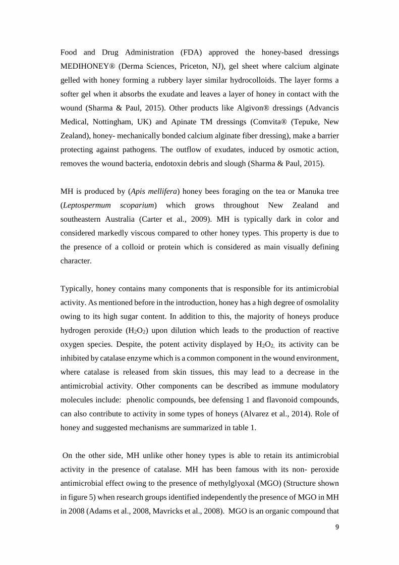
Food and Drug Administration (FDA) approved the honey-based dressings
MEDIHONEY® (Derma Sciences, Priceton, NJ), gel sheet where calcium alginate
gelled with honey forming a rubbery layer similar hydrocolloids. The layer forms a
softer gel when it absorbs the exudate and leaves a layer of honey in contact with the
wound (Sharma & Paul, 2015). Other products like Algivon® dressings (Advancis
Medical, Nottingham, UK) and Apinate TM dressings (Comvita® (Tepuke, New
Zealand), honey- mechanically bonded calcium alginate fiber dressing), make a barrier
protecting against pathogens. The outflow of exudates, induced by osmotic action,
removes the wound bacteria, endotoxin debris and slough (Sharma & Paul, 2015).
MH is produced by (Apis mellifera) honey bees foraging on the tea or Manuka tree
(Leptospermum scoparium) which grows throughout New Zealand and
southeastern Australia (Carter et al., 2009). MH is typically dark in color and
considered markedly viscous compared to other honey types. This property is due to
the presence of a colloid or protein which is considered as main visually defining
character.
Typically, honey contains many components that is responsible for its antimicrobial
activity. As mentioned before in the introduction, honey has a high degree of osmolality
owing to its high sugar content. In addition to this, the majority of honeys produce
hydrogen peroxide (H2O2) upon dilution which leads to the production of reactive
oxygen species. Despite, the potent activity displayed by H2O2, its activity can be
inhibited by catalase enzyme which is a common component in the wound environment,
where catalase is released from skin tissues, this may lead to a decrease in the
antimicrobial activity. Other components can be described as immune modulatory
molecules include: phenolic compounds, bee defensing 1 and flavonoid compounds,
can also contribute to activity in some types of honeys (Alvarez et al., 2014). Role of
honey and suggested mechanisms are summarized in table 1.
On the other side, MH unlike other honey types is able to retain its antimicrobial
activity in the presence of catalase. MH has been famous with its non- peroxide
antimicrobial effect owing to the presence of methylglyoxal (MGO) (Structure shown
in figure 5) when research groups identified independently the presence of MGO in MH
in 2008 (Adams et al., 2008, Mavricks et al., 2008). MGO is an organic compound that

10
can be called also 2-oxopropanal or pyruvaldehyde. MGO has the formula: CH3C (O)
CHO. In the gaseous state, MGO has 2 carbonyl groups, an aldehyde and a ketone.
However, in the presence of water, it is present as hydrates as well as oligomers. MGO
is considered to be a pyruvic acid reduced derivative. MGO is formed as a side-product
of several metabolic pathways in organisms. MGO is a component within MH as it was
shown to have activity against both E. coli and S. aureus and has been shown to be
responsible for most of the manuka honey’s antimicrobial activity (Mavric. et al., 2008,
Israili.et al., 2014) and may help the formation of biofilms formed by P.
aeruginosa (Israili et al., 2014).
Interestingly, the mechanism by which MH exhibits its antibacterial activity has been
investigated in S. aureus and Pseudomonas aeruginosa (see figure 6 and figure 7
respectively). It was shown that MH inhibits S. aureus by interfering with cell division,
whereas it leads to lysis of P. aeruginosa cells due to the reduction of a key structural
protein (Jenkins et al., 2015).
In addition to its non-peroxide antimicrobial activity, it was suggested that MH has a
role in modulating the initial inflammatory response by promoting the production of
cytokines that regulate the production and the angiogenesis of fibroblasts (Molan 2006,
Tonks et al., 2007). MH was shown to help in stimulating toll-like receptor 4 on
monocytes which in turn leads to the stimulation of the production of IL1b, IL6 and
TNFα from monocytes which are important in tissue repair and regeneration (Tonks et
al., 2001, 2003, 2007). Other effects that aid in the healing process were mentioned
such as lowering the pH of wound area. It was reported that MH decreased pH of
cutaneous wounds (Gethin at al., 2008). Raising pH of the wound towards acidic was
suggested to have many effects starting from including a shift to the right to what is
called the oxygen–haemoglobin dissociation curve, leading to an increase in oxygen
release, decreased toxicity of end products of bacteria such as ammonia, decreased
protease activity, activated destruction of abnormal collagen, activated angiogenesis,
enhanced fibroblast and macrophage activity as well as controlled the enzyme activity
(Greener et al. 2003, Githen et al., 2008). Suggested mechanisms for honey as well as
their clinical effects in healing wounds are represented in figure 8.
In a way to rate MH, a grading system called UMF (Unique Manuka Factor) rating was
started by the UMF association. The system is supposed to measure the non-hydrogen

11
peroxide antibacterial potency of MH. The higher the UMF number is, the higher the
efficacy and anti-bacterial activity is. Another rating is (MGO) rating made by the
Active Manuka Honey Association (AMHA), as MGO content increases as the potency
and activity of MH increases. UMF rating typically ranges from 10 to 25 while MGO
rating ranges from 100+ to 850+ (Atrott at al., 2009, Jenkins et al., 2015).
1.8. Electro-spun nanofibrous scaffolds for wound healing
Electro-spun nanofibrous scaffolds have many applications in the biomedical field
especially in wound healing and tissue engineering. Main advantages of these special
fibers include high surface area to volume ratio, high porosity. Other advantages related
to the electro-spinning process include the wide range of electro-spinable polymers that
can be used, versatility and flexibility of the electro-spinning process itself and the
possibility of loading the fibers with bioactive agents, antibacterial agents or functional
nanoparticles. All these advantages contribute to expanding the use of electro-spun
nanofibers in wound healing application (Liu et al, 2017).
A desired feature in nanofibrous mat dressings that they can allows for providing a
temporary structure for skin cells that resembles the native ECM functions, act as an
adhesive layer for cells, guide the growing tissue and act as mechanical support. The
three-dimensional (3D) electro-spun nanofibrous scaffolds have been developed to
produce dressings that resembles the ECM nano sized structure. The highly porous
nature of electro-spun matrices that resemble a natural ECM are important for the cells
to restore their shape and restore their normal behavior (Puppi et al., 2011).
Electro-spinning, is a technology used for the formation for electrostatic fibers. The
resulting fibers usually have diameters that range from several nanometers to several
micrometers where high voltage electric fields are used in the process. A typical electro-
spinner includes the following: high voltage power supply, spinneret system as well as
a collecting screen as shown in figure 9. In the process, there are parameters that affect
the transformation of polymer solutions into nanofibers. These parameters include: a)
variables like flow rate, voltage used and distance from the needle tip to the collecting
surface, b) solution properties such as concentration, viscosity, elasticity, conductivity
and surface tension, (c) ambient parameters like humidity, solution temperature and air
12
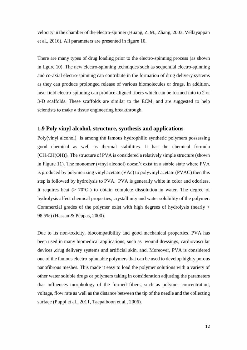
velocity in the chamber of the electro-spinner (Huang, Z. M., Zhang, 2003, Vellayappan
et al., 2016). All parameters are presented in figure 10.
There are many types of drug loading prior to the electro-spinning process (as shown
in figure 10). The new electro-spinning techniques such as sequential electro-spinning
and co-axial electro-spinning can contribute in the formation of drug delivery systems
as they can produce prolonged release of various biomolecules or drugs. In addition,
near field electro-spinning can produce aligned fibers which can be formed into to 2 or
3-D scaffolds. These scaffolds are similar to the ECM, and are suggested to help
scientists to make a tissue engineering breakthrough.
1.9 Poly vinyl alcohol, structure, synthesis and applications
Poly(vinyl alcohol) is among the famous hydrophilic synthetic polymers possessing
good chemical as well as thermal stabilities. It has the chemical formula
[CH2CH(OH)]n. The structure of PVA is considered a relatively simple structure (shown
in Figure 11). The monomer (vinyl alcohol) doesn’t exist in a stable state where PVA
is produced by polymerizing vinyl acetate (VAc) to polyvinyl acetate (PVAC) then this
step is followed by hydrolysis to PVA. PVA is generally white in color and odorless.
It requires heat (> 70℃ ) to obtain complete dissolution in water. The degree of
hydrolysis affect chemical properties, crystallinity and water solubility of the polymer.
Commercial grades of the polymer exist with high degrees of hydrolysis (nearly >
98.5%) (Hassan & Peppas, 2000).
Due to its non-toxicity, biocompatibility and good mechanical properties, PVA has
been used in many biomedical applications, such as wound dressings, cardiovascular
devices ,drug delivery systems and artificial skin, and. Moreover, PVA is considered
one of the famous electro-spinnable polymers that can be used to develop highly porous
nanofibrous meshes. This made it easy to load the polymer solutions with a variety of
other water soluble drugs or polymers taking in consideration adjusting the parameters
that influences morphology of the formed fibers, such as polymer concentration,
voltage, flow rate as well as the distance between the tip of the needle and the collecting
surface (Puppi et al., 2011, Taepaiboon et al., 2006).

13
1.10. Pomegranate peel powder extract
Pomegranate (Punica granatum L.) is one of the fruits that gained researcher’s interest
in recent years due to its enormous biological activities. Pomegranate fruit is rich in
tannins and phenolics which were shown to be responsible for its antioxidant activity.
Pomegranate juice together with its peel contain remarkable amounts of polyphenolic
compounds such as ellagic acid, ellagic tannins, flavonols, anthocyanins, catechin,
procyanidins and gallic acid. The fruit has been used in many pharmaceutical
preparations like cosmetics, tinctures and other therapeutic formulas.
Pomegranate peel constitutes 50% of the pomegranate fruit weight. The peel was shown
to have higher amounts of polyphenolic compounds than pomegranate juice and thereby
possessing even stronger biological activity. Despite this, the peel is often discarded as
waste. That is why it gained concern in many studies being an abundant and cheap
source for natural antioxidants. While there is an increased demand on processing of
pomegranate. Larger amounts of wastes are produced. For each 1 ton of pomegranates
being processed, 500-550 kg waste peels are produced as a major by-product (Qu et al.,
2009, Tenkler et al., 2018). Under European regulations, pomegranate peel waste
should not be disposed neither on land nor in landfills because it holds a significant risk
to watercourses. Therefore, pomegranate peel waste disposal is turning into a major
problem mainly for pomegranate processing factories (Goula & Lazarides, 2015).
Several studies reported that pomegranate peel powder extract (PPP) had markedly
higher antioxidant capacity than the pomegranate juice extract in scavenging against
hydroxyl, peroxyl radicals and superoxide anion. In addition to its antioxidant activity,
PPP was shown to have a range of other biological actions like anti-cancer activity,
antimicrobial activity, anti-inflammatory and anti-diabetic activities. A diagram for the
PPP extracted biomolecules and its suggested enthopharmacological potential in many
health conditions ranging from acting as a cardiovascular protective agent, anti-
inflammatory and anti-allergic, anti-influenza as well as an antimalarial and effective
wound healing agent is shown in figure 12 (Ismail et al., 2012).
In a way to investigate more into PPP wound healing mechanisms, Aslam et al.,
suggested that PPP activated the synthesis of type I procollagen and led to the inhibition

14
of the production of matrix metalloproteinase-1 (MMP-1; interstitial collagenase by
dermal fibroblast. (Aslam et al., 2006).
1.11. Bee Venom
In the past, bee venom (BV) was not only considered as the main way by which bees
defend themselves against predators but also it was popular for therapeutic value in
many ancient civilizations (Babylont, Egypt, Greece and China) (Urtubey, N., 2005).
Apis mellifera honey BV grabbed attention for the first time in modern history in the
late 19th century. In 1950, Aberman and Neuman suggested that the venom’s biological
activity is attributed to the presence of protein and peptide content. In 1963, Benton et
al. presented an ingenious way for the collection of venom in large quantities. Since
then BV has been the focus of several scientists unravelling BV composition and the
biological effects of its components (Banks & Shipolini, R. A.1986).
The main activity of BV is attributed to melittin protein which constitutes from 40% to
50% of the dry weight of the venom. Melittin itself has been the focus of many studies
owing to its potent antimicrobial as well as lytic properties (Banks & Shipolini, 1986).
Mellitin was found to have cytotoxic activity towards cancer cells as well as being
acting as a potent anti-inflammatory and anti-arthritic in very small amounts. However
large doses were reported to be inflammatory and hemolytic. (Oršolić, 2013)
Many recent studies considered bee venom (BV) as a promising candidate for wound
healing owing to its powerful antibacterial effect (Fennell, J. F. et al.,1967 ,Lubke et
al., 1997, Isam et al., 2015, Lazarev, V., 2005) and potent anti-inflammatory properties
(Kang et al., 2002, Lee et al., 2005, Lee et al., 2010). Another study also suggested that
BV in concentration less than 100 μg/ml did not show cytotoxic effects. Bee Venom
also, activates human epidermal keratinocyte migration and proliferation in vitro (Han
et al., 2013).
1.12. Honey powder
Honey powder is a concentrated form of honey lacking the 20% moisture content. This
type of honey captured attention for its use in food industry. In contrast to liquid honey
which is viscous and difficult to disperse, honey powder was a good alternative. Tong
et al., suggested that honey powder can be used to improve the dough quality and shelf
life in bread making (Tong et al., 2010).

15
Among the methods to prepare honey powder is spray drying, vacuum drying and
freeze drying/ lyophilization. Extreme stickiness and high viscosity of liquid honey
have been the main drive for the increasing demand for the production of dried honey
powder by food manufacturers and consumers. However, getting honey into the free
flowing powder form either by freeze or spray drying was reported to be a challenging
process due to its stickiness and strong hygroscopic property (Adhikari et al., 2009,
Kruszewski et al., 2014). Some papers reported that inclusion of high molecular weight
carrier material like whey protein within honey helped to solve this
problem.(Shi et al., 2013, Samborska et al., 2015a,b) as whey protein can modify the
surface properties of the atomised particles as well as the film‐forming ability of the
particles during spray drying (Islam et al., 2013, Wang et al., 2013).
Despite the challenge in achieving the powdered honey form without including
additives, numerous companies sell pure honey powder. Also, although honey powder
use is limited to food industry, its efficacy in other fields like wound healing, needs
investigation. Taking in consideration ease of transportation and storage, being a
concentrated form of honey and relative cheap price compared to other honey types,
potential of this form of honey should be addressed through including it in more studies
to observe its use in fields other than food industry.

16
Chapter 2: Materials and Methods
2.1. Materials
Poly (vinyl alcohol) (PVA) with MW (molecular weight) ~ 125,0000 and commercial
name “Mowiol 20-98” was purchased from Sigma Aldrich, Germany. Lyophilized
multiflora honey was purchased from Xiaocaokeji, China. Manuka honey MGO 550+,
was purchased from Manuka Health, New Zealand. Methanol (Ultra) gradient HPLC
grade was purchased from J.T. Baker, Philipsburg, NJ, USA. Pomegranate peel powder
was purchased from local condiments store. Glutaraldehyde (GH) solution 50% was
purchased from Acros Organics, Belgium. RPMI media with L-glutamine, Alkaline
Phosphate buffer (PBS) and trypsin were purchased from Lonza, Belgium. Thiazolyl
blue tetrazolium bromide (MTT) was purchased from Sigma Aldrich, Germany. Difco
Nutrient broth and Difco Nutrient agar were purchased from Thermo Fisher, Germany.
2.2. Experimental Procedures
2.2.1. Extraction of the pomegranate peel powder
Extraction of the pomegranate peel powder was done according to a previously
published protocol (Singh & Jayaprakasha, 2002). The first step was suspending 25 g
of the pomegranate powder in 100 ml methanol. Stirring was done at room temperature
for 1 h. The extract was then centrifuged at 4000 rpm for 3 min. The supernatant was
then diluted with distilled water to ratio (80:20) to decrease the freezing point from -
97℃ to -20℃. The resulting solution was lyophilized using a freeze drying machine
(TOPT – 10 C Freeze Dryer, China). The process was achieved after keeping the freeze
drying machine operating for 48 h. The resulting powder was weighed and kept in an
eppendorf tubes to be used later on.
2.2.2. Preparation of polymer solutions
Solutions containing different ratios of MH, LH, PPP, BV and PVA were prepared in
the following concentrations: (10% MH/ 1% PPP/ 12% PVA), (20% MH/ 2% PPP/
10.5% PVA), (25% MH/2.5% PPP, 9.7% PVA), (30%/MH/3%PPP/9.7%PVA), (25%
MH/ 2.5% PPP/ 0.01% BV, 9.7% PVA), (25% LH/ 2.5% PPP/ 0.01%/ BV, 9.7% PVA).
Honey concentrations used were chosen according to previous data (Sarhan & Azzazy

17
2016). As for BV, it was shown that in concentration less than 100 μg/ml did not show
cytotoxic effect while activated human epidermal keratinocyte migration and
proliferation in vitro (Han et al., 2013). According to the later suggestion, BV
concentration was fixed at 0.01 % in all samples. In our study, PPP concentration was
used up to 2.5%. The extract was not loaded before into nanofibers. However, it has
been shown that 2.5% PPP gel decreased wound healing time in animal model
(Chidambara Murthy et al., 2004). PVA was dissolved in deionized water by stirring at
90℃ for 3 h and then honey and PPP were added where stirring was maintained at room
temperature for 1h. After adding BV, the polymer jars were covered with aluminum
foil at room temperature until electro-spinning.
2.2.3. Electro-spinning
Different voltages were applied to the polymer solutions (Gamma High Voltage Power
Supply, USA) in order to determine the best voltage for each solution. Adjustment of
the flow rate was carried out at 1ml/min. The Distance between the collecting plate and
the nozzle of the electro-spinner (Sustaincubator, Egypt) was adjusted at 12 cm
throughout the spinning process.
The concentration of the components of each formula was represented in wt%. After
stirring each prepared polymeric solution was taken into a 20 ml syringe that was
attached to a needle. 16-22 KV voltage range was applied to the tip of the needle. The
electro-spun fibers were collected on an aluminum foil sheet that was covering a non-
moving collector. Electro-spinning parameters were adjusted for each solution,
depending on trial and error, to obtain the optimum conditions (Table 3). This is
displayed in the fibers’ morphology as examined by the SEM. All the electro-spinning
processes were performed at ~30% humidity and ~30 o C and. After collection, the fibers
were kept in a dry and cool place to be ready for further steps.
2.2.4. Cross linking of fiber mats
This step was achieved chemically using GH 25% solution. The fibers were placed in
a desiccator. The desiccator was further closed after being saturated with GH vapors.
Fibers’ exposure to the vapors was done at different time intervals (6, 12 and 24 hr).
After this step, heating was done under vacuum in a vacuum oven (Jeiotech, OV-11,
South Korea) at 45°C for 24 h.

18
2.3. Characterization of the prepared electro-spun fibrous meshes
2.3.1. Scanning electron microscopy
Nanofibers’ morphology was observed using scanning electron microscope (SEM:
FESEM, Leo Supra 55, Zeiss Inc., Oberkochen, Germany) at an accelerating voltage of
3 kV. For better quality of the resulting images, some of the samples were sputter-
coated with a gold layer before SEM examination (JEOL JFC-1600 Auto fine coater,
Japan). Some samples were examined using (SEM Model Quanta 250 FEG (Field
Emission Gun), FEI Company, Netherlands). All SEM images were then analyzed
using image analysis software (Image J, National Institutes of Health, USA) where the
average fiber diameter was determined. 50 random nanofibers for each image were used
to determine the mean and the standard deviation of fibers diameters.
2.3.2. Swelling and water retention capacity
Swelling or water retention capacity of each fibrous sample was measured using a
phosphate buffer saline (PBS) solution at (37°C) for 30 min, 3 h and 7 days where thee
replicas were used. The mats were weighed after wiping off excess PBS solution that
adhered on the fibrous mat using a filter paper. Percentages of swelling of the
nanofibrous samples were calculated using the following equation:
𝐷𝑒𝑔𝑟𝑒𝑒 𝑜𝑓 𝑠𝑤𝑒𝑙𝑙𝑖𝑛𝑔 (% 𝑜𝑓 𝑤𝑎𝑡𝑒𝑟 𝑢𝑝 𝑡𝑎𝑘𝑒) = {(Sw− I) /Sw} ×100
Where Sw is the weight of the swollen sample that was dried by the help of a filter
paper and I is the initial mass of sample.
2.3.3. Water loss capacity
Water loss capacity of each sample was measured in a phosphate buffer solution at the
physiological temperature (37 °C) for 1 h, 7 days and 2 months using three replicas.
After immersion, fibrous mats were left to dry on filter paper before weighing. The
percentages of water loss of the fibrous samples were calculated using the following
equation:
𝐷𝑒𝑔𝑟𝑒𝑒 𝑜𝑓 water loss (% 𝑜𝑓 water loss) = {(I – Sd)/I } ×100

19
Where Sd is the dried mass of the sample after being suspended in PBS and dried at
40◦C. I is the initial mass of the sample.
2.4. In vitro antibacterial assessment
Viable cell count method was used in order to evaluate the antibacterial activity of the
electro-spun fibrous mats. The antibacterial activity was evaluated against both
bacterial strains, Staphylococcus aureus and Escherichia coli. Each strain was added
into 20 ml Difco nutrient broth that was adjusted to an optical density of 0.1 at
wavelength of 625 nm. The following samples were tested against S. aureus: (10% MH/
1% PPP), (20% MH/ 2% PPP), (25% MH/2.5% PPP). Based on the results of this
experiment, the following samples: (25% MH/2.5% PPP), (25% MH/ 2.5% PPP/ 0.01%
BV) and (25% LH/ 2.5% PPP/ 0.01%, BV) were tested against both S. aureus and E.
coli where 0.01 g of each fibrous sample was added to each of bacterial organisms’ test
tubes having 1ml from the nutrient media and bacterial strain mixture. All the fibrous
scaffolds were UV sterilized for 1 h on each side before the test. The S. aureus and E.
coli tubes containing the nanofibrous mats and a control tube were incubated at room
temperature with shaking at 150 rpm. The samples from the bacterial broth as well as
the control were serially diluted in the nutrient broth medium. After this step, 25 µL
from some dilutions were spread evenly on nutrient agar plates which were then
incubated at room temperature for one day. Surviving colonies were counted and
compared to the control tube colonies numbers. The experiment was done in triplicate.
2.5. Cytotoxicity assay
L929 mouse fibroblast cells (American Type Culture Collection (ATCC) were used in
this study to evaluate the toxicity of MH/PPP/PVA, MH/PPP/BV/PVA,
LH/PPP/BV/PVA nanofibers. The cross-linked and non-cross-linked nanofibrous
scaffolds were tested. Crosslinking was done through exposing the fibers to GH vapor
for 24 h, then heating was done under vacuum at 40◦C. The scaffolds were cut into 1
mg sheets and sterilized by exposing each side of the fibers to UV light for 1 h. The
extracts were prepared through soaking the scaffolds in culture media containing RPMI
with L-glutamine and 5 % antibiotic (pen-strept) for one day at room temperature. The
fibers’ extracts were prepared at a concentration of 1mg/ml. Cytotoxicity was

20
investigated through adding 1 ml of the scaffolds extracts to the cells cultured in a 24-
well plate. The cytotoxicity was evaluated using 3-(4, 5-dimethylthiazol-2-yl)-2, 5-
diphenyltetrazolium bromide (MTT) assay. L929 fibroblast cells were seeded in a 24
well plate (TCPS; Costar®, Corning, NY, USA) at a density of 10,000 cells/well and
then were incubated in a humidified incubator with 5% CO2 for 2 days at room
temperature before treatment with the fibers’ extracts. Then, the extract for each fibrous
sample was added to the cell monolayer inside each well. This step was followed by 1
day incubation of the cells in CO2 incubator at room temperature and 5% CO2.
Triplicate wells were chosen for each sample. The assay depends on reducing the
yellow tetrazolium salt to the purple crystals of formazan as a result of dehydrogenase
enzymes that are released by the mitochondria of the live and active cells (Vatankhah
et al, 2014). The number of viable cells present in each well is proportional to the
amount of purple formazan crystals in this well. After 24 h of incubation, the medium
in each well was aspirated. Then, 1 ml of MTT solution (5 mg/ml in PBS) was added
to each well. The plate was incubated at 37 ᵒC for 3 hours, the medium was then
aspirated where 1 ml DMSO/well were added as a subsequent step. DMSO was
supposed to dissolve the formazan crystals. The optical absorbance was measured at
595 nm using a plate reader (SPECTROstar Nano, BMG Labtech, Ortenberg,
Germany). Cells not treated with the extracts were used as the negative control. Cell
viability (%) was calculated based on the following equation:
𝑆𝑢𝑟𝑣𝑖𝑣𝑎𝑙 𝑟𝑎𝑡𝑒 %= Ab 𝑠𝑎𝑚𝑝𝑙𝑒 −𝐴b b𝑙𝑎𝑛𝑘 /𝐴b 𝑐𝑜𝑛𝑡𝑟𝑜𝑙−𝐴b 𝑏𝑙𝑎𝑛𝑘 ×100
Where Ab sample is the sample absorbance, Ab blank is the absorbance of blank, Ab
control is the absorbance of the control.
2.6. In vivo wound healing assay
Adult female Sprague-Dawley rats (Rattus norvegicus albinus) weighing ≈150-200 gm
were used and divided into 6 groups. Each group having 4 -7 rats. All groups are
represented in table 4All animal procedures and care were performed in compliance
with National Research Council’s Guide for the Care Use of Laboratory Animals, and
with the national institute of health (NIH) guidelines for the Care and Use of Laboratory
Animals.

21
Rats were anesthetized with the help of intraperitoneal injection of Ketamine/Xylazine
(ketamine 40-100 mg/kg IP, xylazine 5-13 mg/kg), while the dorsal hairs of the rats
were shaved. To create a wound on each animal, a scissors was used to create an
excisional wound on the dorsal side of each rat’s skin. One circular wound with a
surface area of ~ 45 mm2 was created on the back surface of each rat. Four groups of
the rats received treatment dressings while two groups received control dressings (No
treatment) and (PVA). Photographs were taken for the wounds at different time
intervals (0, 3, 5, 10 and 14). The wound areas were then measured with the help of
Image J software (Image J, National Institutes of Health, USA) where the average
surface area for each wound was determined.
For histological analysis, wounded tissues were Hematoxylin and Eosin (H&E) as well
as Masson Trichrome (MT) stained. Their histopathology was observed at days 5 and
10. Tissue samples were evaluated for enhanced healing for all treatment groups and
compared to the PVA control group. In addition, a scoring system was developed for
the groups where the least score was considered the best in terms of enhanced healing
process.
2.7. Statistical analysis
GraphPad PRISM software was used for determining significance using anova for
multiple comparisons among samples/groups and t-test for determining significance
between each two samples/groups. Moreover, all graphs were done by the help of the
software and error bars were added to the bar charts after determining mean and
standard deviation for each sample. The analysis was done on the antibacterial
assessment results as well as the in vivo wound healing assay results.

22
Chapter 3: Results
3.1. Morphological characterization
As shown from the SEM images that under the optimized spinning conditions, the fibers
had random orientation, smooth surface, looked round shaped. No beads were observed
in the fibers. LH sample showed smaller and more uniform diameter than those samples
containing MH under the same spinning conditions. Range of fibers’ diameter and mean
of diameter size is represented in table 4. Figure 13 shows the morphology of the
samples: (10% MH/ 1% PPP/ 12% PVA), (25% MH/2.5% PPP/10.5% PVA), (25%
MH/2.5% PPP/ 9.7% PVA),( 25% MH/ 2.5% PPP/ 0.01% BV, 9.7% PVA) and (25%
LH/ 2.5% PPP/ 0.01%/BV/ 9.7% PVA) and PVA (10%) together with the histogram
graphs showing a relationship between the frequency and fibers’ diameter in nm.
Figure 14 and 15 shows samples (25% MH/2.5% PPP/ 9.7% PVA) and (25% MH/ 2.5%
PPP/ 0.01% BV, 9.7% PVA) respectively after cross linking by exposure to GH vapour
for 24 h followed by drying the samples under vacuum for another 24 h. The images
show the success of crosslinking. Figure 16 shows samples (10% MH/ 1% PPP), (20%
MH/ 2% PPP/) and (25% MH/2.5% PPP) as viewed under SEM.
3.2. Swelling and water retention capacity
The percentages of water uptake capacity of the following samples: (25% MH/2.5%
PPP/9.7% PVA), (25% MH/ 2.5% PPP/ 0.01% BV/9.7% PVA), (25% LH/ 2.5% PPP/
0.01%, BV/9.7% PVA) and PVA (10%) are given in Table 5 The three samples show
moderate capability for water uptake in comparison to PVA scaffolds which had more
swelling capacity (see figure 17).
3.3. Water loss capacity
The percentages of water loss o of the following samples (25% MH/2.5% PPP/ 9.7%
PVA), (25% MH/ 2.5% PPP/ 0.01% BV/9.7% PVA), (25% LH/ 2.5% PPP/ 0.01%,
BV/ 9.7% PVA) are given in Table 3. It was shown from the results, an increase in
water loss capacity in honey incorporated scaffolds in comparison with PVA that
showed less water loss capacity (see figure 18)

23
3.4. In vitro antibacterial assessment
In the starting experiment against S. aureus, it was shown that the number of colonies
decreased with increasing the MH/PPP concentration (see figure 19). Based on this
result, the sample with the highest concentration (25% MH/2.5% PPP) was selected for
further experiments.
In the following experiment, the bacterial colonies were counted in triplicates for each
of the samples: (25% MH/2.5% PPP), (25% MH/ 2.5% PPP/ 0.01% BV), (25% LH/
2.5% PPP/ 0.01%) against S. aureus where the average was determined. All samples
used in the experiments significantly decreased the number of colonies against S.
aureus in comparison to PVA and the control tested (P< 0.0001). Figure 20 a) shows a
graph showing a relationship between the numbers of S. aureus colonies vs. samples
tested while figure 20 b) shows the agar plates after 24 h incubation for each sample
including the control. Samples containing BV were slightly more effective than the
sample without BV (P = 0.0013) while there was no significance observed between
both samples containing BV (MH/PPP/BV), (LH/PPP/BV).
When testing the same samples, the average number of E. coli colonies was also
evaluated in triplicates and a relationship between the average number of E. coli
colonies and different samples was plotted (see figure 21 a). It was clear from the results
that the three samples tested showed significant decrease in the number of colonies
compared to both controls used in the experiments (P< 0.0001). Figure 21 b) shows the
agar plates after 24 h incubation for each sample and the control. There was no
significance between samples (MH/PPP) and (MH/PPP/BV) (P= 0.561). However,
sample (MH/PPP/BV) was slightly more effective than sample (LH/PPP/BV) (P=
0.0382).
3.5. Cytotoxicity assay:
Figure 22 shows the cytotoxicities of the following samples: MH/PPP (uncross linked),
MH/PPP (cross-linked), MH/PPP/BV (uncross linked), MH/PPP/BV (cross-linked),
LH/ PPP/BV (uncross linked), LH/PPP/BV (cross-linked) scaffolds as assessed by the
MTT test.
In the cytotoxicity experiment, the extraction medium of the previously prepared fibers
for each sample was used to evaluate their cytotoxicities against L929 fibroblast cells.
All samples showed percent viability ranging from 120 % to 104 %.

24
3.6. In vivo wound healing assay
The average wound surface area for each group was measured and represented in bar
charts at three time points (day 3, day 5 and day 10) in figure 23. It was shown from
the results that MH 25%/PPP 2.5%, MH 25%/PPP2.5 %/ BV 0.01 % and LH 25%/PPP
2.5 %/BV 0.01 % samples significantly decreased wound surface area compared to both
controls tested at day 3, day 5 and day 10 (P<0.0001). All treatment groups (MH
10%/PPP 1%, MH 25%/PPP 2.5%, MH 25 %/PPP 2.5 %./ BV, LH 25%/PPP 2.5
%/BV) achieved complete healing by day 10 compared to day 14 in case of both
controls tested. The appearance of each wound was examined at different time points
starting with day 0, passing by days 3, 5, 7, 10 and ending with day 14 of treatment.
Figure 24 shows a macroscopic observation for each wound at day 0, day 5 and day 10
for the six groups tested (group 1, 2, 3, 4, 5, 6). Two control groups were used in the
study PVA (group 5) and No treatment group (group 6). A representative figure shows
a delayed healing in the two control groups compared to the treatment groups (see
figure 24).
Histopathological assessment using H&E stain showed that treatment samples had
better healing than the PVA control group. MH/PPP/BV micrograph showed great
resemblance to normal skin at day 10 (see figure 25). MT micrographs revealed that at
day 5 less dense collagen fibers were seen in all groups while denser collagen
deposition of the treated samples was observed at day 10 when compared to the PVA
control group at the same time point (see figure 26). A histological scoring system for
the stained samples is presented in table 8. The scores are represented as follows: − =
absent, + = scanty, ++ = moderate, +++ = profound. The scoring of the histologic data
showed that sample MH/PPP/BV had the best score in terms of most enhanced healing
effect at day 5 and 10 followed by LH/PPP/BV at the same time points while MH/PPP
(25%/2.5%) and sample MH/PPP (10%/1%) the same scores at both day 5 and day 10.
All nanofiber dressings provided decrease in the inflammatory phase, allowed for
earlier granulation tissue formation and earlier epithelialization.

25
Chapter 4: Discussion
Wounds problem together with their complications put a huge economic burden on
health care systems especially in developing countries. The current study is an attempt
to develop an effective nanofibrous wound dressings which meets the demands. Many
natural components were the focus of our project. Honey, pomegranate peel extract,
and bee venom are the three natural products that are known for their antibacterial and
anti-inflammatory properties and have been used throughout history in wound healing.
Among the interesting fruit extracts being known for its superior antioxidant and
antibacterial effect, is pomegranate peel extract (PPP). The fruit peel is usually being
discarded as waste in spite of possessing higher amounts of polyphenolic compounds
than the fruit’s juice and thereby having better biological activity. In the current study,
multiple formulas were prepared using PPP owing to its anti-inflammatory, antioxidant,
and antibacterial characteristics that was loaded in honey/PVA nanofiber scaffold in
different concentrations in order to provide an added wound healing activity to the
PVA/honey formula.
The methanolic extract of PPP was prepared and used in our study. According to
Chidambara Murthy et al., methanol extracted the highest amount of antioxidant poly
phenolic compounds from pomegranate peel in comparison with ethanol and water as
solvents (Chidambara Murthy et al., 2002).
In wound healing, PPP was investigated in enhancing wound healing in several research
projects. One study demonstrated that the methanolic extract of the fruit’s peel
contained a high content of phenolic compounds as well as other constituents. The study
further aimed at preparing a 10% (wt/wt) gel and investigated its efficacy on Wistar
rats excision wounds. The results showed that rats that received 5.0% gel treatment
showed complete healing after 10 days compared to 12 days in those rats treated with
2.5% and 16-18 days in the rats that received a blank gel (Chidambara et al., 2004). In
2011, Hayouni et al., investigated the efficacy 5% (w/w) of the methanolic extract of a
pomegranate peel based-ointment on guinea pigs. The results demonstrated that the PPP

26
ointment significantly promoted wound contraction as well as the period of
epithelialization as assessed by the biochemical, mechanical and histopathological
characteristics (Hayouni et al, 2011).
Another study evaluated the activity of PPP gel in cutaneous wounds in diabetic rat
models. The results showed that the gel significantly shortened healing time as shown
by histological examination as its use promoted in collagen regeneration, fibroblast
infiltration, vascularization, and epithelialization. Moreover, PPP gel-treated diabetic
rats showed promoted the EGF, VGEF and TGF-β1. It also led to a raise in contents of
hydroxyproline and promoted NO production
(Yan et al., 2013).
Later on, MH together with pomegranate peel extract (PPP) were used to prepare four
formulas having an increasing (MH/PPP) concentration. The formulas were electrospun
and characterized using SEM. The samples were (10% MH/ 1% PPP12% PVA), (20%
MH/ 2% PPP/10.5 %PVA), (25%/ 2.5%PPP/9.7%PVA) (30%MH/3%PPP/9.7% PVA).
SEM analysis showed that fibers displayed a good morphology while the fibers’
diameter increased with increasing the MH/PPP concentration. Observing an increase
in fibers diameter as MH concentration increases is matching previous studies (Minden-
Birkenmaier et al, 2015, Yang et al., 2017).
After examining fibers’ morphology of samples (10% MH/ 1% PPP/12% PVA), (20%
MH/ 2% PPP/10.5 %PVA), (25%MH/ 2.5%PPP/9.7% PVA), they were tested against
S. aureus. The results that the antibacterial activity of the MH/PPP combination
increases as the concentration increases. That is why sample (25%MH/ 2.5% PPP/9.7%
PVA) was selected for further tests.
Concerning honey, there were numerous studies that compared conventional drugs used
in wound healing and honey. In these studies honey showed successful results. For
example, in comparison to silver sulfadiazine, the first drug of choice in treatment of
burns, honey lead to shorter healing process in partial thickness burns. Also, honey had
faster healing in comparison with ethoxy- diamino-acridine plus nitrofurazone dressing
in pressure ulcer patients (Günes & Eser, 2007). In comparison to amniotic membrane

27
dressing, honey impregnated gauze showed faster healing in patients with partial
thickness burns (9.4 days vs. 17 days) (Subrahmanyam, 1994).
Manuka honey (MH), was used due to its superior activity over other honey types and
popularity in literature in healing wounds owing to the non-peroxide antimicrobial
activity (Carter et al., 2016) and being a main component in several FDA approved
dressings. In 2017, Yang et al., developed MH/Silk Fibroin (SF) fibrous antimicrobial
wound dressing. The study showed that antibacterial effect of the dressings against, E
.coli, , S. aureus, P. aeruginosa and MRSA increased with increasing the MH
concentration (from 10 wt % to 70 wt %) without affecting the excellent biocompatible
properties of SF and finally enhanced wound healing in animal models while having
similar results to Aquacel Ag dressings (Yang et al, 2017). Although having very high
percent of MH (70%) incorporated within the meshes, MH used in this study was of
low grade MH with UMF 5.
Another study also compared Aquacel Ag dressings to honey/chitosan nanofibers
scaffolds had similar wound closure rates while having better biocompatibility (Sarhan
and Azzazy, 2016). In 2015, another study investigated the potential of MH/ poly(ε-
caprolactone (PCL) scaffolds in terms of in vitro wound healing effects. The study
demonstrated that MH incorporation (10 1, 5, 10, and 20% v/v) within the scaffold
promoted cell infiltration into it and increased fibroblast proliferation when in direct
contact with the scaffold dressing. Disc diffusion test demonstrated significant
antibacterial effect against E. coli (Minden-Birkenmaier et al, 2015)
We also tested a second type of honey, lyophilized multiflora honey powder (LH).
Using lyophilized honey was interesting in terms of being a concentrated form of honey
that its use is capturing food industry interest to increase quality of some products.
Many companies sell the pure form of LH where the price is considered to be cheap in
comparison to other more expensive honey types like MH. Although it was not reported
that honey powder was used in field other than food industry. Its interesting features in
terms of ease of storing and transportation, relative cheap price and being more
concentrated captured our attention to include in our study. We wanted to formulate
both MH and LH and observe the differences in the results

28
Another interesting component that captured attention to include in our study was Bee
Venom (BV). The venom has been the focus of many research groups for years for its
powerful therapeutic effect in various pathological conditions ranging from pain,
rheumatic arthritis and skin diseases as well as tumors. In spite of offering a great
potential in dealing with many incurable conditions, the venom is considered as
cytotoxic to eukaryotic cells. That’s why adopting new attempts to move around these
limitations is important in order to offer effective solutions for treating many
challenging disorders all over the world. For example, nanotechnology provides
promising techniques aiming at providing solutions by which cytotoxic effects of BV
to eukaryotic cells can be controlled (Oršolić et al, 2012).
BV was added at the least concentration of (0.01 %) was selected for fear of possible
cytotoxicity. According to one study, BV in concentration >100 μg/ml did not show
cytotoxic effects. Moreover, it promoted human epidermal keratinocyte migration and
proliferation (Han et al., 2013).
In a subsequent characterization step, fibrous samples were further tested for swelling
as well as water loss capacity. All samples showed moderate swelling capacity when
compared to PVA which had higher swelling capability while honey containing
scaffolds showed better water loss capacity than PVA. The reason to the observed
moderate swelling might be due to the fact that honey has a high solubility in water. At
first, Despite honey is supposed to promote water uptake (MohdZohdi et al., 2011), its
high solubility in water causes an increase in fibers’ degradation rate. Similar to this a
study done by Wang et al., 2012 were given the same results in honey incorporated
gelatine/chitosan hydrogel (Wang, Zhu, Xue, & Wu, 2012). Also, Wessam Sarhan and
Hassan Azzazy obtained similar results when investigating swelling capacity as well as
water loss capacity of Honey/Chitosan/ PVA nanofibrous scaffolds. Where honey
inclusion within the scaffold increased the water loss capacity of the PVA scaffolds
while showing moderate water uptake capacity in the swelling test in comparison to
honey free PVA scaffolds (Sarhan and Azzazy, 2015)
In the current study we report that honey based nanofibers showed strong antibacterial
activity against both gram positive and gram negative organisms tested. Having strong

29
activity of honey based dressings against S. aureus is consistent with a previous study
that demonstrated that honey/chitosan nanofibers showed pronounced antibacterial
activity against S. aureus. However, honey/chitosan nanofibers had mild antibacterial
activity against E. coli although honey used was at higher concentration (40%) than our
current study (Sarhan and Azzazy, 2015). An explanation to this may be attributed to
the possible synergistic effect between honey and PPP or may be due to MH
incorporation. It was reported that MH has a strong antibacterial activity towards E.
coli (Jenkins et al., 2015). This is consistent with our study’s results where MH
incorporated samples were slightly more effective than LH incorporated samples.
However, since a good antibacterial effect was also observed in LH samples, that’s why
we suggest a possible synergistic effect between honey and PPP that led to a strong
antibacterial effect against E. coli.
Also it was observed that despite the significant antibacterial activity in comparison to
both controls tested. Samples containing BV showed a slight increase of activity against
S. aureus but had no added effect against E. coli. This can be explained from the
previously reported from data supporting the increased activity of BV against gram
positive bacteria than gram negative bacteria (Fennell et al., 1967).
In crosslinking using GH, 12, 24 h exposures were favored over 6 h exposure. The
reason to this is attributed to the observed easy dissolution of the fibers in PBS when
exposed to GH vapors for 6 h. While 12-24 h exposure assured the desired crosslinking
result as fibers retained their structure and did not dissolve instantly when submerged
in PBS.
The three samples were further evaluated for cytotoxicity using MTT assay. Our results
show that, all samples had 100- 120 % cell viability which indicated that the produced
fibrous dressings have no significant cytotoxicity towards skin cells.
Based on the cytotoxicity tests, the following samples were chosen for further testing
on animals: (10% MH/ 1% PPP), (25% MH/2.5%PPP), (25% MH/2.5%PPP/ 0.01
%BV) (25% LH/2.5 %PPP/ 0.01% BV) (no treatment) and (PVA).

30
In the wound healing assay that was done using excision wound models. Treatment
groups ((25% MH/2.5%PPP), (25% MH/2.5%PPP/ 0.01 %BV) (25% LH/2.5 %PPP/
0.01% BV)) led to a significant decrease in wound surface areas when compared to
both controls used at day 3 and day 5. However, all treatment groups had wound surface
area < 2mm and thereby reached complete healing by day 10 compared to day 14 in
case of both controls.
On the histological side, all treatment groups showed better healing when compared to
the PVA control group which displayed poor healing at the two time points tested (day
5 and day 10). MH/PPP/BV sample showed excellent healing at day 10 where the
epidermis showed mature collagen deposition resembling normal intact skin.
Taking together our results and previous data, based on the antibacterial test and the
clinical assessment of the in vivo wound healing assay, we suggest that the use of
lyophilized honey can also be adopted for the perpetration of future dressings. LH
incorporated nanofibers were smaller than those having MH and had more uniform
diameter. Taking in consideration the difference in price, LH can be considered as a
much cheaper alternative for MH. We also suggest that MH/PPP/BV combination can
be considered as an effective combination to be used for wound healing. BV at 0.01%
led to a slight increase in activity against S. aureus and in combination with MH, it
displayed better healing pattern histologically (MH/PPP/BV). We suggest that more
experiments should be done in order to confirm the enhanced healing effect of
MH/PPP/BV combination histologically. Also in further work, BV can be added at
higher concentrations, with caution and samples should be monitored for possible
cytotoxicity. Despite both types of honey MH and LH showed similar results in terms
of decreasing the wound surface area; however a slight increase in activity was
observed against E. coli in the MH incorporated sample (MH/PPP/BV) than the sample
containing LH (LH/PPP/BV) in the antibacterial assessment. Moreover, based on the
histology results, MH/PPP/BV showed slightly better healing pattern than LH/PPP/BV.
However, further studies are necessary to investigate this effect.

31
Future directions
Further analysis is needed to investigate the potential of honey based nanofibrous
dressings on more challenging wound types like chronic and diabetic wounds taking in
consideration comparing MH to other alternative honey types. Further studies are also
needed for addressing the potential of using LH powder in preparing electro-spun
nanofiber dressings and wound healing. Natural products used in this study were
promising. Interestingly, varying the concentration of each component might be having
a direct impact on both fibers’ morphology as well as, dressings’ effectiveness and
toxicity. For example, BV concentration can be increased while assessing possible
cytotoxicity. Also, increasing PPP concentration while decreasing honey concentration
might display fibers with different morphology and possible increased or decreased
antibacterial activity. In addition, each component can be loaded alone in increasing
concentrations within each nanofibrous dressing to have better understanding of each
component potential in terms of accelerating wound healing and fighting infections.
Taking in consideration histology results, MH/PPP/BV sample showed the best healing
pattern. However, further histological analysis should be done to confirm this effect.

32
Table 1. Role of honey in wound healing and suggested mechanisms (Oryan et al., 2016)
Role of honey Mechanism
Anti-microbial H2O2 in all honey types and MGO in case of MH were shown to
responsible for the antimicrobial activity
High osmolality
High osmotic pressure of honey withdraws wound exudate into the
surface
Nitric oxide (NO) production
Presence of NO in honey was suggested to aid in antimicrobial,
antifungal and antiviral effects of honey
Increase lymphocytic activity and phagocytosis
Honey was shown to promote T- lymphocytes and B -lymphocytes
generation as well as activate neutrophils’ phagocytosis in vitro. In
addition, it was shown that honey also stimulates monocytes to produce
cytokines,
interleukin-6 (IL6), interlukin-1 (ILType equation here.1) and tumor
necrosis
factor-a (TNFα) which thereby stimulates the immune response to
infection.
Antibody production
Honey was shown to increase antibodies production
Prostaglandins
Honey reduces prostaglandin
concentrations and its inhibitory effect was found to
increase with time
Anti-
inflammatory
Honey was found to decrease the damage caused by reactive oxygen
species during the inflammatory phase of wound healing thus reducing
further tissue necrosis and scarring. Moreover, it was shown that honey
reduces inflammation via inhibiting the NF-kB pathway.
Wound
debridement
Applying honey dressing over a wound allows for creating a moist
environment allowing rapid debridement where high osmotic pressure
of honey and stimulation of proteases by H2O2 may be among the main
factors contributing to this effect.

33
Boosting the
immune system
Proteins and peptides within honey can help in boosting the immune
system like the 55 kDa glycoprotein called major royal jelly protein 1
and another protein called type II arabinogalactan. They were suggested
to promote the release of TNFα from monocytes. In additon, NO
present in honey enables it to enhance the immune system. Another
factor that contributes to this effect is that honey causes a reduction in
prostaglandins’ concentration which in turn promotes antibody
production. Moreover, prebiotic oligosaccharides in honey help to
enhance the immune responses. Recently, a study by Tonks et al.
suggested that a identified a 5.8 kDa protein within MH honey and
reported that it is responsible for stimulating the immune system.

34
Table 2. Manuka honey antibacterial strength and their corresponding values using
MGO and UMF ratings. (Atrott et al., 2009)
Strength MGO UMF
Mild 120+ 10-12
Medium 220 + 13-5
High 400 + 18-20
Ultra - high 900+ 32-35

35
Table 3. Electro-spinning parameters that were used for fabricating the nanofibrous
samples
Concentration (wt %) Voltage
(KV)
Flow rate
(ml/h)
Nozzle
(mm)
Distance
(cm)
10% MH/ 1% PPP 16 1 25 12
20% MH/ 2% PPP, 12% PVA 22 1 25 12
25% MH/2.5% PPP/ 10.5% PVA 22 1 25 12
25% MH/ 2.5% PPP/ 0.01% BV/9.7 % PVA 22 1 25 12
25% LH/ 2.5% PPP/ 0.01%/PVA 9.7% 22 1 25 12
PVA 10% 16 1 25 12

36
Table 4. All groups of rats used in the in vivo wound healing assay
The table shows four treatment groups and two control groups (PVA and No treatment), the
number of rats used per group and the corresponding samples used for each group
Groups Samples
Group 1 ( 5 rats) MH/PPP (25/2.5) wt%
Group 2 (5 rats) MH/PPP (10/1) wt%
Group 3 (7 rats) MH/PPP/BV (25/2.5/0.01) wt%
Group 4 (6 rats) LH/PPP/BV (25/2.5/0.01) wt%
Group 5 (7 rats) PVA (10) wt%
Group 6 (4 rats) No treatment
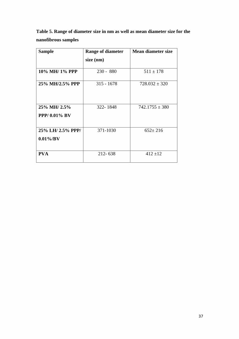
37
Table 5. Range of diameter size in nm as well as mean diameter size for the
nanofibrous samples
Sample Range of diameter
size (nm)
Mean diameter size
10% MH/ 1% PPP 230 - 880 511 ± 178
25% MH/2.5% PPP 315 - 1678 728.032 ± 320
25% MH/ 2.5%
PPP/ 0.01% BV
322- 1848 742.1755 ± 380
25% LH/ 2.5% PPP/
0.01%/BV
371-1030 652± 216
PVA 212- 638 412 ±12

38
Table 6. Swelling capacity of selected nanofibrous samples
Time (h) 25% MH/2.5%
PPP/ 0.01BV
25% LH/ 2.5%
PPP/ 0.01%,
BV
PVA 10%
1 96 % 50 % 218 %
4 101 % 52 % 222 %
24 106.6 % 68 % 250 %

39
Table 7. Water loss capacity of selected nanofibrous samples
Time 25% MH/ 2.5%
PPP/ 0.01% BV
25% LH/ 2.5%
PPP/ 0.01%, BV
PVA 10%
1 h 28.5 % 32% 15.5 %
7 days 66% 55.5% 41%
2 months 73% 75.5 % 48%

40
Table 8. Histological scoring system for H&E and MT stained wounded tissues
The scores are represented as follows: Necrosis: − = (0) absent, + = (1) scanty, ++ = (2)
moderate, +++ (3) = profound. Inflammatory cells − = (0) absent, + = (1) scanty, ++ = (2)
moderate, +++ (3) = profound. Hemorrhage: − = (0) absent, + = (1) scanty, ++ = (2)
moderate, +++ (3) = profound. Granulation tissue maturation: − = (3) absent, + = (2)
scanty, ++ = (1) moderate, +++ (0) = profound. Epithelialization: − = (3) absent, + = (2)
scanty, ++ = (1) moderate, +++ (0) = profound. Collagen fibers maturation: − = (3) absent,
+ = (2) scanty, ++ = (1) moderate, +++ (0) = profound. Activated hair follicles: − = (3)
absent, + = (2) scanty, ++ = (1) moderate, +++ (0) = profound.
Sa
mp
les
Nec
rosi
s
Infl
am
m
ato
ry
cell
s H
emo
rrh
ag
e
Gra
nu
lat
ion
tiss
ue
ma
tura
ti
on
Ep
ith
elia
liza
tio
n
Co
lla
gen
fib
ers
ma
tura
ti
on
Act
ivate
d
ha
ir
foll
icle
s T
ota
l
sco
re
Normal − − − +++ +++ +++ +++ 0
PVA (10%)
Day 5
+++ +++ ++ + − − − 19
PVA (10%)
Day 10
++ +++ + + − − − 17
MH/PPP (25%/2.5%)
Day 5
+++ ++ ++ ++ + + − 15
MH/PPP (25%/2.5%)
Day 10
− ++ + ++ + + + 10
MH/PPP (10%/1%)
Day 5
+++ ++ + + + + − 15
MH/PPP (10%/1%)
Day 10
− ++ + ++ ++ ++ − 10
MH/PPP/BV (25%/2.5%/0.01%)
Day 5
− ++ ++ ++ +++ + − 10
MH/PPP/BV 25%/2.5%/0.01%)
Day 10
− − − +++ +++ +++ ++ 1
LH/PPP/BV 25%/2.5%/0.01%)
Day 5
++ ++ + ++ + + − 13
LH/PPP/B (25%/2.5%/0.01%)
Day 10
− + − +++ +++ ++ ++ 3

41
Figure 1. The global prevelance of wounds by type in 2011 and that expected for 2025. It
is expected that there will be a significant increase in number of chronic wounds whose wounds
requires an advanced wound care products (MedMarket diligence).

42
Figure 2. Stages of wound healing: (a) Inflammation step, followed by proliferation step
and finally (c) remodeling. Inflammation step includes injury of a blood vessel followed by
coagulation, and acute local inflammatory response with the formation of a clot that helps to
protect against threatening infections). Proliferation step is characterized by cell migration,
angiogenesis as well as granulation tissue formation (c) Tissue remodeling step involves the
formation of a disorganized ECM as is shown in the figure in addition to a slightly elevated
area with the lack of the normal skin appendages (Stein & Küchle, 2013).

43
Figure 3. Tissue injury of acute wound vs. chronic wounds. On the left, acute injury activates
blood clotting, aggregation of platelets and migration of macrophages and neutrophils to the injury
site and a blood clot is formed. On the upper right of the figure, by day 5 after injury and under
normal conditions, the wound continue the normal healing process with the help of the growing
granulation tissue. On the lower right of the figure, the wound fails to heal properly due to an
underlying pathobiology or as a result of a microorganism invasion where the healing process is
interrupted leading to a chronic wound or ulcer formation (Clark et al., 2007).

44
Figure 4. Methylglyoxal structure.

45
Figure 5. Suggested mechanism by which MH inhibits S. aureus. MH was found to
influence cell division later stages where after speta formation is obtained, decreased
production of murine hydrolase together with being sequestered into an inactive form lead to
having the two daughter cells remaining attached as the septa are unable to become degraded.
This eventually leads to cell death (Jenkins et al., 2015).

46
Figure 6. A suggested mechanism by which MH inhibits P. aeruginosa. MH initially causes
cell envelope destabilization via the down-regulation of Oprf which a key proteins that is
involved in maintaining cell shape and cell wall stability. Loss of this structural protein leads
to what is called, membrane blebbing which eventually leads to cell lysis (Jenkins et al., 2015).

47
Figure 7. Suggested modulatory effects of honey throughout the stages of wound healing.
Clinical effects are on the left of the figure while their suggested mechanisms are on the right
of the figure (Lee at al., 2011).

48
Figure 8. A diagram showing the process of polymeric nanofibers formation via electro-
spinning, different techniques for electro-spinning and different types of collectors. A
typical electro-spinner include a high voltage power supply, spinneret system as well as a
collecting screen. Based on the configuration of the syringe required for loading. There are
different electro-spinning techniques ranging from co-electro-spinning and passing by side by
side electro-spinning, coaxial spinning, multi-jet spinning, emulsion spinning and the technique
of surface immobilization. Different collectors are displayed also which may range from fixed
or static collector, rotating drum, grid and a rotating disc (Vellayappan et al., 2016).
Polymer solution
Drug solution A
Drug solution B

49
Figure 9 . Parameters affecting the electrospinning process. Parameters include parameters
related to either the solution, process or surrounding conditions (Vellayappan et al., 2016).

50
Figure 10. PVA structure.

51
Figure 11. Potential of pomegranate peel extract (PPP) in treating various health conditions.
The figure shows graphical representation for the PPP extracted biomolecules and its suggested
enthopharmacological potential as a cardiovascular protective agent, anti- inflammatory and anti-
allergic, anti-influenza as well as an antimalarial and effective wound healing agent. Moreover, PPP
was shown to have anti-cancer properties (Ismail, T. et al., 2012).

52
Sample SEM images Histogram
10%MH/
1.5 PPP
25%MH/
2.5 %
PPP
25%MH/
2.5 %
PPP/
0.01%
BV
0
5
10
15
Freq
uen
cy
Diameter(nm)
Histogram
0
2
4
6
8
10
Freq
uen
cy
Diameter(nm)
Histogram
02468
101214
Freq
uen
cy
Diameter (nm)
Histogram

53
25% LH/
2.5 %
PPP/
0.01%
BV
PVA
10%
Figure 12. Representative SEM images of the corresponding fiber mats, including
histogram for the diameters of fibers (n≈50).
0
5
10
15
30
0-4
00
40
0-5
00
50
0-6
00
60
0-7
00
70
0-8
00
80
0-9
00
90
0-1
00
0
10
00
-11
00
11
00
-12
00
14
00
-15
00
Freq
uen
cy
Diameter (nm)
Histogram
0
5
10
15
20
Freq
uen
cy
Diameter (nm)
Histogram

54
Figure 13. Representative SEM images of the crosslinked (MH/PPP) sample. (a) Cross
linked MH/PPP mat viewed by SEM: FESEM, Leo Supra 55, Zeiss Inc., Oberkochen,
Germany) at magnification 1500 X. (b) Cross linked MH/PPP mat viewed by SEM
Model Quanta 250 FEG (Field Emission Gun), FEI company, Netherlands) at magnification
1.500 X. (c) Cross linked MH/PPP mat viewed by SEM Model Quanta 250 FEG (Field
Emission Gun), FEI company, Netherlands) at magnification of 3000 X.

55
Figure 14. Representative SEM image for MH/PPP/BV sample. The image reveals the
success of the crosslinking process. The sample was viewed by SEM ((SEM: FESEM, Leo
Supra 55, Zeiss Inc., Oberkochen, Germany)) SEM at magnification OF 1500 X.

56
Figure 15. Representative SEM images of the samples: a) (MH 10%/PPP 1%), b) (MH
20%/PPP 2%), and (MH 25%/PPP 2.5 %). Samples were viewed using SEM Model Quanta
250 FEG (Field Emission Gun), FEI Company, Netherlands.

57
MH
/PP
/BV
(1 h
r )
MH
/PP
P/B
V (
4 h
rs)
MH
/PP
P/B
V (
24 h
rs)
LH
/PP
P/B
V (
1 h
r )
LH
/PP
P/B
V (
4 h
rs)
LH
/PP
P/B
V (
24 h
rs)
PV
A (
1h
r )
PV
A (
4 h
rs)
PV
A (
24 h
rs)
0
1 0 0
2 0 0
3 0 0
S w e llin g c a p a c ity
S a m p le s
Sw
ell
ing
(%
)M H /P P /B V (1 h r)
M H /P P P /B V ( 4 h rs )
M H /P P P /B V ( 2 4 h rs )
L H /P P P /B V (1 h r)
L H /P P P /B V (4 h rs )
L H /P P P /B V (2 4 h rs )
P V A (1 h r )
P V A (4 h rs )
P V A (2 4 h rs )
Figure 16. A bar chart showing a comparison between the swelling behaviors of the
electro-spun scaffolds. Honey incorporated samples showed moderate capability for water
uptake in comparison to PVA scaffolds which had more swelling capacity.

58
MH
/PP
/BV
(1 h
r )
MH
/PP
P/B
V (
1 w
eek)
MH
/PP
P/B
V (
2 m
on
ths)
LH
/PP
P/B
V (
1 h
r )
LH
/PP
P/B
V (
1 w
eek)
LH
/PP
P/B
V (
2 m
on
ths)
PV
A (
1h
r )
PV
A (
1 w
eek)
PV
A (
2 m
on
ths)
0
2 0
4 0
6 0
8 0
1 0 0
B io d e g ra d ib ility
S a m p le s
Bio
de
gra
dib
ilit
y (
%)
M H /P P /B V (1 h r)
M H /P P P /B V ( 1 w e e k )
M H /P P P /B V ( 2 m o n th s )
L H /P P P /B V (1 h r)
L H /P P P /B V (1 w e e k )
L H /P P P /B V (2 m o n th s )
P V A (1 h r )
P V A (1 w e e k )
P V A (2 m o n th s )
Figure 17. A bar chart showing a comparison between the water loss behaviors of the
electrospun samples. It was shown from the results, an increase in water loss capacity in honey
incorporated scaffolds in comparison with PVA that showed less water loss capacity.
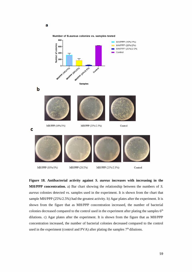
59
Figure 18. Antibacterial activity against S. aureus increases with increasing in the
MH/PPP concentration. a) Bar chart showing the relationship between the numbers of S.
aureus colonies detected vs. samples used in the experiment. It is shown from the chart that
sample MH/PPP (25%/2.5%) had the greatest activity. b) Agar plates after the experiment. It is
shown from the figure that as MH/PPP concentration increased, the number of bacterial
colonies decreased compared to the control used in the experiment after plating the samples 6th
dilutions. c) Agar plates after the experiment. It is shown from the figure that as MH/PPP
concentration increased, the number of bacterial colonies decreased compared to the control
used in the experiment (control and PVA) after plating the samples 7th dilutions.

60
Figure 19. Antibacterial activity against S. aureus. a) Bar chart showing the antibacterial
activity against S. aureus. It is shown from the figure that the three scaffolds decreased number
of bacterial colonies compared to both controls used in the experiment (control and PVA) (P <
0.0001). Samples containing BV were slightly more effective than the sample without BV (P <
0.05) b) Agar plates after the 24 h incubation. All honey based samples showed significant
decrease in number of colonies in comparison to both controls tested.

61
Figure 20. Antibacterial activity against E. coli. a) Bar chart showing the antibacterial
activity against E. coli. It is shown from the figure that the three scaffolds decreased the number
of bacterial colonies compared to both controls used in the experiment (control and PVA) (P <
0.0001). Sample (MH/PPP/BV) was slightly more effective than sample (LH/PPP/BV) (P <
0.05). b) Agar plates after 24 h incubation. It is shown from the figure that the three scaffolds
decreased the number of bacterial colonies compared to both controls used in the experiment
(control and PVA).

62
MH
/PP
P U
NC
RO
SS
MH
/PP
P C
RO
SS
MH
/PP
P/B
V U
NC
RO
SS
MH
/PP
P/B
V C
RO
SS
LH
/PP
P/B
VU
N C
RO
SS
LH
/PP
P/B
VC
RO
SS
0
5 0
1 0 0
1 5 0
V ia b ility p e rc e n t re la t iv e to th e c o n tro l
S a m p le s
Via
bil
ity
(%)
M H /P P P U N C R O S S
M H /P P P C R O S S
M H /P P P /B V C R O S S
L H /P P P /B V U N C R O S S
L H /P P P /B V C R O S S
M H /P P P /B V U N C R O S S
Figure 21. Cytotoxicity of electro-spun scaffolds. Samples included the following samples
MH/PPP (uncross-linked), MH/PPP (cross-linked), MH/PPP/BV (uncross-linked),
MH/PPP/BV (cross-linked), LH/ PPP/BV (uncross-linked), LH/PPP/BV (cross-linked)
scaffold. All samples did not affect cell viability.

63
Figure 22. Representative graphs showing average wound surface area at day 3, 5 and 10. a) Bar chart
showing average wound surface areas at day 3. It is shown from the figure that samples MH/PPP (25/2.5) wt %,
MH/PPP/BV (25/2.5/0.01) wt%, LH/PPP/BV (25/2.5/0.01) wt%, significantly decreased wound surface area
compared to both controls tested (P< 0.0001). b) Bar chart of the average wound surface areas at day 5. It is
shown from the figure that samples MH/PPP (25/2.5) wt%, MH/PPP/BV (25/2.5/0.01) wt%, LH/PPP/BV
(25/2.5/0.01) wt%, significantly decreased wound surface area compared to both controls tested (P<0.0001). c)
Bar chart of the average wound surface areas at day 10. It is shown from the figure that all treatment groups
(samples MH/PPP (25/2.5) wt%, MH/PPP(10/1) wt%, MH/PPP/BV (25/2.5/0.01) wt%, LH/PPP/BV
(25/2.5/0.01) wt%) had a significantly decreased wound surface areas and almost achieved complete healing
compared to both controls tested (P<0.0001).

64
Figure 23. Images showing healing progression. The images show healing progression from
day 0 to day 10 for wounds treated with all treatment scaffolds vs. the 2 control scaffolds used
in this study. It is clear from the pictures that healing was achieved by day 10 in all treatment
groups unlike slower healing in PVA and no treatment groups.
GROUP 1
MH/PPP
(25%/2.5%)
GROUP 2
MH/PPP
(10%/1%)
GROUP 3
MH/PPP/BV
(25%/2.5%/0
.01%)
GROUP 4
LH/PPP/B
V
25%/2.5%/
0.01%)
GROUP 5
PVA
GROUP 6
NO TTT
Day 0
Day 5
Day 10

65
Normal
(Intact
skin)
Day 5 Day 10
PVA

66
MH/PP
P
(25%/2.
5%)
.
MH/PP
P
(10%/1
%)

67
MH/PP
P/BV
(25%/2.
5%/0.01
%)
LH/PP
P/BV
(25%/2.
5%/0.01
%)
Figure 24. Representative histological assessment for all treatment groups vs. PVA
control group at day 5 and day 10 as shown by H&E staining (magnification 100 x). a) A
section in normal skin covered by an intact epidermis is shown. Normal histological structures
of skin including intact cellular elements in epidermis (arrow), dermis (black star) and
subcutaneous tissue are shown (red star). b) PVA control group at 5 days showed severe
epidermal necrosis, ulceration with many necrotic tissue debris infiltrated with inflammatory
cells (arrow) along with dermal layer severe inflammation. Cells infiltration with congested
blood vessels and focal areas of hemorrhage were also observed. c) At 10 days the group

68
showed delayed healing with ulcer formation and necrotic tissue debris (arrow) and granulation
tissue formation (star). d) MH/PPP (25%/2.5%) at day 5 demonstrated incomplete epidermal
re-epithelialization (arrow) as well as ulcer formation. Diffuse dermal and subcutaneous
infiltration of inflammatory cells were also observed. e) The same group at 10 days
demonstrated enhanced healing with complete epithelial bridging across wound gap (arrow). A
smaller area of highly cellular granulation tissue (star) was observed with moderate
inflammatory cells infiltrations. f) MH/PPP (10%/1%) group showed ulcer formation and
necrotic tissue debris (arrow) as well as granulation tissue formation in dermal layer at 5 days.
At 10 days, the same group (g) demonstrated enhanced healing process with complete epithelial
bridging across wound gap (arrow). A decreased area of highly cellular granulation tissue (star)
was observed with moderate inflammatory cells infiltrations. Some hair follicles were also
observed. MH/PPP/BV (25%/2.5%/0.01%) group showed good healing at day 5 with complete
epithelial bridging across wound gap (arrow). However; focal hemorrhagic areas (star) at
dermo-epidermal junction with extensive dermal inflammatory cells infiltration were observed.
i) At 10 days the wound of this group revealed accelerated wound healing with complete
epithelial bridging (arrow), minimal inflammatory cells were observed in dermal layer and
more activated hair follicles growth (star). j) LH/PPP/BV (25%/2.5%/0.01%) group at 5 days
showed ulcer formation and necrotic tissue debris (arrow) and granulation tissue formation
(star). At 10 days (k) demonstrated enhanced healing, complete epithelial bridging (arrow). A
small area of highly cellular granulation tissue (star) was observed with moderate inflammatory
cells infiltrations along with some hair follicles growth.

69
Normal
(Intact
skin)
Day 5 Day 10
PVA

70
MH/PPP
(25%/2.5
%)
MH/PPP
/
(10%/1
%)

71
MH/PPP
/BV
(25%/2.5
%/0.01
%)
LH/PPP/
BV
(25%/2.5
%/0.01
%)
Figure 25. Representative histological assessment for all treatment groups vs. PVA
control group at day 5 and day 10 as shown by Masson Trichrome staining (magnification
100 x). Abundant mature collagen fibers deposition in dermal layer can be observed in normal
skin in (a). It is shown from the pictures that at day 5 less dense collagen fibers were seen in all
groups as seen by grey shades instead of blue color while denser collagen deposition of the
treated samples: e, g, I, k was observed at day 10 when compared to the control group (c) at the
same time point.

72
References
Adams, C. J., Boult, C.H., Deadman,B.J., Farr, J. M.,Grainger, M.N.C., Manley
Harris,M.,et al.(2008). Isolation by HPL Candcharacterisation of the bioactive fraction
of NewZealand manuka (Leptospermumscoparium) honey. Carbohydr. Res. 343, 651–
659.doi:10.1016/j.carres.2007.12.011.
Adhikari, B., Howes, T., Wood, B.J. & Bhandari, B.R. (2009). The effect of low
molecular weight surfactants and proteins on surface stickiness of sucrose during
powder formation through spray drying. Journal of Food Engineering, 94, 135–143.
Alrubaiy, L., & Al-Rubaiy, K. K. (2009). Skin Substitutes: A Brief Review of Types
and Clinical Applications. Oman Medical Journal, 24(1), 4–6.
Alvarez-Suarez, J. M., Gasparrini, M., Forbes-Hernández, T. Y., Mazzoni, L., &
Giampieri, F. (2014). The composition and biological activity of honey: a focus on
Manuka honey. Foods, 3(3), 420-432.
American Type Culture Collection (ATCC): http://www.atcc.org
Andreu, V., Mendoza, G., Arruebo, M., & Irusta, S. (2015). Smart dressings based on
nanostructured fibers containing natural origin antimicrobial, anti-inflammatory, and
regenerative compounds. Materials, 8(8), 5154-5193.
Aslam, M. N., Lansky, E. P., & Varani, J. (2006). Pomegranate as a cosmeceutical
source: pomegranate fractions promote proliferation and procollagen synthesis and
inhibit matrix metalloproteinase-1 production in human skin cells. Journal of
ethnopharmacology, 103(3), 311-318.
Atrott, J., & Henle, T. (2009). Methylglyoxal in manuka honey—correlation with
antibacterial properties. Czech Journal of Food Sciences, 27(Spec.), S163-S165.
Bagde, A. B., Sawant, R. S., Bingare, S. D., Sawai, R. V., & Nikumbh, M. B. (2013).
Therapeutic and nutritional values of honey (madhu). International Research Journal
of Pharmacy, 4(3), 19-22.

73
Chang, J., & Cuellar, N. G. (2009). The use of honey for wound care management: A
traditional remedy revisited. Home Healthcare Now, 27(5), 308-316.
Balaji, A., Vellayappan, M. V., John, A. A., Subramanian, A. P., Jaganathan, S. K.,
Supriyanto, E., & Razak, S. I. A. (2015). An insight on electrospun-nanofibers-inspired
modern drug delivery system in the treatment of deadly cancers. RSC Advances, 5(71),
57984-58004.
Banks, B. E., & Shipolini, R. A. (1986). Chemistry and pharmacology of honey-bee
venom. Venoms of the Hymenoptera: Biochemical, pharmacological and behavioral
aspects, 329-416.
Carter, D. A., Blair, S. E., Cokcetin, N. N., Bouzo, D., Brooks, P., Schothauer, R., &
Harry, E. J. (2016). Therapeutic Manuka Honey: No Longer So Alternative. Frontiers
in Microbiology, 7, 569. http://doi.org/10.3389/fmicb.2016.00569.
Chidambara Murthy, K. N., Reddy, V. K., Veigas, J. M., & Murthy, U. D. (2004). Study
on wound healing activity of Punica granatum peel. Journal of Medicinal Food, 7(2),
256-259.
Clark, R. A., Ghosh, K., & Tonnesen, M. G. (2007). Tissue engineering for cutaneous
wounds. Journal of Investigative Dermatology, 127(5), 1018-1029.
Dhivya, S., Padma, V. V., & Santhini, E. (2015). Wound dressings – a
review. BioMedicine, 5(4), 22. http://doi.org/10.7603/s40681-015-0022-9
doi:10,2147/RRB.S75754.
Fennell, J. F., Shipman, W. H., & Cole, L. J. (1967). Antibacterial action of a bee venom
fraction (melittin) against a penicillin-resistant staphylococcus and other
microorganisms (No. USNRDL-TR-67-101). Naval Radiological Defense Lab San
Francisco CA.

74
Gethin, G. T., Cowman, S., & Conroy, R. M. (2008). The impact of Manuka honey
dressings on the surface pH of chronic wounds. International Wound Journal, 5(2),
185-194.
Global advanced wound care market outlook 2018 - smart dressings: Adding a new
dimension to wound healing. (2015). Biotech Week, , 77.
Gorzalski, P. (2009). Medicinal Use of Honey. American Bee Journal, 149(6), 518-518.
Greener, B., Hughes, A. A., Bannister, N. P., & Douglass, J. (2005). Proteases and pH
in chronic wounds. Journal of wound care, 14(2), 59-61.
Goula, A. M., & Lazarides, H. N. (2015). Integrated processes can turn industrial food
waste into valuable food by-products and/or ingredients: The cases of olive mill and
pomegranate wastes. Journal of Food Engineering, 167, 45-50.
Günes, Ü. Y., & Eser, I. (2007). Effectiveness of a honey dressing for healing pressure
ulcers. Journal of Wound Ostomy & Continence Nursing,34(2), 184-190.
Han, S. M., Lee, K. G., Park, K. K., & Pak, S. C. (2013). Skin sensitization study of
bee venom (Apis mellifera L.) in guinea pigs and rats. Cutaneous and ocular
Toxicology, 32(1), 27-30.
Hassan, C. M., & Peppas, N. A. (2000). Structure and applications of poly (vinyl
alcohol) hydrogels produced by conventional crosslinking or by freezing/thawing
methods. In Biopolymers· PVA Hydrogels, Anionic Polymerisation
Nanocomposites (pp. 37-65). Springer, Berlin, Heidelberg.
Huang, Z. M., Zhang, Y. Z., Kotaki, M., & Ramakrishna, S. (2003). A review on
polymer nanofibers by electrospinning and their applications in
nanocomposites. Composites science and technology, 63(15), 2223-2253.
Islam, M.I., Edrisi, M. & Langrish, T. (2013). Improving process yield by adding WPI
to lactose during crystallization and spray drying under high-humidity conditions.
Drying Technology: An International Journal, 31, 393–404.

75
Ismail, T., Sestili, P., & Akhtar, S. (2012). Pomegranate peel and fruit extracts: a review
of potential anti-inflammatory and anti-infective effects. Journal of
ethnopharmacology, 143(2), 397-405.
Issam, A. A., Zimmermann, S., Reichling, J., & Wink, M. (2015). Pharmacological
synergism of bee venom and melittin with antibiotics and plant secondary metabolites
against multi-drug resistant microbial pathogens.Phytomedicine, 22(2), 245-255.
Jenkins, R., Roberts, A., & Browm, H. L. (2015). On the antibacterial effects of Manuka
honey: Mechanistic insights. Research and Reports in Biology, 2015, 215-224.
Kang, S. S., Pak, S. C., & Choi, S. H. (2002). The effect of whole bee venom on
arthritis. The American journal of Chinese medicine, 30(01), 73-80.
Khazaeli, P., Karamouzian, M., Rohani, S., Sadeghirad, B., & Ghalekhani, N. (2014).
Effects of minoxidil gel on burn wound healing in rats. Iranian Journal of
Pharmaceutical Research : IJPR, 13(1), 243.
Kruszewski, B., Jedlinska, A., Antczak, M., Lipinska, E. & WrowaRajchert, D. (2014).
Novel methods of producing honey powders and assessment of their biological activity.
Food Science Technology Quality, 1, 160–172.
Lazarev, V. N., Shkarupeta, M. M., Titova, G. A., Kostrjukova, E. S., Akopian, T. A.,
& Govorun, V. M. (2005). Effect of induced expression of an antimicrobial peptide
melittin on Chlamydia trachomatis and Mycoplasma hominis infections in
vivo. Biochemical and biophysical research communications, 338(2), 946-950.
Lee, David S., Sammy Sinno, and Amor Khachemoune. "Honey and wound
healing." American journal of clinical dermatology 12.3 (2011): 181-190.
LEE, J. Y., KANG, S. S., KIM, J., BAE, C. S., & CHOI, S. H. (2005). Inhibitory effect
of whole bee venom in adjuvant-induced arthritis. In Vivo, 19(4), 801.
Lee, W. R., Kim, S. J., Park, J. H., Kim, K. H., Chang, Y. C., Park, Y. Y., ... & Park,
K. K. (2010). Bee venom reduces atherosclerotic lesion formation via anti-
76
inflammatory mechanism. The American journal of Chinese medicine, 38(06), 1077-
1092.
Liu, M., Duan, X., Li, Y., Yang, D., & Long, Y. (2017). Electrospun nanofibers for
wound healing. Materials Science & Engineering C, 76, 1413-1423.
doi:10.1016/j.msec.2017.03.034
Liu, X., Chen, D., Xie, L., & Zhang, R. (2002). Effect of honey bee venom on
proliferation of K1735M2 mouse melanoma cells in-vitro and growth of murine B16
melanomas in-vivo. Journal of Pharmacy and Pharmacology, 54(8), 1083–1089.
Maleki, H., Gharehaghaji, A. A., & Dijkstra, P. J. (2013). A novel honey‐based
nanofibrous scaffold for wound dressing application. Journal of Applied Polymer
Science, 127(5), 4086-4092.
Mavric, E., Wittmann, S., Barth, G., & Henle, T. (2008). Identification and
quantification of methylglyoxal as the dominant antibacterial constituent of Manuka
(Leptospermum scoparium) honeys from New Zealand. Molecular nutrition & food
research, 52(4), 483-489.
Michelet, J. F., Commo, S., Billoni, N., Mahé, Y. F., & Bernard, B. A. (1997).
Activation of cytoprotective prostaglandin synthase-1 by minoxidil as a possible
explanation for its hair growth-stimulating effect. Journal of investigative
dermatology, 108(2), 205-209.
Molan, P.C. (2006) The evidence supporting the use of honey as a wound dressing. Int.
J. Lower Extrem. Wounds 5, 40-54.
Oršolić, N. (2012). Bee venom in cancer therapy. Cancer and Metastasis
Reviews, 31(1), 173-194. doi:10.1007/s10555-011-9339-3
Oryan, A., Alemzadeh, E., & Moshiri, A. (2016). Biological properties and therapeutic
activities of honey in wound healing: a narrative review and meta-analysis. Journal of
tissue viability, 25(2), 98-118.

77
Pieper, B. (2009). Honey‐Based Dressings and Wound Care: An Option for Care in the
United States. Journal of Wound Ostomy & Continence Nursing,36(1), 60-66.
Priestley, G. C., LORD, R., & Stavropoulos, P. (1991). The metabolism of fibroblasts
from normal and fibrotic skin is inhibited by minoxidil in vitro.British Journal of
Dermatology, 125(3), 217-221.
Puppi, D., Piras, A. M., Detta, N., Ylikauppila, H., Nikkola, L., Ashammakhi, N., ... &
Chiellini, E. (2011). Poly (vinyl alcohol)-based electrospun meshes as potential
candidate scaffolds in regenerative medicine. Journal of Bioactive and Compatible
Polymers, 26(1), 20-34.
Qu, W., Pan, Z., Zhang, R., Ma, H., Zhu, B., Wang, Z., & Atungulu, G. G. (2009).
Integrated extraction and anaerobic digestion process for recovery of nutraceuticals and
biogas from pomegranate marc. Transactions of the ASABE, 52(6), 1997-2006.
Samborska, K., Langa, E., Kaminska-Dworznicka, A. & WitrowaRajchert, D. (2015).
The influence of sodium caseinate on the physical properties of spray-dried honey.
International Journal of Food Science and Technology, 50, 256–262.
Shi, Q., Fang, Z. & Bhandari, B. (2013). Effect of addition of whey protein isolate on
spray-drying behavior of honey with maltodextrin as a carrier material. Drying
Technology, 31, 1681– 1692.
Sarhan, W. A., & Azzazy, H. M. (2015). High concentration honey chitosan electrospun
nanofibers: Biocompatibility and antibacterial effects.Carbohydrate polymers, 122,
135-143.
Sen, C. K., Gordillo, G. M., Roy, S., Kirsner, R., Lambert, L., Hunt, T. K.. . Longaker,
M. T. (2009). Human skin wounds: A major and snowballing threat to public health
and the economy. Wound Repair and Regeneration : Official Publication of the Wound
Healing Society [and] the European Tissue Repair Society, 17(6), 763.
doi:10.1111/j.1524-475X.2009.00543.x
Sharma, C. P., & Paul, W. (2015) Advances in Wound Healing Materials: Science and
Skin Engineering. Shewsbury: Smithers Rapra.

78
Singh, R. P., Chidambara Murthy, K. N., & Jayaprakasha, G. K. (2002). Studies on the
antioxidant activity of pomegranate (Punica granatum) peel and seed extracts using in
vitro models. Journal of agricultural and food chemistry, 50(1), 81-86.
Stein, C., & Küchler, S. (2013). Targeting inflammation and wound healing by
opioids. Trends in pharmacological sciences, 34(6), 303-312.
Structure and applications of poly (vinyl alcohol) hydrogels produced by conventional
crosslinking or by freezing/thawing methods. In Biopolymers· PVA Hydrogels, Anionic
Polymerisation Nanocomposites (pp. 37-65). Springer, Berlin, Heidelberg.
Subrahmanyam, M. (1994). Honey-impregnated gauze versus amniotic membrane in
the treatment of burns. Burns, 20(4), 331-333.
Subrahmanyam, M. (1998). A prospective randomised clinical and histological study
of superficial burn wound healing with honey and silver sulfadiazine. Burns, 24(2),
157-161.
Taepaiboon, P., Rungsardthong, U., & Supaphol, P. (2006). Drug-loaded electrospun
mats of poly (vinyl alcohol) fibres and their release characteristics of four model
drugs. Nanotechnology, 17(9), 2317.
Talekar, S., Patti, A. F., Vijayraghavan, R., & Arora, A. (2018). An integrated green
biorefinery approach towards simultaneous recovery of pectin and polyphenols coupled
with bioethanol production from waste pomegranate peels. Bioresource Technology.
Tonks, A., Cooper, R.A., Price, A.J., Molan, P.C. and Jones, K.P. (2001) Stimulation
of TNF- alpha release in monocytes by honey. Cytokine 14, 240-242.
Tonks, A.J., Cooper, R.A., Jones, K.P., Blair, S., Parton, J. and Tonks, A. (2003) Honey
stimulates inflammatory cytokine production from monocytes. Cytokine 21, 242-247.
Tonks, A.J., Dudley, E., Porter, N.G., Parton, J., Brazier, J., Smith, E.L. and Tonks,
A.A. (2007) 5.8-kDa component of manuka honey stimulates immune cells via TLR4.

79
J. Leukocyte Biol. Urtubey, N. "Apitoxin: from bee venom to apitoxin for medical
use." Termas de Rio Grande Santiago del Estero, Argentina (2005).
Vellayappan, M. V., Venugopal, J. R., Ramakrishna, S., Ray, S., Ismail, A. F., Mandal,
M. & Jaganathan, S. K. (2016). Electrospinning applications from diagnosis to
treatment of diabetes. RSC Advances, 6(87), 83638-83655.
Wang, P., & He, J. H. (2013). Electrospun polyvinyl alcohol-honey
nanofibers. Thermal Science, 17(5), 1549-1550.
Yahia, I. S., & Keshk, S. M. (2017). Preparation and characterization of PVA/Congo
red polymeric composite films for a wide scale laser filters. Optics & Laser
Technology, 90, 197-200.

80
Appendix

81
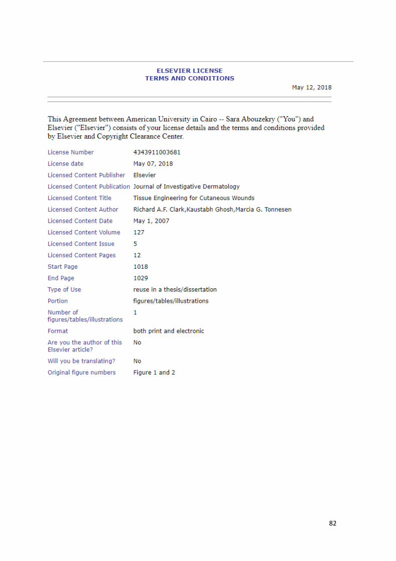
82

83

84

85

86

![Effect of Pomegranate Peel Extract on Submandibular ... · Diminished salivary secretion can be result from multiple situations: the salivary glands radiotherapy [4], drugs [5], in](https://static.fdocuments.in/doc/165x107/5f144407d49cd328962a5056/effect-of-pomegranate-peel-extract-on-submandibular-diminished-salivary-secretion.jpg)


![New Application of Pomegranate Peel Waste: The ... · flocculation [3], reverse osmosis [4] and activated carbon absorption [5]. These technologies do not show significant successful](https://static.fdocuments.in/doc/165x107/5eb2cffa0f8f9d41710c76d9/new-application-of-pomegranate-peel-waste-the-flocculation-3-reverse-osmosis.jpg)
