Fabrication of electrospun polyacrylonitrile ion-exchange ...Fabrication of electrospun...
10
1. Introduction A conventional ion exchange resin usually has a gel or granular structure, composed of styrene or acrylic material [1]. However, micropore structures within the resin result in lower molecular adsorp- tion/desorption rates, due to diffusion distance. With an increase in the pace of processing, the vol- ume of adsorbed molecules increased, and an increase in the frequency of its application rapidly decreased the efficiency of its ion exchange capa- bility, to the point where it was suitable only for smaller molecules. In addition, micropores in tradi- tional resins are easily blocked, which reduces the effectiveness of purification and decreases the regeneration response rate. Furthermore, the resin itself is vulnerable to damage due to pressure [2–3]. To tackle the above problems, the fiber structure of the ion exchange filters has undergone significant development in recent years. Compared to resins of other types of gel or granular structure, fiber struc- tured resins are simple to use, thanks to their large surface area, and possess high adsorption capacity, fast ion exchange and regeneration rate, as well as other unique features [4–6]. It is widely used in dyes, precious metals, protein pharmaceuticals and other high value-added materials during the enrichment and purification; it is also used in wastewater and gas decontamination processes. The potential bene- fits and value to researchers cannot be overlooked [7–11]. 308 Fabrication of electrospun polyacrylonitrile ion-exchange membranes for application in lysozym H. T. Chiu 1* , J. M. Lin 1 , T. H. Cheng 2 , S. Y. Chou 2 1 Department of Materials Science and Engineering National Taiwan University of Science and Technology, No.43, Keelung Rd., Section 4, Taipei, Taiwan 2 Department of Products, Taiwan Textile Research Institute, No. 6, Chengtian Rd., Tucheng City, Taipei County 23674, Taiwan Received 18 August 2010; accepted in revised form 7 November 2010 Abstract. Ion exchange (IEX) chromatography is commonly used in separation and purification systems. However, micro- pore blockage within its resin structure can easily lead to a reduction in the effectiveness of purification. To tackle this prob- lem, we adopted the concept of membrane separation by combining electrospinning techniques with rapid alkaline hydrolysis to prepare a weak acid IEX nanofibrous membrane (AEA-COOH), consisting of polyethyleneterephthalate (PET) meltblown fabric as a supporting layer, with upper and lower IEX layers consisting of polyacrylonitrile (PAN) nanofibrous membranes. To determine the characteristics of the AEA-COOH membrane, we used the commercial product Sartobind® C IEX membrane as the standard of comparison. Results showed that the base weight and thickness of AEA- COOH were 33 and 64%, relative to Sartobind ® C membrane. The thermo-degradable temperature of AEA-COOH mem- brane (320°C) was far higher than that of Sartobind ® C (115°C), indicating high thermal stability. Finally, comparisons between the lysozyme adsorption rates and capacity of various IEX membranes confirmed that AEA-COOH was lighter, thinner, faster, possessing higher protein adsorption efficiency than Sartobind ® C membrane. Keywords: polymer membranes, electrospun, nanofiber, ion-exchange, lysozymes adsorption eXPRESS Polymer Letters Vol.5, No.4 (2011) 308–317 Available online at www.expresspolymlett.com DOI: 10.3144/expresspolymlett.2011.31 * Corresponding author, e-mail: [email protected] © BME-PT
Transcript of Fabrication of electrospun polyacrylonitrile ion-exchange ...Fabrication of electrospun...

1. IntroductionA conventional ion exchange resin usually has a gelor granular structure, composed of styrene oracrylic material [1]. However, micropore structureswithin the resin result in lower molecular adsorp-tion/desorption rates, due to diffusion distance.With an increase in the pace of processing, the vol-ume of adsorbed molecules increased, and anincrease in the frequency of its application rapidlydecreased the efficiency of its ion exchange capa-bility, to the point where it was suitable only forsmaller molecules. In addition, micropores in tradi-tional resins are easily blocked, which reduces theeffectiveness of purification and decreases theregeneration response rate. Furthermore, the resin
itself is vulnerable to damage due to pressure [2–3].To tackle the above problems, the fiber structure ofthe ion exchange filters has undergone significantdevelopment in recent years. Compared to resins ofother types of gel or granular structure, fiber struc-tured resins are simple to use, thanks to their largesurface area, and possess high adsorption capacity,fast ion exchange and regeneration rate, as well asother unique features [4–6]. It is widely used in dyes,precious metals, protein pharmaceuticals and otherhigh value-added materials during the enrichmentand purification; it is also used in wastewater andgas decontamination processes. The potential bene-fits and value to researchers cannot be overlooked[7–11].
308
Fabrication of electrospun polyacrylonitrile ion-exchangemembranes for application in lysozymH. T. Chiu1*, J. M. Lin1, T. H. Cheng2, S. Y. Chou2
1Department of Materials Science and Engineering National Taiwan University of Science and Technology, No.43,Keelung Rd., Section 4, Taipei, Taiwan
2Department of Products, Taiwan Textile Research Institute, No. 6, Chengtian Rd., Tucheng City, Taipei County 23674,Taiwan
Received 18 August 2010; accepted in revised form 7 November 2010
Abstract. Ion exchange (IEX) chromatography is commonly used in separation and purification systems. However, micro-pore blockage within its resin structure can easily lead to a reduction in the effectiveness of purification. To tackle this prob-lem, we adopted the concept of membrane separation by combining electrospinning techniques with rapid alkalinehydrolysis to prepare a weak acid IEX nanofibrous membrane (AEA-COOH), consisting of polyethyleneterephthalate(PET) meltblown fabric as a supporting layer, with upper and lower IEX layers consisting of polyacrylonitrile (PAN)nanofibrous membranes. To determine the characteristics of the AEA-COOH membrane, we used the commercial productSartobind® C IEX membrane as the standard of comparison. Results showed that the base weight and thickness of AEA-COOH were 33 and 64%, relative to Sartobind® C membrane. The thermo-degradable temperature of AEA-COOH mem-brane (320°C) was far higher than that of Sartobind® C (115°C), indicating high thermal stability. Finally, comparisonsbetween the lysozyme adsorption rates and capacity of various IEX membranes confirmed that AEA-COOH was lighter,thinner, faster, possessing higher protein adsorption efficiency than Sartobind® C membrane.
Keywords: polymer membranes, electrospun, nanofiber, ion-exchange, lysozymes adsorption
eXPRESS Polymer Letters Vol.5, No.4 (2011) 308–317Available online at www.expresspolymlett.comDOI: 10.3144/expresspolymlett.2011.31
*Corresponding author, e-mail: [email protected]© BME-PT

According to previous studies [12–18], electrospin-ning is the only direct, efficient, and continuousmethod for producing polymer nanofibers showingthe most promise and greatest efficiency. Hundredsof polymer nanofibers of various kinds have beendeveloped using electrospinning. Electrospinningpolymer processing systems deliver polymer solu-tions to a spin feeder, to form liquid droplets. Withthe application of a high voltage charge, the electri-cal charge within the polymer mutually excludesand overcomes the surface tension of the solution.The surface of the droplets spray a charged liquidcolumn, which, under the continuous effect of theelectric field, quickly spiral downward to form alashing extension. The resulting nanofiber is a thou-sand times thinner than fibers manufactured usingnormal micro-spinning technology. As the processcontinues, the solvent in the solution rapidly evapo-rates. Finally, non-woven membranes of nanometerfiber diameter are collected in the collector platebeneath. By electrospinning, fibers with a diameterbetween a few dozen to several hundred nanome-ters could be manufactured. When compared to tra-ditional fibers (diameter of 50 microns) under thesame volumetric conditions, these fibers had asmaller diameter pore aperture, more densely inter-connected pores and increased porosity. In addition,the specific surface area of nanofibers is approxi-mately 100 times that of normal fibers. Thus, nano -fiber membrane filters, when compared to tradi-tional filters, have a higher throughput, lower loss,lower energy consumption, shorter transmissiondistance, higher adsorption, faster reaction, as wellas other advantages [19–21].Polyacrylonitrile (PAN) possesses high mechanicalstrength thanks to the strong hydrogen bonding ofits molecular structure. It also exhibits strong chem-ical resistance, excellent acid-proof alkalinity,resistance to sunlight, a diminished susceptibility tohumidity, and low cost [22–23]. Thus, it has caughtthe attention of many researchers, in the past10 years. It is widely used in the preparation ofmicrofiltration (MF), ultrafiltration (UF) and hol-low fiber membranes [24–26]. In addition, PANmolecular chains carry a cyano group, which can bemodified. It is hydrolysable and can be adjusted toachieve functionality in a number of applications. Ifpart of the hydrolysis process could be performedwith carboxyl functional groups (–COOH) in a solu-
tion, it could quickly adsorb metal ions, lysozymes,and other molecules. Its unique features includehigh absorption capacity, fast absorption speed, andgood dynamic performance. It is an excellent mate-rial for use in the purification of heavy metal waste-water and lysozymes [27–30].In this study, we first used electrospinning technol-ogy to prepare PAN/PET/PAN nanofiber compositemembrane. The nanofiber was stacked into a 3Dreticular fiber structure through cross laying. Thisgave the membrane a high specific surface area,higher porosity (>80%), and micron size membranepores. With the use of alkaline hydrolysis tech-niques, high-density carboxyl groups appeared onthe surface of PAN nanofibers. This prepared thenanofiber membrane for weak acid ion exchange.During the exchange of ions through the nanofibermembrane shortened the distance in molecular dif-fusion, thereby accelerating the rate of ion exchangeand improving adsorption capacity. Scanning elec-tron microscopy, porosity analysis, and ToluidineBlue O (TBO) functional groups detector were usedto analyze micro-porous PAN/PET/PAN structure,patterns, pore size, distribution, and density of nano -fiber composite membrane and its functional groups.Finally, the nanofiber composite membrane wascompared to commercial product Sartobind® Cmembrane in order to further explore the differention exchange membranes in the lysozyme adsorp-tion.
2. Experiments2.1. MaterialsPolyacrylonitrile (PAN) yarn (Mw = 120 000, con-taining 93% acrylonitrile and 7% vinylacetate) anddimethylacetamide (DMAc) were purchased fromFortune Industries Inc (Tao-Yuan, Taiwan) and I-Chang Chemical Co., Ltd (Taipei, Taiwan), respec-tively. Sodium hydroxide (NaOH) was from J.T.Baker. Lysozyme and Toluidine Blue O (TBO)were from Sigma. The reagents and solvents wereof analytical grade and used without further purifi-cation.
2.2. Electrospinning of polyacrylonitrileElectrospinning device was purchased from JyiGoang Enterprise Co., Ltd. (Taipei, Taiwan). PANyarn was dissolved in DMAc at a concentration of15% (w/v). PAN/DMAc solution was loaded into a
Chiu et al. – eXPRESS Polymer Letters Vol.5, No.4 (2011) 308–317
309

syringe and delivered to the tip of a 21-gauge stain-less steel nozzle with a syringe pump. The nozzlewas connected to a power supply and charged to26.5 kV. The electrospinning mats were collectedon nonwoven PET (basis weight 15 g/m2) andwrapped on a grounded steel sheet. The distancebetween the tip of the nozzle and the substrate was15.8 cm, the flow rate was 1ml/h, and the collectorrotation rate was 24 cm/s. The nozzle was movingalong the y-axis (20 cm), and the frequency was12 times/min.
2.3. Introduction of anionic moieties into PANnanofiber membranes
Electrospun PAN nanofiber mats were chemicallyactivated by substituting the nitrile groups withanion carboxylic acid groups as an ion exchangematerial. First, PAN nanofiber mats were alkalinehydrolyzed by immersion in various concentrations(1N, 2N, 3N) of sodium hydroxide stirred (150 rpm)at different temperatures (25–85°C) for 0–40 min.They were washed out with deionized water untilno change in pH was apparent. Finally, the carboxy-lated electrospun PAN nanofiber mat was treatedwith 0.1M HCl, followed by drying in an oven, at60°C.
2.4. Characterization of fabricsThe morphology and diameter of electrospun PANnanofiber mats before and after the introduction ofthe ion-exchange groups were observed using ascanning electron microscope (SEM, JEOL, andJSM 6510). All samples were sputter-coated withPt.Thermogravimetric analysis (TGA) was conductedwith TGA Q50 from TA Instruments at a heatingrate of 20°C/min under nitrogen. To characterizethe crystallinity of the fibers, wide-angle X-ray dif-fraction (WAXD, SIEMENS, and D5005) patternsof PAN nanofiber membranes under various alka-line hydrolysis conditions were obtained using anX-ray goniometer operating at 40 kV and 30 mAwith a scanning speed of 1°/min.The distribution of pore size and the mean pore sizeof electrospun PAN nanofiber mats were deter-mined with a measuring instrument (Perm-Porome-ter, PMI, USA). The porosity and the specific sur-face area were measured with fully automatic densityanalyzer (AccuPyc 1330 Pycnometer) and BET
apparatus (Micrometritics, ASPS 2000), respec-tively.
2.5. Adsorption behavior [31–32]Subsequent to alkaline hydrolysis, the concentra-tion of carboxyl group on the surface of the PANnanofiber was measured using Toluidine Blue O(TBO) dye. We placed the modified PAN nanofibermembrane in 2 ml 1mM TBO dyeing agent (dis-solved in 1 mM NaOH solution, pH = 10), at roomtemperature for 6 hours. It was cleaned severaltimes with de-ionized water. This was followed byadditional cleaning of the membrane using 0.1 mMNaOH to remove TBO dye adsorbed on the surfaceof the membrane. Then, 2 ml of 50% acetic acidsolution was added to remove the TBO dye of car-boxyl ionic bond. Finally, UV spectrum absorptionwas set to a wavelength of 633 nm for quantitativeanalysis. Using TBO/COOH = 1:1, the number ofcarboxyl groups on the membrane surface was cal-culated. Under lysozyme adsorption conditions, invarious time frames, at 298 K, with stirring speed of150 rpm, the lysozyme initial concentration was0.5 mg/ml (in 20 mM glycine buffer (pH 9)), andthe 595 nm absorbance wavelength was measuredusing Bio-Rad lysozyme concentration analysis. Byusing the standard concentration curve of lysozyme,the adsorption capacity of lysozyme nanofibercould be obtained.
3. Results and discussion3.1. Polyacrylonitrile (PAN) nanofiber
membranesThis study combines electrospinning nanofiberswith rapid alkaline hydrolysis technology, to developan ion-exchange nanofiber membrane with high
Chiu et al. – eXPRESS Polymer Letters Vol.5, No.4 (2011) 308–317
310
Figure 1. Effect of electrospinning duration on pore sizeand thickness of PAN nanofiber membranes

surface area and high porosity. Figure 1 shows therelationship between the duration of electrospin-ning and the thickness and average pore size of theelectrospun PAN nanofiber membrane. These resultsindicated that the thickness of nanofiber membraneincreased linearly and proportionally to increases inelectrospinning duration. Conversely, the averagepore diameter declined rapidly over time within the1–3 h of the spinning time frame, whereupon it wasstabilized. The duration of the electrospinning hadno significant effect on the pore size of membranes.SEM photos in Figure 2 show electrospun PANmembrane with a loose structure comprising a matof network of cross-laid nanofibers, with large dia-metric pores between the fibers. After the heatpressing process, the structure of the membraneincreased in density, with an increased in the con-centration and distribution of pores. Figure 2b illus-trates that variations in the diameter distribution ofthe PAN nanofiber was uniform, with the averagediameter falling between 200–250 nm. Therefore,we selected a PAN nanofiber membrane (thickness:55.26±2.87 µm, an average pore size: 0.79±0.04 µm),electrospun over a 10 hr period, to serve as the basisfor ion-exchange membrane.
3.2. Alkaline hydrolysis of polyacrylonitrile(PAN) nanofiber membranes
Partial hydrolysis is one of the most commonlyused methods for PAN-based membranes in thesubstitution of nitrile groups (–C!N) with car-boxylic acid groups (–COOH) subject to base con-
ditions. The chemical mechanism of alkali hydroly-sis reaction is described in detail by Wang et al. [27]We used NaOH to quickly and easily alkalinehydrolyze –C!N bond on the surface of the PANnanofiber, converting it into COOH functionalgroups. Figure 3 shows the effects of alkaline hydrol-ysis on the surface density of COOH PAN nano -fibers. Under conditions of fixed alkalization time(10 min) and temperature (25, 40, 55, 70, 85°C), thedensity of COOH increased, following an increasein the concentration of alkaline hydrolyzed NaOH.The trend of increasing became clear as the temper-ature of the alkalization increased. When the con-centration of NaOH had reached 3N, the COOHdensity on the PAN nanofiber surface showed themost significant increase.Figure 4 shows the effect of NaOH alkaline hydrol-ysis temperature on COOH density of the surface of
Chiu et al. – eXPRESS Polymer Letters Vol.5, No.4 (2011) 308–317
311
Figure 2. SEM images of electrospun fibers of polyacrylonitrile: (a) electrospinning a PAN nanofiber mat; (b) compactingthe mat to increase the volume density of fibers. The insert shows the morphology of nanofibers at a magnifica-tion of 50 000.
Figure 3. Effect of alkali (NaOH) concentration (N) on car-boxylic acid ligand density of AEA-COOH com-posite membranes

PAN nanofiber. Under conditions of fixed alkaliza-tion time (10 min) and concentration (1N, 2N, 3N),COOH density following an increase in tempera-ture to above 60°C in the alkaline hydrolyzedNaOH. Particularly with a high concentration ofNaOH lye, the rate of increase became even moreapparent. In addition, the curves in Figure 4 show acritical alkaline hydrolysis temperature in AEA-COOH composite nanofiber membrane. When thattemperature was below 60°C, this had little effecton the density of the functional group, but when itincreased to above 60°C, COOH the densityincreased exponentially as a function of tempera-ture.By TBO dye test analysis, the set COOH density incommercial standard Sartobind® C membrane wasdetermined to be 135.1 µeq/g. Figure 5 shows theeffect of NaOH alkaline hydrolysis duration on theCOOH density of the PAN nanofiber surface. Underfixed alkalization temperature (85°C) and concen-tration (3N), when alkaline hydrolysis time for AEA-
COOH composite nanofiber membrane was lessthan 13 minutes, the COOH group density was lessthan Sartobind® C. However, with a further increasein alkaline hydrolysis duration, PAN nanofiber sur-face density of COOH groups rapidly increasedafter 35 minutes. The density of the ionic group was3.3 times that of the commercial product. Afteralkaline hydrolysis had continued for more than60 minutes, the COOH density decreased rapidly toabout 1.9 µeq/g. This was due to an overreaction inthe hydrolysis reaction, resulting in alkali-solubleconditions in PAN nanofiber (data not shown).Thus, under appropriate reaction conditions, and asthe need arose, the density of the functional groupAEA-COOH composite nanofiber membrane wasmodified in order to adjust the duration of alkalinehydrolysis. Yet if Sartobind® C membranes wereprocessed through phase separation, their func-tional group density could not be adjusted for time,and its operating flexibility was thus limited.
3.3. Physical properties of differention-exchange membranes
Different SEM microstructures of ion exchangemembrane are shown in Figure 6. Due to the phaseseparation process, the surface of the Sartobind® Cmembrane had an uneven pore distribution; most ofthe surface area of the membranes was occupied bypolymer. Observed by a SEM cross-section view,the Sartobind® C membrane was found to possessan asymmetric structure, which increased its liquidcirculation. In this study, the weak acid ion exchangemembrane was prepared by electrospinning of PANnanofiber membrane using NaOH alkaline hydroly-sis. Under conditions of fixed alkalization tempera-ture (85°C), concentration (3N), and time (35 min),the membranes were able to maintain the integrityof its nanofiber structure; its surface pore distribu-tion was uniform and dense. With PET serving asan intermediate substrate, it was synthesized into aPAN-PET-PAN (AEA-COOH) asymmetric mem-brane structure.Results from PMI pore data test on different mem-branes showed the relationship between pore distri-bution [%] and size [µm], as shown in Figure 7. Thedata confirmed that the pore size distribution oncommercial product Sartobind® C membranes wasvery broad, and its distribution ranged widely from0.5 to 2.5 µm. The PAN-PET, PAN-PET-PAN ion-
Chiu et al. – eXPRESS Polymer Letters Vol.5, No.4 (2011) 308–317
312
Figure 4. Effect of alkaline hydrolysis temperature on car-boxylic acid ligand density of AEA-COOH com-posite membranes
Figure 5. Effect of alkaline hydrolysis time on carboxylicacid ligand density in AEA-COOH compositemembranes

exchange composite membranes, with their cross-laid, densely packed nanofiber mesh pattern, had amore concentrated arrangement of their pore sizeand distribution than Sartobind® C membrane.Figure 8 shows the relationship between the num-ber of PAN-COOH membrane layers and its aver-
age pore size; it was similar to control group Sarto-bind® C membrane in its average pore size of1.29±0.04 µm. A single PAN-COOH layer com-bined with PET composite membrane had an aver-age pore size of 0.70±0.05 µm. However, followingan increase in the number of PAN-COOH layers,
Chiu et al. – eXPRESS Polymer Letters Vol.5, No.4 (2011) 308–317
313
Figure 6. SEM micrographs of ion-exchange membranes. (a) and (b) are surface and cross-sectional images of Sartoriusweak acidic cation exchanger (Sartobind® C). (c) and (d) are surface and cross-sectional SEM images of electro-spun polyacrylonitrile composite membrane hydrolyzed with NaOH (AEA-COOH).
Figure 7. Pore size distribution [%] of different ion-exchange membranes
Figure 8. The relationship between average pore size andnumber of PAN-COOH membrane layers
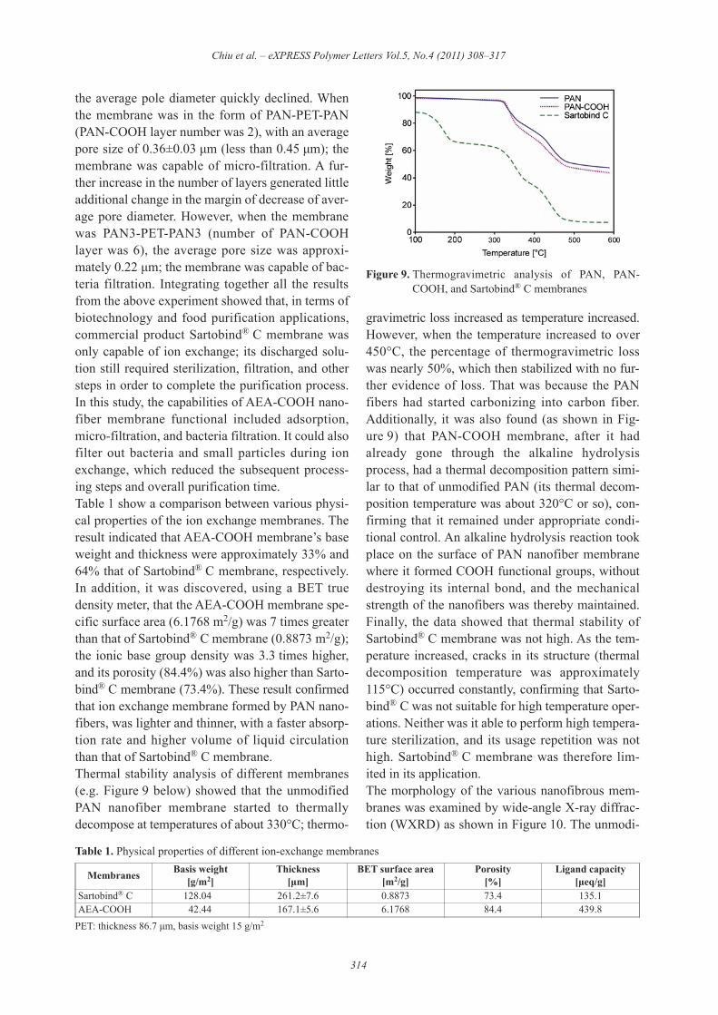
the average pole diameter quickly declined. Whenthe membrane was in the form of PAN-PET-PAN(PAN-COOH layer number was 2), with an averagepore size of 0.36±0.03 µm (less than 0.45 µm); themembrane was capable of micro-filtration. A fur-ther increase in the number of layers generated littleadditional change in the margin of decrease of aver-age pore diameter. However, when the membranewas PAN3-PET-PAN3 (number of PAN-COOHlayer was 6), the average pore size was approxi-mately 0.22 µm; the membrane was capable of bac-teria filtration. Integrating together all the resultsfrom the above experiment showed that, in terms ofbiotechnology and food purification applications,commercial product Sartobind® C membrane wasonly capable of ion exchange; its discharged solu-tion still required sterilization, filtration, and othersteps in order to complete the purification process.In this study, the capabilities of AEA-COOH nano -fiber membrane functional included adsorption,micro-filtration, and bacteria filtration. It could alsofilter out bacteria and small particles during ionexchange, which reduced the subsequent process-ing steps and overall purification time.Table 1 show a comparison between various physi-cal properties of the ion exchange membranes. Theresult indicated that AEA-COOH membrane’s baseweight and thickness were approximately 33% and64% that of Sartobind® C membrane, respectively.In addition, it was discovered, using a BET truedensity meter, that the AEA-COOH membrane spe-cific surface area (6.1768 m2/g) was 7 times greaterthan that of Sartobind® C membrane (0.8873 m2/g);the ionic base group density was 3.3 times higher,and its porosity (84.4%) was also higher than Sarto-bind® C membrane (73.4%). These result confirmedthat ion exchange membrane formed by PAN nano -fibers, was lighter and thinner, with a faster absorp-tion rate and higher volume of liquid circulationthan that of Sartobind® C membrane.Thermal stability analysis of different membranes(e.g. Figure 9 below) showed that the unmodifiedPAN nanofiber membrane started to thermallydecompose at temperatures of about 330°C; thermo-
gravimetric loss increased as temperature increased.However, when the temperature increased to over450°C, the percentage of thermogravimetric losswas nearly 50%, which then stabilized with no fur-ther evidence of loss. That was because the PANfibers had started carbonizing into carbon fiber.Additionally, it was also found (as shown in Fig-ure 9) that PAN-COOH membrane, after it hadalready gone through the alkaline hydrolysisprocess, had a thermal decomposition pattern simi-lar to that of unmodified PAN (its thermal decom-position temperature was about 320°C or so), con-firming that it remained under appropriate condi-tional control. An alkaline hydrolysis reaction tookplace on the surface of PAN nanofiber membranewhere it formed COOH functional groups, withoutdestroying its internal bond, and the mechanicalstrength of the nanofibers was thereby maintained.Finally, the data showed that thermal stability ofSartobind® C membrane was not high. As the tem-perature increased, cracks in its structure (thermaldecomposition temperature was approximately115°C) occurred constantly, confirming that Sarto-bind® C was not suitable for high temperature oper-ations. Neither was it able to perform high tempera-ture sterilization, and its usage repetition was nothigh. Sartobind® C membrane was therefore lim-ited in its application.The morphology of the various nanofibrous mem-branes was examined by wide-angle X-ray diffrac-tion (WXRD) as shown in Figure 10. The unmodi-
Chiu et al. – eXPRESS Polymer Letters Vol.5, No.4 (2011) 308–317
314
Table 1. Physical properties of different ion-exchange membranes
PET: thickness 86.7 µm, basis weight 15 g/m2
Membranes Basis weight[g/m2]
Thickness[µm]
BET surface area[m2/g]
Porosity[%]
Ligand capacity[µeq/g]
Sartobind® C 128.04 261.2±7.6 0.8873 73.4 135.1AEA-COOH 42.44 167.1±5.6 6.1768 84.4 439.8
Figure 9. Thermogravimetric analysis of PAN, PAN-COOH, and Sartobind® C membranes

fied PAN membrane exhibited three diffractionpeaks at 17, 23, and 26°, which correspond to thecrystalline structure of linear PAN. The diffrac-togram also showed peaks at 17 and 23° for PAN-COOH nanofibrous membranes under differentalkaline hydrolysis conditions. The diffraction peakat 26º disappeared and only a minor decrease of rel-ative intensity was observed when the PAN nanofi-brous mat was modified with NaOH to substitutethe nitrile groups with anion carboxylic acid groups,indicating minor interference of the crystalline for-mation for PAN-COOH. Combining TGA thermalstability data in Figure 9 with XRD results in Fig-ure 10, this study confirmed that the rapid alkalinehydrolysis process only formed COOH functionalgroups on the surface of the PAN nanofiber mem-brane while under appropriate conditional control;the process did not cause any damage to its mainstructure. Thus, the PAN nanofiber membrane main-tained a certain degree of mechanical integritywhile simultaneously maintaining high-density ionfunctional groups.
3.4. Adsorption behavior of different ion-exchange membranes
Figure 11 shows comparisons of ion exchange mem-branes and their lysozyme adsorption volume. Theresults indicated that, using unit weight of mem-brane as a benchmark, under the adsorption condi-tions set with temperature of 298 K, stirring speed
of 150 rpm, and lysozyme initial concentration of0.5 mg/ml (in 20 mM glycine buffer (pH 9)), themaximum lysozyme adsorption capacity of Sarto-bind® C membrane was 28.6 mg/g. The AEA-COOHmembrane was 83.2 mg/g, or about 2.9 times thatof the commercial product. Under condition of fixedlysozyme concentration and adsorption time, com-parison could be made on the adsorption rate amongdifferent ion exchange membranes, due to its initiallinear absorption characteristic. Results of the exper-iment showed that the adsorption rate of AEA-COOH> Sartobind® C. Nanofiber structure basedion exchange membrane had an adsorption time3.2 times faster than that of the commercial prod-uct.
4. ConclusionsThis study combines electrospinning and alkalihydrolysis processes to create a weak acid ionexchange membrane possessing high surface func-tional group density, high porosity, and 3D nano -fiber structure. The membrane facilitated an increasein lysozyme adsorption, improved fluid throughput,reduced pressure loss, enhanced overall efficiencyof lysozyme purification, increased membrane lifespan, and reduced the cost of the adsorption mem-brane. Therefore, compared with the commerciallyavailable ion exchange membranes, the AEA-COOH nanofiber membrane was lighter and thin-ner, with a high adsorption capacity. It also hadother unique characteristics such as low-pressure
Chiu et al. – eXPRESS Polymer Letters Vol.5, No.4 (2011) 308–317
315
Figure 10. Wide-angle X-ray diffraction patterns of PANnanofiber membranes at different alkaline hydrol-ysis conditions. (a) electrospun PAN nanofibermat; (b) PAN-COOH at the alkaline hydrolysiscondition of 85°C, 3N, 10 imn; (c) PAN-COOHat the alkaline hydrolysis condition of 85°C, 3N,30 imn
Figure 11. Lysozyme adsorption capacity on the (—)AEA-COOH and (– –) Sartobind® C. Temperature:298 K; shaking rate: 150 rpm; adsorption solu-tion: 0.5 mg/ml lysozyme in 20 mM glycinebuffer (pH 9). The insert shows the initial linearadsorption rate.

drop and rapid adsorption rate. Its functional groupdensity adjusted to changes in the duration of basichydrolysis. It simultaneously possessed capabilitiessuch as adsorption, micro-filtration, and bacteriafiltration functions. During ion exchange, it canalso filter out bacteria and small particles in order toreduce subsequent processing steps and overallpurification time.
References [1] An H., Shin C., Chase G. G.: Ion exchanger using elec-
trospun polystyrene nanofibers. Journal of MembraneScience, 283, 84–87 (2006).DOI: 10.1016/j.memsci.2006.06.014
[2] Dominguez L., Benak K. R., Economy J.: Design ofhigh efficiency polymeric cation exchange fibers. Poly-mers for Advanced Technologies, 12, 197–205 (2001).DOI: 10.1002/pat.125
[3] Kariduraganavar M. Y., Nagarale R. K., Kittur A. A.,Kulkarni S. S.: Ion-exchange membranes: Preparativemethods for electrodialysis and fuel cell applications.Desalination, 197, 225–246 (2006).DOI: 10.1016/j.desal.2006.01.019
[4] Kumar A., Gurian P. L., Bucciarelli-Tieger R. H.,Mitchell-Blackwood J.: Iron oxide – Coated fibroussorbents for arsenic removal. Journal of the AmericanWater Works Association, 100, 151–164 (2008).
[5] Greenleaf J. E., SenGupta A. K.: Environmentallybenign hardness removal using ion-exchange fibersand snowmelt. Environmental Science and Technol-ogy, 40, 370–376 (2006).DOI: 10.1021/es051702x
[6] Ma Z., Lan Z., Matsuura T., Ramakrishna S.: Electro-spun polyethersulfone affinity membrane: Membranepreparation and performance evaluation. Journal ofChromatography B, 877, 3686–3694 (2009).DOI: 10.1016/j.jchromb.2009.09.019
[7] Ding Z. J., Qi L., Ye J. Z.: Research on preparation ofsheath-core bicomponent composite ion exchangefibers and absorption properties to metal ion. Macro-molecular Research, 16, 21–30 (2008).
[8] Stanelle R. D., Straut C. M., Marcus R. K.: Nylon-6capillary-channeled polymer fibers as a stationaryphase for the mixed-mode ion exchange/reversed-phase chromatography separation of proteins. Journalof Chromatographic Science, 45, 415–421 (2007).
[9] Greenleaf J. E., Lin J-C., Sengupta A. K.: Two novelapplications of ion exchange fibers: Arsenic removaland chemical-free softening of hard water. Environ-mental Progress, 25, 300–311 (2006).DOI: 10.1002/ep.10163
[10] Matsumoto H., Wakamatsu Y., Minagawa M., TaniokaA.: Preparation of ion-exchange fiber fabrics by elec-trospray deposition. Journal of Colloid and InterfaceScience, 293, 143–150 (2006).DOI: 10.1016/j.jcis.2005.06.022
[11] Wakamatsu Y., Matsumoto H., Minagawa M., TaniokaA.: Effect of ion-exchange nanofiber fabrics on watersplitting in bipolar membrane. Journal of Colloid andInterface Science, 300, 442–445 (2006).DOI: 10.1016/j.jcis.2006.03.077
[12] Park S. A., Park K., Yoon H., Son J. G., Min T., KimG-H.: Apparatus for preparing electrospun nanofibers:Designing an electrospinning process for nanofiberfabrication. Polymer International, 56, 1361–1366(2007).DOI: 10.1002/pi.2345
[13] Barhate R. S., Ramakrishna S.: Nanofibrous filteringmedia: Filtration problems and solutions from tinymaterials. Journal of Membrane Science, 296, 1–8(2007).DOI: 10.1016/j.memsci.2007.03.038
[14] Teo W. E., Ramakrishna S.: A review on electrospin-ning design and nanofibre assemblies. Nanotechnol-ogy, 17, 89–106 (2006).DOI: 10.1088/0957-4484/17/14/R01
[15] Subbiah T., Bhat G. S., Tock R. W., Parameswaran S.,Ramkumar S. S.: Electrospinning of nanofibers. Jour-nal of Applied Polymer Science, 96, 557–569 (2005).DOI: 10.1002/app.21481
[16] Huang Z-M., Zhang Y-Z., Kotaki M., Ramakrishna S.:A review on polymer nanofibers by electrospinningand their applications in nanocomposites. CompositesScience and Technology, 63, 2223–2253 (2003).DOI: 10.1016/S0266-3538(03)00178-7
[17] Frenot A., Chronakis I. S.: Polymer nanofibers assem-bled by electrospinning. Current Opinion in Colloidand Interface Science, 8, 64–75 (2003).DOI: 10.1016/S1359-0294(03)00004-9
[18] Ma Z., Ramakrishna S.: Electrospun regenerated cel-lulose nanofiber affinity membrane functionalizedwith protein A/G for IgG purification. Journal ofMembrane Science, 319, 23–28 (2008).DOI: 10.1016/j.memsci.2008.03.045
[19] Dhanalakshmi M., Jog J. P.: Preparation and character-ization of electrospun fibers of Nylon 11. ExpressPolymer Letters, 2, 540–545 (2008).DOI: 10.3144/expresspolymlett.2008.65
[20] Demir M. M.: Investigation on glassy skin formationof porous polystyrene fibers electrospun from DMF.Express Polymer Letters, 4, 2–8 (2010).DOI: 10.3144/expresspolymlett.2010.2
[21] Jayakumar R., Prabaharan M., Nair S. V., Tamura H.:Novel chitin and chitosan nanofibers in biomedicalapplications. Biotechnology Advances, 28, 142–150(2010).DOI: 10.1016/j.biotechadv.2009.11.001
[22] Heikkliä P., Harlin A.: Electrospinning of polyacry-lonitrile (PAN) solution: Effect of conductive additiveand filler on the process. Express Polymer Letters, 3,437–445 (2009).DOI: 10.3144/expresspolymlett.2009.53
Chiu et al. – eXPRESS Polymer Letters Vol.5, No.4 (2011) 308–317
316

[23] Chang Z. J., Zhao X., Zhang Q. H., Chen D. J.:Nanofibre-assisted alignment of carbon nanotubes inmacroscopic polymer matrix via a scaffold-basedmethod. Express Polymer Letters, 4, 47–53 (2010).DOI: 10.3144/expresspolymlett.2010.8
[24] Li S-F., Chen J-P., Wu W-T.: Electrospun polyacry-lonitrile nanofibrous membranes for lipase immobi-lization. Journal of Molecular Catalysis B: Enzimatic,47, 117–124 (2007).DOI: 10.1016/j.molcatb.2007.04.010
[25] Wang T., Kumar S.: Electrospinning of polyacryloni-trile nanofibers. Journal of Applied Polymer Science,102, 1023–1029 (2006).DOI: 10.1002/app.24123
[26] Yoon K., Kim K., Wang X., Fang D., Hsiao B. S., ChuB.: High flux ultrafiltration membranes based on elec-trospun nanofibrous PAN scaffolds and chitosan coat-ing. Polymer, 47, 2434–2441 (2006).DOI: 10.1016/j.polymer.2006.01.042
[27] Wang Z-G., Wan L-S., Xu Z-K.: Surface engineeringsof polyacrylonitrile-based asymmetric membranestowards biomedical applications: An overview. Jour-nal of Membrane Science, 304, 8–23 (2007).DOI: 10.1016/j.memsci.2007.05.012
[28] Shunkevich A. A., Akulich Z. I., Mediak G. V., Solda-tov V. S.: Acid-base properties of ion exchangers. III.Anion exchangers on the basis of polyacrylonitrilefiber. Reactive and Functional Polymers, 63, 27–34(2005).DOI: 10.1016/j.reactfunctpolym.2005.02.002
[29] Dai Z-W., Nie F-Q., Xu Z-K.: Acrylonitrile-basedcopolymer membranes containing reactive groups:Fabrication dual-layer biomimetic membranes by theimmobilization of biomacromolecules. Journal ofMembrane Science, 264, 20–26 (2005).DOI: 10.1016/j.memsci.2005.04.022
[30] Yang M-C., Lin W-C.: Surface modification and bloodcompatibility of polyacrylonitrile membrane withimmobilized chitosan-heparin conjugate. Journal ofPolymer Research, 9, 201–206 (2002).DOI: 10.1023/A:1021347810130
[31] Lin W-C., Liu T-Y., Yang M-C.: Hemocompatibility ofpolyacrylonitrile dialysis membrane immobilized withchitosan and heparin conjugate. Biomaterials, 25,1947–1957 (2004).DOI: 10.1016/j.biomaterials.2003.08.027
[32] Sasai Y., Matsuzaki N., Kondo S-I., Kuzuya M.: Intro-duction of carboxyl group onto polystyrene surfaceusing plasma techniques. Surface and Coatings Tech-nology, 202, 5724–5727 (2008).DOI: 10.1016/j.surfcoat.2008.06.085
Chiu et al. – eXPRESS Polymer Letters Vol.5, No.4 (2011) 308–317
317


![Fe-aminoclay-entrapping electrospun polyacrylonitrile ...on bio-medical applications such as (bone) tissue engineering [1-3] and drug delivery [4,5] applications, owing to the availability](https://static.fdocuments.in/doc/165x107/5f61932d6f82bc43f567b8be/fe-aminoclay-entrapping-electrospun-polyacrylonitrile-on-bio-medical-applications.jpg)