Fabrication of Biomimetically Patterned Surfaces and … of... · 28 KEYWORDS: plant−bacteria...
12
1 Fabrication of Biomimetically Patterned Surfaces and Their 2 Application to Probing Plant−Bacteria Interactions 3 Boce Zhang, †,‡ Yaguang Luo,* ,‡,§ Arne J. Pearlstein, ∥ Jesse Aplin, †,‡ Yi Liu, ⊥ Gary R. Bauchan, # 4 Gregory F. Payne, ⊥,△ Qin Wang, † Xiangwu Nou, ‡ and Patricia D. Millner ‡ 5 † Department of Nutrition and Food Science, ⊥ Institute for Bioscience and Biotechnology Research, △ Fischell Department of 6 Bioengineering, University of Maryland, College Park, Maryland 20742, United States 7 ‡ Environmental Microbial and Food Safety Lab, Agricultural Research Service, § Food Quality Lab, Agricultural Research Service, and 8 # Electron and Confocal Microscopy Unit, Agricultural Research Service, United States Department of Agriculture, Beltsville, 9 Maryland 20705, United States 10 ∥ Department of Mechanical Science and Engineering, University of Illinois at Urbana−Champaign, Urbana, Illinois 61801, United 11 States 12 ABSTRACT: We have developed a two-step replica molding 13 method for rapid fabrication of biomimetically patterned plant 14 surfaces (BPS) using polydimethylsiloxane (PDMS-BPS) and 15 agarose (AGAR-BPS). Beyond providing multiple identical speci- 16 mens that faithfully reproduce leaf surface microstructure, this 17 approach also offers unique chemical, physical, and biological 18 features. PDMS-BPS provide good structural durability for SEM 19 examination, have surface wettability comparable to plant surfaces 20 for coating development, and allow for real-time monitoring of 21 biosynthesis through incorporation into microfluidic devices. 22 AGAR-BPS are compatible with bacterial growth, recovery, and 23 quantification, and enable investigation of the effects of surface 24 topography on spatially varying survival and inactivation of 25 Escherichia coli cells during biocide treatment. Further development and application of these biomimetically patterned surfaces 26 to study (and possibly modify) other aspects of plant−bacteria interactions can provide insight into controlling pathogen 27 contamination in a wide range of applications. 28 KEYWORDS: plant−bacteria interaction, biomimetically-patterned surfaces, replica molding, PDMS, agarose, surface topography 29 ■ INTRODUCTION 30 Consumption of pathogen-contaminated food is a major cause 31 of human illness and mortality. The Centers for Disease 32 Control and Prevention (CDC) reports that nearly 48 million 33 illnesses, more than 128 000 hospitalizations, and more than 34 3000 deaths are attributable to foodborne disease each year in 35 the U.S. alone. 1 Fresh leafy green vegetables (e.g., lettuce, 36 spinach, cabbage) have emerged as a substantial vehicle of 37 foodborne bacterial pathogens, despite use of bactericidal 38 sanitizers (chiefly chlorine) in processing wash water. 2 Evidence 39 strongly indicates that bacterial cells persist to varying degrees 40 on and in tissues of leafy greens. Although internalized 41 contamination originating from seeds and roots is reportedly 42 rare, 3−6 leaves, with their rough and hydrophobic surface 43 microstructures, including stomata, hydathodes, and trichomes, 44 provide protected harborage for bacterial cells in disinfecting/ 45 sanitizing washing processes. 3−10 46 The importance of surface attachment, and the extent to 47 which local topography and hydrophobicity affect attachment, 48 is evident in the kinetics of bacterial disinfection by chemical 49 sanitizers, where unattached and loosely attached bacteria are 50 easily inactivated, and bacteria strongly attached to a surface are 51 far less vulnerable. 7−9 Unfortunately, the specific mechanisms 52 of attachment/detachment involved in these microscale plant− 53 bacteria surface interactions, and the surface attributes and 54 interfacial forces that affect attachment/detachment, are not 55 understood. 3,7−9 As a result, development of improved 56 mitigation approaches (e.g., involving nonchlorinated sanitizers, 57 surfactants, and ultrasound) is highly empirical. 58 A significant impediment to understanding plant−bacteria 59 surface interactions is that the surface microstructure of leaves 60 varies with species, cultivar, plant, and location on the plant, 61 and is also influenced by growing conditions and maturity stage. 62 These factors make it difficult to replicate experiments, and to 63 interpret variation as a function of experimental parameters. 64 Development of biomimetically patterned surfaces (BPS) that 65 faithfully and reproducibly capture the microstructural top- 66 ography of plant leaves provides a means to precisely replicate Received: April 19, 2014 Accepted: July 9, 2014 Research Article www.acsami.org © XXXX American Chemical Society A dx.doi.org/10.1021/am502384q | ACS Appl. Mater. Interfaces XXXX, XXX, XXX−XXX clp00 | ACSJCA | JCA10.0.1465/W Unicode | research.3f (R3.6.i5 HF01:4227 | 2.0 alpha 39) 2014/03/19 08:04:00 | PROD-JCAVA | rq_3724287 | 7/14/2014 14:19:26 | 12 | JCA-DEFAULT
Transcript of Fabrication of Biomimetically Patterned Surfaces and … of... · 28 KEYWORDS: plant−bacteria...

1 Fabrication of Biomimetically Patterned Surfaces and Their2 Application to Probing Plant−Bacteria Interactions3 Boce Zhang,†,‡ Yaguang Luo,*,‡,§ Arne J. Pearlstein,∥ Jesse Aplin,†,‡ Yi Liu,⊥ Gary R. Bauchan,#
4 Gregory F. Payne,⊥,△ Qin Wang,† Xiangwu Nou,‡ and Patricia D. Millner‡
5†Department of Nutrition and Food Science, ⊥Institute for Bioscience and Biotechnology Research, △Fischell Department of
6 Bioengineering, University of Maryland, College Park, Maryland 20742, United States
7‡Environmental Microbial and Food Safety Lab, Agricultural Research Service, §Food Quality Lab, Agricultural Research Service, and
8#Electron and Confocal Microscopy Unit, Agricultural Research Service, United States Department of Agriculture, Beltsville,
9 Maryland 20705, United States
10∥Department of Mechanical Science and Engineering, University of Illinois at Urbana−Champaign, Urbana, Illinois 61801, United
11 States
12 ABSTRACT: We have developed a two-step replica molding13 method for rapid fabrication of biomimetically patterned plant14 surfaces (BPS) using polydimethylsiloxane (PDMS-BPS) and15 agarose (AGAR-BPS). Beyond providing multiple identical speci-16 mens that faithfully reproduce leaf surface microstructure, this17 approach also offers unique chemical, physical, and biological18 features. PDMS-BPS provide good structural durability for SEM19 examination, have surface wettability comparable to plant surfaces20 for coating development, and allow for real-time monitoring of21 biosynthesis through incorporation into microfluidic devices.22 AGAR-BPS are compatible with bacterial growth, recovery, and23 quantification, and enable investigation of the effects of surface24 topography on spatially varying survival and inactivation of25 Escherichia coli cells during biocide treatment. Further development and application of these biomimetically patterned surfaces26 to study (and possibly modify) other aspects of plant−bacteria interactions can provide insight into controlling pathogen27 contamination in a wide range of applications.
28 KEYWORDS: plant−bacteria interaction, biomimetically-patterned surfaces, replica molding, PDMS, agarose, surface topography
29 ■ INTRODUCTION
30 Consumption of pathogen-contaminated food is a major cause31 of human illness and mortality. The Centers for Disease32 Control and Prevention (CDC) reports that nearly 48 million33 illnesses, more than 128 000 hospitalizations, and more than34 3000 deaths are attributable to foodborne disease each year in35 the U.S. alone.1 Fresh leafy green vegetables (e.g., lettuce,36 spinach, cabbage) have emerged as a substantial vehicle of37 foodborne bacterial pathogens, despite use of bactericidal38 sanitizers (chiefly chlorine) in processing wash water.2 Evidence39 strongly indicates that bacterial cells persist to varying degrees40 on and in tissues of leafy greens. Although internalized41 contamination originating from seeds and roots is reportedly42 rare,3−6 leaves, with their rough and hydrophobic surface43 microstructures, including stomata, hydathodes, and trichomes,44 provide protected harborage for bacterial cells in disinfecting/45 sanitizing washing processes.3−10
46 The importance of surface attachment, and the extent to47 which local topography and hydrophobicity affect attachment,48 is evident in the kinetics of bacterial disinfection by chemical49 sanitizers, where unattached and loosely attached bacteria are
50easily inactivated, and bacteria strongly attached to a surface are51far less vulnerable.7−9 Unfortunately, the specific mechanisms52of attachment/detachment involved in these microscale plant−53bacteria surface interactions, and the surface attributes and54interfacial forces that affect attachment/detachment, are not55understood.3,7−9 As a result, development of improved56mitigation approaches (e.g., involving nonchlorinated sanitizers,57surfactants, and ultrasound) is highly empirical.58A significant impediment to understanding plant−bacteria59surface interactions is that the surface microstructure of leaves60varies with species, cultivar, plant, and location on the plant,61and is also influenced by growing conditions and maturity stage.62These factors make it difficult to replicate experiments, and to63interpret variation as a function of experimental parameters.64Development of biomimetically patterned surfaces (BPS) that65faithfully and reproducibly capture the microstructural top-66ography of plant leaves provides a means to precisely replicate
Received: April 19, 2014Accepted: July 9, 2014
Research Article
www.acsami.org
© XXXX American Chemical Society A dx.doi.org/10.1021/am502384q | ACS Appl. Mater. Interfaces XXXX, XXX, XXX−XXX
clp00 | ACSJCA | JCA10.0.1465/W Unicode | research.3f (R3.6.i5 HF01:4227 | 2.0 alpha 39) 2014/03/19 08:04:00 | PROD-JCAVA | rq_3724287 | 7/14/2014 14:19:26 | 12 | JCA-DEFAULT
67 experiments, allowing interpretation of the results when68 parameters are systematically varied, without confounding69 influences of natural variation. Moreover, the capability to70 tailor and characterize surface microstructures (and other71 surface properties, such as hydrophobicity) will enable72 experiments that enhance understanding of the interactions73 between plant surfaces and human pathogens (including those74 interactions relevant to mitigation strategies), as well as with75 phytopathogens.76 Although artificial surfaces with deliberately patterned micro-77 or nanoscale texture are frequently used to study attachment,78 growth, and migration of mammalian cells, a recent review11
79 mentions very few previous reports on the use of reproducible80 surfaces in studies of microbial attachment. Apoga et al.12
81 studied appressorial attachment of fungal germlings to regular82 arrays of pillars on a silicon substrate, as a function of pillar83 area, and found that essentially no appressoria were induced84 below a minimum area of the flat pillar end. Held et al.13
85 studied attachment of wild-type and mutant Neurospora crassa86 to unpatterned surfaces in microfluidic channels. De la Fuente87 et al.14 used an unpatterned polydimethylsiloxane (PDMS)88 surface in a microfluidic channel to study attachment of a89 pathogenic grapevine bacterium, and measured very different90 detachment forces for wild-type and mutant organisms. Finally,91 Sirinutsomboon et al.15 used microfabricated silicon surfaces to92 study attachment of Escherichia coli to trichome and stomatal-93 like structures, and grooves between epidermal cells. Each 2 cm94 square Si specimen had a spatially periodic array of only one of95 these features, and each feature type had a geometrically simple96 topography. These authors found strong localization of97 attached bacteria at the bases of the trichome-like structures.
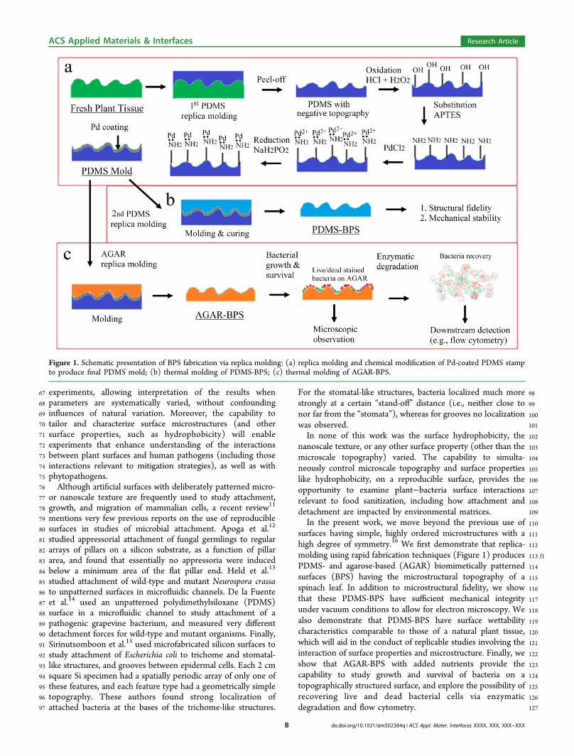
98For the stomatal-like structures, bacteria localized much more99strongly at a certain “stand-off” distance (i.e., neither close to100nor far from the “stomata”), whereas for grooves no localization101was observed.102In none of this work was the surface hydrophobicity, the103nanoscale texture, or any other surface property (other than the104microscale topography) varied. The capability to simulta-105neously control microscale topography and surface properties106like hydrophobicity, on a reproducible surface, provides the107opportunity to examine plant−bacteria surface interactions108relevant to food sanitization, including how attachment and109detachment are impacted by environmental matrices.110In the present work, we move beyond the previous use of111surfaces having simple, highly ordered microstructures with a112high degree of symmetry.16 We first demonstrate that replica-113 f1molding using rapid fabrication techniques (Figure 1) produces114PDMS- and agarose-based (AGAR) biomimetically patterned115surfaces (BPS) having the microstructural topography of a116spinach leaf. In addition to microstructural fidelity, we show117that these PDMS-BPS have sufficient mechanical integrity118under vacuum conditions to allow for electron microscopy. We119also demonstrate that PDMS-BPS have surface wettability120characteristics comparable to those of a natural plant tissue,121which will aid in the conduct of replicable studies involving the122interaction of surface properties and microstructure. Finally, we123show that AGAR-BPS with added nutrients provide the124capability to study growth and survival of bacteria on a125topographically structured surface, and explore the possibility of126recovering live and dead bacterial cells via enzymatic127degradation and flow cytometry.
Figure 1. Schematic presentation of BPS fabrication via replica molding: (a) replica molding and chemical modification of Pd-coated PDMS stampto produce final PDMS mold; (b) thermal molding of PDMS-BPS; (c) thermal molding of AGAR-BPS.
ACS Applied Materials & Interfaces Research Article
dx.doi.org/10.1021/am502384q | ACS Appl. Mater. Interfaces XXXX, XXX, XXX−XXXB

128 ■ RESULTS AND DISCUSSION
129 Microstructural Characterization of PDMS Stamps130 and Biomimetically Patterned Surfaces. After preparation131 of these artificial surfaces, as described in Materials and132 Methods, it was important to establish that they faithfully133 reproduced the surface topography of spinach leaves. A Hirox134 3D microscope was used to perform 3D imaging of these
f2 135 surfaces. Figure 2 shows optical images and color topographic136 renderings of (a) the original spinach leaf, and the137 corresponding areas of (b) the final Pd-coated PDMS stamp138 (hereinafter referred to as the PDMS mold), (c) the PDMS-139 BPS, and (d) the AGAR-BPS. The PDMS mold in Figure 2b140 showed structure and topography mirroring (i.e., opposite to
141that of) the spinach leaf (Figure 2a). Panels c and d in Figure 2142show the PDMS-BPS and AGAR-BPS replicas of the leaf,143respectively, made using the PDMS mold. They are mirror144images of the PDMS mold, and clearly provide true replicas of145the surface topographical features (e.g., valleys between built-up146cellular structures, etc.) of the original spinach leaf (Figure 2a).147These images suggest that surface microstructures of epithelial148cells were faithfully reproduced via replica molding. The root-149mean-square roughness (RRMS) of the surfaces (examples of150which are shown in Figure 2) was measured using the standard151deviation of the z-values of all pixels (1.92 million pixels over an152area of 300 μm by 200 μm). The results are quite similar for all153four surface types, consistent with the hypothesis that, at this
Figure 2. Hirox 3D microscopy images and 3D topographical models show faithful replication of surface topography. (a) Spinach leaf; (b) Pd-coatedPDMS stamp; (c) PDMS-BPS; (d) AGAR-BPS. Red circles and arrow highlight the near-identical plant cell morphology, and stomatal structure,respectively, on the surfaces.
ACS Applied Materials & Interfaces Research Article
dx.doi.org/10.1021/am502384q | ACS Appl. Mater. Interfaces XXXX, XXX, XXX−XXXC
154 resolution, the topographies of the real leaf and of the PDMS155 and AGAR biomimetically patterned surfaces, are very similar.156 Much of the variation of RRMS among the different surfaces can
157be attributed to glare on a glossy surface (e.g., through large158apparent peaks or depressions at the edges of the images); and159environmental humidity, which can also affect leaf plumpness
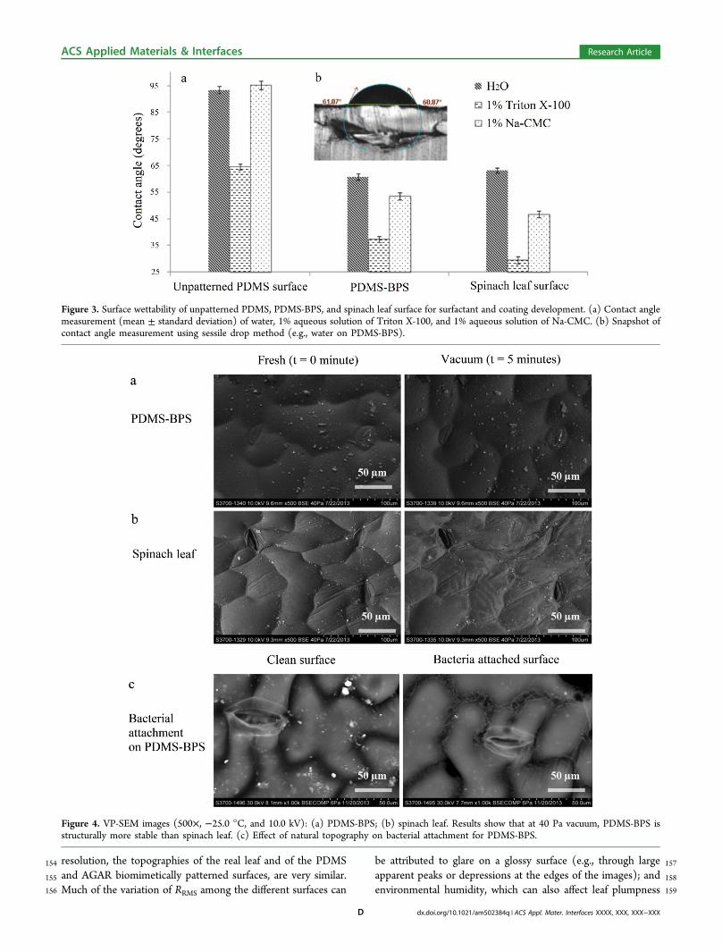
Figure 3. Surface wettability of unpatterned PDMS, PDMS-BPS, and spinach leaf surface for surfactant and coating development. (a) Contact anglemeasurement (mean ± standard deviation) of water, 1% aqueous solution of Triton X-100, and 1% aqueous solution of Na-CMC. (b) Snapshot ofcontact angle measurement using sessile drop method (e.g., water on PDMS-BPS).
Figure 4. VP-SEM images (500×, −25.0 °C, and 10.0 kV): (a) PDMS-BPS; (b) spinach leaf. Results show that at 40 Pa vacuum, PDMS-BPS isstructurally more stable than spinach leaf. (c) Effect of natural topography on bacterial attachment for PDMS-BPS.
ACS Applied Materials & Interfaces Research Article
dx.doi.org/10.1021/am502384q | ACS Appl. Mater. Interfaces XXXX, XXX, XXX−XXXD
160 and the AGAR-BPS surfaces because of either saturation or161 dehydration.162 Wettability of PDMS Biomimetically Patterned Surfa-163 ces. PDMS offers the possibility of altering surface physical164 characteristics while maintaining durable mechanical and165 transparent optical properties, thus providing insight into166 plant−bacteria interaction. We first evaluated the potential of167 PDMS-BPS for in vitro study of plant−bacteria interaction in168 terms of the potential to provide surface wettability comparable169 to the natural plant tissue surface, and structural stability in170 vacuum (VP-SEM).171 The surface wettability of spinach leaf, unpatterned PDMS,172 and PDMS-BPS was evaluated by contact angle measurement173 on static sessile drops using an optical tensiometer. The inset
f3 174 image in Figure 3 shows contact angles of water, aqueous175 solutions of Triton X-100 (a nonionic surfactant), and Na-176 CMC (sodium carboxymethylcellulose, a cellulosic gum used as177 an edible coating material, thickener, and emulsion stabilizer)178 on these solid surfaces. We can identify three possible179 contributors to variance in the contact angle measurements:180 (a) contact angle hysteresis, in which the measured contact181 angle of a static drop might differ according to whether the182 interface has most recently advanced or receded; (b) random or183 systematic experimental error, including error associated with184 measurement precision, electronics, vibration, temperature185 variation, etc.; and (c) for the patterned surfaces, precise186 location of the drop on the surface. The fact that the variance is187 small for each combination of surface and liquid suggests that188 contact angle hysteresis is not very important for any of these
189combinations, and that small variations in drop position on the190patterned surface and leaf are not important for those cases.191The unpatterned PDMS surface was hydrophobic to water192and to the Na-CMC solution, with contact angles near 90°.193Triton X-100 improves the surface wettability of hydrophobic194surfaces by reducing the contact angle to 65°, consistent with195previous work.17 Compared to unpatterned PDMS, biomimeti-196cally patterned PDMS surfaces showed much better wettability197(corresponding to reductions of 32, 42, and 43% for water,198Triton X-100, and Na-CMC, respectively) for drops of all three199liquids, probably due to the Cassie impregnating wetting state200(the “petal” effect, in which liquid wets large but not small201grooves, where adhesive forces between the liquid and solid are202very high),18−20 and the formation of air pockets in the valleys203between asperities. For these three liquids, the considerably204smaller differences between the wettability of PDMS-BPS and205that of spinach leaf (about 5, 12, and 22% for water, Triton X-206100, and NaCMC, respectively) show the importance of207topography, and suggest significant potential for use of208biomimetically patterned surfaces of PDMS in rapid screening209of surfactants and coating materials of interest in applications.210The ability to replicate different leaf microstructures provides211the capability to better understand how topographical features212of real biological materials affect wettability. Since both surface213biochemistry and topography are critical to bacterial attach-214ment, growth, and inactivation, the present approach allows for215a relatively “clean” separation of the effects of surface216microstructure from those of surface chemistry and nanoscale217texturing. Independent control of the chemical composition
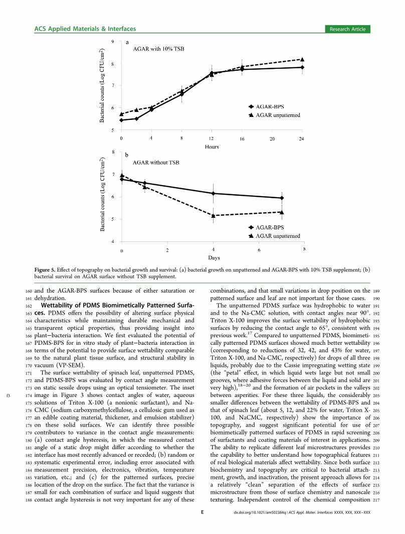
Figure 5. Effect of topography on bacterial growth and survival: (a) bacterial growth on unpatterned and AGAR-BPS with 10% TSB supplement; (b)bacterial survival on AGAR surface without TSB supplement.
ACS Applied Materials & Interfaces Research Article
dx.doi.org/10.1021/am502384q | ACS Appl. Mater. Interfaces XXXX, XXX, XXX−XXXE
218 and properties of the polymeric surface, while maintaining219 identical microstructures in the replica-molding process, will be220 the next step.221 SEM Compatibility of PDMS Biomimetically Patterned222 Surfaces. Because studies of surface topography and structure223 using SEM and other imaging technologies typically require a224 high-vacuum environment, we evaluated the structural stability225 of PDMS-BPS under vacuum conditions in a variable-pressure226 SEM (VP-SEM). The SEM images indicate that PDMS-BPS
f4 227 (Figure 4a) were robust and very durable in the VP-SEM, and228 that all topographic features were preserved during vacuum229 exposure, while the epithelial cells of actual spinach leaf (Figure230 4b) collapsed after 5 min at low (40 Pa) vacuum.231 The SEM compatibility of PDMS-BPS was used to232 investigate bacterial attachment, and to provide insight into233 how natural topography affects bacterial distribution on plant234 surfaces. Figure 4c shows that bacterial cells tend to concentrate235 in valleys. This result suggests that the solution in which236 bacteria are suspended can provide sufficient wetting, despite237 the hydrophobic nature of both the PDMS surface and the238 natural spinach tissues. Therefore, transition to the Cassie239 impregnating wetting state could explain how an aqueous240 bacterial suspension interacts with natural topography on plant241 surfaces.21 The in vacuo stability of the PDMS-BPS strongly242 suggests that such surfaces can be a valuable tool for detailed243 SEM examination of plant surface topography, by significantly244 shortening and simplifying sample preparation. Such studies245 have the potential to provide insight into spatial distributions of246 bacterial cells on surfaces, and into the physical and chemical247 mechanisms that lead to nonuniformity.248 Bacterial Growth, Aggregation, and Survival on AGAR249 Biomimetically Patterned Surfaces. Because of high250 moisture content (∼98% by weight), AGAR-BPS are inferior251 to natural plant tissue in terms of mechanical strength and252 vacuum stability. However, AGAR-BPS have two major253 advantages compared to PDMS and natural leaf surfaces.254 First, when prepared with suitably controlled nutrient mixtures,255 they can be used to study the effect of natural surface256 topography on bacterial growth and survival, and to identify257 interactions between nutrients and topography, using both258 wild-type and mutant bacteria. Second, when coupled to259 downstream detection capability, the enzymatic biodegrad-260 ability of AGAR-BPS can provide unique capabilities to study261 cellular attachment, detachment, and surface effects on growth.
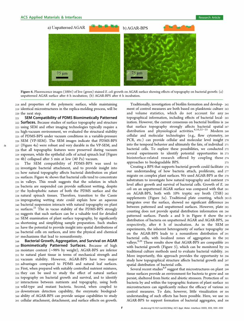
262Traditionally, investigation of biofilm formation and develop-263ment of control measures are both based on planktonic culture264and volume statistics, which do not account for any265topographical information, including effects of bacterial local-266ization. However, the current consensus on bacterial biofilms is267that surface topography strongly affects bacterial spatial268distribution and physiological activities.8,16,22−25 Modern269cellular and molecular technologies (e.g., flow cytometry,270PCR, etc.) can provide cellular and molecular level insight271into the temporal behavior and ultimately the fate, of individual272bacterial cells. To explore these possibilities, we conducted273several experiments to identify potential opportunities in274biointerface-related research offered by coupling these275approaches to biodegradable BPS.276Creating a BPS that supports bacterial growth could facilitate277our understanding of how bacteria attach, proliferate, and278migrate on complex plant surfaces. We used AGAR-BPS as the279substratum to investigate how natural topography and nutrient280level affect growth and survival of bacterial cells. Growth of E.281coli on an unpatterned AGAR surface was compared with that282on AGAR-BPS, both with 10% tryptic soy broth (TSB)283 f5supplements (Figure 5a). Traditional plate counting, which284integrates over the surface, showed no significant difference285between patterned and unpatterned surfaces. However, plate286counting does not provide spatial distribution information on287 f6patterned surfaces. Panels a and b in Figure 6 show the288distribution of bacteria on unpatterned AGAR and AGAR-BPS,289respectively, after 6 h of incubation. As in the SEM290experiments, the inherent heterogeneity of surface topography291on the AGAR-BPS leads to a nonuniform distribution of292bacterial cells, with localized zones of aggregation in the293valleys.8,26 These results show that AGAR-BPS are compatible294with bacterial growth (Figure 5), which can be monitored by295traditional culture methods used to evaluate bacterial viability.296More importantly, this approach provides the opportunity to297study how topographical structure affects bacterial growth and298spatial distribution of bacterial cells.299Several recent studies8,9 suggest that microstructures on plant300tissue surfaces provide an environment for bacteria to grow and301persist, sheltered from biotic and abiotic stressors. Protection of302bacteria by and within the topographic features of plant surface303microstructures can significantly reduce the efficacy of various304control measures. To date, however, little visualization or305understanding of such effects has been possible. Here, we use306AGAR-BPS to support formation of bacterial aggregates, and
Figure 6. Fluorescence images (100×) of live (green) stained E. coli growth on AGAR surface showing effects of topography on bacterial growth: (a)unpatterned AGAR surface after 6 h incubation; (b) AGAR-BPS after 6 h incubation.
ACS Applied Materials & Interfaces Research Article
dx.doi.org/10.1021/am502384q | ACS Appl. Mater. Interfaces XXXX, XXX, XXX−XXXF

307 then determine the effects of surface topography on bacterial308 survival in response to dehydration and biocide treatments.309 Nutrient supplementation of AGAR supports bacterial310 growth, which allows investigation of bacterial survival on311 patterned surfaces. Figure 5b shows that no later than the312 fourth day, bacterial survival on AGAR-BPS significantly313 exceeds survival on an unpatterned AGAR surface. These314 results indicate that the niches on AGAR-BPS (e.g., replicated315 valleys and stomatal structures) can offer significant protection316 to bacterial cells against dehydration. A similar result was317 previously reported for real plant tissue.8
318 Similarly, BPS can also be used to study how the topography319 of plant surfaces influences the efficacy of biocide treatment.
f7 320 On an unpatterned AGAR surface (Figure 7a), a relatively thick
321bacterial growth covered the surface uniformly before biocide322treatment. Treatment with chlorinated water (200 mg/L for 1323min) killed most of the bacterial cells, and no viable cells were324observed in the enlarged images in Figure 7a. However, on325AGAR-BPS (Figure 7b) before biocide treatment, individual326bacterial cells are strongly clustered in the valleys, with larger327aggregates along the ridges. After biocide treatment with328chlorinated water, most bacterial cells were killed. The cell329aggregates were no longer observed and the background was330covered with red fluorescence, likely due to lysis of bacterial331cells during chlorine treatment. Viable cells were still observed332in the valleys; a result that can be attributed to the “steric”333protection afforded to bacterial aggregates against biocide334treatment.8 The enlarged images in Figure 7b also show a few
Figure 7. Effect of topography on E. coli survival and inactivation during a biocide treatment with 200 mg/L free chlorine for 1 min: (a) unpatternedAGAR surface; (b) AGAR-BPS. The surfaces were subjected to Live/Dead bacterial stain before and after biocide treatment. Green and red indicatelive and dead bacteria, respectively.
ACS Applied Materials & Interfaces Research Article
dx.doi.org/10.1021/am502384q | ACS Appl. Mater. Interfaces XXXX, XXX, XXX−XXXG

335 surviving bacterial cells. Besides providing information on336 surface topography and bacterial cell viability, AGAR-BPS can337 also be used for flow cytometry after enzymatic degradation, as
t1 338 shown schematically in Figure 1c. Results shown in Table 1
339 indicate that biocide treatment has significantly different340 outcomes for bacterial inactivation on unpatterned surfaces341 and AGAR-BPS, as expected. The biocide provides approx-342 imately 3.80 vs 2.12 log10 reduction in viable counts on the343 unpatterned and patterned surfaces, respectively. Similar344 differential inactivation of pathogens has also been reported345 with real plant tissues having different surface topographies.8
346 Therefore, bactericidal efficacy on BPS is lower than on347 chemically identical unpatterned surfaces, suggesting that348 topography provides “steric” protection to bacterial cells. The349 results also suggest the potential to combine topographical and350 spatial distribution information using BPS (e.g., SEM and351 fluorescence microscopy) and downstream flow cytometry352 detection.353 The 3D distribution of bacterial cells (viable and nonviable)354 was also investigated by orthographic projection from Z-stack
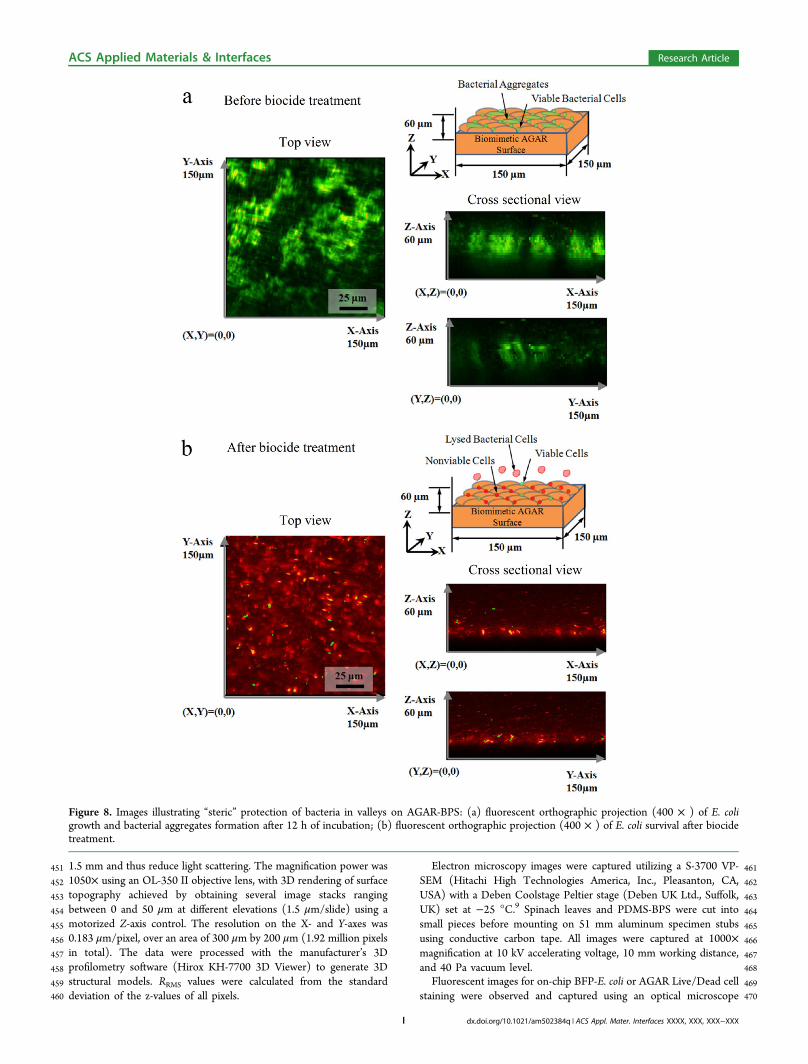
f8 355 measurements using confocal microscopy. Figure 8a shows the356 orthographic projections of viable bacterial aggregates on357 AGAR-BPS before biocide treatment. In the X-Y projection,358 aggregates of viable bacterial cells were observed, consistent359 with Figure 7a. The X-Z and Y-Z projections show nearly360 uniform distributions of bacterial cells in the Z direction. After361 biocide treatment, most of the cells were dead (Figure 8b), and362 bacterial aggregates were no longer observed in the X-Y363 projection. Isolated viable cells were observed in the Z-axis in364 X-Z and Y-Z projections, with some in areas corresponding to365 valleys. This distribution strongly suggests that AGAR-BPS can366 provide information on how topographical features influence367 survival of bacterial cells.
368 ■ MATERIALS AND METHODS369 Materials and Chemicals. All chemicals and buffers were370 purchased from Sigma-Aldrich (St. Louis, MO, USA), except for:371 SU-8 2050 photoresist and developer (MicroChem, Newton, MA,372 USA); Sylgard 184 elastomer kit (PDMS) (Dow Chemical Company,373 Midland, MI, USA); and agarose gel-digesting enzyme GELase374 (Epicenter Biotechnologies, Madison, WI, USA). Bacterial viability375 was determined using a Live/Dead BacLight kit (Invitrogen, Grand376 Island, NY, USA). Fresh spinach leaves (Spinacia oleracea) were377 purchased from a local produce wholesale market (Jessup, Md., USA).378 The E. coli cell-harboring plasmid bearing pRSET/BFP (BFP-E. coli)379 was provided by the Fischell Department of Bioengineering, University380 of Maryland (College Park, MD, USA).
381Replica Molding of PDMS Stamps. For either PDMS or AGAR,382BPS were prepared in two steps. The first step was to produce a383PDMS stamp with reversed microstructure via replica molding. The384second step involved thermal molding of PDMS- and AGAR-BPS385from the PDMS molds (Figure 1). To prepare the PDMS molds,386spinach leaves were securely taped to the bottom of a 100 mm (ID)387aluminum dish. The PDMS mixture (50 g; base:curing agents =10:1)388was cast in the dish, followed by degassing in low vacuum for 15 min389and curing at 40 °C for 12 h. (The low curing temperature avoids390thermal damage to plant tissue.) Cured PDMS stamps were then391chemically modified with a layer of Pd nanoparticles (serving as a392nonadhesive layer in the thermal molding step) as described below393(see Figure 1a).18,20,27 The PDMS stamps were first oxidized for 10394min in an aqueous solution containing 5.2% (v/v) hydrochloric acid395(HCl) and 4.3% (v/v) hydrogen peroxide (H2O2) while subjected to396ultrasonic treatment (35 kHz), followed by rinsing with H2O and397100% ethanol. The PDMS stamps were then treated with ultrasound398for 45 min in an ethanolic solution (50%, v/v) of (3-aminopropyl)399triethoxysilane (APTES) at 22 °C, followed by rinsing in ethanol and400H2O.
18,27 The silylamine-modified PDMS was then shaken overnight401at 120 rpm in 0.2 g/L of PdCl2 in 0.2 N HCl aqueous solution,402followed by 1 h treatment with 2 g/L NaH2PO2 aqueous solution to403form the nonadhesive layer of Pd0 nanoparticles (Figure 1a). The Pd-404coated PDMS molds were reusable, and were stored at 4 °C after each405thermal molding process.406Thermal Molding of PDMS and AGAR Biomimetically407Patterned Surfaces. The PDMS- and AGAR-BPS were prepared408from PDMS molds via thermal molding. The PDMS-BPS were409molded and cured at 125 °C for 20 min following the manufacturer’s410protocol (Figure 1b). Replication of AGAR-BPS was achieved by first411dissolving 2.5% (w/v) agarose (Type I−B) in water, or in 10% TSB412containing 15 g/L tryptone, 5g/L soytone, and 5 g/L NaCl (Figure4131c). Immediately after sterilization, 15 mL of hot liquid medium was414cast on PDMS molds (preheated at room temperature and sterilized415by 100% ethanol), followed by immediate low-vacuum degassing for41610 s, and transfer to (and rapid gelling in) an ice bath for 5 min. After417solidification, the AGAR-BPS was collected and the PDMS mold was418recovered.419Bacterial Culture. The BFP-E. coli bearing pRSET/BFP plasmid420was inoculated into TSB from a frozen stock culture, and incubated for42124 h at 37 °C.28,29 Cells were harvested by centrifugation at 6000 g for42210 min at 4 °C, followed by two pellet rinses with sterile phosphate423buffered saline (PBS). After resuspension and dilution in PBS, each424aliquot was determined to contain approximately 1 × 10⁷ CFU/mL of425bacterial cells.8,30
426The bacterial attachment assays on PDMS- and AGAR-BPS were427accomplished by inoculating 100 μL of the suspension containing428BFP-E. coli over a 1 cm2 area. For PDMS- BPS assays, inocula were429incubated for 12 h at 37 °C, and the surface was gently rinsed with430PBS for 30 s to remove loosely attached bacterial cells before further431characterization. For bacterial growth studies, inoculated AGAR432containing 10% TSB was incubated at 25 °C. Growth was examined433by traditional plate counting as previously described8,30−32 and by434fluorescence microscopy, at 2-h intervals for 24 h. To study bacterial435survival, inoculated AGAR without nutrient supplements was436incubated at 4 °C, and plate counts were recorded at 0, 1, 4, and 7437days. Before each microscopic observation, bacteria on the AGAR438surface were stained at 22 °C for 15 min in the dark with 500 μL of the439Live/Dead bacterial viability stain. Buffer containing 6 μM SYTO 9440stains live cells green and 30 μM propidium iodide stains dead cells441red. After 12 h of incubation, biocide (200 mg/L chlorinated water)442was applied to the surface for 1 min. The bactericidal effect was also443examined using the bacterial viability staining kit.444Microscopy. Analyses of surface topography, roughness, and 3D445imaging of spinach leaf, PDMS molds, PDMS-BPS, and AGAR-BPS446were performed using a 3D digital optical microscope (Hirox KH-4477700, Hackensack, NJ, USA). Specimen preparation of PDMS molds448and PDMS-BPS was as previously described in the thermal molding449section. AGAR-BPS were prepared by molding the sample between450the PDMS mold and a glass slide, in order to limit the gel thickness to
Table 1. Effects of Surface Topography on Bacterial SurvivalDetermined by Flow Cytometry
cell counts (log cell/cm2)a
unpatterned AGAR surface AGAR-BPS
beforebiocidetreatment
after biocidetreatment
beforebiocidetreatment
after biocidetreatment
viablecounts
7.92 ± 1.01 4.42 ± 0.91 8.52 ± 1.25 6.11 ± 0.75
dead counts 5.36 ± 1.12 7.60 ± 0.88 6.40 ± 0.63 8.31 ± 1.21reduction ofviablecounts
3.80 ± 0.85 2.12 ± 0.67
aLog cell counts are shown as mean ± standard error.
ACS Applied Materials & Interfaces Research Article
dx.doi.org/10.1021/am502384q | ACS Appl. Mater. Interfaces XXXX, XXX, XXX−XXXH
451 1.5 mm and thus reduce light scattering. The magnification power was452 1050× using an OL-350 II objective lens, with 3D rendering of surface453 topography achieved by obtaining several image stacks ranging454 between 0 and 50 μm at different elevations (1.5 μm/slide) using a455 motorized Z-axis control. The resolution on the X- and Y-axes was456 0.183 μm/pixel, over an area of 300 μm by 200 μm (1.92 million pixels457 in total). The data were processed with the manufacturer’s 3D458 profilometry software (Hirox KH-7700 3D Viewer) to generate 3D459 structural models. RRMS values were calculated from the standard460 deviation of the z-values of all pixels.
461Electron microscopy images were captured utilizing a S-3700 VP-462SEM (Hitachi High Technologies America, Inc., Pleasanton, CA,463USA) with a Deben Coolstage Peltier stage (Deben UK Ltd., Suffolk,464UK) set at −25 °C.9 Spinach leaves and PDMS-BPS were cut into465small pieces before mounting on 51 mm aluminum specimen stubs466using conductive carbon tape. All images were captured at 1000×467magnification at 10 kV accelerating voltage, 10 mm working distance,468and 40 Pa vacuum level.469Fluorescent images for on-chip BFP-E. coli or AGAR Live/Dead cell470staining were observed and captured using an optical microscope
Figure 8. Images illustrating “steric” protection of bacteria in valleys on AGAR-BPS: (a) fluorescent orthographic projection (400 × ) of E. coligrowth and bacterial aggregates formation after 12 h of incubation; (b) fluorescent orthographic projection (400 × ) of E. coli survival after biocidetreatment.
ACS Applied Materials & Interfaces Research Article
dx.doi.org/10.1021/am502384q | ACS Appl. Mater. Interfaces XXXX, XXX, XXX−XXXI

471 (Nikon E400, Nikon Instruments, Melville, NY, USA) with a472 fluorescent illuminator (Intensilight C-HGFI, Nikon) and fluorescent473 filter cubes of BFP, FITC (SYTO 9), and TRITC (for propidium474 iodide). A Cool Snap HQ camera (Photometrics, Tucson, AZ, USA)475 and NIS Elements software (version 3.0, Nikon) were used to visualize476 fluorescent signals. Nikon Plan 10×/0.25 and Nikon Plan 40×/0.65477 objective lenses, and a 10× ocular lens were used in the study.6
478 Confocal scanning microscopy (Zeiss LSM-700, Jena, Germany)479 was used to study the role of surface topography on bacterial growth480 and survival.9 The Z-stack function was used to scan a 150 μm × 150481 μm area at Z-axis resolution of 2 μm/slice. The excitation laser had a482 wavelength of 488 nm, and a FITC filter was used to capture the483 SYTO 9 signal (green fluorescence), while a PI filter was used for484 propidium iodide stained cells (red fluorescence). An EC Plan-485 Neofluar 40×/0.9 was used as the objective lens. Z-stack images were486 analyzed and constructed using Zen software (2012, Zeiss).487 Contact Angle Measurement. Contact angle measurements488 were made by the sessile drop method using an Attension Theta489 optical tensiometer system (Biolin Scientific, Linthicum Heights, MD,490 USA), with a drop volume of 50 μL (5 drops on a 1 cm × 10 cm491 strip). Placement of drops of either water, or an aqueous solution of492 Triton X-100 or Na-CMC, on each surface was controlled by an493 automatic liquid dispenser (C201, Biolin Scientific). Data recording494 (60 fps) was triggered by the initial contact of a liquid drop with a495 solid surface (i.e., an unpatterned PDMS surface, PDMS-BPS, or496 spinach leaf). We selected leaf portions with relatively low gross497 curvature, which were quite flat on the scale of the relatively large498 drops (equivalent spherical diameter of ∼4.6 mm, and equivalent499 hemispherical diameter of ∼5.8 mm). For each surface type, 20500 specimens were used. All measurements were performed with sessile501 “advanced” drops (i.e., static drops for which the contact surface had502 most recently advanced), under ambient conditions. Captured images503 were analyzed automatically (OneAttension software, Version 1.8,504 Biolin Scientific, Linthicum Heights, MD, USA) to identify the505 baseline and calculate contact angles.506 Flow Cytometry. Flow downstream of the AGAR-BPS (Figure 1c)507 was quantified with a flow cytometer to demonstrate the possibility of508 counting cells and monitoring their viability.33 After microscopic509 examination, AGAR-BPS (1 cm2) were enzymatically degraded in 10510 mL of GELase solution (20 units/mL) at 45 °C for 40 min. Aliquots511 of 1 mL of the resulting suspension containing live and dead bacterial512 cells were then analyzed by flow cytometry (FACS Canto II, BD513 Biosciences, San Jose, CA, USA) for fluorescence expression and514 viability counts. The negative control was prepared using a bacterial515 suspension of cells grown on an unpatterned AGAR surface, and516 sterilizing the suspension with 200 mg/L chlorinated water for 1 min.517 Flow cytometry data were analyzed by FACS Canto clinical software518 (BD Biosciences, Sparks, MD) to calculate means and standard errors519 of cell counts and survival rates.520 Statistical Analysis. Surface roughness, contact angle, and flow521 cytometry experiments were conducted with five replications, and the522 data were reported as mean ± standard error. Analysis of variance523 (ANOVA) was performed using SAS software (Version 9.2, SAS524 Institute Inc., Cary, NC). Surface roughness of PDMS molds and BPS525 were tested against that of spinach leaf by ANOVA Dunnett’s test. The526 contact angles of water, and of the aqueous solutions of Triton X-100527 and Na-CMC, on unpatterned PDMS, PDMS-BPS, and spinach leaves528 were ranked using ANOVA Tukey’s test. The probability (P) of all test529 statistics was set at 0.05.
530 ■ COMMENTS AND CONCLUSIONS531 In this study, a two-step replica molding method was developed532 for rapid fabrication of polymer-based biomimetically patterned533 surfaces (BPS) having the surface microstructure of plant tissue.534 Surfaces of PDMS- and AGAR-BPS replicating spinach leaf535 microstructure demonstrate a high degree of topographical536 fidelity to the original plant tissue. PDMS surfaces provide537 structural stability under vacuum for SEM-associated applica-538 tions, and have surface wettability similar to natural leaf
539surfaces, which will facilitate development of coating and540biocide-related intervention technologies. The possibility of541chemically functionalizing PDMS27 allows for potential542tailoring of chemical properties important to bacterial attach-543ment, with independent control of microstructural topography.544For AGAR surfaces, simple adjustment of nutrient levels545facilitates investigation of how natural topography affects546bacterial growth and survival. Exploratory experiments show547that high-fidelity topography, structural stability, and the548capability to integrate with instrumentation for studying549bacterial growth and survival on PDMS- and AGAR-BPS,550provide potentially valuable tools for plant−bacteria interaction551studies, including those relevant to food safety. AGAR-BPS can552also be enzymatically degraded to recover bacterial cells for553subsequent studies using flow cytometry and other microbial554detection and enumeration technologies. For AGAR-based555BPS, the ease with which composition is modifiable provides556the opportunity to independently study the effects of surface557chemistry, microstructure, and nutrients on bacterial attach-558ment, growth, and survival for a wide variety of bacteria.559Surfaces with simple, spatially periodic microstructures in560which each feature has a high degree of symmetry can be561extremely useful for understanding certain basic mechanisms of562attachment. However, their use as testbeds for studying the563details of surface interactions (including attachment and564detachment) for specific bacteria/plant pairs, and in the565evaluation and optimization of sanitization techniques and566other postharvest treatments, is severely limited by the lack of567geometric complexity. Real plant surfaces are highly complex at568the micro- and nanoscale, and so the value of an approach that569provides for reproducible studies of geometrically complex570surfaces is evident. A key advantage of the present approach is571that it avoids two pitfalls of conventional microfabrication572techniques for producing geometrically complex surfaces. First,573existing approaches require laborious clean-room fabrication574processes involving multiple high-vacuum (e.g., ion sputter575coating) and high-temperature (e.g., nickel stamp electroplating576at 55−70 °C) treatments.34−36 Second, those methods577reproduce plant surface structures on a self-cleaning super-578hydrophobic surface, which is generally incompatible with579cellular attachment.34,36,37 An additional advantage of the580present approach is that it allows for a relatively “clean”581separation of the effects of surface microstructure from those of582surface chemistry and nanoscale texturing, since one can583independently control the chemical composition and properties584of the polymeric surface, while maintaining identical micro-585structures in the replica molding process.586Initial applications tested in this study demonstrate the587robustness of biomimetically patterned surfaces and their588potential application to other areas of plant or animal tissue-589microbe interface research. Because surface biochemistry is also590critical to bacterial attachment, growth, and inactivation,591establishing biochemical similarity would be the next step.592Systematic evaluation of the interaction between living bacterial593cells and surfaces is essential to development of possible594interventions directed at reducing or eliminating attachment595and microbial survival. Although spinach leaves were chosen as596the plant surface in this study, the method developed has great597potential for replicating the surfaces of other plant and animal598tissues. We anticipate that this approach will provide an599important research tool for understanding surface−bacteria600interactions and facilitating development of technology to
ACS Applied Materials & Interfaces Research Article
dx.doi.org/10.1021/am502384q | ACS Appl. Mater. Interfaces XXXX, XXX, XXX−XXXJ
601 enhance inactivation of foodborne human pathogens and602 improve public health.
603 ■ AUTHOR INFORMATION
604 Corresponding Author605 *E-mail: [email protected]. Tel: 301.504.6186. Fax:606 301.504.5107.
607 Notes608 The authors declare no competing financial interest.
609 ■ ACKNOWLEDGMENTS
610 This work was supported by USDA-NIFA Specialty Crop611 Research Initiative Grant Award 2010-01165. We also acknowl-612 edge grant HDTRA1-13-0037 and BO085PO008 from the U.S.613 Defense Threat Reduction Agency. We are grateful for the614 support of the FabLab at the Maryland NanoCenter. We thank615 Dr. William Bentley and Ms. Jessica Terrell of the Fischell616 Department of Bioengineering at the University of Maryland,617 College Park, for providing BFP-E. coli strains and for generous618 technical support. Access to the FACS equipment at the Flow619 Cytometry Core Facility (Maryland Pathogen Research620 Institute, University of Maryland) is gratefully acknowledged.
621 ■ REFERENCES(1)622 CDC, CDC Estimates of Foodborne Illness in the United States
623 2011, CS218786-A.(2)624 Lynch, M. F.; Tauxe, R. V.; Hedberg, C. W. The Growing Burden
625 of Foodborne Outbreaks due to Contaminated Fresh Produce: Risks626 and Opportunities. Epidemiol. Infect. 2009, 137, 307−315.
(3)627 Erickson, M. C. Internalization of Fresh Produce by Food borne628 Pathogens. Annu. Rev. Food Sci. Technol. 2012, 3, 283−310.
(4)629 Erickson, M. C.; Webb, C. C.; Diaz-Perez, J. C.; Davey, L. E.;630 Payton, A. S.; Flitcroft, I. D.; Phatak, S. C.; Doyle, M. P. Internalization631 of Escherichia coli O157:H7 Following Spraying of Cut Shoots when632 Leafy Greens are Regrown for a Second Crop. J. Food Prot. 2013, 76,633 2052−2056.
(5)634 Erickson, M. C.; Webb, C. C.; Diaz-Perez, J. C.; Davey, L. E.;635 Payton, A. S.; Flitcroft, I. D.; Phatak, S. C.; Doyle, M. P. Absence of636 Internalization of Escherichia coli O157:H7 into Germinating Tissue of637 Field-Grown Leafy Greens. J. Food Prot. 2014, 77, 189−196.
(6)638 Sharma, M.; Ingram, D. T.; Patel, J. R.; Millner, P. D.; Wang, X.;639 Hull, A. E.; Donnenberg, M. S. A Novel Approach to Investigate the640 Uptake and Internalization of Escherichia coli O157:H7 in Spinach641 Cultivated in Soil and Hydroponic Medium. J. Food Prot. 2009, 72,642 1513−1520.
(7)643 Whitehead, K. A.; Verran, J. The Effect of Surface Topography on644 the Retention of Microorganisms. Food Bioprod. Process. 2006, 84,645 253−259.
(8)646 Wang, H.; Feng, H.; Liang, W.; Luo, Y.; Malyarchuk, V. Effect of647 Surface Roughness on Retention and Removal of Escherichia coli648 O157:H7 on Surfaces of Selected Fruits. J. Food Sci. 2009, 74, E8−649 E15.
(9)650 Macarisin, D.; Patel, J.; Bauchan, G.; Giron, J. A.; Ravishankar, S.651 Effect of Spinach Cultivar and Bacterial Adherence Factors on Survival652 of Escherichia coli O157:H7 on Spinach Leaves. J. Food Prot. 2013, 76,653 1829−1837.
(10)654 Seo, K. H.; Frank, J. F. Attachment of Escherichia coli O157:H7655 to Lettuce Leaf Surface and Bacterial Viability in Response to Chlorine656 Treatment as Demonstrated by Using Confocal Scanning Laser657 Microscopy. J. Food Prot. 1999, 62, 3−9.
(11)658 Warning, A.; Datta, A. K. Interdisciplinary Engineering659 Approaches to Study How Pathogenic Bacteria Interact with Fresh660 Produce. J. Food Eng. 2013, 114, 426−448.
(12)661 Apoga, D.; Barnard, J.; Craighead, H. G.; Hoch, H. C.662 Quantification of Substratum Contact Required for Initiation of
663Colletotrichum graminicola Appressoria. Fungal Genet. Biol. 2004, 41,6641−12.
(13) 665Held, M.; Edwards, C.; Nicolau, D. V. Probing the Growth666Dynamics of Neurospora crassa with Microfluidic Structures. Fungal667Biol. 2011, 115, 493−505.
(14) 668De la Fuente, L.; Montanes, E.; Meng, Y. Z.; Li, Y. X.; Burr, T.669J.; Hoch, H. C.; Wu, M. M. Assessing Adhesion Forces of Type I and670Type IV Pili of Xylella fastidiosa Bacteria by Use of a Microfluidic Flow671Chamber. Appl. Environ. Microbiol. 2007, 73, 2690−2696.
(15) 672Sirinutsomboon, B.; Delwiche, M. J.; Young, G. M. Attachment673of Escherichia coli on Plant Surface Structures Built by Micro-674fabrication. Biosyst. Eng. 2011, 108, 244−252.
(16) 675Friedlander, R. S.; Vlamakis, H.; Kim, P.; Khan, M.; Kolter, R.;676Aizenberg, J. Bacterial Flagella Explore Microscale Hummocks and677Hollows to Increase Adhesion. Proc. Natl. Acad. Sci. U.S.A. 2013, 110,6785624−5629.
(17) 679Seo, J.; Lee, L. P. Effects on Wettability by Surfactant680Accumulation/Depletion in Bulk Polydimethylsiloxane (PDMS).681Sens. Actuators, B 2006, 119, 192−198.
(18) 682Zhang, B.; Feldman, A.; Wang, Q. A Novel Insight in Rapid683Allergen Detection in Food Systems: from Threshold Dose to Real-684World Concentration. Sens Actuators, B 2013, 186, 597−602.
(19) 685Anselme, K.; Davidson, P.; Popa, A. M.; Giazzon, M.; Liley, M.;686Ploux, L. The Interaction of Cells and Bacteria with Surfaces687Structured at the Nanometre Scale. Acta Biomater. 2010, 6, 3824−6883846.
(20) 689McDonald, J. C.; Duffy, D. C.; Anderson, J. R.; Chiu, D. T.; Wu,690H. K.; Schueller, O. J. A.; Whitesides, G. M. Fabrication of Microfluidic691Systems in Poly(dimethylsiloxane). Electrophoresis 2000, 21, 27−40.
(21) 692Feng, L.; Zhang, Y. A.; Xi, J. M.; Zhu, Y.; Wang, N.; Xia, F.;693Jiang, L. Petal Effect: a Superhydrophobic State with High Adhesive694Force. Langmuir 2008, 24, 4114−4119.
(22) 695Hall-Stoodley, L.; Costerton, J. W.; Stoodley, P. Bacterial696Biofilms: from the Natural Environment to Infectious Diseases. Nat.697Rev. Microbiol. 2004, 2, 95−108.
(23) 698Debeer, D.; Stoodley, P.; Roe, F.; Lewandowski, Z. Effects of699Biofilm Structures on Oxygen Distribution and Mass-Transport.700Biotechnol. Bioeng. 1994, 43, 1131−1138.
(24) 701Huang, C. T.; Xu, K. D.; McFeters, G. A.; Stewart, P. S. Spatial702Patterns of Alkaline Phosphatase Expression within Bacterial Colonies703and Biofilms in Response to Phosphate Starvation. Appl. Environ.704Microbiol. 1998, 64, 1526−1531.
(25) 705Rizzello, L.; Sorce, B.; Sabella, S.; Vecchio, G.; Galeone, A.;706Brunetti, V.; Cingolani, R.; Pompa, P. P. Impact of Nanoscale707Topography on Genomics and Proteomics of Adherent Bacteria. ACS708Nano 2011, 5, 1865−1876.
(26) 709Lawrence, J. R.; Korber, D. R.; Hoyle, B. D.; Costerton, J. W.;710Caldwell, D. E. Optical Sectioning of Microbial Biofilms. J. Bacteriol.7111991, 173, 6558−6567.
(27) 712Yu, L.; Li, C.; Liu, Y. S.; Gao, J.; Wang, W.; Gan, Y. Flow-713through Functionalized PDMS Microfluidic Channels with Dextran714Derivative for ELISAs. Lab Chip 2009, 9, 1243−1247.
(28) 715Tsao, C. Y.; Hooshangi, S.; Wu, H. C.; Valdes, J. J.; Bentley, W.716E. Autonomous Induction of Recombinant Proteins by Minimally717Rewiring Native Quorum Sensing Regulon of E. coli.Metab. Eng. 2010,71812, 291−297.
(29) 719Cheng, Y.; Luo, X. L.; Tsao, C. Y.; Wu, H. C.; Betz, J.; Payne, G.720F.; Bentley, W. E.; Rubloff, G. W. Biocompatible Multi-Address 3D721Cell Assembly in Microfluidic Devices using Spatially Programmable722Gel Formation. Lab Chip 2011, 11, 2316−2318.
(30) 723Shen, C.; Luo, Y.; Nou, X.; Bauchan, G.; Zhou, B.; Wang, Q.;724Millner, P. Enhanced Inactivation of Salmonella and Pseudomonas725Biofilms on Stainless Steel by Use of T-128, a Fresh-Produce Washing726Aid, in Chlorinated Wash Solutions. Appl. Environ. Microb. 2012, 78,7276789−6798.
(31) 728Zhang, B.; Luo, Y.; Wang, Q. Development of Silver-Zein729Composites as a Promising Antimicrobial Agent. Biomacromolecules7302010, 11, 2366−2375.
ACS Applied Materials & Interfaces Research Article
dx.doi.org/10.1021/am502384q | ACS Appl. Mater. Interfaces XXXX, XXX, XXX−XXXK
(32)731 Zhang, B.; Luo, Y.; Wang, Q. Development of Silver/Alpha-732 Lactalbumin Nanocomposites: A New Approach to Reduce Silver733 Toxicity. Int. J. Antimicrob. Agents 2011, 38, 502−509.
(33)734 Gupta, A.; Terrell, J. L.; Fernandes, R.; Dowling, M. B.; Payne,735 G. F.; Raghavan, S. R.; Bentley, W. E. Encapsulated Fusion Protein736 Confers ″Sense and Respond″ Activity to Chitosan-Alginate Capsules737 to Manipulate Bacterial Quorum Sensing. Biotechnol. Bioeng. 2013,738 110, 552−562.
(34)739 Lee, S. M.; Kwon, T. H. Mass-producible Replication of Highly740 Hydrophobic Surfaces from Plant Leaves. Nanotechnology 2006, 17,741 3189−3196.
(35)742 Lee, S. M.; Upping, J.; Bielawny, A.; Knez, M. Structure-Based743 Color of Natural Petals Discriminated by Polymer Replication. ACS744 Appl. Mater. Interfaces 2011, 3, 30−34.
(36)745 Liu, B.; He, Y. N.; Fan, Y.; Wang, X. G. Fabricating Super-746 hydrophobic Lotus-leaf-like Surfaces through Soft-lithographic Im-747 printing. Macromol. Rapid Commun. 2006, 27, 1859−1864.
(37)748 Lampin, M.; Warocquier-Clerout, R.; Legris, C.; Degrange, M.;749 Sigot-Luizard, M. F. Correlation between Substratum Roughness and750 Wettability, Cell Adhesion, and Cell Migration. J. Biomed. Mater. Res.751 1997, 36, 99−108.
ACS Applied Materials & Interfaces Research Article
dx.doi.org/10.1021/am502384q | ACS Appl. Mater. Interfaces XXXX, XXX, XXX−XXXL


![REVIEW Open Access Biomimetic patterned surfaces for ...hibits high friction/wear due to its soft nature [7]. In the past, various chemical modifications of surfaces such as boundary](https://static.fdocuments.in/doc/165x107/5faf0bacf29365493373f346/review-open-access-biomimetic-patterned-surfaces-for-hibits-high-frictionwear.jpg)