MaxGel Hydrogel Dressing MaxGel Dressing Novel and Unique Sterile Hydrogel Product Introduction.
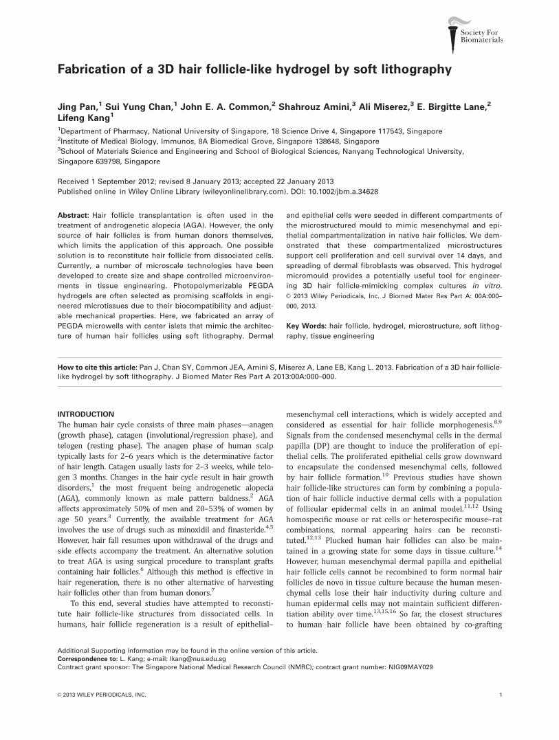
Fabrication of a 3D hair follicle-like hydrogel by soft lithography
Jing Pan,1 Sui Yung Chan,1 John E. A. Common,2 Shahrouz Amini,3 Ali Miserez,3 E. Birgitte Lane,2
Lifeng Kang1
1Department of Pharmacy, National University of Singapore, 18 Science Drive 4, Singapore 117543, Singapore2Institute of Medical Biology, Immunos, 8A Biomedical Grove, Singapore 138648, Singapore3School of Materials Science and Engineering and School of Biological Sciences, Nanyang Technological University,
Singapore 639798, Singapore
Received 1 September 2012; revised 8 January 2013; accepted 22 January 2013
Published online in Wiley Online Library (wileyonlinelibrary.com). DOI: 10.1002/jbm.a.34628
Abstract: Hair follicle transplantation is often used in the
treatment of androgenetic alopecia (AGA). However, the only
source of hair follicles is from human donors themselves,
which limits the application of this approach. One possible
solution is to reconstitute hair follicle from dissociated cells.
Currently, a number of microscale technologies have been
developed to create size and shape controlled microenviron-
ments in tissue engineering. Photopolymerizable PEGDA
hydrogels are often selected as promising scaffolds in engi-
neered microtissues due to their biocompatibility and adjust-
able mechanical properties. Here, we fabricated an array of
PEGDA microwells with center islets that mimic the architec-
ture of human hair follicles using soft lithography. Dermal
and epithelial cells were seeded in different compartments of
the microstructured mould to mimic mesenchymal and epi-
thelial compartmentalization in native hair follicles. We dem-
onstrated that these compartmentalized microstructures
support cell proliferation and cell survival over 14 days, and
spreading of dermal fibroblasts was observed. This hydrogel
micromould provides a potentially useful tool for engineer-
ing 3D hair follicle-mimicking complex cultures in vitro.
VC 2013 Wiley Periodicals, Inc. J Biomed Mater Res Part A: 00A:000–
000, 2013.
Key Words: hair follicle, hydrogel, microstructure, soft lithog-
raphy, tissue engineering
How to cite this article: Pan J, Chan SY, Common JEA, Amini S, Miserez A, Lane EB, Kang L. 2013. Fabrication of a 3D hair follicle-like hydrogel by soft lithography. J Biomed Mater Res Part A 2013:00A:000–000.
INTRODUCTION
The human hair cycle consists of three main phases—anagen(growth phase), catagen (involutional/regression phase), andtelogen (resting phase). The anagen phase of human scalptypically lasts for 2–6 years which is the determinative factorof hair length. Catagen usually lasts for 2–3 weeks, while telo-gen 3 months. Changes in the hair cycle result in hair growthdisorders,1 the most frequent being androgenetic alopecia(AGA), commonly known as male pattern baldness.2 AGAaffects approximately 50% of men and 20–53% of women byage 50 years.3 Currently, the available treatment for AGAinvolves the use of drugs such as minoxidil and finasteride.4,5
However, hair fall resumes upon withdrawal of the drugs andside effects accompany the treatment. An alternative solutionto treat AGA is using surgical procedure to transplant graftscontaining hair follicles.6 Although this method is effective inhair regeneration, there is no other alternative of harvestinghair follicles other than from human donors.7
To this end, several studies have attempted to reconsti-tute hair follicle-like structures from dissociated cells. Inhumans, hair follicle regeneration is a result of epithelial–
mesenchymal cell interactions, which is widely accepted andconsidered as essential for hair follicle morphogenesis.8,9
Signals from the condensed mesenchymal cells in the dermalpapilla (DP) are thought to induce the proliferation of epi-thelial cells. The proliferated epithelial cells grow downwardto encapsulate the condensed mesenchymal cells, followedby hair follicle formation.10 Previous studies have shownhair follicle-like structures can form by combining a popula-tion of hair follicle inductive dermal cells with a populationof follicular epidermal cells in an animal model.11,12 Usinghomospecific mouse or rat cells or heterospecific mouse–ratcombinations, normal appearing hairs can be reconsti-tuted.12,13 Plucked human hair follicles can also be main-tained in a growing state for some days in tissue culture.14
However, human mesenchymal dermal papilla and epithelialhair follicle cells cannot be recombined to form normal hairfollicles de novo in tissue culture because the human mesen-chymal cells lose their hair inductivity during culture andhuman epidermal cells may not maintain sufficient differen-tiation ability over time.13,15,16 So far, the closest structuresto human hair follicle have been obtained by co-grafting
Additional Supporting Information may be found in the online version of this article.
Correspondence to: L. Kang; e-mail: [email protected]
Contract grant sponsor: The Singapore National Medical Research Council (NMRC); contract grant number: NIG09MAY029
VC 2013 WILEY PERIODICALS, INC. 1
foreskin-derived human keratinocytes and murine DP cellsonto nude mice 13. However, hair follicles formed in xeno-grafts of human and murine cell components are not suita-ble for hair transplantation due to the immune rejection.17,18
The failure to form better differentiated and organized fol-licles is believed to be due to the lack of communicationbetween the mesenchymal and epithelial cells.3,19
With the increasing progress in microscale technologies,new approaches have been developed to investigate cellbehaviors in microenvironments for cellular biology and tis-sue engineering applications.20 Microscale technologies suchas soft lithography, photolithography, flow lithography, andbioprinting have enabled the construction of diversesynthetic microstructures to incorporate cells.21–26 Micro-structures are expected to provide cells with a suitablemicroenvironment, sufficient nutrient transport, and me-chanical integrity.20,27,28 In particular, three dimensional(3D) microstructures can be readily made using photocros-slinkable polymers with adjustable mechanical properties,microarchitecture, and alterable chemical compositions.23,29
Soft lithography, which employs elastomeric stamps fabri-cated from patterned silicon wafers to print or mold materi-als, is commonly used in 3D microstructure fabrication.30
In this study, microstructured scaffolds were fabricatedin which hair follicle inductive dermal cells can be posi-tioned and grown close to, but separated from epidermalcell populations by soft lithography. The mould resemblesthe physiological architecture of hair follicle [Fig. 1(A)]. Pol-y(dimethylsiloxane) (PDMS) stamps were fabricated frompatterned silicon masters and then the stamps wereemployed to mold poly(ethylene glycol) diacrylate (PEGDA)microwells with center islets on a glass slide by UV cross-linking [Fig. 1(B)]. Epithelial cells and dermal cells were im-mobilized in different locations of the microstructure for tis-sue culturing. Such a scaffold can serve as a potentialplatform for hair follicle regeneration in vitro.
EXPERIMENTAL
Master fabricationPhotomasks were designed using AutoCAD 2010 andprinted on chromium coated soda lime glasses at InfinateGraphics PTE LTD (Singapore). Silicon wafers were spin-coated with the epoxy negative photoresist SU-8 2050(MicroChem Corp., Newton, MA) at 2200 rpm, yielding thedesired film thickness of 50 lm. Wafers were soft-baked at65�C for 1 min, followed by a second baking at 95�C for10 min. For crosslinking of the photoresist, the coatedwafers were exposed to UV light of 350–400 nm for 65 sthrough a photomask on a single-side mask aligner (SVC,Model H94-25). Subsequently, the wafers were post-expo-sure baked at 65�C for 1 min and then at 95�C for 6 min.The photoresist-patterned silicon masters were developedusing SU-8 developer, rinsed with isopropyl alcohol for 10 s,and air dried with pressurized nitrogen. The pattern of pro-truding rods was analyzed using an optical microscopeequipped on the aligner. For secondary spin-coating, pat-terned silicon masters were spin-coated with negative pho-toresist SU-8 2075 (MicroChem Corp., Newton, MA) at
1000 rpm, yielding the desired film thickness about 200lm. Wafers were soft-baked at 65�C for 7 min, followed bya second soft-baking at 95�C for 60 min. After aligning thepatterned master with the second photomask by the crosseson each of them, the coated wafers were exposed to UVlight of 350–400 nm for 90 s through the second photo-mask by using the aligner. Subsequently, the wafers werepost-exposure baked at 65�C for 6 min and then at 95�C for15 min. The photoresist-patterned silicon masters weredeveloped using SU-8 developer, rinsed with isopropyl alco-hol for 10 s, and air dried with pressurized nitrogen. Fourdifferent dimensions of microwells were obtained with cen-ter islets in accordance with the design of photomasks (50lm with 16 lm islet, 100 lm with 33 lm islet, 200 lmwith 66 lm islet, and 400 lm with 133 lm islet).
PDMS-stamp fabricationPoly(dimethylsiloxane) (PDMS) stamps were fabricated bycuring a 10:1 mixture of silicone elastomer base solutionand curing agent Sylgard 184 (Dow Corning Corporation,Midland, USA) on a patterned silicon master. The PDMSelastomer solution was degassed for 20–30 min in a vac-uum chamber and cured at 70�C for 2–4 h before the PDMSstamps were peeled from the silicon masters. The generatedPDMS replicas had patterns corresponding to the siliconmaster with protruding columns and were subsequentlyused for molding of PEGDA microwells.
To identify outlines of PDMS stamps, a slice of the PDMSstamp was cut using a blade and treated the surface byusing oxygen plasma for 3 min (Harrick Scientific, USA).Then, the slice was immersed in 5 lg/ml Rhodamine B(Alfa Aesar, Lancaster, UK) and observed under a fluorescentmicroscope (Nikon Ti, Japan, ex: 545–565 nm).
Microwell fabricationMicrowell arrays were fabricated using UV-photocrosslink-able PEGDA (Aldrich Chemistry, USA and Jenkem Technol-ogy, USA) of different average molecular weights (MWs;575, 700, and 3500 Da) mixed in a 0.2% (w/v) ratio of thephotoinitiator 2-hydroxy-4’-(2-hydroxy-ethoxy)-2-methylpro-piophenone (Irgacure 2959, Aldrich Chemistry, USA) on a 3-(trimethoxysilyl) propyl methacrylate (TMS-PMA, Sigma,USA) treated glass slide. A patterned PDMS stamp wasplaced on an evenly distributed film of precursor solutionon a glass slide. To optimize the conditions for PEGDAhydrogel photopolymerization, we determined the minimumduration of UV exposure required for the formation ofdesigned microwell arrays at various UV intensities. Afterpolymerization, the PDMS stamp was peeled from the sub-strate. All photopolymerizations were performed using theOmniCureV
R
Series 2000 curing station (320–500 nm; LumenDynamics, Canada).
Microwell stabilityTo find stable microwell arrays molded on TMSPMA-treatedglass slides, prepolymer solutions of various PEGDA concen-trations (10, 20, 40, and 80% w/v) were used to fabricatemicrowell arrays. The stability of microwells on the glass
2 PAN ET AL. FABRICATION OF A 3D HAIR FOLLICLE-LIKE HYDROGEL

slides was assessed by immersing microarrays in phos-phate-buffered saline (PBS, Vivantis, KL, Malaysia) in 37�C,5% CO2 humidified incubator and analyzing the integrity ofthe arrays over time. In all cases, dilutions were made in1� PBS. Experiments were conducted in triplicates.
Mechanical testingPolymerization was performed as described for microwellfabrication. Samples were incubated in PBS at 37�C, 5%CO2 humidified incubator for 24 h to make gels swell toreach equilibrium.31 Young’s modulus of PEGDA hydrogels
were obtained by probing flat surfaces by nanoindentation,using a Triboindenter (Hysitron, Minneapolis, MN). Wechose a spherical indenter tip (R � 50 lm) for nanoinden-tation studies, using a peak load of 25 lN, a loading/unloading rate of 5 lN/s, and a holding time at peak loadof 2 s. The Young’s modulus was determined as the slopeof the linear region upon unloading. Sixteen indentationcurves were performed within a 600 lm � 600 lm areaat a lateral separation of 150 lm. During the nanoindenta-tion test, the hydrogels were kept in PBS solution to avoiddehydration.
FIGURE 1. Schematic representation of hair follicle-like mould fabrication. A: There are two types of cells which are necessary for hair follicle
generation. Blue dots represent mesenchymal cells which can induce the proliferation of epithelial cells (red dots). The scale bar represents
100 lm. (David A. Whiting. Histology of the Human Hair Follicle. In: Ulrike Blume-Peytavi, Antonella Tosti, David A. Whiting, Ralph M. Trueb,
editor. Hair Growth and Disorders: Springer-Verlag Berlin Heidelberg; 2008. p 107–123. With kind permission of Springer ScienceþBusiness
Media.) B: Microwell fabrication: (i) silicon master manufacturing, (ii) PDMS stamp production, and (iii) hydrogel wells fabrication. [Color figure
can be viewed in the online issue, which is available at wileyonlinelibrary.com.]
ORIGINAL ARTICLE
JOURNAL OF BIOMEDICAL MATERIALS RESEARCH A | MONTH 2013 VOL 00A, ISSUE 00 3
Cell cultureHuman dermal fibroblast (HDF) and human adult low cal-cium high temperature (HaCaT) keratinocyte cells weremanipulated under aseptic conditions and maintained in ahumidified incubator at 37�C with a 5% CO2 atmosphere.Media components were filtered through 0.22 lm poreCorning filter units (Corning Incorporated, USA). Culturemedia consisted of Dulbecco’s modified Eagle’s medium(DMEM, Invitrogen Corporation, USA) supplemented with10% fetal bovine serum (FBS, Invitrogen Corporation, USA),1% 10,000 U/mL penicillin and 10 mg/mL streptomycin(PAN-Biotech GmbH, Germany).
Cell seedingUsing a previously reported method, cells were seeded intothe microwells.32 Briefly, 20 lL of cell media (1–12 millioncells per mL) was pipetted along the edge of a microscopyglass coverslip which was then slowly wiped across a mi-crowell array. The coverslip was wiped across the array at1.0 mm/s and the array was placed in a humid enclosure toavoid evaporation of the isolated droplets in the microwells.Cell viability after seeding process was assessed using aLive/Dead stain kit (Invitrogen Corporation, USA). Cellswere incubated in 4 lM ethidium homodimer (Ethd) and2 lM calcein-AM in PBS for 10 min at 37�C. Live cells werestained green due to enzymatic conversion of the non-fluorescent cell-permeant calcein-AM to fluorescent calcein.Dead cells were stained red after binding of Ethd to nucleicacids of membrane-compromised cells. The number ofcells was counted manually using ImageJ (http://rsbweb.-nih.gov/ij/).
Cell encapsulationTo fabricate cell-laden microwells, HDF cells were trypsi-nized and mixed with 10% (w/v) PEGDA prepolymer solu-tions with different average MW (PEGDA 575, PEGDA 700,and PEGDA 3500) containing 0.2% (w/v) photoinitiator at 2� 106 cells/mL. Then, following the microwell fabricationprocess, 60 lL cell suspensions were transferred on a TMS-PMA treated glass slide and a patterned PDMS stamp wasplaced on the cell suspension, followed by UV photocros-slinking. After photopolymerization, the cell-laden microgelswere transferred into tissue culture petri-dishes containingDMEM culture medium. Cell-laden microgels were culturedover 14 days in a humidified incubator at 37�C with 5%CO2 atmosphere and fed with medium every 2–3 days. Cellviability was assessed by the Live/Dead assay.
Cell distribution in 3D microstructurePolymerization was performed as described for cell encap-sulation. Immediately following hydrogel formation, glassslides with patterned microstructures were transferred inDMEM culture medium at 37�C with 5% CO2 atmospherefor 1 h. Then, the cell-laden hydrogels were incubated in 4lM Ethd and 2 lM calcein-AM in PBS for 30 min and themicrowells were imaged using a Nikon SMZ 1500 stereomi-croscope (Nikon, Japan) to characterize cell distribution inthe 3D microstructure.
StatisticsData were compared using ANOVA followed by Bonferroni’spost-hoc test using a GraphPad Prism 5.0 software (SanDiego, USA).
RESULTS AND DISCUSSION
Microwell fabricationThe silicon wafer was patterned with the photoresist SU-8to form microwells with center islets resembling the archi-tecture of hair follicles [Fig. 1(B)]. Four dimensions (50,100, 200 and 400 lm) were chosen based on histologicalstudies of the hair follicle, which showed that a human hairfollicle is approximately 150–200 lm in diameter at itsroot.2 The ratio between the diameter of the center isletand the diameter of the microwell is 1:3, since the diameterof the mesenchymal condense enclosed by hair matrix cellsis nearly one third of the diameter of lower hair bulb. Theside views of PDMS patterned from the silicon master wereshown in Figure 2(A) (i–iv). The PDMS stamps filled withrhodamine B solutions showed the microwell diameters(MD), the microwell heights (MH), and the height of centerislets (IH) [Fig. 2(A)]. Microwells of various diameters werefabricated from PEGDA precursor solutions by using thesePDMS stamps [Fig. 2(B) (i–iv)]. As a widely used biomate-rial, PEGDA is hydrophilic and photocrosslinkable.33 In addi-tion, the porous form of PEGDA hydrogel was reported notto confine the mobility of cells and support epithelial–mes-enchymal cell interactions.20 To increase the bonding of thepolymer–glass interface, the glass substrates were acrylatedusing TMS-PMA. This surface treatment introduced terminalacrylate functional groups on the glass, providing anchoringsites for the PEG acrylates.34
The design of microstructure may influence cell function.Eukaryotic cells contain geometry-sensing tools in theircytosol which recognize the change of membrane curvature.35Membrane curvature is closely related to cell growth, divi-sion, and movement.36 In our study, although the microwellis not exactly the same as the architecture of hair follicle,our design follows the dimension of hair follicle and facili-tates the cell distribution in a similar way to that in thehair follicle. In the early anagen, the mesenchymal cells liebeneath the epithelial cells while the rapidly proliferatingepithelial cells enclose the mesechymal cells. The use of themicrostructure described here will immobilize these twotypes of cells at designated locations, which can bettermimic their spatial relationship in vivo.
Microwell stabilityTo test the stability of microwells over time, the microwellswere immersed in 1� PBS buffer and monitored daily. Mi-crowells were deemed as ‘‘unstable’’ if they detached fromthe underlying glass slide. During the study, cracks devel-oped in some microwell arrays but these cracks did notaffect all microwells on an array. Hence, two different crite-ria were established to describe the microwell stability,namely ‘‘stability by counting’’ and ‘‘overall stability’’. Foroverall stability, microwell arrays were deemed as ‘‘unsta-ble’’ once one or more cracks or detachments occurred from
4 PAN ET AL. FABRICATION OF A 3D HAIR FOLLICLE-LIKE HYDROGEL

underlying glass substrate [Supporting Information Fig.1(A)]. Overall stability may be employed when the integrityof whole microwell arrays is essential in this study. For sta-bility by counting, the number of individual damaged micro-wells was counted every day and the overall percentage ofstable microwells was calculated [Supporting Information
Fig. 1(B)]. Partially detached microwell arrays were stilluseful if only a few microwells are damaged because of thecracks. Most microwells made of 80% (w/v) PEGDA solu-tion detached partially when incubated in 1� PBS for 1 or2 days, while microwells made of 10 and 20% (w/v) PEGDAremained stable for up to 10 days. Microwells made of 40%
FIGURE 2. Different dimensions of PDMS stamps and corresponding hydrogel microwells. A: Cross-sectional images of PDMS stamps (i–iv mi-
crowell diameters: 56, 93, 180, and 388 lm) stained by rhodamine B, where MD represents microwell diameter; MH represents microwell height;
IH represents islet height. B: i–iv: images of microwells with various diameters fabricated by 10% (w/v) PEGDA. All scale bars represent 100 lm.
[Color figure can be viewed in the online issue, which is available at wileyonlinelibrary.com.]
ORIGINAL ARTICLE
JOURNAL OF BIOMEDICAL MATERIALS RESEARCH A | MONTH 2013 VOL 00A, ISSUE 00 5

(w/v) PEGDA solution showed inconsistent stability for dif-ferent dimensions of microwell arrays.
Diluted prepolymer solution [<40% (w/v) PEGDA]formed stable microwells. The reason for the results can beexplained by the gel swelling upon exposure to an aqueousenvironment. When hydrophilic polymeric networks areplaced in contact with water, they usually swell due tofavorable thermodynamic interaction of macromolecularsegments with water molecules.37–39 Thus, higher concen-trations of prepolymer solutions [^40% (w/v) PEGDA]allowed rapid water uptake and swelling which createdstress across the glass–polymer interface and led to detach-ment of the microwell array. The results are consistent withthose reported by Hannes-Christian Moeller et al. 37
Mechanical propertiesTo further understand the mechanical properties of micro-well arrays, nanoindentation studies were performed tomeasure the stiffness of PEGDA hydrogels. After the stabilitytest, only 10 and 20% (w/v) PEGDA were chosen for thecell-encapsulation study due to their high stability. There-fore, the stiffness of these hydrogels were measured. Toassess the homogeneity of the hydrogel, the stiffness ofmicrowell bottom and hydrogel surface were tested. Inden-tation results showed that there were no significant differ-ences between the stiffness of microwell bottoms andhydrogel surfaces (Fig. 3). It was also shown that theYoung’s modulus of 20% (w/v) PEGDA was significantlyhigher than that of 10% (w/v) PEGDA, as increasing thePEGDA concentration increased the number of reactive dia-crylate groups in the polymerization, thereby leading toincrease crosslink densities of hydrogel samples.
Tissue cultureIn this study, HDF and HaCaT cells were used instead of DPand primary keratinocytes which are involved in epithelial–mesenchymal interactions in the hair follicle in humans. TheDP is a group of specialized dermal fibroblast cells, derivedfrom the embryonic mesoderm.40 However, compared withHDFs, papilla cells exhibit a shorter in vitro survival timeand papilla cells may lose their hair inductivity during cul-ture.13 It was reported that HDFs may also exert DP-like ac-tivity including hair inductivity.41 On the other hand, theimmortalized HaCaT cell line was employed as a keratino-cyte model in this study due to its ease of propagation andto establish our hydrogel tissue culturing system.42
To fabricate complex tissues, co-culture of different celltypes in physiologically relevant geometrical patterns isrequired. In addition, quantitative control of these cellswithin scaffold is also important.43 The number of HDF cellsin the gel was controlled by preparing different densities ofcell suspensions prior to microwell fabrication. For thequantitative control of HaCaT cells, a wiping technique,established in our previous study, was employed. 32 Thiswiping method produced relatively uniform distribution ofcells in the microstructures and accurately predicted cellseeding densities.32 Using this method, we seeded variousdensities of HaCaT cells (1–12 million cells per mL) inside
microwells [Fig. 4(A–E)]. Similar to previous study, the num-ber of cells in the microwells increased with the cell seedingdensity [Fig. 4(F)]. The difference is that a broader range ofcell densities was selected. Hence, this wiping method wasverified to be also useful in high cell densities when morecells were retained within the microwells. From Figure 4(F),the linear least-squares fit has a slope of 4.00 for d ¼ 200lm while the slope in previous study was 7.62 for d ¼ 229lm.32 This may be due to the difference in microwell diam-eters and patterns. The new design has a center islet in themiddle of the microwell which may have influenced thecapacity of the microwell in docking cells.
Some center islets were covered by HaCaT cells when ini-tial cell solution concentration was increased to 12 millioncells per mL. It was shown in Figure 4 that the number of
FIGURE 3. Mechanical properties of PEGDA hydrogels with varying
gel percentage and thickness. A: Representative nanoindentation
curves from 10% (w/v) PEGDA microwell bottom, 10% (w/v) PEGDA
hydrogel, 20% (w/v) PEGDA microwell bottom, and 20% (w/v) PEGDA
hydrogel. B: Young’s modulus for 10% (w/v) PEGDA microwell bot-
tom, 10% (w/v) PEGDA hydrogel, 20% (w/v) PEGDA microwell bottom,
and 20% (w/v) PEGDA hydrogel. Young’s modulus of 20% (w/v)
PEGDA was significantly higher than that of 10% (w/v) PEGDA (***p <
0.001) while there were no significant differences between Young’s
modulus of microwell bottoms and surfaces for both concentrations of
PEGDA. The scale bar represents 100 lm. [Color figure can be viewed
in the online issue, which is available at wileyonlinelibrary.com.]
6 PAN ET AL. FABRICATION OF A 3D HAIR FOLLICLE-LIKE HYDROGEL
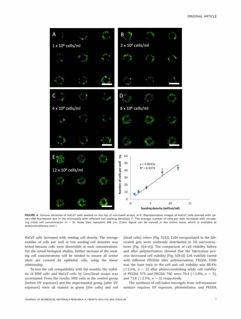
HaCaT cells increased with seeding cell density. The averagenumber of cells per well at low seeding cell densities wastested because cells were discernible at such concentrations.For the actual biological studies, further increase of the seed-ing cell concentrations will be needed to ensure all centerislets are covered by epithelial cells, using the linearrelationship.
To test the cell compatibility with the moulds, the viabil-ity of HDF cells and HaCaT cells by Live/Dead assays wasascertained. From the results, HDF cells in the control group(before UV exposure) and the experimental group (after UVexposure) were all stained in green (live cells) and red
(dead cells) colors [Fig. 5(A)]. Cells encapsulated in the fab-ricated gels were uniformly distributed in 3D microstruc-tures [Fig. 5(A-vi)]. The comparison of cell viability beforeand after polymerization showed that the fabrication pro-cess decreased cell viability [Fig. 5(B-i)]. Cell viability variedwith different PEGDAs after polymerization. PEGDA 3500was the least toxic to the cell and cell viability was 88.4%(62.6%, n ¼ 3) after photocrosslinking while cell viabilityof PEGDA 575 and PEGDA 700 were 70.4 (61.8%, n ¼ 3),and 73.8 (62.9%, n ¼ 3) respectively.
The synthesis of cell-laden microgels from cell-monomermixture requires UV exposure, photoinitiator, and PEGDA
FIGURE 4. Various densities of HaCaT cells seeded on the top of microwell arrays. A–E: Representative images of HaCaT cells stained with cal-
cein-AM fluorescent dye in the microwells with different cell seeding densities. F: The average number of cells per well increased with increas-
ing initial cell concentration (n ¼ 3). Scale bars represent 200 lm. [Color figure can be viewed in the online issue, which is available at
wileyonlinelibrary.com.]
ORIGINAL ARTICLE
JOURNAL OF BIOMEDICAL MATERIALS RESEARCH A | MONTH 2013 VOL 00A, ISSUE 00 7
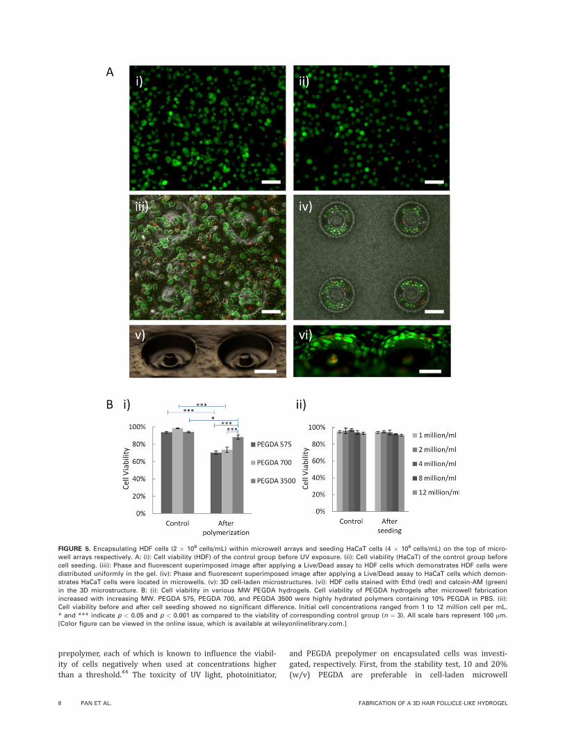
prepolymer, each of which is known to influence the viabil-ity of cells negatively when used at concentrations higherthan a threshold.44 The toxicity of UV light, photoinitiator,
and PEGDA prepolymer on encapsulated cells was investi-gated, respectively. First, from the stability test, 10 and 20%(w/v) PEGDA are preferable in cell-laden microwell
FIGURE 5. Encapsulating HDF cells (2 � 106 cells/mL) within microwell arrays and seeding HaCaT cells (4 � 106 cells/mL) on the top of micro-
well arrays respectively. A: (i): Cell viability (HDF) of the control group before UV exposure. (ii): Cell viability (HaCaT) of the control group before
cell seeding. (iii): Phase and fluorescent superimposed image after applying a Live/Dead assay to HDF cells which demonstrates HDF cells were
distributed uniformly in the gel. (iv): Phase and fluorescent superimposed image after applying a Live/Dead assay to HaCaT cells which demon-
strates HaCaT cells were located in microwells. (v): 3D cell-laden microstructures. (vi): HDF cells stained with Ethd (red) and calcein-AM (green)
in the 3D microstructure. B: (i): Cell viability in various MW PEGDA hydrogels. Cell viability of PEGDA hydrogels after microwell fabrication
increased with increasing MW. PEGDA 575, PEGDA 700, and PEGDA 3500 were highly hydrated polymers containing 10% PEGDA in PBS. (ii):
Cell viability before and after cell seeding showed no significant difference. Initial cell concentrations ranged from 1 to 12 million cell per mL.
* and *** indicate p < 0.05 and p < 0.001 as compared to the viability of corresponding control group (n ¼ 3). All scale bars represent 100 lm.
[Color figure can be viewed in the online issue, which is available at wileyonlinelibrary.com.]
8 PAN ET AL. FABRICATION OF A 3D HAIR FOLLICLE-LIKE HYDROGEL
fabrication. Therefore, HDF cells were subjected to 10 and20% (w/v) PEGDA solutions (PEGDA 575, PEGDA 700, andPEGDA 3500) for 2 h. It was found that PEGDA solution oflower molecular weight was more cytotoxic to HDF andHDF cells in PEGDA 3500 solution survived longer [Support-ing Information Fig. 2(A)]. Secondly, to minimize the toxicityof UV light, the minimum duration of UV exposure at vari-ous UV intensities was established, which was deemed to bethe time required for the microwell array formation with nodeformation. Then, HDF cells in PBS solution were exposedto UV for minimum duration to analyze the effect of UValone. For the analysis of photoinitiator, HDF cells were sus-pended in 0.2% (w/v) photoinitiator for 2 h. The resultsshowed that UV exposure and photoinitiator did not affectHDF viability on their own [Supporting Information Fig. 2(B,
C)]. Subsequently, each combination of UV intensity, PEGDAsolution and photoinitiator were tested and the optimal con-ditions for cell-laden microwell fabrication were found to be10% (w/v) PEGDA in 0.2% (w/v) photoinitiator under 4.96W/cm2 for 30 s. After cell encapsulation, the difference ofcell viability among different MW PEGDA may be due to anincrease in the free radical concentration produced from theshorter chained PEGDA 575 and PEGDA 700 during thecrosslinking process. Furthermore, PEGDA 575 and PEGDA700 allowed higher diffusion rates into the cells comparedto PEGDA 3500 which can adversely affect cell viability.45
For HaCaT cells, various cell densities (1–12 million cellsper mL) were applied on the top of microwells. Cells wereoriginally dispersed in the cell solution before cell seeding[Fig. 5(A-ii)] while cells were retained in microwells after
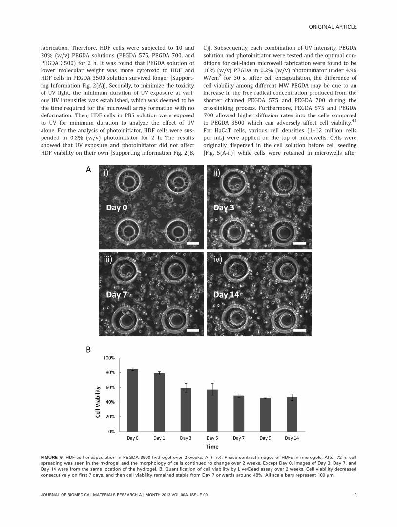
FIGURE 6. HDF cell encapsulation in PEGDA 3500 hydrogel over 2 weeks. A: (i–iv): Phase contrast images of HDFs in microgels. After 72 h, cell
spreading was seen in the hydrogel and the morphology of cells continued to change over 2 weeks. Except Day 0, images of Day 3, Day 7, and
Day 14 were from the same location of the hydrogel. B: Quantification of cell viability by Live/Dead assay over 2 weeks. Cell viability decreased
consecutively on first 7 days, and then cell viability remained stable from Day 7 onwards around 48%. All scale bars represent 100 lm.
ORIGINAL ARTICLE
JOURNAL OF BIOMEDICAL MATERIALS RESEARCH A | MONTH 2013 VOL 00A, ISSUE 00 9

cell seeding [Fig. 5(A-iv)]. After Live/Dead assay, cell viabil-ity of control group (before cell seeding) and experimentgroup (after cell seeding) had no significant difference [Fig.5(B-ii)]. From the superimposed image, it was shown thatHaCaT cells were localized inside the microwells after seed-ing [Fig. 5(A-iv)]. The diameter of HDF cells (17.37 6 3.30lm) was larger than that of HaCaT cells (10.77 6 2.04 lm)and the diameter of HDF cells after cell encapsulation was19.75 6 3.46 lm (Supporting Information Fig. 3). HDF cellattachment may be developed inside the micro-mould whichleads to the size difference before and after cell encapsula-tion. It was demonstrated that two different types of cellscan be controlled at the designated locations of microwellarrays, where HDF cells were uniformly distributed insidethe fabricated gels and HaCaT cells were seeded on the topof microwells.
After microwell fabrication, the cells encapsulated in thehydrogels were monitored for up to 14 days at the samelocations. Cell spreading was observed at the bottom of mi-crowells after 72 h incubation and the morphology of cellschanged at Day 7 and Day 14 [Fig. 6(A)]. The reason thatcell spreading only occurred at the bottom of the microwellmay be that the bottom was made of a thinner layer ofPEGDA hydrogel which can minimize diffusion limitationsand provide more effective nutrient transport.20,37 PEGDA3500 in our study showed long-term viability for cellsencapsulated over 14 days. From Live/Dead assays, it wasshown that cell viability decreased quickly in first 3 daysfrom 84.4 to 59.2%, while it was stabilized around 48%from Day 7 to Day 14 [Fig. 6(B)]. The initial decrease in cellviability could be due to cell damage caused by residualphotocrosslinking factors encapsulated in hydrogels, such astoxic free radicals formed during crosslinking process, unpo-lymerized photoinitiator, and PEGDA monomers. These toxicfactors may continuously diffuse into outside culture me-dium and eventually be removed after fresh medium wasadded repeatedly. As a result, the cell viability was main-tained at the same level after 7 days. It has been reportedthat the addition of other factors into PEGDA gels can fur-ther improve cell development46,47 and this could beexplored for future studies.
CONCLUSION
In this study, a 3D microstructure was fabricated byusing a patterned PDMS stamp on a glass substrate. Wedemonstrated that 10 and 20% (w/v) PEGDA hydrogelmicrostructures are stable on the glass substrate andtheir mechanical properties were characterized by nano-indentation. HDF and HaCaT cells were immobilized atthe designated locations of the microstructure, respec-tively. The number of HaCaT cells in the microwellincreased with increasing cell density and cell seedingprocess did not compromise HaCaT cell viability. We alsodemonstrated that HDF survived inside the hydrogelsover 14 days with observable cell spreading. Hence, thismicrostructure can be potentially used for human hairfollicle regeneration in vitro.
ACKNOWLEDGMENTS
J. Pan is a recipient of the National University of SingaporeResearch Scholarship. We thank SBIC-Nikon Imaging Centrefor providing the SMZ 1500microscope for microwell imaging.A. Miserez and S. Amini thank the support of the SingaporeNational Research Foundation (NRF Fellowship to A. Miserez).The authors declare no conflict of interest.
REFERENCES1. Krause K, Foitzik K. Biology of the hair follicle: the basics. Semin
Cutan Med Surg 2006;25:2–10.
2. Annika Vogt KJM, Ulrike Blume-Peytavi. Biology of the hair fol-
licle. In: David A, Whitting UB-P, Antonella Tosti and Ralph M.
Trueb, editor. Hair Growth and Disorders. Springer-Verlag: Berlin;
2008. p 1–22.
3. Stenn K, Parimoo S, Zheng Y, Barrows T, Boucher M, Washenik
K. Bioengineering the hair follicle. Organogenesis 2007;3:6–13.
4. Leyden J, Dunlap F, Miller B, Winters P, Lebwohl M, Hecker D,
Kraus S, Baldwin H, Shalita A, Draelos Z. Finasteride in the treat-
ment of men with frontal male pattern hair loss. J Am Acad Der-
matol 1999;40:930–937.
5. Messenger AG, Rundegren J. Minoxidil: Mechanisms of action on
hair growth. Br J Dermatol 2004;150:186–194.
6. Orentreich N. Autografts in alopecias and other selected dermato-
logical conditions. Ann N Y Acad Sci 1959;83:463–479.
7. Epstein JS. Evolution of techniques in hair transplantation: A 12-
year perspective. Facial Plast Surg 2007;23:51–60.
8. Millar SE. Molecular mechanisms regulating hair follicle develop-
ment. J Invest Dermatol 2002;118:216–225.
9. Stenn KS, Paus R. Controls of hair follicle cycling. Physiol Rev
2001;81:449–494.
10. Chuong CM, Cotsarelis G, Stenn K. Defining hair follicles in the
age of stem cell bioengineering. J Invest Dermatol 2007;127:
2098–2100.
11. Weinberg WC, Goodman LV, George C, Morgan DL, Ledbetter S,
Yuspa SH, Lichti U. Reconstitution of hair follicle development in
vivo: Determination of follicle formation, hair growth, and hair
quality by dermal cells. J Invest Dermatol 1993;100:229–236.
12. Zheng Y, Du X, Wang W, Boucher M, Parimoo S, Stenn K. Orga-
nogenesis from dissociated cells: Generation of mature cycling
hair follicles from skin-derived cells. J Invest Dermatol 2005;124:
867–876.
13. Ehama R, Ishimatsu-Tsuji Y, Iriyama S, Ideta R, Soma T, Yano K,
Kawasaki C, Suzuki S, Shirakata Y, Hashimoto K. Hair follicle
regeneration using grafted rodent and human cells. J Invest Der-
matol 2007;127:2106–2115.
14. Jone LN, Pope FM. Isolation of intermediate filament assemblies
from human hair follicles. J Cell Biol 1985;101:1569–1577.
15. Higgins CA, Richardson GD, Ferdinando D, Westgate GE, Jahoda
CA. Modelling the hair follicle dermal papilla using spheroid cell
cultures. Exp Dermatol 2010;19:546–548.
16. Hashimoto T, Kazama T, Ito M, Urano K, Katakai Y, Yamaguchi N,
Ueyama Y. Histologic and cell kinetic studies of hair loss and sub-
sequent recovery process of human scalp hair follicles grafted
onto severe combined immunodeficient mice. J Invest Dermatol
2000;115:200–206.
17. Auchincloss H Jr, Sachs DH. Xenogeneic transplantation. Annu
Rev Immunol 1998;16:433–470.
18. Cicchetti F, Fodor W, Deacon TW, van Horne C, Rollins S, Burton
W, Costantini LC, Isacson O. Immune parameters relevant to neu-
ral xenograft survival in the primate brain. Xenotransplantation
2003;10:41–49.
19. Havlickova B, Biro T, Mescalchin A, Tschirschmann M, Mollenkopf
H, Bettermann A, Pertile P, Lauster R, Bodo E, Paus R. A human
folliculoid microsphere assay for exploring epithelial–mesenchy-
mal interactions in the human hair follicle. J Invest Dermatol
2009;129:972–983.
20. Khademhosseini A, Langer R, Borenstein J, Vacanti JP. Micro-
scale technologies for tissue engineering and biology. Proc Natl
Acad Sci USA 2006;103:2480–2487.
10 PAN ET AL. FABRICATION OF A 3D HAIR FOLLICLE-LIKE HYDROGEL
21. Barron JA, Wu P, Ladouceur HD, Ringeisen BR. Biological laser
printing: A novel technique for creating heterogeneous 3-dimen-
sional cell patterns. Biomed Microdevices 2004;6:139–147.
22. Rolland JP, Maynor BW, Euliss LE, Exner AE, Denison GM,
DeSimone JM. Direct fabrication and harvesting of monodisperse,
shape-specific nanobiomaterials. J Am Chem Soc 2005;127:
10096–10100.
23. Pan J, Chan SY, Lee WG, Kang L. Microfabricated particulate
drug-delivery systems. Biotechnol J 2011;6:1477–1487.
24. Kane RS, Takayama S, Ostuni E, Ingber DE, Whitesides GM. Pat-
terning proteins and cells using soft lithography. Biomaterials
1999;20:2363–2376.
25. Koh WG, Revzin A, Pishko MV. Poly(ethylene glycol) hydrogel
microstructures encapsulating living cells. Langmuir 2002;18:
2459–2462.
26. Dendukuri D, Gu SS, Pregibon DC, Hatton TA, Doyle PS. Stop-
flow lithography in a microfluidic device. Lab Chip 2007;7:
818–828.
27. Langer R, Vacanti JP. Tissue engineering. Science 1993;260:
920–926.
28. Place ES, George JH, Williams CK, Stevens MM. Synthetic poly-
mer scaffolds for tissue engineering. Chem Soc Rev 2009;38:
1139–1151.
29. Zhu J. Bioactive modification of poly(ethylene glycol) hydrogels
for tissue engineering. Biomaterials 2010;31:4639–4656.
30. Xia Y, Whitesides GM. Soft lithography. Annu Rev Mater Sci
1998;28:153–184.
31. Nichol JW, Koshy ST, Hojae B, Hwang CM, Yamanlar S, Khadem-
hosseini A. Cell-laden microengineered gelatin methacrylate
hydrogels. Biomaterials 2010;31:5536–5544.
32. Kang L, Hancock MJ, Brigham MD, Khademhosseini A. Cell con-
finement in patterned nanoliter droplets in a microwell array by
wiping. J Biomed Mater Res A 2010;93:547–557.
33. Zhang H, Wang L, Song L, Niu G, Cao H, Wang G, Yang H, Zhu S.
Controllable properties and microstructure of hydrogels based on
crosslinked poly(ethylene glycol) diacrylates with different molec-
ular weights. J Appl Polym Sci 2011;121:531–540.
34. Revzin A, Russell RJ, Yadavalli VK, Koh WG, Deister C, Hile DD,
Mellott MB, Pishko MV. Fabrication of poly(ethylene glycol)
hydrogel microstructures using photolithography. Langmuir 2001;
17:5440–5447.
35. Antonny B. Mechanisms of membrane curvature sensing. Annu
Rev Biochem 2011;80:101–123.
36. McMahon HT, Gallop JL. Membrane curvature and mechanisms
of dynamic cell membrane remodelling. Nature 2005;438:590–596.
37. Moeller HC, Mian MK, Shrivastava S, Chung BG, Khademhosseini
A. A microwell array system for stem cell culture. Biomaterials
2008;29:752–763.
38. Peppas NA, Brannon-Peppas L. Hydrogels at critical conditions.
Part 1. Thermodynamics and swelling behavior. J Membr Sci
1990;48:281–290.
39. Temenoff JS, Athanasiou KA, LeBaron RG, Mikos AG. Effect of
poly(ethylene glycol) molecular weight on tensile and swelling
properties of oligo(poly(ethylene glycol) fumarate) hydrogels for
cartilage tissue engineering. J Biomed Mater Res 2002;59:
429–437.
40. Jahoda CA, Horne KA, Oliver RF. Induction of hair growth by im-
plantation of cultured dermal papilla cells. Nature 1984;311:
560–562.
41. Shimizu H, Morgan BA. Wnt signaling through the beta-catenin
pathway is sufficient to maintain, but not restore, anagen-phase
characteristics of dermal papilla cells. J Invest Dermatol 2004;122:
239–245.
42. Deyrieux AF, Wilson VG. In vitro culture conditions to study kera-
tinocyte differentiation using the HaCaT cell line. Cytotechnology
2007;54:77–83.
43. Zorlutuna P, Jeong JH, Kong H, Bashir R. Stereolithography-
based hydrogel microenvironments to examine cellular interac-
tions. Adv Funct Mater 2011;21:3642–3651.
44. Panda P, Ali S, Lo E, Chung BG, Hatton TA, Khademhosseini A,
Doyle PS. Stop-flow lithography to generate cell-laden microgel
particles. Lab Chip 2008;8:1056–1061.
45. Mazzoccoli JP, Feke DL, Baskaran H, Pintauro PN. Mechanical and
cell viability properties of crosslinked low- and high-molecular
weight poly(ethylene glycol) diacrylate blends. J Biomed Mater
Res A 2010;93:558–566.
46. Chan V, Zorlutuna P, Jeong JH, Kong H, Bashir R. Three-dimen-
sional photopatterning of hydrogels using stereolithography for
long-term cell encapsulation. Lab Chip 2010;10:2062–2070.
47. Xu K, Fu Y, Chung W, Zheng X, Cui Y, Hsu IC, Kao WJ. Thiol-ene-
based biological/synthetic hybrid biomatrix for 3-D living cell cul-
ture. Acta Biomater 2012;8:2504–2516.
ORIGINAL ARTICLE
JOURNAL OF BIOMEDICAL MATERIALS RESEARCH A | MONTH 2013 VOL 00A, ISSUE 00 11