Fabrication and characterisation of Extracellular matrix...
43
Fabrication and characterisation of Extracellular matrix based composite films for wound healing application Thesis submitted in partial fulfillment of the requirements for the degree Of Master of Technology in Biomedical Engineering By Susanta Basuri Roll No: 212BM1349 Under the guidance of Prof. Sirsendu Sekhar Ray Assistant Professor Department of Biotechnology & Medical Engineering National Institute of Technology Rourkela 2012-2014
Transcript of Fabrication and characterisation of Extracellular matrix...
Fabrication and characterisation of Extracellular
matrix based composite films for wound healing
application
Thesis submitted in partial fulfillment of the requirements for the degree
Of
Master of Technology
in
Biomedical Engineering
By
Susanta Basuri
Roll No: 212BM1349
Under the guidance of
Prof. Sirsendu Sekhar Ray Assistant Professor
Department of Biotechnology & Medical Engineering
National Institute of Technology
Rourkela
2012-2014
NATIONAL INSTITUTE OF TECHNOLOGY, ROURKELA
CERTIFICATE
This is to certify that the thesis entitled, “Fabrication and characterisation of Extracellular
matrix based composite films for wound healing application” submitted by Mr. Susanta
Basuri in partial fulfillment of the requirements for the award of degree of Master of
Technology in Biotechnology & Medical Engineering with specialization in “Biomedical
Engineering” at National Institute of Technology, Rourkela is an authentic work carried out
by him under my supervision and guidance. To the best of my knowledge, the matter embodied in the thesis has not been submitted to
any other university/institute for the award of any Degree.
Date: 30/05/2014 Prof. Sirsendu Sekhar Ray
Assistant Professor
Department of Biotechnology &
Medical Engineering
NIT Rourkela
NATIONAL INSTITUTE OF TECHNOLOGY, ROURKELA
ACKNOWLEDGMENT
I would like to express my gratitude to my supervisor Prof. Sirsendu Sekhar Ray for his
patience, motivation, enthusiasm, immense knowledge and constant support. His guidance
has helped me throughout my project work and in writing my thesis at NIT, Rourkela.
Besides my advisor, I would like to thank Prof.K. Pramanik, Prof. Kunal Pal, Prof.
IndranilBanarjee for their encouragement and insightful comments. I would like to thank all
faculty members and staff of the Department of Biotechnology & Medical Engineering,
N.I.T. Rourkela for their generous help in various ways for the completion of this thesis.
I would like to thank all my friends and especially my lab mates Somya Asthana, Mohit
Gangwar and Alisha Prasad for all the discussions and help. I’ve enjoyed their
companionship during my stay at NIT, Rourkela. I am especially indebted to my parents for
their love, sacrifice, and support. My full dedication to the work would have not been
possible without their blessings and moral support. This thesis is a dedication to them.
Date: 30.05.2014
SUSANTA BASURI
212BM1349
Department of Biotechnology &
Medical Engineering
NIT Rourkela
Abstract – Wound healing, occurs naturally upon inflammation following a programmed
process such as hemostasis, inflammation, proliferation, and remodeling. But at times this
sequence is not followed due to lack of sufficient oxygenation, nutrition, stress etc. thus
causing improper or impaired wound healing. To suffice this need, implants such as scaffolds
are prepared which mimics the extracellular matrix (ECM) thereby providing a proper
environment for growth, maintenance and adherence of the cells present in it. In the present
work, we propose ECM derived from porcine omentumto be used as a xenogeneic
biomaterial for designing our scaffolds. For this study porcine omentum was decellularised
following a series of processes including both physical and chemical method such
as repeated freeze thawingand SDS washing of the tissues. Composite films of both
Alginate-ECM and Chitosan –ECM in different compositions were prepared by solvent
casting method. These films were further characterized by Light Transmission, Swelling,
hemocompatibility, moisture absorption and water vapour permeability, X-ray diffraction
(XRD) and Fourier Transform Infrared (FTIR) spectroscopy. Alginate films without ECM
were found to be less thick than the chitosan films without ECM. Upon swelling the films
differed in the hydrophilicity as uptake of water by chitosan films were more than alginate
films. This accounts to the moisture retention capacity of films Apart from this since ECM is
more hydrophilic than alginate and chitosan and addition of higher proportion of ECM in
films imparts them more hydrophilicity. Alginate films were found to have more thickness,
more hemocompitibility, more crystallinity and higher rate of moister absorption, lesser
swelling ratio and more water vapour permeability than chitosan films
Keywords: Porcine omentum, Decellularization, Extracellular matrix, Chitosan, Alginate,
Chronic wounds
TABLE OF CONTENTS
Chapter 1. Introduction ...................................................................................................... 1-3
Chapter 2. Review of Literature ......................................................................................... 4-9
Chapter 3. Materials and Methods................................................................................. 10-17
3.1 Decellularization and solubilisation of Adipose Tissue: .............................................. 11
3.2 Preparation of Solutions: ............................................................................................... 12
3.2.1 Polymer solution preparation: ......................................................... 12
3.2.2 ECM solution preparation: .............................................................. 12
3.2.3 Preparation of Composite Films: ....................................................... 12
3.4 Characterizations of ECM Films: ............................................................................ 13-17
3.4.1 Thickness of the films: ......................................................................... 13
3.4.2 Film Transparency/ Film Light Transmission Test: ........................ 13
3.4.3 Swelling Test: ....................................................................................... 14
3.4.4 Moisture Test: ...................................................................................... 14
3.4.5 Water Vapor Transmission Permeability Test: ................................ 15
3.4.6 Hemocompatibility Test: ..................................................................... 15
3.4.7 Fourier Transform Infrared Spectroscopy: ...................................... 16
3.4.8 X- Ray Diffraction: .............................................................................. 16
Chapter 4. Result and Discussions ................................................................................. 18-30
4.1 Thickness: ................................................................................................ 19
4.2 Film Transparency/ Film Light Transmission: .................................... 20
4.3 Swelling: ................................................................................................... 20
4.4 Moisture absorption: .............................................................................. 23
4.5 Hemocompatibility: ................................................................................ 24
4.6 Water Vapor Permeability: ................................................................... 26
4.7 X-Ray Diffraction..................................................................................... 28
4.8 Fourier Transform Infrared Spectroscopy ........................................... 29
Chapter 5. Conclusion and Future works ..................................................................... 31-32
References ...................................................................................................................................
LIST OR TABLES
Table 1: Specifications of UV-Spectrophotometer.............................................................. 13
Table 2: Description of Materials used for Project work ................................................... 17
Table 3: Thickness of Chitosan- ECM Film ........................................................................ 19
Table 4: Thickness of Alginate- ECM Film ........................................................................ 19
Table 5: Swelling of Chitosan- ECM Film ........................................................................... 21
Table 6: Swelling of Chitosan- ECM Film ........................................................................... 21
Table 7: Swelling of Alginate- ECM Film............................................................................ 22
Table 8: Moisture of Chitosan- ECM Film.......................................................................... 23
Table 9: Moisture of Alginate- ECM Film .......................................................................... 24
Table 10: Hemocompatibility of Chitosan-ECM Film Alginate- ECM Film ................... 25
Table 11: Water Vapor Permeability of Chitosan- ECM Film ......................................... 26
Table 12: Water Vapor Permeability of Alginate- ECM Film .......................................... 26
Table 13: Wave number of different functional groups in Chitosan, Sodium Alginate
and ECM ................................................................................................................................. 29
LIST OR FIGURES
Fig 1: Decellularization of omentum .................................................................................... 11
Fig 2: Film Light Thickness of Chitosan-ECM Film Alginate- ECM Film ..................... 19
Fig 3: Film Light Transmission of Chitosan-ECM Film Alginate- ECM Film ................ 20
Fig 4: Swelling of Chitosan-ECM Film Alginate- ECM Film ............................................ 22
Fig 5: Moisture absorption of Chitosan-ECM Film Alginate- ECM Film ....................... 24
Fig 6: Hemocompatibilityof Chitosan-ECM Film Alginate- ECM Film .......................... 25
Fig 7: Water Vapor Permeability of Chitosan-ECM Film Alginate- ECM Film ............ 27
Fig 8: XRD Pattern of Chitosan-ECM Film Alginate- ECM Film ................................... 28
Fig 9: FTIR Spectroscopy of Chitosan-ECM Film Alginate- ECM Film ......................... 30
1 | P a g e
Chapter 1
INTRODUCTION
2 | P a g e
Chronic wounds are the wounds that does not heal or improve significantly within predictable
amount of time. Chronic wounds occur due to insufficient blood supply, abnormal ECM deposition
and because of the dry conditions occurring in the body [1]. Since these chronic wounds take a lot of
time to heal and thus it causes physical and emotional stress in many patients, so it is necessary to
find a proper treatment for this.
Chitosan is a Polymer that is acquired from the hard skeleton of shell fish, including crab,
lobster, and shrimp. It is utilized to treat obesity, high cholesterol, and Crohn's infection. It is
aexcellent biopolysaccharides [2].Chitosan is profoundly hydrophobic and is insoluble in water. It is
dissolvable in hexafluoroisopropanol, hexafluoroacetone and chloroalcoholsin conjugation with
water results of mineral acids [3]. It is suitable as a transporter for its high thickness, charge
appropriation and discharge systems. Further, because of its crystalline, hydrophobic nature and
dynamic –OH and –NH2 groups, Chitosan is novelpractical bio-macromolecules that have the
aggregate impacts as pharmaceutical excipients, the biocompatibilityand biodegradability [4].
Alginate is a commonly happening anionic polymer normally gotten from tan ocean growth, and has
been widely explored and utilized for some biomedical application, because of its biocompatibility,
gentle gelation by expansion of divalent cations [5]. Alginate hydrogels have been especially alluring
in wound recuperating, pill conveyance, and tissue building applications [6]. Alginate wound
dressings keep up a physiologically sodden microenvironment, minimize bacterial contamination at
the injury site, and encourage wound recuperating. Drug particles, from little compound pills to
macromolecular proteins, could be discharged from alginate gels in a controlled way [7]. Alginate
dressings keep up a physiologically sodden microenvironment that pushes recuperating and the
framing of granulation tissue. Alginates could be washed away with saline watering system, so
evacuation of the dressing does not meddle with recuperating granulation tissue. Alginate dressings
are extremely helpful for moderate to intensely exuding wounds [8].
3 | P a g e
The extracellular matrix (ECM) is the non-cell part introduce inside all tissues and organs, and gives
vital physical platform for the cell constituents. ECM is made out of proteins and polysaccharides
[9]. The ECM is made out of two fundamental classes of macromolecules:proteoglycans and sinewy
proteins. The fundamental stringy ECM proteins are collagens, elastins, fibronectins and laminin
[10]. All cells in strong tissues are encompassed by extracellular matrix. Both plants and creatures
have ECM. The cell divider of plant cells is a sort of extracellular matrix[11]. ECM gives: rigidity
for tendons,compressive quality for cartilage,hydraulic security for some sorts of cells,elasticity to
the dividers of blood vessels . We use ECM based bandage for chronic wounds because it can
replace of abnormal ECM and growth factor attached with it [12].
In this study we worked for the treatment of chronic wounds and fabricated ECM composite
bandages. The ECM derived from the porcine omentum was composited with Chitosan and Alginate
in various proportion and those were characterized by various methods.
Objective
Fabrication and characterisation of Chitosan-ECM composite films for wound healing
application
Fabrication and characterisation of Alginate-ECM composite films for wound healing
application
4 | P a g e
Chapter 2
REVIEW OF LITARETURE
5 | P a g e
Moustafa M.G. Fouda*, R. Wittke, D. Knittel, “Use of chitosan/polyamine biopolymers based
cotton as a model system to prepare antimicrobial wound dressing”
Theamainaaim ofatheastudy was toaexploreaandacompareathe antibacterial propertiesaof
chitosanaandalinear polyvinyl amine, as aabiopolymer, withathe prepared dressing based cotton.
These treated cotton were further characterized by monitoring the susceptibility of the amino groups
created on the surface of the fabric. The produced dressing based cotton can be used as a model
system to treat wounds, ulcers as well as diabetic ulcers. Chitosan is afbiopolymer that hasfbeen
known asabeing able toaaccelerate the healing ofawound inahuman. Chitosanasimulatedathe
migration ofapolymorph nuclear (PMN) asawellaas mononuclearacells and acceleratedathe re-
epithelizationaand normal skinaregeneration. Chitosan have antibacterialaactivity against a broad
spectrum. The bindingaofachitosan withaDNAaand
inhibitionaofamRNAasynthesisaoccursaviaatheapenetrationaofachitosanaintoatheanuclei of the
microorganismsaandainterferingawithatheasynthesis ofamRNA andaproteins.aChitosan also
facilitates wound repair. According to the results inathis work, theaantimicrobialaactivityaof
chitosan/polyvinyl amine system showed promising results[13].
Rupesh Gajanan Nawalakhe, et al “Development of Electrospun Iminochitosan for
Improved Wound Healing Application”
Toaexploreatheapropertiesaofachitosanaderivatives, nanofibrousaiminochitosanawas prepared by
electrospinning technique. The solvent used for dissolving iminochitosan before being electrospun is
trifluoroacetic acid (TFA). Apart from this several other parameters including
polymeraconcentration,aelectricafieldaandaextrusionarateawere alsoainvestigated. Goodafiber
6 | P a g e
formationaoccurred within aarangeaofa3%-8% of iminochitosanaconcentration.aThe
electrospinningaconcentrationsainathearangeaofa1%-5% wereastudiedaforaantibacterialatesting. The
resultsaindicateathat the nanofiber webs exhibitaexcellent antimicrobial behavior[14].
Ali Demir Sezer,1 Fatih Hatipoğlu, et al “Chitosan Film Containing Fucoidan as a Wound
Dressing for Dermal Burn Healing: Preparation and In Vitro/In Vivo Evaluation”
Thisastudy was done toadevelopachitosan based filmsacontaining fucoidanaand toainvestigate its
suitabilityaforatheatreatmentaofadermalaburnsaonarabbits. The prepared films were tested on the
basis of porosity, thickness, swelling tests, tensile strength, water vapor permeability and bio
adhesion of the films. It was observed thathigherachitosanaconcentrationasignificantlyaincreased
tensile strengthaof theafilms. Also more porous the films more would be its water uptakeaproperty.
The swelling test accounted to the retention property of the films. Furthermore,
dermalaburnahealingaexperiments using rabbit haveashown that theaapplication of fucoidan
chitosanafilmaonto anaopen burnawound induces significantawoundacontraction, and accelerates the
wound closure and healing process. Thus, theafucoidan-chitosan filmamayabe
aapromisinganewadressingaforawoundaocclusionaand tissuearepairing[15].
Immanuel M. Sebastine and David J. Williams “The Role of Mechanical Stimulation in
Engineering of Extracellular Matrix (ECM) “
7 | P a g e
Mechanotransduction is aacomplexaphenomenonarequiring theaselectiveainvolvementaof many
different signaling pathwaysainaresponse to mechanicalastimuli .The critical component of the
mechanotransduction process is the ECM and the initial responses to mechanicalastimuliaare
recorded at the proximities of cell-ECM contacts. This review focuses on the study of these pathways
involved in engineering the ECM. Since, cellsarespond to mechanical stimuli and
regulateatheametabolicafunctions viaamechanotransduction and synthesize ECM, in-vitro studies of
mechanotransduction using automatedabioreactors that are capable of mimickingathe
physiologicalaenvironment by applyingadifferentaloadsawill help us to examine how mechanical
loads influence intracellular signaling, pathway and their behavior on ECM. Although the
experimentsaconducted in microscopicatissues haveademonstrated a strongacorrelationabetween
mechanicalaforces andachanges in cell behaviors, betteraunderstanding ofamechanotransduction
willahelpaus to applyaappropriate mechanicalastimulation on cells inascaffolds in vitroafor the
expressionaof a specific gene of interest oracreation ofaparticular constructs.aDevelopment of new
tools or sensors toaobserve the changes in cells duringamechanotransduction and toanalyses the
genes,amRNA, and proteinsaexpressed within the tissue construct will open newaavenues of
research[16].
Biji Balakrishnana, M. Mohanty et al “Evaluation of an in situ forming hydrogel wound
dressing based on oxidized alginate and gelatin”
In situ wound dressings are better and has more advantages than the preformed dressings as it offers
comfort withoutawrinkling orafluting in the woundabed, ease ofaapplication and improved
patientacompliance. This paper describes an in situ based wound dressing applications using
hydrogels and use of certain biomaterials like gelatin, oxidized alginate and borax. As we know that
8 | P a g e
periodateaoxidizedaalginate rapidlyacross-links proteins such asagelatin in theapresence of borax
toagive in situ formingahydrogels that are bothanon-toxic andabiodegradable. This compositeamatrix
has the haemostaticaeffect of gelatin, the woundahealing-promotingafeature of alginateaand
theaantiseptic property ofaborax which makes it a potential woundadressing material. From the study
conducted, the hydrogelawasafound to have a fluidauptake ofa90% of itsaweight which would
prevent the wound bed fromaaccumulation ofaexudates. The water vapor transmission ratea(WVTR)
of theahydrogel was found to be 26867124 g/m2 day indicatingathat the hydrogel can maintain a
moistaenvironment over woundabed in moderate to heavily exuding wound which
wouldaenhanceaepithelial cellamigration during theahealing process. A rat model was used for
demonstrating, the efficacy of hydrogel in wound healing and it was found that withina2 weeks, the
woundacovered with gel wasacompletely filled with newaepithelium without any significantaadverse
reactions. These in situaforming hydrogelsafulfil many critical elements desirable in a wound
dressingamaterial. Thus, this can act as a promisingaapproach serving theapurpose[17].
R. Jayakumar1, M. Prabaharan, P. T. et al, “Novel Chitin and Chitosan Materials in Wound
Dressing”
As Skin plays an important role inahomeostasis and theaprevention of invasion by microorganisms,
use of a biomaterial as a support material can help solve the issue. This review particularly, focuses
on the affectivity of chitin and chitosan as wound dressing material and mechanisms of such action
in the molecular,acellular, and systemicalevels. Chitin and its derivative, chitosan, are
biocompatible,abiodegradable,anontoxic,aanti-microbial and hydrating agents. Due to
theseaproperties, they show goodabiocompatibility and positiveaeffects on wound Healing. Chitin is
an abundant polysaccharide and chitosan is aadeacetylated product of chitin. Both chitin and chitosan
9 | P a g e
has beneficial biological andaantimicrobial properties and has potential for wound healing. Healing
pattern gets altered in chronic wounds which lead to scars and unwanted tissue damage which might
be a result of environmental irritants as well as water and electrolyte disturbances. The
orderedaregeneration of woundedatissues requiresathe use of chitin and chitosanain the form of non-
woven, Nano fibrils, composites, films,ascaffolds andasponges. Thus, these naturally occurring
biomaterials can help restore the damaged tissue, thereby serving in the field of tissue
engineering[18].
10 | P a g e
Chapter 3
MATERIALS AND METHODS
11 | P a g e
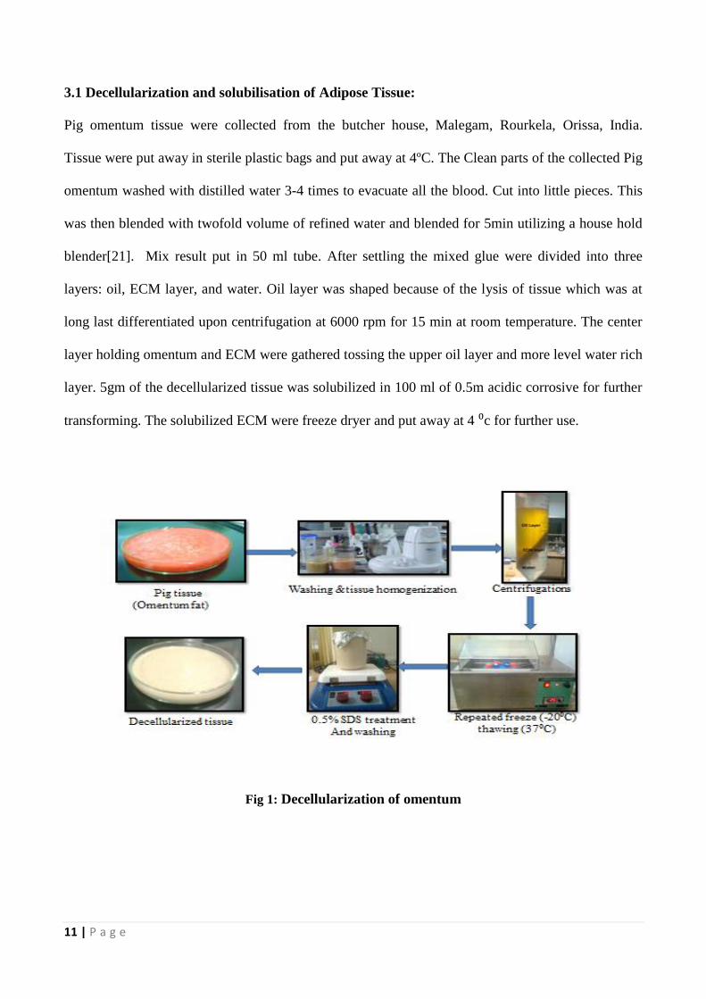
3.1 Decellularization and solubilisation of Adipose Tissue:
Pig omentum tissue were collected from the butcher house, Malegam, Rourkela, Orissa, India.
Tissue were put away in sterile plastic bags and put away at 4ºC. The Clean parts of the collected Pig
omentum washed with distilled water 3-4 times to evacuate all the blood. Cut into little pieces. This
was then blended with twofold volume of refined water and blended for 5min utilizing a house hold
blender[21]. Mix result put in 50 ml tube. After settling the mixed glue were divided into three
layers: oil, ECM layer, and water. Oil layer was shaped because of the lysis of tissue which was at
long last differentiated upon centrifugation at 6000 rpm for 15 min at room temperature. The center
layer holding omentum and ECM were gathered tossing the upper oil layer and more level water rich
layer. 5gm of the decellularized tissue was solubilized in 100 ml of 0.5m acidic corrosive for further
transforming. The solubilized ECM were freeze dryer and put away at 4 ⁰c for further use.
Fig 1: Decellularization of omentum
12 | P a g e
3.2 Preparation of Solutions:
3.2.1 Polymer solution preparation: For arrangement of polymer result, 2gm/dl of chitosan
solution was mixing in 0.5 Maacetic acid . Chitosan powder was gradually added to 0.5 M
aceticaacidasolution under stirring condition for 24hrs until a homogenous solution was formed.
For preparation of Alginate solution, we take 2gm Sodium Alginate. It was slowly added to
Distilled water for 1 day until a homogenous solution was formed.
3.2.2 ECM solution preparation:
For ECM solution, 5gm of ECM was dissolved in acetic acid solution .It wasadissolved in 0.5 M
acetic acid solution (4.5ml/150ml). Thus, ECM solution were prepared. Other hand another 150
mg/ml of ECM solution was prepared. In this time ECM was dissolved in NaOH solution.
3.2.3 Preparation of Composite Films:
Casting/aSolvent evaporation Technique was emulated toaprepare the blend films in different ratios.
Arranged Chitosan & ECM solutions were taken in 5:0, 4:1, 3:2, 2:3, 1:4, (w/w) ratios individually.
The mixture was mixed until homogenous solutions was formed which was trailed by 4 hrs of
degassing. The chitosan control specimen was additionally ready in the same path, aside from it had
refined water rather than ECM. 30 gm of the arranged chitosan – ECM solutions were spilled in the
little Petri plates (90 mm width) then dried for 24 hours in the vacuum oven at temperature of 37°c.
After drying we get chitosan-ECM composite films.
For prepared Alginate and ECM solution also take in 5:0, 4:1,3:2,2:3,1:4 (w/w) ratios respectively.
In case use ECM which was dissolved in NaOH solution.
13 | P a g e
3.4 Characterizations of ECM Films:
3.4.1 Thickness of the films:
Digital caliper (Traceable® Digital Calipers 6in, Fischer Scientific) was utilized to measure the
thickness of the films at different portions of the film. The final thickness was calculated as mean
ofthe estimations at 3 locations of the film.
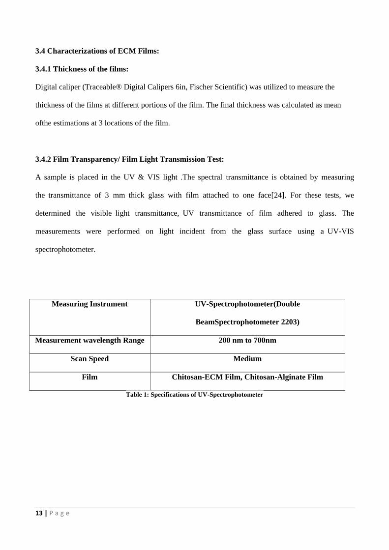
3.4.2 Film Transparency/ Film Light Transmission Test:
A sample is placed in the UV & VIS light .The spectralatransmittance is obtained byameasuring
theatransmittance of 3 mmathick glass with film attached to one face[24]. For these tests, we
determined the visiblealight transmittance,aUV transmittance of film adhered to glass. The
measurements were performed on light incident from the glass surface using aaUV-VIS
spectrophotometer.
Measuring Instrument UV-Spectrophotometer(Double
BeamSpectrophotometer 2203)
Measurement wavelength Range 200 nm to 700nm
Scan Speed Medium
Film Chitosan-ECM Film, Chitosan-Alginate Film
Table 1: Specifications of UV-Spectrophotometer
14 | P a g e
3.4.3 Swelling Test:
The Films(chitosan-ECM Films and Alginate-ECM Films) of 1x1 cm2 size were taken in a 6 well
plate. Dry weight of the films were noted. These films in the wake of peeling were weighed and
drenched in PBS (pH 7.4)aat roomatemperature. The filmsawere expelled from PBS and smeared
on filter paper to uproot approximately bound water and wet weights of the films (W t) were
noted after customary interim of time (5, 10, 20, 30, 60, 120 min) separately[25]. The experiment
was performed in triplicates and mean worth was taken. The swelling ratio (%) might be defined as
the degree of the weight increment to the introductory dry weight films.
Swelling ratio (%) = [(Wet weight of films-Dry weight of films)/Dry weight of films] %
3.4.4 Moisture Test:
Moisture tests are due to check the amount of moisture absorb in materials when they were exposed
to environmental condition. In our study we chose 84% of humidity and 30°C temperature keeping
in view the atmosphere of India. The 84% humidity was created by supersaturated solution of Kcl
inside the desiccator and the desiccator was kept outside environment. The filmswere weighed again
after 24 hrs of drying and rate of water misfortune was computed which shows the dampness
substance introduce at first in the film[26].
Percentage moisture absorbed (%) = [(Weight (final) of the films after drying – Initial weight of the
films)/finalWeightof the films after drying] %
15 | P a g e
3.4.5 Water Vapor Transmission Permeability Test:
The property of films to penetrate is proportional to the surface geography of the film, which is
controlled by Water vapor transmission test utilizing the water system. As indicated by this
technique, the Chitosan-ECM films were kept totally stuck on to the highest point of a barrel shaped
glass tubes utilizing a Teflon tape. At first filled with 30 ml PBS and the introductory weight and
stature of the water was recorded and after that the tubes were kept at 37°c. The readings were taken
in regular interval 6hrs, 12 hrs, 1 day, 2 days to 6 days. The transmission of water vapor was
calculated[27].
Water Vapor Transmission rate (g/day-m2) = (Change in weight × Test area)÷ Time
3.4.6 Hemocompatibility Test:
The hemocompatibility test were performed utilizing leachants of the ECM–chitosan Films and
ECM-alginate Films. The film of 1x1 cm2size were taken in a 12 well plate. The films drenched into
10 ml of PBS. pH value of PBS is 7.4. Then it kept in shaking incubator, under stirring rate of 60
rpm at 37°c for 10 minutes. For This test, fresh goat blood was taken. Than it diluted in 1:1 ratio with
0.9% normal saline. 0.5ml of leachants was included blood suspension took after by the sample
incubationaat 37°c for 1 hour. From that point, the samples were centrifuged ata4000 rpm and the
supernatant was investigated for the optical thickness. For making the positive control, we add the
0.1N HCL. And for negative control we use normal saline. Than The % hemolysis was measured.
% Hemolysis = [(Test sample-Negative Control) ÷(Positive Control – Negative Control)]×100
16 | P a g e
3.4.7 Fourier Transform Infrared Spectroscopy:
Fourier Transform Infrared Spectroscopy is a method which is used to obtainainfraredaspectrum of
transmission, absorption of materials. FTIR is maybe the most compelling device for recognizing
sorts of chemical bonds[28]. The wavelength of light absorbed is characteristic of the chemical
bond.The chitosan-ECM composite films and alginate-ECM films were scanned for spectroscopic
analysis using FTIR spectroscopy ATR mode. These specimens were examined keeping air as the
reference. Reading of air was at first taken by the machine for foundation subtraction and afterward
the samples were set in machine to record FTIR readings, accordingly subtracting the crests acquired
via air.Scanning range was 4000 cm-1
to 500 cm-1
with a resolution of 4 cm-1
.
3.4.8 X- Ray Diffraction:
X-Ray Diffraction is a technique that is used to study for crystalline material. The 3D stricter of non-
amorphous material is defined by fixed, repeating planes ofaatoms which form a crystalline lattice.
When a X-ray beam interacts with planes of atoms, some part of the X-Ray beam is transmitted,
some part isaabsorbed by the sample, some part is deflected andascattered[29]. When anaX-ray hits a
sample and isadiffracted, we can measure the distances between the planes of the atoms that Present
in the sample by using Bragg's Law.
n λ =2dsinθ
[Where, λ= wavelength of theaincident X-Ray, n= order of the diffracted beam, d= distance
betweenaadjacent plane of the atoms]
The Chitosan – ECM and Alginate – ECM composite films were examined using X-ray
diffractometer(PW3040, XRD – PANanalytical, Philips, Holland). Cu – Kα use as a radiation source.
17 | P a g e
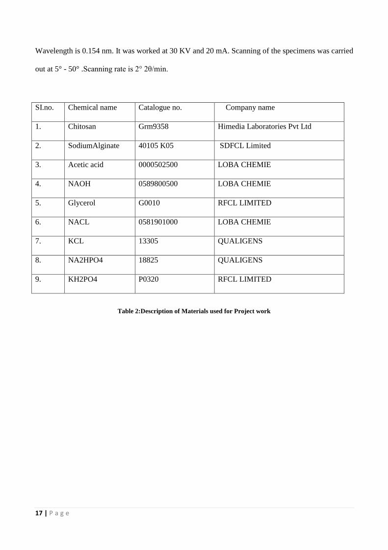
Wavelength is 0.154 nm. It was worked at 30 KV and 20 mA. Scanning of the specimens was carried
out at 5° - 50° .Scanning rate is 2° 2θ/min.
SI.no. Chemical name Catalogue no. Company name
1. Chitosan Grm9358 Himedia Laboratories Pvt Ltd
2. SodiumAlginate 40105 K05 SDFCL Limited
3. Acetic acid 0000502500 LOBA CHEMIE
4. NAOH 0589800500 LOBA CHEMIE
5. Glycerol G0010 RFCL LIMITED
6. NACL 0581901000 LOBA CHEMIE
7. KCL 13305 QUALIGENS
8. NA2HPO4 18825 QUALIGENS
9. KH2PO4 P0320 RFCL LIMITED
Table 2:Description of Materials used for Project work
18 | P a g e
Chapter 4
RESULTS & DISCUSSIONS
19 | P a g e
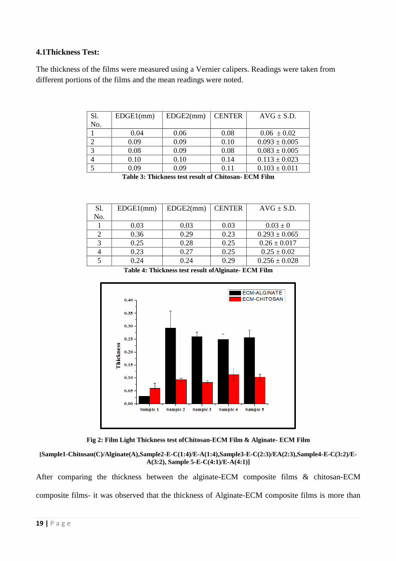
4.1Thickness Test:
The thickness of the films were measured using a Vernier calipers. Readings were taken from
different portions of the films and the mean readings were noted.
Sl.
No.
EDGE1(mm) EDGE2(mm) CENTER AVG ± S.D.
1 0.04 0.06 0.08 0.06 ± 0.02
2 0.09 0.09 0.10 0.093 ± 0.005
3 0.08 0.09 0.08 0.083 ± 0.005
4 0.10 0.10 0.14 0.113 ± 0.023
5 0.09 0.09 0.11 0.103 ± 0.011
Table 3: Thickness test result of Chitosan- ECM Film
Sl.
No.
EDGE1(mm) EDGE2(mm) CENTER AVG ± S.D.
1 0.03 0.03 0.03 0.03 ± 0
2 0.36 0.29 0.23 0.293 ± 0.065
3 0.25 0.28 0.25 0.26 ± 0.017
4 0.23 0.27 0.25 0.25 ± 0.02
5 0.24 0.24 0.29 0.256 ± 0.028
Table 4: Thickness test result ofAlginate- ECM Film
Fig 2: Film Light Thickness test ofChitosan-ECM Film & Alginate- ECM Film
[Sample1-Chitosan(C)/Alginate(A),Sample2-E-C(1:4)/E-A(1:4),Sample3-E-C(2:3)/EA(2:3),Sample4-E-C(3:2)/E-
A(3:2), Sample 5-E-C(4:1)/E-A(4:1)]
After comparing the thickness between the alginate-ECM composite films & chitosan-ECM
composite films- it was observed that the thickness of Alginate-ECM composite films is more than

20 | P a g e
the thickness of Chitosan-ECM composite films. This may be because of higher amount of NaOH
solubilized ECM (approx. 100mg) in alginate films than acetic acid solubilized ECMin case of
chitosan films. Further it was observed than the Acetic Acid solubilized ECM turn into crystalline
pattern leads to more compactness than the alginate-composite films. Otherwise, the only alginate
films without ECM are of lower thickness than only Chitosan Film without ECM.
4.2 Film Transparency/ Film Light Transmission Test:
Normally %transmission of chitosan films is more than the alginate films but in all the films addition
of the ECM leads to more opacity or reducedtransparency. This is obvious as ECM by itself is an
opaque product.
(a) (b)
Fig 3: Film Light Transmission test of Chitosan-ECM Film (a) Alginate- ECM Film (b)
[1-Chitosan(C)/Alginate(A),2-E-C(1:4)/EA(1:4),3-E-C(2:3)/E-A(2:3),4-E-C(3:2)/E-A(3:2),5-E-C(4:1)/E-A(4:1)]
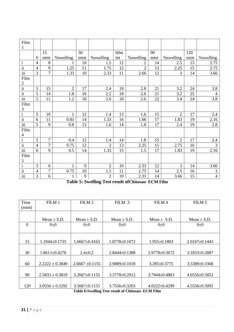
4.3 Swelling Test:
In the swelling test it was observed that with increase in the proportion of ECM. The % swelling also
increases. This might be because of more hydrophilicity of ECM in comparison in to the polymers.
21 | P a g e
Film
1
0
15
mint %swelling
30
mint %swelling
60m
int %swelling
90
mint %swelling
120
mint %swelling
i 4 8 1 10 1.5 12 2 14 2.5 15 2.75
ii 4 9 1.25 11 1.75 12 2 13 2.25 15 2.75
iii 3 7 1.33 10 2.33 11 2.66 12 3 14 3.66
Film
2
ii 5 15 2 17 2.4 19 2.8 21 3.2 24 3.8
ii 5 14 1.8 16 2.2 18 2.6 21 3.2 25 4
iii 5 11 1.2 18 2.6 18 2.6 22 3.4 24 3.8
Film
3
i 5 10 1 12 1.4 13 1.6 15 2 17 2.4
ii 6 11 0.83 14 1.33 16 1.66 17 1.83 19 2.16
iii 5 9 0.8 13 1.6 14 1.8 17 2.4 19 2.8
Film
4
i 5 7 0.4 12 1.4 14 1.8 15 2 17 2.4
ii 4 7 0.75 12 2 13 2.25 15 2.75 16 3
iii 6 9 0.5 14 1.33 15 1.5 17 1.83 19 2.16
Film
5
i 3 6 1 9 2 10 2.33 12 3 14 3.66
ii 4 7 0.75 10 1.5 11 1.75 14 2.5 16 3
iii 3 6 1 9 2 10 2.33 14 3.66 15 4
Table 5: Swelling Test result ofChitosan- ECM Film
Time
(mint)
FILM 1 FILM 2 FILM 3 FILM 4 FILM 5
Mean ± S.D. Mean ± S.D. Mean ± S.D. Mean ± S.D. Mean ± S.D.
0 0±0 0±0 0±0 0±0 0±0
15 1.1944±0.1735 1.6667±0.4163 1.8778±0.1072 1.955±0.1803 2.0167±0.1443
30 1.8611±0.4276 2.4±0.2 2.8444±0.1388 2.9778±0.3672 3.1833±0.2887
60 2.2222 ± 0.3849 2.6667 ±0.1155 2.9889±0.1018 3.285±0.3775 3.5389±0.3368
90 2.5833 ± 0.3819 3.2667±0.1155 3.5778±0.2912 3.7944±0.4883 4.0556±0.5853
120 3.0556 ± 0.5292 3.5667±0.1155 3.7556±0.3203 4.0222±0.4299 4.5556±0.5092
Table 6:Swelling Test result of Chitosan- ECM Film
22 | P a g e
Film 1
0 15 mint %swelling Mean±S.D. 30 mint %swelling Mean±S.D.
i 6 10 0.66
0.64±0.207
15 1.5 1.49±0.345
ii 7 10 0.42 15 1.14
iii 6 11 0.83 17 1.83
Film 2
ii 7 12 0.71
0.72±0.021
18 1.57
1.5±0.071
ii 8 14 0.75 20 1.5
iii 7 12 0.71 17 1.42
Film 3
i 7 12 0.71
0.74±0.094
19 1.71
1.624±0.155
ii 7 13 0.85 19 1.71
iii 9 15 0.66 22 1.44
Film 4
i 9 16 0.77
0.81±0.056
21 1.33
1.351±0.139
ii 8 15 0.87 20 1.5
iii 9 16 0.77 20 1.22
Film5
i 9 17 0.88
0.92±0.064
24 1.66
1.6±0.038
ii 9 17 0.88 24 1.66
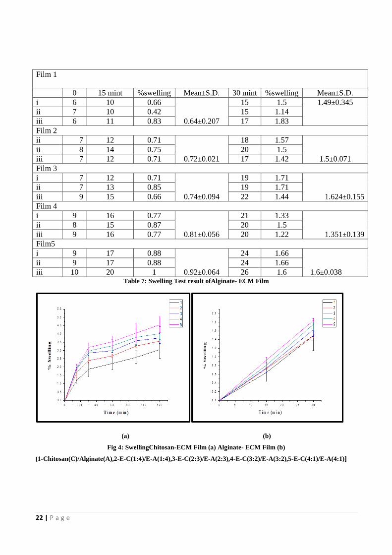
iii 10 20 1 26 1.6 Table 7: Swelling Test result ofAlginate- ECM Film
(a) (b)
Fig 4: SwellingChitosan-ECM Film (a) Alginate- ECM Film (b)
[1-Chitosan(C)/Alginate(A),2-E-C(1:4)/E-A(1:4),3-E-C(2:3)/E-A(2:3),4-E-C(3:2)/E-A(3:2),5-E-C(4:1)/E-A(4:1)]
23 | P a g e
Generally the % swelling of chitosan films are more than alginate films. Another interesting finding
in the alginate films lasts less than the chitosan films in PBS before getting disintegrated. This may
be because of the uncross linking nature of the films. So increase ECM concentration will lead to
more retention of water in the films. Hence will be more useful for the dry wound for which the
moisture or water is necessary for better wound healing.
Alginate films were found to disintegrate within 30 mints whereas Chitosan films were found to not
disintegrate even after 2 hours. This may be because of more ECM in the Chitosan films which
within themselves or with Chitosan films some short of bonds. Whereas in Alginate-ECM chitosan
films might not have participated in any type of bond formation.
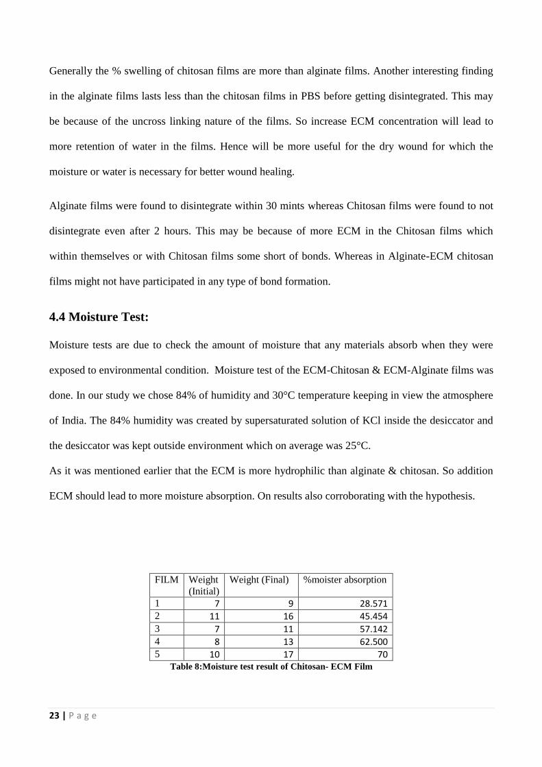
4.4 Moisture Test:
Moisture tests are due to check the amount of moisture that any materials absorb when they were
exposed to environmental condition. Moisture test of the ECM-Chitosan & ECM-Alginate films was
done. In our study we chose 84% of humidity and 30°C temperature keeping in view the atmosphere
of India. The 84% humidity was created by supersaturated solution of KCl inside the desiccator and
the desiccator was kept outside environment which on average was 25°C.
As it was mentioned earlier that the ECM is more hydrophilic than alginate & chitosan. So addition
ECM should lead to more moisture absorption. On results also corroborating with the hypothesis.
FILM Weight
(Initial)
Weight (Final) %moister absorption
1 7 9 28.571 2 11 16 45.454 3 7 11 57.142 4 8 13 62.500 5 10 17 70
Table 8:Moisture test result of Chitosan- ECM Film
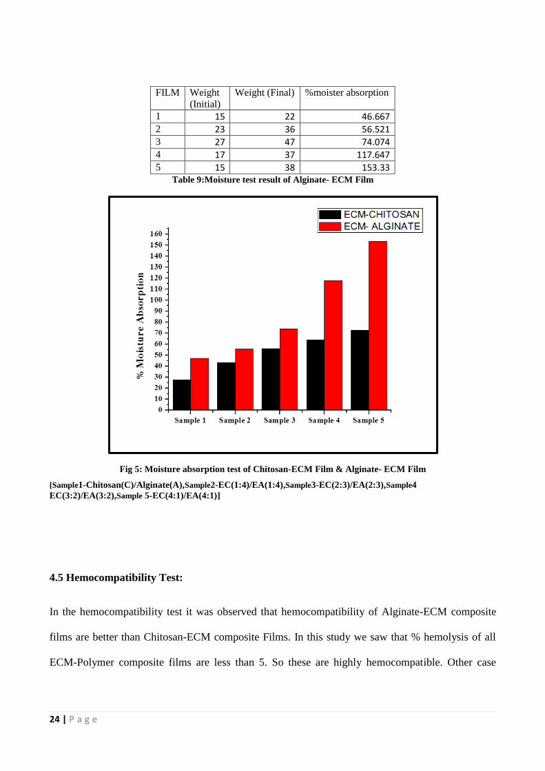
24 | P a g e
FILM Weight
(Initial)
Weight (Final) %moister absorption
1 15 22 46.667 2 23 36 56.521 3 27 47 74.074 4 17 37 117.647 5 15 38 153.33
Table 9:Moisture test result of Alginate- ECM Film
Fig 5: Moisture absorption test of Chitosan-ECM Film & Alginate- ECM Film
[Sample1-Chitosan(C)/Alginate(A),Sample2-EC(1:4)/EA(1:4),Sample3-EC(2:3)/EA(2:3),Sample4
EC(3:2)/EA(3:2),Sample 5-EC(4:1)/EA(4:1)]
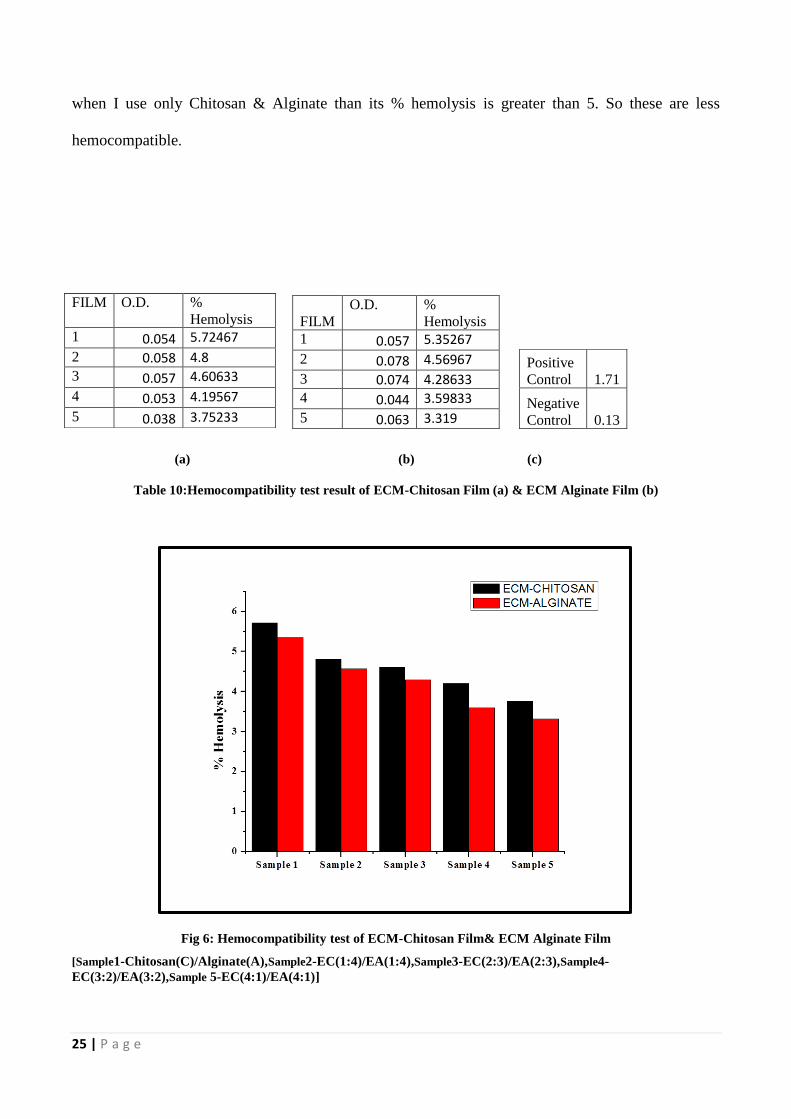
4.5 Hemocompatibility Test:
In the hemocompatibility test it was observed that hemocompatibility of Alginate-ECM composite
films are better than Chitosan-ECM composite Films. In this study we saw that % hemolysis of all
ECM-Polymer composite films are less than 5. So these are highly hemocompatible. Other case
25 | P a g e
when I use only Chitosan & Alginate than its % hemolysis is greater than 5. So these are less
hemocompatible.
(a) (b) (c)
Table 10:Hemocompatibility test result of ECM-Chitosan Film (a) & ECM Alginate Film (b)
Fig 6: Hemocompatibility test of ECM-Chitosan Film& ECM Alginate Film
[Sample1-Chitosan(C)/Alginate(A),Sample2-EC(1:4)/EA(1:4),Sample3-EC(2:3)/EA(2:3),Sample4-
EC(3:2)/EA(3:2),Sample 5-EC(4:1)/EA(4:1)]
FILM O.D. %
Hemolysis
1 0.054 5.72467
2 0.058 4.8
3 0.057 4.60633
4 0.053 4.19567
5 0.038 3.75233
FILM
O.D. %
Hemolysis
1 0.057 5.35267
2 0.078 4.56967
3 0.074 4.28633 4 0.044 3.59833
5 0.063 3.319
Positive
Control 1.71
Negative
Control 0.13
26 | P a g e
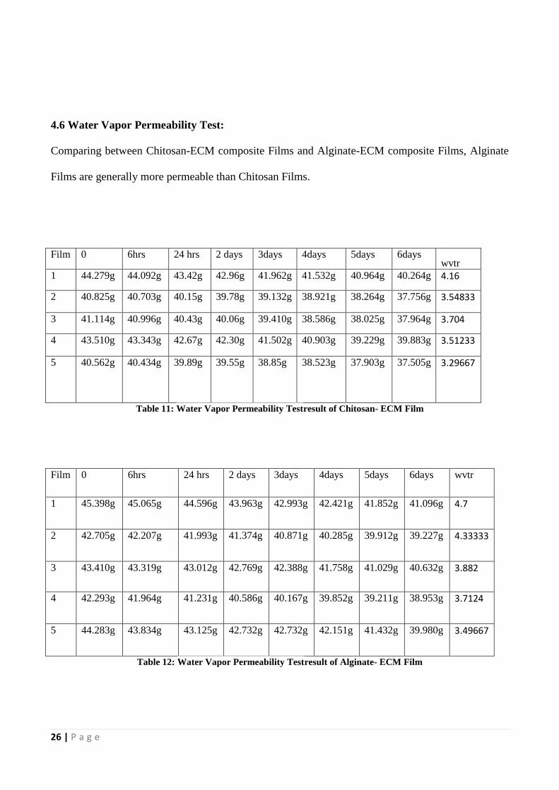
4.6 Water Vapor Permeability Test:
Comparing between Chitosan-ECM composite Films and Alginate-ECM composite Films, Alginate
Films are generally more permeable than Chitosan Films.
Film 0 6hrs 24 hrs 2 days 3days 4days 5days 6days wvtr
1 44.279g 44.092g 43.42g 42.96g 41.962g 41.532g 40.964g 40.264g 4.16
2 40.825g 40.703g 40.15g 39.78g 39.132g 38.921g 38.264g 37.756g 3.54833
3 41.114g 40.996g 40.43g 40.06g 39.410g 38.586g 38.025g 37.964g 3.704
4 43.510g 43.343g 42.67g 42.30g 41.502g 40.903g 39.229g 39.883g 3.51233
5 40.562g 40.434g 39.89g 39.55g 38.85g 38.523g 37.903g 37.505g 3.29667
Table 11: Water Vapor Permeability Testresult of Chitosan- ECM Film
Film 0 6hrs 24 hrs 2 days 3days 4days 5days 6days wvtr
1 45.398g 45.065g 44.596g 43.963g 42.993g 42.421g 41.852g 41.096g 4.7
2 42.705g 42.207g 41.993g 41.374g 40.871g 40.285g 39.912g 39.227g 4.33333
3 43.410g 43.319g 43.012g 42.769g 42.388g 41.758g 41.029g 40.632g 3.882
4 42.293g 41.964g 41.231g 40.586g 40.167g 39.852g 39.211g 38.953g 3.7124
5 44.283g 43.834g 43.125g 42.732g 42.732g 42.151g 41.432g 39.980g 3.49667
Table 12: Water Vapor Permeability Testresult of Alginate- ECM Film
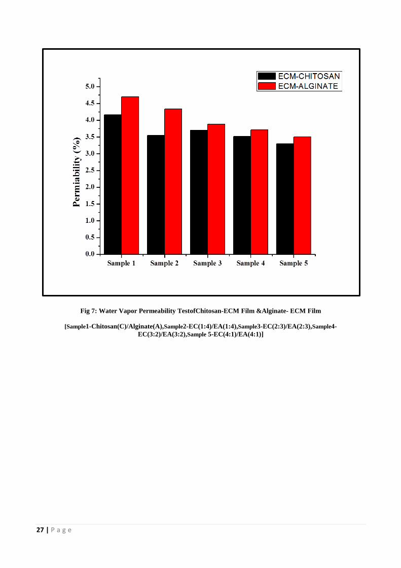
27 | P a g e
Fig 7: Water Vapor Permeability TestofChitosan-ECM Film &Alginate- ECM Film
[Sample1-Chitosan(C)/Alginate(A),Sample2-EC(1:4)/EA(1:4),Sample3-EC(2:3)/EA(2:3),Sample4-
EC(3:2)/EA(3:2),Sample 5-EC(4:1)/EA(4:1)]
28 | P a g e
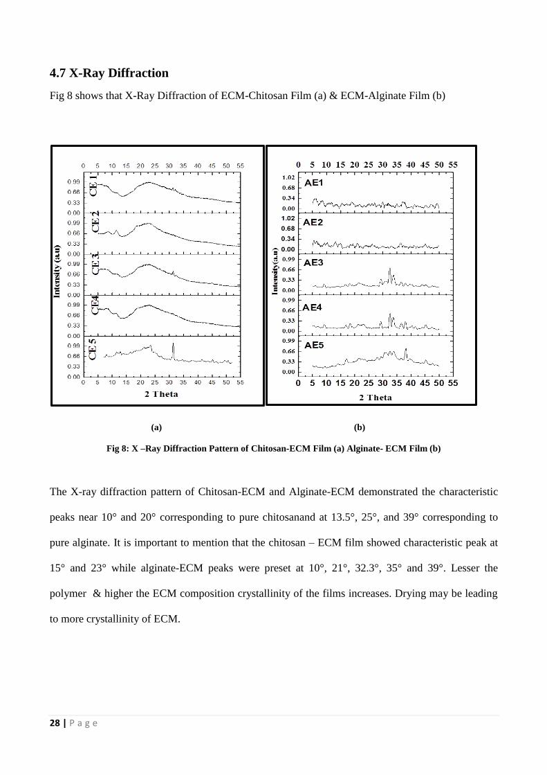
4.7 X-Ray Diffraction
Fig 8 shows that X-Ray Diffraction of ECM-Chitosan Film (a) & ECM-Alginate Film (b)
(a) (b)
Fig 8: X –Ray Diffraction Pattern of Chitosan-ECM Film (a) Alginate- ECM Film (b)
The X-ray diffraction pattern of Chitosan-ECM and Alginate-ECM demonstrated the characteristic
peaks near 10° and 20° corresponding to pure chitosanand at 13.5°, 25°, and 39° corresponding to
pure alginate. It is important to mention that the chitosan – ECM film showed characteristic peak at
15° and 23° while alginate-ECM peaks were preset at 10°, 21°, 32.3°, 35° and 39°. Lesser the
polymer & higher the ECM composition crystallinity of the films increases. Drying may be leading
to more crystallinity of ECM.
29 | P a g e
4.8 Fourier Transform Infrared Spectroscopy
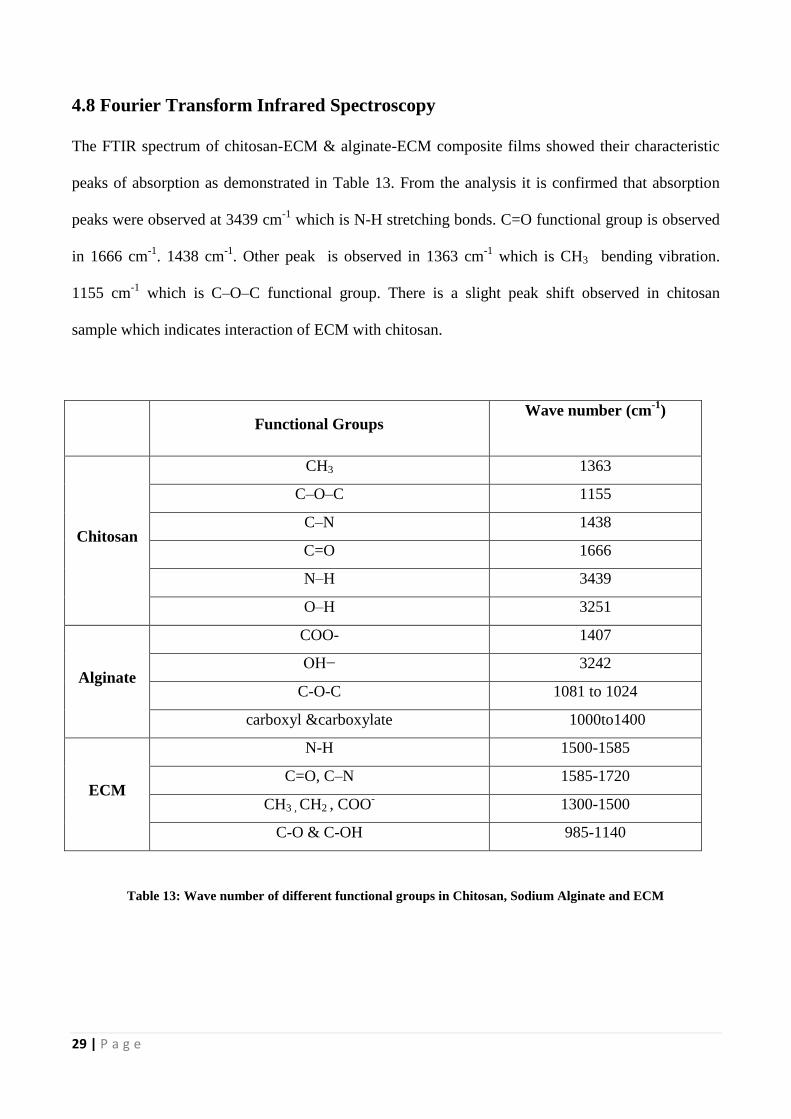
The FTIR spectrum of chitosan-ECM & alginate-ECM composite films showed their characteristic
peaks of absorption as demonstrated in Table 13. From the analysis it is confirmed that absorption
peaks were observed at 3439 cm-1
which is N-H stretching bonds. C=O functional group is observed
in 1666 cm-1
. 1438 cm-1
. Other peak is observed in 1363 cm-1
which is CH3 abending vibration.
1155 cm-1
which isaC–O–C functional group. There is a slight peak shift observed in chitosan
sample which indicates interaction of ECM with chitosan.
Functional Groups Wave number (cm
-1)
Chitosan
CH3 1363
C–O–C 1155
C–N 1438
C=O 1666
N–H 3439
O–H 3251
Alginate
COO- 1407
OH− 3242
C-O-C 1081 to 1024
carboxyl &carboxylate 1000to1400
ECM
N-H 1500-1585
C=O, C–N 1585-1720
CH3 , CH2 , COO- 1300-1500
C-O & C-OH 985-1140
Table 13: Wave number of different functional groups in Chitosan, Sodium Alginate and ECM
30 | P a g e
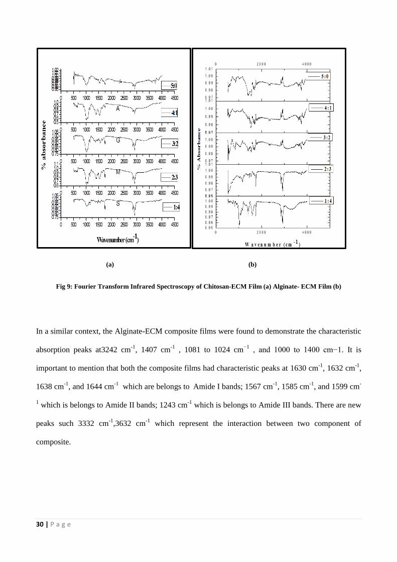
(a) (b)
Fig 9: Fourier Transform Infrared Spectroscopy of Chitosan-ECM Film (a) Alginate- ECM Film (b)
In a similar context, the Alginate-ECM composite films were found to demonstrate the characteristic
absorption peaks at3242 cm-1
, 1407 cm-1
, 1081 to 1024 cm−1
, and 1000 to 1400 cm−1. It is
important to mention that both the composite films had characteristic peaks at 1630 cm-1
, 1632 cm-1
,
1638 cm-1
, and 1644 cm-1
which are belongs toaAmide I bands; 1567 cm-1
, 1585 cm-1
, and 1599 cm-
1 which is belongs to Amide II bands; 1243 cm
-1 which is belongs to Amide III bands. There are new
peaks such 3332 cm-1
,3632 cm-1
which represent the interaction between two component of
composite.
31 | P a g e
Chapter 5
CONCLUSION AND FUTURE WORKS
32 | P a g e
Alginate films without ECM were found to be less thick than the chitosan films without ECM. Upon
swelling the films differed in the hydrophilicity as uptake of water by chitosan films were more than
alginate films. This accounts to the moisture retention capacity of films Apart from this since ECM is
more hydrophilic than alginate and chitosan and addition of higher proportion of ECM in films
imparts them more hydrophilicity. Another interesting point is that greater the proportion of ECM in
the films, higher is the crystallinity. Further in-vitro and in-vivo characterizations will give more
information regarding its utility.
33 | P a g e
References
[1] M. N. Ravi Kumar, "A review of chitin and chitosan applications," Reactive and functional
polymers, vol. 46, pp. 1-27, 2000.
[2] P. Terrill, et al., "Absorption of blood by moist wound healing dressings," Primary Intention:
The Australian Journal of Wound Management, vol. 11, p. 7, 2003.
[3] K. Harish Prashanth and R. Tharanathan, "Chitin/chitosan: modifications and their unlimited
application potential—an overview," Trends in food science & technology, vol. 18, pp. 117-
131, 2007.
[4] W. R. Gombotz and S. F. Wee, "Protein release from alginate matrices," Advanced drug
delivery reviews, vol. 64, pp. 194-205, 2012.
[5] P. K. Kreeger, et al., "The in vitro regulation of ovarian follicle development using alginate-
extracellular matrix gels," Biomaterials, vol. 27, pp. 714-723, 2006.
[6] T. Gilchrist and A. Martin, "Wound treatment with Sorbsan—an alginate fibre dressing,"
Biomaterials, vol. 4, pp. 317-320, 1983.
[7] G. Motta, "Calcium alginate topical wound dressings: a new dimension in the cost-effective
treatment for exudating dermal wounds and pressure sores," Ostomy/wound management,
vol. 25, pp. 52-56, 1988.
[8] G. M. Edwards, et al., "Regulation of mammary differentiation by extracellular matrix
involves protein-tyrosine phosphatases," Journal of Biological Chemistry, vol. 273, pp. 9495-
9500, 1998.
[9] F. T. Bosman and I. Stamenkovic, "Functional structure and composition of the extracellular
matrix," The Journal of pathology, vol. 200, pp. 423-428, 2003.
[10] J. H. Hamman, "Composition and applications of Aloe vera leaf gel," Molecules, vol. 13, pp.
1599-1616, 2008.
34 | P a g e
[11] N. Yun, et al., "Protective effect of Aloe vera on polymicrobial sepsis in mice," Food and
chemical toxicology, vol. 47, pp. 1341-1348, 2009.
[12] S.-Y. Lin, et al., "Design and evaluation of drug-loaded wound dressing having
thermoresponsive, adhesive, absorptive and easy peeling properties," Biomaterials, vol. 22,
pp. 2999-3004, 2001.
[13] M. G. Fouda, et al., "Use of chitosan/polyamine biopolymers based cotton as a model system
to prepare antimicrobial wound dressing," International Journal of Diabetes Mellitus, vol. 1,
pp. 61-64, 2009.
[14] Rupesh Gajanan Nawalakhe, et al., " Development of Electrospun Imino chitosan for
Improved Wound Healing Application"
[15] A. D. Sezer, et al., "Chitosan film containing fucoidan as a wound dressing for dermal burn
healing: preparation and in vitro/in vivo evaluation," AAPS PharmSciTech, vol. 8, pp. E94-
E101, 2007.
[16] Immanuel M. Sebastine and David J. Williams, et al., " The Role of Mechanical Stimulation
in Engineering of Extracellular Matrix (ECM)," in Molecular, Cellular and Tissue
Engineering, 2006. Proceedings of the 28th IEEE-EMBS Annual International Conference
on, 2006, pp. 3648-3651.
[17] Biji Balakrishnana, M. Mohanty et al., ―Evaluation of an in situ forming hydrogel wound
dressing based on oxidized alginate and gelatin‖ Biomaterials 6335–6342,2005
[18] R. Jayakumar, et al., "Novel chitin and chitosan nanofibers in biomedical applications,"
Biotechnology advances, vol. 28, pp. 142-150, 2010.
[19] A. Kirichenko, et al., "Morphological study of burn wound healing with the use of collagen-
chitosan wound dressing," Bulletin of experimental biology and medicine, vol. 154, pp. 692-
696, 2013.
35 | P a g e
[20] S. Şenel and S. J. McClure, "Potential applications of chitosan in veterinary medicine,"
Advanced drug delivery reviews, vol. 56, pp. 1467-1480, 2004.
[21] R. Rezende, et al., "Experimental Characterisation of the Alginate Gelation Process for Rapid
Prototyping," CHEMICAL ENGINEERING, vol. 11, 2007.
[22] R. F. Pereira, et al., "Novel Alginate/Aloe Vera Hydrogel Blends as Wound Dressings for the
Treatment of Several Types of Wounds," CHEMICAL ENGINEERING, vol. 32, 2013.
[23] C. Simpliciano, et al., "Cross-Linked Alginate Film Pore Size Determination Using Atomic
Force Microscopy and Validation Using Diffusivity Determinations," Journal of Surface
Engineered Materials and Advanced Technology, vol. 3, p. 1, 2013.
[24] S. N. Syahirah, et al., "Optimization of Aloe vera and alginate as encapsulating matrices for
Lactobacillus acidophilus using FCCD-RSM approach," in System Engineering and
Technology (ICSET), 2013 IEEE 3rd International Conference on, 2013, pp. 43-47.
[25] J. Alvarado-González, et al., "Optical microstructrual crostructrual, functional and
nanomechanical properties of Aloe vera gel/gellan gum edible films propiedades
opticas,microestructurales, funcionales nanomecanic as depeliculas comestibles gel Aloe
vera/ Goma Gelano."
[26] K. Yamashita, et al., "Intact extracellular matrix can be utilized for bio-integrated materials
and tissue engineering," in Molecular, Cellular and Tissue Engineering, 2002. Proceedings
of the IEEE-EMBS Special Topic Conference on, 2002, pp. 116-119.
[28] D. Kim, et al., "Combined effects of shear stress and extracellular matrices on vascular
differentiation of mouse embryonic stem cells," in Bioengineering Conference (NEBEC),
2011 IEEE 37th Annual Northeast, 2011, pp. 1-2.
[29] Y. Chen, et al., "Measuring collective cell movement and extracellular matrix interactions
using magnetic resonance imaging," Scientific reports, vol. 3, 2013.