EyePACS DIGITAL RETINAL IMAGE GRADING · EyePACS DIGITAL RETINAL IMAGE GRADING PROTOCOL NARRATIVE...
12
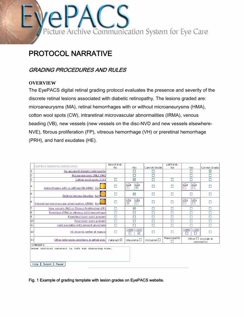
EyePACS DIGITAL RETINAL IMAGE GRADING PROTOCOL NARRATIVE GRADING PROCEDURES AND RULES OVERVIEW The EyePACS digital retinal grading protocol evaluates the presence and severity of the discrete retinal lesions associated with diabetic retinopathy. The lesions graded are: microaneurysms (MA), retinal hemorrhages with or without microaneurysms (HMA), cotton wool spots (CW), intraretinal microvascular abnormalities (IRMA), venous beading (VB), new vessels (new vessels on the disc-NVD and new vessels elsewhere- NVE), fibrous proliferation (FP), vitreous hemorrhage (VH) or preretinal hemorrhage (PRH), and hard exudates (HE). Fig. 1 Example of grading template with lesion grades on EyePACS website.
Transcript of EyePACS DIGITAL RETINAL IMAGE GRADING · EyePACS DIGITAL RETINAL IMAGE GRADING PROTOCOL NARRATIVE...
EyePACS DIGITAL RETINAL IMAGE GRADING
PROTOCOL NARRATIVE
GRADING PROCEDURES AND RULES
OVERVIEW The EyePACS digital retinal grading protocol evaluates the presence and severity of the discrete retinal lesions associated with diabetic retinopathy. The lesions graded are: microaneurysms (MA), retinal hemorrhages with or without microaneurysms (HMA), cotton wool spots (CW), intraretinal microvascular abnormalities (IRMA), venous beading (VB), new vessels (new vessels on the disc-NVD and new vessels elsewhere-NVE), fibrous proliferation (FP), vitreous hemorrhage (VH) or preretinal hemorrhage (PRH), and hard exudates (HE).
Fig. 1 Example of grading template with lesion grades on EyePACS website.
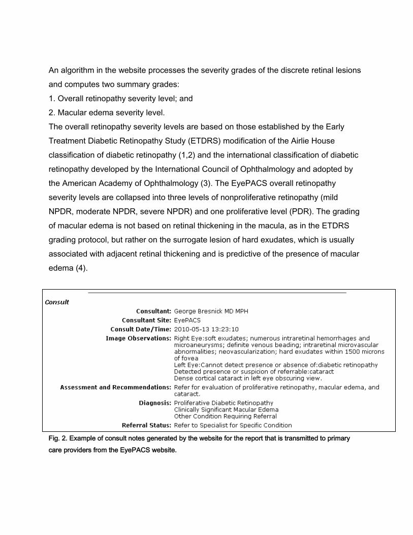
An algorithm in the website processes the severity grades of the discrete retinal lesions and computes two summary grades: 1. Overall retinopathy severity level; and 2. Macular edema severity level. The overall retinopathy severity levels are based on those established by the Early Treatment Diabetic Retinopathy Study (ETDRS) modification of the Airlie House classification of diabetic retinopathy (1,2) and the international classification of diabetic retinopathy developed by the International Council of Ophthalmology and adopted by the American Academy of Ophthalmology (3). The EyePACS overall retinopathy severity levels are collapsed into three levels of nonproliferative retinopathy (mild NPDR, moderate NPDR, severe NPDR) and one proliferative level (PDR). The grading of macular edema is not based on retinal thickening in the macula, as in the ETDRS grading protocol, but rather on the surrogate lesion of hard exudates, which is usually associated with adjacent retinal thickening and is predictive of the presence of macular edema (4).
Fig. 2. Example of consult notes generated by the website for the report that is transmitted to primary care providers from the EyePACS website.
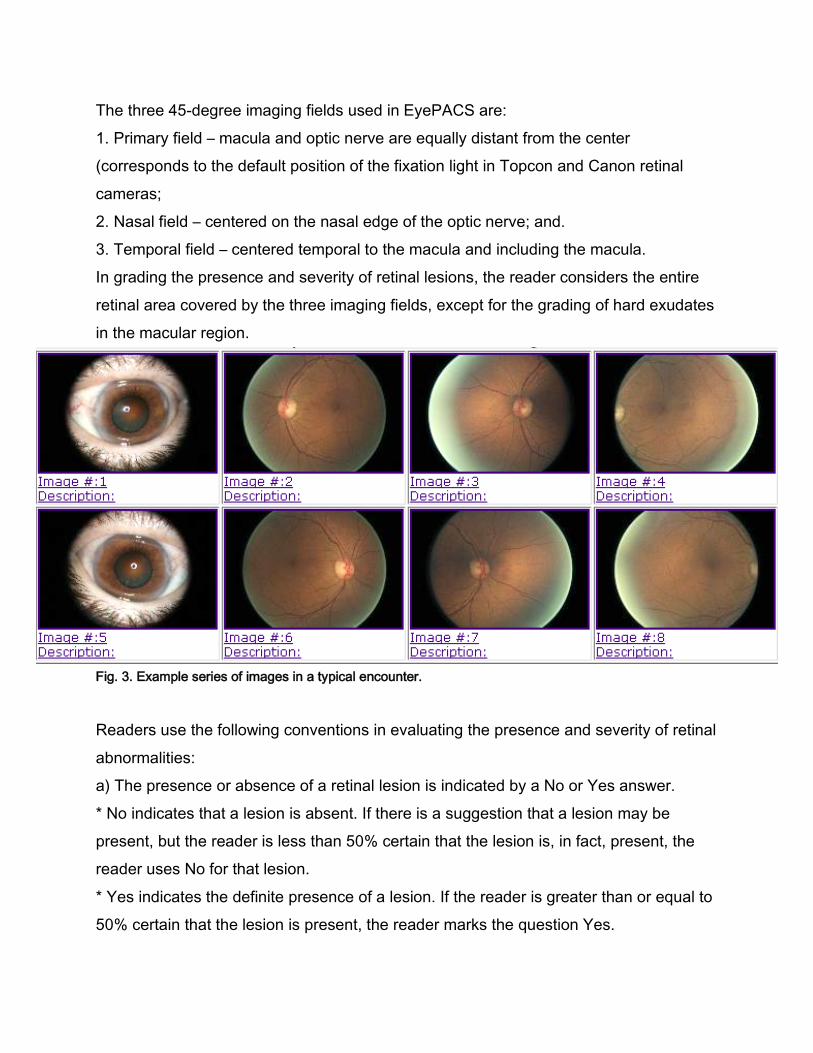
The three 45-degree imaging fields used in EyePACS are: 1. Primary field – macula and optic nerve are equally distant from the center (corresponds to the default position of the fixation light in Topcon and Canon retinal cameras; 2. Nasal field – centered on the nasal edge of the optic nerve; and. 3. Temporal field – centered temporal to the macula and including the macula. In grading the presence and severity of retinal lesions, the reader considers the entire retinal area covered by the three imaging fields, except for the grading of hard exudates in the macular region.
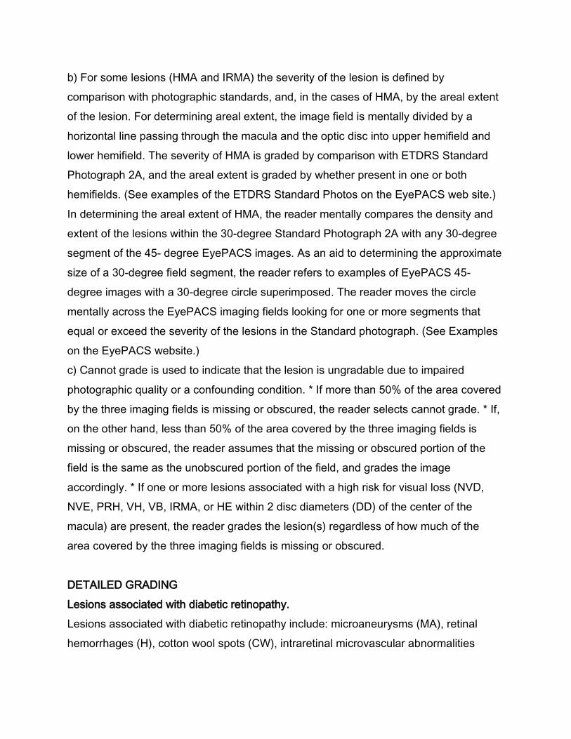
Fig. 3. Example series of images in a typical encounter. Readers use the following conventions in evaluating the presence and severity of retinal abnormalities: a) The presence or absence of a retinal lesion is indicated by a No or Yes answer. * No indicates that a lesion is absent. If there is a suggestion that a lesion may be present, but the reader is less than 50% certain that the lesion is, in fact, present, the reader uses No for that lesion. * Yes indicates the definite presence of a lesion. If the reader is greater than or equal to 50% certain that the lesion is present, the reader marks the question Yes.
b) For some lesions (HMA and IRMA) the severity of the lesion is defined by comparison with photographic standards, and, in the cases of HMA, by the areal extent of the lesion. For determining areal extent, the image field is mentally divided by a horizontal line passing through the macula and the optic disc into upper hemifield and lower hemifield. The severity of HMA is graded by comparison with ETDRS Standard Photograph 2A, and the areal extent is graded by whether present in one or both hemifields. (See examples of the ETDRS Standard Photos on the EyePACS web site.) In determining the areal extent of HMA, the reader mentally compares the density and extent of the lesions within the 30-degree Standard Photograph 2A with any 30-degree segment of the 45- degree EyePACS images. As an aid to determining the approximate size of a 30-degree field segment, the reader refers to examples of EyePACS 45-degree images with a 30-degree circle superimposed. The reader moves the circle mentally across the EyePACS imaging fields looking for one or more segments that equal or exceed the severity of the lesions in the Standard photograph. (See Examples on the EyePACS website.) c) Cannot grade is used to indicate that the lesion is ungradable due to impaired photographic quality or a confounding condition. * If more than 50% of the area covered by the three imaging fields is missing or obscured, the reader selects cannot grade. * If, on the other hand, less than 50% of the area covered by the three imaging fields is missing or obscured, the reader assumes that the missing or obscured portion of the field is the same as the unobscured portion of the field, and grades the image accordingly. * If one or more lesions associated with a high risk for visual loss (NVD, NVE, PRH, VH, VB, IRMA, or HE within 2 disc diameters (DD) of the center of the macula) are present, the reader grades the lesion(s) regardless of how much of the area covered by the three imaging fields is missing or obscured. DETAILED GRADING Lesions associated with diabetic retinopathy. Lesions associated with diabetic retinopathy include: microaneurysms (MA), retinal hemorrhages (H), cotton wool spots (CW), intraretinal microvascular abnormalities
(IRMA), venous beading (VB), hard exudates (HE), new vessels (NV), fibrous proliferation (FP), vitreous hemorrhage (VH) and preretinal hemorrhage (PRH). (Other lesions, such as venous loops and venous or arteriolar sheathing are not graded, but can be described in the Comments Section.) 1. No apparent diabetic retinopathy Mark "Yes" if there is no apparent diabetic retinopathy, then skip to Question 13. Mark "Cannot Grade" If the presence or absence of diabetic retinal lesions in the photographed fields cannot be assessed. Microaneurysms (MA) Microaneurysms (MA), usually the first changes that define diabetic retinopathy that are seen on fundus photographs, appear as small circular red dots with well defined borders, varying in size from about 20µ in diameter to125µ in diameter (125µ is approximately the diameter of a branch vein within the optic nerve head). Dot hemorrhages of a similar size cannot be distinguished from microaneurysms without fluorescein angiography. Therefore, small circular red dot, which may represent a combination of microaneurysms and dot hemorrhages, are combined in the grading process as MA. The reader answers: 2. Microaneurysms ONLY Answer "Yes" if no other diabetic retinal lesions are present. Answer "Cannot Grade" if presence or absence of microaneurysms (as defined above) in the photographed fields cannot be assessed. Cotton wool spots (CW) Cotton wool spots (CW) or soft exudates are a sign of localized retinal ischemia. The reader evaluates the presence of CW anywhere in the three imaging fields. 3. Cotton wool spots (CW) Hemorrhages with or without Microaneurysms (HMA) In addition to MA, the reader evaluates retinal hemorrhages (H) which appear as red spots of varying size with irregular margins and uneven densities. They are classically divided into blot hemorrhages (usually located in the middle retinal layers) and flame
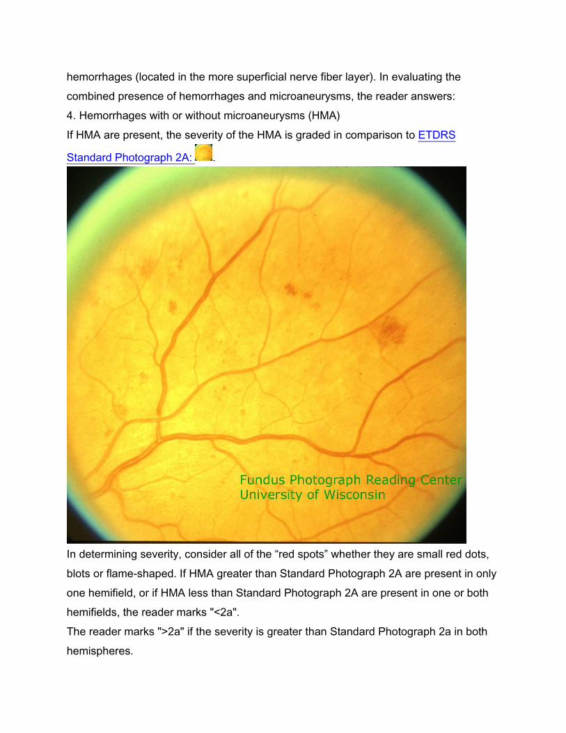
hemorrhages (located in the more superficial nerve fiber layer). In evaluating the combined presence of hemorrhages and microaneurysms, the reader answers: 4. Hemorrhages with or without microaneurysms (HMA) If HMA are present, the severity of the HMA is graded in comparison to ETDRS
Standard Photograph 2A: .
In determining severity, consider all of the “red spots” whether they are small red dots, blots or flame-shaped. If HMA greater than Standard Photograph 2A are present in only one hemifield, or if HMA less than Standard Photograph 2A are present in one or both hemifields, the reader marks "<2a". The reader marks ">2a" if the severity is greater than Standard Photograph 2a in both hemispheres.
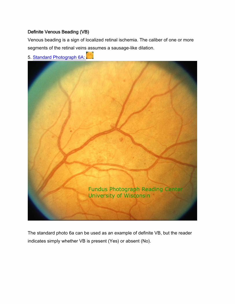
Definite Venous Beading (VB) Venous beading is a sign of localized retinal ischemia. The caliber of one or more segments of the retinal veins assumes a sausage-like dilation.
5. Standard Photograph 6A:
The standard photo 6a can be used as an example of definite VB, but the reader indicates simply whether VB is present (Yes) or absent (No).
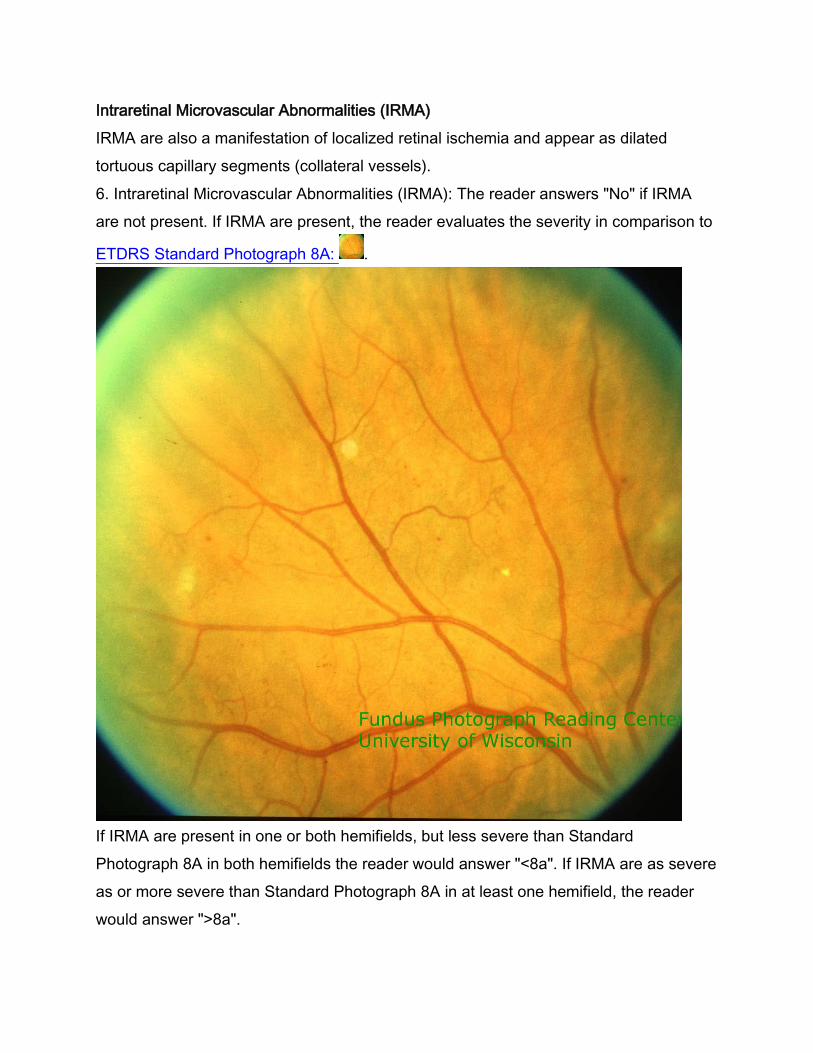
Intraretinal Microvascular Abnormalities (IRMA) IRMA are also a manifestation of localized retinal ischemia and appear as dilated tortuous capillary segments (collateral vessels). 6. Intraretinal Microvascular Abnormalities (IRMA): The reader answers "No" if IRMA are not present. If IRMA are present, the reader evaluates the severity in comparison to
ETDRS Standard Photograph 8A: .
If IRMA are present in one or both hemifields, but less severe than Standard Photograph 8A in both hemifields the reader would answer "<8a". If IRMA are as severe as or more severe than Standard Photograph 8A in at least one hemifield, the reader would answer ">8a".
New vessels (NV) and/or Fibrous Proliferation (FP) Proliferative diabetic retinopathy is characterized by growth of abnormal blood vessels and fibrous tissue from the optic nerve head or from the inner retinal surface (usually on or near retinal veins). The new vessels, which appear initially as fine tufts on the inner surface of the retina subsequently grow into the outermost layer of the vitreous. Initially they consist of fine “naked” vessels, and, as they progress, a fibrous tissue component is added. Ultimately, the vessel component regresses and a predominately fibrous frond may be seen. Therefore, the presence of fibrous tissue without a visible vascular component may indicate proliferative retinopathy. If the reader believes that a fibrous tissue frond originated from neovascular growth (based on its location and configuration), then the question regarding new vessels and/or fibrous proliferation should be answered "Yes". If the reader believes that fibrous (or glial) tissue is from another condition (e.g., epiretinal membrane or optic disc congenital glial remnant), then the question should be answered "No". The reader answers: 7. New vessels (NV) and/or fibrous proliferation (FP) Preretinal (PRH) and/or Vitreous Hemorrhage (VH) Hemorrhage on the surface of the retina (preretinal) and/or in the vitreous is assessed. In the grading algorithm, VH or PRH are considered a manifestation of proliferative diabetic retinopathy, even if no NV or FP is seen. The reader answers: 8. Preretinal hemorrhage (PRH) and/or vitreous hemorrhage (VH) Laser scars (PRP or Focal) If laser scars are present, the reader is asked to distinguish between panretinal laser scars (usually between 200 microns and 500 microns in diameter and located beyond the posterior pole in all quadrants) or focal laser scars (usually less than 200 microns in diameter and located in the macular region.) The reader answers: 9. Panretinal laser scars present 10. Focal laser scars present
Hard Exudates (HE) Retinal hard exudates (HE) are variable in size, sharply defined, and yellow. They may be scattered, aggregated, or “ring-like” in their distribution. The reader indicates the presence or absence of HE in relation to the center of the macula. The reader answers: 11. Hard exudates (HE) present anywhere. 12. Hard exudates close center of the macula. The reader evaluates the proximity of any hard exudate to the center of the macula. Mark "No" If HE are not present or are present, but further than 2 disc diameters from the center of the macula; mark "<2DD" if hard exudates are within 2 disc diameters but further than 1 disc diameter from the center of the macula; mark "<1DD" if hard exudates are within 1 disc diameter from the center of the macula. Other Conditions Requiring Referral 13. Mark appropriate boxes for cataract, glaucoma, vascular occlusion, any type of maculopathy, and any other condition in either eye that should be followed by an eye care provider. Indicate nature of referable condition in Comments section. IMAGE QUALITY After completion of the detailed grading of lesions, the reader assesses the overall quality of the images in both eyes. Image quality factors to consider are: Focus: Is the focus good enough to perform adequate grading of the smaller retinal lesions such as MA and IRMA? Illumination: Is the illumination adequate (not too dark, not too light)? Are there dark areas or washed-out areas that interfere with detailed grading? Image field definition: Does the primary field include the entire optic nerve head and macula? Are the nasal and temporal fields adequately centered to include at least 80% of the non-overlapping portion of the field (i.e., the nasal portion of the nasal field, temporal portion of the temporal field)? Artifacts: Are the images sufficiently free of artifacts (such as dust spots, arc defects, and eyelash images) to allow adequate grading?
In the dropdown menu in the Image Quality field, the reader chooses one of the following: Excellent: There were no problems in any of the image quality factors listed above, and all of the detailed retinopathy questions were gradable. Good: There were some problems in one or two of the image quality factors, and all of the detailed retinopathy questions were gradable. Adequate: There were some problems in three or four of the image quality factors listed above, and all of the detailed retinopathy questions were gradable. Insufficient for Full Interpretation: One or more of the detailed retinopathy questions were marked “cannot grade,” but some of the questions were gradable. Insufficient for Any Interpretation: None of the questions were gradable. Other: Some other factor(s) in the quality of the images interfered with the grading. The reader should specify the nature of the problems in the Comments Section.
EYEPACS GRADING LEVEL DEFINITIONS Overall diabetic retinopathy severity: Each EyePACS overall diabetic retinopathy severity level is followed by a corresponding numerical level as defined by the Early Treatment Diabetic Retinopathy Study (ETDRS) (1, 2) and an ICD-9-CM diagnostic code: No retinopathy (level 10)(250.00-1): No lesions associated with diabetic retinopathy Mild NPDR (level 20)(362.04): Microaneurysms (MA) only -- No blot or flame hemorrhages or other retinal lesions associated with diabetic retinopathy [including cotton wool spots (CW), intraretinal microvascular abnormalities (IRMA), venous beading (VB), hard exudates (HE), new vessels (NV), fibrous proliferation (FP), vitreous hemorrhage (VH) and preretinal hemorrhage (PRH)] are present. Moderate NPDR (level 35, 43, 47)(362.05): Hemorrhages with or without microaneurysms (HMA) present, but less than the criteria for severe NPDR. If cotton wool spots (CW) and/or hard exudates (HE) are present, but there are no HMA, the eye is classified “no diabetic retinopathy” (level 10), and the presence of these lesions is noted in the Comments Section. IRMA or venous beading (VB) less than the severity described under “severe NPDR” (level 53) may be present.
Severe NPDR (level 53)(362.06): Any one or more of the following (4-2-1-rule): • Hemorrhages/microaneurysms (H/MA)>=Standard 2A in both hemifields (all four quadrants) • Definite venous beading (VB) present in one or more hemifields (two or more quadrants) (see Standard 6A for example of definite VB) • IRMA >=Standard 8A in one or more hemifields (one or more quadrants) PDR (levels 60, 65)(362.02): Any one or more of the following: • New vessels on the disc (NVD) or new vessels elsewhere (NVE) • Fibrous proliferation (FP) on the disc or elsewhere likely to be inactive fibrovascular tissue • Preretinal hemorrhage (PRH) or vitreous hemorrhage (VH) Macular edema severity: No macular edema (no ME): No hard exudates within 2 disc diameters of the center of the macula Macular edema, not clinically significant (ME not CSME)(362.07): Hard exudates within 2 disc diameters of the center of the macula, but not within 1 disc diameter. Macula edema, clinically significant (CSME)(362.07): Hard exudates within 1 disc diameter of the center of the macula
REFERENCES
1. ETDRS Research Group. Grading diabetic retinopathy from stereoscopic color fundus photographs-an extension of the Airlie House classification. ETDRS Report 10. Ophthalmology 1991;98:786-806. 2. ETDRS Research Group. Fundus photographic risk factors for progression of diabetic retinopathy. ETDRS Report 12. Ophthalmology 1991;98:823-833. 3. International Clinical Diabetic Retinopathy Disease Severity Scale; International Council of Ophthalmology.; http://www.icoph.org/standards/pdrclass.html 4. Bresnick, GH, et al. A screening approach to the surveillance of patients with diabetes for the presence of vision-threatening retinopathy. Ophthalmology 2000; 107:19-24 * Reference Images Courtesy of: Fundus Photograph Reading Center Department of Ophthalmology and Visual Sciences, University of Wisconsin