EYE2EYE...Eye2Eye Quarter 3 2019 5 Censor-in-Chief’s Update QEC and Board approve programmatic...
68
EYE2EYE the magazine of the leaders in collaborative eye care 2 Quarter 3 2019 VOLUME 22 ISSUE 3 IN THIS ISSUE: Flexibility during training pg.18 Congress Social Events pg.26 50 years of ophthalmic innovation & development pg.41 RANZCO Congress 2019 speakers pg.28
Transcript of EYE2EYE...Eye2Eye Quarter 3 2019 5 Censor-in-Chief’s Update QEC and Board approve programmatic...
EYE2EYEthe magazine of the leaders in collaborative eye care
2Quarter 3 2019VOLUME 22
ISSUE 3
IN THIS ISSUE:
Flexibility during training pg.18
Congress Social Events pg.26
50 years of ophthalmic innovation & development pg.41
RANZCO Congress 2019 speakers pg.28
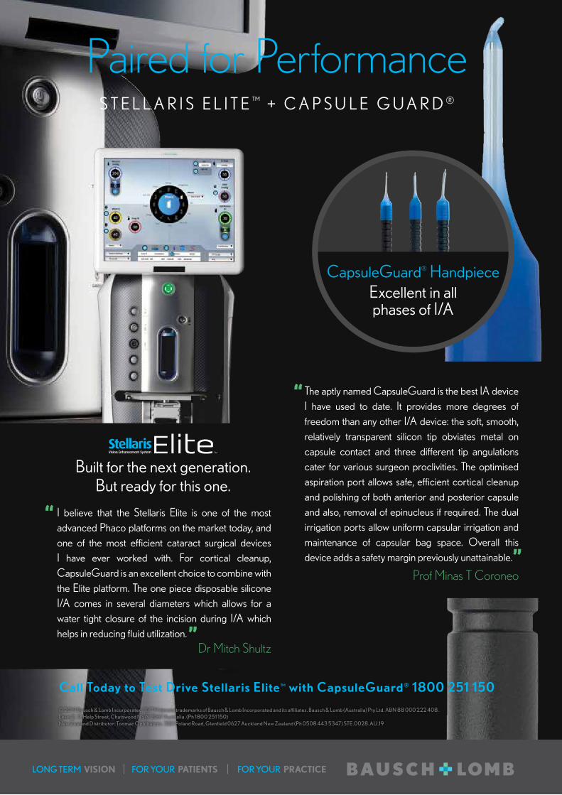
CapsuleGuard® HandpieceExcellent in all phases of I/A
Call Today to Test Drive Stellaris Elite™ with CapsuleGuard® 1800 251 150
© 2019 Bausch & Lomb Incorporated. ®/TM denote trademarks of Bausch & Lomb Incorporated and its affiliates. Bausch & Lomb (Australia) Pty Ltd. ABN 88 000 222 408. Level 2, 12 Help Street, Chatswood NSW 2067 Australia. (Ph 1800 251 150)New Zealand Distributor: Toomac Ophthalmic. 32D Poland Road, Glenfield 0627 Auckland New Zealand (Ph 0508 443 5347) STE.0028.AU.19
Built for the next generation.But ready for this one.
Paired for PerformanceS T E L L A R I S E L I T E ™ + C A P S U L E G U A R D ®
I believe that the Stellaris Elite is one of the most advanced Phaco platforms on the market today, and one of the most efficient cataract surgical devices I have ever worked with. For cortical cleanup, CapsuleGuard is an excellent choice to combine with the Elite platform. The one piece disposable silicone I/A comes in several diameters which allows for a water tight closure of the incision during I/A which helps in reducing fluid utilization.
Dr Mitch Shultz
“
“
The aptly named CapsuleGuard is the best IA device I have used to date. It provides more degrees of freedom than any other I/A device: the soft, smooth, relatively transparent silicon tip obviates metal on capsule contact and three different tip angulations cater for various surgeon proclivities. The optimised aspiration port allows safe, efficient cortical cleanup and polishing of both anterior and posterior capsule and also, removal of epinucleus if required. The dual irrigation ports allow uniform capsular irrigation and maintenance of capsular bag space. Overall this device adds a safety margin previously unattainable.
Prof Minas T Coroneo
“
“
Eye2Eye is published by The Royal Australian and New Zealand College of Ophthalmologists as information for its members. The views expressed in the publication are those of the authors and not necessarily of the College. The inclusion of advertising in this publication does not constitute College endorsement of the products or services advertised.
Editor: Emma Carr Design and layout: Francine DuttonThe Royal Australian and New Zealand College of Ophthalmologists A.C.N 000 644 40494-98 Chalmers Street Surry Hills NSW 2010 Australia Ph: +61 2 9690 1001 Fax: +61 2 9690 1321 E-mail: [email protected] Website: www.ranzco.edu
EYE2EYEthe magazine of the leaders in collaborative eye care
2Message from the President ..........................................................................4
Censor-in-Chief’s Update ..............................................................................5
CEO’s Corner .................................................................................................8
ANZEF Update ............................................................................................10
Membership Spotlight ................................................................................11
Health and WellbeingFlexibility During Training - The Way of the Future ...................................18
Active Bystanders - Stepping Up Now for the Future We Want ................21
RANZCO Office ............................................................................................23
Annual Scientific Congress ..........................................................................24
Policy and Advocacy Matters ......................................................................34
International Development .........................................................................37
Feature Articles 50 Years of Ophthalmic Innovation ..........................................................40
Introduction and Scope: The Future of Ophthalmology Task Force ..........43
Branch Musings ..........................................................................................47
Special Interest Groups ...............................................................................50
RANZCO Affiliates........................................................................................52
Ophthal News .............................................................................................54
Obituary ......................................................................................................62
Calendar of Events ......................................................................................64
Classifieds ...................................................................................................66
Contents
24
37Front cover: 19th century Retinoscopy Training apparatus from Professor Ian McAllister’s collection
CapsuleGuard® HandpieceExcellent in all phases of I/A
Call Today to Test Drive Stellaris Elite™ with CapsuleGuard® 1800 251 150
© 2019 Bausch & Lomb Incorporated. ®/TM denote trademarks of Bausch & Lomb Incorporated and its affiliates. Bausch & Lomb (Australia) Pty Ltd. ABN 88 000 222 408. Level 2, 12 Help Street, Chatswood NSW 2067 Australia. (Ph 1800 251 150)New Zealand Distributor: Toomac Ophthalmic. 32D Poland Road, Glenfield 0627 Auckland New Zealand (Ph 0508 443 5347) STE.0028.AU.19
Built for the next generation.But ready for this one.
Paired for PerformanceS T E L L A R I S E L I T E ™ + C A P S U L E G U A R D ®
I believe that the Stellaris Elite is one of the most advanced Phaco platforms on the market today, and one of the most efficient cataract surgical devices I have ever worked with. For cortical cleanup, CapsuleGuard is an excellent choice to combine with the Elite platform. The one piece disposable silicone I/A comes in several diameters which allows for a water tight closure of the incision during I/A which helps in reducing fluid utilization.
Dr Mitch Shultz
“
“
The aptly named CapsuleGuard is the best IA device I have used to date. It provides more degrees of freedom than any other I/A device: the soft, smooth, relatively transparent silicon tip obviates metal on capsule contact and three different tip angulations cater for various surgeon proclivities. The optimised aspiration port allows safe, efficient cortical cleanup and polishing of both anterior and posterior capsule and also, removal of epinucleus if required. The dual irrigation ports allow uniform capsular irrigation and maintenance of capsular bag space. Overall this device adds a safety margin previously unattainable.
Prof Minas T Coroneo
“
“
4
Message from the President
Message from the President
Impacts on practice by the Medicare review
The Medicare review is a complex review process of all Medicare item numbers. It is stated to be a modernisation process and not a cost-cutting exercise. Under the guidance of Professor Bruce Robinson, the MBS schedule was divided into craft-based sections and committees appointed for each craft group to work with MBS staff.
While the review intentionally did not work directly with RANZCO or other colleges, the clinical committees included Fellows of appropriate colleges. Brad Horsburgh was Chair of the Ophthalmology Committee. Other members included Paul Healey, Angus Turner, Hugh Taylor, Alex Hunyor and Stephanie Watson. One of the consumer representatives was Susi Tegen, a former RANZCO CEO.
At the time of writing, the ophthalmology report was not released for consultation. Other reports that have been released and are relevant to ophthalmology include reports on optometry, specialist and consultant physicians, general practice and nurse practitioners. Highlights of these reports, which, RANZCO has provided feedback for, are: • The Optometry Committee recommended a new item
number for nursing home visits, where perimetry would be performed, that could be used twice a year. RANZCO recommended that tablet-based perimetry has not yet been shown to be equivalent to formal perimetry in the detection and monitoring of field defects. We stated that this item number would be likely to blow out and to generate unreliable clinical data of questionable clinical utility.
• The optometry review did not recommend an increase in scope of practice to laser or intra-vitreal injections. In Oklahoma, optometrists were very recently awarded the ability to perform laser, incisional surgery (lids, chalazia) and intravitreal injections. Previous experience in other US states have shown that optometrists do not always perform treatments that they are entitled to do under legislative acts. The American Academy of Ophthalmology has the ability to monitor optometry billing in real time, and at the time of writing no optometrists had billed for any intravitreal injections across the USA.
• The Specialist and Consultant Physicians Committee recommended that all specialist consultations be time based (<6 minutes, 6-20 minutes, 20-40 minutes, 40-60 minutes). No draft rebates were supplied so it is not possible to model financial changes for ophthalmologists or their patients. Telemedicine items were to be billed in the same manner (i.e. no recognition of ‘store-and-forward’ methodology that is very suited to ophthalmology). Further, there was no recognition that different specialties had different cost bases for routine consulting or telemedicine consulting. The role of referrals was unclear, and it is unclear whether ongoing referrals will be necessary, and whether patients will migrate permanently to specialty care for conditions previously managed by GPs such as dry eye.
• The Nurse Practitioners (NP) Committee recommended that NP be no longer required to work collaboratively with medical practitioners. This does not affect ophthalmology directly but suggests the same may occur with optometry in the future.
It is clear from the Medicare review and the work of other regulators, such as the Australian Commission for Quality and Safety in Health Care and the coming national workforce study to be performed for the Chief Medical Officer, that the practice of ophthalmology is likely to change. One of RANZCO’s roles is to position us to provide meaningful feedback to regulators that they view as important information from the leaders in the eye care world.
To this end I attended the American Academy of Ophthalmology Mid-year Forum (MYF) in April in Washington DC. The goals of the MYF are to provide a mechanism for discussion and debate of proposed policies, to identify new issues to be addressed by the Academy, to receive input from constituent state societies (equivalent of branches), to educate ophthalmology leaders about critical issues facing ophthalmology and to educate and update leaders about critical issues facing ophthalmology. This is very similar to the role of RANZCO Council. In addition, MYF participants are trained in how to best represent our profession to members of Congress and their staff. They then participate in a meeting with their relevant House and Senate Representatives.
A/Prof Heather Mack President, RANZCO
5Eye2Eye Quarter 3 2019
Censor-in-Chief’s UpdateQEC and Board approve programmatic assessment framework for new VTP
RANZCO – through both the Curriculum Committee, Chaired by Dr Catherine Green, and the Chairs of the examination boards – has been working towards a major revision of the vocational training program (VTP). This is expected to further our aim of creating competent general ophthalmologists able to work in a variety of clinical settings including regional areas.
In May 2019, the QEC and Board approved, in principle, a programmatic assessment framework for the new VTP. The framework is designed to fully assess whether a trainee has developed the competencies required to undertake safe, comprehensive practice as a general ophthalmologist and is ready to transition to independent practice. Equal emphasis is placed on learning and assessing foundation skills and knowledge and applied ophthalmology and professional capabilities.
We are reinforcing the importance of the non-clinical CanMeds domains, and also simplifying their assessment, by combining communicator, collaborator, manager/leader, health advocate and professional under a single domain of professional capabilities. Importantly, cultural competency is also included.
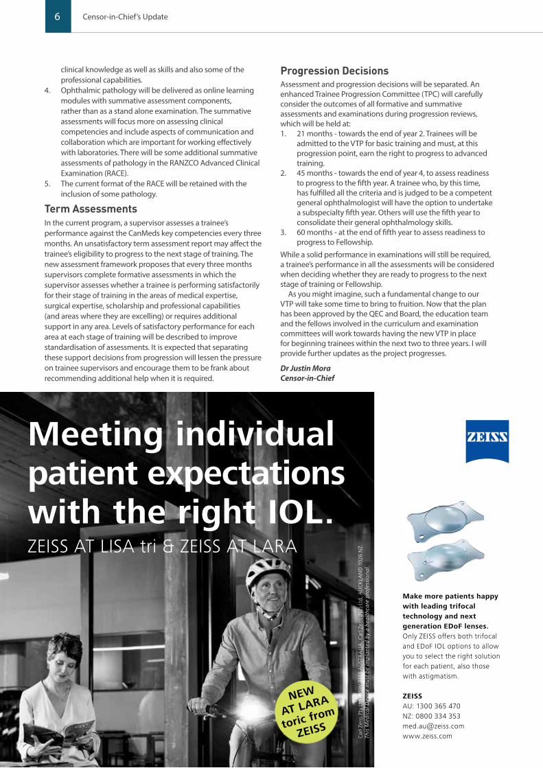
The RANZCO competencies ‘flower’ A range of assessment tools will be used to determine a trainee’s readiness to progress through training, including online assessments in conjunction with learning modules, end of term reports, surgical assessment through Objective Structured Assessments of Technical Skills (OSATS) and logbook audits, surgical simulator assessment modules and examinations. Assessment outcomes will be stored in a trainee’s online portfolio, which they can access at any time and supervisors can access while a trainee is under their supervision.
Examinations A consolidation of the examinations is proposed. 1. COPEM 1 and 2 will be incorporated into an induction
workshop which will welcome trainees to the program, explain policies, processes and expectations and provide training on:
• ocular pharmacology; • management of ophthalmic emergencies; • communication; • cultural competence; • accepting and using feedback to guide learning; and • Royal Australasian College of Surgeons’ (RACS’)
Operating with Respect (anti bullying, harassment and discrimination training).
The feasibility of including a microsurgical skills course and having trainees consolidate their skills in wet lab training in their network is being considered.
2. The Ophthalmic Sciences (OS) examinations will be combined into a single examination which will assess aspects of anatomy, physiology and optics that are clinically relevant to ophthalmology and in a clinical context. Trainees will sit this examination after their first year of training to ensure they have some clinical knowledge. It is hoped that this will encourage trainees to focus more on clinical work during first year rather than on passing the OS examinations.
3. Trainees will sit the Ophthalmic Basic Competencies and Knowledge (OBCK) examination after 15 months of training and this examination will have a stronger focus on assessing
Ophthalmic knowledge & surgical skill
Scholar
Competentgeneral
ophthalmologist
Professional, communicator, collaborator, leader/manager,
health advocate, culturally competent
Fig 1. RANZCO competencies ‘flower’
6 Censor-in-Chief’s Update
clinical knowledge as well as skills and also some of the professional capabilities.
4. Ophthalmic pathology will be delivered as online learning modules with summative assessment components, rather than as a stand alone examination. The summative assessments will focus more on assessing clinical competencies and include aspects of communication and collaboration which are important for working effectively with laboratories. There will be some additional summative assessments of pathology in the RANZCO Advanced Clinical Examination (RACE).
5. The current format of the RACE will be retained with the inclusion of some pathology.
Term Assessments In the current program, a supervisor assesses a trainee’s performance against the CanMeds key competencies every three months. An unsatisfactory term assessment report may affect the trainee’s eligibility to progress to the next stage of training. The new assessment framework proposes that every three months supervisors complete formative assessments in which the supervisor assesses whether a trainee is performing satisfactorily for their stage of training in the areas of medical expertise, surgical expertise, scholarship and professional capabilities (and areas where they are excelling) or requires additional support in any area. Levels of satisfactory performance for each area at each stage of training will be described to improve standardisation of assessments. It is expected that separating these support decisions from progression will lessen the pressure on trainee supervisors and encourage them to be frank about recommending additional help when it is required.
Progression Decisions Assessment and progression decisions will be separated. An enhanced Trainee Progression Committee (TPC) will carefully consider the outcomes of all formative and summative assessments and examinations during progression reviews, which will be held at: 1. 21 months - towards the end of year 2. Trainees will be
admitted to the VTP for basic training and must, at this progression point, earn the right to progress to advanced training.
2. 45 months - towards the end of year 4, to assess readiness to progress to the fifth year. A trainee who, by this time, has fulfilled all the criteria and is judged to be a competent general ophthalmologist will have the option to undertake a subspecialty fifth year. Others will use the fifth year to consolidate their general ophthalmology skills.
3. 60 months - at the end of fifth year to assess readiness to progress to Fellowship.
While a solid performance in examinations will still be required, a trainee’s performance in all the assessments will be considered when deciding whether they are ready to progress to the next stage of training or Fellowship.
As you might imagine, such a fundamental change to our VTP will take some time to bring to fruition. Now that the plan has been approved by the QEC and Board, the education team and the fellows involved in the curriculum and examination committees will work towards having the new VTP in place for beginning trainees within the next two to three years. I will provide further updates as the project progresses.
Dr Justin Mora Censor-in-Chief
NEW
AT LARA
toric from
ZEISS
Make more patients happy with leading trifocal technology and next generation EDoF lenses.Only ZEISS offers both trifocal and EDoF IOL options to allow you to select the right solution for each patient, also those with astigmatism.
ZEISSAU: 1300 365 470NZ: 0800 334 [email protected] www.zeiss.com
Meeting individual patient expectations with the right IOL.ZEISS AT LISA tri & ZEISS AT LARA
Car
l Zei
ss P
ty L
td, N
SW 2
113
AU
STR
ALI
A. C
arl Z
eiss
(NZ)
Ltd
, AU
CK
LAN
D 1
026
NZ.
Th
is M
edic
al D
evic
e m
ust
be
imp
lant
ed b
y a
hea
lthc
are
pro
fess
iona
l.

7Eye2Eye Quarter 3 2019
8
CEO’s Corner
Celebrating RANZCO’s 50th year as a college
At the RANZCO Council meeting on 15 June sustainability was discussed. Sustainability can mean different things, but for RANZCO it is about the sustainability of the organisation and what role we play in ensuring a sustainable future for the planet.
In addressing the first point I will say that there is no question that RANZCO is in a strong financial position with good governance, so there is no immediate need for concern by members. In fact, for a charity/educational organisation with our remit we are in an excellent position thanks to careful governance and management over the last 50 years. But looking well forward we need to consider how we remain so. The Council heard about the increasing cost, both time and money, needed for regulatory compliance as a medical education entity. This is not going to decrease, whatever we may say or wish.
The increase in personal ongoing, lifelong education was discussed, and again this is not going to decrease, so how can RANZCO best support our members? The number of members remains largely static as those entering Fellowship roughly equal those retiring. With no additional members we are expected to do more, and at a higher level. The number of committees at RANZCO has steadily grown over the last few years, driven largely by the need to respond to outside entities. The staff to support these committees has therefore grown. But we will probably reach a point, soon, where members are unwilling or unable to provide more necessary hours of work needed for all these committees. The current way of working is not sustainable. While technology does make things easier, and we are investing heavily to help the process, ultimately it is the intellectual time and energy that makes a difference. I am conscious that Fellows in particular have pressure not only from their work, but other charities or health departments wanting more of their precious time to do their own important work, and still need to have a life beyond ophthalmology. For some individuals now I cannot see how this is possible. For their own health it is not sustainable.
The Council heard about the 17 United Nations Sustainable Development Goals, and how RANZCO is touching on 9 of these as part of our current activities. There is no doubt many around the world, including our members, have grave concerns for the sustainability of the planet. Of course, it is not realistic to think that
RANZCO can solve this problem, but through careful thinking and some actions we can play a part in creating a sustainable future. We will be looking closely at what impacts we can realistically make and ensure our strategic plan and activities reflect these. In some cases, it may be direct action, such as investing in stocks which operate under an ethical, sustainable and strong governance framework (an industry recognised standard). In many situations it may be the influence RANZCO or our Fellows have on others, for example in trying to reduce the huge amount of single use materials associated with surgery in the developed world.
The Council endorsed the Board and me with exploring how we can consider our strategic plan and activities through a sustainability lens. This will be done with expert help and as part of our strategic planning process for 2021 and beyond. It will also be a consideration in setting a long term vision for RANZCO. While we celebrate our 50th anniversary this year, we need to think about how we will still be active and relevant in another 50 years and consider the legacy we are creating for future generations.
Dr David Andrews CEO, RANZCO
CEO’s Corner

9Eye2Eye Quarter 3 2019
AVANT Advertorial
Consent for treatment decisions involving children This can be a difficult issue for medical practitioners. In the case of very young children, generally a parent or guardian can give consent for the child to receive medical treatment. If the treating team and parents cannot agree, a court may be asked to consent on behalf of the child. In such cases, the overriding consideration is what is in the child’s best interests.
As a recent case illustrates, complex clinical situations involving multiple treating teams and a changing prognosis may mean it can be extraordinarily difficult to determine how the child’s interests are best served. In this case, a public hospital sought urgent court authorisation for consent to ophthalmic treatment for an infant, which they argued was urgently required to partially save the infant’s vision.
Background The infant (C) was born prematurely by emergency caesarean section, with extremely low birthweight. She required immediate resuscitation, intubation and mechanical ventilation. From her second week of life there were multiple documented medical issues including bowel obstructions, feed intolerance and retinopathy of prematurity. The parents consented to six surgeries each under general anaesthesia in her first 12 weeks. At 16 weeks (chronological age) a decision was made by the neonatology team, a paediatric surgeon and C’s parents to cease active medical treatment and palliate her. Four weeks later she unexpectedly recovered sufficiently for the hospital to cease palliation.
Consent to ophthalmic surgery With medical decision making now shifting to consider C’s future quality of life, her deteriorating vision became an urgent concern. Her right retina was already fully detached, and with her left retina at risk, a pars plana vitrectomy was urgently proposed. The surgical opinion stated the procedure was at least four weeks overdue, and that at best there was a 40 per cent chance of reattaching her retina and retaining some navigational vision. Without surgery, there was a near absolute risk of blindness.
The surgical risks were stated to include: • loss of the eye (1 in 10000) • total loss of vision (if retina detaches) • infection (1 in 3000) • sympathetic ophthalmia (1 in 3000 to 6000) • cataract (rare in children) • progression to total retinal detachment (60%) • elevated intraocular pressure (usually transient and responsive
to local measures).
According to the case report, C’s parents “were asked on multiple occasions by members of the medical team to consent to the retinal re-attachment surgery taking place, but they did not provide their consent”. Given the time sensitive nature of the surgery, the hospital sought court authorisation.
After the parents had been summoned to appear at a court hearing the next day, another meeting about the proposed surgery was organised between the parents, a general paediatrician, two registrars and a child protection consultant about the proposed surgery. The judgment quoted extensively from notes of that meeting and these offer a key insight into the consent process.
Court hearing The judge noted C’s parents had already authorised six operations for their daughter and numerous other treatments. They felt they had received mixed messages around surgical risk. They had been told five weeks earlier that without retinal surgery their daughter would already be totally blind and were told varying percentages for the expected chance of success.
Given C’s experiences with surgery to date, her mother expressed concerns about anaesthetic risks, in particular, neurological risks and the effects of intubation. She cited issues with extubation, prolonged post-operative intubation requiring two weeks of CPAP, and sepsis after earlier eye surgery. She only received reassurances about anaesthetic, neurological and cardiac risks in telephone calls with specialists the night before the hearing.
With her daughter now making slow developmental gains from being in a palliative care situation weeks earlier, C’s mother was concerned this surgery could lead to a serious setback. Based on the medical information they had received over many weeks, C’s parents were very conflicted about what was best for their daughter, but they were “not against” the procedure.
Best interests The judge concluded surgery would be in C’s best interests and ordered the vitrectomy and any associated procedures be performed the next day. He considered that medical opinion was overwhelmingly in favour of the surgery and that the potential benefit to C substantially outweighed the known risks. He also concluded the concerns raised by the parents about other medical issues affecting the proposed surgery were surmountable in C’s current stable medical state.
Conclusion There was little doubt all involved were acting in what they believed to be for the best interests of the child. It is also clear that the highly specialised nature of the procedure, which was time sensitive but not an emergency situation, made the consent process particularly complex. In such cases, where parents are physically and emotionally overwhelmed by the challenges of caring for a desperately ill infant, they may welcome the decision being taken by an independent third party.
However, some lessons can be learned about better ways to support patients and parents making extremely difficult treatment decisions. Establishing the family’s trust in the treating team was clearly key to this decision. C’s mother is quoted as saying that she was finding it hard to develop a new relationship with a new team and to trust them. Both parents seemed to feel they were receiving conflicting information and that their concerns were not heard, or addressed, until after they experienced the additional distress of receiving a court summons.
From the details included in the reported case, it seems the urgency of getting consent forms signed did get in the way of the decision making process. Finding ways of focusing more on the decision maker’s needs and less on the form signatures could help in similar situations.
For more information on children and consent see https://www.avant.org.au/Resources/Public/20160531-children-and-consent/
Georgie Haysom - Head of Research, Education and Advocacy, Avant
10 ANZEF Update
While the Australian and New Zealand Eye Foundation (ANZEF) was being set up as an internal committee within RANZCO, fundraising efforts focussed on RANZCO members, who have given generously to support the Foundation in its efforts to raise funding for important research and education projects.
With the initial administration of the charity now secured –including applying for and being granted charitable fundraising licences across the states and territories of Australia and in New Zealand
– JulEye provided a good opportunity for ANZEF to broaden our fundraising to target the public.
With this in mind, and with the aim of drawing on the goodwill and support of RANZCO members, ANZEF produced public awareness flyers, focussing on the importance of education and capacity building projects to support people in the Asia Pacific region. These flyers were then distributed to all RANZCO Fellows in Australia and New Zealand to distribute as they see fit. Some Fellows chose to display the flyers in their waiting rooms or hand them out to patients while some choose to hand them out or mail them to specific supporters. The result has been an upturn in donations – including a series of donations in honour of one supporter’s 90th birthday.
Another significant piece of public awareness raising came through the
generosity of Qantas and our our professional conference organisers Think Business. Thanks to our connection with Qantas as an official airline partner for RANZCO’s 2019 Congress – a partnership which was developed and set up by Think Business and RANZCO’s Senior Manager of Events and Industry Relationships, Sarah Stedman – we were granted free a half page advert in the Qantas inflight magazine in July. This was an excellent opportunity to promote ANZEF to a wider group of people. See below for an example of the excellent advert, which was expertly designed by RANZCO’s inhouse graphic designer Francine Dutton.
Thank you for your support of ANZEF. Please continue to spread the word to your patients who might be interested in supporting ophthalmic research and education.
ANZEF launches fundraising campaign for JulEye
The exc lus ive a i r l ine par tner o f RANZCO 51 st Annual Sc ient i f i c Congress
Australian and New Zealand Eye Foundation
Help end preventable blindness!
ANZEF aims to build sustainable eye care in the Asia Pacific region by funding programs that educate doctors and nurses to deliver much needed eye care and also raises funds for ophthalmic research and for outreach projects in Australia and New Zealand.For more information and to make a tax deductible donation visit www.ranzco.edu or email [email protected]
ANZEF is the charitable arm of The Royal Australian and New Zealand College of Ophthalmologists (RANZCO)
Qantas_Mag_Half_Page_Horizontal_Ad_Template_135x210.indd 1 16/07/2019 1:08:58 PM
If you didn’t receive your ANZEF flyers or would like some more, please email [email protected] to request some.
ANZEF Update

11
Membership Spotlight
More than 5000 delegates arrived in Bangkok earlier this year to attend the 34th Congress of the Asia-Pacific Academy of Ophthalmology. The APAO President, and Clinical and Experimental Ophthalmology (CEO) Associate Editor, Professor Charles McGhee invited the CEO editorial team to present a research course aimed at educating and inspiring young clinicians and scientists in the core skills required to undergo successful research. The three chairs, Professor McGhee, CEO Editor-in-Chief Associate Professor Salmaan Al-Qureshi and CEO Managing Editor Ms Victoria Cartwright, all presented in the session. The topics included talks on how to start out in eye research, how to give a successful presentation, how to spot and avoid predatory publishers and how to review a paper and the importance of doing so. One of the highlights of the session was when renowned ophthalmologist and researcher Professor Tien Wong gave his top 10 pearls on how to write a paper and get it published.
The audience turnout was excellent and the talks were followed up with a lively Q&A discussion. The CEO editorial team hopes that a new generation of researchers were able to take away some valuable tips and we look forward to publishing their research papers in the future.
Ms Victoria Cartwright, Managing Editor, Clinical and Experimental Ophthalmology
Clinical and Experimental Ophthalmology Research Course at APAO Congress
Editor-in-Chief Associate Professor Salmaan, CEO Managing Editor Ms Victoria Cartwright and Professor Charles McGhee
Queens Birthday HonorsOfficer (AO) in the General Division
Professor David Mackey for distinguished service to medicine, and to medical education, in the field of ophthalmology, as a clinician scientist and academic.
Member (AM) in the General DivisionDr Joseph Reich for significant service to ophthalmology.
Professor Dao-Yi Yu for significant service to ophthalmology, and to education.
Medal (OAM) in the General DivisionDr Michael Scobie for service to ophthalmology, and to the community.Dr William Nardi for service to medicine in the field of ophthalmology.
The New Zealand Order of Merit (ONZM)Professor Charles McGhee for services to ophthalmology.
Eye2Eye Quarter 3 2019
12 Membership Spotlight
GENEYE Inaugural Ophthalmic Microsurgical Skills Conference
“We devote time for unstructured and deliberate practice. We care for ourselves first, so that we can serve our patients better.“We are a group of individuals who aspire to learn in new and different ways. Ophthalmic surgery is our game. We range from medical students to the most advanced and experienced consultants. We aspire to improve. We identify with progress, access the latest technology and invite the most compelling speakers. We improve through collaboration. We allow ourselves opportunities to learn from our mistakes in positive environments.”
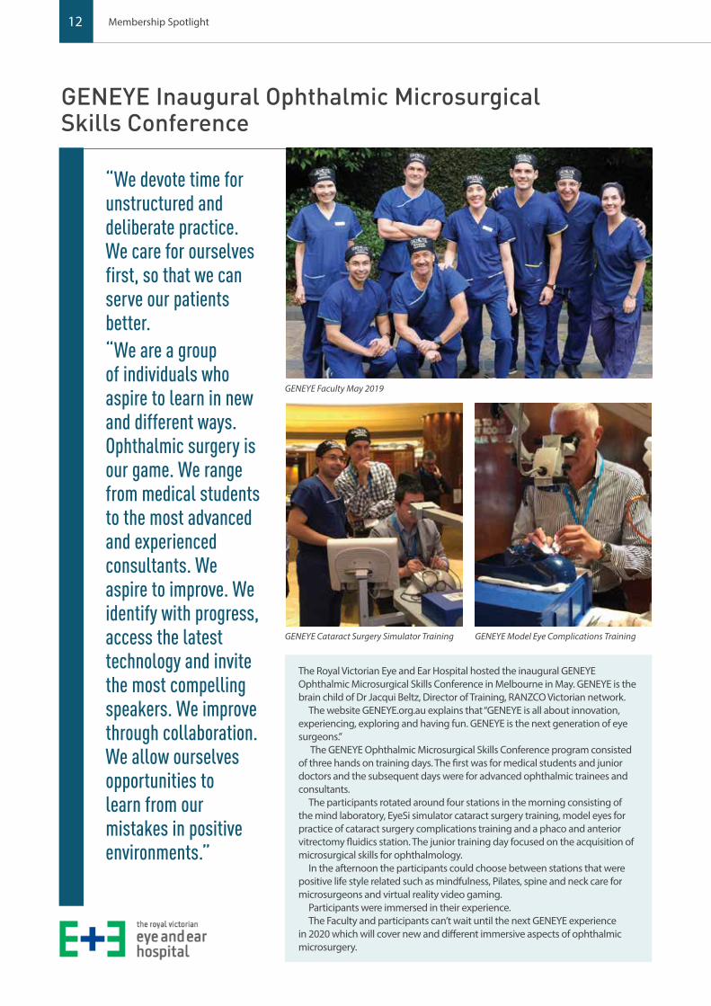
GENEYE Faculty May 2019
GENEYE Cataract Surgery Simulator Training GENEYE Model Eye Complications Training
The Royal Victorian Eye and Ear Hospital hosted the inaugural GENEYE Ophthalmic Microsurgical Skills Conference in Melbourne in May. GENEYE is the brain child of Dr Jacqui Beltz, Director of Training, RANZCO Victorian network.
The website GENEYE.org.au explains that “GENEYE is all about innovation, experiencing, exploring and having fun. GENEYE is the next generation of eye surgeons.”
The GENEYE Ophthalmic Microsurgical Skills Conference program consisted of three hands on training days. The first was for medical students and junior doctors and the subsequent days were for advanced ophthalmic trainees and consultants.
The participants rotated around four stations in the morning consisting of the mind laboratory, EyeSi simulator cataract surgery training, model eyes for practice of cataract surgery complications training and a phaco and anterior vitrectomy fluidics station. The junior training day focused on the acquisition of microsurgical skills for ophthalmology.
In the afternoon the participants could choose between stations that were positive life style related such as mindfulness, Pilates, spine and neck care for microsurgeons and virtual reality video gaming.
Participants were immersed in their experience.The Faculty and participants can’t wait until the next GENEYE experience
in 2020 which will cover new and different immersive aspects of ophthalmic microsurgery.
13Eye2Eye Quarter 3 2019
Well to my adopted home, at least: a home that I had previously adopted by accident. Some speculated that it may have been the result of a divine fate, as some of my religiously minded friends had proclaimed that the Hunter was “God’s own country”. I think that I was following more earthy (and somewhat aquatic) instincts.
Very basic simple mathematics revealed that the ratio of population numbers to ophthalmologist numbers was very much more favourable to the practice of ophthalmology in the rural and regional areas of the land. A less commercial competitive milieu would intuitively suggest a more collegial and cooperative interaction with colleagues and that indeed proved to be the case.
For reasons that I do not know, the early to mid-1990s witnessed a significant migration of city bred and raised newly minted ophthalmologists to the bush. I think that it was more than just simple economic mathematical equation.
Perhaps it was the result of a deep-seated fear instilled when were studied pathology at medical school. Who could fail to be impressed by the clean pink tissues of the lungs of the rural dweller, as opposed to the black, carbon impregnated lungs with bulging nodular hilar lymph nodes so characteristic of the city dwellers?
I think that another factor was the pathway onto the training program was much quicker, and people could finish their training before they reached middle age. Thus, they had less time to develop deeply embedded roots firmly anchored in the Sydney sandstone rock.
I would venture that surgical training was more diverse and comprehensive, probably more consistent with how the founding fathers of the training program had designed the education system.
Yes, Virginia, there is more to the surgical spectrum of ophthalmology than
just a conveyor belt of cataract surgery. I guess that cataracts are very reliable – everyone gets them as we get older and they come in pairs – but surely there is that wonderful thrill of being able to offer our patients a reasonable repertoire of surgical remedies. The broad base is essential for the bush-based practice.
Enough of waxing lyrical about the romance of the bush. If you want anymore read AB Paterson’s Clancy of the Overflow.
The collegiality of the Hunter region ophthalmology fraternity was strong in 1994. There was plenty of work for everyone and practitioners relied on one another to look after problem patients when they were on leave. There were social gatherings and Christmas functions.
It was a time before the competitive entrepreneurial ophthalmology era of high volume cataract surgery and intravitreal injection clinics, and the merchant bank approach of value adding as many MBS item numbers to each patient encounter as possible. Also, before the hypocrisy of feigned devoted interest in educating optometrists (send
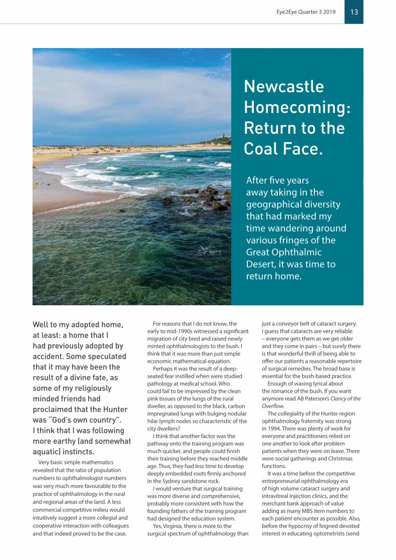
Newcastle Homecoming: Return to the Coal Face.
After five years away taking in the geographical diversity that had marked my time wandering around various fringes of the Great Ophthalmic Desert, it was time to return home.
14 Membership Spotlight
“A stroke of political luck happened in 1995, when the Premier and Health Minister of the great and prosperous state of New South Wales boldly proclaimed that they would resign if they did not get the surgical waiting list down to under 12 months. Amazingly, the old economic orthodoxies of health administration were magically changed overnight, and two new public hospital ophthalmology positions were created.”
your procedural patients to me, and the dry eyes elsewhere), yet having little interest in helping with public hospital training of registrars or medical students.
The Hunter ophthalmologists were generalists, practicing with a broad range of skills. The only subspecialist in town was a retinal surgeon – note not a vitreoretinal surgeon, because vitrectomies were still relatively uncommon at that stage and the approach to ERM and macular holes remained very conservative. We covered most of the surgical base, and the referrals down to Sydney were mainly for vitreoretinal and complex uveitis.
Colleagues were very helpful in providing steady locum sessions to help pay the mortgage and feed the kids. In those days, surgery was not part of the deal, but the consulting work paid considerably better than the registrar’s salary.
After about six months into locum land, I decided to establish my own practice. I picked an area at the top end of Lake Macquarie, which seemed to offer a patient flow from both the eastern and western sides of the lake as well as the surrounding residential areas. Importantly I chose a location which I did not think would impose much on the existing patches of colleagues. I informed my colleagues of my decisions, and they were unfazed and, indeed, supportive. I continued to work as locums in three practices for the next 12 months.
My new surgery was a small unit in an arcade, about 100 metres from the foreshore. A series of gyprock and timber walls were constructed to divide it into a waiting room, a consultation room and another, shall we call it, multipurpose room.
My wife Wendy was also multipurpose as receptionist, secretary, visual field technician and painter. She would ascend and descend the painting ladder in a synchrony dictated by the phone.
The plan was to start off in a minimalist fashion and slowly expand. Equipment was supplied through OPSM, dutifully delivered and installed by the amazingly helpful and enthusiastic Lee Morris. Indeed, it was a great thrill to remove the plastic wrappers from the oculars of the Haig Street slit lamp, turn on the lamp and move the joystick to command the slit lamp column to move through its X, Y and Z axes. There was something sensual in closely tolerance engineering and its precision movement.
I picked the Medmont field machine over the Humphry-Zeiss because it was cheaper, quicker and Australian. I chose speed over a superior statistical analysis
on the logic that no amount of statistical analysis would overcome a patient who has fallen asleep on the chin rest.
The other great investment was a Coherent Ultima Argon laser, which is still working 25 years later.
A very old, third- or fourth-hand Zeiss fluorescein camera completed the diagnostic equipment array.
The first consulting session had four patients. One was a young adult, recently returned from Africa, who was very unwell, with headaches. She had only a day or so before being discharged from our city hospital. Her bilaterally swollen optic discs suggested that she should promptly be readmitted for further investigation and treatment. How about that! I had done something life saving on my very first afternoon, even before I had seen my first cataract or dry eye!
The first private hospital operating list was a collection of ocular plastic cases including a DCR. Not one bread or butter cataract operation. I was hoping for a bit more variety in the future!
Public hospital consultant positions were not that readily available and at that time positions generally became available only when someone died or retired. The public hospital administrators figured that Parkinson’s Law applied and that the workload and costs would expand or contract in proportion to the number of consultants employed. As payment was on a fee for service basis, they worried that demand may become doctor driven rather than patient driven.
A stroke of political luck happened in 1995, when the Premier and Health Minister of the great and prosperous state of New South Wales boldly proclaimed that they would resign if they did not get the surgical waiting list down to under 12 months. Amazingly, the old economic orthodoxies of health administration were magically changed overnight, and two new public hospital ophthalmology positions were created.
The collegiality of the ophthalmology group has sadly diminished over the past two decades. There has been a generational change in attitudes. Also, the advent of a more entrepreneurial business model of some has created disharmony. The fight for market share has led to petty and entirely unprofessional squabbles amongst those protagonists and caused disillusionment amongst the others who abhor that type of behaviour. Fortunately, all is not lost and we have seen the influx of some wonderful people of the traditional ethical style in the past couple of years.

15
1800 225 307dfv.com.au
DV990-1118
Experience titanium quality, at a price comparable to stainless steel!
NEW - Core Values Range
NEW - E - Range
20% OFFFor a limited time...
Nidek OPD-Scan III
THE LEADING CATARACT REFRACTION WORKSTATION
Features:• Wavefront Aberrometer – upto 2520 points• Topographer – 33 ‘blue’ mires, 11,880 data points. Measure irregular
astigmatism• Auto Refractometer/Keratometer - even through Multifocal IOLs• Pupillometer and Pupillographer – Mesopic and Photopic analyses• Retroillumination of IOLs and lens opacities• TORIC IOL correction – measure IOL lens rotation and correction angles• Determine IOL Suitability – measures Angles Alpha & Kappa• Customisable displays and review stations available• Fast & accurate objective measure of patient’s refractive status.• Analyse and determine patient’s aberrations and corrective strategy.• Links to Nidek EXCIMER Laser for custom abblations using FormFit
software
Industry leading 5-in-1 Total Eye Objective Refractive AnalysisUsing ‘dynamic skeoscopy’ & placido topography, measures corneal, internal and total eye aberrations with a multitude of Refractive, Topography, and Pupilometry Analyses.Determine & provide real world explanation of a patient’s total refractive status, even though Multifocals!Improve Refractive outcomes and Clinic workflow.An essential tool that every practice should and can have.
5
A Map and Guide for Optimal Clinical Decisions
A number of summaries are available in the OPD-Scan III, customizable to the clinician’s preference.
White to White summary Comparison map
Cataract summary Toric IOL summary Optical Quality summary
1
2
3
4
6
Irregularity helps determine the best strategy for vision correction. Separation into Total, Corneal and Internal components allows determination of the source of the optical pathology.
PSF images of OPD, Axial, and Internal OPD map simulate objective retinal visual qualityfrom each component of the eye for easy clinical assessment and patient education.
Corneal Spherical Aberration aids in the selection of aspheric IOLs and contact lenses.
Color coded Classification Indices help identify post-LASIK corneas and Keratoconus.
The Astigmatism index aids the implantation of toric IOLs such as incision placement and lens alignment.
A retroillumination image of cataracts captured during the OPD exam allows better understanding of pupillary effects on vision and in patient education.
1
2 2 2
3
5
4 6
Interpreting the Overview summary:
The Overview summary provides refractive data and incorporates corneal disease analysis software and data for cataract and refractive surgery.
Retroillumination image
5
A Map and Guide for Optimal Clinical Decisions
A number of summaries are available in the OPD-Scan III, customizable to the clinician’s preference.
White to White summary Comparison map
Cataract summary Toric IOL summary Optical Quality summary
1
2
3
4
6
Irregularity helps determine the best strategy for vision correction. Separation into Total, Corneal and Internal components allows determination of the source of the optical pathology.
PSF images of OPD, Axial, and Internal OPD map simulate objective retinal visual qualityfrom each component of the eye for easy clinical assessment and patient education.
Corneal Spherical Aberration aids in the selection of aspheric IOLs and contact lenses.
Color coded Classification Indices help identify post-LASIK corneas and Keratoconus.
The Astigmatism index aids the implantation of toric IOLs such as incision placement and lens alignment.
A retroillumination image of cataracts captured during the OPD exam allows better understanding of pupillary effects on vision and in patient education.
1
2 2 2
3
5
4 6
Interpreting the Overview summary:
The Overview summary provides refractive data and incorporates corneal disease analysis software and data for cataract and refractive surgery.
Retroillumination image

16
Q There’s a lot of women in ophthalmology who are inspired by you and see you as a role model. How do you feel about that? What do you think are some of the challenges women face in the profession today?
A Thank you, that is such a lovely thing to hear! I like to support other doctors.
Recently, I’ve realised that when I was younger, I was one of the few women around – even in medical school – and that was fine, it’s never been a problem. As a woman, I’ve always thought, “Well you just make it on your own. You define your own life and goals, and hopefully you achieve them.” But as I have become older, I’ve come to realise that it’s actually not that easy.
Being on the Board for the past few years, I’ve seen some of the challenges other women face and heard a lot more discussion about the shortage of women in senior positions. I believe that with hard work and determination you can often achieve what you want but I think I have also been lucky. I have had wonderful support from our male colleagues. That doesn’t always happen and it is important to support our female colleagues.
While on the Board, I believed strongly that reaching 35% women on the executive would be an advantage to the Board. That was achieved. I’m on the Diversity and Inclusion Committee and everyone was very supportive on having a goal. While I understand some don’t like the idea of quotas, the current situation just wasn't working. In the past, women who were just as good as
the men were not getting into senior positions and were held back because of their gender. So, I believed that the only way to overcome that was by setting a goal and making sure we were choosing the right women who were the equivalent [to men] in terms of their knowledge and experience – and there are plenty around.
Q As a female ophthalmologist what has your own experience been like in the profession, especially early on in your career?
A Back when I started, when I was in med school, I think we [women] made up about 20 per cent of the population. It was really unusual to be a female med student back in those days and I used to get some really weird comments. But, overall, it was a great experience. I mean you’d just finished school and you were thrust into this incredible world of being a medical student. We had a wonderful time. As a cohort, we were all the same and gender was fortunately irrelevant. I guess because perhaps medicine attracted the sort of woman who felt confident enough to be able to do it and be in male company. The guys were very respectful. I was never, ever aware of any discrimination or had any concern as a medical student. I just loved what I did and I loved all my friends. And it was the same when I started ophthalmology.
I’ve always had wonderful friends that have been male and they’ve always been incredibly supportive. I’ve been lucky; I’ve never had a problem and I’ve always just thought, “Yeah I’m one of the guys!” But it’s only in recent years that I’ve been thinking, “Hang on a minute. That’s not working for all women!”
On another note, one of the reasons we have a Diversity and Inclusion Committee is so that we can encourage men as well as women to call out behaviour that is inappropriate, especially among their peers.
Q How did you get involved in medicine and ophthalmology in particular?
A I decided to do medicine to be a psychiatrist. I really enjoyed Freud and Jung and behavioural psychology at school so I wanted to be a psychiatrist. Once I did a psychiatric term as a med student, I realised that it wasn’t quite what I wanted. Once I became an intern I realised that I loved surgery. So, when I was doing a casualty term and someone
came in with a laceration or a dislocated shoulder I found that I wanted to do it; I’d put my hand up to sew up the wound or fix whatever was going on! Then I was really just looking for a surgical speciality. I met some ophthalmologists and thought that looked like a good surgical speciality to go in to and here I am!
Q Did you have any role models while you were studying or training as an ophthalmologist?
A While I was studying, there weren’t really any female ophthalmologists around. I did look up to many of the senior ophthalmologists that were around while I was training. I had an enormous amount of support from all of my ophthalmology teachers – they were all fantastic! I loved my training at the Sydney Eye Hospital. I enjoyed all of my terms and all of the men were incredibly supportive. Some were more supportive than others but perhaps that was just their personalities. There are certainly some who have, throughout my life, become my mentors.
Q Can you outline some of the challenges you faced throughout your career and how you overcame them?
A I think some of the obvious challenges when you start out relate to the study and getting through the exams, as well as the long hours. And then, ongoing, are the challenges of having a practice, dealing with difficult patients and working through complicated patients or cases. But I think the added challenge, specifically for women – and for some men as well – is having a family. Women specifically, especially in this day and age, can have the pressure of needing to be a full time carer and a full time ophthalmologist simultaneously, which can be very challenging and difficult. I have worked full time my entire life and I had six weeks off with the birth of each child and went back afterwards. But, in saying that, it was my own choosing and it’s what I wanted to do. Then again, I don’t think I realised how hard it was going to be trying to run your own practice and work the hours while being there for your kids. But again, I think we’re so lucky as women to be able to have it all: to have our own practice, have all our wonderful patients and have this great career, and we can have children and get them through their lives. We can have
Member Profile
Member Profile: Diana Semmonds

17Eye2Eye Quarter 3 2019
this very busy but incredibly productive life, which is fantastic. You do it because you want to and because you can.
Q It’s obvious you do it because you’re passionate about it.
A That’s the beauty of ophthalmology, it’s such a great profession. I think we’re all so lucky to be ophthalmologists; to actually be able to help people and for people to then be able to see better. I’ve done thousands of cataract surgeries but I still love each day after the operating theatre, after the procedure, when patients come in and you take off their patch and they say “Wow.” I still get a kick out of that… every single time. It’s incredibly rewarding and we’re very, very lucky to have the jobs we have.
Q Can you tell us a little bit about your previous role as Vice President?
A I spent my last two years on the Board as Vice President and I really enjoyed the extra responsibility in representing the College. It was a real honour.
Q As you know Heather Mack recently took on the role of RANZCO President and is the first ever female President. As former Vice President, why did you choose not to run?
A It would have been a great honour to have been elected President. However, I didn’t think I could run my practice as the sole practitioner and fulfil the responsibilities of a President. Being on the Board for a number of years and seeing the work the President is involved in, I know how demanding the role is and how difficult it would be to run your own practice – especially if you’re the only ophthalmologist and the owner of the business. The only way to do it, in my opinion, is when you have a bigger practice where you’ve got a lot of support or when you do a lot of public hospital work and can take the time. Otherwise, I would have loved to have done it and it would have been a great honour. The College has been a passion of mine from day one. From the day I graduated, I was asked to join the NSW Branch and I came on as secretary. They were the early days and the secretary did everything. It was all manual work. We didn’t have computers and there wasn’t even a fax machine so everything would be typed up and placed into envelopes and sent out, and I had the
kids running around – but I absolutely loved it! I loved being part of the College and I stayed, going on to roles on the Board, the Council and on various committees. It was like my hobby. When I wasn’t working or looking after family, I was doing College stuff. However, I love my practice and all my patients and I didn’t want to lose that. But it’s wonderful to see Heather in the role and as the first ever female President. She is doing a fantastic job!
Q Over the next 20 years, what changes do you hope to see in ophthalmology or eye healthcare in general?
A The future for ophthalmology or for vision science and how we’re going to be able to treat our patients is going to be amazing! From the types of surgeries we’ll be able to do, the type of lenses we’ll be able to implant, to the treatment for macular degeneration and other eye diseases. We’ll hopefully be able to cure a lot of the diseases that we can’t treat at the moment. That’s the future. It’s very bright!
Q What has been your most striking memory as an ophthalmologist or a Fellow?
A There’s one particular patient that I’ve never forgotten. It was a lady in her 40s who as a child had very poor vision but was thought to just be a bit simple. If she dropped something on the ground, she wouldn’t see it and she wouldn’t be able to pick it up so she would be hit. She even used to wet her pants trying to find her way to the toilet and was often in trouble from her parents, until they realised that she needed glasses. But she was so short sighted that even when she got glasses they were very, very thick and she still didn’t have good vision. She eventually wore contact lenses but then developed problems and couldn’t wear them anymore. She came to see me and I put on what you call ‘implantable contact lenses’ – a little lens that you can implant into the eye which gives really good vision. Following the surgery, it was the first time that she could actually see well and she said, “I had to learn at 40 that if I dropped something on the ground I could look down and pick it up.” She was really inspirational. This is just one example of what you can achieve as an ophthalmologist and how you can change peoples’ lives. It was just a wonderful story and I’m sure that
all ophthalmologists have that one (or more) special patient that they’ve really, really helped.
Q What is an important lesson that you have learnt over the course of your career?
A I think the most important lesson that I’ve learnt is respecting people; respecting your fellow ophthalmologists, respecting your patients and everyone around you. Everyone is different and everyone has a different way of approaching things. It’s so important to respect diversity and not judge – to respect the differences and enjoy it! It [respect] is such a powerful thing. If everyone in the world just respected each other – you don’t have to agree or like what the other person is saying, you just have to respect the other person’s views – then it would be a great world!
Q Is there anything else you’d like to highlight or share with our readers?
A I’d like to encourage everyone to be part of the College! There are so many committees – such diverse committees – and so much you could learn outside of your normal clinical practise. All the College committees I have been on have extended my understanding of the world of ophthalmology and hopefully I have given a little back. You just learn another facet of ophthalmology outside of your day-to-day, which is really worthwhile. I encourage everybody to get involved in some way!
To get involved with College Committees, check the fortnightly e-news for expressions of interest.
18 Health and Wellbeing
There are many reasons why part time training and flexible employment are on the rise globally, including parental leave, carers’ responsibilities, mental health and greater focus on work/life balance. Eye2Eye talks to three RANZCO trainees about their experiences with part time training and job sharing, including some of the challenges and what worked for them.
Part time training Dr James English was about to move from Sydney to Melbourne to start the ophthalmology training program when his wife went into premature labour and their baby was born at 31 weeks. A tumultuous few months followed, with the baby in neonatal intensive care (NICU) for five weeks and the move to Melbourne postponed.
“I had to delay the start of my training for a month and, while our baby was in the NICU, I was trying to support my spouse and study for my first exam at the same time.” After arriving in Melbourne, life remained hectic. “I had countless exams in that first year. It was a strain on my spouse and unpleasant for me. I felt shackled – wanting to be there to support my partner and baby while needing to study and do exams. It made us consider part time training and we started to explore the possibility.”
It was not a straightforward process and quite time-consuming. “The option to do some of your training part time should be more the norm, rather than the exception,”
says Dr English. “At the moment, the training program is fairly inflexible for young families or for others with extenuating circumstances.”
Dr English’s second child was also born premature, at 34 weeks, and had a very low birth weight. It was another stressful period for the family. “From 27 weeks onwards were the scariest times of my life,” he says. This encouraged him to pursue part time training, which ended up being quite a process to negotiate.
“I wanted to go to 0.8 full-time equivalent (FTE) for 18 months. The process included having to speak to each director of the six terms, usually at least two medical workforce representatives from each network, the Director of Training, the head of QEC for the state (Victoria), and the Censor-in-Chief of the College. In the end, I talked to more than 15 different people, which was an onerous and time consuming exercise.”
Dr English has observed from his own experience and that of other trainees that there seems to be a carry over culture that full time training is the only type of training. “Anecdotally, a lot of people want to go part time for their training, but it’s perceived as being too big a hurdle. While the mindset is shifting, flexible training has not yet fully been embraced in ophthalmology,” he says.
In the end Dr English was granted nine months of the 18 months part time training he requested. “If I’m honest, there was a mix of receptiveness. Generally, College Fellows were supportive. However, when it came down to the nuts and bolts of service delivery, it was often deemed too hard by medical workforce to implement. Surgical lists were always the first to be proposed to be cut – given the supervising consultant could continue service delivery. This required significant
Flexibility During Training – The Way of the Future
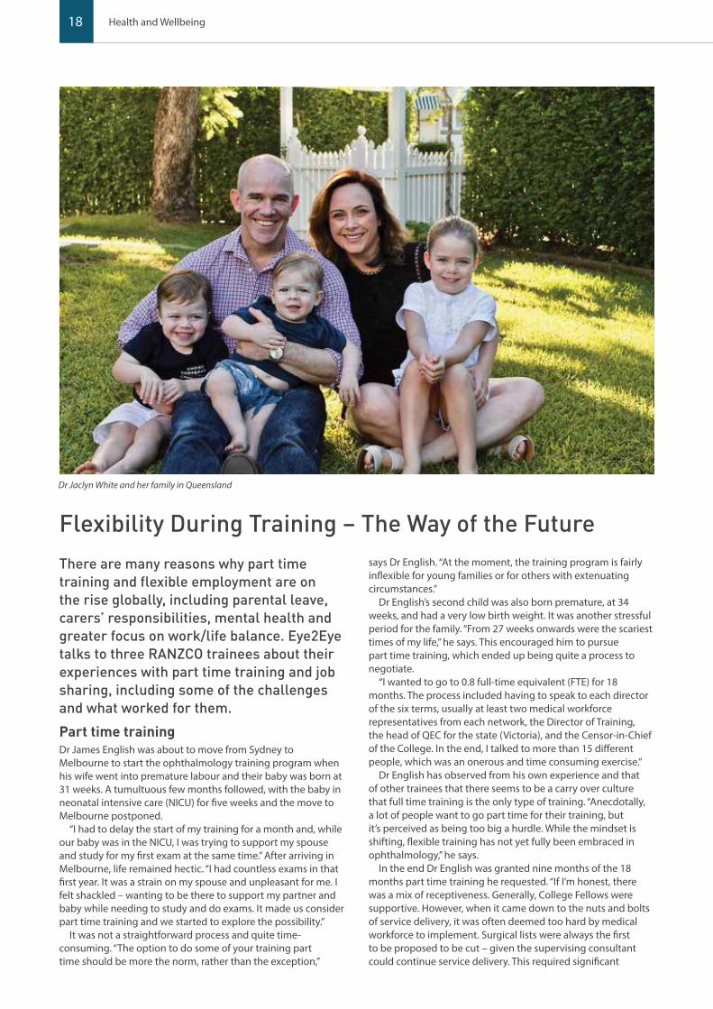
Dr Jaclyn White and her family in Queensland
19Eye2Eye Quarter 3 2019
self-advocacy to resist. The networks best positioned to absorb the 0.2 FTE deficit were, surprisingly, often those that felt unable to acquiesce. After the birth of our second child, we were rotated to Albury (a city on the border of New South Wales and Victoria). The initial phone call reception from Albury’s Dr Paul Giles, I felt, was a model answer to a request for part-time training: ‘Absolutely, let’s make that happen. How can we help?’”
Dr English says some trainees won’t ask for part-time status because they’re worried about being perceived as an inconvenience because they’re asking for something different to the norm. “However, I felt I had to push for it and advocate for my family. Without a doubt those nine months going part-time were the best of my training. I was able to support my spouse, see my two girls, support the family and it made me a more effective trainee.”
Smoothing the way Dr English, now in his fifth year of training, says there are a number of things that could make part time training easier in the future. “Having to pitch your request to 15 different people is onerous. Once a trainee requests part time training, ideally a representative could liaise on their behalf and advocate for the trainee to each hospital network. Having leadership from the College, despite not having local hospital network control, would also provide significant political clout to agitate for change.”
Different legislation in different states makes it difficult for trainees to know their rights. In Melbourne, for example, only recently has legislation passed that ensures a trainee’s re-employment for the rest of their contract after taking time out for maternity/paternity leave. Prior to this, the hospital network was not obliged to extend a trainee’s contract to incorporate this leave. The same protections, however, do not extend to part time trainees, even if it is for family reasons. “I think Fellows would be concerned that, for example, a Victorian hospital employing a trainee who takes maternity leave for six months at the end of her contract the hospital was under no obligation to provide six months of employment upon her return – until recently. In a similar vein, without legislative protection, some trainees will see going part time as running the risk of not being re-employed. This is a significant hurdle. The colleges could support their trainees by requesting public policies from network hospitals to support re-employment for part timers.”
Dr English says the key to facilitating part time training is to have a contingency plan to plug the holes that invariably happen in any training program. “Each year, there are trainees who take long term leave, be it parental or otherwise. While I am oversimplifying the sometimes very difficult task of training network committees in balancing training with service provision, my observation has been that, without an ongoing network contingency plan in place to fill the gaps, this causes significant chaos and distress across the network. This cascades to trainees often asked to shift rotations and take on other roles to compensate. Quite unintentionally, this inevitably takes a toll on the trainee who has decided to take the leave in the first place. I would argue that each network must have a more robust contingency plan to accommodate this repeated scenario.”
Some of the ways things have always been done can seem rigid and draconian, and parts that can be made flexible are perceived – because of some people’s experiences – as being too hard to negotiate. “It’s clear to trainees that RANZCO is taking this seriously and this is very much welcomed. Support for flexible training, beyond the individual, speaks of a culture shift to one of inclusivity, gender equality and a recognition that healthier, happier trainees provide better patient care and are fine ambassadors for our profession.”
Job-sharing – It can be done During a play date with their new babies, two trainees decided that a job-share return-to-work arrangement was the path to continue their ophthalmology training in a way that worked for them, their young families, the hospital and the College. Forward planning, good communication and considered solutions to potential problems were some of the keys to a good outcome.
Dr Jaclyn White and Dr Juanita Pappalardo were doing their training in Queensland when their babies were born three weeks apart. “It was my first child,” says Dr White. “I was absolutely besotted – a feeling well known to new parents – and I was struggling to find motivation to return to work. Reflecting on how busy I had been in my first two years on the program with on-call commitments, teaching and exams; the Pathology and RACE exams still ahead of me; and with a medical husband who was also very busy, I was concerned about achieving – and sustaining – an appropriate work-life balance.”
Dr Pappalardo was in a similar situation. She was 18 months into her vocational training, already had two school-age children and had planned for 12 months of maternity leave after the birth of her third child. “My husband had recently completed his own surgical training and was working as a full time staff specialist and director of a surgical unit at a major tertiary hospital. We were a busy family, and I faced the daunting reality of returning to a demanding surgical training job, with the Ophthalmic Pathology examination looming ahead. I’m naturally a bit of a perfectionist and was well aware that keeping up with the demands of both family and training was going to be quite a task.”
Dr White and Dr Pappalardo decided to share one full time equivalent (FTE) position – 0.5 FTE each – and began to work out how to make it happen.
“Job-sharing as ophthalmology trainees in Queensland had not previously been undertaken, so there was no clear
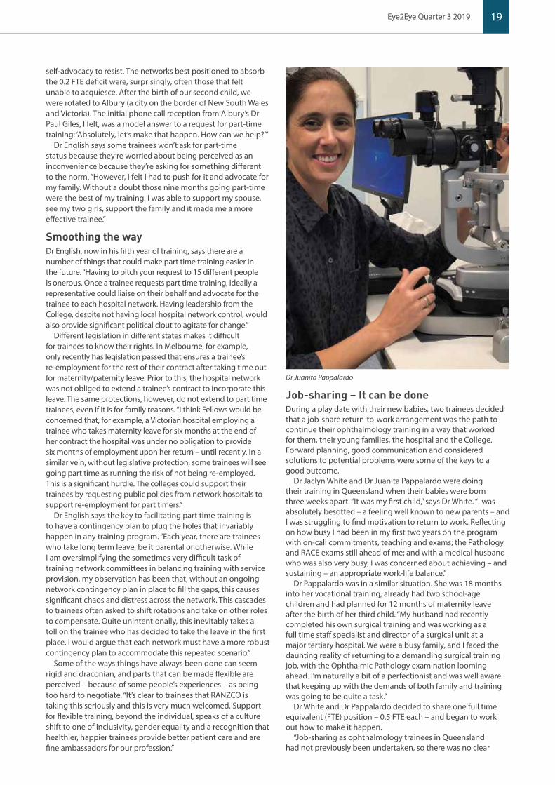
Dr Juanita Pappalardo
20 Health and Wellbeing
process to follow,” says Dr White. “I was fortunate as Juanita had previous experience in job-sharing as a resident. About three months prior to our return to work, we drafted a proposal to present to our local QEC. We proposed a model of part time training that provided an appropriate standard of ophthalmic care and training within a flexible framework. It was important to us to present a proposal that would benefit all stakeholders – i.e. not only ourselves, but the patients, other registrars and our supervising consultant. Where potential issues were anticipated, we outlined our proposed solutions. Once RANZCO’s QEC was happy for us to proceed, we sought approval from the College’s Censor-In-Chief, and finally with Queensland Health.”
The next step involved a lot of problem solving. “As a partnership, we were committed to ensuring we were able to provide the same standard and continuity of care to our patients as we would otherwise have done as individual full time trainees,” says Dr Pappalardo. “We had to find ways of seamlessly working with each other to do this, and we discussed this aspect in detail together. The challenges of job sharing are unique, as each individual not only has the utmost responsibility to their patients and colleagues, but also to one another. There is a constant and inherent drive to not want to let your ‘other half’ down, which served as self-motivation to be a better trainee and communicator.”
Both Dr Pappalardo and Dr White were very aware they were entering unchartered territory at the time. They knew one of their big challenges was in foreseeing issues that might arise from sharing a full time position and troubleshooting these in a proactive manner.
“Working so closely with Jaclyn in managing common patients – together with the fact that we spent one to two hours on the phone to one another on handover night – was incredibly beneficial,” says Dr Pappalardo. “We learned so much from each other and were able to share different opinions and ideas in the context of managing difficult or interesting clinical scenarios.”
Dr White says the job-sharing arrangement worked out better than she initially expected. “I was fortunate enough to have an exceptional job-share partner and I think I can speak for both of us when I say we achieved what we set out to do – which was to optimise our work-life balance and enrich our training experience. Job-sharing or part time vocational training isn’t the path for everybody, but if you’re even considering it, it is well worth exploring.”
Dr Pappalardo says job-sharing gave her the ability to enjoy, and the resilience to cope with, the cumulative stresses of training, studying and raising children. “I was able to job share for three years of my vocational training (1.5 years FTE) from mid-second year to the commencement of my fourth year. During this time, I was able to successfully complete both the Ophthalmic Pathology and RACE examinations. While studying for the latter, I was also pregnant with my fourth child. I cannot even begin to imagine how those years would have looked and felt had I not been in a flexible arrangement.”
Flexible training arrangements can take many forms, of which job sharing is a unique subset. Both trainees agree job sharing is certainly worth considering as a viable working arrangement. “I recently read a wonderful article discussing the phenomenon of high-performing women having a tight knit circle of other women surrounding them,” says Dr Pappalardo. “These women collaborate in order to collectively succeed. I certainly feel as though the time is ripe for anyone with determination to follow this pathway. The first step is to find a colleague to work with, which I think is probably still the main hurdle for those who would like to consider this path. When Jaclyn floated the idea of job sharing with me, I knew it was a workable solution – we were both hardworking and good communicators and, from my previous experience, I knew both of these attributes were the keys to a successful arrangement.”
“As a partnership, we were committed to ensuring we were able to provide the same standard and continuity of care to our patients as we would otherwise have done as individual full time trainees,” says Dr Pappalardo. “We had to find ways of seamlessly working with each other to do this, and we discussed this aspect in detail together. The challenges of job sharing are unique, as each individual not only has the utmost responsibility to their patients and colleagues, but also to one another. There is a constant and inherent drive to not want to let your ‘other half’ down, which served as self-motivation to be a better trainee and communicator.”
21Eye2Eye Quarter 3 2019
“The standard you walk past is the standard you accept” became one of the most memorable phrases of a landmark speech delivered in 2013 by the then Australian Chief of Army Lieutenant General David Morrison. He doesn’t take credit for coming up with the line, attributing it as a phrase originating from General David Hurley, former Chief of the Australian Defence Force. The powerful phrase is as applicable to ophthalmology as the armed forces.
The previous issue of Eye2Eye magazine included an article on ‘Bystander Intervention’ – the concept of speaking up or taking action when potentially harmful incidents or situations are observed. A ‘bystander’ is a person who witnesses an incident between a perpetrator and a target but is not an active participant. An ‘active bystander’ is someone who – when they witness or become aware of an incident – speaks up or takes action.
Most people like to think of themselves as someone who wouldn’t tolerate bullying, discrimination or harassment. However, in the heat of the moment, being an active bystander is not always straightforward, particularly if the bystander feels unsafe to intervene or risks adverse consequences themselves.
In this issue of Eye2Eye we explore some of the approaches and strategies of active bystanders. Stepping up can enhance respectful, inclusive training and workplace cultures in which everyone can thrive.
Active Bystanders have a plan Victims of bullying, discrimination and harassment (BDH) sometimes go on to inflict that behaviour on others, while some who’ve experienced BDH become the staunchest advocates for safe, supportive workplaces. Education, reflection, self awareness and planning ahead can help you gain the skills and confidence to become change-agents in your workplace.
Often it is hard to know exactly how to respond – whether during an event you witness as it occurs, or something you hear about later. Even before an event occurs, it can help to make a plan as to how you might respond in different scenarios. For example: If I see this, then I’ll do that – know what behaviour you will and won’t tolerate, plan out what you’ll say or do.
An active bystander role models behaviours that make workplaces safer for everyone. Berkeley University of California defines an ‘Active Bystander’ as someone who sees the potential for harm to occur and takes action to interrupt the situation and prevent harm.
Active Bystanders assess risk and ask for help We’ve all heard of scenarios where intervening, or not intervening, has ended badly for some or all involved. Sometimes in these situations there seems too much at stake to stick your neck out. Intervening could inadvertently make the victim more of a target or expose them to further, hidden, abuse or harm. Getting involved could mean you are threatened in some way or face a risk to your position
Active Bystanders
Berkeley’s C.A.R.E model outlines four strategies for active bystanders:
C onfront the situation A lert others R edirect attention E ngage peers
You can use multiple strategies for a given situation depending on your comfort level and personal style. Some of these are outlined below. It is important to keep your own safety in mind any time you are choosing to be an active bystander.
HAVE A PLAN
– Stepping Up Now for the Future We Want
22 Health and Wellbeing
or reputation. Calling out the behaviour of a perpetrator may mean they could lose their job. Or bringing attention to workplace BDH can sully the reputation of a department or organisation. While doing nothing does harm, it can be daunting getting involved; the temptation for silence or inaction is great.
Active Bystanders speak up When you become aware of incidents of BDH, you may experience a range of feelings including anxiety about how to best help the target. Speaking up can be difficult. Before stepping in, try the A.B.C approach: • Assess for safety: If you see someone in trouble, ask yourself
if you can help safely in any way. Remember, your personal safety is a priority – never put yourself at risk
• Be in a group: It’s safer to call out behaviour or intervene in a group. If this is not an option, report it to others who can act
• Care for the victim. Talk to the person who you think may need help. Ask them if they are OK, and if they want you to support them in any way.
Active Bystanders take action Many of us have been in situations where something just didn’t feel right, and yet we weren’t sure how to respond. Adequately equipped, active bystanders have the power to transform workplaces into communities that are safer and more inclusive for everyone. Here are some tips for taking action: • Support the victim; offer resources and information without
pressure or judgment about their decisions. • Derail a conversation as it happens or disrupt someone from
taking any further steps that are problematic.
• If you feel unsafe to take action at the time, wait for the heat to die down and then have a chat with the perpetrator.
• Address the behaviour, not the person – focusing on the behaviour can seem less like a personal attack. Try saying “Those ideas aren’t cool” instead of saying “You’re wrong”.
• Respond with: “You may not have meant to offend, but here is how I experienced that joke”, or “I don’t get it” or “C’mon, that’s not ok”, or “You used the term 'x' and many people find that derogatory – is there another way to say what you mean?”
GET HELP AND SUPPORT: RANZCO’s Employee Assistance Program (EAP) offers confidential support provided by qualified external counsellors – free to all members and their immediate families. To make an appointment or speak with an EAP provider, call:
NZ – 0800 666 367 AU – 1300 687 327
RANZCO’s policy on bullying, discrimination and harassment and RANZCO’s complaints procedures can be found on the RANZCO website.
Here are some tips to lessen the threat of speaking up or taking action:
9 There’s safety in numbers. Get a buddy (preferably another witness) to commit to backing you up when you speak up.
9 Carefully document and keep records where possible.
9 Know who your allies are.
9 Seek advice from a trusted source or reputable support service.
9 Report it to a superior, to HR, to the College.
The website OurWatch.org.au has some tips on speaking up at the time you notice an incident. Speak up and show it’s not ok:
9 Roll your eyes.
9 Shake your head.
9 Don’t laugh along.
9 Respond to a sexist joke with “I don’t get it.”
9 Purposely change the topic: “Ok, let’s move on.”
9 Gently tease them: “Are you still in the 1950s?”
9 Ask them to stop: “Can you not? C’mon.”
BDH that occurs online can be just as destructive as if it occurred in person. If you observe or experience online abuse or harassment, OurWatch.org.au suggests some ways to take action online, including:
9 Retweet and like comments that are respectful and call out disrespect.
9 Message the disrespectful person with a meme or an article, or let them know you’re not onboard with what they’ve posted.
9 Report disrespect or abuse to Facebook, Instagram or Twitter.

23
www.anterion.com
2003
86-0
01 IN
T.A
E18
© H
eid
elb
erg
En
gin
eeri
ng
Gm
bH
METRICSCATARACTCORNEA
Experience the power of swept-source OCT technology
at its best. Perform the most relevant anterior segment
exams in one modular, upgradeable platform.
Introducing ANTERION®.
Take the lead with a dynamic, workflow-efficient
imaging platform that delivers powerful results.
Call +61 3 9639 2125 for more information or contact
Axial length
mm 20 22 24
BEAT YOUR PERSONAL BEST.FAST. EFFICIENT. POWERFUL.
Jan FrodenoTriathlon Olympic ChampionDouble IRONMAN World Champion
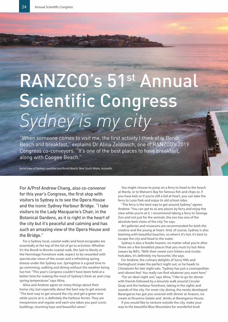
24 Annual Scientific Congress
“When someone comes to visit me, the first activity I think of is Bondi Beach and breakfast,” explains Dr Alina Zeldovich, one of RANZCO’s 2019 Congress co-conveyors. “It’s one of the best places to have breakfast, along with Coogee Beach.”
Aerial view of Sydney coastline and Bondi Beach, New South Wales, Australia
RANZCO’s 51st Annual Scientific Congress Sydney is my city
For A/Prof Andrew Chang, also co-convenor for this year’s Congress, the first stop with visitors to Sydney is to see the Opera House and the iconic Sydney Harbour Bridge. “I take visitors to the Lady Macquarie’s Chair, in the Botanical Gardens, as it is right in the heart of the city but it’s peaceful and calming and has such an amazing view of the Opera House and the Bridge.”
For a Sydney local, coastal walks and food escapades are essentially at the top of the list of go to activities. Whether it’s the Bondi to Bronte coastal walk, the Spit to Manly or the Hermitage Foreshore walk, expect to be rewarded with spectacular views of the ocean and a refreshing spring breeze under the Sydney sun. Springtime is a good time to go swimming, walking and dining without the weather being too hot. “This year’s Congress couldn’t have been held at a better time for making the most of Sydney’s fresh air and crisp spring temperature,” says Alina.
Alina and Andrew agree on many things about their home city, but especially about the best way to get around. “The best way to get around the city and get a great view while you’re at it, is definitely the harbour ferries. They are inexpensive and regular and each one takes you past iconic buildings, stunning bays and beautiful views.”
You might choose to jump on a ferry to head to the beach at Manly, or to Watson’s Bay for famous fish and chips or, if you have kids or if you’re still a kid at heart, you can take the ferry to Luna Park and enjoy its old school rides.
“The ferry is the best way to get around Sydney,” agrees Andrew. “You can get to so any places by ferry and enjoy the view while you’re at it. I recommend taking a ferry to Taronga Zoo and not just for the animals; the zoo has one of the absolute best views of the city,” he says.
Art galleries and museums are recommended for both the creative and the young at heart. And, of course, Sydney is also teeming with beautiful beaches, so when’s it’s hot, it’s best to escape the city and head to the water.
Sydney is also a foodie heaven, no matter what you’re after. There are a few breakfast places that you must try but Alina swears by Bill’s, “With their sweet corn fritters and ricotta hotcakes, it’s definitely my favourite,’ she says.
For Andrew, the culinary delights of Surry Hills and Darlinghurst make the perfect night out, or he heads to Chinatown for late night eats. “Sydney has just a cosmopolitan and vibrant feel. You really can find whatever you want here.”
“For an ideal night out,” says Alina, “I like to go for dinner with friends followed by a leisurely walk around Circular Quay and the harbour foreshore, taking in the sights and sounds of the city. For inner city dining, the newly developed Barangaroo has got you covered with dinner at Anason, ice cream at Rivareno Gelato and drinks at Barangaroo House.
If you would like to venture outside the city, make your way to the beautiful Blue Mountains for wonderful bush

25Eye2Eye Quarter 3 2019
The Royal Botanic Gardens Sydney
walking, mountain bike riding, cute boutique shopping and great food. You could start off with breakfast at the Hominy bakery in Katoomba, for their sourdough yeasted bread and financier cake, lunch at Leura Garage and dinner at Silk’s Brassiere. You can earn your food with a few hours on some of the beautiful mountain walks.
Whether or not you do one or all of these things, be sure to explore Sydney’s mix of modernity and nostalgia when you’re here for RANZCO’s 2019 Congress in November.
We look forward to catching up with you and experiencing the amazing scientific program that is planned for this year’s Congress.
“With this being RANZCO’s 50th year,” says Andrew, “there is sure to be plenty of celebration. The Congress dinner in particular should be a lot of fun, and a great opportunity to celebrate with colleagues young and not-so-young anymore! We look forward to seeing you there!”
Dr Alina ZeldovichA/Prof Andrew ChangCo-convenors

26 Annual Scientific Congress
Graduation & Awards Ceremony and President’s Reception 5.45-7.15pm International Convention Centre, Sydney 14 Darling Drive, Sydney Take part in this momentous ceremony celebrating our new Fellows, college award recipients, trainers of excellence and examination medallists. Come onsite to ICC Sydney at 5pm for pre drinks followed by the recognition ceremony where we welcome our new Fellows and highlight our colleagues’ excellence in the field. Celebrate with everybody afterwards at the President’s Reception set at the convention centre’s foyer overlooking the stunning night lights at Darling Harbour.
Cost: $75
Saturday 9 November 2019
RANZCO’s 51st Congress Social Events With the remaining months leading up to the 51st Annual Scientific Congress in 8 to 12 November, the College is gearing up towards an eventful five days of bringing together ophthalmologists, trainees and allied health professionals from Australia, New Zealand and around the world. Along with the line up of prominent and prestigious speakers, the College also has a program of exciting social events, each of which provide a good opportunity to celebrate RANZCO’s 50th anniversary and recognise everyone’s participation and contribution throughout the years.
Welcome Reception6.00-8.00pm Museum of Contemporary Art 140 George Street, The Rocks Begin the festivities by attending the official Welcome Reception, which will be set at Australia’s premier contemporary art gallery. Get the chance to mingle and connect with colleagues whilst listening to one of the highlights of the night: Key note speaker Prof Lisa Harvey-Smith, an astronomer who will deliver the Congress Opening Lecture about how astronomers study the universe by creating special “eyes”. Enjoy canapés and drinks with friends old and new as you enjoy the beautiful panoramic backdrop of the iconic Sydney Harbour Bridge and Sydney Opera House.
Cost: $75
Friday 8 November 2019

27Eye2Eye Quarter 3 2019
Congress Dinner 7.00-11.00pm Doltone House, Jones Bay Wharf Level 3/26-32 Pirrama Road, Pyrmont Cap off the entire conference by attending the grand finale, the event that marks RANZCO’s 50th year, the Congress Dinner. All RANZCO Fellows, members and associates are highly encouraged to participate in this special night. The picturesque view of the city skyline and the Sydney Harbour Bridge will be the stunning backdrop for this memorable dinner where various forms of celebrations and commemorations will ensue. Participants may take the light rail, alighting at the “Star City” light rail stop and walk the expanse of the beautiful wharf to the venue.
Cost: $175
Sunday 10 November 2019 Monday 11 November 2019
Younger Fellows’ Dinner 7.00-10.00pm Ventuno Restaurant 21 Hickson Road, Walsh Bay Situated at a rustic location overlooking the marina, this dinner at Ventuno Restaurant is designed for Younger Fellows to celebrate and have a relaxing night in Sydney. Share a delectable three course dinner of modern Italian cuisine whilst making the most of this excellent networking opportunity. Some drinks will be provided and a wide variety of wines and boutique Italian beers are available at bar prices.
Cost: $80
Practice Managers’ Dinner 7.00-10.00pm Blackbird Café Wheat Road Balcony Level 1, Cockle Bay Wharf, Darling Harbour NSW 2000
Practice managers’ are invited to a relaxed evening at what is boasted to be the restaurant with the best view of Darling Harbour. Enjoy the complimentary drinks upon arrival while networking with colleagues. Feast on a delicious three course dinner of modern Australian cuisine while taking in the waterfront view of Cockle Bay Wharf and Darling Harbour. Included in the package are shared entrees and desserts, while additional drinks are available at bar prices.
Cost: $50
Senior and Retired Fellow’s Dinner 7.00-10.00pm Establishment Ballroom 252 George Street, Sydney Senior and retired Fellows are invited to partake in an exclusive three course dinner at The Establishment, an iconic heritage building set in the heart of Sydney’s CBD. There’s no better location to reconnect with friends than at this beautiful 19th century venue. Reminisce with colleagues at the stunning ballroom as you enjoy your welcome drinks and your scrumptious dinner spread afterwards. Take the conversation further into the New York-style terrace overlooking a magnificent backdrop of the city. There will be complimentary bus shuttles from ICC Sydney so participants need not worry about transportation. Take this opportunity to reunite and share stories with old friends and colleagues about the different journeys you have had as individuals and how ophthalmology has progressed in that time.
Cost: $115
These social events have been tailored to cater to everyone and serve as additional networking opportunities for all participants whilst presenting some of the best Sydney has to offer. If you would like to register to these social events, you may when registering for the Congress. If you have already registered and have seen a social event that you are interested to be a part of, please email Think Business on [email protected]
We hope that you take the opportunity to participate in these excellent networking events to exchange news on the latest developments discussed in the Congress lectures while taking in the beautiful sights and scenes of our stunning city. We look forward to seeing you at the 51st Annual Scientific Congress this November. We are confident it will be the most memorable and exciting to date.

28 Annual Scientific Congress
Meet our RANZCO Congress 2019 Speakers
Dr Emily Chew – Retina Update Lecture Emily Chew is the Director of the Division of Epidemiology and Clinical Applications (DECA) at the National Eye Institute, the National Institutes of Health in Bethesda, Maryland. She is also the Chief of the Clinical Trials Branch within the Division. Emily received her medical degree and her ophthalmology training at the University of Toronto, School of Medicine, in Toronto, Canada. She completed her fellowship in Medical Retina at the Wilmer Eye Institute, the Johns Hopkins Medical Institutes and the University of Nijmegen, the Netherlands.
Q How did you become involved in ophthalmology?
A I was blessed with wonderful role models such as my friend and colleague Dr Brenda Gallie who was very passionate about her research in retinoblastoma. She impressed upon me the need to do research to improve the lives of many individuals.
Q Tell us a little bit more about the work/research you’re involved in.
A My key focus has been randomised clinical trials in mostly retinovascular diseases. Epidemiologic studies are also important to help us understand the natural history and the associated risk factors. I have also been interested in the area of genetic associations of diseases, particularly in age-related macular degeneration.
Q What prompted you to specialise in this area?
A I was interested in medical retina because one develops a long term relationship with our patients who require chronic care (AMD, diabetic retinopathy). The research possibilities seemed endless when I started and a great deal of progress has been made.
Q What has been the highlight of your career?
A There are many highlights and perhaps my proudest achievement is to reflect on the wonderful achievements of our mentees.
Q Have there been any game changing research, techniques or technologies in this area recently? Can you tell us a bit more about this?
A The use of the optical coherence tomography and its continued development with angiography, etc. is truly amazing. We are developing objective outcome measurements based on OCT and now, with the use of artificial intelligence, specifically deep learning, I believe we will indeed improve our ability to care for patients and to conduct more objective outcomes for clinical trials.
Q What do you hope to achieve in the next 10 years?
A I hope we will have more primary prevention for diseases such as AMD and we already know a great deal of early prevention of diabetic retinopathy. We will hopefully improve our understanding and be able to target the early stages of disease instead of waiting to intervene at the late stages.
Q Can you give us a sneak peek of your presentation? What will be the focus of your lecture?
A This is truly an Australian story as we have partnered with philanthropy based in Australia and have collaborated with our esteemed Australian colleague to gain better understanding of a somewhat rare condition known as macular telangiectasia type 2 (formerly known as juxta-foveal macular telangiectasia). I will describe how over the last 15 years, the knowledge base has expanded with a natural history study conducted with a large internal collaboration and have completed phase 1 and 2 trials of neuroprotection for this condition. I believe this is a story of team science and big data.
Q What are some of the challenges you’ve faced over the course of your career and how have you overcome these?
A There are many challenges that we still need to be mindful of. The under representation of minorities and women in our speciality, especially in leadership positions, has been an important cause for me. I am very heartened by the recent statement from my NIH director, Dr Francis Collins, who refuses to participate in any panel that does not include women. I am also grateful to see the many young women and more minorities entering our field. Diversity will only improve our current institutions, our work environment and ultimately, our research as we take into account many different perspectives.
Q What advice would you give to those starting out in ophthalmology?
A Find your passion and run with it.
Dr Santosh Honavar - Oculoplastic Update Lecture
Dr Honavar completed his basic medical education at the Bangalore Medical College, Bangalore, India. He received residency training in ophthalmology, followed by subspecialty training in ophthalmic plastic surgery and ocular oncology at the Dr Rajendra Prasad Center for Ophthalmic Sciences, All India Institute of Medical Sciences, New Delhi, India. Dr Honavar further trained in ocular oncology and was mentored by Profs JA Shields and CL Shields at the Wills Eye Hospital, Philadelphia, PA, USA. He thereafter
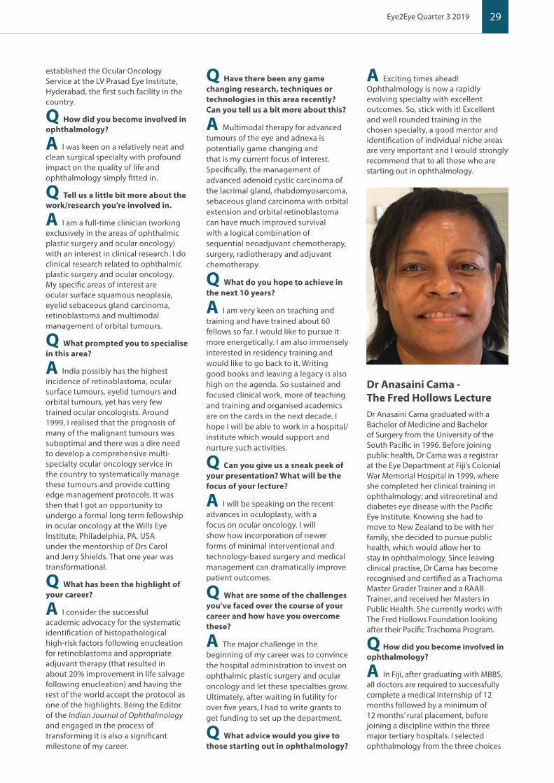
29
established the Ocular Oncology Service at the LV Prasad Eye Institute, Hyderabad, the first such facility in the country.
Q How did you become involved in ophthalmology?
A I was keen on a relatively neat and clean surgical specialty with profound impact on the quality of life and ophthalmology simply fitted in.
Q Tell us a little bit more about the work/research you’re involved in.
A I am a full-time clinician (working exclusively in the areas of ophthalmic plastic surgery and ocular oncology) with an interest in clinical research. I do clinical research related to ophthalmic plastic surgery and ocular oncology. My specific areas of interest are ocular surface squamous neoplasia, eyelid sebaceous gland carcinoma, retinoblastoma and multimodal management of orbital tumours.
Q What prompted you to specialise in this area?
A India possibly has the highest incidence of retinoblastoma, ocular surface tumours, eyelid tumours and orbital tumours, yet has very few trained ocular oncologists. Around 1999, I realised that the prognosis of many of the malignant tumours was suboptimal and there was a dire need to develop a comprehensive multi-specialty ocular oncology service in the country to systematically manage these tumours and provide cutting edge management protocols. It was then that I got an opportunity to undergo a formal long term fellowship in ocular oncology at the Wills Eye Institute, Philadelphia, PA, USA under the mentorship of Drs Carol and Jerry Shields. That one year was transformational.
Q What has been the highlight of your career?
A I consider the successful academic advocacy for the systematic identification of histopathological high-risk factors following enucleation for retinoblastoma and appropriate adjuvant therapy (that resulted in about 20% improvement in life salvage following enucleation) and having the rest of the world accept the protocol as one of the highlights. Being the Editor of the Indian Journal of Ophthalmology and engaged in the process of transforming it is also a significant milestone of my career.
Q Have there been any game changing research, techniques or technologies in this area recently? Can you tell us a bit more about this?
A Multimodal therapy for advanced tumours of the eye and adnexa is potentially game changing and that is my current focus of interest. Specifically, the management of advanced adenoid cystic carcinoma of the lacrimal gland, rhabdomyosarcoma, sebaceous gland carcinoma with orbital extension and orbital retinoblastoma can have much improved survival with a logical combination of sequential neoadjuvant chemotherapy, surgery, radiotherapy and adjuvant chemotherapy.
Q What do you hope to achieve in the next 10 years?
A I am very keen on teaching and training and have trained about 60 fellows so far. I would like to pursue it more energetically. I am also immensely interested in residency training and would like to go back to it. Writing good books and leaving a legacy is also high on the agenda. So sustained and focused clinical work, more of teaching and training and organised academics are on the cards in the next decade. I hope I will be able to work in a hospital/institute which would support and nurture such activities.
Q Can you give us a sneak peek of your presentation? What will be the focus of your lecture?
A I will be speaking on the recent advances in oculoplasty, with a focus on ocular oncology. I will show how incorporation of newer forms of minimal interventional and technology-based surgery and medical management can dramatically improve patient outcomes.
Q What are some of the challenges you’ve faced over the course of your career and how have you overcome these?
A The major challenge in the beginning of my career was to convince the hospital administration to invest on ophthalmic plastic surgery and ocular oncology and let these specialties grow. Ultimately, after waiting in futility for over five years, I had to write grants to get funding to set up the department.
Q What advice would you give to those starting out in ophthalmology?
A Exciting times ahead! Ophthalmology is now a rapidly evolving specialty with excellent outcomes. So, stick with it! Excellent and well rounded training in the chosen specialty, a good mentor and identification of individual niche areas are very important and I would strongly recommend that to all those who are starting out in ophthalmology.
Dr Anasaini Cama - The Fred Hollows Lecture
Dr Anasaini Cama graduated with a Bachelor of Medicine and Bachelor of Surgery from the University of the South Pacific in 1996. Before joining public health, Dr Cama was a registrar at the Eye Department at Fiji’s Colonial War Memorial Hospital in 1999, where she completed her clinical training in ophthalmology; and vitreoretinal and diabetes eye disease with the Pacific Eye Institute. Knowing she had to move to New Zealand to be with her family, she decided to pursue public health, which would allow her to stay in ophthalmology. Since leaving clinical practise, Dr Cama has become recognised and certified as a Trachoma Master Grader Trainer and a RAAB Trainer, and received her Masters in Public Health. She currently works with The Fred Hollows Foundation looking after their Pacific Trachoma Program.
Q How did you become involved in ophthalmology?
A In Fiji, after graduating with MBBS, all doctors are required to successfully complete a medical internship of 12 months followed by a minimum of 12 months’ rural placement, before joining a discipline within the three major tertiary hospitals. I selected ophthalmology from the three choices
Eye2Eye Quarter 3 2019
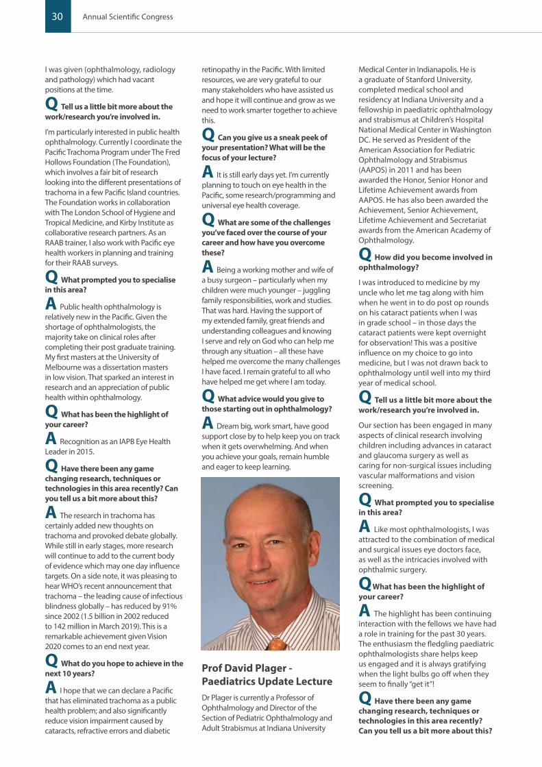
30 Annual Scientific Congress
I was given (ophthalmology, radiology and pathology) which had vacant positions at the time.
Q Tell us a little bit more about the work/research you’re involved in.
I’m particularly interested in public health ophthalmology. Currently I coordinate the Pacific Trachoma Program under The Fred Hollows Foundation (The Foundation), which involves a fair bit of research looking into the different presentations of trachoma in a few Pacific Island countries. The Foundation works in collaboration with The London School of Hygiene and Tropical Medicine, and Kirby Institute as collaborative research partners. As an RAAB trainer, I also work with Pacific eye health workers in planning and training for their RAAB surveys.
Q What prompted you to specialise in this area?
A Public health ophthalmology is relatively new in the Pacific. Given the shortage of ophthalmologists, the majority take on clinical roles after completing their post graduate training. My first masters at the University of Melbourne was a dissertation masters in low vision. That sparked an interest in research and an appreciation of public health within ophthalmology.
Q What has been the highlight of your career?
A Recognition as an IAPB Eye Health Leader in 2015.
Q Have there been any game changing research, techniques or technologies in this area recently? Can you tell us a bit more about this?
A The research in trachoma has certainly added new thoughts on trachoma and provoked debate globally. While still in early stages, more research will continue to add to the current body of evidence which may one day influence targets. On a side note, it was pleasing to hear WHO’s recent announcement that trachoma – the leading cause of infectious blindness globally – has reduced by 91% since 2002 (1.5 billion in 2002 reduced to 142 million in March 2019). This is a remarkable achievement given Vision 2020 comes to an end next year.
Q What do you hope to achieve in the next 10 years?
A I hope that we can declare a Pacific that has eliminated trachoma as a public health problem; and also significantly reduce vision impairment caused by cataracts, refractive errors and diabetic
retinopathy in the Pacific. With limited resources, we are very grateful to our many stakeholders who have assisted us and hope it will continue and grow as we need to work smarter together to achieve this.
Q Can you give us a sneak peek of your presentation? What will be the focus of your lecture?
A It is still early days yet. I’m currently planning to touch on eye health in the Pacific, some research/programming and universal eye health coverage.
Q What are some of the challenges you’ve faced over the course of your career and how have you overcome these?
A Being a working mother and wife of a busy surgeon – particularly when my children were much younger – juggling family responsibilities, work and studies. That was hard. Having the support of my extended family, great friends and understanding colleagues and knowing I serve and rely on God who can help me through any situation – all these have helped me overcome the many challenges I have faced. I remain grateful to all who have helped me get where I am today.
Q What advice would you give to those starting out in ophthalmology?
A Dream big, work smart, have good support close by to help keep you on track when it gets overwhelming. And when you achieve your goals, remain humble and eager to keep learning.
Prof David Plager - Paediatrics Update Lecture Dr Plager is currently a Professor of Ophthalmology and Director of the Section of Pediatric Ophthalmology and Adult Strabismus at Indiana University
Medical Center in Indianapolis. He is a graduate of Stanford University, completed medical school and residency at Indiana University and a fellowship in paediatric ophthalmology and strabismus at Children’s Hospital National Medical Center in Washington DC. He served as President of the American Association for Pediatric Ophthalmology and Strabismus (AAPOS) in 2011 and has been awarded the Honor, Senior Honor and Lifetime Achievement awards from AAPOS. He has also been awarded the Achievement, Senior Achievement, Lifetime Achievement and Secretariat awards from the American Academy of Ophthalmology.
Q How did you become involved in ophthalmology?
I was introduced to medicine by my uncle who let me tag along with him when he went in to do post op rounds on his cataract patients when I was in grade school – in those days the cataract patients were kept overnight for observation! This was a positive influence on my choice to go into medicine, but I was not drawn back to ophthalmology until well into my third year of medical school.
Q Tell us a little bit more about the work/research you’re involved in.
Our section has been engaged in many aspects of clinical research involving children including advances in cataract and glaucoma surgery as well as caring for non-surgical issues including vascular malformations and vision screening.
Q What prompted you to specialise in this area?
A Like most ophthalmologists, I was attracted to the combination of medical and surgical issues eye doctors face, as well as the intricacies involved with ophthalmic surgery.
Q What has been the highlight of your career?
A The highlight has been continuing interaction with the fellows we have had a role in training for the past 30 years. The enthusiasm the fledgling paediatric ophthalmologists share helps keep us engaged and it is always gratifying when the light bulbs go off when they seem to finally “get it”!
Q Have there been any game changing research, techniques or technologies in this area recently? Can you tell us a bit more about this?

31
A One of the important practice-altering research findings has come from the 20+ years we have been studying how infant eyes react to cataract/IOL surgery, principally through the efforts of the Infant Aphakia Treatment Study group. This has caused us to dampen any enthusiasm we may have been building for extending the incredible success story that intraocular lenses have been over the past four decades, first in older adults and later extending down into your childhood. We have discovered that we are not quite ready to extend this modality down into infants in the first six months of life.
Q What do you hope to achieve in the next 10 years?
A I hope that I am still able to do what I do in 10 years!
Q Can you give us a sneak peek of your presentation? What will be the focus of your lecture?
A The focus will be on what was described in #5 above – how far we have progressed in the care of cataracts in children in my lifetime since Dr Costenbader remarked in 1957 that he had never seen a child with a congenital cataract benefit from surgery for it.
Q What are some of the challenges you’ve faced over the course of your career and how have you overcome these?
A I have been blessed to work at an institution and with people who have been very supportive of what we have wanted or needed to do in order to provide the best care we could to our young patients. I have some concerns about the evolving corporatisation and depersonalisation of practicing medicine in the US, but this may just be the typical lament of an almost senior citizen who is watching the world change. Technological advances have been dramatic and for the most part beneficial, but physicians becoming more technology dependent probably has some downsides in terms of the personal side of medicine – an evolution that demands we pay it some attention regarding the training of the next generation of physicians (or providers!)
Q What advice would you give to those starting out in ophthalmology?
A Go for it. It remains one of the great subspecialties in medicine – full of wonderful, exciting technology and grateful, appreciative patients – a tough combination to beat!
Prof John Marshall - The Dame Ida Mann Memorial Lecture Professor John Marshall is the Frost Professor of Ophthalmology at the Institute of Ophthalmology in association with Moorfield’s Eye Hospital, University College London. He is Emeritus Professor of Ophthalmology at Kings College London, Honorary Distinguished Professor at the University of Cardiff; Honorary Professor at City University and Honorary Professor at Glasgow Calendonian University. His research over the past forty years has ranged over a number of ocular problems but has concentrated on the inter-relationships between light and ageing; the environmental mechanisms underlying age-related, diabetic and inherited retinal disease and the development of lasers for use in ophthalmic diagnosis and surgery. This work has resulted in over five hundred research papers, 44 book chapters and seven books. He invented and patented the revolutionary Excimer laser for the correction of refractive disorders and also created the world’s first Diode laser for treating eye problems of diabetes, glaucoma and ageing. He is editor and co-editor of numerous international journals.
Q How did you become involved in ophthalmology?
A I entered ophthalmology in 1965 shortly after the advent of lasers and was headhunted for a PhD project in conjunction with the Royal Air Force to determine the damaging effects of lasers on the retina. Their concern was these novel systems could perhaps be developed as weapons and it was my job to determine what wavelengths pulse duration and energy levels would
be required to damage the retina. Obviously, knowledge of such data would provide the ability to draw up codes of practice to make both military personnel and the public safe.
Q Tell us a little bit more about the work/research you’re involved in.
A When I found out that there was a split between those within the Ministry of Defence who wanted to render systems safe and those who wanted to perhaps develop weapon systems I immediately started working with the International Committee of the Red Cross and chaired a committee which eventually obtained a Geneva convention banning the development, distribution and use of antipersonnel laser weapons. Understanding the mechanisms by which lasers damage tissue I was able to utilise such mechanisms in developing new therapeutic devices for treating all aspects of ocular pathologies in different tissues.
Q What prompted you to specialise in this area?
A I guess two things committed me to research into lasers. The first was the growing public awareness due predominantly to the James Bond film Goldfinger and the second was the apparent limitless budget of the Ministry of Defence. It was great to be working in a totally new field with the ability to raise funds for lots of different research projects.
Q What has been the highlight of your career?
A I guess in the past a highlight was drawing up the original patents for Excimer based refractive surgery and cofounding the first company to see such refractive surgery through the bureaucracy of the FDA. It took 10 years from an idea to FDA approval. A second highlight was the first diode laser for use in ophthalmology, which has resulted in a technology change throughout the field. More recently I’m delighted to have carried out research on the aetiology of AMD and to have the game patent the device which may well slow down the disease process and prevent conversion from atrophic to neovascular disease.
Q Have there been any game changing research, techniques or technologies in this area recently? Can you tell us a bit more about this?
A I guess I have answered the game changes in question four, but I should
Eye2Eye Quarter 3 2019
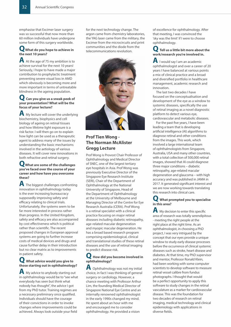
32
emphasise that Excimer laser surgery was so successful that now more than 60 million individuals have undergone some form of this surgery worldwide.
Q What do you hope to achieve in the next 10 years?
A At the age of 75 my ambition is to achieve survival for the next 10 years! Seriously, I hope to have made a major contribution to prophylactic treatment preventing severe visual loss in AMD which obviously is becoming more and more important in terms of untreatable blindness in the ageing population.
Q Can you give us a sneak peek of your presentation? What will be the focus of your lecture?
A My lecture will cover the underlying biochemistry, biophysics and cell biology of ageing on retinal tissues and how lifetime light exposure is a risk factor. I will then go on to explain how light can be used as a therapeutic agent to address many of the issues by understanding the basic mechanisms involved in the aetiology of various diseases. It will cover new innovations in both refractive and retinal surgery.
Q What are some of the challenges you’ve faced over the course of your career and how have you overcome these?
A The biggest challenges confronting innovation in ophthalmology today is the ever increasing bureaucracy supposedly improving safety and efficacy relating to clinical trials. Unfortunately, the systems seem to be far more interested in process rather than progress. In the United Kingdom, safety and efficacy are also accompanied by cost effectiveness which is political rather than scientific. The recent proposed changes in European approval systems are going to further increase costs of medical devices and drugs and cause further delay in their introduction but no clear matrix as to improvements in patient safety.
Q What advice would you give to those starting out in ophthalmology?
A My advice to anybody starting out in ophthalmology would be to “see what everybody has seen but think what nobody has thought”, the advice I got from my PhD tutor. Training regimes are a necessary preliminary once qualified. Individuals should have the courage of their convictions in order to invoke changes where improvements could be achieved. Always look outside your field
for the next technology change. The argon came from chemistry laboratories, the YAG laser came from the military, the Excimer from the microcircuits and print communities and the diode from the telecommunications revolution.
Prof Tien Wong - The Norman McAlister Gregg Lecture Prof Wong is Provost Chair Professor of Ophthalmology and Medical Director of SNEC, one of the largest tertiary eye hospitals in Asia. Prof Wong was previously Executive Director of the Singapore Eye Research Institute (SERI), Chair of the Department of Ophthalmology at the National University of Singapore, Head of the Department of Ophthalmology at the University of Melbourne and Managing Director of the Centre for Eye Research Australia (CERA). Prof Wong is a retinal specialist with a clinical practice focusing on major retinal diseases including diabetic retinopathy, age-related macular degeneration and myopic macular degeneration. He has a broad based research program comprising epidemiological, clinical and translational studies of these retinal diseases and the use of retinal imaging to predict disease risk.
Q How did you become involved in ophthalmology?
A Ophthalmology was not my initial choice, in fact I was thinking of general surgery or cardiology. However, a chance meeting with Professor Arthur Lim, the founding Medical Director of Singapore National Eye Centre and an internally renowned ophthalmologist in the early 1990s changed my mind. He spent about an hour with me and encouraged me to consider ophthalmology. He provided a vision
of excellence for ophthalmology. After that meeting, I was convinced the ‘sky was the limit’ if I were to choose ophthalmology.
Q Tell us a little bit more about the work/research you’re involved in.
A I would say I am an academic ophthalmologist and over a career of 20 years I have balanced at various points a mix of clinical practice and a broad and diversified portfolio in healthcare management, academic research and innovation.
The last two decades I have focused on the conceptualisation and development of the eye as a window to systemic diseases, specifically the use of retinal imaging as a novel diagnostic platform to detect various eye, cardiovascular and metabolic diseases.
For the past five years, I have been leading a team that is developing artificial intelligence (AI) algorithms to diagnose retinal and other conditions from the images. This work, which involved a large international team of ophthalmologists from Singapore, Australia, USA and many other countries, with a total collection of 500,000 retinal images, showed that AI could diagnose three major conditions – diabetic retinopathy, age-related macular degeneration and glaucoma – with high accuracy and was published in JAMA in 2017. It generated significant interest and we are now working towards translating this research into clinical care.
Q What prompted you to specialise in this area?
A My decision to enter this specific area of research was totally serendipitous - meeting the right people at the right place at the right time. As an ophthalmologist, in choosing a PhD project, I was very intrigued by the concept that our eyes provide a unique window to study early disease processes before the occurrence of clinical systemic diseases such as stroke, heart disease and diabetes. At that time, my PhD supervisor and mentor, Professor Ronald Klein, had been working with some computer scientists to develop software to measure retinal vessel calibre from fundus photographs. I thought that would be a perfect opportunity to apply this software to study changes in the retinal vasculature as a marker for cardiovascular disease. This was the foundation for two decades of research on retinal imaging, medical technology and clinical epidemiology with applications in diverse fields.
Annual Scientific Congress
33
More importantly, due to its cross-disciplinary nature and frontier science status, our work has attracted very wide coverage across various media platforms and is not solely confined to eye-related journals.
Q What has been the highlight of your career?
A Perhaps the most important highlights have been the ability to translate my research into clinical and commercial outcomes. For example, my research on the burden and risk factors of diabetic retinopathy, a leading cause of blindness in Singapore, allowed me to set up a national telemedicine screening program for diabetic retinopathy, the Singapore Integrated Diabetic Retinopathy Programme (SiDRP). The SIDRP now screens >150,000 persons with diabetes in Singapore annually across 18 primary care clinics, improving screening coverage and quality of referrals to tertiary eye hospitals. SiDRP has won multiple national IT and healthcare awards in Singapore.
My research on retinal imaging, which has shown that measurements of changes in blood vessels in the retina may be a unique non-invasive test for a person’s cardiovascular risk, has led to development of a novel imaging software (SIVA), now licensed and used by >30 international clinical, research and industry partners.
Q Have there been any game changing research, techniques or technologies in this area recently?
A In the past few years, I have led a team that is developing artificial intelligence (AI) algorithms to diagnose retinal and other conditions from the images. The team comprises ophthalmologists from SNEC and SERI and computer science experts from the National University of Singapore’s School of Computing. We are also collaborating with top centres from around the world such as Moorfields Eye Hospital and University College London.
This has led to formation of two start-up companies, plano and EyRIS, of which I am a co-founder.
Q Can you tell us a bit more about this?
A AI has been suggested to be the fourth industrial revolution. In the diagnosis of eye diseases, we typically rely on human ophthalmologists. However, AI can improve accuracy and speed of diagnosis and reduce costs, so this can increase the delivery of eye care
with (eventually) minimal dependence on specialist ophthalmologists for screening of common conditions.
We and others have shown AI could diagnose diabetic retinopathy, age-related macular degeneration and glaucoma with better accuracy than humans.
Q What do you hope to achieve in the next 10 years?
A Enjoy the work, the network, the new ideas, and continue the innovation journey to translate the research findings into actual clinical care and practice!
Q Can you give us a sneak peek of your presentation? What will be the focus of your lecture
A My talk on ‘Artificial Intelligence in Ophthalmology: Concepts, Progress, Challenges and Myths’ will cover the following: • What is AI? How is it different from
previous fields? How is it applied in ophthalmology and in particular for diabetic retinopathy screening?
• What have we achieved in using AI for diabetic retinopathy screening and other diseases? What are the latest findings and how is that relevant to ophthalmologists?
• What does the future hold? What will move this field forward? Will ophthalmologists lose their jobs? Will patients accept AI? Can AI solve all our problems?
Q What are some of the challenges you’ve faced over the course of your career and how have you overcome these?
A I would say that the career of an academic ophthalmologist is difficult in terms of struggling to balance clinical medicine and science in my career. To do well in both, you need to have an active clinical practice that provides excellent care for patients, but combine this with the precision, intensity and rigour of cutting edge science. The danger is that you cannot be good in either.
Many ophthalmologists will choose a simpler route of being a pure clinician. Career pathways for academic ophthalmologists and clinician scientists are erratic. You do not know if you will succeed. Unfortunately, I do not see many young people taking this route of balancing clinical work with research. Most people would want a safer career which provides a better work life balance.
Q What advice would you give to those starting out in ophthalmology?
A First, we cannot plan our lives and career too much. We should take new opportunities that come and do the best we can with those opportunities.
Second, for budding ophthalmologists, ask yourselves the following questions when thinking about career: Are you ambitious and do you want to influence and change ophthalmology? Do you want to do something in your life that is different from the well trodden path? Do you want to be a thought leader who can influence clinical practice? Do you want to make a major breakthrough in ophthalmology? If the answers are all yes, you may wish to explore an academic ophthalmologist career.
Eye2Eye Quarter 3 2019
CONGRESS LECTURES
Saturday 9 November
RANZCO Congress Opening LectureProf Lisa Harvey-Smith
The Norman McAlister Gregg Lecture
Prof Tien Y Wong
Paediatrics Update LectureProf David A Plager MD
Sunday 10 November
Retina Update LectureDr Emily Y Chew MD
The Council LectureA/Prof Penelope Allen
Monday 11 November
Cataract Update LectureProf Soon-Phaik Chee
The Fred Hollows LectureDr Anasaini Cama
Tuesday 12 November
Glaucoma Update LectureProf David (Ted) Garway-Heath
The Dame Ida Mann Memorial Lecture
Prof John MarshallOculoplastics Update Lecture
Dr Santosh G Honavar
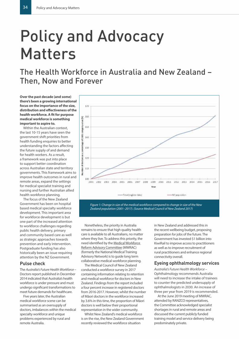
34
Policy and Advocacy Matters
Policy and Advocacy Matters
The Health Workforce in Australia and New Zealand – Then, Now and Forever
Over the past decade (and some) there’s been a growing international focus on the importance of the size, distribution and effectiveness of the health workforce. A fit for purpose medical workforce is something important to aspire to.
Within the Australian context, the last 10-15 years have seen the government shift priorities from health funding enquiries to better understanding the factors affecting the future supply of and demand for health workers. As a result, a framework was put into place to support better coordination across Australian state and territory governments. This framework aims to improve health outcomes in rural and remote areas, expand the settings for medical specialist training and nursing and further Australian allied health workforce planning.
The focus of the New Zealand Government has been on hospital based medical specialty workforce development. This important area for workforce development is but one part of the increased attention to workforce challenges regarding public health delivery, primary and community based care as well as strategic approaches towards prevention and early intervention. Postgraduate funding has also historically been an issue requiring attention by the NZ Government.
Pulse check The Australia’s Future Health Workforce – Doctors report published in December 2014 indicated that Australia’s health workforce is under pressure and must undergo significant transformations to meet future demands for healthcare.
Five years later, the Australian medical workforce scene can be summarised as an oversupply of doctors, imbalances within the medical specialty workforce and unique problems experienced by rural and remote Australia.
Nonetheless, the priority in Australia remains to ensure that high quality health care is available to all Australians, no matter where they live. To address this priority, the need identified by the Medical Workforce Reform Advisory Committee (MWRAC) (formerly the National Medical Training Advisory Network) is to guide long term collaborative medical workforce planning.
The Medical Council of New Zealand conducted a workforce survey in 2017 containing information relating to retention and medical workforce for doctors in New Zealand. Findings from the report included a four percent increase in registered doctors from 2016-2017. However, whilst the number of Māori doctors in the workforce increased by 3.6% in this time, the proportion of Māori doctors is well below their proportional representation in the wider community.
Whilst New Zealand’s medical workforce is on the rise, the New Zealand Government recently reviewed the workforce situation
in New Zealand and addressed this in the recent wellbeing budget, proposing preparation for jobs of the future. The Government has invested $1 billion into KiwiRail to improve access to practitioners as well as to improve recruitment of rural practitioners and enhance regional connectivity overall.
Eyeing ophthalmology services Australia’s Future Health Workforce – Ophthalmology recommends Australia will need to increase the intake of trainees to counter the predicted undersupply of ophthalmologists in 2030. An increase of three per year from 2019 is recommended.
At the June 2019 meeting of MWRAC, attended by RANZCO representatives, the Committee acknowledged specialist shortages in rural and remote areas and discussed the current publicly funded training model and service delivery being predominately private.
Figure 1: Change in size of the medical workforce compared to change in size of the New Zealand population (2001–2017). (Source Medical Council of New Zealand 2017)

35Eye2Eye Quarter 3 2019
Fig 2: Location of clinicians by remoteness. In 2016, the majority (83.6%) of clinicians were in a major city or a location considered as MMM1 under the Modified Monash Model classification system. (Source: Department of Health 2016)
Fig 3: Ophthalmology specialist clinicians (headcount and sector: proportion of specialist clinical FTE in public and private) by state and territory. (Source: NHWDS, Medical Practitioner 2016). A craft like ophthalmology would always have a bigger private sector.
Location of clinicians by remoteness, Modified Monash Model (MMM*)
MMMCategory 1 2 3 4 5 6 7
% 83.6 7.7 7.0 0.8 0.2 0.2 0.5
State & Territory
Headcount Specialist clinical FTE
% Public % Private
NSW 289 10.7 89.3
VIC 188 20.4 79.2
QLD 121 12.3 87.7
SA 51 22.9 77.1
WA 57 19.4 80.6
TAS 18 9.5 90.5
NT 5 77.6 22.4
ACT 9 35.9 64.1
Total 738 15.6 84.3
In New Zealand, the demand for ophthalmic work done per ophthalmologist is set to rise by 20% by 2028 if the number of new specialists entering the workforce remains at 5.6, which is the average over the past six years. To maintain workloads at present levels, this requires eight new entries each year from 2019 to 2022, nine new entries from 2023 to 2024 and 10 new entries from 2025 to 2028.
Where to next? Effective planning for the future medical workforce is essential to meet the complex requirements for a successful healthcare system in both Australia and New Zealand. Variables such as social, cultural, economic, ageing populations and gender representation all impact recruitment and retention in Australia and New Zealand. Alternative service delivery versus established models, and international and private verses public recruitment and retention are all variables to consider when forward planning. It is thus imperative for the Australian and New Zealand governments to strategically plan to meet the future healthcare needs of individuals in both populations whilst considering unique variable sets.
For Aotearoa it is retention. Retention is important to optimise the current workforce and thereby ensure a stable and accessible multidisciplinary healthcare team. This could be fostered by offering appropriate career development and employment opportunities, remuneration and a sense of community to rural practitioners.
New Zealand Government funding may improve numbers of ophthalmologists entering the workforce, especially when considering the growing needs of an ageing population. Future planning of ophthalmology departments and DHBs can ensure current and future vacancies well known to NZ trained fellows.
In Australia, the need to address maldistribution is key. Further changes in technology may increase the viability of telemedicine for service provision but a strengthening of the rural health outreach fund may well be a more opportune mechanism. Air transfer services may help to address comorbidities as well as reduce the burden of patients with better service coordination.
Co-claiming MBS consultation items with T8 ophthalmology procedural items In some circumstances, patients require a review for a different condition in one eye on the same day they need to undergo surgery, such as cataract surgery, on the other eye. This is a cause for concern for patients residing in rural areas of Australia as they may otherwise be required to come back the following day which, given their lengthy travel, may be an impractical option. It is appropriate for the patient to attend a review consultation followed by their planned surgery. Medicare have refused claims like this, even if different eyes are considered.
From 1 November 2017, the Government commenced changes to the MBS restricting the co-claiming of specialist and consultant physician consultation items which are performed on the same day as operations/procedures that have a schedule fee of $300 or more. As a result of the change in November 2017, ophthalmologists are thus unable to co-claim subsequent attendance items such as 105 the same day a claim is made for any Group T8 item with a schedule fee of equal to or greater than $300.
The Department of Health (DoH) considers that a procedure with a schedule fee of less than $300 is inclusive of any consultation provided at the time of the service.
In April 2019, DoH advised there are some scenarios where an exemption to the co-claiming restriction is possible. The following scenarios include: • subsequent attendance item 111 can be billed if
an urgent unpredicted procedure (with a schedule fee over $300) during an attendance is clinically necessary to be performed on the same day; or
• item 115 can be billed if, at the time of a scheduled operation (with a schedule fee of $300 or greater), an attendance for an unpredicted, unrelated matter is required.
It is inappropriate to claim item 115 if there is a planned attendance where an unscheduled procedure is performed. Item 115 should not also be claimed for routine extended consultations with procedures.
When using MBS item 115, the attendance should be unpredicted and deferring the unplanned consultation would be a clinical risk to the patient. Item numbers 111 and 115 are only to be used in exceptional circumstances. In all instances, the ophthalmologist’s clinical records should include contemporaneous notes outlining the clinical situation. It should be clearly identified why the consultation is necessary and include the clinical risks.
The DoH has advised item numbers 111 and 115 are only to be claimed in exceptional circumstances.
The increase of training positions outside major cities and surrounding areas – with a focus on rural and remote posts – may assist in addressing the current and projected workforce shortage.
References are available upon request. Please contact Stephanie Mulholland on [email protected]
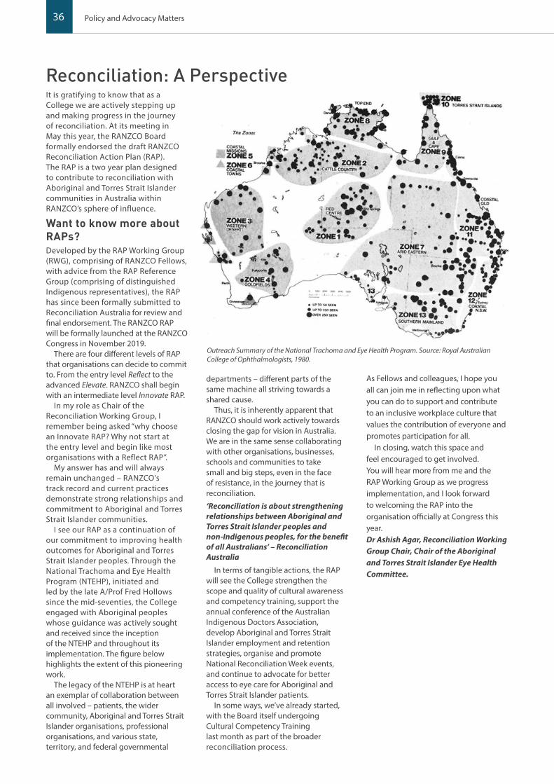
36 Policy and Advocacy Matters
It is gratifying to know that as a College we are actively stepping up and making progress in the journey of reconciliation. At its meeting in May this year, the RANZCO Board formally endorsed the draft RANZCO Reconciliation Action Plan (RAP). The RAP is a two year plan designed to contribute to reconciliation with Aboriginal and Torres Strait Islander communities in Australia within RANZCO’s sphere of influence.
Want to know more about RAPs?Developed by the RAP Working Group (RWG), comprising of RANZCO Fellows, with advice from the RAP Reference Group (comprising of distinguished Indigenous representatives), the RAP has since been formally submitted to Reconciliation Australia for review and final endorsement. The RANZCO RAP will be formally launched at the RANZCO Congress in November 2019.
There are four different levels of RAP that organisations can decide to commit to. From the entry level Reflect to the advanced Elevate. RANZCO shall begin with an intermediate level Innovate RAP.
In my role as Chair of the Reconciliation Working Group, I remember being asked “why choose an Innovate RAP? Why not start at the entry level and begin like most organisations with a Reflect RAP”.
My answer has and will always remain unchanged – RANZCO's track record and current practices demonstrate strong relationships and commitment to Aboriginal and Torres Strait Islander communities.
I see our RAP as a continuation of our commitment to improving health outcomes for Aboriginal and Torres Strait Islander peoples. Through the National Trachoma and Eye Health Program (NTEHP), initiated and led by the late A/Prof Fred Hollows since the mid-seventies, the College engaged with Aboriginal peoples whose guidance was actively sought and received since the inception of the NTEHP and throughout its implementation. The figure below highlights the extent of this pioneering work.
The legacy of the NTEHP is at heart an exemplar of collaboration between all involved – patients, the wider community, Aboriginal and Torres Strait Islander organisations, professional organisations, and various state, territory, and federal governmental
departments – different parts of the same machine all striving towards a shared cause.
Thus, it is inherently apparent that RANZCO should work actively towards closing the gap for vision in Australia. We are in the same sense collaborating with other organisations, businesses, schools and communities to take small and big steps, even in the face of resistance, in the journey that is reconciliation.
‘Reconciliation is about strengthening relationships between Aboriginal and Torres Strait Islander peoples and non-Indigenous peoples, for the benefit of all Australians’ – Reconciliation Australia
In terms of tangible actions, the RAP will see the College strengthen the scope and quality of cultural awareness and competency training, support the annual conference of the Australian Indigenous Doctors Association, develop Aboriginal and Torres Strait Islander employment and retention strategies, organise and promote National Reconciliation Week events, and continue to advocate for better access to eye care for Aboriginal and Torres Strait Islander patients.
In some ways, we’ve already started, with the Board itself undergoing Cultural Competency Training last month as part of the broader reconciliation process.
As Fellows and colleagues, I hope you all can join me in reflecting upon what you can do to support and contribute to an inclusive workplace culture that values the contribution of everyone and promotes participation for all.
In closing, watch this space and feel encouraged to get involved. You will hear more from me and the RAP Working Group as we progress implementation, and I look forward to welcoming the RAP into the organisation officially at Congress this year. Dr Ashish Agar, Reconciliation Working Group Chair, Chair of the Aboriginal and Torres Strait Islander Eye Health Committee.
Outreach Summary of the National Trachoma and Eye Health Program. Source: Royal Australian College of Ophthalmologists, 1980.
Reconciliation: A Perspective

37Eye2Eye Quarter 3 2019
International Development
Following on from the first education training workshop for ophthalmologists in Papua New Guinea held in 2018, a second Train the Trainer workshop was conducted in June this year by RANZCO Fellow Dr Neil Murray and Dr Harris Ansari (Pacific Eye Institute) together with Geoffrey Wabulembo UPNG Senior Lecturer. The workshop was enthusiastically received by participants who gained insight into tools and skills and confidence to provide effective and appropriate programmatic assessment for the registrars.
Dr Wabulembo, who has been instrumental in supporting the PNG Ophthalmology Capacity Building Program, commented that the teaching visits are indeed a paradigm shift in the training of ophthalmologists in PNG – explaining further that the exposure of the registrars to the international
community has already triggered a desire to learn more and venture into collaborative research.
“When we held the second workshop on assessment, we developed skills to make assessment more objective and more informative about the true abilities of the trainee being examined,” explained Dr Wabulembo. “We are now in a better position to set good multiple choice questions that will explore a wide range of topics in ophthalmology. Thanks to RANZCO for providing excellent facilitators.”
Collaboration between RANZCO and the University of Papua New Guinea (UPNG) has also been solidified through the signing of an updated MOU, whereby UPNG and RANZCO agreed to explore collaborative activities including examiner visits; exploring teaching and learning opportunities for collaboration;
organisation of joint academic activities, such as courses, conferences, seminars, symposia or lectures and exchange of research outcomes and other academic information.
Previous collaboration has included the completion of the ophthalmology curriculum review and the development of a curriculum implementation plan. This implementation plan involved a series of both teaching the teachers workshops and one week sub-specialty teaching visits to upskill local faculty and train registrars.
The second teaching workshop and one week subspecialty teaching visits kick off this year. These series of seven teaching visits have been scheduled for 2019. Thanks to the following contributors for their time, expertise and contribution to the RANZCO UPNG capacity building program:
RANZCO and Papua New Guinea: Capacity Building in PNG
Speciality Visiting Lecturer DatePost-operative endophthalmitis
Mathew Green 25 February 2019
Retina Anthony Bennett Hall 20-21 March 2019
Oculoplastics Sarah Crowe 25-29 March 2019
Paediatric Susan Carden 4-7 June 2019
Optics and Refraction Sophie Koh 10-14 June 2019
Medical Retina Rohan Essex 17-21 June 2019
Cornea Gerrard Sutton 1-5 July 2019
The teaching visits are designed to upskill trainees in subspecialty areas and to build the capacity of the local ophthalmologist. These teaching visit positions are all undertaken at the University of Papua New Guinea (UPNG) and the Port Moresby General Hospital, whereby visiting doctors will deliver a teaching program of up to a one week duration with regards to the PNG curriculum. RANZCO will also be supporting scholarships from eye care doctors and ophthalmologists in PNG to attend the RANZCO Congress scheduled in Sydney in November.
Anyone wanting to become involved as we look to plan activities for 2020, please contact Gail van Heerden at [email protected]
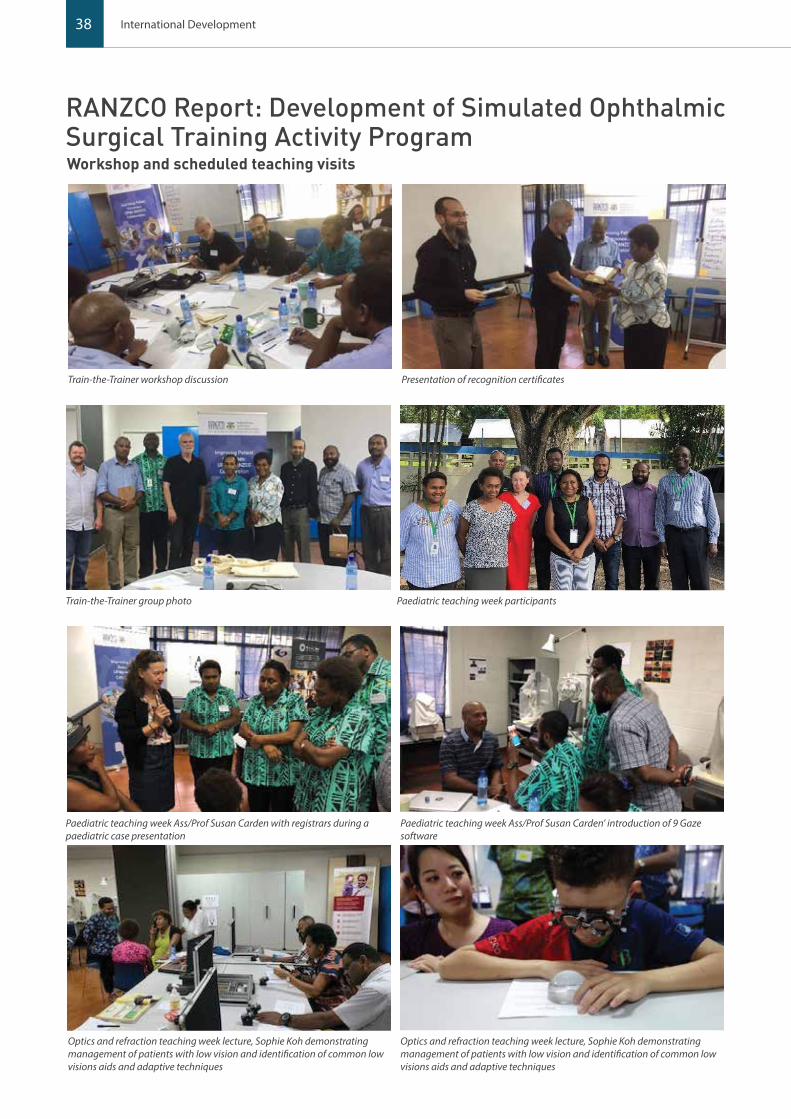
38 International Development
RANZCO Report: Development of Simulated Ophthalmic Surgical Training Activity Program Workshop and scheduled teaching visits
Train-the-Trainer workshop discussion
Train-the-Trainer group photo Paediatric teaching week participants
Paediatric teaching week Ass/Prof Susan Carden with registrars during a paediatric case presentation
Paediatric teaching week Ass/Prof Susan Carden’ introduction of 9 Gaze software
Optics and refraction teaching week lecture, Sophie Koh demonstrating management of patients with low vision and identification of common low visions aids and adaptive techniques
Optics and refraction teaching week lecture, Sophie Koh demonstrating management of patients with low vision and identification of common low visions aids and adaptive techniques
Presentation of recognition certificates
39Eye2Eye Quarter 3 2019
Collaborating partners UNICEF, the University of Papua New Guinea (UPNG) and RANZCO have come together to tackle retinopathy of prematurity (ROP), a potentially blinding disease that primarily affects premature infants.
An MOU, signed in June, marks a key milestone in a partnership that began in 2018 where UNICEF, RANZCO and UPNG supported training to upskill ophthalmologists, paediatricians, nurses and registrars in the identification and treatment of ROP. Work undertaken through this initiative will increase awareness around ROP and promote prevention and management in Papua New Guinea. Further planning and the procurement of retinal cameras and lasers are key aspects of this collaboration.
The joint UNICEF-UPNG-RANZCO collaboration is a unique intervention that will form part of a bigger picture: the Saving Lives - Spreading Smiles program to ensure that preterm babies have access to screening and treatment of ROP to prevent vision impairment and blindness.
“Without awareness, prevention and management, an estimated 20,000 to 25,000 babies born preterm each year in PNG are at risk of succumbing to this disorder,” UNICEF Representative, David Mcloughlin said.
RANZCO Fellow A/Prof Susan Carden has been instrumental in the RANZCO ROP Program work thus far, dedicating her time and expertise to the project. Susan has also been involved in the scheduled paediatric teaching week at UPNG, involvement of which has enabled increased skills and knowledge of the registrars.
Tackling Retinopathy of Prematurity (ROP)
Comment received from a Trainer-the-Trainer workshop participant: “Planning and teaching in an organised manner is a big challenge so having RANZCO input [through conducting the Training-the-Trainer workshop] is very much appreciated”.
Comments received from participants attending the Visiting Lecturer sessions: “I found the training very helpful and am so thankful I attended. Now I can comfortably refract my patients esp. strabismus and squint pts plus accommodating esotropia and convergence excess”.“[It is] always nice to have retinal visits to backup whatever knowledge we have from reading books on treatment options usually limited in relation to retinal disease. This has given me a boost in laser PRP and Intravitreal injections”.
I will implement in my practice as a result of participating in the teaching visits: - Applying principles in management
of most or retina cases, esp. diabetic/hypertension retinopathy, Eales disease
- Diagnose diabetic retinopathy (grading) and be able to diagnose which patient need Laser Rx
- Be able to order low vision assistive devices and refract and prescribe glasses for squint and strabismus patients. Not give up on low vision patients but to go an extra mile to assist them with refraction and assistive devices.
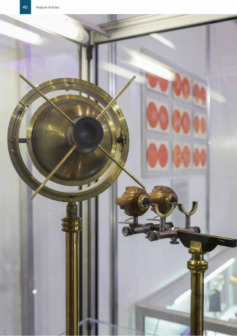
40 Feature Articles
41
50 Years in Vitreoretinal SurgeryUndoubtedly the development of pars plana vitrectomy in the 1970s transformed retinal reattachment surgery and also opened the way to the development of macular surgery and surgery for the complications of diabetic retinopathy.
Robert Machemer and Jean-Marie Parel, recruited from Melbourne where he worked with Prof Gerard Crock, developed the prototype device with Storz Instrument Company and conducted surgical training courses at Bascom Palmer Eye Institute in Miami to introduce retinal surgeons to the new instrumentation and techniques.
In 1975 Conor O’Malley and Ralph Heintz introduced the concept of three port pars plana vitrectomy with a 20 gauge Ocutome. All the ports were of the same gauge and hence interchangeable. This advantage with the smaller instrumentation meant that this system rapidly became adopted worldwide.
During the 1980s, Stanley Chang described the use of perfluropropane gas C3F8 for longer acting internal tamponade and then in 1988 he described the use of “heavy liquids” perflurocarbon liquids for intraoperative management of retinal detachment.
The development of smaller gauge instrumentation progressed through the late 1990s and in 2002 Gildo Fujui, Eugene De Juan and Mark Humayun published regarding a 25 gauge vitrectomy system. The flexibility of these instruments limited the adoption of them initially and Claus Eckhardt in 2005 published an account of 23 gauge system which combined the positive feature of being transconjunctival but with increased rigidity and safety compared to the 25 system. Subsequent to this the 25 gauge instruments have been strengthened and now are very widely used.
Macular surgery has now become routine practice and the results from full thickness macular hole repair and epiretinal membrane peel have improved significantly with smaller gauge surgery and the development of dyes to stain the epiretinal and internal limiting membrane to facilitate peeling. The primary success rate of full thickness macular hole repair is now routinely 85-90% and with transconjunctival sclerostomies patients are more comfortable and day surgery predominates.
Machemer and the early adopters of vitrectomy would be astounded to learn that now the vitrectomy approach is being used to deliver gene therapy to patients with retinal dystrophies. Luxterna® (voretigene neparvovec-rzyl) developed by Spark
Therapeutics Inc. has been given FDA approval for the treatment of biallelic RPE65-mediated inherited retinal dystrophy. This is just the first approved retinal gene therapy, with many more on the way.
In addition, the recent development of both epiretinal and subretinal retinal prostheses for patients with severe visual loss due to retinal dystrophic disease utilises the vitrectomy approach. These devices, which aim to provide visual information to patients who are non-navigational, have proven successful in aiding these patients with navigation and identification of objects. More recently, a subretinal device has been developed aiming to aid patients with severe geographic atrophy due to age related macular degeneration.
Interestingly within the last decade a previously avoided space has become a surgical target. The suprachoroidal space is now being investigated for a number of interventions including our own suprachoroidal retinal prosthesis, developed in Melbourne.
We can only look forward to the next 50 years and what that brings in advancing technology, leading to improved outcomes for our patients.
A/Prof Penny Allen
50 Years of Paediatric Cataract SurgeryChanges in cataract surgery in children over the last 50 years have produced significantly better visual outcomes.
Prior to the invention of YAG-laser for posterior capsular opacity and after cataract remnants due to extracapsular cataract surgery, needling discission was the usual approach.
The use of discission to remove cataracts in children was first described around 1960. This was an alternative to the standard intracapsular procedures being performed in adults, which required large incisions and significant postoperative care to allow for wound healing in an age without microsurgical techniques and the operating microscope.
A paper by Ira S Jones in 1960, in the Transactions of the American Ophthalmic Society, discussed whether different surgical procedures for cataract removal were applicable to congenital cataracts of different aetiologies. His final analysis confirmed that needling could be used for all aetiologies. This technique has its advantages as it was through a small incision which did not require suturing. However, it often needed to be repeated to “clear” the visual axis.
Eye2Eye Quarter 3 2019
50 Years of Ophthalmic Innovation and Development
The developments in ophthalmology in the 50 years since the foundation of the College are breathtaking and ongoing. To explore how ophthalmology has changed during this time, I have asked experts in their field to highlight pivotal changes and innovations. Many of these developments will be shown in the Museum exhibit at RANZCO Congress in November.
David Kaufman, Museum CuratormuseumSharing our heritage
Pictured left: Donders physiological eye from the Museum exhibition held at the Perth 2017 RANZCO Annual Scientific Congress

42 Feature Articles
Fast forward to 2019 and paediatric cataracts are performed using adult phaocoemulsification techniques, with some technical modifications to allow for the decreased scleral rigidity and increased capsular elasticity found in children.
Now the biggest dilemma in paediatric cataract surgery is the choice of IOL power and the theoretically desired refractive error to allow for future eye growth, and likely myopic shift, versus the risk of induced hypermetropia and resultant amblyopia.
Dr Wendy Marshman
50 Year of the RetinaThe 1971 Diabetic Retinopathy Study (DRS) showed that argon laser photocoagulation of the retina was a successful treatment for proliferative diabetic retinopathy. This was followed shortly after by the ETDRS which demonstrated efficacy of macular laser for oedema. Prior to this, a variety of treatments were used including pituitary ablation which carried a significant mortality rate. Despite being decades old, laser still has a role to play in the management of diabetic retinopathy as evidenced by the recent DRCRNet data from Protocol S in its five year results 2018.
Retinal imaging evolved from the development of fluorescein angiography, which initially allowed retinal specialists to view posterior segment functionality as well as the structural information provided by colour photography. From this information, gifted clinicians such as J. Donald Gass and Alan Bird were able to describe a range of diseases and characterise their features.
More recently we have seen the rise of the digital age. Since 1991 OCT has revolutionised the viewing of the macula and eliminated any controversy about the presence of oedema. It has also allowed us to view in-vivo tissue to an almost cellular level. Further developments have followed, including scanning laser ophthalmoscopes, fundus autofluorescence, oct-angiography, adaptive optics and widefield imaging. It is now possible to take dozens of images of the retina in both en-face and cross-sectional views. If anything, we have gone from too little information to perhaps an excess. Diseases that were thought to be distinct entities have been shown to have overlapping features and likewise previously linked diseases have been shown to be separate conditions. The challenge now is to choose the correct imaging to display the pathological features and target treatment. Artificial intelligence systems are already being developed that use these digital images to help screen vast numbers of patients.
Recognition of the role played by VEGF in subretinal choroidal neovascularization in ARMD initiated the use of anti VEGF agents delivered by intravitreal injection. The efficacy of the treatment was confirmed in the MARINA and ANCHOR trials in 2006. The vitreous cavity was once considered a space to be violated at one’s peril. Intravitreal injections are now the most commonly performed procedure by ophthalmologists and provide treatment for a range of ever increasing pathologies. These include macular degeneration, retinal infections, retinal vascular disease and, more recently, genetic therapies. With hindsight, the vitreous is almost the perfect location for local treatment. It is easily accessed with a small gauge needle, provides a reservoir for treatment, can be easily visualised and minimally traumatic to the eye. With increasing knowledge of the procedure and causes of post injection infection, guidelines have been developed to make the risk of infection minimal. The scale of the anti-VEGF revolution is hard to over-state. It has provided treatment for blinding conditions and revolution of anti-VEGF agents. It is likely more treatments will be trialled and released for use as intravitreal agents.
Dr Xavier Fagan
50 Years of Cataract SurgeryThe last five decades have seen what must be one of surgery’s most astonishing transformations. Cataract surgery was a high risk operation performed under a general anaesthetic, requiring several days in hospital and leaving the patient with an oedematous cornea for several days to weeks, high astigmatism and thick aphakic glasses. It is now a low risk operation often performed with only sedation and topical anaesthetic as a day procedure with rapid recovery where the patient’s new refractive situation is often spectacle independence for near and distance.
It is not just ophthalmic hubris to say that modern cataract surgery is now the most successful of all surgeries, one of the few where the patient frequently is left better functioning than they were prior to the development of the disease. What other common surgery can claim this, especially on a matter of such importance as sight?
This progress from intra-capsular, through extra-capsular to modern small incision phacoemulsification cataract surgery has been built on hundreds of small, incremental advances and several large jumps forward. It is fair to say that it is also one of the great success stories of a partnership between surgeons and medical companies.
The number of advances are way too numerous to list here but extend across several fields; operative techniques and equipment (especially the introduction of phacoemulsification, improved fluidics and torsional phaco power), improved wound profiles and reduced size (and with this the elimination of the need for sutures), anaesthesia (with the introduction of sedation plus topical anaesthesia) and medication (with intracameral antibiotics). Then there is the jewel in the crown, the introduction of IOLs and decades of improvements not only in IOL technology (with foldable, aspheric, toric, multifocal and EDOF IOLs to name but a few) but also in IOL formulae and biometry.
In conjunction with the developments in cataract surgery there has been a commensurate improvement in vitreoretinal and corneal grafting surgery to often allow full rehabilitation of patients that have either pre-existing concurrent ophthalmic disease or experience an operative complication.
Other changes have been introduced to safeguard patients with specific risk factors, such as intracameral phenylephrine and dilation devices for small pupils and intraoperative floppy iris syndrome. This is an excellent example of the profession meeting the challenges of new risks to maintain patient safety.
The result has been surgery where the trauma to the eye has been reduced so dramatically that frequently the day following surgery the vision is already greatly improved and the eye, to the casual observer, is normal, with no external signs of inflammation.
As always one change is built on the back of another and then allows the next development, such as the replacement of the capsulotomy with a capsulorhexis allowing for the introduction of safe phacoemulsification surgery and foldable IOLs driving more astigmatically neutral small incision wounds.
I feel privileged to have had a career that has spanned most of these improvements. Change does not stop and I am sure in 20 years we will look back on present day surgery and feel it was somewhat primitive. But, at almost 60 years of age and with my own cataract surgery not seeming that distant, I am incredibly grateful that I will be having modern, small-incision surgery with topical anaesthesia and a near vision IOL strategy. Now if only we have fully accommodative IOLs before my surgery is due!
Dr Michael Loughnan
43Eye2Eye Quarter 3 2019
50 Years of Corneal SurgeryIn the seventies corneal diseases were all treated with one operation: the penetrating keratoplasty. Whether the indication was tectonic repair of a leaking cornea, removal of a worsening infection or attempting to regularise an ectatic cornea to improve vision, the abnormal cornea was removed and donor tissue carefully sown into position. Arguments raged mainly over suture techniques and other tricks to improve outcomes.
Real patient data on outcomes, via Coster and Williams’ work on the ACGR, highlighted high risk patients that might not do well with surgery and applied some rigor to our decision to resist offering unlimited repeat grafts after failure. Despite these suture related complications and rejection and prolonged recovery times after surgery limited the effectiveness of transplant surgery.
The revolution in lamellar surgery has transformed our outlook.
Endothelial keratoplasty techniques, developed by Terry and Melles and standardised by Price, Kruse and others, arose from the unanticipated ability of the graft to attach to the posterior stroma without sutures or glue, only the steady pressure of an intraocular air bubble.
The realisation that the transplanted donor stroma led to interface irregularities and haze, and so reduced the vision as well as increasing the risk of rejection, led to minimalist operation; transplants of just the endothelial layer with its supporting basement membrane (DMEK).
The ability to expand endothelial cells in the laboratory has opened the way for the possibility of cell therapy or a truly tissue engineered cornea.
Keratoconus remains a mysterious condition, affecting young people with poorly defined diagnosis, unclear aetiology and highly variable severity. Therapy 50 years ago was restricted to glasses, the newly developed rigid gas permeable contact lenses and graft surgery if best corrected vision remained poor.
Progression was carefully observed and eye rubbing was discouraged, but no really effective intervention was offered.
The engineering principle of cross linking a polymer to reduce elasticity, stiffen the material and so set the long term shape of the cornea was explored by a group in Dresden. They took up the challenge of developing an effective but safe cross linking protocol that minimised scar formation, maintained vision and was safe to the endothelial cells but still halted progression. The initial recommendation was epithelial removal followed by 30 minutes’ soaking with riboflavin and then a 30 minute session of irradiation with UV light at 3mW. This protocol stressed safety at the cost of both surgeon efficiency and the patient. A successful randomised control trial and widespread adoption has made a dramatic difference to sufferers, keeping most with good vision in glasses or contact lenses and dramatically reducing the need for corneal graft surgery.
The discussion has now moved on to optimising the treatment regimen. Transepithelial treatment reduces pain and the risk of complications but at the cost of lower efficacy. Shorter soak times and higher intensity but reduced treatment times may have equivalent effect as the Dresden protocol. Evaluation of definite signs of progression, earlier diagnosis and access to care are the current challenges. Deeper understanding of the fundamental basis of the condition will provide the next breakthrough.
A/Prof Mark Daniell
50 Years of Refractive and Corneal SurgeryOver the past half century, ophthalmology has seen the establishment and continued refinement of the refractive surgery subspecialty. Prior incisional refractive corneal surgical techniques lacked accuracy and predictability but planted the seeds for the development of a more refined method for modifying corneal curvature. This came in the form of the Excimer laser, which was developed as a result of trans-Atlantic collaborations between London and New York. Phototherapeutic keratectomy (PTK) emerged as a safe and precise method of correcting refractive error, which continues to be used until this day. Twenty year follow up data confirms the safety and efficacy of PTK, which remains an excellent procedure for laser vision correction in many patients.
Parallel to the improvements in corneal refractive surgery, we have benefited from advances in anterior segment imaging. A key development has been an appreciation of the importance of imaging the posterior cornea. This has resulted in a shift from classic Placido based corneal topography to more three dimensional Scheimpflug corneal tomography. No refractive surgeon would consider laser vision correction without the reassurance of a normal tomographic map of the cornea, which is essential to exclude keratoconus and confirm that a safe residual stromal bed will remain after surgery.
The transition to lamellar corneal surgery has been a theme over recent decades. For laser refractive surgery this meant the evolution from surface treatment with PTK to LASIK, characterised by creation of a corneal flap followed by Excimer laser ablation of the exposed stroma. Flap creation by a mechanical microkeratome has largely been superseded by the introduction of the femtosecond laser, reducing serious intraoperative flap related complications and affording precise control over flap dimensions. More recently, SMILE is an exciting addition to the refractive armamentarium. Using a femtosecond laser, a small lenticule of corneal stroma is cut and then manually excised from the cornea. This method avoids potential flap related complications and is growing in use.
Lamellar corneal transplant techniques have also evolved considerably. Fifty years ago, full thickness penetrating keratoplasty (PK) was used for most corneal transplants. While tried and tested, PK is far from a perfect procedure and transplants have become targeted to replace only the layer of the cornea which is diseased. Thus endothelial disease is now treated with endothelial keratoplasty (EK), with the implanted lamella of tissue becoming ever thinner as the years pass. We have moved on from Descemet’s stripping (automated) EK (DS(A)EK), which includes a layer of corneal stroma to Descemet’s membrane EK (DMEK). Anterior lamellar keratoplasty is preferred over PK in eyes with normal corneal endothelium, for example keratoconus.
A truly pivotal event for corneal surgery has been the development of corneal cross linking (CXL) for the treatment of progressive keratoconus. Early evidence for its efficacy comes from a landmark randomised controlled trial performed in Melbourne. Corneal transplant surgery for keratoconus will perhaps become a thing of the past as we move into the next 50 years as a College. It is with great interest that we corneal surgeons see what the future brings for our patients.
Dr Georgia Cleary

44 Feature Articles
RANZCO’s newly established Future of Ophthalmology Task Force explores how eyes will be looked after in the next few decades.
Artificial intelligence (AI) is not only a thing of the future, it is already being used in medicine. Therefore, we have begun our task by looking first at how AI will shape the future of not only ophthalmology but medicine as a whole.
Objectives of the Task Force: As a Task Force, our first job is to produce a white paper on AI which will define the technology and address various important associated issues: ethical, privacy, financial and relevant legislative aspects.
This paper will be a resource to educate and inform our membership on these matters as well as to explore the cost of delivery of artificial intelligence systems and the impact on some current and future government initiatives such as the Digital Health Record. Issues of data ownership will also be addressed.
While this is the initial phase of this exercise, we think that the final paper will be a living document which will serve as an important reference for all of us.
Being leaders in collaborative eye care, RANZCO sees this white paper being the foundation to provide advice to our own Board, as well as to government, regulatory authorities and other related bodies.
There is no doubt that change in the way we practice medicine is already here. One example is the use of artificial intelligence in the diagnosis and management of diabetic retinopathy. This has already been implemented in many countries including India
and the USA and is now being rolled out in neighbouring Timor Leste.
We will be monitoring all these changes closely and will advise the College on how these new technologies should be approached and managed in line with the current and future RANZCO strategic plans. We will communicate with our members to provide evidence based advice as to what impact changes might have on the profession to ensure we all continue to deliver the highest quality of eye health for all.
In the coming months you will see various task force members presenting at branch and federal meetings to outline the progress of this project and provide briefing on new concepts and technologies that are supported by scientific evidence. The aim of this is to help us all make informed decisions about which innovations to adopt.
Other issues that the task force is looking into include creating a ‘vision for the future’. This means exploring what ophthalmology and eye care will look like in the next few decades and informing the board so that we can ‘steer the ship’ in the proper direction. Obviously, current training pathways and teaching programs need to be appropriately modified and new approaches developed.
This fits in with the strategic plan of the College to provide contemporary education and professional development to our Fellows and make sure we remain the leaders in the field of eye care.
What was Ophthalmology like 50 years ago? Over the past 50 years ophthalmology has changed dramatically in that we are now treating and managing conditions proactively rather than reactively as we did in the past. A prime example
The task force has the following members: Professor Nitin Verma (chair), Associate Professor Peter Van Wijngaarden, Dr Andrew Crawford, Dr Narme Deva, Professor Margaret Otlowski, Professor Alex Hewitt, Dr Chadran Pereira, Professor John Grigg, Dr Weng Onn Chan and Dr David Andrews.
Introduction and Scope: The Future of Ophthalmology Task force

45Eye2Eye Quarter 3 2019
was the timing of cataract surgery where a few decades ago cataract surgery was performed only when the patient was severely visually disabled, and the philosophy was that it was best to intervene surgically when the patient would have nothing to lose should the cataract surgery go wrong. Techniques for intracapsular cataract extraction included “tumbling”, the Smith Indian technique, instrument assisted extraction with the erysiphake, capsule forceps, cryotherapy with or without the use of zonulysis. Even when cataract surgery was performed, it was very often without the use of a microscope using 8-0 silk sutures to close the wound and leaving the patient aphakic. By today’s standards, the visual results were often poor and the complication rates high.
With the advent of intraocular lenses, small incision cataract surgery with phacoemulsification and better intraocular lens power calculation formulae, cataract surgery has evolved into refractive surgery, which is being offered earlier and earlier to patients once they start complaining of visual disability. Cataracts are often in the earlier stages in their development when surgical intervention is planned.
As you would expect, looking more broadly we can see that, in every area of ophthalmology, the treatments and methods we use today produce significantly better results than were achieved 50 years ago.
The ophthalmic therapeutic armament was very limited and most of the drugs we used had serious side effects. An example of this was the use of topical Pilocarpine and Eserine in the management of glaucoma. These were often made in hospital pharmacies. Surgical techniques in the earlier days included cyclodialysis, iridenclesis and sector iridectomy. The complications of these medical and surgical therapies were very serious and were well known.
Screening programs for diseases such as glaucoma, diabetic retinopathy and childhood eye disorders were limited. Very often when disease was detected, it was fairly advanced. In addition, blindness from infectious diseases such as tuberculosis, syphilis and leprosy were common.
Changes in the past few decadesOver the years a lot of disruptive medical technologies have come in (“disruptive” is a word used to define a technology that radically changes the way we do things; in this instance practice our profession). Examples of disruptive ophthalmic technologies include optical coherence tomography, phacoemulsification and the use of intravitreal anti-VEGF agents for the management of ocular diseases. The YAG and Argon lasers are other examples of disruptive technologies that have come into common use over the past few decades.
From being a speciality that not many people were interested and being low on the “scale” of popular medical disciplines, ophthalmology now is a sought after field of specialisation. To enter the training program today is a very competitive process simply because the speciality has evolved to a very precise art and the results we can achieve today are nothing short of spectacular.
The advent and incorporation of artificial intelligence in our day-to-day work will only make things better for our patients, the profession and, of course, the community at large.
What will be the biggest, most impactful change in Ophthalmology in the coming years? Artificial intelligence is already being incorporated in algorithms for the screening and grading of diabetic retinopathy and maculopathy.
Glaucoma, macular degeneration and uveitis management algorithms will also soon be using this technology. In addition, rare diseases which often get left behind in research programs
and therefore treatment development will certainly be easier to address using artificial intelligence for diagnosis and management.
Gene and stem cell therapies will also be much easier to implement and will use artificial intelligence to guide the process. Diseases such as Stargardt’s disease, retinitis pigmentosa and even glaucoma could be converted from the category of incurable to curable. No aspect of eye care will be left untouched.
What will be the biggest challenges for Ophthalmologists over the next 50 years?While we continue to strive towards perfection, the biggest challenge currently is to demystify and understand the potential and limitations of emerging technologies. Immediately, we need to understand the process of integration of artificial intelligence into health care platforms at the right time and stage. It will also be very important to ensure the wellbeing of our patients, not only from the medical point of view but also in terms of protecting their privacy by ensuring that the data that gets into the system is safe and secure and that it is used exclusively for the purposes it was intended for. The ethical and moral values of the profession need to be upheld at all times.
How will the profession of Ophthalmology change in 50 years time? The skill set of an ophthalmologist over the past 50 years has changed dramatically with more and more ophthalmologists practicing a subspecialty. There are very few ophthalmologists who consider themselves as general ophthalmologists, as was the norm in the past.
Normally, a significant proportion of an ophthalmologist’s task is to amalgamate the history findings and the results of investigations of a patient into a diagnosis/management plan. I believe the more one does this, the more experienced one becomes. An experienced ophthalmologist is one who has worked out his or her own ‘formulae’ to successfully handle the problems that patients present them with.
However, when an ophthalmologist stops practicing, the experience is lost and there is no way to transfer this to junior colleagues in a comprehensive manner unless the two have worked together for a long time. There is no comprehensive way that the profession as a whole can keep building on this knowledge and experience base. Books are one possibility, but this is a tedious and resource intensive option.
The use of artificial intelligence allows data and experience to be shared like never before. The whole system gets better and more experienced with time and is not dependent on the availability of a particular (experienced) ophthalmologist. An artificial intelligence enabled system could soon help in guiding the management of diseases not only for an individual patient but also for the disease subset. Machines will be able to recognise the problem and suggest treatment. The more people use artificial intelligence all over the world, the greater the accuracy of the system will be and soon this level of accuracy will be demanded by the patient as well as governments worldwide. It is clear that the use of artificial intelligence in ophthalmology is inevitable and we need to ensure that we are prepared for it so that it can be implemented in a proper, orderly, safe way.
The role of the doctor will keep changing as artificial intelligence takes on a greater role. At least in the near future, I believe that this will be related to recognition of patterns and therefore on the diagnostic side of our day-to-day work. The availability of robots will change things when it comes to treatment, but his is not yet an immediate priority to address.
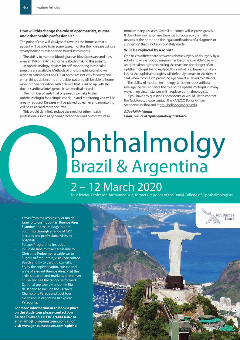
46
Brazil & ArgentinaOphthalmolgy2 – 12 March 2020 Tour leader: Professor Harminder Dua, former President of the Royal College of Ophthalmologists
• Travel from the iconic city of Rio de Janeiro to cosmopolitan Buenos Aires.
• Examine ophthalmology in both countries through a range of CPD lectures and professional visits to hospitals.
• Partner Programme included• In Rio de Janeiro take a train ride to
Christ the Redeemer, a cable car to Sugar Loaf Mountain, visit Copacabana Beach and fly to vast Iguazu Falls.
• Enjoy the sophistication, cuisine and wine of elegant Buenos Aires, visit the artist's quarter and markets, take a river cruise and see the tango performed.
• Optional pre tour extension in Rio de Janeiro to include the Carnival Champions Parade and post tour extension in Argentina to explore Patagonia.
For more information or to book a place on the study tour please contact Jon Baines Tours on + 61 (0)3 9343 6367 or email [email protected] or visit www.jonbainestours.com/ophthal
Feature Articles
How will this change the role of optometrists, nurses and other health professionals?The point of care will slowly shift towards the home so that a patient will be able to, in some cases, monitor their disease using a smartphone or similar device-based instruments.
The ability to monitor blood glucose, blood pressure and now even an INR or HbA1c at home is slowly making this a reality
In ophthalmology, devices for self-monitoring intraocular pressure are available. Methods of photographing one’s own retina or carrying out an OCT at home are not very far away and when things do become affordable, patients will be able to home monitor their condition with a device that is linked up with the doctor’s artificial intelligence based medical record.
The number of visits that one needs to make to the ophthalmologist for a simple check-up and monitoring visit will be greatly reduced. Diseases will be picked up earlier and monitoring will be easier and more accurate.
This would definitely reduce the need for other health professionals such as general practitioners and optometrists to
monitor many diseases. Overall outcomes will improve greatly. It does, however, also raise the issues of accuracy of smaller devices at the home and the legal ramifications of a diagnosis or suggestion that is not appropriately made.
Will I be replaced by a robot?One has to differentiate between robotic surgery and surgery by a robot and while robotic surgery may become available to us with an ophthalmologist controlling the machine, the danger of an ophthalmologist being replaced by a robot is extremely unlikely. I think that ophthalmologists will definitely remain in the driver’s seat when it comes to providing eye care at all levels to patients.
The ability of modern technology, which includes artificial intelligence, will enhance the role of the ophthalmologist in many ways. In no circumstances will it replace ophthalmologists.
If you have any questions or concerns or would like to contact the Task Force, please contact the RANZCO Policy Officer, Stephanie Mulholland at [email protected]
A/Prof Nitin Verma Chair, Future of Ophthalmology Taskforce
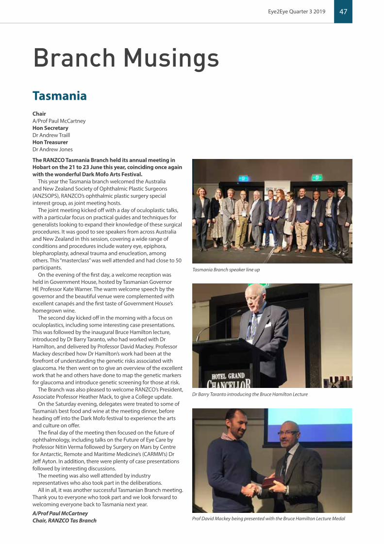
47
Brazil & Argentina
Branch Musings
ChairA/Prof Paul McCartneyHon SecretaryDr Andrew TraillHon TreasurerDr Andrew Jones
The RANZCO Tasmania Branch held its annual meeting in Hobart on the 21 to 23 June this year, coinciding once again with the wonderful Dark Mofo Arts Festival.
This year the Tasmania branch welcomed the Australia and New Zealand Society of Ophthalmic Plastic Surgeons (ANZSOPS), RANZCO’s ophthalmic plastic surgery special interest group, as joint meeting hosts.
The joint meeting kicked off with a day of oculoplastic talks, with a particular focus on practical guides and techniques for generalists looking to expand their knowledge of these surgical procedures. It was good to see speakers from across Australia and New Zealand in this session, covering a wide range of conditions and procedures include watery eye, epiphora, blepharoplasty, adnexal trauma and enucleation, among others. This “masterclass” was well attended and had close to 50 participants.
On the evening of the first day, a welcome reception was held in Government House, hosted by Tasmanian Governor HE Professor Kate Warner. The warm welcome speech by the governor and the beautiful venue were complemented with excellent canapés and the first taste of Government House’s homegrown wine.
The second day kicked off in the morning with a focus on oculoplastics, including some interesting case presentations. This was followed by the inaugural Bruce Hamilton lecture, introduced by Dr Barry Taranto, who had worked with Dr Hamilton, and delivered by Professor David Mackey. Professor Mackey described how Dr Hamilton’s work had been at the forefront of understanding the genetic risks associated with glaucoma. He then went on to give an overview of the excellent work that he and others have done to map the genetic markers for glaucoma and introduce genetic screening for those at risk.
The Branch was also pleased to welcome RANZCO’s President, Associate Professor Heather Mack, to give a College update.
On the Saturday evening, delegates were treated to some of Tasmania’s best food and wine at the meeting dinner, before heading off into the Dark Mofo festival to experience the arts and culture on offer.
The final day of the meeting then focused on the future of ophthalmology, including talks on the Future of Eye Care by Professor Nitin Verma followed by Surgery on Mars by Centre for Antarctic, Remote and Maritime Medicine’s (CARMM’s) Dr Jeff Ayton. In addition, there were plenty of case presentations followed by interesting discussions.
The meeting was also well attended by industry representatives who also took part in the deliberations.
All in all, it was another successful Tasmanian Branch meeting. Thank you to everyone who took part and we look forward to welcoming everyone back to Tasmania next year.
A/Prof Paul McCartneyChair, RANZCO Tas Branch
Tasmania
Eye2Eye Quarter 3 2019
Tasmania Branch speaker line up
Dr Barry Taranto introducing the Bruce Hamilton Lecture
Prof David Mackey being presented with the Bruce Hamilton Lecture Medal

48 Branch Musings
ChairDr Robert GriffitsVice ChairDr Diana FarlowHon SecretaryDr Alina ZeldovichHon TreasurerDr Nisha SachdevCountry Vice ChairpersonDr Neale Mulligan
The 2019 NSW Annual Scientific Meeting (ASM) was successfully held over the weekend of 16-17 March with the theme ‘Ophthalmology: Current Waves, and the View over the Horizon’. The two day conference dealt with the evolving technologies and innovations across the spectrum of ophthalmology. We also looked outside of our speciality to what is also transforming other surgical and medical specialties.
Robotics, nanotechnologies, new drug delivery systems, targeted immune therapies and the ever expanding application of monoclonal antibody application to medicine continue to evolve and are impressive new developments.
Ron Adelman from Yale University, Tina Wong from Singapore Eye Centre and Lyndell Lim from CERA were the invited keynote speakers and they all delivered excellent presentations from their diverse subspecialties and research.
Planning is well underway for the 2020 ASM. It will be held at the Newcastle Exhibition Centre and, as it will be an out of Sydney event, the decision was made to revert to the Friday/Saturday format (one day out of work and one day out of the weekend).
The city of Newcastle has been absolutely transformed over the past three decades and it has well and truly moved away from its industrial image. Hopefully delegates will be able to spend a little extra time to enjoy the revitalised city.
The 2020 ASM title will be ‘The Changing Landscope of Urban, Regional and Rural Ophthalmology’. We do know how to spell and ‘Landscope’ is a play on words combining ‘landscape’ with ‘scope’. The scope will once again focus on emerging technologies and ophthalmic techniques and will also splice in the defining and differing elements of urban, regional and rural practice.
The NSW Branch Committee is very keen to be much more inclusive of our registrars at our ASM. We are looking at adding special registrar sessions and planning to minimise the financial impacts to facilitate registrar attendance.
At our last committee meeting, the Branch decided to once again continue to support Australian Ophthalmology Research and Education (ORIA). Rather than give our customary direct grant to ORIA, $100,000 has been donated to the fledging Australian and New Zealand Eye Foundation (ANZEF). Fund raising is not easy, particularly given the strong competition that already exists with other charitable eye organisations. We strongly encourage NSW Fellows to directly support ANZEF financially, as well as encourage our patients to do the same.
The Branch has been enacting initiatives to promote and support rural and regional ophthalmology with a view to improving services outside the capital cities. This includes remote and very remote ophthalmology for both Indigenous and non-Indigenous remote communities. The maldistribution of specialist providers has been clear for a long time and needs a multifaceted approach. We hope to foster relations and communications with the NSW Minister for Health.
Dr Robert GriffitsChair, RANZCO NSW Branch
New South WalesVictoriaChairDr David van der StraatenHon SecretaryDr Xavier FaganHon TreasurerDr Lewis Levitz
The Victorian Branch Scientific Meeting, held on 2 March, heard a broad range of presentations, ranging from effects of pregnancy on the eye to systemic management of diabetes to a debate on iStents. We were fortunate to receive addresses from several invited ophthalmologists. A/Prof Clare Fraser provided insights into the community impact of concussion and treatable aspects of its effect upon ocular motility disorders. Dr Jennifer Arnold discussed updates on clinical outcomes in AMD and interpretation of fundus autofluorescence imaging. Prof Bill Morgan outlined the aspects of the Xen implant. Recently appointed Ringland Anderson Professor Keith Martin spoke on gene therapy for glaucoma, providing a brief taste of what was to come later that month with the highly successful World Glaucoma Congress, of which he was a key organiser.
We are grateful to Drs Elaine Chong and Anu Mathew for taking over the main role of arranging scientific content for the 2020 meeting, scheduled for 21 March 2020 and to be held again at the Woodward Centre.
The Melbourne Ophthalmic Alumni meeting in May, led by Prof Keith Martin, continued the glaucoma theme. It was followed by dinner at the State Library where Peter Meagher was the guest of honour.
In May, a team led by Dr Jacqui Beltz ran an innovative program “Geneye” to allow medical students, HMOs, trainees and consultant ophthalmologists to use surgical simulators and wet lab facilities and prepare themselves mentally and physically for surgery. The simulators allow a virtual reality experience of performing intra-ocular surgery, such as cortical aspiration, and performing anterior vitrectomy after rupturing the posterior capsule. Other sessions included mindfulness, ergonomics of sitting at a microscope and core strength exercises.
The Victorian Branch has continued to support trainees by giving them exposure to Indigenous health, funding three registrars each year to spend a week assisting the Lions Outback Vision Van service. One trainee was sponsored to attend the Closing the Gap for Vision Conference in Alice Springs, reporting this to be a particularly helpful experience.
Dr David van der Straaten Chair, RANZCO Vic Branch
49Eye2Eye Quarter 3 2019
Western Australia QueenslandChairDr Nigel MorletHon SecretaryDr David DelahuntyHon TreasurerDr Tom Cunneen
The Branch was delighted that Professor David Mackey was awarded an Order of Australia for his significant contribution to ophthalmic teaching and research. He recently stepped down as the head of the Lions Eye Institute, a role now filled by Professor Bill Morgan.
The Branch teaching program continues to be well supported with this year’s inter-hospitals meeting in March convened by Fremantle Hospital. It provided a great update on OCT and ocular electrophysiology and launched this year’s Branch collective audit on OCT use in clinical practice, organised expertly by Jo Richards as usual.
The Branch Annual Meeting was held in Albany, in keeping with the Branch’s current regional focus. We were privileged to have Prof Justin Smith from Flinders University deliver the Eye Surgery Foundation Lecture on inflammatory eye disease, and Prof Nitin Verma talk about the Timor Eye Program and the College’s diabetic referral guidelines. The topic of focus this year was Artificial Intelligence in Ophthalmology with Peter van Wijngaarden, along with trainee registrar Chandra Perera and RMO Matt Watson, who provided great insights into how these emerging technologies will impact clinical practice in the future. Chandra and Matt were both awarded best presentations for their efforts.
A/Prof Angus Turner has made great progress with developing the Northwest eye services. A regional eye care hub is now likely with the recent land donation to establish a full time clinic in Broome. State government funding for the provision of consultant salaries and support staff is looking good at this stage as well. Once up and running, the service would enable the establishment of a rural ophthalmology training program rotating through the Northwest, Bunbury, Hobart and Perth. In time we hope that an Albany post may also be established as part of the rotation.
The WA Health Department requested feedback regarding the proposed Cataract Clinic Standard from the Australian Commission on Safety and Quality in Health Care. We outlined our concerns about the hard visual acuity cut off at 6/12, suggesting that many patients would be disadvantaged if that was implemented.
Our next meeting will be the Pathology Imaging Meeting on Iris Diseases planned for Friday 20 September.
Dr Nigel Morlet Chair, RANZCO WA Branch
ORIA Grant Review Process The 2020 grant review process is underway with a total of 51 applications across Australia and New Zealand including five New Investigator applications. 17 Fellows of the College have confirmed participation on the review panel, made up of members of the Research Advisory Committee (RAC). Mirroring the NHMRC protocols, panel discussion of each application will be informed by feedback obtained through an external review process. Once the panel has met to assess the applications, based on the RAC's advice, the ORIA Board will allocate funding to the highest ranking applicants. Successful applications will be announced at the ORIA AGM held at RANZCO’s Congress in Sydney, November 2019.
O R I AADVANCING EYE RESEARCH
ChairDr Stephen GodfreyHon SecretaryDr Mark ChiangHon TreasurerDr Oben Candemir
As our southern states brace themselves for the third Arctic blast of winter, it is a timely reminder that the Queensland Branch Scientific Meeting is upon us and promises to be an entertaining and informative meeting of glaucoma advances and controversies. The weather will also be just about perfect.
I have to retract my statement from the last Eye2Eye magazine regarding the Director’s position at Guide Dogs Association Australia. I was premature in stating that Phung Vu had been appointed, as the position is still under consideration. I will update the final determination when it is made.
Bill Glasson, a previous RANZCO and AMA President, has handed over the reins of his Central Western Queensland practice to Sunil Warrier and Todd Goodwin. Bill is no stranger to places like Longreach, Barcaldine, Blackall, Bedourie and Boulea and has given 30 years of incredible service to these towns to ensure they have quality ophthalmic cover. I know Bill, and now Sunil and Todd, regard this as a privilege, not a task.
Queensland, along with all the other states, is grappling with the introduction of AI (artificial intelligence) and virtual training. The scope and application of these technologies are rapidly finding a place in our Vocational Training Program and should see positive outcomes for selection and training for the next generation of ophthalmologists.
Dr Stephen GodfreyChair, RANZCO QLD Branch
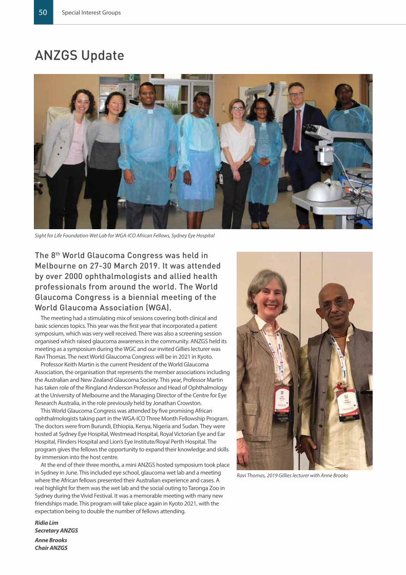
50 Special Interest Groups
The 8th World Glaucoma Congress was held in Melbourne on 27-30 March 2019. It was attended by over 2000 ophthalmologists and allied health professionals from around the world. The World Glaucoma Congress is a biennial meeting of the World Glaucoma Association (WGA).
The meeting had a stimulating mix of sessions covering both clinical and basic sciences topics. This year was the first year that incorporated a patient symposium, which was very well received. There was also a screening session organised which raised glaucoma awareness in the community. ANZGS held its meeting as a symposium during the WGC and our invited Gillies lecturer was Ravi Thomas. The next World Glaucoma Congress will be in 2021 in Kyoto.
Professor Keith Martin is the current President of the World Glaucoma Association, the organisation that represents the member associations including the Australian and New Zealand Glaucoma Society. This year, Professor Martin has taken role of the Ringland Anderson Professor and Head of Ophthalmology at the University of Melbourne and the Managing Director of the Centre for Eye Research Australia, in the role previously held by Jonathan Crowston.
This World Glaucoma Congress was attended by five promising African ophthalmologists taking part in the WGA-ICO Three Month Fellowship Program. The doctors were from Burundi, Ethiopia, Kenya, Nigeria and Sudan. They were hosted at Sydney Eye Hospital, Westmead Hospital, Royal Victorian Eye and Ear Hospital, Flinders Hospital and Lion’s Eye Institute/Royal Perth Hospital. The program gives the fellows the opportunity to expand their knowledge and skills by immersion into the host centre.
At the end of their three months, a mini ANZGS hosted symposium took place in Sydney in June. This included eye school, glaucoma wet lab and a meeting where the African fellows presented their Australian experience and cases. A real highlight for them was the wet lab and the social outing to Taronga Zoo in Sydney during the Vivid Festival. It was a memorable meeting with many new friendships made. This program will take place again in Kyoto 2021, with the expectation being to double the number of fellows attending.
Ridia LimSecretary ANZGS
Anne Brooks Chair ANZGS
ANZGS Update
Sight for Life Foundation Wet Lab for WGA-ICO African Fellows, Sydney Eye Hospital
Ravi Thomas, 2019 Gillies lecturer with Anne Brooks
51Eye2Eye Quarter 3 2019
This July, the Paediatric Special Interest Group (PSIG) met in sunny Noosa for their annual meeting. As always with the PSIG annual meeting, each delegate was asked to present two cases for discussion. This gives attendees the opportunity to share interesting or unusual cases and to get feedback from others about diagnosis and treatment options.
With this meeting having the highest attendance ever – obviously word is getting out about how useful and engaging these meetings are – we had to be very strict about times to fit everyone in twice over the two days. Thankfully we managed because each case study provided food for thought, useful perspectives and engaging discussion. Cases ranged from serious trauma to ROP and congenital cataracts, among a host of other things. There was an interesting focus in many cases on the genetic cause of impact on conditions. Lab based genetic diagnosis is becoming increasingly useful to paediatric ophthalmologists faced with an unusual case.
The business portion of the meeting focussed on plans to raise awareness about paediatric ophthalmology in the lead up to and during the upcoming RANZCO Congress in Sydney, which is happening in conjunction with the American Association for Pediatric Ophthalmology and Strabismus (AAPOS) and Asia-Pacific Strabismus and Paediatric Ophthalmology Society (APSPOS) meetings. This focus on paediatric ophthalmology gives us an excellent opportunity to showcase the innovations and developments within the supspecialty , to promote awareness of child eye health and to advocate for policy developments. Discussions in this area focussed on eye screening, myopia and trauma prevention.
We look forward to launching awareness activity later in the year and to seeing our colleagues in Sydney for RANZCO 2019.
Paediatric Special Interest Group – Annual Meeting RANZCO Practice Managers’
Conference 2019 – What to expect this year The Practice Managers Committee is in the throes of planning the 14th Practice Managers’ Conference and a list of speakers is currently being assembled to ensure that this year’s program is thought provoking, engaging and inspiring for all who attend.
Set in the vibrant city of Sydney, over three days from 9-11 November, there will be many opportunities to network with colleagues old and new. Sessions will take on various formats including discussions, panels, workshops and presentations. Time will be allocated for Q&A at the end of each plenary to ensure maximum participation. Delegates will also be given a useful toolkit to take home at the end of the conference, made up of resources provided by each presenter.
We are delighted to announce that multiple Walkley Award winning author, journalist and broadcaster Tracey Spicer will be this year’s keynote speaker, ensuring that the conference gets off to an exciting start. So far, the following topics have also been confirmed:
9 Current Risk Evolution in Private Practice
9 Managing Unsatisfactory Performance
9 The Lifecycle of a Practice Manager
9 Time Management Skills, Stressor Balancing and Leadership Strategies for Practice Managers
9 Staffing Benchmarking
9 Q+A with the RANZCO Medicare Advisory Committee
9 Emerging HR and Legal Issues
9 The Workforce of the Future – How will it differ from today and how will the manager need to Evolve?
Representatives from the AAPM will be on hand, and there will be opportunities to connect with our sponsors Avant Mutual, Inservio, Novartis and Wageloch over the course of the conference.
Who should attend?• Practice managers • Practice support staff - receptionists, office managers, team
leaders, administrators, allied health professionals. • The organising committee also welcomes other professions to
attend, such as Fellow delegates, who share their passion for excellence in healthcare and management.
Please check the practice managers’ page on the RANZCO Congress website for updates regarding registration and program details once they become available. http://www.ranzco2019.com/practice-managers-conference/

52
Representing the interests of ophthalmologists and their patients throughout New Zealand
The Wellbeing Budget 2019ONZ recently brought to the attention of RANZCO readers the continuing issues related to providing private ophthalmological services in New Zealand, in particular in the payment of services by anaesthetists.
ONZ notes that related concerns have been voiced by the New Zealand Association of Plastic Surgeons and the New Zealand Dermatological Society Inc. in a submission to the Ministry of Business, Innovation and Employment (MBIE). These concerns relate to issues that ONZ has voiced regarding unfair contractual terms and limitations on services to consumers that are non-transparent to consumers in New Zealand. As per ONZ, these organisations identify the major issue at stake here, as these limitations and terms have “the potential to adversely affect consumer choice and patient care”.
Whilst the insurers concerned are acting to limit their liability in costs, and implicitly sending patients towards the District Health Boards, it is interesting to contrast these actions with the Wellbeing Budget 2019.
ONZ applauds the emphasis on health contained within this budget, in particular the initiatives into mental health, but notes the devil may be in the detail. According to the estimates, the injection of funds into the District Health Boards operational budgets and capital expenditure budgets may just be at the cost of purchasing services. If this is the case, it will impact on the current contracts to private providers to service unmet needs at District Health Boards, in particular cataract surgery.
With the Wellbeing Budget aiming to cater for the provision of further services to the increasing population of New Zealand via the District Health Boards, at the same time as taking back contractual work, ONZ asks how will the public health system cope with an influx of patients whose needs are not met by their private health insurer?
On behalf of the full membership, ONZ would like to congratulate Professor Charles McGhee, MB, BSc, PhD, DSc, FRCS, FRANZCO, FRCOphth, on being awarded the honour of Officer of New Zealand (ONZM) for his services to ophthalmology.
Many of our members were taught by Professor McGhee, and many more had relatives and friends treated by him.
Professor Charles McGhee
RANZCO Affiliates

53Eye2Eye Quarter 3 2019
Quality Eye Nutrition
1300 95 2001 (Aus) e: [email protected] 0800 443 652 (NZ) f: +61 7 3056 0969
For more information, samples & brochures, please contact MD EyeCare:
© MD EyeCare 2019.
Australian-made in a world-class pharmaceutical facility.
✔ Concentrated Omega-3, GLA, Vitamin D3 & Antioxidants✔ Patented, Research-Based Formula for Systemic Relief✔ Improves Tear Quality & Production✔ Reduces Inflammation
SYSTEMICRELIEF
OMEGA 3 + Vit DGLA + ANTIOXIDANTS
MACULAR
ONCE DAILY
FORMULA
✔ Available as a Softgel Capsule or Powdered Orange Drink✔ Premium Ingredients✔ Compliance Prompting Packaging✔ Unbeatable Value - 6 Packs for $99
Proudly supporting
PHAH24151 EYE2EYE AD HALF PAGE UPDATE JUL19.indd 1 24/7/19 3:29 pm
New RANZCO WebsiteWe hope you have all found your way to the new RANZCO website. You will see it has a much cleaner look with easier to find sections, and better search functionality to drill down to specific information. Sitting behind the Member Login is a brand new dashboard with a cleaner look. Over the coming weeks and months, new elements of the dashboard will become live. The dashboards are personalised to Members, so if you are a Fellow you will see your CPD button with an easy to use CPD platform. If you are a Trainee you will see links to all the training and assessment opportunities, and be easily able to track your progress. The Surgical Logbook will also be much easier to use and Supervisors will find it easy to track the Trainees under their care. The website and the dashboard will in time be mobile enabled which means they will reconfigure to whatever device you use. As the system quickly expands, more detailed communications will come your way.
Part of the reason for a staged process is that one thing needs to happen before another. We wanted the new website and dashboard operating smoothly before migrating all members’ details, including CPD activity, in case any issues were encountered. So far things are great and we plan to have the new CPD diary fully functional well before Congress.
Another thing you may have noticed is the new look of the Learning Management Site. Over many years RANZCO has built up a huge number of learning resources, and the system was bloated. We have started the tidying process but there is a lot more to come. As we move to a more digitally enhanced teaching and supervisor support program, the LMS will continue to evolve.
New RANZCO website
If you have any feedback or questions about the new website, dashboard and LMS please contact us via [email protected]
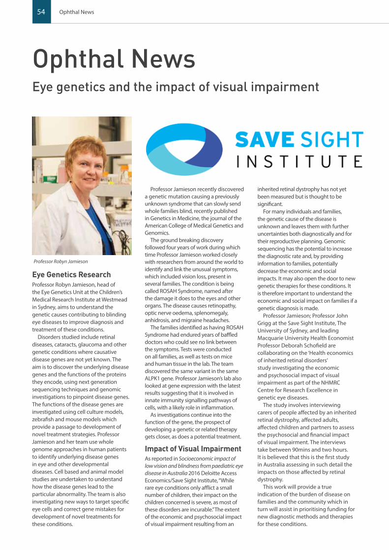
54
Ophthal News
Eye Genetics Research Professor Robyn Jamieson, head of the Eye Genetics Unit at the Children’s Medical Research Institute at Westmead in Sydney, aims to understand the genetic causes contributing to blinding eye diseases to improve diagnosis and treatment of these conditions.
Disorders studied include retinal diseases, cataracts, glaucoma and other genetic conditions where causative disease genes are not yet known. The aim is to discover the underlying disease genes and the functions of the proteins they encode, using next generation sequencing techniques and genomic investigations to pinpoint disease genes. The functions of the disease genes are investigated using cell culture models, zebrafish and mouse models which provide a passage to development of novel treatment strategies. Professor Jamieson and her team use whole genome approaches in human patients to identify underlying disease genes in eye and other developmental diseases. Cell based and animal model studies are undertaken to understand how the disease genes lead to the particular abnormality. The team is also investigating new ways to target specific eye cells and correct gene mistakes for development of novel treatments for these conditions.
Professor Jamieson recently discovered a genetic mutation causing a previously unknown syndrome that can slowly send whole families blind, recently published in Genetics in Medicine, the journal of the American College of Medical Genetics and Genomics.
The ground breaking discovery followed four years of work during which time Professor Jamieson worked closely with researchers from around the world to identify and link the unusual symptoms, which included vision loss, present in several families. The condition is being called ROSAH Syndrome, named after the damage it does to the eyes and other organs. The disease causes retinopathy, optic nerve oedema, splenomegaly, anhidrosis, and migraine headaches.
The families identified as having ROSAH Syndrome had endured years of baffled doctors who could see no link between the symptoms. Tests were conducted on all families, as well as tests on mice and human tissue in the lab. The team discovered the same variant in the same ALPK1 gene. Professor Jamieson’s lab also looked at gene expression with the latest results suggesting that it is involved in innate immunity signalling pathways of cells, with a likely role in inflammation.
As investigations continue into the function of the gene, the prospect of developing a genetic or related therapy gets closer, as does a potential treatment.
Impact of Visual Impairment As reported in Socioeconomic impact of low vision and blindness from paediatric eye disease in Australia 2016 Deloitte Access Economics/Save Sight Institute, “While rare eye conditions only afflict a small number of children, their impact on the children concerned is severe, as most of these disorders are incurable.” The extent of the economic and psychosocial impact of visual impairment resulting from an
inherited retinal dystrophy has not yet been measured but is thought to be significant.
For many individuals and families, the genetic cause of the disease is unknown and leaves them with further uncertainties both diagnostically and for their reproductive planning. Genomic sequencing has the potential to increase the diagnostic rate and, by providing information to families, potentially decrease the economic and social impacts. It may also open the door to new genetic therapies for these conditions. It is therefore important to understand the economic and social impact on families if a genetic diagnosis is made.
Professor Jamieson; Professor John Grigg at the Save Sight Institute, The University of Sydney, and leading Macquarie University Health Economist Professor Deborah Schofield are collaborating on the ‘Health economics of inherited retinal disorders’ study investigating the economic and psychosocial impact of visual impairment as part of the NHMRC Centre for Research Excellence in genetic eye diseases.
The study involves interviewing carers of people affected by an inherited retinal dystrophy, affected adults, affected children and partners to assess the psychosocial and financial impact of visual impairment. The interviews take between 90mins and two hours. It is believed that this is the first study in Australia assessing in such detail the impacts on those affected by retinal dystrophy.
This work will provide a true indication of the burden of disease on families and the community which in turn will assist in prioritising funding for new diagnostic methods and therapies for these conditions.
Eye genetics and the impact of visual impairment
Professor Robyn Jamieson
Ophthal News
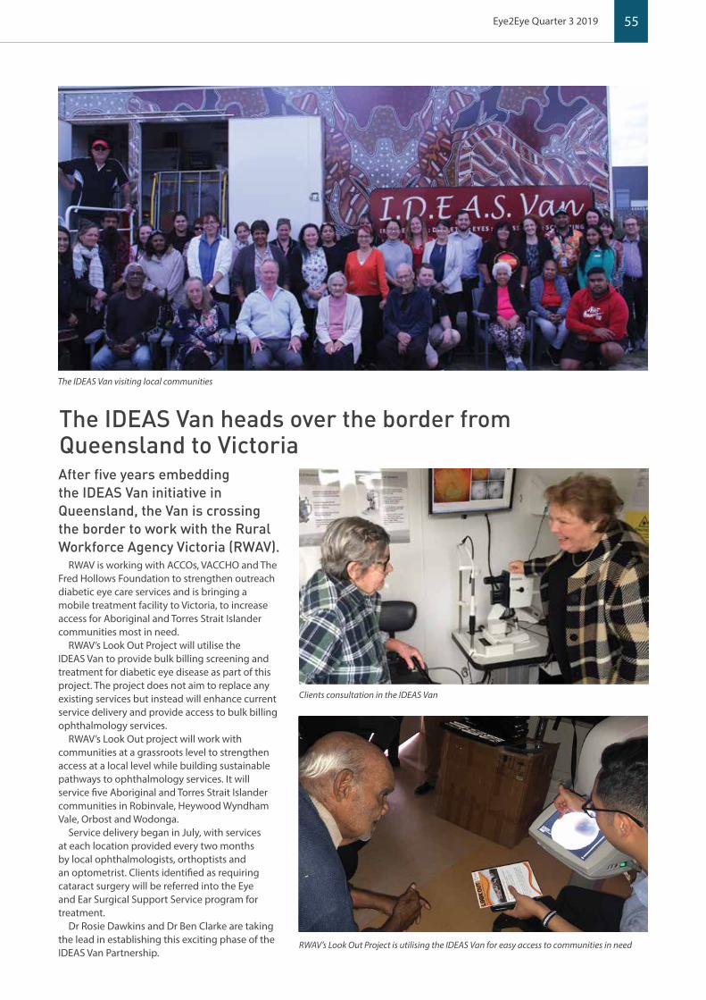
55Eye2Eye Quarter 3 2019
The IDEAS Van visiting local communities
Clients consultation in the IDEAS Van
RWAV’s Look Out Project is utilising the IDEAS Van for easy access to communities in need
After five years embedding the IDEAS Van initiative in Queensland, the Van is crossing the border to work with the Rural Workforce Agency Victoria (RWAV).
RWAV is working with ACCOs, VACCHO and The Fred Hollows Foundation to strengthen outreach diabetic eye care services and is bringing a mobile treatment facility to Victoria, to increase access for Aboriginal and Torres Strait Islander communities most in need.
RWAV’s Look Out Project will utilise the IDEAS Van to provide bulk billing screening and treatment for diabetic eye disease as part of this project. The project does not aim to replace any existing services but instead will enhance current service delivery and provide access to bulk billing ophthalmology services.
RWAV’s Look Out project will work with communities at a grassroots level to strengthen access at a local level while building sustainable pathways to ophthalmology services. It will service five Aboriginal and Torres Strait Islander communities in Robinvale, Heywood Wyndham Vale, Orbost and Wodonga.
Service delivery began in July, with services at each location provided every two months by local ophthalmologists, orthoptists and an optometrist. Clients identified as requiring cataract surgery will be referred into the Eye and Ear Surgical Support Service program for treatment.
Dr Rosie Dawkins and Dr Ben Clarke are taking the lead in establishing this exciting phase of the IDEAS Van Partnership.
The IDEAS Van heads over the border from Queensland to Victoria
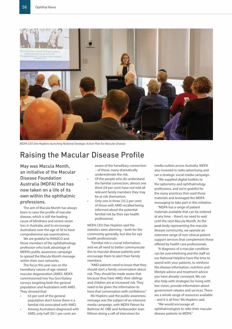
56 Ophthal News
May was Macula Month, an initiative of the Macular Disease Foundation Australia (MDFA) that has now taken on a life of its own within the ophthalmic professions.
The aim of Macula Month has always been to raise the profile of macular disease, which is still the leading cause of blindness and severe vision loss in Australia, and to encourage Australians over the age of 50 to have comprehensive eye examinations.
We are grateful to RANZCO and those members of the ophthalmology profession who took advantage of MDFA’s public awareness campaign to spread the Macula Month message within their own networks.
The focus this year was on the hereditary nature of age-related macular degeneration (AMD). MDFA commissioned two You Gov/Galaxy surveys targeting both the general population and Australians with AMD. They showed that:• 63 per cent of the general
population don’t know there is a familial risk associated with AMD.
• Among Australians diagnosed with AMD, only half (50.1 per cent) are
aware of the hereditary connection – of those, many dramatically underestimate the risk.
• Of the people who do understand the familial connection, almost one third (29 per cent) have not told all relevant family members they may be at risk themselves.
• Only one in three (33.2 per cent) of those with AMD recalled being informed about the potential familial risk by their eye health professional.
MDFA CEO Dee Hopkins said the statistics were alarming – both for the community generally, but also for eye health professionals:
“Familial risk is crucial information, and we all need to better communicate this to macular disease patients and encourage them to alert their family members.
“AMD patients need to know that they should start a family conversation about risk. They should be made aware that because they have AMD, their siblings and children are at increased risk. They need to be given the information to have that conversation with confidence.”
Ms Hopkins said the public awareness message was the subject of an intensive media campaign, with MDFA Patron Ita Buttrose AC OBE and Ambassador Jean Kittson doing a raft of interviews for
media outlets across Australia. MDFA also invested in radio advertising and ran a strategic social media campaign.
“We supplied digital toolkits to the optometry and ophthalmology professions, and we’re grateful for the many practices that used those materials and leveraged the MDFA messaging to take part in this initiative.
“MDFA has a range of patient materials available that can be ordered at any time – there’s no need to wait until the next Macula Month. As the peak body representing the macular disease community, we operate an extensive range of non-clinical patient support services that complement those offered by health care professionals.
“A diagnosis of a macular condition can be overwhelming and the staff on our National Helpline have the time to spend with your patients, to reinforce the disease information, nutrition and lifestyle advice and treatment advice you have already conveyed. We can also help with strategies for living with low vision, provide information about government rebates and services. There are a whole range of resources available – and it is all free,” Ms Hopkins said.
“We would encourage all ophthalmologists to refer their macular disease patients to MDFA.”
Raising the Macular Disease Profile
MDFA CEO Dee Hopkins launching National Strategic Action Plan for Macular Disease

57Eye2Eye Quarter 3 2019
National Strategic Action Plan for Macular DiseaseIt is an exciting new era for everyone working in the macular disease sector, with the federal government’s recent release of the National Strategic Action Plan for Macular Disease.
The federal Government’s commitment to the Action Plan, developed by MDFA – and backed by the firm commitment of funding – is recognition of the enormous impact macular disease has on our community.
MDFA CEO Dee Hopkins said the plan outlines a national, coordinated response and informs how limited health care resources can be better coordinated and targeted across all levels of government.
“Australia is a world leader in fighting macular disease. Over the past decade, we’ve seen key stakeholders from across the health sector and industry unite to prevent, detect and manage macular disease.
“The roadmap outlined in this 44-point action plan will allow this partnership to continue in a coordinated, strategic way. It is built on
four ‘pillars’ of action – prevention and early detection, treatment, support for people with macular disease, and data and research,” she said.
“These action points will help reduce – and ideally prevent – the economic, social and emotional costs of vision loss and blindness as a result of macular disease, greatly assisting the members of our community living with macular disease, their families and carers,” Ms Hopkins said.
MDFA’s National Research Advisor and world leading ophthalmologist, Professor Paul Mitchell AO, said the action plan addresses issues relating to treatment, research and better access and use of available aggregate data.
“Australia is recognised as a world leader in macular disease research and the action plan articulates a framework and agenda to enable that sight saving work to continue in a coordinated way.
“As our population ages and becomes more at risk of age-related macular degeneration, and with a rising prevalence of diabetic eye disease in working aged Australians, the incidence and impact of macular disease will only
increase and put additional strain on our healthcare system, which is why this action plan is so important,” Professor Mitchell said.
The Strategic National Action Plan is available online at: http://bit.ly/2WvhoP0
MDFA Research GrantsApplications for MDFA Research Grants have now closed. Applications will be assessed following rigorous evaluation based on the National Health and Medical Research Council (NHMRC) processes, along with peer review, to ensure successful applicants meet the highest standards.
Since the inception of the MDFA’s Research Grant’s Program in 2011, MDFA has committed almost $3.6m to 18 research projects.
A total grant pool of up to $600,000 plus GST will be available for the next round of research to start in 2020. The grant recipients will be publicly announced on World Sight Day in October.
Dee Hopkins, CEO Macular Disease Foundation Australia

58 Ophthal News
Associate Professor Angus Turner is actively involved in the delivery of specialist outreach services to remote and Indigenous communities in the Kimberley, Pilbara, Goldfields, Midwest and Great Southern regions of Western Australia. As an Associate Professor at the University of Western Australia (UWA), Dr Turner is actively engaged in several research projects at the Lions Eye Institute (LEI), focusing on service delivery for remote and Indigenous people. He is also a clinical lecturer for the Rural Clinical School at UWA and Notre Dame.
Associate Professor Lyndall Lim is a uveitis and medical retina subspecialist and leads the Clinical Trials Research at the Centre for Eye Research (CERA) in Melbourne. After completing her Ophthalmology training in Melbourne, A/Prof Lim then completed two Fellowships – one in Medical Retina at the Royal Victorian Eye and Ear Hospital, and one in Uveitis/Ocular Inflammatory Disease at the Casey Eye Institute, Portland, Oregon, USA.
Associate Professor Samantha Fraser-Bell is an Associate Professor in the discipline of Ophthalmology, University of Sydney, and a consultant Medical Retinal subspecialist at the Royal North Shore and Sydney Eye Hospitals. Her expertise is in the diagnosis and treatment of diseases of the macula and retina, particularly age-related macular degeneration, diabetic retinopathy, retinal vein occlusion and inflammatory eye disease.
Clinical Professor Nitin Verma specialises in General Ophthalmology, Cataract, Oculoplastic, Glaucoma, and Medical Retina. He is based at Hobart Eye Surgeons and heads a number of research projects concerning new treatments for AMD as well as diabetic macular oedema.
Associate Professor Anthony Kwan is a Director of Vitreoretinal Surgery at the Queensland Eye Institute and Director of the Queensland Electro-Diagnostic and Imaging Centre. He is also the Senior Staff Specialist at the Mater Hospital Brisbane. He undertook his resident and registrar training at the Moorfields Eye Hospital in London, He spent another two years in research at the Institute of Ophthalmology in London before completing his fellowships in Moorfields and in Perth, Australia.
KEEPSIGHT – Zeroing in on people not receiving diabetic eye care
Diabetes is one of the fastest growing conditions in Australia with 1.7 million people living with diabetes and an additional 280 people diagnosed every day. By 2030, the number of Australians with diabetes is expected to be close to 2.5 million.
These concerning statistics, coupled with the fact that too many Australians with diabetes are losing their sight simply because they aren’t having their eyes checked within recommended timeframes, catalysed the development of KeepSight, a new national program to remind people with diabetes when it’s time to have an eye check.
The premise of KeepSight is simple. By encouraging people with diabetes to get their eyes checked more regularly, we can help them to keep their sight.
The recall and reminder program will aim to engage with the 1.3 million people registered on the National Diabetes Services Scheme (NDSS) to urge them to have regular eye examinations using existing eye health and primary care practitioners and existing MBS item numbers.
We talked to a number of Australian ophthalmologists about the challenges of improving the eye health of people with diabetes and how KeepSight may play a role.
59Eye2Eye Quarter 3 2019
Associate Professor Anthony Kwan trained in the United Kingdom at the time it was spearheading its approach to diabetic eye care through its national screening program.
The NHS-led program was established in 2003 to offer two-field mydriatic digital photographic screening annually to all people with diabetes. By 2008, the program had reached the whole population with more than 2.14 million people with diabetes screened in that year. By 2010, diabetic retinopathy and maculopathy was no longer the leading cause of blindness registration for working age people in England.
According to A/Prof Kwan, this program was a great model which we can emulate in Australia.
“We are leaders in health care and technology, yet we don’t yet have a national diabetic retinopathy screening program.
“KeepSight really provides a platform and a strong starting point to get a coordinated screening program established. We have public and private support for KeepSight and from it, we can establish who is not being screened. This is a great place to start.
“It took a long time for the UK to get its program up and running and it was largely pre-internet and mobile, so we have a real advantage with our ability to automate,” A/Professor Kwan said.
Clinical Professor Verma Nitin said one of the biggest challenges was helping people with diabetes to understand the importance of regular screening of their eyes.
“Eye disease doesn’t hurt and you can have a lot of disease but still have good vision which means people delay being tested until the problem is irreparable.”
Prof Verma said that people with diabetes had many health issues to manage and other diabetes-related health issues often took priority.
“And don’t forget, for the most part, these are working-aged people – they work, they have families. It’s not like macular degeneration where patients are generally much older.
“But the moment someone with diabetes becomes symptomatic, then the whole concept changes. It becomes a treatment regime, not a screening regime.
“A reminder program like KeepSight is a really important place to start, but we also need to have a coordinated, integrated and funded screening program,” Prof Verma said.
Associate Professor Lyndell Lim, who sees upwards of 40 patients with diabetes each week, many of whom are pregnant women, agreed one of the biggest challenges was that people with diabetes were not getting their eyes checked until they started to experience issues with their sight.
“Often then the damage has been done. People with diabetes have so many other health priorities to manage, that their eye health is almost at the bottom of the list. They need to see an endocrinologist, a dietician, a podiatrist, if they’re pregnant, an obstetrician. Less than 50 per cent of women with diabetes come in for their eye checks during pregnancy.
“The ease of registering with KeepSight via phone or PC and then the constant reminding is what’s needed to get people to come in for screening.
“We need to tap into people’s everyday dependency on technology.
“Nationally, our diabetic retinopathy screening rates need improving. We can’t just scare people; we need to give them practical tools to help them remember and remind them why regular screening is so important,” A/Prof Lim said.
Associate Professor Angus Turner runs a service that sees over 10,000 patients each year, with the majority having diabetes, and has a long history of providing diabetic screening using retinal cameras, including in remote outreach areas of Western Australia.
One of the priorities for A/Prof Turner is ensuring the National Diabetes Services Scheme (NDSS) continues to accurately reflect a true picture of the prevalence of diabetes, particularly in remote Aboriginal communities.
“In many regional communities we have a constant rotation of general practitioners at the frontline who are focused solely on the acute issues. They just don’t have the capacity to be dealing with registering people as well. There is high turnover and limited continuity of care. We need to address this.”
A/Prof Turner said a key challenge was recognising that not all people with diabetes living in remote Aboriginal communities are registered with the NDSS. “In some communities the number of people registered with the NDSS is a tenth of the actual number of people with diabetes in that community.
“KeepSight is a great concept and an important step in the right direction, but we have some work to do to enable it to achieve its goal, particularly in the rural and remote regions,” says A/Prof Turner.
Associate Professor Samantha Fraser-Bell runs a specialist diabetic retinopathy clinic in Sydney and said in New South Wales many of the people with vision threatening eye disease were managed within the public system.
“It’s an excellent system, but it is only capable of dealing with the more serious end of the spectrum – the people who already have eye disease. We just don’t have the capacity to be offering an early stage screening program as well.”
Dr Fraser-Bell said one of the challenges in Australia was a lack of continuity in our screening processes and even in diabetes care.
“People are very prepared to chop and change providers based on retail influences or convenience. This presents a challenge in continuity of care in early and regular screening.
“One of the programs I’m involved in that works really well is at Royal North Shore where we work within the endocrinology program. The service is much more holistic and covers the gamut of diabetic health support – nutrition, podiatry, eye care.
“There is no doubt people can access great health care in Australia, but we have to work out how to coordinate it better,” A/Prof Fraser-Bell said.
According to Deputy Director of CERA and KeepSight steering committee member Associate Professor Peter van Wijngaarden, KeepSight is a once in a generation opportunity to significantly reduce rates of diabetes-related vision loss and blindness in Australia.
“The involvement of eye care professionals, diabetes health care professionals and general practitioners in the KeepSight program is central to its success. The program has a real opportunity to make a step change in the eye health of Australians with diabetes.”
60
&
Ophthal News
Why do we need KeepSight? 9 One in three Australians with diabetes presently has diabetic retinopathy (DR) and all are at risk of developing DR.
9 Evidence shows that early detection and timely treatment can prevent most diabetes-related vision loss, but the lack of a coordinated and systematic approach means a significant proportion of Australians with diabetes do not have an eye examination when required.
9 Australia has ample existing capacity in optometry, ophthalmology and general practices to support early detection and treatment.
9 A national diabetic retinopathy screening program has been proven to work in the UK and it can work in Australia. The National Diabetes Services Scheme (NDSS) database, managed by Diabetes Australia, is a resource we can use to increase eye examination uptake.
What will KeepSight change? 9 KeepSight aims to demonstrate that a coordinated and systematic approach to preventative and timely healthcare delivers
improved outcomes for people with diabetes.
9 The program will provide new capacity to measure the burden of diabetes-related eye disease in Australia and will serve as a vehicle to reduce avoidable vision loss and blindness.
9 The KeepSight program is not intended to disrupt existing arrangements between eye care providers and their patients, but supplement this with an additional recall and reminder system. Further, public facing materials will note that if a person already has arrangements for periodic reviews with an existing eye care provider in place, these should continue.
9 This program will encourage the 50% of people with diabetes in Australia who currently aren’t accessing regular eye examinations to visit their local eye care provider.
How can ophthalmologists participate? 9 Register as a provider and record your patient details. Refer to www.keepsight.org.au on how to register.
- 589 – over half of all ophthalmologists are registered as KeepSight providers
- 2769 – half of all optometrists
- 193 – eye care providers have registered patients with KeepSight.
9 Eye care providers are critical in helping to identify how many of the 1.3 million people registered on the NDSS are receiving diabetic eye care. Having this information will allow us to then identify which people with diabetes are not receiving eye care.
9 Developing strategies to target people who are not receiving diabetic eye care is the way forward, however it is contingent on your participation in the program.
KeepSight is being implemented by Diabetes Australia in partnership with Vision 2020 Australia with technical support from Oculo and the Centre for Eye Research Australia. The program is funded by the Australian Government and corporate partners including Specsavers, Novartis and Bayer, with a free web based portal open to all clinicians and capacity for patients to self-register to the program.
KEYFACTS
61Eye2Eye Quarter 3 2019
Every hero needs asidekick.
Glaucoma Australia, is keen to be the Robin to your Batman in the fi ght to save the sight of glaucoma patients Australia wide.
Our aim is to encourage early detection, provide knowledge and support for continuing treatments and ongoing medical check ups.
Refer your patients to us today and together we can save sight and say goodbye to glaucoma blindness.
glaucoma.org.au
GA_11040_2019_INSIGHT_HPC_AD_SIDEKICK_HORIZ_235x144mm_FA.indd 1 27/2/19 12:34 pm
Topics include: → What, when and how to locum → Business structure, finance, insurance and asset
protection → Marketing essentials → Medicare, billing and item numbers → Staffing and HR → To buy in or go it alone → Turning income into wealth
THRIVE
SURVIVE
Younger Fellows’ Professional Development Workshop
&
Supporters of the 2019 Younger Fellows’ Educational Grant
Survive and Thrive is an initiative of the RANZCO Younger Fellows. It is a one-day workshop in Sydney in September for recently graduated Fellows and 5th year Trainees. The event came about after a survey of recently graduated Fellows found that they felt they lacked business and real-world knowledge.
Survive and Thrive will be taking place on Saturday 28 September at The Amora in Sydney and is accredited for CPD points.Contact the College for more information Email: [email protected]:+61 2 9690 1001
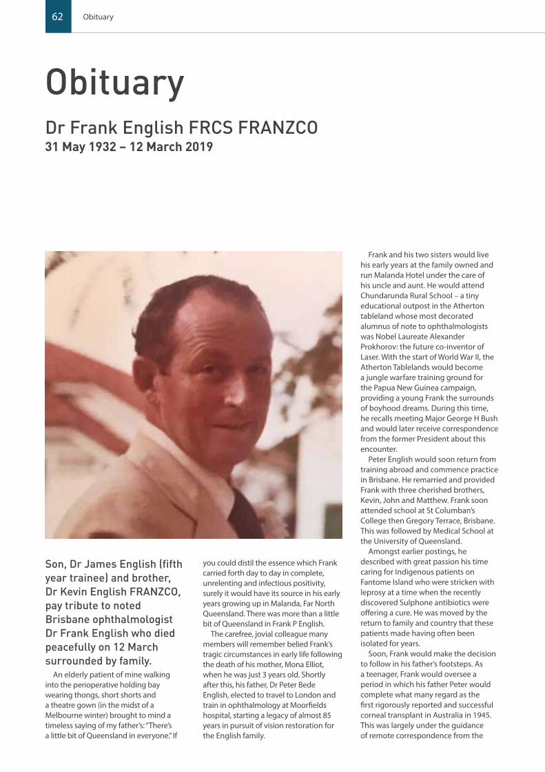
62 Obituary
ObituaryDr Frank English FRCS FRANZCO 31 May 1932 – 12 March 2019
Son, Dr James English (fifth year trainee) and brother, Dr Kevin English FRANZCO, pay tribute to noted Brisbane ophthalmologist Dr Frank English who died peacefully on 12 March surrounded by family.
An elderly patient of mine walking into the perioperative holding bay wearing thongs, short shorts and a theatre gown (in the midst of a Melbourne winter) brought to mind a timeless saying of my father’s: “There’s a little bit of Queensland in everyone.” If
you could distil the essence which Frank carried forth day to day in complete, unrelenting and infectious positivity, surely it would have its source in his early years growing up in Malanda, Far North Queensland. There was more than a little bit of Queensland in Frank P English.
The carefree, jovial colleague many members will remember belied Frank’s tragic circumstances in early life following the death of his mother, Mona Elliot, when he was just 3 years old. Shortly after this, his father, Dr Peter Bede English, elected to travel to London and train in ophthalmology at Moorfields hospital, starting a legacy of almost 85 years in pursuit of vision restoration for the English family.
Frank and his two sisters would live his early years at the family owned and run Malanda Hotel under the care of his uncle and aunt. He would attend Chundarunda Rural School – a tiny educational outpost in the Atherton tableland whose most decorated alumnus of note to ophthalmologists was Nobel Laureate Alexander Prokhorov: the future co-inventor of Laser. With the start of World War II, the Atherton Tablelands would become a jungle warfare training ground for the Papua New Guinea campaign, providing a young Frank the surrounds of boyhood dreams. During this time, he recalls meeting Major George H Bush and would later receive correspondence from the former President about this encounter.
Peter English would soon return from training abroad and commence practice in Brisbane. He remarried and provided Frank with three cherished brothers, Kevin, John and Matthew. Frank soon attended school at St Columban’s College then Gregory Terrace, Brisbane. This was followed by Medical School at the University of Queensland.
Amongst earlier postings, he described with great passion his time caring for Indigenous patients on Fantome Island who were stricken with leprosy at a time when the recently discovered Sulphone antibiotics were offering a cure. He was moved by the return to family and country that these patients made having often been isolated for years.
Soon, Frank would make the decision to follow in his father’s footsteps. As a teenager, Frank would oversee a period in which his father Peter would complete what many regard as the first rigorously reported and successful corneal transplant in Australia in 1945. This was largely under the guidance of remote correspondence from the

63Eye2Eye Quarter 3 2019
cornea and cataract pioneer Ramon Castroviejo, who Frank would also later work alongside.
Frank then set forth on no fewer than six fellowships across most aspects of ophthalmic practice. Key themes would appear to be mentorship from well-regarded international pioneers, internationally reputed destinations in their own right, world class ophthalmic institutions, and proximity to a cold beer (very possibly in reverse order of priority):• Boston – Research fellow in
Glaucoma: Harvard Medical School. Frank would become lifelong friends with Dr Charles Schepens, the vitreoretinal pioneer during this time.
• London – General, Guys Hospital; Moorfields, Oculoplastics (JRO Collin).
• Houston, Texas – Vitreoretinal (Richard Ruiz).
• New York, Manhattan Eye, Ear and Throat Hospital – Oculoplastics (Byron Smith) & Cornea (Ramon Castroviejo).
• Boston – Cornea.
Frank’s love of travel led him on many adventures, including instigating an investigation into Eskimo Keratopathy in Labrador and Newfoundland after receiving a travel award with the Grenfell Association.
A chance encounter in Boston with Professor Bill Nutting, a world authority on Demodectic mites, sparked a lifelong
passion for understanding these enigmatic parasites. Frank published an article in TIME magazine on these creatures, complete with gruesome electron-microscopic pictures that would not have been misplaced in a Hollywood horror film.
Frank published widely with significant contributions to literature. He published the textbook ‘Reconstructive and Plastic Surgery of the Eyelids’, the foreword of which was written by his colleague and mentor, Byron Smith.
Upon returning to Brisbane, Frank married Leonie Jones and raised two sons.
My father’s home life was one of sunshine, “expeditions”, reading, backyard cricket, beach retreats, body surfing and travel. Despite his busy surgical life, my father was an omnipresent figure in my upbringing, always available and encouraging, heavily supported by my mother who provided an essential balance to family and professional life.
Frank had consultant presences at the Mater Public Hospital, Greenslopes Repatriation General Hospital and St Andrew’s Hospital (Brisbane) and held positions as Member of Board of Directors of the International Oculoplastic Society (New York) and Research Associate, Queensland Institute of Medical Research, Brisbane, amongst other appointments.
My inspiration to pursue ophthalmology is clearly borne of my father’s lifelong passion. My brother,
Dr Lawrence English, a celebrated sound artist, composer, producer and entrepreneur, also recalls being inspired by my father to listen whilst bird-watching: an act which started his now lifelong interest in listening. In his own words:
“His simple request to close my eyes and listen provided me with what was a cursory engagement with my audition that has since resonated throughout my life.”
Many fellows have related similar experiences to those that Kevin and I remember from during my father’s life.
Perhaps the greatest single privilege as a doctor has been to look after patients who have been cared for by three generations of the English family. When I ask them, patients’ recollections across the generations share themes of kindness, commitment, joviality and varying degrees of hairline recession – all attributes worn with pride by my father who practised ophthalmology into his 80s.
We would like to take this opportunity to thank those who have supported my father and the family over many years. Frank is survived by his devoted wife, Leonie, two sons and five grandchildren: Frances, Theodore, Augustine, Georgia and Xanthe.
Drs James English (son) & Dr Kevin English FRANZCO (brother)

64
Calendar of EventsEVENT DETAILS CONTACT19th European Society of Retina Specialists Congress (EURETINA) 2019 5-8 September 2018 E: [email protected]
Neuro-Ophthalmology Society of Australia (NOSA) 35th Clinical & Scientific Meeting and NeuroVision Training Weekend
5-8 September 2018
P: 0402 891 804E: [email protected]: 0417 544 310 E: [email protected]
European Society of Cataract & Refractive Surgeons (ESCRS) Congress 2019 14 September 2019 E: [email protected]
Asia Pacific Tele-Ophthalmology Society’s 4th Annual Symposium 21-22 September 2019 W: http://2019.asiateleophth.org/registration.php
The 2019 Royal Children’s Hospital Paediatric Ophthalmology Seminar 4-6 October 2019 E: [email protected]
American Academy of Ophthalmology (AAO) 2019 Annual Meeting 12-15 October 2019 W: www.ranzco.edu/view-all-events
Dunedin Ophthalmology Course 14-25 October 2019E: [email protected]: www.events4you.co.nz/docc
Leaders in Indigenous Medical Education (LIME) Network Connection 5-8 November W: https://www.limeconnection.net.au/about
ANZ Microsurgical Course Teaching 6-8 November 2019 P: +61 29690 1001
AAPOS APSPOS RANZCO Joint Meeting 7-8 November 2019 W: www.ranzco.edu/view-all-events
RANZCO 51st Annual Scientific Congress 7-12 November 2019 W: www.ranzco2019.com
Trends in RetinaBarcelona Spain 15-16 November 2019 W: www.fundacionimo.org/en/trends-retina#top
AVAN New Zealand Meeting 2019 6-7 December 2019 W: https://outshine.eventsair.com/avan-19/
33rd Annual Registrars’ Conference and Teaching Course – Glaucoma 8-11 January 2020 W: www.ranzco.edu/view-all-events
1st Asia-Pacific Ocular Imaging Society Congress in Conjunction with 35th Singapore-Malaysia Joint Meeting in Ophthalmology
17-19 January 2020 W: www.ranzco.edu/view-all-events
Australian and New Zealand Glaucoma Society (ANZGS) Scientific Meeting 21-22 February 2020
E: [email protected]: [email protected]
Ophthalmology in Brazil and Argentina – study tour 2-12 March 2020
W: https://www.jonbainestours.com.au/tours/medical-and-professional/ophthal/
NSW Branch ASM 2020 13-14 March 2020 W: www.ranzco.edu/view-all-events
Victoria Branch Annual Scientific Meeting 21 March 2020E: [email protected]: [email protected]
Asia-Pacific Academy of Ophthalmology (APAO) 2020 22-26 April 2020 E: [email protected]
Calendar of Events
65
24-25 August 2019Westin Hotel, Sydney Register at www.ophthalmologyupdates.com
Convenor | Adrian Fung
Medical Retina | Lawrence YannuzziMyopia | Ian Morgan
Uveitis | Peter McCluskeyPaediatrics | Frank Martin
Ocular Oncology & Sight For All | James MueckeOculo-plastics | Brett O'Donnell
VR Surgery | Rohan EssexCataract | Rick Wolfe
Neuro-ophthalmology | Clare FraserGlaucoma | Paul Healey
Medical Retina | Paul MitchellMedical Retina | Mark Gillies
IOL Opacification | Matt SpargoCornea | Chameen Samarawickrama
Eye2Eye Quarter 3 2019
EVENT DETAILS CONTACT
ARVO 2020 3-7 May 2020 E: [email protected]
ASCRS ASOA Annual Meeting 2020 15-19 May 2020 E: [email protected]
Australian and New Zealand Society of Retinal Specialists (ANZSRS) Meeting 2020 23-24 May 2020 W: www.ranzco.edu/view-all-events
14th European Glaucoma Society Congress 2020 30 May to 2 June 2020 P: +39 055 50351
ASO Expo 2020 20-21 June 2020 E: [email protected]
World Ophthalmology Congress 2020 26-29 June 2020 W: [email protected]
The International Agency for the Prevention of Blindness (IAPB) 12th General Assembly 12-14 October 2020 W: www.ranzco.edu/view-all-events
American Academy of Ophthalmology (AAO) 2020 14-17 November 2020 E: [email protected]

66 Calendar of Events
ClassifiedsPositions vacantGeneral Ophthalmologist in NE VictoriaGeneral Ophthalmologist required to take over long standing existing practice, two hours from Melbourne.
Currently two days a fortnight, with one full day operating every four weeks, mixed private and fee for service public.
Extremely well equipped with: SD OCT, FFA, YAG laser , SLT, Double freq YAG, Perimetry, IOL Master, contact AScan etc. The new ophthalmologist must be comfortable with managing General Medical Retina, Glaucoma, Cataract Surgery and basic Oculoplastics procedures.
The patients are a delight and deserve further professional and caring management.
Applicants must be an AHPRA registered medical specialist with RANZCO Fellowship.
Ophthalmologist Required - Ballina
A unique opportunity to join the largest eye clinic in Ballina due to the sudden retirement of a full time associate.
The eye clinic is situated in a large, Tuscan Manor House which incorporates a Day Hospital fully equipped for ophthalmic surgery and staffed by ophthalmic trained nurses.
Ballina is a rapid growth area and a favoured town for retirees. It is 20 minutes south of Byron Bay and has its own airport.
C: Dr Pamela Weir P: 0438 011 963 E: [email protected]
Locum Ophthalmologist - Tamworth NSWAssistant or locum wanted for busy rural practice in Tamworth NSW.
Very well equipped practice with large workload. Good direct air connections to Sydney and Brisbane.
Ophthalmologist Sunshine CoastSeeking Ophthalmologist to work with general ophthalmologist wishing to transition to retirement. The practice is a long established large solo Sunshine Coast practice. Operating from a well equiped stand alone facility the practice caters for general ophthalmology and has an emphasis on cataract surgery, medical retina, glaucoma and occulo-plastics. There is considerable potential for expansion and subspeciality interests.
Applicants must be APHRA registered and a Fellow of RANZCO.
Theatre time available - Forest Road Day Surgery, Hurstville Forest Road Day Surgery is a purpose built Day Surgery and Laser Suite, specialising in ophthalmology.
Our facility offers a dedicated laser suite (LenSx Laser system) within the theatre complex, Centurion Vision System, Verion Image Guided System and ceiling mounted microscope. We have highly experienced ophthalmology trained nursing staff led by an on-site Director of Nursing.
We provide free secure onsite parking for patients and doctors. Our facility is part of a group of 10 day surgeries throughout Australia.
C: Carmel Kennedy P: 0407 206 146 or +61 2 9330 6900 E: [email protected]
Ophthalmology Position Sunshine CoastOpportunity exists for an Ophthalmologist to join an established growing practice on the Sunshine Coast. The practice is modern, purpose built, well equipped and well staffed to cater for expansion in both general ophthalmology and subspecialty interests.
Ophthalmologist - Nowra Southern NSWEstablished busy rural practice with a large workload of cataract, glaucoma and medical retina patients. The practice needs the help of a general or sub-specialty trained Ophthalmologist to share the load.
Rural Ophthalmologist PositionGeneral or Sub-Specialist position available in Devonport and Burnie. All modern diagnostic facilities and Day Surgery.
C: Dr Michael HaybittelE: [email protected]
Ophthalmologist Northern Rivers NSW
Dr Anne Malatt has just opened new rooms in Alstonville, Northern NSW and is looking for an associate to join her.
The rooms are large, light, lovely and well equipped in a beautiful, newly renovated freestanding building with ample parking and disabled access and facilities.
P: +61 2 6687 2433 E: [email protected]
General Ophthalmologist in Orange
Ophthalmologist assistants required for sessions in Orange and Lithgow, NSW. Will suit general ophthalmologists or those with subspecialty interest in glaucoma, oculoplastics paediatrics or cornea.
• Great working environment with experienced support staff.
• Must be RANZCO member and AHPRA registered.
• All expressions of interest considered.
5th Year Position - The Townsville HospitalAn opportunity exists for a 5th year/post-exam trainee who wants excellent exposure to comprehensive ophthalmology in Queensland’s largest regional hospital. There is exposure to all the ophthalmic subspecialties, as well as a high surgical load in cataract and plastics in particular; Indigenous eye health is a particular focus of our unit. Surgical and medical retina is also an option, if this is an interest.
The eye clinic at TTH has recently undergone a $6M redevelopment and its equipment is state of the art. Townsville is a diverse community of 200,000 people, serving a wider population approaching 1 million.
RANZCO SYDNEY 2019in conjunction with AAPOS APSPOS
�������������������������
8-12 November 2019, ICC Sydneywww.ranzco2019.com
Celebrating our 50th year

* IMPORTANT: Professional indemnity insurance products are issued by Avant Insurance Limited, ABN 82 003 707 471, AFSL 238 765. The information provided here is general advice only. You should consider the appropriateness of the advice having regard to your own objectives, financial situation and needs before deciding to purchase or continuing to hold a policy with us. For full details including the terms, conditions, and exclusions that apply, please read and consider the policy wording and PDS, which is available at avant.org.au or by contacting us on 1800 128 268. 2258.6 02/19 (0811)
As a respected ophthalmologist, you stand by your reputation and our reputation is built on protecting yours. The fact is, no medical indemnity insurer has more resources or expertise to safeguard your reputation than Avant. With Avant, you’ll have the support of award winning Avant Law, Australia’s largest specialist medico-legal firm. And more doctors on staff delivers
the unique support and understanding that only a peer can provide. The depth of our experience and expertise gives us knowledge of your specialty that’s simply unmatched. We’ve got your back.
Don’t risk your reputation. Talk to us today about Professional Indemnity Insurance.
More specialty expertise.More ophthalmologists protected.
We’ve got your back.
Dr Jagjit Gilhotra OphthalmologistSouth Australia
1800 128 268
avant.org.au