Extrinsic and intrinsic signals converge on the · PDF filethe Runx1/CBFb transcription factor...
24
*For correspondence: [email protected] Present address: † Department of Chemistry and Life Sciences, United States Military Academy, West Point, United States Competing interest: See page 21 Funding: See page 21 Received: 14 August 2015 Accepted: 28 September 2015 Published: 29 September 2015 Reviewing editor: Ben Barres, Stanford School of Medicine, United States Copyright Huang et al. This article is distributed under the terms of the Creative Commons Attribution License, which permits unrestricted use and redistribution provided that the original author and source are credited. Extrinsic and intrinsic signals converge on the Runx1/CBFb transcription factor for nonpeptidergic nociceptor maturation Siyi Huang 1,2 , Kevin J O’Donovan 3† , Eric E Turner 4 , Jian Zhong 3 , David D Ginty 1,2 * 1 Department of Neurobiology, Howard Hughes Medical Institute, Harvard Medical School, Boston, United States; 2 Department of Neuroscience, Howard Hughes Medical Institute, Johns Hopkins School of Medicine, Baltimore, United States; 3 Burke Medical Research Institute, Weill Medical College of Cornell University, White Plains, United States; 4 Seattle Children’s Hospital, Seattle Children’s Research Institute, Seattle, United States Abstract The generation of diverse neuronal subtypes involves specification of neural progenitors and, subsequently, postmitotic neuronal differentiation, a relatively poorly understood process. Here, we describe a mechanism whereby the neurotrophic factor NGF and the transcription factor Runx1 coordinate postmitotic differentiation of nonpeptidergic nociceptors, a major nociceptor subtype. We show that the integrity of a Runx1/CBFb holocomplex is crucial for NGF-dependent nonpeptidergic nociceptor maturation. NGF signals through the ERK/MAPK pathway to promote expression of Cbfb but not Runx1 prior to maturation of nonpeptidergic nociceptors. In contrast, transcriptional initiation of Runx1 in nonpeptidergic nociceptor precursors is dependent on the homeodomain transcription factor Islet1, which is largely dispensable for Cbfb expression. Thus, an NGF/TrkA-MAPK-CBFb pathway converges with Islet1-Runx1 signaling to promote Runx1/CBFb holocomplex formation and nonpeptidergic nociceptor maturation. Convergence of extrinsic and intrinsic signals to control heterodimeric transcription factor complex formation provides a robust mechanism for postmitotic neuronal subtype specification. DOI: 10.7554/eLife.10874.001 Introduction Neuronal cell fate specification is a multistep process that can be broadly divided into two stages, early specification of neural progenitor cell identity and postmitotic differentiation of neuronal sub- types. A recurring theme in the early phase of neural progenitor specification of the vertebrate ner- vous system is that the interplay between intrinsic determinants and extrinsic signals governs neural cell fate decisions (Fishell and Heintz, 2013). Indeed, cell fate choice of progenitor cells is deter- mined by a combination of intrinsic genetic programs, which define the differentiation potential or the competence state of progenitors, and extrinsic cues, which influence the relative proportions of each cell type generated within the confines of a given competent state (Livesey and Cepko, 2001). Our understanding of the mechanisms of postmitotic differentiation of neuronal subtypes, on the other hand, is rather limited, and thus the relative contributions of extrinsic and intrinsic signals, and their mechanisms of interplay during late phases of cell fate determination, remain poorly understood. Primary sensory neurons of the dorsal root ganglion (DRG) are pseudo-unipolar neurons that extend one axonal branch to the spinal cord and the other to peripheral targets such as the skin. The existence of a large number of functionally specialized DRG sensory neuron subtypes, each endowed with unique morphological and physiological properties enables the somatosensory sys- tem to encode diverse sensory information, including nociception, temperature, light touch, limb Huang et al. eLife 2015;4:e10874. DOI: 10.7554/eLife.10874 1 of 24 RESEARCH ARTICLE
Transcript of Extrinsic and intrinsic signals converge on the · PDF filethe Runx1/CBFb transcription factor...

*For correspondence:
Present address: †Department
of Chemistry and Life Sciences,
United States Military Academy,
West Point, United States
Competing interest: See
page 21
Funding: See page 21
Received: 14 August 2015
Accepted: 28 September 2015
Published: 29 September 2015
Reviewing editor: Ben Barres,
Stanford School of Medicine,
United States
Copyright Huang et al. This
article is distributed under the
terms of the Creative Commons
Attribution License, which
permits unrestricted use and
redistribution provided that the
original author and source are
credited.
Extrinsic and intrinsic signals converge onthe Runx1/CBFb transcription factor fornonpeptidergic nociceptor maturationSiyi Huang1,2, Kevin J O’Donovan3†, Eric E Turner4, Jian Zhong3, David D Ginty1,2*
1Department of Neurobiology, Howard Hughes Medical Institute, Harvard MedicalSchool, Boston, United States; 2Department of Neuroscience, Howard HughesMedical Institute, Johns Hopkins School of Medicine, Baltimore, United States;3Burke Medical Research Institute, Weill Medical College of Cornell University,White Plains, United States; 4Seattle Children’s Hospital, Seattle Children’sResearch Institute, Seattle, United States
Abstract The generation of diverse neuronal subtypes involves specification of neural
progenitors and, subsequently, postmitotic neuronal differentiation, a relatively poorly understood
process. Here, we describe a mechanism whereby the neurotrophic factor NGF and the
transcription factor Runx1 coordinate postmitotic differentiation of nonpeptidergic nociceptors, a
major nociceptor subtype. We show that the integrity of a Runx1/CBFb holocomplex is crucial for
NGF-dependent nonpeptidergic nociceptor maturation. NGF signals through the ERK/MAPK
pathway to promote expression of Cbfb but not Runx1 prior to maturation of nonpeptidergic
nociceptors. In contrast, transcriptional initiation of Runx1 in nonpeptidergic nociceptor precursors
is dependent on the homeodomain transcription factor Islet1, which is largely dispensable for Cbfb
expression. Thus, an NGF/TrkA-MAPK-CBFb pathway converges with Islet1-Runx1 signaling to
promote Runx1/CBFb holocomplex formation and nonpeptidergic nociceptor maturation.
Convergence of extrinsic and intrinsic signals to control heterodimeric transcription factor complex
formation provides a robust mechanism for postmitotic neuronal subtype specification.
DOI: 10.7554/eLife.10874.001
IntroductionNeuronal cell fate specification is a multistep process that can be broadly divided into two stages,
early specification of neural progenitor cell identity and postmitotic differentiation of neuronal sub-
types. A recurring theme in the early phase of neural progenitor specification of the vertebrate ner-
vous system is that the interplay between intrinsic determinants and extrinsic signals governs neural
cell fate decisions (Fishell and Heintz, 2013). Indeed, cell fate choice of progenitor cells is deter-
mined by a combination of intrinsic genetic programs, which define the differentiation potential or
the competence state of progenitors, and extrinsic cues, which influence the relative proportions of
each cell type generated within the confines of a given competent state (Livesey and Cepko, 2001).
Our understanding of the mechanisms of postmitotic differentiation of neuronal subtypes, on the
other hand, is rather limited, and thus the relative contributions of extrinsic and intrinsic signals, and
their mechanisms of interplay during late phases of cell fate determination, remain poorly
understood.
Primary sensory neurons of the dorsal root ganglion (DRG) are pseudo-unipolar neurons that
extend one axonal branch to the spinal cord and the other to peripheral targets such as the skin.
The existence of a large number of functionally specialized DRG sensory neuron subtypes, each
endowed with unique morphological and physiological properties enables the somatosensory sys-
tem to encode diverse sensory information, including nociception, temperature, light touch, limb
Huang et al. eLife 2015;4:e10874. DOI: 10.7554/eLife.10874 1 of 24
RESEARCH ARTICLE

movement, and body position. DRG neurons can be classified into three major classes: nociceptors,
which preferentially respond to noxious stimuli, pruritogenic stimuli, and temperature; mechanore-
ceptors, which respond to innocuous tactile sensations; and proprioceptors, which detect position
and movement of the trunk and limbs. Each functional class is further subdivided into subtypes
exhibiting distinct physiological, morphological, and molecular properties (Lallemend and Ernfors,
2012; Usoskin et al., 2015). Since the same Neurogenin1-dependent progenitor population gives
rise to the three principal DRG neuron types within a relatively short time window (Frank and Sanes,
1991; Ma et al., 1999), specification of the diverse DRG neuron subtypes mainly takes place in post-
mitotic neurons. Therefore, DRG sensory neurons are particularly well suited for mechanistic studies
of postmitotic differentiation of neuronal subtypes.
The differentiation of nociceptor precursors into nonpeptidergic and peptidergic nociceptors is a
well-studied example of postmitotic sensory neuron subtype specification. These two nociceptor
subtypes are molecularly, morphologically and functionally distinct. Peptidergic nociceptors are tra-
ditionally defined by expression of neuropeptides, such as CGRP and Substance P, while the majority
of nonpeptidergic nociceptors have binding sites for the lectin IB4 (Mulderry et al., 1988;
Silverman and Kruger, 1990). More recently, a single cell transcriptome analysis led to the identifi-
cation of four molecularly defined nonpeptidergic neuron subtypes (Usoskin et al., 2015). Peptider-
gic and nonpeptidergic nociceptor subtypes are further distinguished by their central and peripheral
axonal projection patterns (Molliver et al., 1995; Zylka et al., 2005). Functionally, nonpeptidergic
and peptidergic nociceptors are preferentially required for mechanical and thermal nociception,
respectively (Cavanaugh et al., 2009; McCoy et al., 2013; Mishra and Hoon, 2010;
Vulchanova et al., 2001).
Despite these differences, nonpeptidergic and peptidergic nociceptors derive from a common
nociceptor precursor that expresses tropomyosin-receptor kinase A (TrkA), the receptor for nerve
eLife digest Animals detect and respond to their environment using their sensory nervous
system, which forms through a complex, multi-step process. A precursor nerve cell’s fate is set early
in its development, and determines the different nerve types it can become. As development
progresses, sensory nerve cells develop further into distinct subtypes that perform particular tasks,
such as responding to touch or pain.
Nociceptors are the specialised sensory nerves that respond to potentially harmful stimuli. They
form two distinct subtypes: peptidergic nerves detect potentially dangerous temperatures, whereas
non-peptidergic nerves detect potentially dangerous mechanical sensations. Both subtypes originate
from the same precursor nerve cell and both initially depend on an external molecule called NGF for
their development and survival. During their development, non-peptidergic neurons stop
responding to NGF and start producing a protein called Runx1, considered to be the ‘master
regulator’ of non-peptidergic nerve cell development. Runx1 works by forming a complex with
another protein called CBFbb, and this complex activates a program of gene expression that is
specific to non-peptidergic nerves. However it was unclear how an external signal, like NGF, can
coordinate with or influence a nerve cell’s internal genetic program during the nerve’s development.
It was also not known whether NGF and Runx1 interact with each other.
By studying non-peptidergic nerve cell development in mice that lack NGF, Runx1 and other
associated proteins, Huang et al. have now established the sequence of events that regulate the
development of this nerve cell subtype. Two signalling pathways converge to switch on non-
peptidergic nerve cell development. An NGF-driven signalling pathway activates the production of
CBFb, while another protein binds to the Runx1 gene to switch it on. This leads to the production of
the Runx1 and CBFb proteins that complex together to activate the non-peptidergic neuronal
genetic program.
These findings demonstrate how two different mechanisms converge to produce the component
parts of a complex, which then activates a genetic program that drives the development of a
particular neuronal subtype. Whether this mechanism is involved in determining the fate of other cell
types remains a question for future work.
DOI: 10.7554/eLife.10874.002
Huang et al. eLife 2015;4:e10874. DOI: 10.7554/eLife.10874 2 of 24
Research article Developmental biology and stem cells Neuroscience

growth factor (NGF), and both nociceptor subtypes require NGF–TrkA signaling for survival and
development beginning at ~E12.5 (Crowley et al., 1994; Luo et al., 2007; Patel et al., 2000; Silos-
Santiago et al., 1995; Smeyne et al., 1994). A key developmental event that defines the segrega-
tion of nociceptor subtypes is the nonpeptidergic nociceptor-specific switch from NGF responsive-
ness to glial-derived neurotrophic factor (GDNF) family ligand (GFL) responsiveness. This occurs by
virtue of the upregulation of Ret and GDNF family receptor alpha receptors (GFRa), receptor com-
ponents for GFL signaling, in nonpeptidergic nociceptors starting from E15.5, as well as postnatal
extinction of TrkA expression (Bennett et al., 1996; Bennett et al., 1998; Molliver and Snider,
1997; Molliver et al., 1997). Interestingly, both NGF–TrkA and GFL–GFRa/Ret signaling are essen-
tial for postmitotic differentiation of nonpeptidergic nociceptors. Indeed, NGF–TrkA signaling is
required for the acquisition of virtually all nonpeptidergic nociceptor-specific features, including
expression of Ret and Gfras, whereas GFL–GFRa/Ret signaling, in turn, plays a critical role in postna-
tal development of nonpeptidergic nociceptors, including expression of a subset of genes character-
istic of mature nonpeptidergic nociceptors (Luo et al., 2007; Patel et al., 2000). The mechanism by
which NGF–TrkA signaling governs differentiation of nonpeptidergic nociceptors is unclear.
In addition to the extrinsic signals NGF and GFLs, intrinsic determinants of nonpeptidergic noci-
ceptor fate have begun to be defined (Chen et al., 2006; Lopes et al., 2012). Most prominent is
Runx1, a Runx family transcription factor whose expression is primarily restricted to nonpeptidergic
nociceptors. Runx1 functions as a master regulator of the nonpeptidergic nociceptor lineage
(Chen et al., 2006; Yoshikawa et al., 2007). In mammals, Runx family proteins, which include
Runx1, Runx2 and Runx3, are key regulators of hematopoietic, osteogenic, and immune cell lineages
(Banerjee et al., 1997; de Bruijn and Speck, 2004; Ducy et al., 1997). In those systems, Runx pro-
teins function as heterodimers by forming complexes with CBFb, a non-DNA-binding cofactor that
enhances the DNA binding affinity and protein stability of Runx proteins (Adya et al., 2000). In the
peripheral nervous system, genetic ablation of Runx1 leads to a near complete loss of nonpeptider-
gic nociceptor-specific features and a concomitant expansion of neurons exhibiting a peptidergic
nociceptor-like phenotype (Chen et al., 2006; Kramer et al., 2006; Yoshikawa et al., 2007). Inter-
estingly, the dramatic consequences of NGF deficiency on nonpeptidergic-specific gene expression
resemble those seen upon Runx1 ablation (Chen et al., 2006; Luo et al., 2007). This observation
suggests a potential interaction between the extrinsic signal NGF and the intrinsic factor Runx1 dur-
ing nonpeptidergic nociceptor development and thus an opportunity to define mechanisms of inter-
play between extrinsic and intrinsic factors during postmitotic neuronal differentiation.
Here, we show that Runx1 controls nonpeptidergic nociceptor development primarily by acting
downstream of NGF–TrkA signaling. NGF/TrkA regulates Runx1 function at least in part by promot-
ing expression of CBFb, which we show is an essential component of the Runx1/CBFb complex dur-
ing nonpeptidergic nociceptor development. Mechanistically, NGF–TrkA signaling controls Runx1/
CBFb complex formation through the ERK/MAPK signaling pathway, which is both necessary and
sufficient for Cbfb expression. On the other hand, Islet1, a LIM-homeodomain transcription factor,
controls transcriptional initiation of Runx1 but not Cbfb. These findings identify a novel NGF/TrkA–
MAPK–CBFb axis critical for the differentiation of nonpeptidergic nociceptors and reveal a mecha-
nism by which convergence of extrinsic and intrinsic signals promotes formation of a heterodimeric
transcription factor complex that instructs postmitotic neuronal subtype specification.
Results
NGF and Runx1 are similarly required for initiation of nonpeptidergicnociceptor-specific gene expressionThe differentiation of nonpeptidergic nociceptors requires the target-derived neurotrophic growth
factor NGF as well as the transcription factor Runx1 (Chen et al., 2006; Luo et al., 2007). The phe-
notypic similarity exhibited by mice lacking NGF and those lacking Runx1 raises the intriguing pos-
sibility that NGF and Runx1 function in a common signaling pathway to control nonpeptidergic
nociceptor maturation. To address this, we assessed the extent to which initiation of the nonpepti-
dergic phenotype is co-dependent on NGF and Runx1 during early stages of nociceptor develop-
ment. We first sought to extend our understanding of Runx1-dependent nonpeptidergic
nociceptor-specific genes using unbiased gene expression profiling of DRGs from Wnt1-Cre;
Huang et al. eLife 2015;4:e10874. DOI: 10.7554/eLife.10874 3 of 24
Research article Developmental biology and stem cells Neuroscience

Runx1f/f (Runx1 CKO) mice, previously characterized neural crest-specific Runx1 conditional mutants
and their littermate controls at E16.5, the onset of nonpeptidergic-specific gene expression
(Supplementary file 1) (Chen et al., 2006). Among the many genes with downregulated expres-
sion in Runx1CKO DRGs compared to controls, Ptprt, Myo1a, and Kif21b were shown to exhibit
strong Runx1 dependence by in situ hybridization analysis (Figure 1O–T). Further co-localization
studies with Runx1 confirmed that their expression is primarily restricted to nonpeptidergic noci-
ceptors (data not shown). We next compared the patterns of expression of these as well as addi-
tional, canonical nonpeptidergic nociceptor-specific genes between Runx1 CKO mice and mice
lacking NGF at the same developmental stages by in situ hybridization. In order to study survival-
independent functions of NGF, nociceptors are kept alive in the absence of NGF by co-deletion of
Ngf and the proapoptotic gene Bax (Ngf-/-; Bax-/- mice) (Patel et al., 2000). In accordance with
previous observations, expression of the nonpeptidergic nociceptor markers Mrgprd and Gfra2 was
Figure 1. The majority of nonpeptidergic nociceptor-specific genes depend on both NGF and Runx1 for expression. (A–J) Expression of Mrgprd
(Control, 12.7% ± 2.4%; Ngf-/-Bax-/-, 0%), Gfra2 (Control, 28.7% ± 2.5%; Ngf-/-Bax-/-, 14.3% ± 1.1%), Ptprt (Control, 19.1% ± 0.3%; Ngf-/-Bax-/-, 10.3% ±
2.1%), Myo1a (Control, 26.4% ± 0.9%; Ngf-/-Bax-/-, 5.8% ± 1.5%) and Kif21b (Control, 27.2% ± 2.7%; Ngf-/-Bax-/-, 14.05% ± 1.8%) in control and Ngf-/-Bax-/-
DRGs at P0, assessed by in situ hybridization. (K–T) Expression of Mrgprd (Control, 26.3% ± 1.0%; Runx1 CKO, 0.3% ± 0.2%), Gfra2 (Control, 40.0% ±
3.7%; Runx1 CKO, 9.6% ± 0.7%), Ptprt (Control, 36.0% ± 3.3%; Runx1 CKO, 10.4% ± 2.3%), Myo1a (Control, 31.1% ± 2.3%; Runx1 CKO, 5.1% ± 1.8%) and
Kif21b (Control, 24.8% ± 1.7%; Runx1 CKO, 10.9% ± 0.8%) in control and Runx1 CKO DRGs at P0, assessed by in situ hybridization. Note that Ngf-/-Bax-/-
and Runx1 CKO mutants display similar deficits in expression of these representative nonpeptidergic-nociceptor specific genes. For Gfra2, Myo1a,
Kif21b, the apparent NGF- and Runx1-independent expression in large diameter neurons can be attributed to their expression in non-nociceptive DRG
neurons. Shown are the means ± SEM for the percentage of DRG neurons expressing indicated genes based on counts from a total of at least 9
sections from three independent animals per genotype. DRG neurons were identified and counted based on combined NeuN immunostaining, which
was not shown. Ngf +/-; Bax-/- or Ngf +/+ ;Bax-/- and Runx1f/f mice were used as control animals for analysis of Ngf-/-;Bax-/- and Runx1 CKO mutants,
respectively. Scale bar, 50 mm. See also Figure 1—figure supplement 1.
DOI: 10.7554/eLife.10874.003
The following figure supplements are available for Figure 1:
Figure supplement 1. The majority of nonpeptidergic nociceptor-specific genes depend on both NGF and Runx1 for initiation of expression.
DOI: 10.7554/eLife.10874.004
Huang et al. eLife 2015;4:e10874. DOI: 10.7554/eLife.10874 4 of 24
Research article Developmental biology and stem cells Neuroscience
almost completely abolished in presumptive nonpeptidergic nociceptors of both Ngf-/-; Bax-/- and
Runx1 CKO mice at P0 (Figure 1A–D, K–N) (Chen et al., 2006; Luo et al., 2007). Moreover, newly
identified nonpeptidergic-specific genes, including Ptprt, Myo1a, and Kif21b, exhibited dramatically
reduced expression in neurons that would normally become nonpeptidergic nociceptors in Ngf-/-;
Bax-/- mice compared to littermate controls at P0 (Figure 1E–J), in a manner similar to that
observed in Runx1 CKO mice (Figure 1O–T). Furthermore, impairment in expression of Mrgprd,
Ptprt, Myo1a, and Kif21b was observed in Ngf-/-; Bax-/- and Runx1 CKO mice as early as E16.5,
which is when their expression is normally initiated (Figure 1—figure supplement 1). As expected,
expression of Gfra2, Myo1a, and Kif21b in non-nociceptors was unaffected in Ngf-/-; Bax-/- and
Runx1 CKO mice, thus explaining the residual expression observed in these mutants (Figure 1D, N
and Figure 1—figure supplement 1F, H, N, P). These results together demonstrate a general
requirement of both NGF and Runx1 for initiation of expression of a large subset of nonpeptidergic
neuron genes.
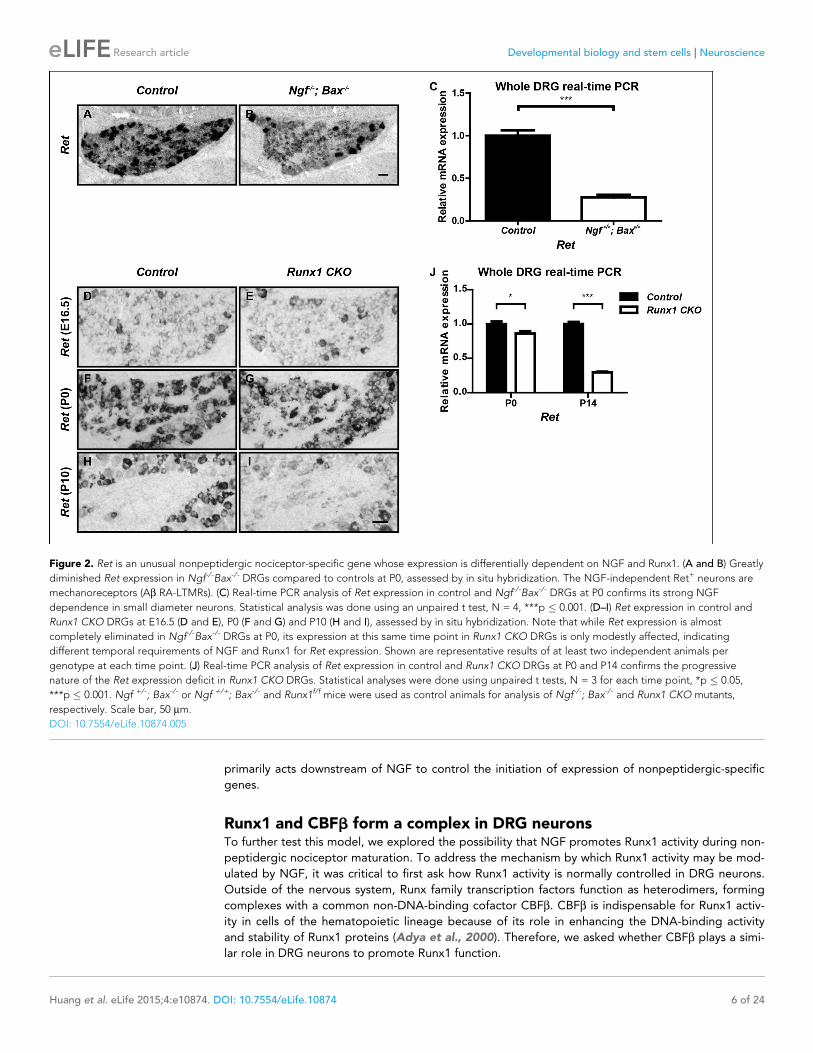
While the large majority of nonpeptidergic-specific genes require both NGF and Runx1 for initia-
tion of expression, expression of the nonpeptidergic marker Ret displays differential dependence on
NGF and Runx1. Although Ret expression is severely impaired in Ngf-/-; Bax-/- DRGs at P0, as previ-
ously described (Figure 2A–C) (Luo et al., 2007), its expression at this time point is only mildly
affected in Runx1 CKO DRGs compared to controls, as shown by both in situ hybridization and real-
time PCR (Figure 2F, G, J). A time-course analysis of Ret expression in control and Runx1 CKO
DRGs using both in situ hybridization and real-time PCR further confirmed the perinatal onset of
Runx1 dependence for Ret expression (Figure 2D–J). Therefore, while NGF is required for initiation
of Ret expression, Runx1 is only required at a later stage, for maintenance of Ret expression. We
conclude that NGF and Runx1 are both required for initiation of expression of a large cohort of non-
peptidergic nociceptor-specific genes, with Ret being a notable exception.
Runx1 acts downstream of NGF signaling to control expression ofnonpeptidergic nociceptor-specific genesThe finding that initiation of expression of most nonpeptidergic nociceptor-specific genes analyzed
so far is dependent on both NGF and Runx1 suggests a model in which Runx1 is a downstream
mediator of NGF signaling during maturation of nonpeptidergic nociceptors. Alternatively, Runx1
may indirectly control nonpeptidergic nociceptor maturation by facilitating NGF signaling. In fact,
the level of NGF signaling, as assessed by the fluorescent intensity of phospho-Trk (pTrk) immunos-
taining, is lower in Runx1 CKO DRG neurons relative to controls at P0 (Figure 3—figure supplement
1A–F). This ability of Runx1 to sustain normal levels of TrkA activity is not simply due to an effect on
TrkA expression, as TrkA levels in control and Runx1 CKO DRGs are indistinguishable, as determined
by immunohistochemistry (Figure 3—figure supplement 1G, H). However, since Ret expression,
which is strongly dependent on NGF signaling in nonpeptidergic nociceptors, appears normal at
E16.5 (Figure 2D, E), it is unlikely that diminished NGF signaling observed at P0 accounts for the
profound deficits in initiation of expression of nonpeptidergic-specific genes in E16.5 Runx1 CKO
mice (Figure 1—figure supplement 1). Nevertheless, to directly test the possibility that Runx1 con-
trols maturation of nonpeptidergic nociceptors solely by modulating NGF signaling, we asked
whether exogenous NGF can rescue nonpeptidergic nociceptor gene expression deficits in Runx1
CKO DRGs. This was addressed in two ways. First, the consequence of excess NGF was evaluated in
vivo by in situ hybridization and real-time PCR analysis following intraperitoneal injections of NGF
into neonatal mice. With the notable exception of Ret, whose expression was partially restored fol-
lowing NGF injection into Runx1 CKO mice (Figure 3J–L and Figure 3—figure supplement 2D), we
found a general and complete lack of effect of NGF administration on expression of nonpeptidergic-
specific genes in Runx1 CKO DRGs (Figure 3A–I and Figure 3—figure supplement 2A–C). In a sec-
ond series of experiments, dissociated DRG cultures from P0 control and Runx1 CKO animals were
incubated in the presence or absence of NGF, and the effects of NGF on nonpeptidergic-specific
gene expression were assessed by real-time PCR. Importantly, NGF treatment led to a robust
increase in expression of the nonpeptidergic-specific genes, Mrgprd, Gfra2, and Ptprt, in wildtype
neurons but not in Runx1-deficient neurons (Figure 2M–O). In contrast, Ret expression was NGF-
dependent in both control and Runx1-deficient neurons, albeit to a lesser extent in the absence of
Runx1 (Figure 3P). Taken together, these findings are most consistent with a model in which Runx1
Huang et al. eLife 2015;4:e10874. DOI: 10.7554/eLife.10874 5 of 24
Research article Developmental biology and stem cells Neuroscience
primarily acts downstream of NGF to control the initiation of expression of nonpeptidergic-specific
genes.
Runx1 and CBFb form a complex in DRG neuronsTo further test this model, we explored the possibility that NGF promotes Runx1 activity during non-
peptidergic nociceptor maturation. To address the mechanism by which Runx1 activity may be mod-
ulated by NGF, it was critical to first ask how Runx1 activity is normally controlled in DRG neurons.
Outside of the nervous system, Runx family transcription factors function as heterodimers, forming
complexes with a common non-DNA-binding cofactor CBFb. CBFb is indispensable for Runx1 activ-
ity in cells of the hematopoietic lineage because of its role in enhancing the DNA-binding activity
and stability of Runx1 proteins (Adya et al., 2000). Therefore, we asked whether CBFb plays a simi-
lar role in DRG neurons to promote Runx1 function.
Figure 2. Ret is an unusual nonpeptidergic nociceptor-specific gene whose expression is differentially dependent on NGF and Runx1. (A and B) Greatly
diminished Ret expression in Ngf-/-Bax-/- DRGs compared to controls at P0, assessed by in situ hybridization. The NGF-independent Ret+ neurons are
mechanoreceptors (Ab RA-LTMRs). (C) Real-time PCR analysis of Ret expression in control and Ngf-/-Bax-/- DRGs at P0 confirms its strong NGF
dependence in small diameter neurons. Statistical analysis was done using an unpaired t test, N = 4, ***p � 0.001. (D–I) Ret expression in control and
Runx1 CKO DRGs at E16.5 (D and E), P0 (F and G) and P10 (H and I), assessed by in situ hybridization. Note that while Ret expression is almost
completely eliminated in Ngf-/-Bax-/- DRGs at P0, its expression at this same time point in Runx1 CKO DRGs is only modestly affected, indicating
different temporal requirements of NGF and Runx1 for Ret expression. Shown are representative results of at least two independent animals per
genotype at each time point. (J) Real-time PCR analysis of Ret expression in control and Runx1 CKO DRGs at P0 and P14 confirms the progressive
nature of the Ret expression deficit in Runx1 CKO DRGs. Statistical analyses were done using unpaired t tests, N = 3 for each time point, *p � 0.05,
***p � 0.001. Ngf +/-; Bax-/- or Ngf +/+; Bax-/- and Runx1f/f mice were used as control animals for analysis of Ngf-/-; Bax-/- and Runx1 CKO mutants,
respectively. Scale bar, 50 mm.
DOI: 10.7554/eLife.10874.005
Huang et al. eLife 2015;4:e10874. DOI: 10.7554/eLife.10874 6 of 24
Research article Developmental biology and stem cells Neuroscience

Figure 3. Runx1 functions downstream of NGF to mediate expression of the majority of nonpeptidergic nociceptor-specific genes, whereas it controls
Ret expression at least in part by enhancing NGF signaling. (A–L) In situ hybridization analysis of expression of Mrgprd (A–C), Gfra2 (D–F), Ptprt (G–I)
and Ret (J–L) in DRGs of P2 control animals that received BSA injections, Runx1CKO animals that received BSA injections or Runx1 CKO animals that
received NGF injections. Note that exogenous NGF administration fails to activate expression of nonpeptidergic-specific genes in Runx1 CKO animals,
with the notable exception of Ret, suggesting that Runx1 is a downstream mediator of NGF signaling that is required for expression of the majority of
nonpeptidergic-specific genes. The ability of exogenous NGF to upregulate Ret expression in Runx1 CKO animals is most consistent with an indirect
Figure 3. continued on next page
Huang et al. eLife 2015;4:e10874. DOI: 10.7554/eLife.10874 7 of 24
Research article Developmental biology and stem cells Neuroscience

To determine whether CBFb is expressed in DRG neurons at the appropriate time to form a com-
plex with Runx1, its expression patterns at both the mRNA and protein levels during nociceptor
development were assessed. In situ hybridization analysis revealed that Cbfb is expressed at varying
levels, in the majority, if not all, DRG neurons over the entire time course of our analysis (Figure 4—
figure supplement 1B–D). Due to a lack of CBFb antibodies that work reliably for immunohis-
tochemistry, we generated a CbfbFlag knockin mouse line, in which N-terminally Flag-tagged CBFb
protein (Flag-CBFb) is produced from the endogenous Cbfb locus (Figure 4—figure supplement
1A). In CbfbFlag mice, the Flag antibody allows for specific detection of endogenous Flag-CBFb,
which appears to be expressed in nearly all DRG neurons, at varying levels (Figure 4A, B). Double
labeling of Flag and Runx1 showed overlap between CBFb and Runx1 throughout nociceptor devel-
opment suggesting a potential interaction between these two proteins (Figure 4C–E and Figure 4—
figure supplement 1E–G). It is noteworthy that CBFb is broadly expressed and found in more than
just Runx1+ neurons, suggesting additional Runx1-independent CBFb functions in the DRG
(Figure 4C–E). To test for a direct physical interaction between Runx1 and CBFb in DRG neurons,
co-immunoprecipitation experiments were done using a Flag antibody and extracts from DRGs of
P0 CbfbFlag/Flag animals and wildtype littermate controls. Indeed, Runx1 is enriched in Flag immuno-
precipitates from CbfbFlag/Flag animals but not wildtype animals (Figure 4F). Thus, Runx1 and CBFb
are co-expressed and form a complex in developing DRG neurons.
CBFb is required for the development of Runx1-dependentnonpeptidergic populationsTo assess the function of CBFb in DRG development and, in particular, the role of CBFb in Runx1-
dependent nonpeptidergic nociceptor maturation, which has not previously been feasible because
of the early lethality of Cbfb null embryos, we next generated a conditional Cbfb allele (Cbfbf) by
flanking the putative promoter sequence and the first two exons of this gene with LoxP sites (Fig-
ure 5—figure supplement 1A). Effective gene ablation in the DRG was achieved by crossing mice
harboring the Cbfbf allele to a Wnt1-Cre mouse line, which drives recombination specifically in the
dorsal neural tube and neural crest derivatives (Danielian et al., 1998) (Figure 5—figure supplement
1B, C). Analysis of Wnt1-Cre; Cbfbf/f (Cbfb CKO) animals revealed a wide range of nonpeptidergic
nociceptor phenotypes that are virtually identical to those seen in Runx1 CKO mice. Specifically,
those genes found to be Runx1-dependent similarly require CBFb for normal expression, as shown
by in situ hybridization and real-time PCR analysis at P0 (Figure 5A–J and Figure 5—figure supple-
ment 1D, E). As has been shown for Runx1 CKO animals, expression of CGRP, a marker of peptider-
gic nociceptors, was de-repressed in Cbfb CKO animals (data not shown). Furthermore, like Runx1
CKO mice, Cbfb CKO animals at P0 exhibited a marked reduction of sensory innervation of the epi-
dermis (Figure 5K–P). This defect was primarily due to a deficiency in the peripheral projections of
nonpeptidergic nociceptors, considering the sparseness of epidermal innervation by peptidergic
nociceptors at P0 as well as their unperturbed innervation pattern in the absence of CBFb (data not
shown). We also found that the subepidermal nerve plexus is unaffected in Cbfb CKO mice,
Figure 3. Continued
role for Runx1 in regulating Ret expression, through enabling NGF signaling. Shown are results representative of at least three independent injection
experiments. See also Figure 3—figure supplements 1, 2. (M–P) Real-time PCR analysis of expression of Mrgprd (M), Gfra2 (N), Ptprt (O) and Ret (P) in
dissociated DRG neurons from P0 control and Runx1 CKO animals cultured in the presence or absence of NGF. Note that, with the exception of Ret,
NGF-dependent expression of these nonpeptidergic-specific genes is completely abolished in the absence of Runx1, further supporting Runx1 as a
downstream mediator of NGF in regulating expression of most nonpeptidergic-specific genes. Statistical analyses were done using two-way ANOVA
with a Bonferroni post-test, N = 5 for M and P, N = 7 for the rest. *p � 0.05, **p � 0.01, ***p � 0.001, ns non-significant. Runx1f/f mice were used as
control animals for analysis of Runx1 CKO mutants. Scale bar, 50 mm.
DOI: 10.7554/eLife.10874.006
The following figure supplements are available for Figure 3:
Figure supplement 1. Runx1 potentiates TrkA activity without regulating TrkA expression.
DOI: 10.7554/eLife.10874.007
Figure supplement 2. Runx1 controls expression of the majority of nonpeptidergic-specific genes independent of its stimulatory effect on NGF
signaling.
DOI: 10.7554/eLife.10874.008
Huang et al. eLife 2015;4:e10874. DOI: 10.7554/eLife.10874 8 of 24
Research article Developmental biology and stem cells Neuroscience

indicating a developmental defect in the final stage of peripheral target innervation. Therefore, both
molecular and morphological features of nonpeptidergic nociceptors critically depend on both
Runx1 and CBFb during embryonic development.
We next asked whether CBFb is required at postnatal times for development of nonpeptidergic
nociceptors. Since Wnt1Cre; Cbfb CKO animals die perinatally due to craniofacial defects, a
Figure 4. Runx1 and CBFb are co-expressed and form a complex in DRG neurons. (A and B) Double
immunostaining of Flag and Runx1 in wildtype and CbfbFlag/+ DRGs at P0 confirms the specificity of the Flag
antibody. Note that Flag immunoreactivity in wildtype DRGs is nearly undetectable. (C–E) Double immunostaining
of Flag and Runx1 in CbfbFlag/+ DRGs at P0 shows extensive colocalization between Flag-CBFb and Runx1
(arrows). Note that CBFb is expressed in many more DRG neurons than those that are Runx1+ . See more
examples in Figure 4—figure supplement 1E–G. (F) Co-immunoprecipitation experiments using a Flag antibody
for immunoprecipitation from DRGs lysates from P0 wildtype and CbfbFlag/Flag animals. Subsequent detection with
Runx1 and CBFb antibodies shows that Runx1 associates with Flag-CBFb from DRGs of CbfbFlag/Flag animals, but
not wildtype controls, indicating the formation of a Runx1/CBFb complex in the DRG. Scale bar, 50 mm.
DOI: 10.7554/eLife.10874.009
The following figure supplements are available for Figure 4:
Figure supplement 1. Generation of the CbfbFlag allele and more detailed characterization of the temporal and
spatial patterns of Cbfb expression.
DOI: 10.7554/eLife.10874.010
Huang et al. eLife 2015;4:e10874. DOI: 10.7554/eLife.10874 9 of 24
Research article Developmental biology and stem cells Neuroscience

Figure 5. CBFb is required for acquisition of molecular and morphological features of nonpeptidergic nociceptors. (A–J) Expression of Mrgprd
(Control, 26.9% ± 2.8%; Cbfb CKO, 0%), Gfra2 (Control, 38.8% ± 2.8%; Cbfb CKO, 11.7% ± 1.9%), Ptprt, (Control, 31.9% ± 3.2%; Cbfb CKO, 7.1% ±
2.8%), Myo1a (Control, 26.9% ± 3.2%; Cbfb CKO, 5.6% ± 0.6%) and Kif21b (Control, 20.2% ± 0.1%; Cbfb CKO, 2.4% ± 0.5%) in control and Cbfb CKO
DRGs at P0 by in situ hybridization analysis. The gene expression deficits in Cbfb CKO animals phenocopy those observed in Runx1 CKO animals
except for Kif21b expression. The discrepancy likely reflects Kif21b expression in proprioceptors where it presumably depends on Runx3 and CBFb for
expression. Shown are the means ± SEMs for the percentage of neurons expressing indicated genes based on counts from a total of at least 9 sections
from three independent animals per genotype. DRG neurons were identified and counted based on combined NeuN immunostaining, which was not
shown. See also Figure 5—figure supplement 1D, E. (K–N) GFP immunostaining of P0 hairy skin to visualize sensory innervation of the epidermis in
control and Runx1 CKO animals (K and L) or control and Cbfb CKO animals (M and N) that also carry the TaumGFP allele. The TaumGFP allele was
Figure 5. continued on next page
Huang et al. eLife 2015;4:e10874. DOI: 10.7554/eLife.10874 10 of 24
Research article Developmental biology and stem cells Neuroscience

Runx1CreER knockin allele (Samokhvalov et al., 2007) combined with postnatal tamoxifen adminis-
tration was used to ablate Cbfb postnatally. A similar strategy was used to generate a Runx1 induc-
ible knockout mouse model for direct comparison. Both Runx1CreER; Cbfbf/f and Runx1CreER/f mice
treated with postnatal tamoxifen were viable and indistinguishable from control littermates. How-
ever, following postnatal deletion of either Runx1 or Cbfb, we observed, during the third postnatal
week, few or no tyrosine hydroxylase (TH)+ C-low threshold mechanoreceptors (C-LTMRs), a non-
peptidergic neuronal subtype (Li et al., 2011; Seal et al., 2009). Specifically, key features of C-
LTMRs, including TH expression and longitudinal lanceolate endings in the periphery, as marked by
expression of a Cre-dependent GFP reporter (Hippenmeyer et al., 2005), are nearly completely
absent in mice lacking Runx1 or CBFb at postnatal time points (Figure 5—figure supplement 1F–
M). Similar phenotypes were previously reported in a different Runx1 conditional mutant (Lou et al.,
2013). Therefore, both Runx1 and CBFb are required during early development for initiation of the
nonpeptidergic nociceptor fate and at postnatal times for maturation of at least one specific non-
peptidergic neuronal subtype, the C-LTMR.
The phenocopy of Runx1 and Cbfb mutants may be partly attributed to a dramatic defect in
Runx1 protein expression in Cbfb CKO DRGs, as shown by immunostaining and western blot analysis
at P0 (Figure 5Q–S). This Runx1 protein deficit was evident as early as E13 (data not shown). Runx1
mRNA expression, on the other hand, remained unchanged, if not increased, in Cbfb CKO DRGs
compared to controls, as determined by both in situ hybridization and real-time PCR (Figure 5T–V).
These findings demonstrate a key role for CBFb in the post-transcriptional regulation of Runx1
expression, most likely at the level of protein stability. Together, these findings indicate that CBFb
and Runx1 are both essential for induction of the nonpeptidergic nociceptor fate and postnatal non-
peptidergic neuron subtype maturation, and that the CBFb requirement may reflect its role in con-
trolling the level and activity of Runx1 proteins.
NGF regulates expression of Cbfb but not Runx1 at early stages ofnociceptor maturationHow does NGF control Runx1/CBFb activity? Since Runx1 and CBFb are both necessary for nonpep-
tidergic neuron maturation, NGF could, in principle, control Runx1/CBFb function by promoting the
activity of Runx1, CBFb or both. Considering the profound requirement of NGF for nonpeptidergic
nociceptor gene expression, one possibility is that NGF promotes expression of Runx1, Cbfb, or
both, thereby enabling Runx1/CBFb complex formation, stabilization and function, and thus initiation
of the nonpeptidergic nociceptor fate. We therefore examined expression of both Cbfb and Runx1
in DRGs of control and Ngf-/-; Bax-/- animals before and during the acquisition of the nonpeptidergic
nociceptor fate. Interestingly, Cbfb expression was found to be sensitive to loss of NGF prior to
specification of nonpeptidergic nociceptors. In fact, at E14.5, a time prior to expression of nonpepti-
dergic nociceptor-specific genes, Cbfb mRNA expression was significantly reduced in Ngf-/-; Bax-/-
Figure 5. Continued
introduced to label all Cre-expressing neurons including all sensory neurons. Note that there is a dramatic reduction in fiber density specifically in the
epidermis in both Runx1 CKO and Cbfb CKO animals relative to their littermate controls. The yellow dotted line denotes the epidermal-dermal junction
which was drawn based on TOPRO3 counterstain (blue). (O and P) Quantification of sensory innervation of the epidermis in control and Runx1 CKO
animals (O) or control and Cbfb CKO animals (P) reveals a remarkably similar reduction in the innervation density in both mutants at P0. The innervation
density is defined as the fraction of area occupied by GFP+ fibers in the epidermis. An unpaired t test was performed on data from three independent
animals per genotype. ***p � 0.001. (Q and R) Runx1 immunostaining of control and Cbfb CKO DRGs at P0 shows almost complete loss of Runx1
proteins in the absence of CBFb. Shown are representative images from at least three independent experiments. (S) Immunoblot analysis of expression
of Runx1 and Cbfb in control and Cbfb CKO DRGs at P0 shows dramatic loss of Runx1 proteins as a result of CBFb depletion. bIII-Tubulin serves as a
loading control. Shown are results from three independent experiments. (T and U) In situ hybridization analysis of Runx1 expression in control and Cbfb
CKO DRGs at P0 shows comparable levels of Runx1 transcripts in control and mutant animals. (V) Real-time PCR analysis of Runx1 expression in control
and Cbfb CKO DRGs at P0 shows increased Runx1 mRNA expression in Cbfb CKO DRGs compared to control, which likely reflects an increased ratio
of nociceptors to proprioceptors (data not shown). An unpaired t test was performed on data from four independent pairs of control and mutant
animals, **p � 0.01. Cbfbf/f mice were used as control animals for analysis of Cbfb CKO mutants. Scale bar, 50 mm.
DOI: 10.7554/eLife.10874.011
The following figure supplements are available for Figure 5:
Figure supplement 1. Generation of the Cbfbf allele and demonstration of a postnatal requirement for both CBFb and Runx1 in C-LTMR development.
DOI: 10.7554/eLife.10874.012
Huang et al. eLife 2015;4:e10874. DOI: 10.7554/eLife.10874 11 of 24
Research article Developmental biology and stem cells Neuroscience

DRGs relative to controls, specifically in small-diameter nociceptor precursors (Figure 6A, B, E). This
Cbfb expression deficit in Ngf-/-; Bax-/- small diameter DRG neurons becomes much more pro-
nounced at later time stages, such as E16.5, the onset of observable deficits in nonpeptidergic-spe-
cific gene expression (Figure 6C–E). Similarly, Ntrk1-/-/Bax-/- DRGsdisplayed reduced levels of Cbfb
expression at P0 (Figure 7H, I). The deficit of Cbfb mRNA expression was confirmed by real-time
PCR analysis (Figure 6—figure supplement 1A). Considering the inability of this assay to distinguish
between Cbfb expressed in nociceptors and that expressed in NGF-independent DRG neurons, such
as proprioceptors, and non-neuronal cells of the ganglion, it is noteworthy that the real-time PCR
measurements are an underestimation of the NGF dependence of Cbfb expression in developing
nociceptors. We further addressed NGF-dependence of Cbfb expression at the protein level in two
ways. First, Flag immunostaining on dissociated DRG cultures from P0 CbfbFlag/+ animals showed
that NGF is essential for CBFb protein expression in vitro (Figure 6F–H). Second, when DRGs from
P0 control and Ngf-/-; Bax-/- animals that were also heterozygous for the CbfbFlag allele were acutely
dissociated, the level of Flag immunoreactivity was significantly lower in Ngf-/-; Bax-/- neurons com-
pared to controls, suggesting NGF-dependence of CBFb protein expression in vivo (Figure 6—fig-
ure supplement 1B–D). Cbfb is also sensitive to the dose of NGF as exogenous NGF administered
to wildtype neonates via intraperitoneal injection further potentiated Cbfb expression (Figure 6—
figure supplement 1E). Together, these findings identify NGF as a key regulator of Cbfb expression
prior to the initiation of NGF-dependent nonpeptidergic-specific gene expression. On the other
hand, Runx1 mRNA levels are normal in Ngf-/-; Bax-/- DRGs at E14.5, as determined by both in situ
hybridization and real-time PCR analysis (Figure 6I, J, M), consistent with our previous observation
(Luo et al., 2007). It is only at later times, beginning at E16.5, that Runx1 expression becomes
affected, suggesting a late requirement of NGF for maintenance of Runx1 expression (Figure 6K–
M). Consistent with the late NGF dependence of Runx1 mRNA expression, Runx1 protein expression
is unaffected in Ngf-/-; Bax-/- DRGs at E14.5 (Figure 6N, O, R, S). At E16.5, the level of Runx1 protein
is significantly diminished without any change in the number of Runx1+ neurons (Figure 6P–S). In
view of the discrepancy between the dramatic nonpeptidergic phenotypes at E16.5 and the rela-
tively modest deficit in Runx1 expression at this time, upregulation of Cbfb expression by NGF is
likely to be an important mechanism by which NGF enables Runx1 function during nonpeptidergic
nociceptor development.
NGF stimulates Cbfb expression in a MAPK-dependent mannerTo better understand the mechanism by which NGF controls Cbfb expression in nociceptor precur-
sors, we next sought to identify NGF–TrkA signaling cascades that control Cbfb expression. The
canonical ERK1/2 signaling cascade represents a likely candidate because this is a major NGF–TrkA
effector pathway, and animals deficient in components of this pathway exhibit nonpeptidergic phe-
notypes, including reduced Ret expression, impaired innervation of the epidermis, and impaired
adult CBFb protein expression (Newbern et al., 2011; Zhong et al., 2007). To directly assess the
role of MAPK signaling in NGF-dependent Cbfb expression during development, both in vitro and
in vivo approaches were used. Through immunostaining and immunoblot analysis, we found that
pharmacological inactivation of Mek1/2, direct activators of ERK1/2, greatly attenuated the ability of
NGF to promote CBFb protein expression in vitro (Figure 7A–E and Figure 7—figure supplement
1A–C). A Nestin-Cre-based conditional knockout mouse model, which targets all four Mapk3,
Mapk1, Map2k1and Map2k2 alleles, was next used to determine the in vivo role of MAPK signaling
for Cbfb expression. Using this in vivo model system, we found that in 4 out of 5 P0 Nes-Cre;
Map2k1f/f;Map2k2-/-; Mapk3-/-; Mapk1f/f mutants (Quadruple), Cbfb mRNA expression was severely
disrupted, demonstrating a strong dependence of Cbfb expression on MAPK signaling in vivo
(Figure 7F, G). The phenotypic variation in this analysis likely reflects incomplete excision of all four
floxed alleles. We next asked whether MAPK signaling is sufficient to promote Cbfb expression in
vivo, in the absence of NGF or activation of other NGF-dependent signaling pathways. For this anal-
ysis, a constitutively active form of B-Raf (V600E) was expressed exclusively in the nervous system to
drive MAPK signaling in animals that were null for both Ntrk1 and Bax (O’Donovan et al., 2014).
Remarkably, while Cbfb expression in Ntrk1-/-; Bax-/- DRGs was severely impaired, expression of B-
RafV600E in Ntrk1-/-; Bax-/- mice to promote constitutive MAPK signaling restored Cbfb expression to
near normal levels (Figure 7H–J). Real-time PCR analysis was carried out as an independent measure
of Cbfb expression to further confirm the necessity and sufficiency of MAPK signaling for Cbfb
Huang et al. eLife 2015;4:e10874. DOI: 10.7554/eLife.10874 12 of 24
Research article Developmental biology and stem cells Neuroscience

Figure 6. NGF regulates the Runx1/CBFb complex through differential control of Cbfb and Runx1 expression. (A and B) In situ hybridization analysis of
Cbfb expression in control and Ngf-/-Bax-/- DRGs at E14.5 shows a significant reduction in the level of transcripts in small diameter neurons that
correspond to prospective nociceptors in Ngf-/-Bax-/- DRGs compared to controls. The insets focus on nociceptor-rich regions. Scale bar for the insets,
10mm. Note that Cbfb in situ hybridization was combined with Runx3 immunostaining to exclude the Runx3+ Cbfb population from the analysis. (C and
Figure 6. continued on next page
Huang et al. eLife 2015;4:e10874. DOI: 10.7554/eLife.10874 13 of 24
Research article Developmental biology and stem cells Neuroscience

expression in vivo (Figure 7K). Together, these findings indicate that the ERK/MAPK signaling cas-
cade is both necessary and sufficient to mediate NGF/TrkA-dependent expression of Cbfb.
Islet1, a LIM-homeodomain transcription factor, is required forinitiation of expression of Runx1 but not CbfbWhile Cbfb expression at early stages of nonpeptidergic nociceptor development is dependent on
NGF, the apparent NGF-independence of Runx1 expression during early development prompted us
to ask whether initiation of Cbfb and Runx1 expression are differentially controlled by intrinsic cues.
We tested the involvement of Islet1, a LIM-homeodomain transcription factor, because in a neural
crest derivative-specific Islet1 mutant (Isl1 CKO), a Runx1 protein deficit was noted as early as E12.5,
the time when Runx1 proteins are first detected in lumbar DRGs (Dykes et al., 2011; Sun et al.,
2008). To define the level at which Runx1 expression is regulated by Islet1, Runx1 expression was
evaluated by in situ hybridization analysis in DRGs of E12.5 control and Isl1 CKO animals. Consistent
with a central role for Islet in initiating Runx1 expression at the transcriptional level, Runx1 transcripts
were virtually undetectable in Isl1 CKO DRGs (Figure 8A, B). In contrast, Cbfb expression was only
minimally affected by the same genetic perturbation (Figure 8C, D). The distinct dependence of
Runx1 and Cbfb expression on Islet1 was confirmed by a microarray analysis of control and Isl1 CKO
DRGs at E12.5 (Figure 8E). Thus, expression of Runx1 and CBFb, obligatory components of a tran-
scription factor holocomplex, are under differential control of Islet1 and NGF; Runx1 and Cbfb at
early stages of nonpeptidergic nociceptor differentiation are preferentially regulated by the intrinsic
cue Islet1 and the extrinsic cue NGF, respectively. Thus, a convergence of intrinsic and extrinsic sig-
naling events in nonpeptidergic nociceptor progenitors enables formation of the Runx1/CBFb tran-
scription factor complex, a key event required for nonpeptidergic nociceptor differentiation.
DiscussionThis study defines a gene regulatory mechanism underlying the specification of nonpeptidergic noci-
ceptors. At the core of this process is the formation of the Runx1/CBFb transcription factor complex,
both components of which are essential for directing the nonpeptidergic nociceptor-specific
Figure 6. Continued
D) In situ hybridization analysis of Cbfb expression in control and Ngf-/-Bax-/- DRGs at E16.5 shows more pronounced Cbfb mRNA deficit in Ngf-/-Bax-/-
DRGs. (E) Quantification of Cbfb expression deficits in nociceptors in Ngf-/-Bax-/- DRGs based on experiments described in (A–D). Intensity of in situ
signal in areas devoid of Runx3+ neurons was measured. An unpaired t test was performed using data collected from 3 independent experiments for
each time point. *p � 0.05. See also Figure 6—figure supplement 1A. (F and G) Double staining of Flag and bIII-Tubulin in dissociated DRG neurons
from P0 CbfbFlag/+ animals that were cultured without or with NGF. Note that NGF application robustly stimulates CBFb protein expression as
indicated by increased Flag immunoreactivity. (H) Quantification of the effect of NGF treatment on CBFb protein levels based on experiments
described in (F and G). CBFb protein abundance was quantified using the average fluorescence intensity of Flag immunoreactivity per cell. An unpaired
t test was performed using data collected from four independent experiments. ***p< 0.0001. See also Figure 6—figure supplement 1B–D. (I and J) In
situ hybridization analysis of Runx1 expression in control and Ngf-/-Bax-/- DRGs at E14.5 shows comparable levels of Runx1 transcripts in control and
mutant DRGs. Means ± SEM for relative intensity of in situ signals after normalization to the level in control DRGs is as follows: Control, 1.00 ± 0.16;
Ngf-/-Bax-/-, 0.73 ± 0.12. p = 0.2079, based on an unpaired t test. (K and L) In situ hybridization analysis of Runx1 expression in control and
Ngf-/-Bax-/-DRGs at E16.5 shows a reduction in the level of signal per cell in Ngf-/-Bax-/- DRGs compared to controls. Control, 1.00 ± 0.07; Ngf-/-Bax-/-,
0.49 ± 0.06. p = 0.0003, based on an unpaired t test. (M) Real-time PCR analysis of Runx1 expression in control and Ngf-/-Bax-/- DRGs at E14.5 and P0
reveals a late requirement of NGF for Runx1 expression. An unpaired t test was performed on data collected from three independent animals per
genotype at each time point. *p � 0.05, ns non-significant. (N–Q) Runx1 immunostaining in control and Ngf-/-Bax-/- DRGs at E14.5 (N and O) and E16.5
(P and Q) shows that the Runx1 protein deficit becomes evident in Ngf-/-Bax-/- DRGs at E16.5, which coincides with the onset of nonpeptidergic
nociceptor deficits in Ngf-/-Bax-/- DRGs. (R and S) Quantification of Runx1 protein expression in control and Ngf-/-Bax-/- DRGs at E14.5 and E16.5 based
on the percentage of Runx1+ neurons or the fluorescence intensity of Runx1 immunoreactivity. Note that loss of NGF specifically affects the level of
Runx1 expression per cell without altering the number of Runx1+ neurons. An unpaired t test was performed on data collected from three independent
animals per genotype. *p � 0.05, ns non-significant. Ngf +/-; Bax-/- or Ngf +/+; Bax-/- mice were used as control animals for analysis of Ngf-/-; Bax-/-
mutants. Scale bar, 50 mm.
DOI: 10.7554/eLife.10874.013
The following figure supplements are available for Figure 6:
Figure supplement 1. Cbfb expression is NGF-dependent in vivo.
DOI: 10.7554/eLife.10874.014
Huang et al. eLife 2015;4:e10874. DOI: 10.7554/eLife.10874 14 of 24
Research article Developmental biology and stem cells Neuroscience

Figure 7. NGF promotes Cbfb expression through the ERK/MAPK signaling pathway. (A–D) Double staining of Flag (green) and bIII-Tubulin (blue) in
DMSO or U0126-treated dissociated DRG neurons from P0 CbfbFlag/+ animals that were cultured without or with NGF. U0126 is a selective inhibitor of
MEK1/2, the direct activators of ERK1/2. Note that CBFb protein expression, as defined by Flag immunoreactivity is greatly diminished in U0126-treated
neurons as compared to vehicle-treated neurons, all grown in the presence of NGF. (E) Quantification of the effect of U0126 treatment on CBFb protein
levels based on experiments as described in (A–D). CBFb protein abundance was quantified as the average fluorescence intensity of Flag
immunoreactivity per cell. Statistical analysis was done using a two-way ANOVA with a Bonferroni post-test, based on data from four independent
experiments. ***p � 0.001, ns non-significant. See also Figure 7—figure supplement 1. (F and G) In situ hybridization analysis of Cbfb expression in
control and quadruple DRGs at P0 reveals a severe deficit in Cbfb mRNA expression in DRGs when MAPK signaling is disrupted in the nervous system.
A similar phenotype of varied severity was observed in 4 out of 5 mutant animals. (H–J) In situ hybridization analysis of Cbfb expression in Ntrk1 +/-;
Bax-/-, Ntrk1-/-; Bax-/- and Ntrk1-/-; Bax-/-; Nes-Cre; V600E DRGs at E18.5 shows that constitutive activation of MAPK signaling leads to a dramatic
increase in Cbfb expression in TrkA-deficient animals. Shown are representative images from two independent experiments. (K) Real-time PCR analysis
of Cbfb expression in the same set of loss-of-function and gain-of-function mouse models as described in (F and G) and (H–J) at P0 further
demonstrates the necessity and sufficiency of MAPK signaling for NGF-dependent Cbfb expression in vivo. Shown are averages from two independent
experiments after normalization to littermate control. Scale bar, 50 mm.
DOI: 10.7554/eLife.10874.015
The following figure supplements are available for Figure 7:
Figure supplement 1. In vitro evidence for the necessity of MAPK signaling for NGF-dependent CBFb expression.
DOI: 10.7554/eLife.10874.016
Huang et al. eLife 2015;4:e10874. DOI: 10.7554/eLife.10874 15 of 24
Research article Developmental biology and stem cells Neuroscience

transcriptional program. Importantly, expression of each component is independently regulated at
the transcriptional level. Cbfb expression is controlled by the extrinsic cue NGF through the MAPK
signaling pathway. On the other hand, Runx1 expression is initiated by a mechanism that is critically
dependent on Islet1, an intrinsic factor that controls early development of sensory neurons. Further-
more, the Runx1/CBFb complex, through an unknown mechanism, maintains a high level of NGF-
TrkA signaling, which is essential for at least one defining feature of nonpeptidergic nociceptors:
postnatal expression of Ret. Thus, the initiation of a lineage-specific differentiation program is tightly
controlled by a convergence of extrinsic and intrinsic factors at the level of formation of the hetero-
dimeric Runx1/CBFb transcription factor complex (Figure 8F).
The Runx1/CBFb complex, a coincidence detector for extrinsic andintrinsic cues that direct nociceptor subtype specificationNonpeptidergic nociceptor specification requires proper expression of both Runx1 and Cbfb, whose
transcriptional initiation is differentially dependent on Islet1 and NGF signaling. The Runx1/CBFb
holocomplex therefore serves as a coincidence detector for extrinsic and intrinsic signals that pro-
mote specification of nonpeptidergic nociceptors. The requirement for NGF, an extrinsic cue that is
critical for survival of all nociceptors, ensures that only those nociceptor precursors that gain access
Figure 8. Islet1 is required for initiation of Runx1, but not Cbfb expression. (A–D) In situ hybridization analysis of expression of Runx1 (A and B) and
Cbfb (C and D) in control and Isl1 CKO DRGs at E12.5 shows that Islet1 deficiency abolishes expression of Runx1 but not Cbfb at an early age. Shown
are representative images from two independent experiments. (E) Microarray analysis of E12.5 control and Isl1 CKO DRGs further confirms the
differential dependence of expression of Runx1 and Cbfb on Islet1. Shown are average expression levels from two independent experiments that are
normalized to the control levels for each gene. Expression levels have been normalized using globe scaling. Isl1f/f mice were used as control animals for
analysis of Isl1 CKO mutants. (F) Schematics illustrating a molecular mechanism underlying specification of nonpeptidergic nociceptors and its general
implication in the context of subtype specification. The extrinsic cue NGF and the intrinsic cue Islet1 coordinately regulate the Runx1/CBFb complex, a
nonpeptidergic nociceptor transcription factor complex, by preferentially targeting Cbfb and Runx1 for transcriptional regulation, respectively.
Furthermore, the Runx1/CBFb complex, through an unknown mechanism, enhances the level of NGF-TrkA signaling, resulting in a positive feedback
loop between NGF-TrkA signaling and Runx1/CBFb complex. This gene regulatory mechanism not only underscores the importance of interplay
between extrinsic and intrinsic factors during multilineage differentiation, but also illustrates how such interplay can control cell-fate decisions through
the convergence of extrinsic and intrinsic signals at the level of a heterodimeric, lineage-specific transcription factor complex. Scale bar, 50 mm.
DOI: 10.7554/eLife.10874.017
Huang et al. eLife 2015;4:e10874. DOI: 10.7554/eLife.10874 16 of 24
Research article Developmental biology and stem cells Neuroscience

to a sufficient amount of NGF and survive the period of naturally occurring cell death will undergo
nonpeptidergic nociceptor maturation. Consistent with this, Cbfb expression exhibits NGF depen-
dence at E14.5, immediately following the period of naturally occurring cell death. On the other
hand, the dependence of Runx1 expression on Islet1, a transcription factor required for terminating
expression of genes whose expression is restricted to the early stage of sensory neurogenesis, likely
contributes to the orderly transition from pan-sensory neurogenesis to specification of nonpeptider-
gic nociceptors by ensuring that Runx1 expression and hence Runx1/CBFb-dependent nonpeptider-
gic nociceptor development are initiated after sensory neurogenesis (Sun et al., 2008). Thus the
NGF and Islet1 signals coordinate temporal control of the Runx1/CBFb complex for timely initiation
of nonpeptidergic nociceptor differentiation. It is important to note that the extrinsic and intrinsic
signals described here are unlikely to be sufficient to instruct Runx1/CBFb complex formation and
the nonpeptidergic nociceptor lineage choice, as neither NGF nor Islet1 functions exclusively in non-
peptidergic nociceptors. Indeed, NGF–TrkA signaling is required for survival, target innervation and
normal phenotypic development of both peptidergic and nonpeptidergic nociceptors
(Harrington and Ginty, 2013), and Islet1 is expressed broadly in developing DRG neurons and is
required for the generation of virtually all nociceptors (Sun et al., 2008). Therefore, one or more
additional, unidentified signals must govern the divergence of the two main nociceptive lineages.
Transcriptional activator(s) of Cbfb expression downstream of MAPKsignalingWe found that MAPK signaling is both necessary and sufficient for NGF-dependent expression of
Cbfb in developing nociceptors. The generality of MAPK signaling suggests that it plays a similar
role in controlling Cbfb expression in cells outside of the nervous system and thus MAPK–CBFb sig-
naling may have a general role in cell type specification. Moreover, the identification of MAPK as an
upstream activator of Cbfb expression may provide an explanation for the observation that Cbfb,
unlike Runx genes, is widely expressed (Wang et al., 1993). The identity of nuclear targets of the
MAPK signaling pathway that directly activate Cbfb expression remains unclear. To this end, we
found, through bioinformatic analysis of the putative Cbfb promoter, cAMP-response element (CRE)
consensus motifs within an evolutionally conserved 458 bp enhancer-like element (data not shown),
raising the intriguing possibility that CREB family members, which are well-studied downstream
mediators of MAPK signaling in many cell types (Shaywitz and Greenberg, 1999), are direct tran-
scriptional activators of Cbfb. In view of the role of CREB family members in NGF-dependent growth
and survival of neurons (Liu et al., 1999; Riccio et al., 1999), the possibility of CREB-mediated acti-
vation of Cbfb expression would further suggest that common effectors of growth factor signaling
pathways support distinct biological outcomes, in this case, survival, axon growth and maturation of
nociceptors, presumably by controlling expression of distinct target genes.
A mechanism of interplay between intrinsic and extrinsic signals forpostmitotic differentiation of neuronal subtypesThe formation of the Runx1/CBFb complex downstream of NGF and Islet1 signaling during nonpep-
tidergic nociceptor maturation illustrates a novel mechanism of interplay between extrinsic and
intrinsic factors in controlling postmitotic specification of neuronal subtypes. It is important to note
the distinction between this relatively late developmental process and the specification of progenitor
domains, which takes place prior to cell cycle exit. While it is well established in vertebrate systems
that specification of progenitor domains is coordinately regulated by the intrinsically defined compe-
tence state of a progenitor and spatially and temporally controlled extrinsic signals (Briscoe and
Novitch, 2008; Livesey and Cepko, 2001; Molyneaux et al., 2007), relatively little is known about
the contributions of extrinsic cues, intrinsic factors, and their modes of interaction, during postmi-
totic specification of neuronal subtypes. The gene regulatory mechanism described here leads us to
propose a simple model in which convergence of extrinsic and intrinsic signals onto a single hetero-
dimeric transcription factor complex controls lineage-specific differentiation programs and postmi-
totic specification of neuronal subtypes (Figure 8F). It is conceivable that this simple model is
generally applicable to a broad range of neuronal subtypes that rely on heterodimeric transcription
factor complexes for their specification.
Huang et al. eLife 2015;4:e10874. DOI: 10.7554/eLife.10874 17 of 24
Research article Developmental biology and stem cells Neuroscience

Materials and methods
MiceFor generation of mice harboring the Cbfbf allele, a 2 kb sequence containing a 1 kb sequence
immediately upstream of the transcription start site as well as exon 1 and exon 2 of the Cbfb locus
was flanked by two loxP sites using recombineering technology. For generation of mice harboring
the CbfbFlag allele, the sequence encoding a single Flag epitope was introduced immediately
upstream of the translational start site of the Cbfb gene. Mice were generated using targeted ES
cells and standard blastocyst injection techniques. Cbfbf/f mice were mated to either a mouse strain
expressing Cre recombinase under control of the Wnt1 promoter (Danielian et al., 1998) to gener-
ate Wnt1-Cre; Cbfbf/f mice or to a mouse strain in which the Cre recombinase coding sequence was
inserted into the Runx1 locus (Samokhvalov et al., 2007) to generate Runx1CreER/+; Cbfbf/f mice.
Wnt1-Cre; Runx1f/f and Ngf�/�; Bax�/� mice were generated as described (Chen et al., 2006;
Patel et al., 2000). The mouse lines used to generate Nes-Cre; Map2k1f/f; Map2k2-/-; Mapk3-/-;
Mapk1f/f and Ntrk1-/-; Bax-/-; Nes-Cre; V600E mice were described previously (Mercer et al., 2005;
Moqrich et al., 2004; Newbern et al., 2011). The TaumGFP allele, a neuronal specific Cre-dependent
GFP reporter, was previously described (Hippenmeyer et al., 2005). See supplemental information
for details on generation of the Cbfbf and CbfbFlag alleles.
In situ hybridization and immunohistochemistryDigoxigenin (DIG)-labeled cRNA probes were used for in situ hybridization. Target sequences for
Ptprt, Myo1a, Kif21b probes were amplified using gene specific PCR primers from either cDNA pre-
pared from P0 mouse DRGs or genomic DNA from wildtype ES cells to generate corresponding
plasmids for in situ hybridization. In situ hybridization probes for Mrgprd, Gfra2, Ret and Runx1 were
described previously (Luo et al., 2007). The in situ hybridization probe for Cbfb was generated from
an available cDNA clone (GenBank: BC026749.1). In situ hybridizations were carried out on 14 mm
fresh frozen DRG sections as described previously (Luo et al., 2007). For combined in situ hybridiza-
tion and immunostaining, regular BCIP/NBT-based in situ hybridization was performed prior to the
standard immunostaining procedure. Bright field and fluorescent images were taken under the same
setting. Bright field images were later pseudocolored and merged with fluorescent images.
Immunohistochemistry on DRG and skin sections was performed as described previously
(Li et al., 2011; Luo et al., 2007). The primary antibodies used were: rabbit anti-Runx1 (a gift from
Dr. Thomas Jessell, Columbia University, 1:10,000), rabbit anti-Runx3 (a gift from Dr. Thomas Jessell,
Columbia University, 1:4000), guinea pig anti-Flag (see supplemental information for antibody gen-
eration details, 1:500), rabbit anti-CGRP (Immunostar, 24112, 1:1000), chicken anti-GFP (Aves Labs,
GFP-1020, 1:500), chicken anti-NF200 (Aves Labs, NFH, 1:500), rabbit anti-Tyrosine Hydroxylase
(Millipore, AB152, 1:1000), rabbit anti-pTrk-SHC (Cell Signaling Technology, 4168,1:500), rabbit anti-
pTrk-PLCg (Cell Signaling Technology, 4619,1:500), rabbit anti-TrkA (Millipore, AB1577, 1:1000),
mouse anti-NeuN (Milllipore, MAB377MI, 1:500), and rabbit anti-bIII-Tubulin (Covance, PRB-435P,
1:1000).
Dissociated DRG neuronal culturesDissociated DRG cultures from neonatal mice were prepared using a method that was adapted from
a previously described protocol for sympathetic neuronal cultures (Deckwerth et al., 1996). Briefly,
neurons were obtained by sequential steps of enzymatic digestion and mechanical dissociation of
DRGs harvested from P0 animals. In general, these neurons were plated on Poly-D-lysine and laminin
coated coverslips at a density of ~50,000 neurons per well and cultured for 2 days in growth media
(10% FBS, 1 U/ml penicillin/streptomycin, 5 mm Ara-C (Sigma), 50 mg/ml a pan-caspase inhibitor Boc-
aspartyl (OMe)-fluoromethylketone (BAF) (MP Biomedicals)) supplemented with NGF (100 ng/ml;
either purified from mouse salivary glands or purchased from Millipore), or a neutralizing NGF anti-
body (Sigma) at a dilution of 1:2000. For experiments that used U0126, cultures were treated with
U0126 (50 mm in DMSO; Calbiochem) or DMSO the morning after plating. For experiments that
addressed in vivo dependence of CBFb protein expression on NGF, neurons were plated for 1 hour
in growth media without supplements before being processed for Flag immunostaining.
Huang et al. eLife 2015;4:e10874. DOI: 10.7554/eLife.10874 18 of 24
Research article Developmental biology and stem cells Neuroscience

Real-time PCRRNA was extracted from freshly isolated DRGs using the RNeasy micro kit (Qiagen) according to the
manufacturer’s instructions. First strand cDNA was synthesized using an oligo dT primer and the
SuperScript III system (Invitrogen). Real-time PCR was performed using the QuantiTect SYBR Green
PCR kit (Qiagen) and a 7300 Real-Time PCR System (Applied Biosystems). The amount of individual
transcripts was normalized to that of PGP9.5, a pan-neuronal marker, unless the comparison was
between control and Ngf-/-; Bax-/-, for which GAPDH served as the internal control. Detailed primer
sequences for real-time PCR can be found in supplemental information.
Immunoblotting and co-immunoprecipitationAcutely dissected DRGs were lysed in ice-old FA-M2 Lysis Buffer (50 mM Tris HCl, 150 mM NaCl, 1
mM EDTA, 1% Triton X-100, pH 7.5) supplemented with a protease inhibitor cocktail (Sigma;1:100
dilution) by sonication. Clarified lysates were either subjected to Flag immunoprecipitation for co-
immunoprecipitation experiments or processed directly for SDS-PAGE. Immunoprecipitation of Flag-
CBFb and its associated proteins was done using anti-Flag M2 affinity gel (Sigma) according to the
manufacturer’s instructions. Immunoblotting was performed using antibodies against Runx1 (Abcam,
1:5000), CBFb (1:1000, Santa Cruz), Histone 3 (1:1000, Cell Signaling Technology) and bIII-Tubulin
(1:1000, Covance), as described (Kuruvilla et al., 2000).
Statistical analysisStatistical differences for mean values between two groups and among multiple groups were ana-
lyzed using GraphPad Prism 5 software. The type of test used for statistical analysis is indicated in
the figure legend. The criterion for statistical significance was set at p � 0.05.
Mouse line generationFor the Cbfbf allele, a ~2 kb sequence (chromosome 8: 105169674- 105171592) corresponding to a
1 kb sequence immediately upstream of the transcriptional start site as well as exon 1 and exon 2 of
the Cbfb locus was flanked by two loxP sites. Recombineering technology was used to generate the
targeting vector (Copeland et al., 2001; Liu et al., 2003). Briefly, a 129/SvJ BAC clone containing
the targeted region of the Cbfb gene was obtained from Geneservice. An 11.5 kb region (chromo-
some 8: 105168174-105179387) with homology arms that were 1.5 kb and 8 kb long was inserted
into a PBS-DTA plasmid, the backbone for the final targeting vector, via the first recombineering
step. The 3’ loxP site and the FRT-Neo-FRT-5’ loxP cassette were then introduced sequentially dur-
ing subsequent recombineering steps. A Bstz171 restriction site was engineered 3’ to the 3’ loxP
site to facilitate southern screening of ES cells. The targeting construct was linearized with KpnI and
then electroporated into mouse 129S6SvEvTac ES cells. ES clones were screened by PCR and cor-
rectly targeted ES clones were confirmed by southern blot hybridization using both 5’ and internal
probes following Bstz171 digestion (WT 9.8 kb and Mutant 6.8 kb, data not shown). Chimeric
Cbfbf mice were produced by injection of positive ES cells into C57Bl/6 blastocysts. Mice carrying
the Cbfbf allele were subsequently generated by mating chimeric mice to germ-line FlpE mice to
remove the Neo cassette (Rodriguez et al., 2000). Cbfbf mice were genotyped using a 2-primer
PCR reaction with the following primers: 5’-GCGCGCCAGTCACTTGTT-3’ and 5’-AAACCATCCCAC-
GAACCGAACCAT-3’. The sizes of PCR products from wildtype and mutant alleles are 219 bp and
324 bp, respectively. For the CbfbFlag allele, the targeting vector, which was nearly identical to that
of the Cbfbf vector, was generated using a combination of recombineering and standard subcloning
strategies. The same targeted genomic region was engineered to include the FRT-Neo-FRT-loxP
cassette at the position identical to that in the Cbfbf allele, using recombineering technology.
The sequence encoding one Flag epitope was introduced into the vector immediately upstream of
the translational start site of the Cbfb gene by replacing a 1.1 kb NotI/AvrII fragment containing
the translational start site with the fragment carrying the insertion using standard cloning
techniques. A Bstz171 restriction site was inserted immediately downstream of the Flag sequence
for the purpose of southern screening. Subsequent steps for generation of CbfbFlag mice were the
same as those described above for Cbfbf mice. CbfbFlag mice were genotyped using a 2-primer
PCR reaction with the following primers: 5’-TGAGAGCTGTCTATGGCAAAC-3’ and 5’-
Huang et al. eLife 2015;4:e10874. DOI: 10.7554/eLife.10874 19 of 24
Research article Developmental biology and stem cells Neuroscience

TCAGTTCAAGGATGGCAGGTA-3’. The sizes of PCR products from wildtype and mutant alleles are
232 bp and 336 bp, respectively.
Real-time PCRPrimers used for real-time PCR analysis are provided Table 1.
NGF administration via intraperitoneal injectionsMouse pups of the desired genotype were subjected to a single intraperitoneal injection of either
NGF (2 mg reconstituted in PBS with 1% BSA) or equal volume of 1% BSA in PBS at both P0 and P1.
Animals were sacrificed at P2 and vertebral columns or DRGs were dissected and processed for
analysis.
Tamoxifen injectionsTamoxifen (Toronto Research Chemicals) was dissolved in ethanol (20 mg/ml). 50 ml (1 mg) of tamox-
ifen in ethanol was mixed with 50 ml of sunflower seed oil (Sigma), vortexed for 20 min and centri-
fuged under vacuum for 45 min to remove the ethanol. The tamoxifen solution was delivered at P2
via an intraperitoneal injection into animals harboring the Runx1CreER allele.
Microarray analysisA total of 6 RNA samples (~1 mg each) were prepared using Trizol and the RNeasy micro kit from
DRGs of three pairs of E16.5 control and Runx1 CKO animals, each from different litters. Samples
were labeled and hybridized to Affymetrix mouse 430 2.0 chips and microarray data were analyzed
with Spotfire software. Only genes with a fold change greater than or equal to 1.5, a p-value less
than or equal to 0.05 were considered differentially expressed between control and Runx1 CKO
DRGs and were reported. Methods for the microarray analysis of DRGs from Islet1 conditional
knockout and control embryos have been described (Dykes et al., 2011).
Antibody production and purificationA guinea pig polyclonal antiserum was raised against a MDYKDDDDKLVY peptide that corresponds
to the N terminus of Flag-CBFb encoded by the CbfbFlag allele using a service provided by Covance.
The peptide was synthesized and conjugated at its C-terminus to KLH. Exsanguination bleeds were
enriched for IgG using Protein A agarose chromatography. A sample of the IgG fraction was further
affinity purified using a Flag peptide-conjugated column prepared using the Sulfolink immobilization
kit for peptides (Pierce).
Quantification of epidermal innervation and the intensity of fluorescentor colorimetric signalsFor quantifying the extent of epidermal innervation, three randomly selected regions of the epider-
mis were imaged for each animal. For each image, the epidermal region was defined based on
TOPRO3 nuclear stain and selected as the region of interest. Images were then thresholded based
Table 1. Primers used for real-time PCR analysis
Cbfb F-TCGAGAACGAGGAGTTCTTCAGGA R-AGGCGTTCTGGAAGCGTGTCT
Runx1 F-GCAGGCAACGATGAAAACTACT R-GCAACTTGTGGCGGATTTGTA
Mrgprd F-TGCTGCTGGAAACACTTCTAGGGA R-GCTGCTGTCAAGAGTGGAGTTCAT
Gfra2 F-TCGTACAGACCACTTGTGCC R-ATCAAACCCAATCATGCCAG
Ptprt F-ACCTGCTTCAACACATCACCCAGA R-TTCATCTTCCTTGGCTGTGTCCCA
Myo1a F-ACAGGTGCTTCAACACAGCCAATC R-GCCCTTAAACAGTTCACTGGCACA
Ret F-TCAACCTTCTGAAGACAGGCCACA R-ATGTCAGCAAACACTGGCCTCTTG
PGP9.5 F-CAGACCATCGGAAACTCCTG R-CACTTGGCTCTATCTTCGGG
GAPDH F-ATGCCTGCTTCACCACCTTCTT R-ATGTGTCCGTCGTGGATCTGA
DOI: 10.7554/eLife.10874.018
Huang et al. eLife 2015;4:e10874. DOI: 10.7554/eLife.10874 20 of 24
Research article Developmental biology and stem cells Neuroscience

on bIII-Tubulin or GFP immunostaining, and the area fraction (the percentage of pixels above thresh-
old in the region of interest) was calculated using Image J and reported as a measure of epidermal
innervation density. Area fraction for each image was considered an individual data point for statisti-
cal analysis. For quantifying the intensity of fluorescent images, images except those of cultured neu-
rons were thresholded and regions of interest were defined either on a cell-by-cell basis or as
populations of cells. The mean or total intensity of pixels above threshold was measured. For cul-
tured neurons, the total intensity of pixels within each neuron was measured without thresholding.
For quantifying in situ hybridization signal intensity, the same procedure was done, except that
images were first converted to grayscale.
AcknowledgementsWe thank Dr. Alan Friedman for providing Runx1flox mice; Dr. Qiufu Ma and members of the Ginty
lab for discussions, technical assistance, and comments on the manuscript; Dori Reimert and Holly
Wellington for ES cell work; Lely Quina for preparing the Islet1 conditional knockout embryos, and
Charles Hawkins and the Johns Hopkins Transgenic Facility for blastocyst injections.
Additional information
Competing interestsDDG: Reviewing editor, eLife. The other authors declare that no competing interests exist.
Funding
Funder Grant reference number Author
National Institutes of Health NS34814 David D Ginty
Whitehall Foundation 2010-08-61 Jian Zhong
National Eye Institute 1R01EY022409 Jian Zhong
National Institutes of Health NS064933 Eric E Turner
The funders had no role in study design, data collection and interpretation, or the decision tosubmit the work for publication.
Author contributions
SH, Conception and design, Acquisition of data, Analysis and interpretation of data, Drafting or
revising the article; KJO’D, EET, JZ, Drafting or revising the article, Contributed unpublished essen-
tial data or reagents; DDG, Conception and design, Analysis and interpretation of data, Drafting or
revising the article
Ethics
Animal experimentation: This study was done in accordance with the recommendations in the Guide
for the Care and Use of Laboratory Animals of the National Institutes of Health. All of the animals
were handled according to approved institutional animal care and use committee (IACUC) protocols
of the Johns Hopkins University School of Medicine. The protocols were approved by the Animal
Care and Use Committee of the Johns Hopkins University School of Medicine (Protocol Numbers:
MO11M10).
Additional files
Supplementary files. Supplementary file 1. Microarray analysis of genes that are differentially expressed in E16.5 DRGs
of control and Runx1 CKO animals
DOI: 10.7554/eLife.10874.019
Huang et al. eLife 2015;4:e10874. DOI: 10.7554/eLife.10874 21 of 24
Research article Developmental biology and stem cells Neuroscience

ReferencesAdya N, Castilla LH, Liu PP. 2000. Function of CBFbeta/Bro proteins. Seminars in Cell & Developmental Biology11:361–368. doi: 10.1006/scdb.2000.0189
Banerjee C, McCabe LR, Choi J-Y, Hiebert SW, Stein JL, Stein GS, Lian JB. 1997. Runt homology domainproteins in osteoblast differentiation: AML3/CBFA1 is a major component of a bone-specific complex. Journalof Cellular Biochemistry 66:1–8. doi: 10.1002/(SICI)1097-4644(19970701)66:1<1::AID-JCB1>3.0.CO;2-V
Ennett DLH, Averill S, Clary DO, Priestley JV, McMahon SB. 1996. Postnatal changes in the expression of thetrkA high-affinity NGF receptor in primary sensory neurons. European Journal of Neuroscience 8:2204–2208.doi: 10.1111/j.1460-9568.1996.tb00742.x
Bennett DL, Michael GJ, Ramachandran N, Munson JB, Averill S, Yan Q, McMahon SB, Priestley JV. 1998. Adistinct subgroup of small DRG cells express GDNF receptor components and GDNF is protective for theseneurons after nerve injury. The Journal of Neuroscience : The Official Journal of the Society for Neuroscience18:3059–3072.
Briscoe J, Novitch BG. 2008. Regulatory pathways linking progenitor patterning, cell fates and neurogenesis inthe ventral neural tube. Philosophical Transactions of the Royal Society of London. Series B, Biological Sciences363:57–70. doi: 10.1016/S0092-8674(02)00677-3
Cavanaugh DJ, Lee H, Lo L, Shields SD, Zylka MJ, Basbaum AI, Anderson DJ. 2009. Distinct subsets ofunmyelinated primary sensory fibers mediate behavioral responses to noxious thermal and mechanical stimuli.Proceedings of the National Academy of Sciences of the United States of America 106:9075–9080. doi: 10.1073/pnas.0901507106
Chen C-L, Broom DC, Liu Y, de Nooij JC, Li Z, Cen C, Samad OA, Jessell TM, Woolf CJ, Ma Q. 2006. Runx1determines nociceptive sensory neuron phenotype and is required for thermal and neuropathic pain. Neuron49:365–377. doi: 10.1016/j.neuron.2005.10.036
Copeland NG, Jenkins NA, Court DL. 2001. Recombineering: a powerful new tool for mouse functionalgenomics. Nature Reviews. Genetics 2:769–779. doi: 10.1038/35093556
Crowley C, Spencer SD, Nishimura MC, Chen KS, Pitts-Meek S, Armaninl MP, Ling LH, McMahon SB, Shelton DL,Levinson AD, Phillips HS. 1994. Mice lacking nerve growth factor display perinatal loss of sensory andsympathetic neurons yet develop basal forebrain cholinergic neurons. Cell 76:1001–1011. doi: 10.1016/0092-8674(94)90378-6
Danielian PS, Muccino D, Rowitch DH, Michael SK, McMahon AP. 1998. Modification of gene activity in mouseembryos in utero by a tamoxifen-inducible form of cre recombinase. Current Biology 8:1323–S2. doi: 10.1016/S0960-9822(07)00562-3
de Bruijn MF, Speck NA. 2004. Core-binding factors in hematopoiesis and immune function. Oncogene 23:4238–4248. doi: 10.1038/sj.onc.1207763
Deckwerth TL, Elliott JL, Knudson CM, Johnson EM, Snider WD, Korsmeyer SJ. 1996. BAX is required forneuronal death after trophic factor deprivation and during development. Neuron 17:401–411. doi: 10.1016/S0896-6273(00)80173-7
Ducy P, Zhang R, Geoffroy V, Ridall AL, Karsenty G. 1997. Osf2/Cbfa1: a transcriptional activator of osteoblastdifferentiation. Cell 89:747–754. doi: 10.1016/S0092-8674(00)80257-3
Dykes IM, Tempest L, Lee S-I, Turner EE. 2011. Brn3a and Islet1 act epistatically to regulate the gene expressionprogram of sensory differentiation. Journal of Neuroscience 31:9789–9799. doi: 10.1523/JNEUROSCI.0901-11.2011
Fishell G, Heintz N. 2013. The neuron identity problem: form meets function. Neuron 80:602–612. doi: 10.1016/j.neuron.2013.10.035
Frank E, Sanes JR. 1991. Lineage of neurons and glia in chick dorsal root ganglia: analysis in vivo with arecombinant retrovirus. Development (Cambridge, England) 111:895–908.
Harrington AW, Ginty DD. 2013. Long-distance retrograde neurotrophic factor signalling in neurons. NatureReviews. Neuroscience 14:177–187. doi: 10.1038/nrn3253
Hippenmeyer S, Vrieseling E, Sigrist M, Portmann T, Laengle C, Ladle DR, Arber S. 2005. A developmentalswitch in the response of DRG neurons to ETS transcription factor signaling. PLoS Biology 3:e159 doi: 10.1371/journal.pbio.0030159.sg005
Kramer I, Sigrist M, de Nooij JC, Taniuchi I, Jessell TM, Arber S. 2006. A role for runx transcription factorsignaling in dorsal root ganglion sensory neuron diversification. Neuron 49:379–393. doi: 10.1016/j.neuron.2006.01.008
Kuruvilla R, Ye H, Ginty DD. 2000. Spatially and functionally distinct roles of the PI3-k effector pathway duringNGF signaling in sympathetic neurons. Neuron 27:499–512. doi: 10.1016/S0896-6273(00)00061-1
Lallemend F, Ernfors P. 2012. Molecular interactions underlying the specification of sensory neurons. Trends inNeurosciences 35:373–381. doi: 10.1016/j.tins.2012.03.006
Li L, Rutlin M, Abraira VE, Cassidy C, Kus L, Gong S, Jankowski MP, Luo W, Heintz N, Koerber HR, Woodbury CJ,Ginty DD. 2011. The functional organization of cutaneous low-threshold mechanosensory neurons. Cell 147:1615–1627. doi: 10.1016/j.cell.2011.11.027
Liu P, Jenkins NA, Copeland NG. 2003. A highly efficient recombineering-based method for generatingconditional knockout mutations. Genome Research 13:476–484. doi: 10.1101/gr.749203
Liu YZ, Boxer LM, Latchman DS. 1999. Activation of the bcl-2 promoter by nerve growth factor is mediated bythe p42/p44 MAPK cascade. Nucleic Acids Research 27:2086–2090. doi: 10.1093/nar/27.10.2086
Huang et al. eLife 2015;4:e10874. DOI: 10.7554/eLife.10874 22 of 24
Research article Developmental biology and stem cells Neuroscience

Livesey FJ, Cepko CL. 2001. Vertebrate neural cell-fate determination: lessons from the retina. Nature ReviewsNeuroscience 2:109–118. doi: 10.1038/35053522
Lopes C, Liu Z, Xu Y, Ma Q. 2012. Tlx3 and Runx1 act in combination to coordinate the development of a cohortof nociceptors, thermoceptors, and pruriceptors. Journal of Neuroscience 32:9706–9715. doi: 10.1523/JNEUROSCI.1109-12.2012
Lou S, Duan B, Vong L, Lowell BB, Ma Q. 2013. Runx1 controls terminal morphology and mechanosensitivity ofVGLUT3-expressing c-mechanoreceptors. Journal of Neuroscience 33:870–882. doi: 10.1523/JNEUROSCI.3942-12.2013
Luo W, Wickramasinghe SR, Savitt JM, Griffin JW, Dawson TM, Ginty DD. 2007. A hierarchical NGF signalingcascade controls ret-dependent and ret-independent events during development of nonpeptidergic DRGneurons. Neuron 54:739–754. doi: 10.1016/j.neuron.2007.04.027
Ma Q, Fode C, Guillemot F, Anderson DJ. 1999. Neurogenin1 and neurogenin2 control two distinct waves ofneurogenesis in developing dorsal root ganglia. Genes & Development 13:1717–1728. doi: 10.1101/gad.13.13.1717
McCoy ES, Taylor-Blake B, Street SE, Pribisko AL, Zheng J, Zylka MJ. 2013. Peptidergic CGRPa primary sensoryneurons encode heat and itch and tonically suppress sensitivity to cold. Neuron 78:138–151. doi: 10.1016/j.neuron.2013.01.030
Mercer K, Giblett S, Green S, Lloyd D, Darocha Dias S, Plumb M, Marais R, Pritchard C. 2005. Expression ofendogenous oncogenic V600EB-raf induces proliferation and developmental defects in mice andtransformation of primary fibroblasts. Cancer Research 65:11493–11500. doi: 10.1158/0008-5472.CAN-05-2211
Mishra SK, Hoon MA. 2010. Ablation of TrpV1 neurons reveals their selective role in thermal pain sensation.Molecular and Cellular Neuroscience 43:157–163. doi: 10.1016/j.mcn.2009.10.006
Molliver DC, Radeke MJ, Feinstein SC, Snider WD. 1995. Presence or absence of TrkA protein distinguishessubsets of small sensory neurons with unique cytochemical characteristics and dorsal horn projections. TheJournal of Comparative Neurology 361:404–416. doi: 10.1002/cne.903610305
Molliver DC, Snider WD. 1997. Nerve growth factor receptor TrkA is down-regulated during postnataldevelopment by a subset of dorsal root ganglion neurons. The Journal of Comparative Neurology 381:428–438. doi: 10.1002/(SICI)1096-9861(19970519)381:4<428::AID-CNE3>3.0.CO;2-4
Molliver DC, Wright DE, Leitner ML, Parsadanian AS, Doster K, Wen D, Yan Q, Snider WD. 1997. IB4-bindingDRG neurons switch from NGF to GDNF dependence in early postnatal life. Neuron 19:849–861. doi: 10.1016/S0896-6273(00)80966-6
Molyneaux BJ, Arlotta P, Menezes JRL, Macklis JD. 2007. Neuronal subtype specification in the cerebral cortex.Nature Reviews Neuroscience 8:427–437. doi: 10.1038/nrn2151
Moqrich A, Earley TJ, Watson J, Andahazy M, Backus C, Martin-Zanca D, Wright DE, Reichardt LF, PatapoutianA. 2004. Expressing TrkC from the TrkA locus causes a subset of dorsal root ganglia neurons to switch fate.Nature Neuroscience 7:812–818. doi: 10.1038/nn1283
Mulderry PK, Ghatei MA, Spokes RA, Jones PM, Pierson AM, Hamid QA, Kanse S, Amara SG, Burrin JM, LegonS. 1988. Differential expression of alpha-CGRP and beta-CGRP by primary sensory neurons and entericautonomic neurons of the rat. Neuroscience 25:195–205.
Newbern JM, Li X, Shoemaker SE, Zhou J, Zhong J, Wu Y, Bonder D, Hollenback S, Coppola G, Geschwind DH,Landreth GE, Snider WD. 2011. Specific functions for ERK/MAPK signaling during PNS development. Neuron69:91–105. doi: 10.1016/j.neuron.2010.12.003
O’Donovan KJ, Ma K, Guo H, Wang C, Sun F, Han SB, Kim H, Wong JK, Charron J, Zou H, Son Y-J, He Z, ZhongJ. 2014. B-RAF kinase drives developmental axon growth and promotes axon regeneration in the injuredmature CNS. Journal of Experimental Medicine 211:801–814. doi: 10.1084/jem.20131780
Patel TD, Jackman A, Rice FL, Kucera J, Snider WD. 2000. Development of sensory neurons in the absence ofNGF/TrkA signaling in vivo. Neuron 25:345–357. doi: 10.1016/S0896-6273(00)80899-5
Riccio A, Ahn S, Davenport CM, Blendy JA, Ginty DD. 1999. Mediation by a CREB family transcription factor ofNGF-dependent survival of sympathetic neurons. Science (New York, N.Y.) 286:2358–2361. doi: 10.1126/science.286.5448.2358
Rodrıguez CI, Buchholz F, Galloway J, Sequerra R, Kasper J, Ayala R, Stewart AF, Dymecki SM. 2000. High-efficiency deleter mice show that FLPe is an alternative to cre-loxP. Nature Genetics 25:139–140. doi: 10.1038/75973
Samokhvalov IM, Samokhvalova NI, Nishikawa Shin-ichi. 2007. Cell tracing shows the contribution of the yolk sacto adult haematopoiesis. Nature 446:1056–1061. doi: 10.1038/nature05725
Seal RP, Wang X, Guan Y, Raja SN, Woodbury CJ, Basbaum AI, Edwards RH. 2009. Injury-induced mechanicalhypersensitivity requires c-low threshold mechanoreceptors. Nature 462:651–655. doi: 10.1038/nature08505
Shaywitz AJ, Greenberg ME. 1999. CREB: a stimulus-induced transcription factor activated by a diverse array ofextracellular signals. Annual Review of Biochemistry 68:821–861. doi: 10.1146/annurev.biochem.68.1.821
Silos-Santiago I, Molliver DC, Ozaki S, Smeyne RJ, Fagan AM, Barbacid M, Snider WD. 1995. Non-TrkA-expressing small DRG neurons are lost in TrkA deficient mice. The Journal of Neuroscience : The OfficialJournal of the Society for Neuroscience 15:5929–5942.
Silverman JD, Kruger L. 1990. Selective neuronal glycoconjugate expression in sensory and autonomic ganglia:relation of lectin reactivity to peptide and enzyme markers. Journal of Neurocytology 19:789–801. doi: 10.1007/BF01188046
Huang et al. eLife 2015;4:e10874. DOI: 10.7554/eLife.10874 23 of 24
Research article Developmental biology and stem cells Neuroscience

Smeyne RJ, Klein R, Schnapp A, Long LK, Bryant S, Lewin A, Lira SA, Barbacid M. 1994. Severe sensory andsympathetic neuropathies in mice carrying a disrupted Trk/NGF receptor gene. Nature 368:246–249. doi: 10.1038/368246a0
Sun Y, Dykes IM, Liang X, Eng SR, Evans SM, Turner EE. 2008. A central role for Islet1 in sensory neurondevelopment linking sensory and spinal gene regulatory programs. Nature Neuroscience 11:1283–1293. doi:10.1038/nn.2209
Usoskin D, Furlan A, Islam S, Abdo H, Lonnerberg P, Lou D, Hjerling-Leffler J, Haeggstrom J, Kharchenko O,Kharchenko PV, Linnarsson S, Ernfors P. 2015. Unbiased classification of sensory neuron types by large-scalesingle-cell RNA sequencing. Nature Neuroscience 18:145–153. doi: 10.1038/nn.3881
Vulchanova L, Olson TH, Stone LS, Riedl MS, Elde R, Honda CN. 2001. Cytotoxic targeting of isolectin IB4-binding sensory neurons. Neuroscience 108:143–155. doi: 10.1016/S0306-4522(01)00377-3
Wang S, Wang Q, Crute BE, Melnikova IN, Keller SR, Speck NA. 1993. Cloning and characterization of subunitsof the t-cell receptor and murine leukemia virus enhancer core-binding factor. Molecular and Cellular Biology13:3324–3339. doi: 10.1128/MCB.13.6.3324
Yoshikawa M, Senzaki K, Yokomizo T, Takahashi S, Ozaki S, Shiga T. 2007. Runx1 selectively regulates cell fatespecification and axonal projections of dorsal root ganglion neurons. Developmental Biology 303:663–674. doi:10.1016/j.ydbio.2006.12.007
Zhong J, Li X, Mcnamee C, Chen AP, Baccarini M, Snider WD. 2007. Raf kinase signaling functions in sensoryneuron differentiation and axon growth in vivo. Nature Neuroscience 10:598–607. doi: 10.1038/nn1898
Zylka MJ, Rice FL, Anderson DJ. 2005. Topographically distinct epidermal nociceptive circuits revealed by axonaltracers targeted to mrgprd. Neuron 45:17–25. doi: 10.1016/j.neuron.2004.12.015
Huang et al. eLife 2015;4:e10874. DOI: 10.7554/eLife.10874 24 of 24
Research article Developmental biology and stem cells Neuroscience