Extraoral Radiographic Anatomy DENT 5102 Oral and Maxillofacial Radiology II.
49
Extraoral Radiographic Anatomy DENT 5102 Oral and Maxillofacial Radiology II
-
Upload
regina-west -
Category
Documents
-
view
252 -
download
9
Transcript of Extraoral Radiographic Anatomy DENT 5102 Oral and Maxillofacial Radiology II.
Extraoral Radiographic Anatomy
DENT 5102 Oral and Maxillofacial Radiology II
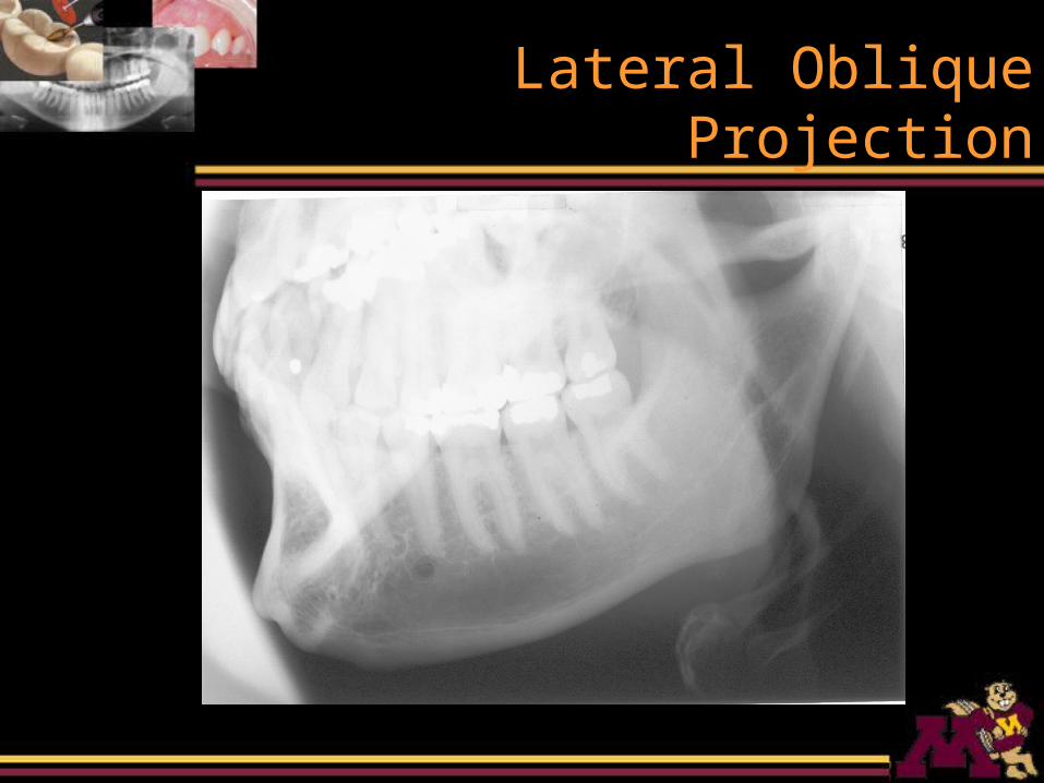
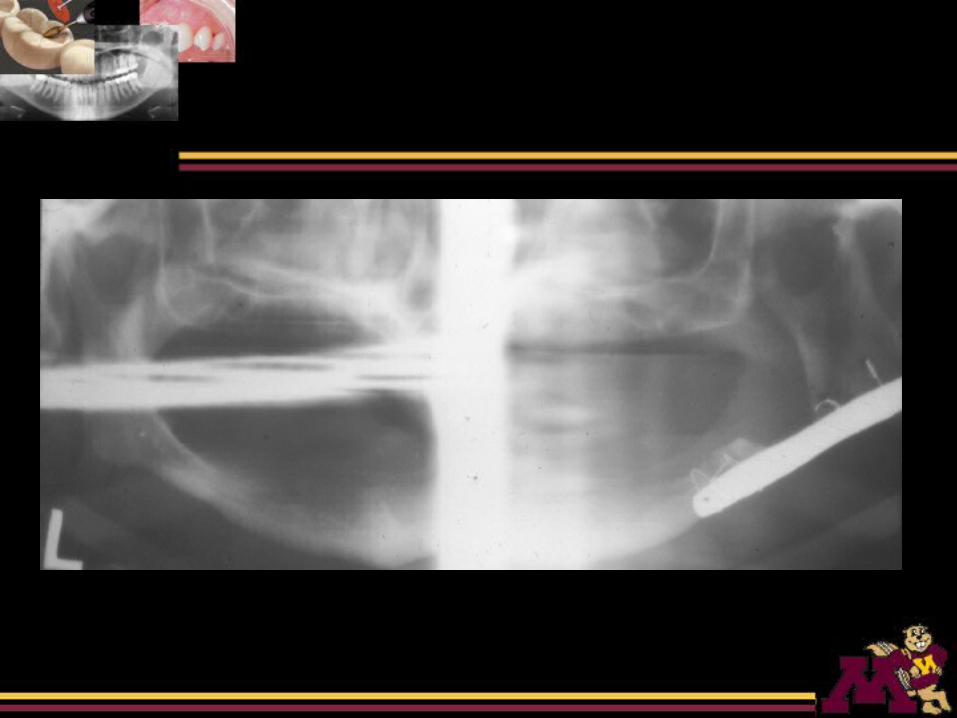
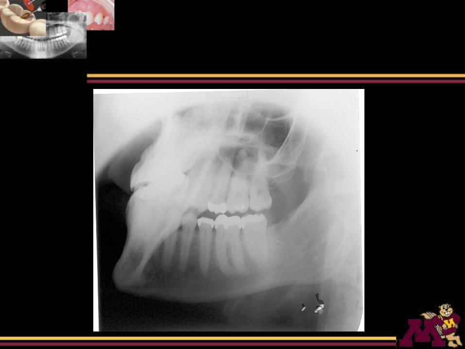
Lateral Oblique Projection
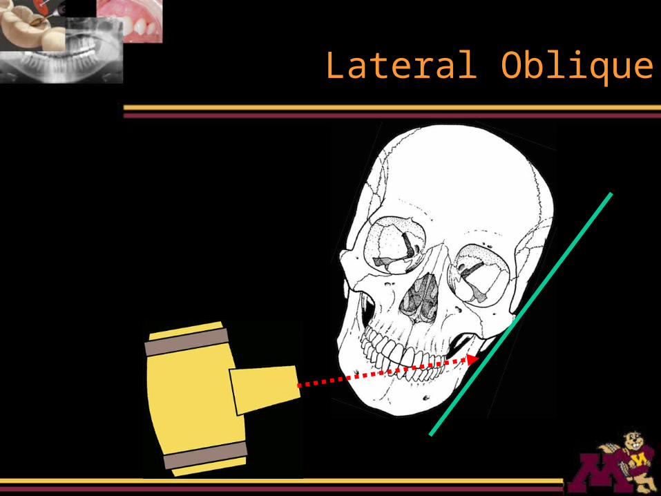
Lateral Oblique
Lateral Oblique Projection
• The film is placed against the side of the patient’s face
• X-ray beam is directed postero-anteriorly from the opposite side
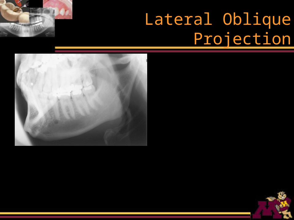
Lateral Oblique Projection
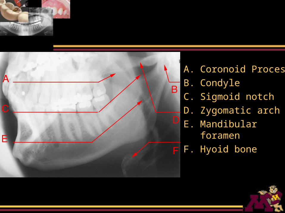
A. Coronoid Process
B. Condyle
C. Sigmoid notch
D. Zygomatic arch
E. Mandibular foramen
F. Hyoid bone
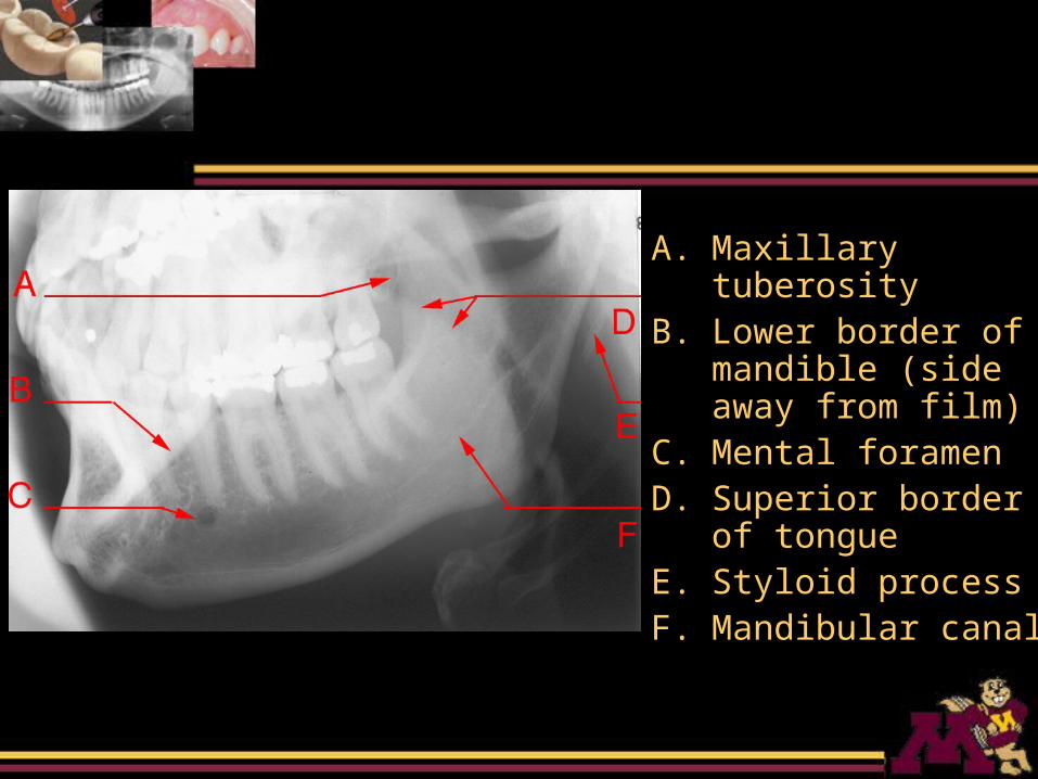
A. Maxillary tuberosityB. Lower border of
mandible (side away from film)
C. Mental foramenD. Superior border of
tongueE. Styloid processF. Mandibular canal
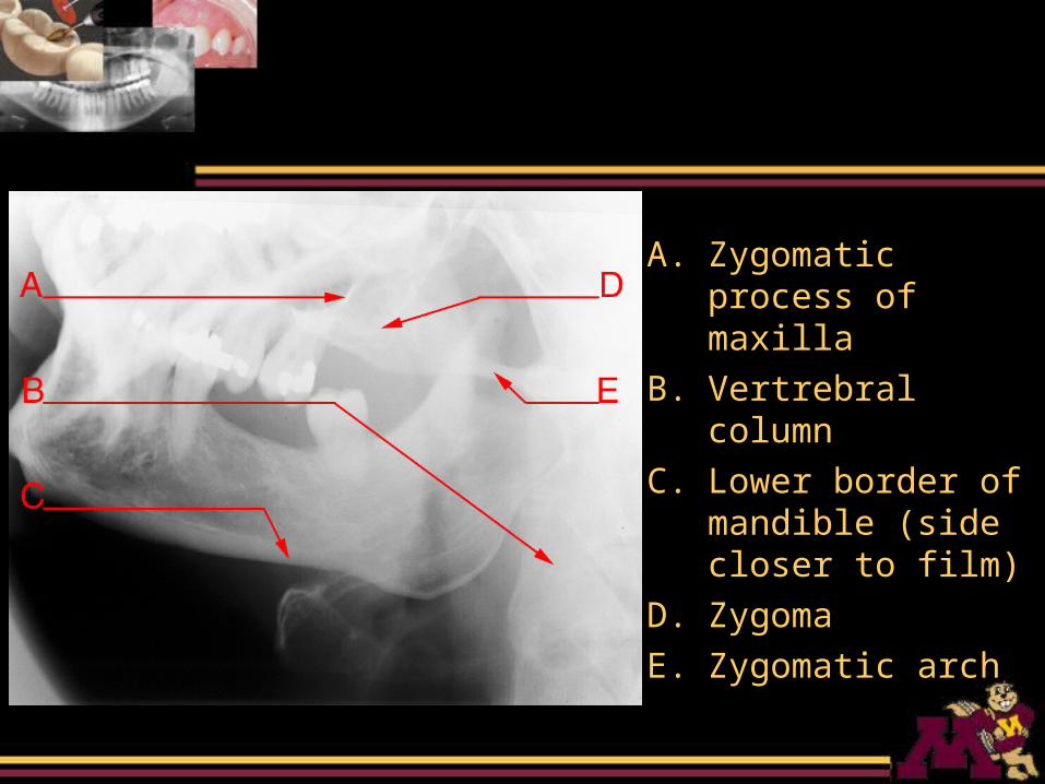
A. Zygomatic process of maxilla
B. Vertrebral column
C. Lower border of mandible (side closer to film)
D. Zygoma
E. Zygomatic arch
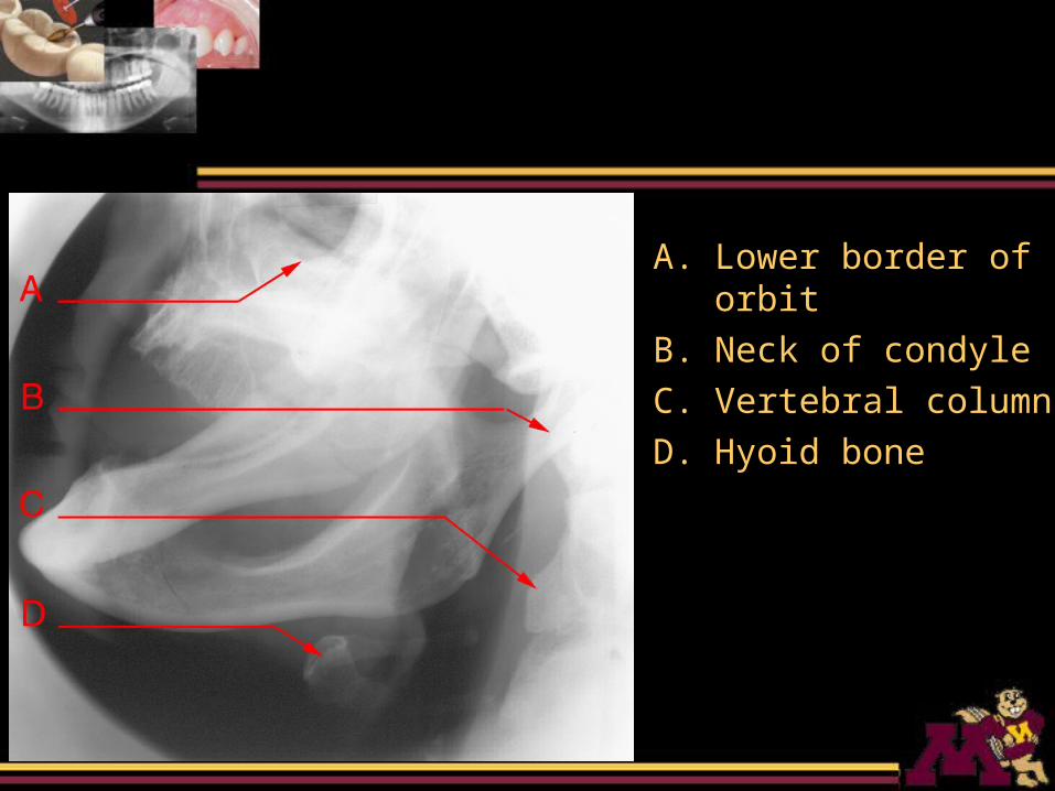
A. Lower border of orbit
B. Neck of condyle
C. Vertebral column
D. Hyoid bone
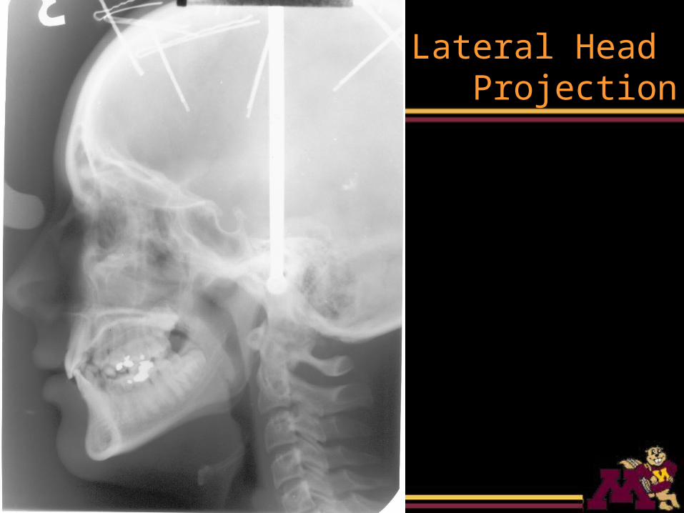
Lateral Head Projection
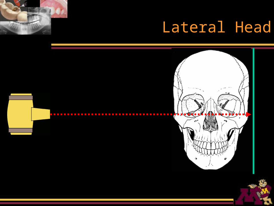
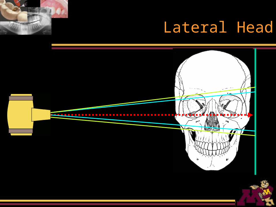
Lateral Head
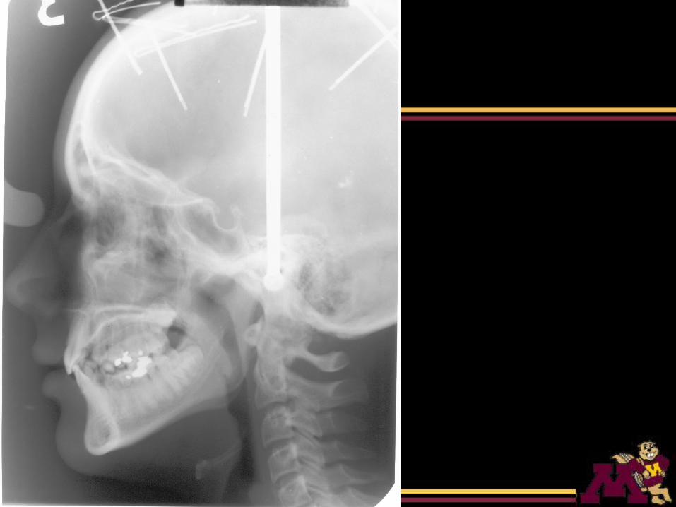
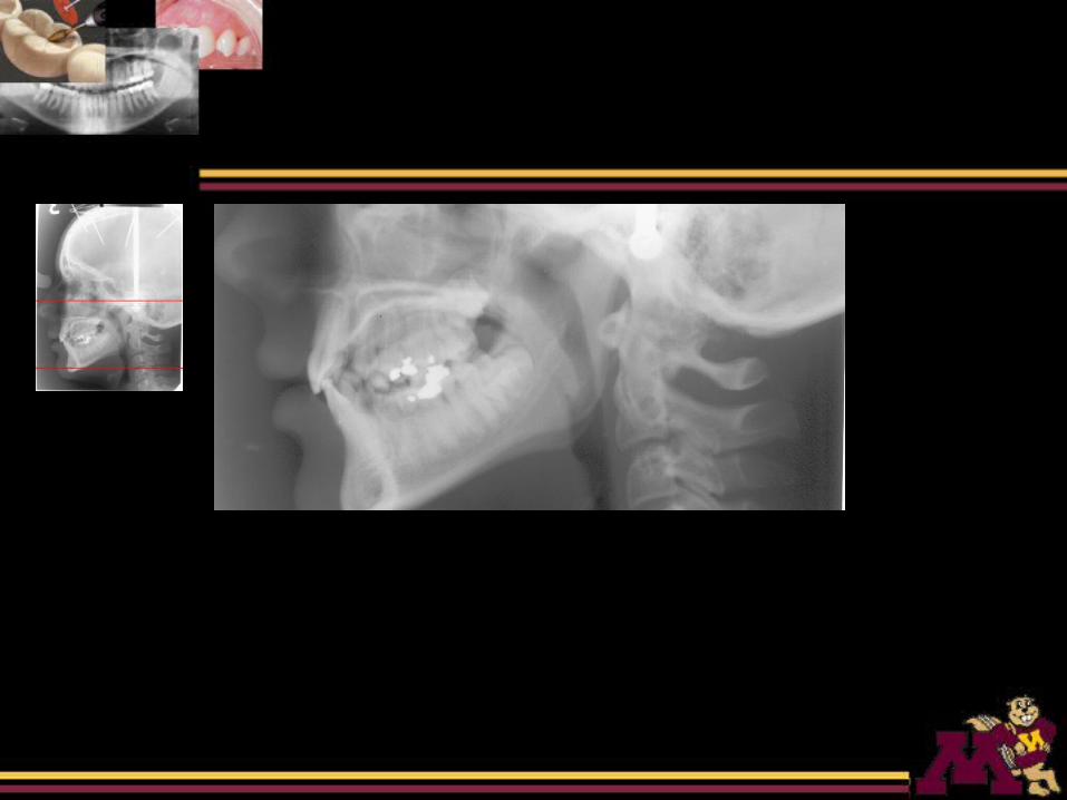
Lateral Skull
• Midsagittal plane of the patient’s head is parallel to the film
• X-ray beam is perpendicular to the film
Lateral Head
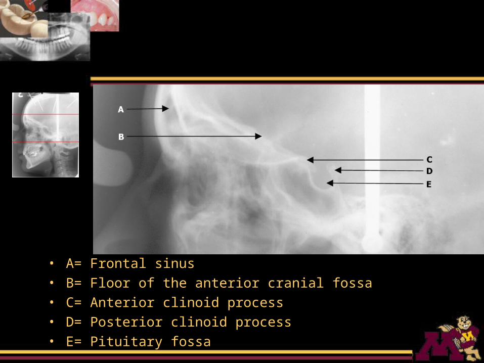
• A= Frontal sinus• B= Floor of the anterior cranial fossa• C= Anterior clinoid process• D= Posterior clinoid process• E= Pituitary fossa
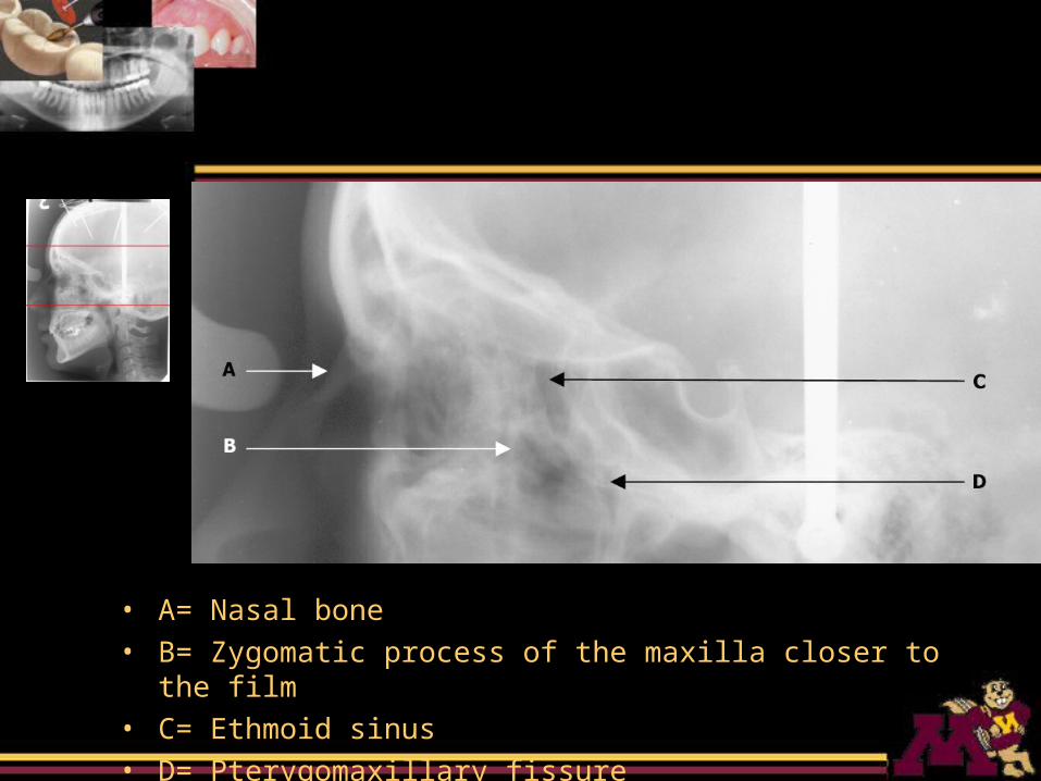
• A= Nasal bone• B= Zygomatic process of the maxilla closer to the film• C= Ethmoid sinus• D= Pterygomaxillary fissure
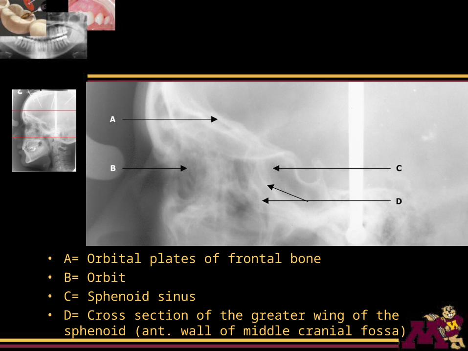
• A= Orbital plates of frontal bone• B= Orbit• C= Sphenoid sinus• D= Cross section of the greater wing of the sphenoid (ant. wall
of middle cranial fossa)
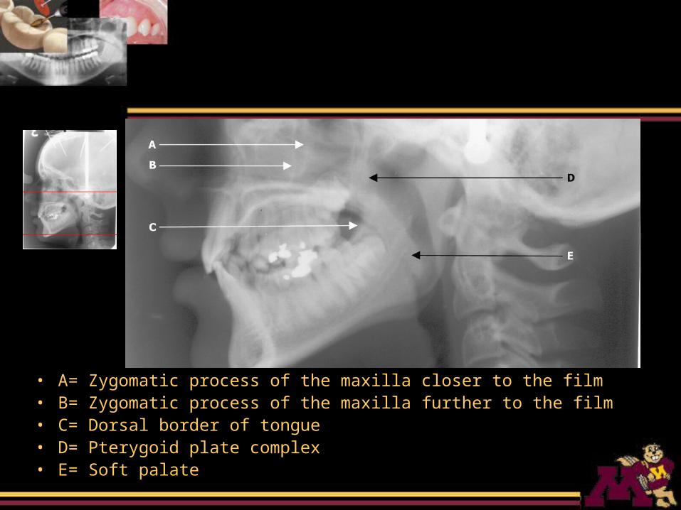
• A= Zygomatic process of the maxilla closer to the film• B= Zygomatic process of the maxilla further to the film • C= Dorsal border of tongue• D= Pterygoid plate complex• E= Soft palate
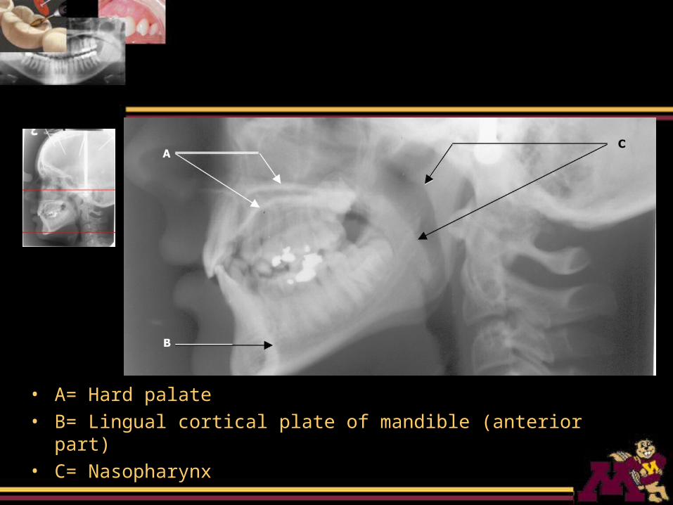
• A= Hard palate• B= Lingual cortical plate of mandible (anterior part)• C= Nasopharynx
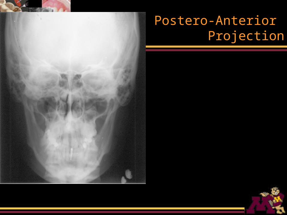
Postero-Anterior Projection
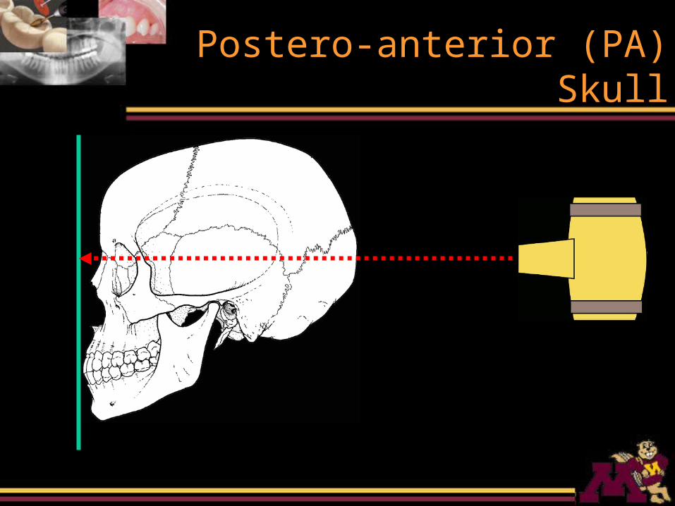
Postero-anterior (PA) Skull
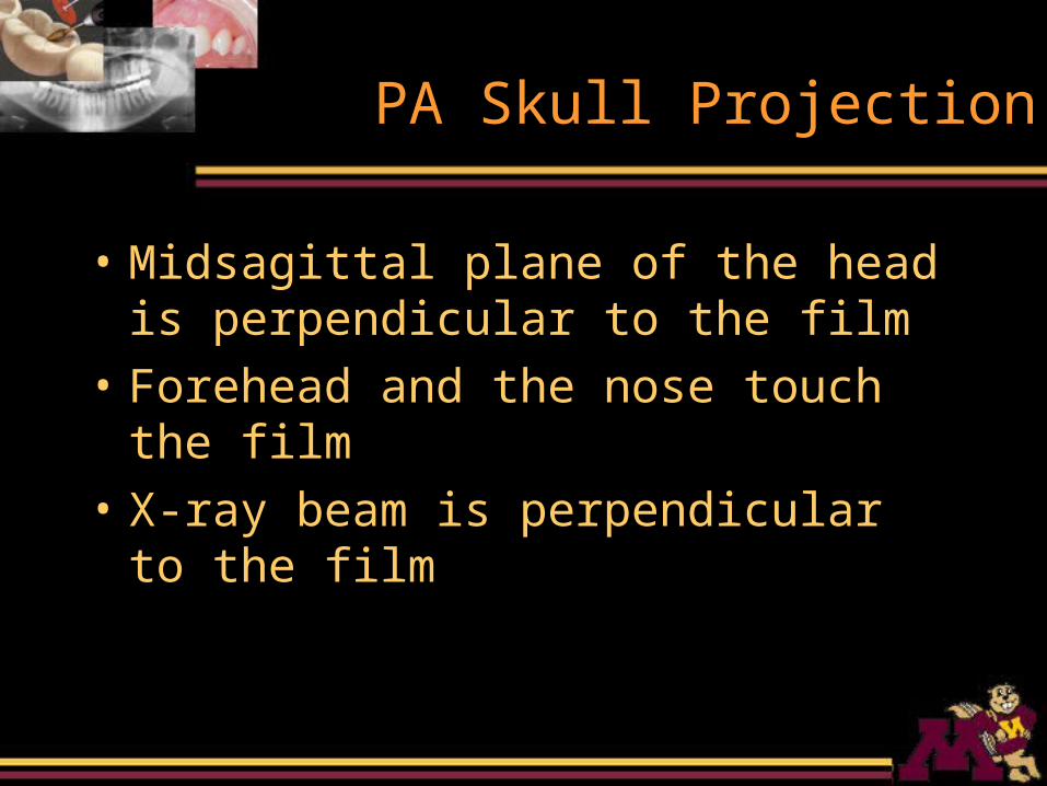
PA Skull Projection
• Midsagittal plane of the head is perpendicular to the film
• Forehead and the nose touch the film
• X-ray beam is perpendicular to the film
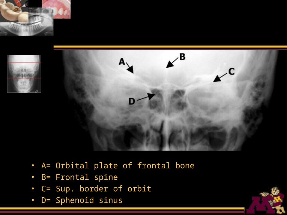
• A= Orbital plate of frontal bone• B= Frontal spine• C= Sup. border of orbit• D= Sphenoid sinus
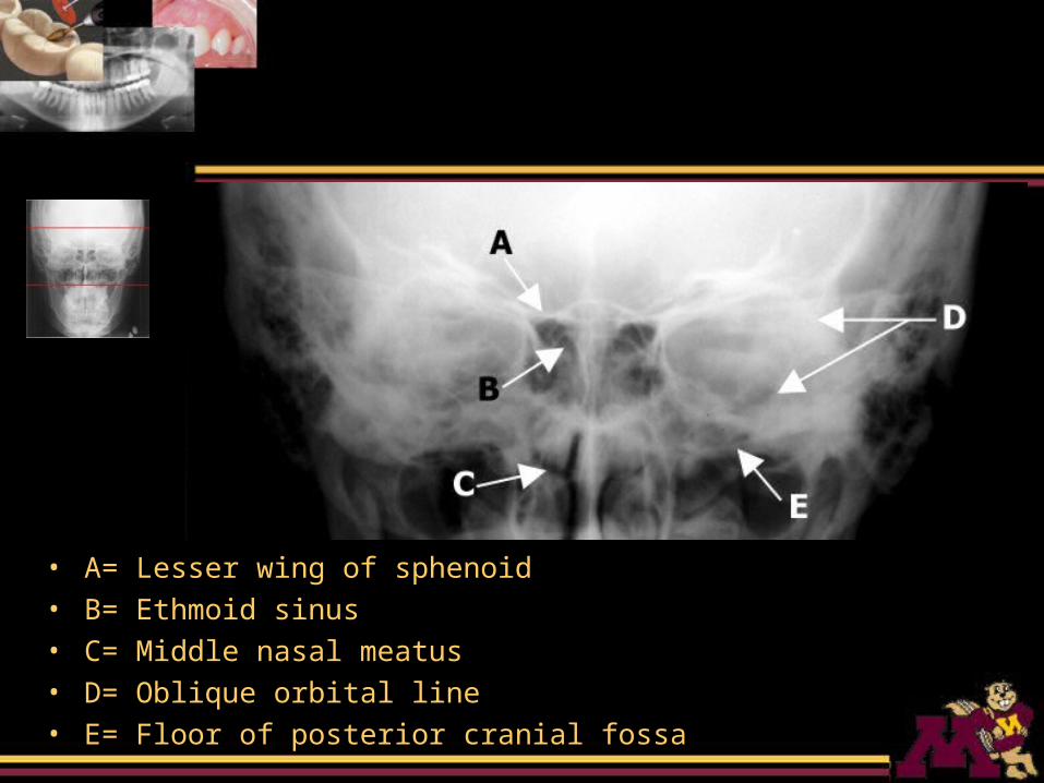
• A= Lesser wing of sphenoid• B= Ethmoid sinus• C= Middle nasal meatus• D= Oblique orbital line• E= Floor of posterior cranial fossa
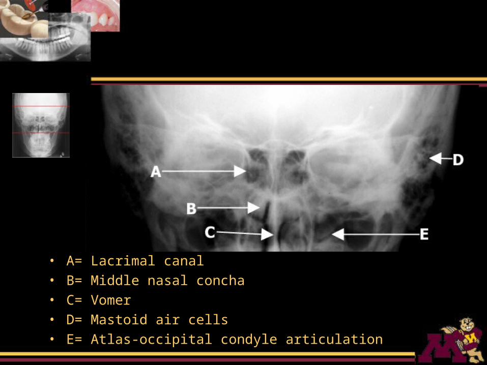
• A= Lacrimal canal• B= Middle nasal concha• C= Vomer• D= Mastoid air cells• E= Atlas-occipital condyle articulation
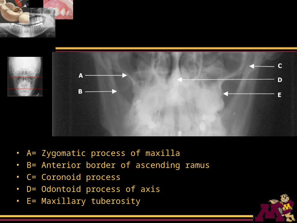
• A= Zygomatic process of maxilla• B= Anterior border of ascending ramus• C= Coronoid process• D= Odontoid process of axis• E= Maxillary tuberosity
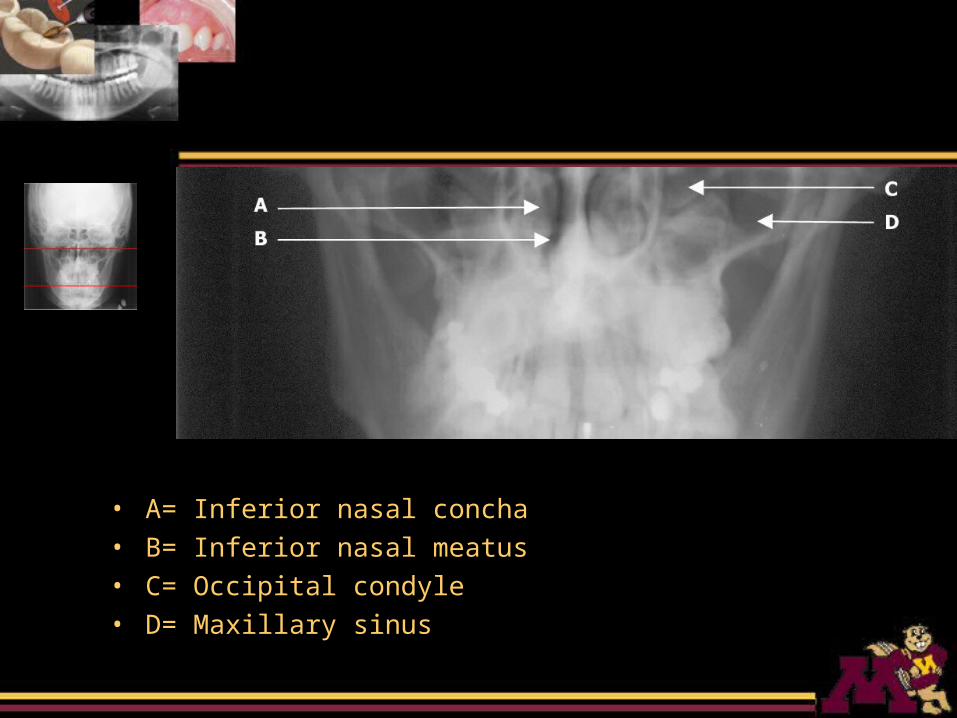
• A= Inferior nasal concha• B= Inferior nasal meatus• C= Occipital condyle• D= Maxillary sinus
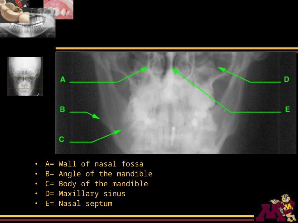
• A= Wall of nasal fossa• B= Angle of the mandible• C= Body of the mandible• D= Maxillary sinus• E= Nasal septum
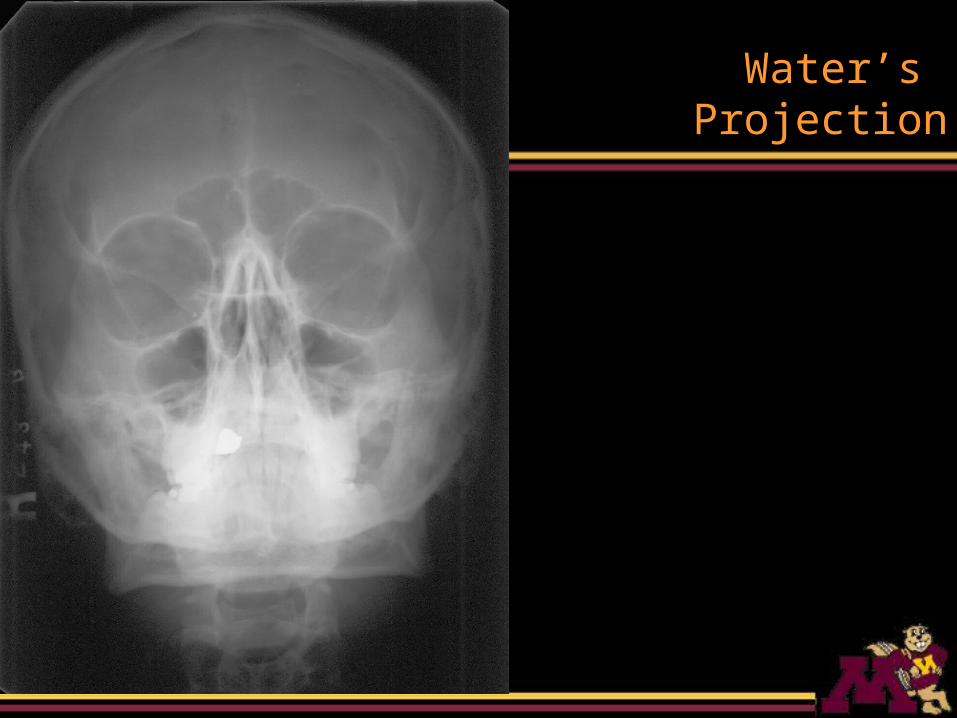
Water’s Projection
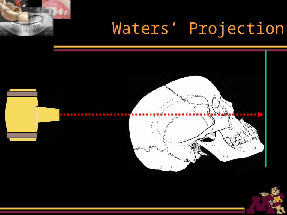
Waters’ Projection
Waters’ Projection
• Midsagittal plane of the head is perpendicular to the film
• chin touches the film• X-ray beam is perpendicular to the
film
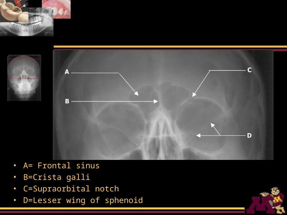
• A= Frontal sinus• B=Crista galli• C=Supraorbital notch• D=Lesser wing of sphenoid
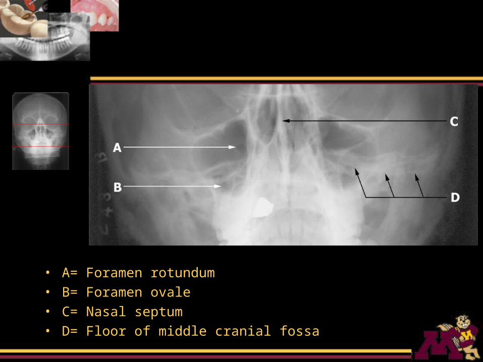
• A= Foramen rotundum• B= Foramen ovale• C= Nasal septum• D= Floor of middle cranial fossa
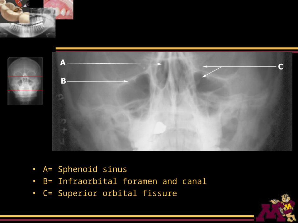
• A= Sphenoid sinus• B= Infraorbital foramen and canal• C= Superior orbital fissure
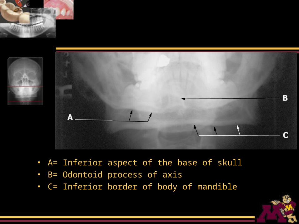
• A= Inferior aspect of the base of skull• B= Odontoid process of axis• C= Inferior border of body of mandible
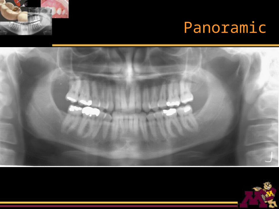
Panoramic
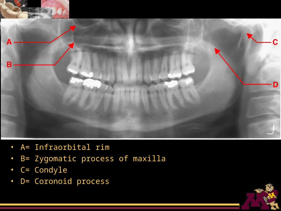
• A= Infraorbital rim• B= Zygomatic process of maxilla• C= Condyle• D= Coronoid process
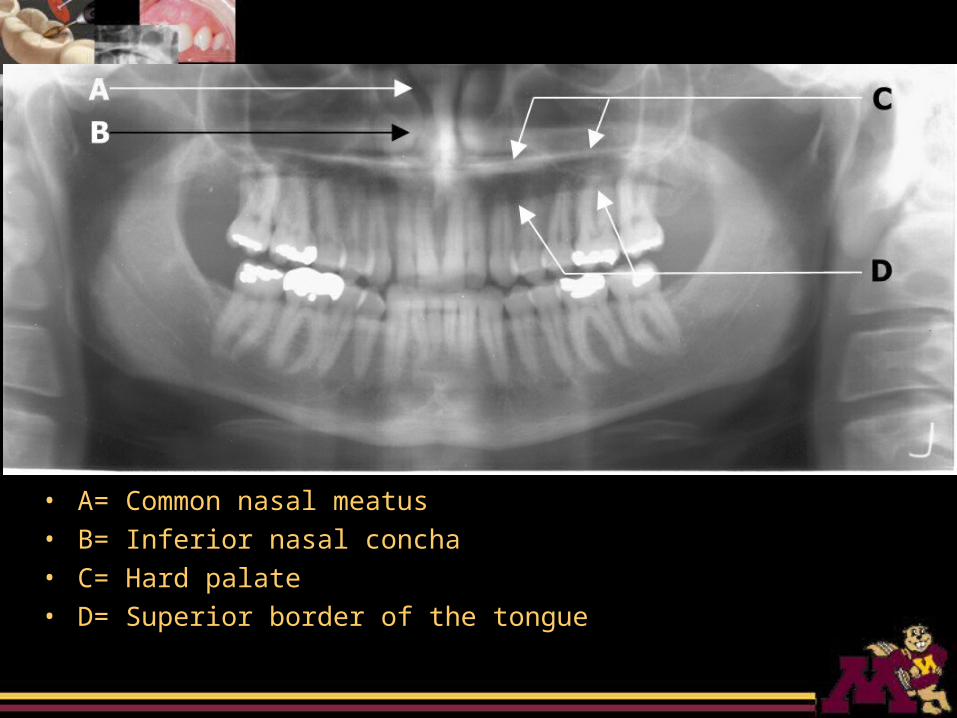
• A= Common nasal meatus• B= Inferior nasal concha• C= Hard palate• D= Superior border of the tongue
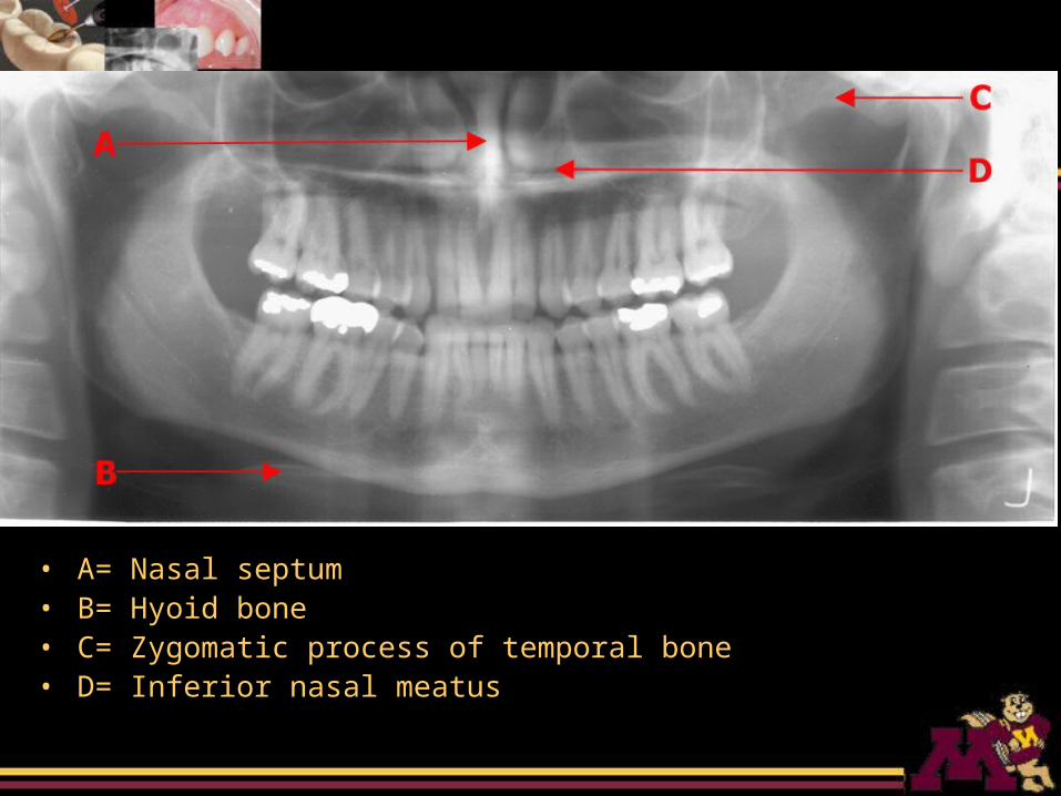
• A= Nasal septum• B= Hyoid bone• C= Zygomatic process of temporal bone• D= Inferior nasal meatus
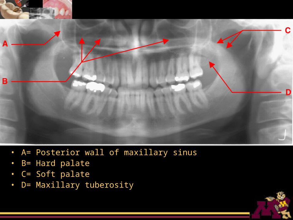
• A= Posterior wall of maxillary sinus• B= Hard palate• C= Soft palate• D= Maxillary tuberosity
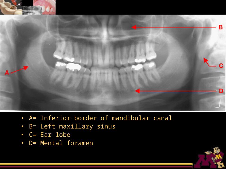
• A= Inferior border of mandibular canal• B= Left maxillary sinus• C= Ear lobe• D= Mental foramen
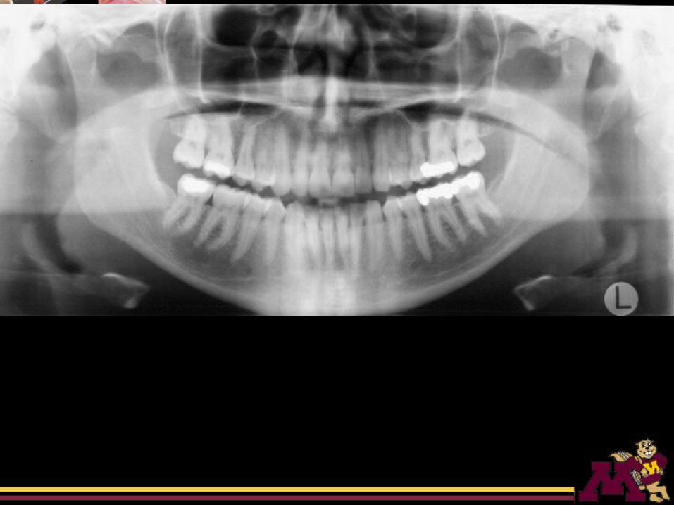
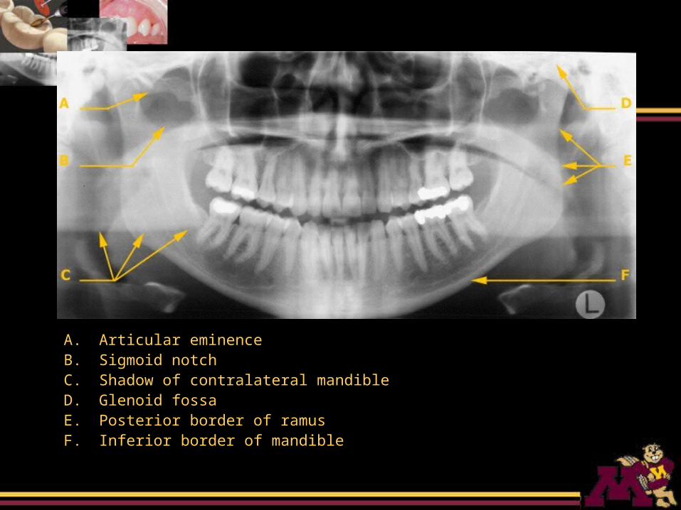
A. Articular eminenceB. Sigmoid notchC. Shadow of contralateral mandibleD. Glenoid fossaE. Posterior border of ramusF. Inferior border of mandible