Extramedullaryhaemopoietic tumours complicating...
6
J Clin Pathol 1988;41:609-614 Extramedullary haemopoietic tumours complicating polycythaemia vera P K MAcCALLUM,* M J NEWBOULD,t P S SAMBROOK,4 I E BURTON* From the Departments of *Haematology, tHistopathology, and tDiagnostic Radiology, Withington Hospital, Manchester suMMARY A 52 year old man with newly diagnosed polycythaemia vera in proliferative phase developed widespread extramedullary haemopoiesis (EMH), and died as a result of cervical cord compression. At necropsy, microscopic areas of primitive cells characteristic of granulocytic sarcoma were found within a large tumour of EMH in the right retroperitoneal region. Ultrastructural analysis showed unusual hexagonal inclusions within the cytoplasm of these primitive cells. Clinicians should be aware of the possibility of discrete haemopoietic tumours, whether EMH or granulocytic sarcoma, in patients with polycythaemia vera as well as in other myeloproliferative disorders. Polycythaemia vera is a myeloproliferative disorder of clonal origin arising at the level of the pluripotential haemopoietic stem cell' and resulting, in neoplastic proliferation of erythroid, myeloid, and mega- karyocytic elements in the bone marrow; an increased red blood cell mass; and, usually, raised blood counts of the three major haemopoietic cell lines. Extramedullary haemopoiesis (EMH) occurring in liver and spleen is a well recognised complication of myeloproliferative disorders, particularly myelo- fibrosis,2 but discrete tumours of EMH are rare and particularly so in the proliferative phase of polycythaemia vera.34 Polycythaemia vera commonly transforms from a proliferative into a spent or myelofibrotic stage,2 but acute myeloblastic leukaemia (AML) can also develop, although this is more common after treat- ment with alkylating agents or radioactive phosphorus (32p) than after venesection alone.5 Granulocytic sar- comas, which are tumours composed of immature cells of the myeloid series, are uncommon, but do arise in AML and myeloproliferative disorders, particularly in chronic granulocytic leukaemia (CGL).67 They are very rare in polycythaemia vera,7 but in this paper we describe a patient with proliferative phase disease in whom such a problem arose. Case report A 52 year old man with a five month history of lower Accepted for publication 9 February 1988 back pain, radiating to the right thigh, was admitted to hospital. For two months he had been aware of a tender swelling in the right loin, at the site of attachment of a right leg prosthesis, which he had worn since a traumatic avulsion of the leg 15 cm below the greater trochanter, in a road traffic accident eight years previously. Examination showed that he had no hepatosplenomegaly, but there was a diffuse tender swelling affecting the right loin. Full blood count was as follows: haemoglobin 201 g/l; packed cell volume 0 65; mean cell volume 73 fl; white cell count 17 5 x I09/l (neutrophils 16 45 x I09/ 1,lymphocytesO88 x 109/l,monocytesO18 x 109/1); and a platelet count of 185 x 109/l. There were no circulating primitive cells. His red cell volume, measured isotopically, was 513 ml/kg (normal 30 (2 SD 5) ml/kg) and plasma volume 54-6 ml/kg (normal 45 (5) ml/kg). His bone marrow was severely hyper- cellular with an increase in erythroid, myeloid, and megakaryocytic activity; iron stores were virtually absent. A trephine biopsy specimen showed pan- myelosis without a large increase in reticulin. Serum vitamin B 12 concentration was greater than 2000 ng/l (normal 150-1000 ng/l) and Bi 2 binding capacity was 3240 ng/l (normal 600-2200 ng/l). On the basis of these results, proliferative phase polycythaemia vera was diagnosed. A computed tomography scan of the abdomen showed a swelling of the right iliopsoas, posterior spinal, and gluteus medius muscles which had the attenuation of normal muscle (fig 1). This was initially 609 on 18 July 2018 by guest. Protected by copyright. http://jcp.bmj.com/ J Clin Pathol: first published as 10.1136/jcp.41.6.609 on 1 June 1988. Downloaded from
Transcript of Extramedullaryhaemopoietic tumours complicating...

J Clin Pathol 1988;41:609-614
Extramedullary haemopoietic tumours complicatingpolycythaemia vera
P K MAcCALLUM,* M J NEWBOULD,t P S SAMBROOK,4 I E BURTON*
From the Departments of*Haematology, tHistopathology, and tDiagnostic Radiology, Withington Hospital,Manchester
suMMARY A 52 year old man with newly diagnosed polycythaemia vera in proliferative phasedeveloped widespread extramedullary haemopoiesis (EMH), and died as a result of cervical cordcompression. At necropsy, microscopic areas ofprimitive cells characteristic ofgranulocytic sarcomawere found within a large tumour ofEMH in the right retroperitoneal region. Ultrastructural analysisshowed unusual hexagonal inclusions within the cytoplasm of these primitive cells.
Clinicians should be aware of the possibility of discrete haemopoietic tumours, whether EMH orgranulocytic sarcoma, in patients with polycythaemia vera as well as in other myeloproliferativedisorders.
Polycythaemia vera is a myeloproliferative disorder ofclonal origin arising at the level of the pluripotentialhaemopoietic stem cell' and resulting, in neoplasticproliferation of erythroid, myeloid, and mega-karyocytic elements in the bone marrow; an increasedred blood cell mass; and, usually, raised blood countsof the three major haemopoietic cell lines.
Extramedullary haemopoiesis (EMH) occurring inliver and spleen is a well recognised complication ofmyeloproliferative disorders, particularly myelo-fibrosis,2 but discrete tumours of EMH are rare andparticularly so in the proliferative phase ofpolycythaemia vera.34
Polycythaemia vera commonly transforms from aproliferative into a spent or myelofibrotic stage,2 butacute myeloblastic leukaemia (AML) can alsodevelop, although this is more common after treat-ment with alkylating agents or radioactive phosphorus(32p) than after venesection alone.5 Granulocytic sar-comas, which are tumours composed ofimmature cellsof the myeloid series, are uncommon, but do arise inAML and myeloproliferative disorders, particularly inchronic granulocytic leukaemia (CGL).67 They arevery rare in polycythaemia vera,7 but in this paper wedescribe a patient with proliferative phase disease inwhom such a problem arose.
Case report
A 52 year old man with a five month history of lowerAccepted for publication 9 February 1988
back pain, radiating to the right thigh, was admitted tohospital. For two months he had been aware of atender swelling in the right loin, at the site ofattachment of a right leg prosthesis, which he hadworn since a traumatic avulsion ofthe leg 15 cm belowthe greater trochanter, in a road traffic accident eightyears previously. Examination showed that he had nohepatosplenomegaly, but there was a diffuse tenderswelling affecting the right loin.
Full blood count was as follows: haemoglobin 201g/l; packed cell volume 0 65; mean cell volume 73 fl;white cell count 17 5 x I09/l (neutrophils 16 45 x I09/1,lymphocytesO88 x 109/l,monocytesO18 x 109/1);and a platelet count of 185 x 109/l. There were nocirculating primitive cells. His red cell volume,measured isotopically, was 513 ml/kg (normal 30 (2SD 5) ml/kg) and plasma volume 54-6 ml/kg (normal45 (5) ml/kg). His bone marrow was severely hyper-cellular with an increase in erythroid, myeloid, andmegakaryocytic activity; iron stores were virtuallyabsent. A trephine biopsy specimen showed pan-myelosis without a large increase in reticulin. Serumvitamin B12 concentration was greater than 2000 ng/l(normal 150-1000 ng/l) and Bi 2 binding capacity was3240 ng/l (normal 600-2200 ng/l). On the basis oftheseresults, proliferative phase polycythaemia vera wasdiagnosed.A computed tomography scan of the abdomen
showed a swelling of the right iliopsoas, posteriorspinal, and gluteus medius muscles which had theattenuation of normal muscle (fig 1). This was initially
609
on 18 July 2018 by guest. Protected by copyright.
http://jcp.bmj.com
/J C
lin Pathol: first published as 10.1136/jcp.41.6.609 on 1 June 1988. D
ownloaded from
MacCallum, Newbould, Sambrook, Burton
f-_ - - _ _ _ _ZA1 '-U}e S ' _ _ _ _ _ ______------------- __----- .---l :..!::
Fig 1 Computed tomography scan ofabdomen showing swelling ofright iliopsoas (IP), posterior spinal (PS), andgluteusmedius (GM) muscles.
presumed to be due to an organised haematoma. Thepatient was treated with venesection (3U) and 32P (300MBq). His haemoglobin concentration fell over twoweeks to 160 g/l, packed cell volume to 0-51, and therewas a slight reduction in the right loin swelling.
Despite this improvement, three weeks after admis-sion he complained of increasingly severe neck painand rapidly developed signs of a progressive quadra-plegia below the C6 level, with sensory loss below T3.After a myelogram a computed tomography scan ofthe cervical region showed a complete extraduralblock with no evidence ofbone destruction at the C4-5level.
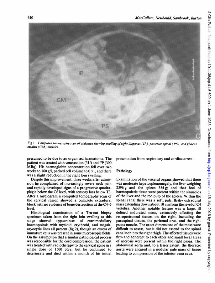
Histological examination of a Tru-cut biopsyspecimen taken from the right loin swelling at thisstage showed appearances of extramedullaryhaemopoiesis with myeloid, erythroid, and megak-aryocytic lines all present (fig 2), though an excess ofimmature cells was present in some microscopic fields.On the assumption that a similar pathological processwas responsible for the cord compression, the patientwas treated with radiotherapy to the cervical spine in asingle dose of 1500 cGy, but he continued todeteriorate and died within a month of his initial
presentation from respiratory and cardiac arrest.
Pathology
Examination of the visceral organs showed that therewas moderate hepatosplenomegaly, the liver weighing2596 g and the spleen 554 g; and that foci ofhaemopoietic tissue were present within the sinusoidsof the liver and the red pulp of the spleen. Within thespinal canal there was a soft, pale, fleshy extraduralmass extending down about 10 cm from the level ofC4vertebra. Another notable feature was a large, illdefined indurated mass, extensively affecting theretroperitoneal tissues on the right, including theparaspinal tissues, the perirenal area, and the rightpsoas muscle. The exact dimensions of the mass weredifficult to assess, but it did not extend to the spinalcanal nor into the right thigh. The affected tissues werefirm and adherent to each other and small focal areasof necrosis were present within the right psoas. Theabdominal aorta and, to a lesser extent, the thoracicaorta were encased in a nodular pale mass of tissueleading to compression of the inferior vena cava.
610
on 18 July 2018 by guest. Protected by copyright.
http://jcp.bmj.com
/J C
lin Pathol: first published as 10.1136/jcp.41.6.609 on 1 June 1988. D
ownloaded from
Extramedullary haemopoietic tumours complicating polycythaemia vera
*AM * ii*t *S;4.1 .~~~~~~~~~~~~~~~~~p
-sO,* ,* _vk" O , tAw %
* - wiS * f & t s LSll~~~~~~~~~~~~~~~W 40i2 T-tiss infiteo inla o g tm ua he p s. em o ia
4P~~~~~~~ V9
Fig Tu-utiosyspeimn frgh rtroertoealmas howngexramdulay hempoesz. Hamatxyinan
eosin.)
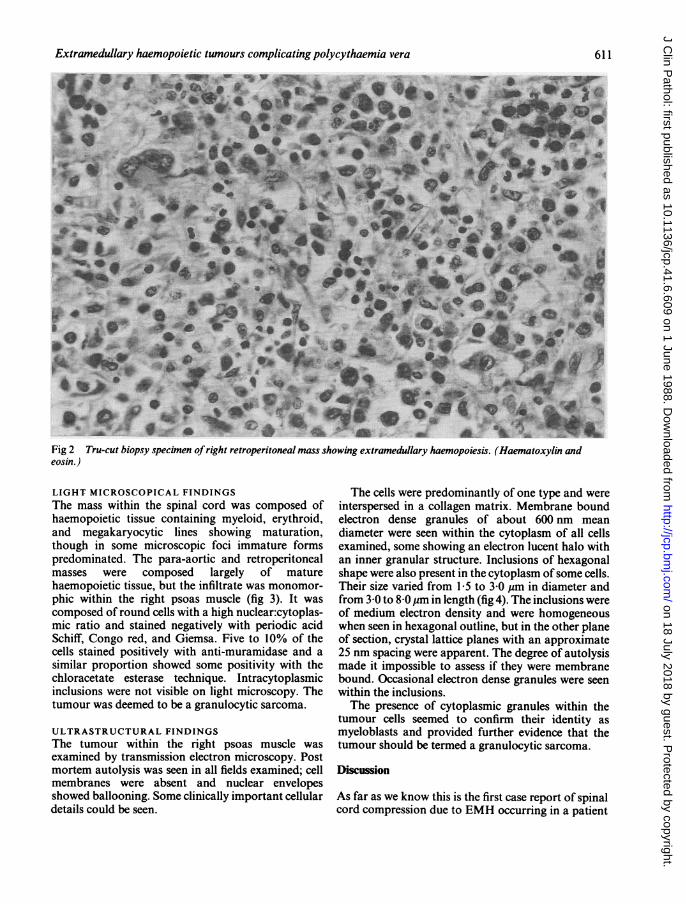
LIGHT MICROSCOPICAL FINDINGSThe mass within the spinal cord was composed ofhaemopoietic tissue containing myeloid, erythroid,and megakaryocytic lines showing maturation,though in some microscopic foci immature formspredominated. The para-aortic and retroperitonealmasses were composed largely of maturehaemopoietic tissue, but the infiltrate was monomor-phic within the right psoas muscle (fig 3). It wascomposed of round cells with a high nuclear:cytoplas-mic ratio and stained negatively with periodic acidSchiff, Congo red, and Giemsa. Five to 10% of thecells stained positively with anti-muramidase and asimilar proportion showed some positivity with thechloracetate esterase technique. Intracytoplasmicinclusions were not visible on light microscopy. Thetumour was deemed to be a granulocytic sarcoma.
ULTRASTRUCTURAL FINDINGSThe tumour within the right psoas muscle wasexamined by transmission electron microscopy. Postmortem autolysis was seen in all fields examined; cellmembranes were absent and nuclear envelopesshowed ballooning. Some clinically important cellulardetails could be seen.
The cells were predominantly of one type and wereinterspersed in a collagen matrix. Membrane boundelectron dense granules of about 600 nm meandiameter were seen within the cytoplasm of all cellsexamined, some showing an electron lucent halo withan inner granular structure. Inclusions of hexagonalshape were also present in the cytoplasm ofsome cells.Their size varied from 1[5 to 3-0 gm in diameter andfrom 3-0 to 8-0 gm in length (fig 4). The inclusions wereof medium electron density and were homogeneouswhen seen in hexagonal outline, but in the other planeof section, crystal lattice planes with an approximate25 nm spacing were apparent. The degree of autolysismade it impossible to assess if they were membranebound. Occasional electron dense granules were seenwithin the inclusions.The presence of cytoplasmic granules within the
tumour cells seemed to confirm their identity asmyeloblasts and provided further evidence that thetumour should be termed a granulocytic sarcoma.
Discussion
As far as we know this is the first case report of spinalcord compression due to EMH occurring in a patient
611
on 18 July 2018 by guest. Protected by copyright.
http://jcp.bmj.com
/J C
lin Pathol: first published as 10.1136/jcp.41.6.609 on 1 June 1988. D
ownloaded from

MacCallum, Newbould, Sambrook, Burton
Fig 3 Mass in right psoas muscle showing granulocytic sarcoma. (Haematoxylin and eosin.)
with polycythaemia vera without accompanyingmyelofibrosis. It is the fourth case in which gran-ulocytic sarcoma developed in polycythaemia vera,but recognition of leukaemic transformation withinEMH in this disease is new, as is the description ofhexagonal crystals within the cytoplasm ofthe tumourcells.Although EMH has been described in various
anatomical sites in other conditions,2 it is not usuallyextensive in uncomplicated polycythaemia vera,though it can be found in splenic biopsy specimenstaken early in the course of the disease.2 Pettit et alsuggest that no, or only minimal, extramedullaryerythropoiesis occurs in uncomplicated polycyth-aemia vera and that its demonstration in such a patientis an indication that the condition is undergoingtransformation to myelofibrosis or to "transitionalmyeloproliferative disorder".8 Our case lackedfeatures suggestive of transformation, such as splen-omegaly, a leucoerythroblastic blood picture, orincreased reticulin in the bone marrow trephine biopsyspecimen. The polycythaemia vera seemed to be inproliferative phase and yet the EMH found atnecropsy was extensive.
There have been 32 reported cases of spinal cordcompression due to EMH."'3 Most of these casesoccurred in hereditary and refractory anaemias, par-ticularly thalassaemia major and intermedia (12cases), and in myeloproliferative disorders, most com-monly myelofibrosis, both primary and post-polycyth-aemic. There is only one previous case report of spinalcord compression due to EMH complicatingpolycythaemia vera,3 and this occurred in a patientwith myelofibrosis which preceded the onset ofpolycythaemia. Intracranial meningeal disease withEMH has also been described in association withmyeloproliferative disorders-predominantly myelo-fibrosis'"'8-but again there has been only one reportin association with polycythaemia vera.4Up to 15% of cases of polycythaemia vera may
terminate in AML, and this is more common inpatients treated with alkylating agents or 32P than withvenesection alone.5 In only three previous cases hasgranulocytic sarcoma been described in patients withpolycythaemia vera.7 As in patients with othermyeloproliferative disorders, particularly CGL, inwhom granulocytic sarcoma develops, the prognosis ispoor6' because of subsequent acute leukaemia. Of the
612
on 18 July 2018 by guest. Protected by copyright.
http://jcp.bmj.com
/J C
lin Pathol: first published as 10.1136/jcp.41.6.609 on 1 June 1988. D
ownloaded from

Extramedullary haemopoietic tumours complicating polycythaemia vera 613
rI
*~i~ ' __..j B, -
Fig 4 Electron micrograph oftumour cell with crystal-like inclusions seen in longitudinal section (arrowheads). Notestructures present within largest crystal matrix.
three cases associated with polycythaemia vera, twodeveloped AML within a month and the thirddeveloped a poorly defined myeloproliferative disor-der one year later.7The development of granulocytic sarcoma within
tumours of EMH is unusual, although there arereports of blast cell infiltration complicating EMH inprimary myelofibrosis.'920 Cytogenetic evidence alsosuggests that blast transformation in chronic gran-ulocytic leukaemia can originate in the spleen alreadyaffected by EMH.``The histological diagnosis of granulocytic sarcoma
can be difficult. When the tumour occurs in theabsence of other evidence of haematological disease itis often misdiagnosed as a high grade lymphoma.72`27Cytochemistry is less helpful in undifferentiatedtumours which are less likely to show extensivechloracetate esterase positivity.27 Ultrastructuralexamination can assist in making the diagnosis, andmyeloblasts show a number of characteristic features.Primary granules of varying appearance are the mostcharacteristic.24 28 Auer rods are found withinmyeloblasts in about 10-20% ofcases. They are needleshaped, peroxidase positive, and have a characteristic
periodicity.28 Hexagonal and other geometricinclusions similar to those noted in this case have beenreported in myeloblasts and macrophages of patientswith myeloid leukaemia and preleukaemic states,2931but this seems to be the first report of such structureswith crystal lattice planes within the tumour cells of agranulocytic sarcoma.
In conclusion, this case shows that discretehaemopoietic tumours, both of EMH and of gran-ulocytic sarcoma, may complicate proliferative phasepolycythaemia vera as well as other myeloproliferativedisorders, and should be considered particularly whenthere are neurological complications.
We thank Professor J C McClure, Dr S S Banerjee,and DrA Curry for helpful advice, Dr P Merry and theArtificial Limb and Appliance Centre, WithingtonHospital for referring the patient and Dr A Jacksonfor performing the Tru-cut biopsy.
Referes
I Adamson JW, Fialkow PJ, Murphy S, Prchal JF, Steinmann L.Polycythemia vera: stem cell and probable clonal origin of thedisease. N Engi J Med 1976;295:913-6.
on 18 July 2018 by guest. Protected by copyright.
http://jcp.bmj.com
/J C
lin Pathol: first published as 10.1136/jcp.41.6.609 on 1 June 1988. D
ownloaded from

614 MacCallum, Newbould, Sambrook, Burton2 Ward HP, Block MH. The natural history of agnogenic mycloid
metaplasia (AMM) and a critical evaluation of its relationshipwith the mycloproliferative syndrome. Medicine 1971;SO:357-420.
3 Rice GPA, Assis LJP, Barr RM, Ebers GC. Extramedullaryhematopoiesis and spinal cord compression complicatingpolycythemia rubra vera. Ann Neurol 1980;7:81-4.
4 Robitaille GA, Eisenberg M, Lehman R. Intracranialextramedullary hematopoiesis in polycythemia vera. Conn Med1985;49:149-51.
5 Berk PD, Goldberg JD, Donovan PB, Fruchtman SM, Berlin NI,Wasserman LR. Therapeutic recommendations in polycyth-emia vera based on Polycythemia Vera Study Group protocols.Semin Hematol 1986;23:132-43.
6 Muss HB, Moloney WC. Chloroma and other myeloblastictumours. Blood 1973;42:721-8.
7 Neiman RS, Barcos M, Berard C, Bonner H, Mann R, Rydell RE,Bennett JM. Granulocytic sarcoma: a clinicopathologic studyof 61 biopsied cases. Cancer 1981;48:1426-37.
8 Pettit JE, Lewis SM, Nicholas AW. Transitionalmyeloproliferative disorder. Br J Haematol 1979;43:167-84.
9 Heffez DS, Sawaya R, Udvarhelyi GB, Mann R. Spinal epiduralextramedullary hematopoiesis with cord compression in apatient with refractory sideroblastic anemia. J Neurosurg1982;57:399-406.
10 Lewkow LM, Shah I. Sickle cell anemia and epiduralextramedullary hematopoiesis. Am J Med 1984;76:748-51.
11 Cromwell LD, Kerber C. Spinal cord compression byextramedullary hematopoiesis in agnogenic myeloid meta-plasia. Radiology 1978;128:118.
12 Crawford DC, Nightingale S, Bates D, Tomlinson BE. Spinal cordcompression by extramedullary haematopoiesis inmyelofibrosis. Postgrad MedJ 1984;60:62-3.
13 Price F, Bell H. Spinal cord compression due to extramedullaryhematopoiesis. Successful treatment in a patient with long-standing myelofibrosis. JAMA 1985;253:2876-7.
14 Ligumski M, Polliack A, Benbassat J. Myeloid metaplasia of thecentral nervous system in patients with myelofibrosis andagnogenic myeloid metaplasia. Report of 3 cases and review ofthe literature. Am J Med Sci 1978;275:99-103.
15 Cameron WR, Ronnert M, Brun A. Extramedullary hem-matopoiesis ofCNS in postpolycythemic myeloid metaplasia. NEnglJ Med 1981;305:765.
16 Lundh B, Brandt L, Cronqvist S, Eyrich R. Intracranial myeloidmetaplasia in myelofibrosis. Scand J Haematol 1982;28:91-4.
17 Cornfield DB, Shipkin P, Alavi A, Becker J, Peyster R. Intracran-ial myeloid metaplasia: diagnosis by CT and Fe52 scans and
treatment by cranial irradiation. Am J Hematol 1983;15:273-8.18 Brown JA, Gomez-Leon G. Subdural hemorrhage secondary to
extramedullary hematopoiesis in postpolycythemic mycloidmetaplasia. Neurosurgery 1984;14:588-91.
19 Clausen KP. Reticulosarcomatosis in primary myelofibrosis.Cancer 1968;22:136-41.
20 Page BM, Watt JL, Reid IN, Davidson RJL, Walker W. Clonalevolution of marker chromosomes in a case of myelofibrosiswith myeloid metaplasia and myeloblastic transformation. ActaHaematol 1979;61:301-9.
21 Mitelman F, Brandt L, Nilsson PG. Cytogenetic evidence forsplenic origin of blastic transformation in chronic myeloidleukaemia. ScandJ Haematol 1974;13:87-92.
22 Gomez G, Hossfield DK, Sokal JE. Removal ofabnormal clone ofleukaemic cells by splenectomy. Br Med J 1975;2:421-3.
23 Anonymous. Chloroma confusion. [Editorial]. Lancet1973;i:1099-100.
24 McCarty KS Jr, Wortman J, Daly J, Rundles RW, Hanker JS.Chloroma (granulocytic sarcoma) without evidence of leuk-aemia: facilitated light microscopic diagnosis. Blood1980;56:104-8.
25 Ersb0ll J, Petri J, Jenson KH, Hansen MM. Granulocytic sarcomapreceding acute myeloid leukaemia. Scand J Haematol1980;24:435-45.
26 Eshghabadi M, Shojania AM, Carr I. Isolated granulocyticsarcoma. Report of a case and review of the literature. J ClinOncol 1986;4:912-7.
27 Meis JM, Butler JJ, Osborne BM, Manning JT. Granulocyticsarcoma in nonleukemic patients. Cancer 1986;58:2697-709.
28 Cawley JC, Hayhoe FGJ. Ultrastructure ofhaemic cells. London:WB Saunders, 1973.
29 Stavem P, Ly B, Bjorneklett A. Light green crystals in May-Gruinwald and Giemsa-stained bone marrow macrophages inpatients with myeloid leukaemia. Scand J Haematol 1977;18:67-72.
30 WolfDJ, Fialk MA, Mouradian J, Gottfried EL, Pasmantier MW.Unusual intracytoplasmic inclusions in acute myeloblasticleukemia. Am J Hematol 1980;9:413-20.
31 Nesland JM, Langholm R, Marton PF. Hexagonal crystals in thebone marrow in patients with myeloproliferative disease andpreleukaemia. Scand J Haematol 1984;32:552-8.
Requests for reprints to: Dr P K MacCallum, Department ofHaematology, Withington Hospital, West Didsbury, Man-chester M20 8LR, England.
on 18 July 2018 by guest. Protected by copyright.
http://jcp.bmj.com
/J C
lin Pathol: first published as 10.1136/jcp.41.6.609 on 1 June 1988. D
ownloaded from