Extensor Digitorum Brevis Free Flap: Anatomic Study and...
10
Extensor Digitorum Brevis Free Flap: Anatomic Study and Further Clinical Applications Francisco del Pin ˜al, M.D., Ph.D., and Francisco Herrero, M.D. Santander, Spain The extensor digitorum brevis muscle flap is reliable, safe, and can be used either as a pedicle or as a free flap with minimal donor site morbidity. To increase the exist- ing knowledge of this flap and to establish further ana- tomic basis for the design and elevation of the extensor digitorum brevis flap, 26 specimens from 13 fresh cadavers were dissected under 3.53 loupes. The lateral tarsal artery was found to be the main blood supply to the muscle. It has an average diameter of 1.83 6 0.35 mm and a length of 1.89 6 0.69 cm. The dorsalis pedis artery has, at the level of the lateral tarsal artery takeoff, a diameter of 3.25 6 0.62 mm. From this point to the origin of the deep plantar branch, the dorsalis pedis artery has minimal branching, and the surgeon has available an artery homogeneous in diameter that is 6.77 6 0.99 cm in length. Related neu- rovascular structures (anterior tibial artery and the venae comitantes, dorsalis pedis and first dorsal metatarsal ar- tery, and deep peroneal nerve) were also studied. A safe and reliable harvesting technique and the “T interposed extensor digitorum brevis” technique for sparing the an- terior tibial artery are presented, as are clinical case ex- amples on the use of this flap as a flow-through, extensor digitorum brevis-vascularized nerve graft, a combined ex- tensor digitorum brevis-deep peroneal nerve graft, and a bilobed extensor digitorum brevis-dorsalis pedis fascio- subcutaneous free flap. (Plast. Reconstr. Surg. 105: 1347, 2000.) The extensor digitorum brevis muscle has been widely used as an island flap, with ante- grade flow to cover defects around the ankle 1–9 and reverse flow for defects on the distal foot, 9 –11 and as a free flap to cover small de- fects 3,7,12 or for functional restoration. 13–16 The gross anatomy of the muscle is well de- scribed in classic anatomy textbooks. 17,18 Landi et al. 1 studied the vascular anatomy of the ex- tensor digitorum brevis in seven cadavers. They found a primary artery (the lateral tarsal ar- tery) and an accessory artery located 1 to 3 cm distal to the main artery. Giordano et al. 4 stated, on the other hand, that “the lateral tarsal artery is the dominant arterial supply of the muscle”; in fact, they advised ligating the dorsalis pedis distal to this branch to preserve the skin blood supply of the dorsum of the foot. Recently, Baltensperger et al. 19 and Ba- khach et al. 11 studied the extensor digitorum brevis, focusing, respectively, on the feasibility of the flap for covering the ankle area and on its use as a distally based flap, even in cases when the dorsalis pedis is interrupted. The purpose of this article is to expand, on the basis of an anatomic study, the applications of the extensor digitorum brevis as a free flap. MATERIALS AND METHODS The anatomic study was performed on 26 feet from 13 fresh cadavers. At time of death, the subjects were 18 to 80 years of age and were apparently free of local or vascular disease. Table I contains the specimen data and mor- phometric findings regarding the muscle itself. Dissections were performed under clear light conditions and with the help of 3.53 loupes. The takeoff of the lateral tarsal vessels was identified and its relationship to the inferior edge of the retinaculum and the talocrural joint was recorded. The dorsalis pedis was tracked distally, as was the first dorsal metatar- sal. The muscle itself was then dissected en bloc from its bed, with care taken to preserve the vascular tree underneath. Finally, the ante- rior tibial neurovascular bundle was traced for From the Section of Hand and Plastic-Reconstructive Surgery, Hospital Mutua Montan ˜ esa, and Private Hand-Wrist and Plastic-Reconstructive Surgery, Santander, Spain. Received for publication February 16, 1999; revised August 6, 1999. 1347
Transcript of Extensor Digitorum Brevis Free Flap: Anatomic Study and...

Extensor Digitorum Brevis Free Flap:Anatomic Study and Further ClinicalApplicationsFrancisco del Pinal, M.D., Ph.D., and Francisco Herrero, M.D.Santander, Spain
The extensor digitorum brevis muscle flap is reliable,safe, and can be used either as a pedicle or as a free flapwith minimal donor site morbidity. To increase the exist-ing knowledge of this flap and to establish further ana-tomic basis for the design and elevation of the extensordigitorum brevis flap, 26 specimens from 13 fresh cadaverswere dissected under 3.53 loupes. The lateral tarsal arterywas found to be the main blood supply to the muscle. Ithas an average diameter of 1.83 6 0.35 mm and a lengthof 1.89 6 0.69 cm. The dorsalis pedis artery has, at the levelof the lateral tarsal artery takeoff, a diameter of 3.25 6 0.62mm. From this point to the origin of the deep plantarbranch, the dorsalis pedis artery has minimal branching,and the surgeon has available an artery homogeneous indiameter that is 6.77 6 0.99 cm in length. Related neu-rovascular structures (anterior tibial artery and the venaecomitantes, dorsalis pedis and first dorsal metatarsal ar-tery, and deep peroneal nerve) were also studied. A safeand reliable harvesting technique and the “T interposedextensor digitorum brevis” technique for sparing the an-terior tibial artery are presented, as are clinical case ex-amples on the use of this flap as a flow-through, extensordigitorum brevis-vascularized nerve graft, a combined ex-tensor digitorum brevis-deep peroneal nerve graft, and abilobed extensor digitorum brevis-dorsalis pedis fascio-subcutaneous free flap. (Plast. Reconstr. Surg. 105: 1347,2000.)
The extensor digitorum brevis muscle hasbeen widely used as an island flap, with ante-grade flow to cover defects around the ankle1–9
and reverse flow for defects on the distalfoot,9–11 and as a free flap to cover small de-fects3,7,12 or for functional restoration.13–16
The gross anatomy of the muscle is well de-scribed in classic anatomy textbooks.17,18 Landiet al.1 studied the vascular anatomy of the ex-tensor digitorum brevis in seven cadavers. Theyfound a primary artery (the lateral tarsal ar-
tery) and an accessory artery located 1 to 3 cmdistal to the main artery. Giordano et al.4
stated, on the other hand, that “the lateraltarsal artery is the dominant arterial supply ofthe muscle”; in fact, they advised ligating thedorsalis pedis distal to this branch to preservethe skin blood supply of the dorsum of thefoot. Recently, Baltensperger et al.19 and Ba-khach et al.11 studied the extensor digitorumbrevis, focusing, respectively, on the feasibilityof the flap for covering the ankle area and onits use as a distally based flap, even in caseswhen the dorsalis pedis is interrupted.
The purpose of this article is to expand, onthe basis of an anatomic study, the applicationsof the extensor digitorum brevis as a free flap.
MATERIALS AND METHODS
The anatomic study was performed on 26feet from 13 fresh cadavers. At time of death,the subjects were 18 to 80 years of age and wereapparently free of local or vascular disease.Table I contains the specimen data and mor-phometric findings regarding the muscle itself.Dissections were performed under clear lightconditions and with the help of 3.53 loupes.
The takeoff of the lateral tarsal vessels wasidentified and its relationship to the inferioredge of the retinaculum and the talocruraljoint was recorded. The dorsalis pedis wastracked distally, as was the first dorsal metatar-sal. The muscle itself was then dissected enbloc from its bed, with care taken to preservethe vascular tree underneath. Finally, the ante-rior tibial neurovascular bundle was traced for
From the Section of Hand and Plastic-Reconstructive Surgery, Hospital Mutua Montanesa, and Private Hand-Wrist and Plastic-ReconstructiveSurgery, Santander, Spain. Received for publication February 16, 1999; revised August 6, 1999.
1347

approximately 10 cm, and an attempt wasmade to cover the Achilles tendon area withthe proximally based island extensor digito-rum brevis, following medial and lateral1 path-ways. The extensor digitorum brevis muscle–neurovascular bundle complex was thenremoved from the body. On a side table, thelength and external diameter of the variousstructures were measured with a ruler and aVernier caliper, respectively. The data wererounded to the nearest 0.1 cm or 0.1 mm, asapplied.
Harvesting Technique
The harvesting technique has been pre-sented in detail previously.1,4,5,7 Nevertheless,through our laboratory and clinical work,we developed the following technical steps(Fig. 1).
Step 1: Lateral tarsal isolation. Through a zig-zag incision, slightly lateral to the anterior tibialbundle, we located the lateral tarsal artery take-off in the cleft between the extensor digitorumlongus and extensor hallucis longus tendons.
We then proceeded laterally, freeing this vesselfirst up to and then under the extensor digito-rum brevis muscle as much as possible. Thedorsalis pedis and deep peroneal nerve are dis-sected at this stage as required.
Step 2: Superficial surface dissection. This partof the procedure is performed in the planebetween the extensor tendons and the muscle.The superficial peroneal nerve and the peri-tenon of the extensor digitorum longus ten-dons should be preserved carefully.
Step 3: Deep surface dissection. After cutting thedistal tendons of the extensor digitorum brevis,the ends are joined together and retracted as agroup to prevent individual slips from separat-ing from one another.7 The undersurface of theextensor digitorum brevis is then dissected.Some concerns may arise here from the pres-ence of myriad thin-walled vessels over the tarsalbones that tear easily and may slow down theprocedure unless they are disregarded. Al-though these vessels can be a source of concernfor the surgeon, it should be remembered thatthe lateral tarsal artery was already partially dis-
TABLE ISummary of Data of the Cadavers and Morphologic Structure of the Extensor Digitorum Brevis
Age SexHeight(cm)
Foot Length(cm) Specimen
Extensor Digitorum Brevis Muscle
Length(cm)*
Width(cm)*
Height(cm)*
Surface(cm2)†
1 80 M 168 25 1R 5 3.8 1 191L 4.6 4.1 0.9 18.86
2 62 M 164 24 2R 4.3 3.3 0.9 14.92L 4.2 3 0.7 12.6
3 18 F 165 22 3R 5.2 4 1 20.83L 5.6 3.9 0.9 21.8
4 79 M 168 24 4R 3.9 3 0.75 11.74L 4 2.9 0.7 12.8
5 70 M 163 24 5R 3.6 2.3 0.6 8.285L 3.9 2.1 0.5 8.19
6 25 M 193 28 6R 6 4.2 1.2 25.26L 6.1 4.7 1 28.67
7 69 F 155 25 7R 5.4 4.6 0.9 24.847L 4.2 3.9 0.8 16.38
8 74 M 163 26 8R 4.6 4.2 0.8 19.328L 5.5 4.6 0.6 25.3
9 44 M 164 24 9R 4.8 4.6 0.9 22.089L 5.6 4.4 0.9 24.64
10 44 M 178 28 10R 6.1 4.2 1 25.6210L 6.7 3.5 1.1 23.45
11 25 M 171 26 11R 5.5 4.3 1.1 23.6511L 6.2 3.7 1.2 22.94
12 49 M 177 26 12R 6 4.4 1.1 26.412L 6 3.8 1 22.8
13 60 F 159 23 13R 4.8 3 0.6 14.413L 5 3.6 0.6 18
Mean 5.13 3.76 0.88 19.72Maximum 6.7 4.7 1.2 28.67Minimum 3.6 2.1 0.5 8.19Standard deviation 0.85 0.70 0.19 5.74
* All measurements were made taking the average between maximum and minimum.† The surface is calculated with the product length 3 width.
1348 PLASTIC AND RECONSTRUCTIVE SURGERY, April 2000

sected (step 1). The key point of the operationis locating and ligating a fairly constant branchof the lateral tarsal artery to the sinus tarsi. Thisis an extremely short and fragile vessel, anddamage to it may put at risk the lateral tarsalartery proper (Fig. 1, below, right). Once thisdifficulty is overcome, the procedure is quicklyterminated by first ligating the lateral tarsal ar-tery at the most lateral aspect and then elevatingthe muscle from its origin.
RESULTS OF THE ANATOMIC STUDY
The data from this study are presented indetail in Tables I and II. It is of note that thelateral tarsal artery is the main blood supply tothe extensor digitorum brevis muscle. It is sig-nificant in diameter (1.83 mm 6 0.35) butsmall in length (1.89 cm 6 0.69). Nevertheless,it can be used with the dorsalis pedis artery,which is of greater diameter (3.25 mm 6 0.62).Although the anatomy may vary after the originof the lateral tarsal artery, we were able to trackthe dorsalis pedis artery distally up to the firstweb space in every case, for a mean length of
6.77 cm 6 0.99.11 The first dorsal metatarsalartery was superficial in 19 of 26 cases (73percent).
The dorsalis pedis venae comitantes werealso examined and, at the level of the lateraltarsal artery, were 1.6 6 0.44 mm in diameter.Just before branching at the muscle, the deepperoneal nerve had an external diameter of2.13 6 0.48 mm. The motor branch itself hada diameter of 1.55 6 0.40 mm and penetratedinto the muscle in the same hilus as the arteryand vein. The latter branch was found to be theonly nerve that entered the muscle.
CASE REPORTS
Case 1: Flow-Through Free FlapA 22-year-old male construction worker was referred for
amputation 3 weeks after sustaining a massive crush of hisright wrist. On presentation the hand was numb, cold, andpainful. Total necrosis of the intrinsic muscles and diffuseinfection were evident. Doppler signal was absent at the wristlevel in the radial, ulnar, and digital arteries. After debride-ment there was a huge dead space and exposure of all meta-carpals, which were devoid of periosteum. Despite the dam-age, the option of hand salvage was discussed with the patient
FIG. 1. (Above) Diagram of the three-step harvesting technique: (1) lateral tarsal artery iso-lation, (2) superficial aspect freed from the paratenon of the extensor digitorum longus tendonsand superficial peroneal nerve branches, and (3) deep surface dissection. EHL, extensor hallucislongus; EDL, extensor digitorum longus. (Below left) Intraoperative view after completion of steps1 and 2. LT, lateral tarsal; DP, dorsalis pedis. (Below right) Anatomic preparation highlighting thebranch to the sinus tarsi. LTA, lateral tarsal artery; STB, sinus tarsi branch.
Vol. 105, No. 4 / EXTENSOR DIGITORUM BREVIS FREE FLAP 1349
and family. The flap procedure was performed 48 hours later.The patient’s hand was revascularized with an extensor digi-torum brevis flow-through free flap that also helped to closethe dead space. Immediately after the procedure the painabated, the hand rewarmed, and sensibility improved. Fur-ther procedures were performed later to restore oppositionwith an abductor hallucis functioning free flap. Infection hadnot recurred at the latest (4-year) follow-up. The extensordigitorum brevis flow-through flap continues to be the mainarterial supply to the hand.
Case 2: Vascularized Nerve GraftA 44-year-old woman had sustained an injury with a heat-
ing press 18 months before referral (Fig. 2). The patient wasmost troubled by an adhered skin graft on her palm, whichwas so tender that she could not grasp any object. Anesthesiaon the radial side of the index finger was also noticed. Duringsurgical exploration, it was found that the second commis-sural nerve and the radial digital nerve were intimately ad-hered to the skin graft, the latter being discontinuous. Toreconstruct the palmar defect (4 3 4 cm) and the gap in theradial digital nerve of the index finger (5-cm), a combinedextensor digitorum brevis-deep peroneal nerve was selected.The former provided padding and coverage. The nerve gap
was resolved by two cable nerve grafts, one of which wasvascularized. Postoperatively, we noticed good progression ofTinel sign up to the pulp; also, sweating and pain responseswere recovered. The patient nevertheless denied any benefitfrom the operation. Compensation claiming was the onlyexplanation we could find for the disagreement betweenexploration and the reported anesthesia.
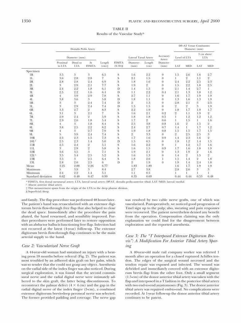
Case 3: The “T Interposed Extensor Digitorum Bre-vis”: A Modification For Anterior Tibial Artery Spar-ing
A 40-year-old male rail company worker was referred 1month after an operation for a closed ruptured Achilles ten-don. The edges of the surgical wound necrosed and thetendon repair was exposed and infected. The wound wasdebrided and immediately covered with an extensor digito-rum brevis flap from the other foot. Only a small segment(1.5-cm) of the donor anterior tibial artery was taken with theflap and interposed in a T fashion in the posterior tibial arterywith two end-to-end anastomoses (Fig. 3). The donor anteriortibial artery was repaired end-to-end. No complications wererecorded. At 1-year follow-up the donor anterior tibial arterycontinues to be patent.
TABLE IIResults of the Vascular Study*
Dorsalis Pedis Artery
FDMTA(S/D)§
Lateral Tarsal Artery AccessoryArtery
Diameter(mm)
DP–AT Venae ComitantesDiameter (mm)
Diameter (mm)
Length(cm)‡
Level of LTA3 cm above
LTA
Proximalto LTA
Distal toLTA
AtFDMTA
Diameter(mm)
Length(cm) LAT MED LAT MED
Specimen1R 3.5 3 3 6.5 S 1.6 2.2 0 1.5 2.6 1.6 2.71L 3.6 2.8 2.8 7 S 2.1 1.5 0 1 2 1.1 22R 2.8 2.8 2.4 6.9 S 1.8 1.6 0 2.4 2.2 2.5 2.32L 3 2.9 2.1 7.7 S 1.9 2 0 1.5 2.2 1.8 2.53R 2.4 2.2 1.8 6.1 D 1.4 1.5 0 2.1 1.4 2.7 13L 2.5 2.2 1.6 6.4 D 1.1 2.2 0.4 2.1 1.3 1.8 1.24R 4 3.9 2.9 7.8 S 2.7 1.1 0 1.6 1.7 1.9 1.94L 3.8 3.6 3 5.8 D 1.8 1.1 0 1.3 1.6 1.3 1.95R 3 3 2.4 7.4 D 2 1.3 0 2.8 2.1 3 2.55L 3 2.9 2.4 7.4 D 1.5 1.5 0 2 2 3 1.96R 3.3 2.7 2 8.3 S 2.2 1.9 0 1.8 1.7 1.9 1.76L 3.1 3 2.1 7 S 1.6 2.1 0.2 2 1.1 1.9 27R 2.9 2.4 2 5.9 S 1.8 1.8 0.5 1 1.2 1.2 1.27L 2.9 2.6 1.8 5.4 S 1.7 2 0.6 1 1.5 1 1.68R 4 3 1.8 8.4 S 2.5 3.8 0.8 1.6 2 1.5 28L 3.8 2.5 2.2 8.2 S 2.4 2.7 0.7 1.4 1 1.7 1.29R 4 3 2.7 7.9 S 1.9 1.8 0.8 1.5 1.5 1.7 1.59L 5 3.9 2.4 7.4 S 2 3.3 0 2 2.5 2.5 310R 2.5 2.3 1.5 7.3 S 1.7 1.6 0.9 1.4 2.3 1.6 210L† 2.5 2.3 1.4 5.6 D 1.5 0.5 0 1.8 1.2 1.4 1.511R 2.5 2.4 2 5.1 S 1.6 2.2 0 1 1.2 1.7 1.611L 3 2.9 2 5.8 S 1.6 1.5 0.9 1.7 1.6 1.8 1.912R 3.6 3.5 3 7.1 S 1.9 2.1 0 1.3 1.9 2 1.612L† 3.5 3.4 2.5 5.2 S 1.6 1.3 0 1.4 2 1.5 213R 3.5 3 2.5 6.4 S 1.8 2.6 1 1.5 1.4 2 1.813L 2.8 2.6 2.5 6 D 2 1.9 0 1.9 1.4 2.4 1.8
Mean 3.25 2.88 2.26 6.77 1.83 1.89 1.6 1.7 1.9 1.9Maximum 5 3.9 3 8.4 2.7 3.8 2.8 2.6 3 3Minimum 2.4 2.2 1.4 5.1 1.1 0.5 1 1 1 1Standard deviation 0.62 0.48 0.47 0.99 0.35 0.69 0.44 0.44 0.53 0.48
* FDMTA, first dorsal metatarsal artery; LTA, lateral tarsal artery; DP-AT, dorsalis pedis–anterior tibial; LAT/MED, lateral/medial.† Absent anterior tibial artery.‡ This measurement spans from the origin of the LTA to the deep plantar division.§ Superficial/deep.
1350 PLASTIC AND RECONSTRUCTIVE SURGERY, April 2000

FIG. 2. Painful skin-grafted area in the palm in case 2 (above, left). (Above, right) Intraoperativephotograph. The extensor digitorum brevis-deep peroneal vascularized nerve graft (asterisks) isstill pedicled on the anterior tibial bundle (AT). (Below, left) Intraoperative photograph of theflap already revascularized in the hand. The extensor digitorum brevis is reflected radially,exposing its undersurface. The gap in the digital nerve is marked by arrows and the deep peronealvascularized nerve graft by asterisks. (Below, right) Early result showing the stable skin-graftedextensor digitorum brevis.
Vol. 105, No. 4 / EXTENSOR DIGITORUM BREVIS FREE FLAP 1351

Case 4: Bilobed Dorsalis Pedis Fasciosubcutaneous-Extensor Digitorum Brevis Muscle Free Flap
A 44-year-old male punch press operator suffered a crushinjury with a heating press to his left hand and forearm (Fig.4). He sustained a third-degree burn to his palm and dorsum;the second finger was charred; and compartment syndromewas diagnosed on his third finger, hand, and distal forearm.After debridement, decompression of the compartments, sec-ond-ray amputation, and third extensor reconstruction, ahuge palmar and dorsal defect was evident. Coverage wasdone immediately to protect the palmar arch and commis-sural arteries, which were exposed and devoid of adventitia.A bilobed dorsalis pedis fasciosubcutaneous-extensor digito-rum brevis combined flap was selected: the muscle for thepalm and the subcutaneous part for the dorsum. The foot wasclosed primarily. Delayed but complete healing of the donorsite was achieved.
DISCUSSION
The extensor digitorum brevis is a trapezoidmuscle that spans from the lateral aspect of thecalcaneus to the first four toes.17,18 We, as wellas other investigators,19 have been unable toconsistently find an independent muscle bellythat may be called the extensor hallucis brevis6;rather, we have noted a uniform proximal mus-cle mass that divides into several bellies in itsdistal third.
The arterial supply on the medial edge ofthe muscle was carefully studied in the cadav-ers. In agreement with other investigators,4,5,19
we found in 100 percent of specimens that thedominant blood supply to the extensor digito-rum brevis was provided by the lateral tarsalartery.
The origin of the lateral tarsal artery hasbeen described as being at or below the loweredge of the inferior extensor retinaculum.1,7,11
Inasmuch as its precise location is critical for asmooth dissecting technique, we investigatedits true relationship to this landmark. Our re-sults indicated that the origin of the lateraltarsal artery is higher than reported above andis better allocated at the level of the talocruraljoint.
When searching for secondary pedicles, wepurposely disregarded the tiny branches of thedorsalis pedis that on their way to the skin havea casual relation to the muscle20; from a surgi-cal standpoint they are irrelevant. This mayexplain why we were unable to find accessory(secondary) arteries in the medial edge of themuscle in 55 percent of the specimens (Fig. 5).Although we did not investigate in detail thelateral blood supply of the muscle,21 we coulddetermine that after passing under the musclethe lateral tarsal artery did anastomose withother arteries on the lateral region of the foot.The lateral tarsal artery is accompanied by ve-nae comitantes, which always drain to the an-terior tibial veins, sometimes as two indepen-dent veins and sometimes forming a rete ofveins. Measurements of the anterior tibial veinswere taken at two levels: where they werejoined by the lateral tarsal veins and 3 cmproximally (Table II). However, it must benoted that these measurements are for descrip-tive guidance only; we know from previous in-vestigations in the deep venous system22 thateven in injected specimens the recorded diam-eters of the veins are very variable and tend tobe smaller than in those of live subjects.
Coverage of the Achilles tendon by the prox-imally pedicled extensor digitorum brevis wasattempted in all of the cadavers in this studyand was found to be inconsistent. Althoughcoverage was uncomplicated in about 40 per-cent of the cadavers, in several the extensordigitorum brevis simply did not reach the cal-caneus–Achilles junction; in others, only thethinnest distal part of the muscle reached thearea.
We found an absence of the anterior tibialartery in two specimens (10L and 12L), whichis in the average 5 percent of results found inlarger studies.23 In both cases the anterior tibialartery was taken over by a large, perforatingbranch of the peroneal artery, which contin-ued on the foot as a dorsalis pedis. Also in bothcases, the lateral tarsal artery and the dorsalispedis were shorter than in the normal side andshorter than the average, which may imposesome limitations when using the T graft and
FIG. 3. Summary of the procedure performed in case 3.From left to right, the extensor digitorum brevis muscle isharvested from the right foot (R) and transferred to the leftfoot (L) with only a small segment of dorsalis pedis artery. Theartery is repaired in the donor side.
1352 PLASTIC AND RECONSTRUCTIVE SURGERY, April 2000

flow-through variants. Although we have notbeen confronted with this scenario in clinicalpractice, we would not hesitate to sacrifice thedorsalis pedis (now dependent of the perforat-ing peroneal) provided that two conditions aremet: (1) the posterior tibial pulses are detectedpreoperatively in the maleollus while occlud-ing the dorsalis pedis on the foot; and (2)upon releasing the tourniquet and havingclamped the dorsalis pedis, normal refilling ofthe foot is seen. Interestingly, when this ana-tomic variant was encountered no problem wasfound in covering the Achilles–calcaneus junc-tion or even the posterior aspect of the calca-neus.
The extensor digitorum brevis muscle hassmall covering area capabilities (about 20 cm2
in our study). It has been widely used as a freeflap for covering small defects3,7,12 or for func-tional restoration.13–16 From our anatomic workwe have been able to expand its use as follows:
Flow-Through Free Flap
The surgeon may at times find it necessary tocover and revascularize during an operation.Taylor and Ham24 introduced the concept ofthe blood carrier flap, and several authors sub-sequently expanded and refined its use.25,26
The extensor digitorum brevis flap is ideallysuited for this purpose, especially in the handtrauma setting, wherein the merits of arterialautografts to reconstruct arterial defects areself-evident.27 Not only are the vessels an excel-lent match for those of the hand, but the size
FIG. 4. Case 4. The condition after debridement and release of the compartments (above, left two panels). (Above, center)Elevated fasciosubcutaneous dorsalis pedis portion of the flap. (Above, right) The bilobed dorsalis pedis-extensor digitorum brevismuscle flap is now pedicled on the vessels. (Below) Result at 8 months.
Vol. 105, No. 4 / EXTENSOR DIGITORUM BREVIS FREE FLAP 1353

of the muscle is quite appropriate for handdefects (Fig. 6). The flap, from the origin ofthe lateral tarsal artery to the deep plantardivision, is benefited by 6.77 cm (SD 6 0.99) ofartery homogeneous in diameter. This lengthis even longer if the patient has a superficialfirst dorsal metatarsal artery (as in 73 percentof cases in our series).
Vascularized Nerve Graft
Controversies exist with regard to the indi-cations for a vascularized nerve graft.28 Thereis, however, no doubt that a prime indicationfor a vascularized nerve graft is a badly scarredbed, such as that found after a deep dermalburn.28,29 The extensor digitorum brevis-deepperoneal vascularized nerve graft adds to thebenefits of providing coverage that better re-sists shearing stresses30 as well as a time-testedvascularized nerve graft.31–33 These propertiesmake this combination the first choice for re-construction of palm (or sole) defects when
there is an associated nerve gap, as in case 2reported here.
The T Interposed Extensor Digitorum Brevis: A Modi-fication for Anterior Tibial Axis Sparing
To avoid the drawback of sacrificing a majorarterial trunk (anterior tibial) to the foot whenharvesting the extensor digitorum brevis, wedevised an alternative procedure: A segment ofonly 1.5 cm of anterior tibial artery, whichincluded the lateral tarsal artery takeoff, is in-terposed in the recipient artery as a T graft,and the donor artery is reconstructed end-to-end. This modification has the disadvantage ofrequiring three anastomoses, but the artery isof large diameter and a more physiologicthrough-flow34 is achieved. The T interposedextensor digitorum brevis modification re-quires a nearby major arterial trunk becausethe pedicle is short (about 2 cm, see Table II).On the other side, an unlimited length of veins
FIG. 5. Anatomic preparation showing the underside ofthe extensor digitorum brevis. The lateral tarsal artery ishighlighted by dots. Note the awkward location of the acces-sory pedicle (aa). DP, dorsalis pedis; LTA, lateral tarsal artery.
FIG. 6. The deep plantar branch and the first dorsal meta-tarsal arteries are to be connected, in this simulation in thecadaver, to the commissural arteries. ATA, anterior tibialartery; LTA, lateral tarsal artery; DPA, dorsalis pedis artery;DB, deep branch of the dorsalis pedis; FDMTA, first dorsalmetatarsal artery.
1354 PLASTIC AND RECONSTRUCTIVE SURGERY, April 2000

(anterior tibial veins) is available for perform-ing the anastomoses distant to the area of in-jury.
Bilobed Dorsalis Pedis Fasciosubcutaneous-ExtensorDigitorum Brevis Free Flap
Combined palmar and dorsal defects in thehand pose a tremendous challenge. A flapcomposed of two independent leaves may beideal for solving the problem. Our anatomicinvestigations allowed us to refine Ismail’s workon the use of the dorsalis pedis myofascialflap.35 First, to increase the size of the fascio-subcutaneous part of the flap, not only thedorsalis pedis but also the first dorsal metatar-sal artery was included, allowing a much largerflap. Second, the full extensor digitorum brevismass was included to increase the coveragearea. Finally, by dissecting the lateral tarsalpedicle from the dorsalis pedis, independentrotation among the two flap components waspermitted, i.e., a true island bilobed flap wascreated.
Because the anterior tibial, the dorsalis pe-dis, and the first dorsal metatarsal arteries are,in essence, on the same vascular axis, and be-cause any of the above named trunks may bethe source of multiple free flaps, countlesscombinations are possible.8,12,14,16 Although itmay seem obvious, we must emphasize that thelimiting factor for these capabilities is the do-nor site sequela, which should be considered atall times so as not to exacerbate the problem.
In conclusion, we think that the extensordigitorum brevis flap is excellent for repairingsmall defects. Future developments and appli-cations will surely expand its use even more.
Dr. Francisco del Pinal, M.D., Dr.Med.Calle Rualasal 23-8 [email protected]
ACKNOWLEDGMENTS
The authors thank Dr. Emilia Jado for her assistance dur-ing the clinical cases and Dr. J. San Jose for his help duringsome of the cadaver dissections. The editorial assistance ofDr. F. Giraldo is greatly appreciated. This work is dedicatedto Dr. G. Ian Taylor of Melbourne, Australia, and Dr. Luis R.Scheker of Louisville, Kentucky, for their lifelong coopera-tion and support. We are indebted to the Hospital MutuaMontanesa de Santander for covering the cost of the colorphotographs.
REFERENCES
1. Landi, A., Soragni, O., and Monteleone, M. The exten-sor digitorum brevis muscle island flap for soft-tissue
loss around the ankle. Plast. Reconstr. Surg. 75: 892,1985.
2. Leitner, D. W., Gordon, L., and Buncke, H. J. The ex-tensor digitorum brevis as a muscle island flap. Plast.Reconstr. Surg. 76: 777, 1985.
3. Hing, D. N., Buncke, H. J., and Alpert, B. S. Applica-tions of the extensor digitorum brevis muscle for softtissue coverage. Ann. Plast. Surg. 19: 530, 1987.
4. Giordano, P. A., Argenson, C., and Pequignot, J. P. Ex-tensor digitorum brevis as an island flap in the recon-struction of soft-tissue defects in the lower limb. Plast.Reconstr. Surg. 83: 100, 1989.
5. Gahhos, F. N., Jaquith, M., and Hidalgo, R. The ex-tended digitorum brevis muscle flap. Ann. Plast. Surg.23: 255, 1989.
6. Crocker, A. D., and Moss, A. L. H. The extensor hallu-cis brevis muscle flap. J. Bone Joint Surg. Br. 71: 532,1989.
7. Buncke, H. J., and Hing, D. N. Extensor Digitorum andHallucis Brevis Muscle Transplantation and Applica-tion as an Island Muscle Flap. In H. J. Buncke (Ed.),Microsurgery: Transplantation-Replantation. Philadel-phia: Lea & Febiger, 1991. Pp. 507–520.
8. Babu, V., Chittaranjan, S., Abraham, G., and Korula, R. J.Single-stage reconstruction of soft-tissue defects in-cluding the Achilles tendon using the dorsalis pedisarterialized flap along with the extensor digitorumbrevis as bridge graft. Plast. Reconstr. Surg. 93: 1090,1994.
9. Gibstein, L. A., Abramson, D. L., Sampson, C. E., andPribaz, J. J. Musculofascial flaps based on the dorsa-lis pedis vascular pedicle for coverage of the foot andankle. Ann. Plast. Surg. 37: 152, 1996.
10. Kurata, S., Hashimoto, H., Terashi, H., Honda, T., andTakayasu, S. Reconstruction of the distal foot dor-sum with a distally based extensor digitorum brevismuscle flap. Ann. Plast. Surg. 29: 76, 1992.
11. Bakhach, J., Demiri, E., Chahidi, N., and Baudet, J. Ex-tensor digitorum brevis muscle flap: New refinements.Plast. Reconstr. Surg. 102: 103, 1998.
12. Hallock, G. G. The conjoint extensor digitorum brevismuscle and dorsalis pedis osteocutaneous island flap.Ann. Plast. Surg. 24: 371, 1990.
13. O’Brien, B. M., Franklin, J. D., and Morrison, W. A.Cross-facial nerve grafts and microneurovascular freemuscle transfer for long established facial palsy. Br. J.Plast. Surg. 33: 202, 1980.
14. Tamai, S., Fukui, A., Shimizu, T., and Yamaguchi, T.Thumb reconstruction with an iliac bone graft and adorsalis pedis flap transplant including the extensordigitorum brevis muscle for restoring opposition: Acase report. Microsurgery 4: 81, 1983.
15. Zhu, S. X., Zhang, B. X., Yao, J. X., et al. Free muscu-locutaneous flap transfer of extensor digitorum brevismuscle by microvascular anastomosis for restorationof function of thenar and adductor pollicis muscles.Ann. Plast. Surg. 15: 481, 1985.
16. Mitz, V. Second toe to thumb transfer with extensordigitorum brevis opponensplasty. Ann. Plast. Surg. 17:259, 1986.
17. Testut, L., and Jacob, O. Pie: Region Dorsal. In L. Testutand O. Jacob (Eds.), Tratado de Anatomıa Topograficacon Aplicaciones Medico-Quirurgicas. Barcelona: Salvat,1927. Pp. 1313–1323.
18. Williams, P. L., Warwick, R., Dyson, M., and Bannister,
Vol. 105, No. 4 / EXTENSOR DIGITORUM BREVIS FREE FLAP 1355

L. H. Gray’s Anatomy, 37th Ed. Edinburgh: ChurchillLivingstone, 1989.
19. Baltensperger, M. M., Ganzoni, M., Jirecek, V., andMeyer, V. E. The extensor digitorum brevis islandflap: Possible applications based on anatomy. Plast.Reconstr. Surg. 101: 107, 1998.
20. Man, D., and Acland, R. D. The microarterial anatomyof the dorsalis pedis flap and its clinical applications.Plast. Reconstr. Surg. 65: 419, 1980.
21. Massin, P., Romana, C., and Masquelet, A. C. Anatomicbasis of a pedicled extensor digitorum brevis muscleflap. Surg. Radiol. Anat. 10: 267, 1988.
22. del Pinal, F., and Taylor, G. I. The deep venous systemand reverse flow flaps. Br. J. Plast. Surg. 46: 652, 1993.
23. Cormack, G. C., and Lamberty, B. G. Lower Leg. InG. C. Cormack and B. G. Lamberty (Eds.), The ArterialAnatomy of Skin Flaps, 2nd Ed. Edinburgh: ChurchillLivingstone, 1994. Pp. 248–257.
24. Taylor, G. I., and Ham, F. J. The free vascularized nervegraft. Plast. Reconstr. Surg. 57: 413, 1976.
25. Foucher, G., van Genechten, F., Merle, N., and Michon,J. A compound radial artery forearm flap in handsurgery: An original modification of the Chinese fore-arm flap. Br. J. Plast. Surg. 37: 139, 1984.
26. Brandt, K., Khouri, R. K., and Upton, J. Free flaps asflow-through vascular conduits for simultaneous cov-erage and revascularization of the hand or digit. Plast.Reconstr. Surg. 98: 321, 1996.
27. Godina, M. Arterial autografts in microvascular sur-gery. Plast. Reconstr. Surg. 78: 293, 1986.
28. Breidenbach, W. C., and Graham, B. VascularizedNerve Grafts. In R. H. Gelberman (Ed.), OperativeNerve Repair and Reconstruction. Philadelphia: Lippin-cott, 1991. Pp. 569–585.
29. Koshima, I., and Harii, K. Experimental study of vas-cularized nerve grafts: Multifactorial analyses of ax-onal regeneration of nerves transplanted into an acuteburn wound. J. Hand Surg. 10A: 64–72, 1985.
30. May, J. W., Jr., Halls, M. J., and Simon, S. R. Free mi-crovascular muscle flaps with skin graft reconstructionof extensive defects of the foot: A clinical andgait analysis study. Plast. Reconstr. Surg. 75: 627,1985.
31. Rose, E. H., and Kowalski, T. A. Restoration of sensi-bility to anesthetic scarred digits with free vascularizednerve grafts from the dorsum of the foot. J. Hand Surg.10A: 514, 1985.
32. Rose, E. H., Kowalski, T. A., and Norris, M. S. The re-versed venous arterialized nerve graft in digital nervereconstruction across scarred beds. Plast. Reconstr.Surg. 83: 593, 1989.
33. Koshima, I., Okumoto, K., Umeda, N., Moriguchi, T.,Ishii, R., and Nakayama, Y. Free vascularized deepperoneal nerve grafts. J. Reconstr. Microsurg. 12: 131,1996.
34. Lister, G. D., and Arnez, Z. Arterial T and Y grafts. Plast.Reconstr. Surg. 88: 319, 1991.
35. Ismail, T. I. A. The dorsalis pedis myofascial flap. Plast.Reconstr. Surg. 86: 573, 1990.
1356 PLASTIC AND RECONSTRUCTIVE SURGERY, April 2000