Expression of Programmed Death 1 Ligands by … of Programmed Death 1 Ligands by Murine T Cells and...
9
of July 15, 2018. This information is current as by Murine T Cells and APC Expression of Programmed Death 1 Ligands and Hideo Yagita Tsuchiya, Drew M. Pardoll, Ko Okumura, Miyuki Azuma Matsuda, Mami Aoki, Yuka Tanno, Tahiro Shin, Haruo Tomohide Yamazaki, Hisaya Akiba, Hideyuki Iwai, Hironori http://www.jimmunol.org/content/169/10/5538 doi: 10.4049/jimmunol.169.10.5538 2002; 169:5538-5545; ; J Immunol References http://www.jimmunol.org/content/169/10/5538.full#ref-list-1 , 6 of which you can access for free at: cites 20 articles This article average * 4 weeks from acceptance to publication Fast Publication! • Every submission reviewed by practicing scientists No Triage! • from submission to initial decision Rapid Reviews! 30 days* • Submit online. ? The JI Why Subscription http://jimmunol.org/subscription is online at: The Journal of Immunology Information about subscribing to Permissions http://www.aai.org/About/Publications/JI/copyright.html Submit copyright permission requests at: Email Alerts http://jimmunol.org/alerts Receive free email-alerts when new articles cite this article. Sign up at: Print ISSN: 0022-1767 Online ISSN: 1550-6606. Immunologists All rights reserved. Copyright © 2002 by The American Association of 1451 Rockville Pike, Suite 650, Rockville, MD 20852 The American Association of Immunologists, Inc., is published twice each month by The Journal of Immunology by guest on July 15, 2018 http://www.jimmunol.org/ Downloaded from by guest on July 15, 2018 http://www.jimmunol.org/ Downloaded from
Transcript of Expression of Programmed Death 1 Ligands by … of Programmed Death 1 Ligands by Murine T Cells and...

of July 15, 2018.This information is current as
by Murine T Cells and APCExpression of Programmed Death 1 Ligands
and Hideo YagitaTsuchiya, Drew M. Pardoll, Ko Okumura, Miyuki AzumaMatsuda, Mami Aoki, Yuka Tanno, Tahiro Shin, Haruo Tomohide Yamazaki, Hisaya Akiba, Hideyuki Iwai, Hironori
http://www.jimmunol.org/content/169/10/5538doi: 10.4049/jimmunol.169.10.5538
2002; 169:5538-5545; ;J Immunol
Referenceshttp://www.jimmunol.org/content/169/10/5538.full#ref-list-1
, 6 of which you can access for free at: cites 20 articlesThis article
average*
4 weeks from acceptance to publicationFast Publication! •
Every submission reviewed by practicing scientistsNo Triage! •
from submission to initial decisionRapid Reviews! 30 days* •
Submit online. ?The JIWhy
Subscriptionhttp://jimmunol.org/subscription
is online at: The Journal of ImmunologyInformation about subscribing to
Permissionshttp://www.aai.org/About/Publications/JI/copyright.htmlSubmit copyright permission requests at:
Email Alertshttp://jimmunol.org/alertsReceive free email-alerts when new articles cite this article. Sign up at:
Print ISSN: 0022-1767 Online ISSN: 1550-6606. Immunologists All rights reserved.Copyright © 2002 by The American Association of1451 Rockville Pike, Suite 650, Rockville, MD 20852The American Association of Immunologists, Inc.,
is published twice each month byThe Journal of Immunology
by guest on July 15, 2018http://w
ww
.jimm
unol.org/D
ownloaded from
by guest on July 15, 2018
http://ww
w.jim
munol.org/
Dow
nloaded from

Expression of Programmed Death 1 Ligands by MurineT Cells and APC1
Tomohide Yamazaki,* Hisaya Akiba,* Hideyuki Iwai, † Hironori Matsuda,* Mami Aoki,*Yuka Tanno,* Tahiro Shin,‡ Haruo Tsuchiya,‡ Drew M. Pardoll, ‡ Ko Okumura,*Miyuki Azuma, † and Hideo Yagita2*
Programmed death 1 (PD-1) is a new member of the CD28/CTLA-4 family, which has been implicated in the maintenance ofperipheral tolerance. Two ligands for PD-1, namely, B7-H1 (PD-L1) and B7-DC (PD-L2), have recently been identified as newmembers of the B7 family but their expression at the protein level remains largely unknown. To characterize the expression ofB7-H1 and B7-DC, we newly generated an anti-mouse B7-H1 mAb (MIH6) and an anti-mouse B7-DC mAb (TY25). MIH6 andTY25 immunoprecipitated a single molecule of 43 and 42 kDa from the lysate of B7-H1 and B7-DC transfectants, respectively.Flow cytometric analysis revealed that B7-H1 was broadly expressed on the surface of mouse tumor cell lines while the expressionof B7-DC was rather restricted. PD-1 was expressed on anti-CD3-stimulated T cells and anti-IgM plus anti-CD40-stimulated Bcells at high levels but was undetectable on activated macrophages or DCs. B7-H1 was constitutively expressed on freshly isolatedsplenic T cells, B cells, macrophages, and dendritic cells (DCs), and up-regulated on T cells by anti-CD3 stimulation on macro-phages by LPS, IFN-�, GM-CSF, or IL-4, and on DCs by IFN-�, GM-CSF, or IL-4. In contrast, B7-DC expression was onlyinducible on macrophages and DCs upon stimulation with IFN-�, GM-CSF, or IL-4. The inducible expression of PD-1 ligands onboth T cells and APCs may suggest new paradigms of PD-1-mediated immune regulation.The Journal of Immunology, 2002, 169:5538–5545.
P rogrammed death 1 (PD-1)3 is a type I transmembraneprotein that was originally identified in a T cell line un-dergoing programmed cell death (1), but further studies
have shown that its expression is associated with lymphocyte ac-tivation rather than cell death (2, 3). We previously generated ananti-mouse PD-1 mAb (J43) and demonstrated that PD-1 was ex-pressed on activated T and B cells and a subset of thymocytes (2,4). Structurally, PD-1 belongs to the CD28/CTLA-4 subfamily ofthe Ig superfamily and contains a single Ig V-like domain in itsextracellular region (5, 6). The PD-1 cytoplasmic region containstwo tyrosine residues, with the N-terminal tyrosine being locatedin an immunoreceptor tyrosine-based inhibitory motif and the C-terminal tyrosine being located in an immunoreceptor tyrosine-based switch motif (ITSM) (7). In vivo studies using PD-1-defi-cient mice have shown that PD-1 plays an important role in theregulation of peripheral tolerance and prevention of autoimmunity.PD-1-deficient C57BL/6 mice developed a lupus-like disease andarthritis (8). In addition, PD-1-deficient BALB/c mice developed
autoantibody-mediated dilated cardiomyopathy (9). Moreover,PD-1-deficient mice crossed with H-2Ld-specific TCR transgenicmice on an H-2b/d background exhibited growth retardation,splenomegaly, and lethal graft-vs-host disease (8). All of theseresults suggested that PD-1 acts as an important negative regulatorof autoimmune responses.
Recently, two new members of the B7 family, B7-H1 (PD-L1)and B7-DC (PD-L2), have been identified to be the ligands forPD-1 (10–13). In vitro studies have shown that the engagement ofPD-1 by B7-H1 or B7-DC inhibited TCR-mediated T cell prolif-eration and cytokine production (IFN-�, IL-10, IL-4, and IL-2)(11, 12). These results indicated that the cross-linking of PD-1 byB7-H1 or B7-DC leads to down-regulation of T cell responses.However, not all studies support the inhibitory role for B7-H1 andB7-DC. Our previous study has shown that resting T cells stimu-lated with immobilized anti-CD3 and B7-DC-Ig exhibited en-hanced proliferation and IFN-� production (13). Another grouphas also indicated that when T cells were stimulated with lowlevels of anti-CD3 and immobilized B7-H1-Ig, proliferation andproduction of IFN-�, GM-CSF, and IL-10 were enhanced (10, 14).These results indicated that B7-H1 and B7-DC could costimulateT cell proliferation and cytokine production. The reason for thesecontradictory results remains unknown. One possible explanationmay be the presence of a second receptor for B7-H1 and B7-DC,which delivers a stimulatory signal. Alternatively, since the ITSMin CD150 has been implicated in both positive and negative sig-naling, the ITSM in PD-1 may also be responsible for positive ornegative signaling via PD-1 (7).
B7-H1 mRNA was detected in various organs, including theheart, lung, thymus, spleen, kidney, and liver, and was up-regu-lated by IFN-� in monocytes and dendritic cells (DCs) (10, 11).B7-DC mRNA was detected in the liver at a high level and in thelung and spleen at lower levels and was preferentially expressed inbone marrow-derived and splenic DCs but not in macrophages (12,
*Department of Immunology, Juntendo University School of Medicine, Tokyo, Ja-pan; †Department of Molecular Immunology, Graduate School, Tokyo Medical andDental University, Tokyo, Japan; and ‡Department of Oncology, Johns Hopkins Uni-versity School of Medicine, Baltimore, MD 21231
Received for publication August 1, 2002. Accepted for publication September3, 2002.
The costs of publication of this article were defrayed in part by the payment of pagecharges. This article must therefore be hereby marked advertisement in accordancewith 18 U.S.C. Section 1734 solely to indicate this fact.1 This work was supported by grants from the Ministry of Education, Culture, Sports,Science and Technology and the Ministry of Health, Labor and Welfare, Japan.2 Address correspondence and reprint requests to Dr. Hideo Yagita, Department ofImmunology, Juntendo University School of Medicine, 2-1-1 Hongo, Bunkyo-ku,Tokyo 113-8421, Japan. E-mail address: [email protected] Abbreviations used in this paper: PD-1, programmed death 1; ITSM, immunore-ceptor tyrosine-based switch motif; DC, dendritic cell; CHO, Chinese hamster ovary;BMDC, bone-marrow derived DC.
The Journal of Immunology
Copyright © 2002 by The American Association of Immunologists, Inc. 0022-1767/02/$02.00
by guest on July 15, 2018http://w
ww
.jimm
unol.org/D
ownloaded from

13). These results suggested the expression of B7-H1 and B7-DCin DCs, which are critical APCs regulating T cell activation andtolerance. However, the cell surface expression of B7-H1 andB7-DC on various APC populations and its regulation remainlargely unknown. In this study, we newly generated mAbs againstmurine B7-H1 and B7-DC and determined the expression of PD-1receptor and ligands on T cells, B cells, macrophages, and DCs.
Materials and MethodsAnimals and cell lines
Six-week-old male C57BL/6 and BALB/c mice and 6-wk-old female SDrats were purchased from Charles River Breeding Laboratories Japan(Atsugi, Japan). Murine T lymphomas L5178Y, WR19L, EL-4, andBW5147; B lymphomas A20.2J, LK35.2, and 2PK-3; macrophage celllines J774A.1, RAW264.7, and P388D1; melanoma B16; myeloma P3U1(P3X63Ag8U.1); and Chinese hamster ovary cell line (CHO) were pur-chased from American Type Culture Collection (Manassas, VA). The hy-bridomas producing mAbs against CD8 (3.155) and Thy1.2 (J1j) were alsoobtained from American Type Culture Collection. The hybridoma produc-ing mAb against CD4 (RL172) was kindly provided by T. Tanaka (OsakaUniversity, Osaka, Japan). T lymphoma MBL-2 and erythroleukemiaFBL-3 were provided by T. Nishimura (Hokkaido University, Sapporo,Japan). A normal rat kidney cell line NRK-52E was kindly provided by T.Otsuka (Institute of Cytosignal Research, Tokyo, Japan). B lymphomaBAL17, renal carcinoma Renca, fibrosarcoma Meth A, and neuroblastomaC1300 were obtained from the Japanese Collection of Research Biore-sources (Osaka, Japan). These cells were cultured in RPMI 1640 mediumcontaining 10% FCS, 10 mM HEPES, 2 mM L-glutamine, 0.1 mg/ml pen-icillin and streptomycin, and 50 �M 2-ME. CHO cells were cultured in�-MEM medium containing 10% FCS, 10 mM HEPES, 2 mM L-glu-tamine, and 0.1 mg/ml penicillin, and streptomycin.
Abs and reagents
Purified anti-CD16/32 (2.4G2) and CD3 (145-2C11) mAbs, FITC-conju-gated anti-CD4 (RM4-4), CD45R/B220 (RA3-6B2), and CD11c (HL3)mAbs, PE-conjugated anti-CD62L (MEL-14) mAb, allophycocyanin-con-jugated anti-CD8� (53–6.7) mAb, biotin-conjugated anti-OX40 (OX86),CD69 (H1.2F3), CD54 (3E2) mAbs, rat IgG isotype control, hamster IgGcontrol, and PE-labeled streptavidin were purchased from BD Biosciences(San Jose, CA). FITC-conjugated F4/80 was purchased from Caltag Lab-oratories (Burlingame, CA). Anti-CD40 (HM40-3) and PD-1 (J43) mAbswere prepared as described previously (2, 15). Anti-CD28 (PV-1) mAbwas kindly provided by R. Abe (Science University of Tokyo, Chiba, Ja-pan). Goat anti-mouse IgM F(ab�)2 Ab was purchased from Jackson Im-munoResearch Laboratories (West Grove, PA). LPS was purchased fromSigma-Aldrich (St. Louis, MO). Recombinant mouse GM-CSF, IL-4, IL-12, and IFN-� were purchased from BD Biosciences.
Preparation of mouse B7-H1 and B7-DC transfectants
A cDNA fragment encoding the entire open reading frame of mouse B7-H1was prepared by RT-PCR from BALB/c peritoneal macrophages. A 5�-CCGCTCGAGCCCAAAACATGAGGATATTTG-3� corresponding to nt9–28 of murine B7-H1 cDNA (11) tagged with an XhoI site and 5�-ATAAGAATGCGGCCGCTTACGTCTCCTCGAATTGTG-3� corresponding tont 870–889 tagged with a NotI site were used as 5� and 3� primers, re-spectively. The PCR product was cloned into a pMKITneo vector, kindlyprovided by K. Maruyama (Tokyo Medical and Dental University, Tokyo,Japan), and transfected into CHO cells with Lipofectin (Invitrogen, Carls-bad, CA) and into L5178Y and NRK by electroporation. After selection by1 mg/ml geneticin (Sigma-Aldrich), transfectants stably expressing B7-H1were identified by staining with PD-1-Ig fusion protein consisting of theextracellular portion of mouse PD-1 (aa 124–564) (1) and the Fc portion ofhuman IgG1. Mouse B7-DC cDNA cloned into a pCAGGS expressionvector (13) was transfected into CHO cells with Lipofectin and intoRAW264.7, L5178Y, and NRK by electroporation. After selection by 8�g/ml blasticidin (Invitrogen), transfectants stably expressing B7-DC wereidentified by staining with PD-1-Ig.
Generation of anti-mouse B7-H1 and B7-DC mAbs
To prepare anti-mouse B7-H1 and B7-DC mAbs, SD rats were immunizedwith B7-H1-transfected L5178Y or B7-DC-transfected RAW264.7 cells.Three days after the final immunization, spleen cells or lymph node cellswere fused with P3U1 myeloma cells. After hypoxanthine-aminopterin-thymidine selection, a hybridoma (MIH6, rat IgG2a/�) producing anti-
B7-H1 mAb and a hybridoma (TY25, rat IgG2a/�) producing anti-B7-DCmAb were selected by their strong reactivity to B7-H1-transfected CHO orB7-DC-transfected NRK, but not to parental cells by flow cytometry andthen cloned by limiting dilution. Both MIH6 and TY25 were purified fromascites by the caprylic acid and ammonium sulfate precipitation method,and purity was verified by SDS-PAGE analysis.
Immunoprecipitation and Western blotting
Cells (1 � 107) were lysed in 1 ml of a lysis buffer containing 1% NonidetP-40, 50 mM HEPES (pH 7.4), 250 mM NaCl, and 2 mM EDTA. Cellulardebris and nuclei were removed by centrifugation, and the supernatant wasprecleared with protein G-Sepharose (Amersham Biosciences, Piscataway,NJ) preincubated with control rat IgG2a for 3 h at 4°C. The cleared lysateswere immunoprecipitated with MIH6-preloaded, TY25-preloaded, or con-trol rat IgG2a-preloaded protein G-Sepharose for 1 h at 4°C. The beadswere washed three times with the lysis buffer, and bound proteins wereeluted with 1% SDS sample buffer, subjected to 10% SDS-PAGE, and thenblotted onto polyvinylidene difluoride membrane (Millipore, Bedford,MA). The blotted proteins were detected using biotin-conjugated MIH6,TY25, or rat IgG2a followed by avidin-biotinylated peroxidase complex(Vector Laboratories, Burlingame, CA) and an ECL Western Blotting De-tection System (Amersham Biosciences) according to the manufacturer’sinstructions.
RT-PCR
Total RNA was extracted from cell lines (1 � 107) using RNA STAT-60(Tel-Test, Friendswood, TX). First-strand cDNA was prepared using a Su-perScript First-Strand Synthesis System (Invitrogen) from 5 �g of totalRNA. cDNA samples were standardized based on the content of �-actin
FIGURE 1. Reactivity of anti-mouse B7-H1 (MIH6) and B7-DC (TY25)mAbs to mouse B7-H1 and B7-DC transfectants. NRK-52E-, CHO-, andL5178Y-derived B7-H1 transfectants (B7-H1/NRK, B7-H1/CHO, and B7-H1/L5178Y), B7-DC transfectants (B7-DC/NRK, B7-DC/CHO, and B7-DC/L5178Y), and parental cells were stained with biotinylated MIH6, TY25, orcontrol rat IgG2a followed by PE-labeled streptavidin. The open histogramsindicate staining with the indicated mAb and the shaded histograms indicatebackground staining with control rat IgG2a.
5539The Journal of Immunology
by guest on July 15, 2018http://w
ww
.jimm
unol.org/D
ownloaded from

cDNA. Primers for mouse �-actin were 5�-GTGGGCCGCTCTAGGCACCAA-3� and 5�-CTCTTTGATGTCACGCACGATTTC-3�. Primers formouse B7-H1 were 5�-ATGAGGATATTTGCTGGCATTAT-3� and 5�-TTACGTCTCCTCGAATTGTGT-3�. Primers for mouse B7-DC were 5�-ATGCTGCTCCTGCCCTGCCG-3� and 5�-CTAGATCCTCTTTCTCTGGAT-3�. PCR was performed in a total volume of 20 �l in PCR buffer inthe presence of 0.2 mM dNTP, 1 �M of each primer, and 1 U of Taq DNApolymerase (Advanced Biotecnologies, Surrey, U.K.). After 35 cycles ofamplification, the PCR products were separated by electrophoresis on a 2%agarose gel and visualized by ethidium bromide staining.
Preparation and stimulation of T cells, B cells, andmacrophages
CD4�CD62L� naive T cells were purified from the spleen of C57BL/6mice by passage through nylon wool columns (Wako Biochemicals, Osaka,Japan) and by using an auto-MACS column with FITC-conjugated anti-CD4, anti-FITC multisort kit, and anti-CD62L-coupled microbeads (Milte-nyi Biotec, Bergisch Gladbach, Germany) according to the manufacturer’s
FIGURE 2. Immunoprecipitation of B7-H1 and B7-DC Ags with MIH6and TY25 mAbs. Immunoprecipitates with control rat IgG2a, MIH6, orTY25 from L5178Y, B7-H1/L5178Y, and B7-DC/L5178Y cell lysateswere subjected to SDS-PAGE under nonreducing conditions, blotted ontopolyvinylidene difluoride membrane, and then detected with biotinylatedcontrol rat IgG2a, MIH6, or TY25, respectively, followed by the avidin-biotinylated peroxidase complex and ECL. The positions of molecularmass markers are indicated at the left in kilodaltons.
FIGURE 3. Expression of B7-H1 and B7-DC bymouse tumor cell lines. A, Reactivity of MIH6 andTY25 with tumor cell lines. T lymphoma cell lines(WR19L, MBL-2, EL-4, and BW5147), B lymphomacell lines (A20.2J, BAL17, LK35.2, and 2PK-3), mac-rophage cell lines (J774A.1, RAW264.7, and P388D1),renal carcinoma cell line Renca, erythroleukemia cellline FBL-3, melanoma cell line B16, fibrosarcoma cellline Meth A, and neuroblastoma cell line C1300 werestained with biotinylated control rat IgG2a, MIH6, orTY25, followed by PE-labeled streptavidin. The openhistograms indicate staining with MIH6 or TY25 andthe shaded histograms indicate background stainingwith control rat IgG2a. B, Expression of B7-H1 andB7-DC mRNAs in tumor cell lines. B7-H1 and B7-DCmRNAs were detected by specific RT-PCR amplifica-tion of total RNA prepared from the indicated tumorcell lines. Amplification of �-actin mRNA was used asa control.
FIGURE 4. Expression of B7-H1, B7-DC, and PD-1 on CD4� T cells.Purified splenic naive CD4� T cells were stimulated by immobilized anti-CD3mAb with (A) or without (B) soluble anti-CD28 mAb and harvested at theindicated periods. Cells were stained with biotinylated anti-B7-H1 mAb(MIH6), anti-B7-DC mAb (TY25), anti-PD-1 mAb, anti-OX40 mAb, or con-trol IgG followed by PE-labeled streptavidin. The open histograms indicatestaining with the indicated mAb and the shaded histograms indicate back-ground staining with control IgG.
5540 CELL SURFACE EXPRESSION OF MURINE PD-1 LIGANDS
by guest on July 15, 2018http://w
ww
.jimm
unol.org/D
ownloaded from
instructions. Purified naive CD4 T cells (3 � 106/ml; �95% CD4�
CD62L�) were stimulated with immobilized anti-CD3 mAb (5 �g/ml) inthe presence or absence of anti-CD28 mAb (3 �g/ml) for 24–96 h. Smallresting B cells were purified as previously described (16). Briefly, spleno-cytes from C57BL/6 mice were treated with a mixture of hybridoma su-pernatants (anti-Thy-1.2, anti-CD4, and anti-CD8) and Low-Tox rabbitcomplement (Cedarlane Laboratories, Hornby, Ontario, Canada). AfterPercoll (Amersham Biosciences) gradient centrifugation, small B cellswere collected from the 60/70% interface. Purified B cells (3 � 106/ml;�95% B220�) were stimulated with anti-IgM Ab (5 �g/ml) and/or anti-CD40 mAb (10 �g/ml), or LPS (5 �g/ml) for 24–72 h. Peritoneal mac-rophages were obtained from C57BL/6 mice that received i.p. 2 ml of 4%thioglycolate (Sigma-Aldrich) 4 days before. Peritoneal exudate cells wereharvested by peritoneal lavage with ice-cold PBS and depleted of nonad-herent cells after a 1-h culture on a plastic dish, and then stimulated withanti-CD40 mAb (5 �g/ml), LPS (5 �g/ml), GM-CSF (10 ng/ml), IL-4 (20ng/ml), or IFN-� (20 ng/ml) for 24–72 h.
Preparation of bone marrow-derived DCs and splenic DCs
Bone marrow-derived DCs (BMDCs) were prepared by culturing bonemarrow cells (5 � 105/ml) from C57BL/6 mice in the presence of GM-CSF(10 ng/ml) for 4–8 days. Nonadherent cells were removed from the cultureand fresh medium containing GM-CSF was fed at days 2, 4, and 6 after theinitiation of culture. For isolating splenic DCs, spleens from C57BL/6 micewere digested with 400 U/ml collagenase (Wako Biochemicals) in the pres-ence of 5 mM EDTA and separated into low- and high-density fractions onan Optiprep gradient (Axis-Shield, Oslo, Norway). Low-density cells wereincubated with anti-CD11c-coupled magnetic beads and the bound cellswere isolated by the auto-MACS column. The purified splenic DCs (2 �106/ml; �95% CD11c�) were cultured with or without anti-CD40 mAb (5�g/ml), GM-CSF (10 ng/ml), IL-4 (20 ng/ml), IFN-� (20 ng/ml), or IL-12(10 ng/ml) at 37°C for 24–72 h.
Flow cytometric analysis
Cells (0.5–1 � 106) were first preincubated with unlabeled anti-CD16/32mAb to avoid nonspecific binding of Abs to Fc�R and then incubated with
FITC- or allophycocyanin-labeled mAbs or biotinylated mAb. After wash-ing with PBS twice, the cells were incubated with PE-labeled streptavidinfor biotinylated mAb. After washing with PBS twice, the stained cells(live-gated on the basis of forward and side scatter profiles and propidiumiodide exclusion) were analyzed on a FACSCalibur (BD Biosciences), anddata were processed using the CellQuest program (BD Biosciences).
ResultsEstablishment of anti-mouse B7-H1 and B7-DC mAbs
To characterize the expression of mouse B7-H1 and B7-DC, wenewly generated specific mAbs. The expression vectors containingmouse B7-H1 or B7-DC cDNA were transfected into L5178Y,RAW264.7, NRK-52E, and CHO cells. Cell surface expression ofB7-H1 and B7-DC on stable transfectants was verified by stainingwith PD-1-Ig fusion protein (data not shown). We immunized SDrats with B7-H1/L5178Y or B7-DC/RAW264.7 cells and screenedthe hybridomas producing mAb that reacted with B7-H1 or B7-DCtransfectants but not parental cells. Two mAbs, designated MIH6and TY25, were selected. As represented in Fig. 1, MIH6 reactedstrongly with B7-H1-transfected cells (B7-H1/NRK, B7-H1/CHO,and B7-H1/L5178Y) but not with B7-DC-transfected or parentalNRK and CHO cells. Parental and B7-DC-transfected L5178Ycells were also stained with MIH6 weakly, which reflected a lowlevel of endogenous B7-H1 expression in L5178Y cells (see be-low). TY25 only reacted with B7-DC-transfected cells (B7-DC/NRK, B7-DC/CHO, and B7-DC/L5178Y) but not with B7-H1-transfected cells or parental cells.
To characterize the Ags recognized by these mAbs, cell lysatesof B7-H1/L5178Y, B7-DC/L5178Y, and L5178Y cells were im-munoprecipitated with MIH6 or TY25. Then the precipitates were
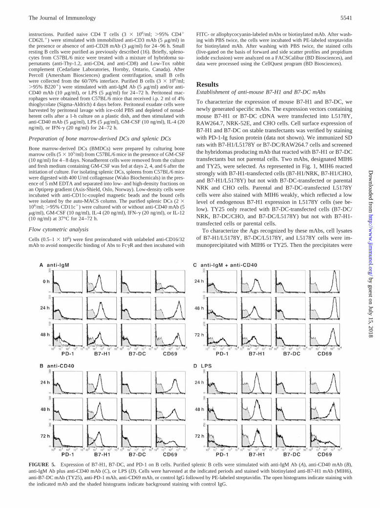
FIGURE 5. Expression of B7-H1, B7-DC, and PD-1 on B cells. Purified splenic B cells were stimulated with anti-IgM Ab (A), anti-CD40 mAb (B),anti-IgM Ab plus anti-CD40 mAb (C), or LPS (D). Cells were harvested at the indicated periods and stained with biotinylated anti-B7-H1 mAb (MIH6),anti-B7-DC mAb (TY25), anti-PD-1 mAb, anti-CD69 mAb, or control IgG followed by PE-labeled streptavidin. The open histograms indicate staining withthe indicated mAb and the shaded histograms indicate background staining with control IgG.
5541The Journal of Immunology
by guest on July 15, 2018http://w
ww
.jimm
unol.org/D
ownloaded from

analyzed by SDS-PAGE under nonreducing condition and immu-noblotting with biotin-conjugated MIH6 or TY25. As shown inFig. 2, MIH6 precipitated a 43-kDa protein from B7-H1/L5178Ycells and TY25 precipitated a 42-kDa protein from B7-DC/L178Ycells. The 43-kDa band was also detected in the MIH6 precipitatesfrom L5178Y and B7-DC/L5178Y cells after a longer exposure,which appeared to represent endogenously expressed B7-H1 inL5178Y cells (data not shown).
Expression of B7-H1 and B7-DC on mouse tumor cell lines
We first examined the correlation between the B7-H1 and B7-DCmRNA expression and the MIH6 and TY25 reactivity in mousetumor cell lines. As shown in Fig. 3A, MIH6 reacted with all testedcell lines which expressed B7-H1 mRNA as estimated by RT-PCR(Fig. 3B). TY25 reacted with MBL-2, BAL17, and RAW264.7cells which expressed B7-DC mRNA, but not with the other cell
lines which did not express B7-DC mRNA (Fig. 3). The strictcorrelation between the B7-H1 and B7-DC mRNA expression andthe MIH6 and TY25 reactivity further substantiated their specific-ity for B7-H1 and B7-DC.
Expression of B7-H1, B7-DC, and PD-1 on T cells
We next examined the expression of B7-H1, B7-DC, and PD-1 onsplenic T cells by flow cytometric analysis using MIH6, TY25, andan anti-mouse PD-1 mAb (J43) we previously generated (2). Ananti-OX40 mAb was also included as an activation marker. Puri-fied naive CD4� T cells were stimulated with immobilized anti-CD3 mAb in the presence or absence of soluble anti-CD28 mAbfor 24–96 h. As shown in Fig. 4A, PD-1 expression was detectedon CD4� T cells after stimulation with anti-CD3 mAb alone,which appeared at 24 h and reached a peak at 48 h. Unexpectedly,naive CD4� T cells constitutively expressed B7-H1, which was
FIGURE 6. Expression of B7-H1, B7-DC, and PD-1 on macrophages. Purified peritoneal macrophages were cultured with medium alone (A) orstimulated with anti-CD40 mAb (B), LPS (C), GM-CSF (D), IL-4 (E), or IFN-� (F). Cells were harvested at the indicated periods and stained withPE-labeled anti-B7-H1 mAb (MIH6), anti-B7-DC mAb (TY25), or anti-PD-1 mAb, FITC-labeled anti-CD54 mAb, or control IgG. The open histogramsindicate staining with the indicated mAb and the shaded histograms indicate background staining with control IgG.
5542 CELL SURFACE EXPRESSION OF MURINE PD-1 LIGANDS
by guest on July 15, 2018http://w
ww
.jimm
unol.org/D
ownloaded from

markedly up-regulated by stimulation with anti-CD3 mAb alone(Fig. 4A). In contrast, B7-DC expression was not found on freshlyisolated naive CD4� T cells, but was marginally detectable afterstimulation with anti-CD3 mAb for 72–96 h (Fig. 4A). Althoughthe addition of anti-CD28 mAb slightly increased the expression ofboth B7-H1 and B7-DC on anti-CD3-stimulated T cells, it accel-erated the reduction of PD-1 expression at 72–96 h (Fig. 4B). Sim-ilar patterns of B7-H1, B7-DC, and PD-1 expression were ob-served when naive CD8� T cells were stimulated with anti-CD3mAb in the presence or absence of anti-CD28 mAb (data notshown).
Expression of B7-H1, B7-DC, and PD-1 on B cells
We next examined the expression of B7-H1, B7-DC, and PD-1 onsplenic B cells. Small resting B cells were stimulated with anti-IgM Ab, anti-CD40 mAb, both anti-IgM Ab and anti-CD40 mAb,or LPS for 24–72 h and stained with MIH6, TY25, or J43. Ananti-CD69 mAb was also included as an activation marker. Ex-pression of PD-1 was found on anti-IgM- or LPS-stimulated Bcells, which was apparent at 48 or 72 h, respectively (Fig. 5, A andD). The combination of anti-IgM Ab and anti-CD40 mAb mark-edly induced the PD-1 expression (Fig. 5C), whereas the stimula-tion with anti-CD40 mAb alone was not effective (Fig. 5B). B7-H1was found on freshly isolated B cells at a substantial level and wasslightly up-regulated by stimulation with anti-IgM Ab (Fig. 5A) orLPS (Fig. 5D) but not with anti-CD40 mAb (Fig. 5, B and C). Incontrast, B7-DC was not found on splenic B cells after anystimulation.
Expression of B7-H1, B7-DC, and PD-1 on macrophages
We also examined the expression of B7-H1, B7-DC, and PD-1 onthioglycolate-elicited peritoneal macrophages. Purified macro-phages were stimulated with anti-CD40 mAb, LPS, or cytokines(GM-CSF, IL-4, or IFN-�) for 24–72 h. Expression of CD54 wasalso monitored as an activation maker. As shown in Fig. 6A, asubstantial level of B7-H1 expression was found on freshly iso-lated macrophages, which was increased by 24–48 h culture inmedium alone and further up-regulated by stimulation with LPS,GM-CSF, IL-4, or IFN-� (Fig. 6, C--F). Although B7-DC expres-sion was not found on freshly isolated macrophages (Fig. 6A), itwas induced by stimulation with GM-CSF, IFN-�, and especiallyIL-4 (Fig. 6, D–F). In contrast, PD-1 expression was not found onmacrophages after any stimulation (Fig. 6).
Expression of B7-H1, B7-DC, and PD-1 on DCs
We previously observed that B7-DC mRNA was preferentiallyexpressed in BMDCs (13). We now examined the cell surfaceexpression of B7-H1 and B7-DC on BMDCs, which were preparedby culturing bone marrow cells with GM-CSF for 4, 6, and 8 daysby staining with MIH6 and TY25 mAbs. As shown in Fig. 7, bothB7-H1 and B7-DC were found on CD11c� BMDCs from days 4to 8 of the culture.
To further examine the expression and regulation of B7-H1,B7-DC, and PD-1 on DCs, we purified CD11c� splenic DCs andstimulated them with anti-CD40 mAb or cytokines (GM-CSF,IL-4, IFN-�, or IL-12) for 24–48 h. CD69 was also included as anactivation marker. As shown in Fig. 8A, while freshly isolatedCD11c�CD8� DCs expressed B7-H1 at a higher level than freshlyisolated CD11c�CD8� DCs, both DC populations up-regulatedthe B7-H1 expression to a similar level after the culture in mediumalone for 24 h. The stimulation with anti-CD40 mAb and cytokines(GM-CSF, IL-4, IFN-�, and IL-12) slightly enhanced the B7-H1expression (Fig. 8, B–F). In contrast, B7-DC was not found onfreshly isolated DCs, but was induced at a low level by culture in
medium alone (Fig. 8A). The B7-DC expression was markedlyincreased by stimulation with GM-CSF, IL-4, and IFN-� on bothCD11c�CD8� and CD11c�CD8� DCs (Fig. 8, C–E). Expressionof PD-1 was not found on either CD11c�CD8� or CD11c�CD8�
DCs even after stimulation (Fig. 8).
DiscussionIn this study, we generated mAbs specific for murine B7-H1(MIH6) and B7-DC (TY25). The B7-H1 and B7-DC moleculesrecognized by MIH6 and TY25 exhibited the molecular mass of 43and 42 kDa, respectively, in SDS-PAGE analysis under nonreduc-ing conditions. Since the molecular mass of the core polypeptidesexpected from the amino acid sequences of B7-H1 and B7-DC are32,780, and 27,819, respectively, these results suggested that bothB7-H1 and B7-DC molecules are glycosylated and expressed as amonomer.
It has been reported that B7-H1 mRNA was broadly expressedwhereas B7-DC mRNA was rather restricted in mouse tumor celllines (12, 13). Consistently, our results demonstrated that cell sur-face expression of B7-H1 was broadly found on both hemopoieticand nonhemopoietic tumors, but that of B7-DC was restricted tocertain leukemias. The engagement of PD-1 by B7-H1 or B7-DChas been reported to inhibit TCR/CD3-mediated T cell prolifera-tion and cytokine production (11, 12, 17). Moreover, Dong et al.(18) recently reported that B7-H1 expressed on tumor cells pro-moted T cell apoptosis. Therefore, the expression of B7-H1 andB7-DC on tumor cells may represent a novel tumor escape mech-anism from immunosurveillance.
Using flow cytometric analysis, we formally determined the ex-pression of B7-H1, B7-DC, and PD-1 on murine T cells and APCsincluding B cells, macrophages, and DCs. Some new findings wereobtained as follows.
Our previous report demonstrated the PD-1 expression onsplenic T cells after anti-CD3, Con A, or PMA plus ionomycinstimulation (2). We now demonstrated that PD-1 could be ex-pressed on naive T cells upon anti-CD3 stimulation alone withoutanti-CD28 costimulation. More importantly, B7-H1 is constitu-tively expressed on naive T cells and markedly up-regulated byanti-CD3 stimulation. It has been known that TCR/CD3 stimula-tion of naive T cells in the absence of CD28 costimulation induces
FIGURE 7. Expression of B7-H1 and B7-DC on BMDCs. BMDCswere prepared by culturing bone marrow cells in the presence of GM-CSF.BMDCs were harvested at the indicated periods and stained with FITC-labeled anti-CD11c mAb and biotinylated anti-B7-H1 mAb (MIH6), anti-B7-DC mAb (TY25), or control IgG followed by PE-labeled streptavidin.The middle and right panels show staining of the electronically gatedCD11c� R1 population in the left panel. The open histograms indicatestaining with the indicated mAb and the shaded histograms indicate back-ground staining with control IgG.
5543The Journal of Immunology
by guest on July 15, 2018http://w
ww
.jimm
unol.org/D
ownloaded from

FIGURE 8. Expression of B7-H1, B7-DC, and PD-1 on splenic DCs. Purified splenic DCs were cultured with medium alone (A) or stimulated withanti-CD40 mAb (B), GM-CSF (C), IL-4 (D), IFN-� (E), or IL-12 (F) and harvested at the indicated periods. Cells were stained with FITC-labeledanti-CD11c mAb, allophycocyanin-labeled anti-CD8 mAb, and biotinylated anti-B7-H1 mAb (MIH6), anti-B7-DC mAb (TY25), anti-PD-1 mAb, anti-CD69 mAb, or control IgG followed by PE-labeled streptavidin. The left four panels show staining of the electronically gated CD11c�CD8� R1 populationand the right four panels show that of the CD11c�CD8� R2 population. The open histograms indicate staining with the indicated mAb and the shadedhistograms indicate background staining with control IgG.
5544 CELL SURFACE EXPRESSION OF MURINE PD-1 LIGANDS
by guest on July 15, 2018http://w
ww
.jimm
unol.org/D
ownloaded from
anergy (19). PD-1-B7-H1 interaction may be involved in thisprocess.
On splenic B cells, PD-1 expression was induced by anti-IgM orPMA plus ionomycin stimulation (2). We here demonstrated ahigher expression of PD-1 by anti-IgM plus anti-CD40 stimulationand no induction by anti-CD40 stimulation alone. This suggeststhat PD-1 is preferentially expressed on surface Ig-stimulated Ag-specific B cells upon interaction with CD40L-expressing Th cells.PD-1�/� C57BL/6 mice developed lupus-like disease with andincreased number of B cells and serum IgG2b, IgG3, and IgA (20).PD-1�/� BALB/c mice developed dilated cardiomyopathy with ahigh level of IgG1 autoantibodies (9). These studies suggested acritical role for PD-1 as a negative regulator of B cell responses. Inthis respect, our present finding of PD-1 on activated B cells andB7-H1 on activated T cells is intriguing, since it suggests that theengagement of PD-1 on B cells by B7-H1 on T cells may consti-tute a novel pathway of T cell-mediated B cell suppression.
In human monocytes, induction of both B7-H1 and B7-DCmRNAs by IFN-� stimulation has been reported (11, 12). We nowdemonstrated that B7-H1 is constitutively expressed on the surfaceof murine peritoneal macrophages and is up-regulated by LPS,IFN-�, and IL-4. In addition, B7-DC expression was also inducedby GM-CSF, IFN-�, and IL-4. These results indicated that B7-H1and B7-DC expression on macrophages can be regulated by bothTh1 (IFN-� and GM-CSF) and Th2 (IL-4) cytokines. Further stud-ies are now under way to explore the costimulatory or suppressivefunction of B7-H1 and B7-DC on macrophages when they act asAPCs for T cells.
We previously demonstrated that B7-DC mRNA was preferen-tially expressed in BMDCs but not in bone marrow-derived mac-rophages (13). We now verified the cell surface expression of B7-DC, as well as B7-H1, on BMDCs. This B7-DC expression onBMDCs seems to result from GM-CSF stimulation rather thandifferentiation into DCs, as observed with peritoneal macrophages.Among freshly isolated splenic DCs, the CD8� myeloid DC pop-ulation exhibited a higher expression of B7-H1 than the CD8�
lymphoid DC population, mechanisms for which are presently un-known. However, both DC populations rapidly up-regulatedB7-H1 expression to a similar level upon in vitro culture, whichmay be mediated by autocrine GM-CSF. Although we previouslyshowed the expression of B7-DC mRNA in splenic DCs (13), cellsurface expression of B7-DC was not found on freshly isolatedsplenic DCs but was rapidly induced upon in vitro culture, whichmay be also induced by autocrine GM-CSF. Alternatively, cellsurface expression of B7-DC on splenic DCs may be posttran-scriptionally modulated in situ. B7-H1 and B7-DC expression onsplenic DCs was further up-regulated by both Th1 (IFN-� andGM-CSF) and Th2 (IL-4) cytokines. This may be relevant to thecostimulation or suppression of both Th1 and Th2 responses bythese molecules.
In summary, we herein characterized the cell surface expressionof B7-H1 and B7-DC on murine T cells and APCs including Bcells, macrophages, and DCs. Further studies are required to explore
the functional roles of B7-H1 and B7-DC on these cells. The regu-lated expression of these two PD-1 ligands on both T cells and APCssuggest new paradigms of PD-1-mediated immune regulation.
AcknowledgmentsWe thank R. Abe for mAb and T. Tanaka, T. Nishimura, and T. Ohtsukafor cells.
References1. Ishida, Y., Y. Agata, K. Shibahara, and T. Honjo. 1992. Induced expression of
PD-1, a novel member of the immunoglobulin gene superfamily, upon pro-grammed cell death. EMBO J. 11:3887.
2. Agata, Y., A. Kawasaki, H. Nishimura, Y. Ishida, T. Tsubata, H. Yagita, andT. Honjo. 1996. Expression of the PD-1 antigen on the surface of stimulatedmouse T and B lymphocytes. Int. Immunol. 8:765.
3. Vibhakar, R., G. Juan, F. Traganos, Z. Darzynkiewicz, and L. R. Finger. 1997.Activation-induced expression of human programmed death-1 gene in T- lym-phocytes. Exp. Cell Res. 232:25.
4. Nishimura, H., Y. Agata, A. Kawasaki, M. Sato, S. Imamura, N. Minato,H. Yagita, T. Nakano, and T. Honjo. 1996. Developmentally regulated expressionof the PD-1 protein on the surface of double-negative (CD4�CD8�) thymocytes.Int. Immunol. 8:773.
5. Nishimura, H., and T. Honjo. 2001. PD-1: an inhibitory immunoreceptor in-volved in peripheral tolerance. Trends Immunol. 22:265.
6. Carreno, B. M., and M. Collins. 2002. The B7 family of ligands and its receptors:new pathways for costimulation and inhibition of immune responses. Annu. Rev.Immunol. 20:29.
7. Sharpe, A. H., and G. J. Freeman. 2002. The B7-CD28 superfamily. Nat. Rev.Immunol. 2:116.
8. Nishimura, H., M. Nose, H. Hiai, N. Minato, and T. Honjo. 1999. Developmentof lupus-like autoimmune diseases by disruption of the PD-1 gene encoding anITIM motif-carrying immunoreceptor. Immunity 11:141.
9. Nishimura, H., T. Okazaki, Y. Tanaka, K. Nakatani, M. Hara, A. Matsumori,S. Sasayama, A. Mizoguchi, H. Hiai, N. Minato, and T. Honjo. 2001. Autoim-mune dilated cardiomyopathy in PD-1 receptor-deficient mice. Science 291:319.
10. Dong, H., G. Zhu, K. Tamada, and L. Chen. 1999. B7–H1, a third member of theB7 family, co-stimulates T-cell proliferation and interleukin-10 secretion. Nat.Med. 5:1365.
11. Freeman, G. J., A. J. Long, Y. Iwai, K. Bourque, T. Chernova, H. Nishimura,L. J. Fitz, N. Malenkovich, T. Okazaki, M. C. Byrne, et al. 2000. Engagement ofthe PD-1 immunoinhibitory receptor by a novel B7 family member leads tonegative regulation of lymphocyte activation. J. Exp. Med. 192:1027.
12. Latchman, Y., C. R. Wood, T. Chernova, D. Chaudhary, M. Borde, I. Chernova,Y. Iwai, A. J. Long, J. A. Brown, R. Nunes, et al. 2001. PD-L2 is a second ligandfor PD-1 and inhibits T cell activation. Nat. Immunol. 2:261.
13. Tseng, S. Y., M. Otsuji, K. Gorski, X. Huang, J. E. Slansky, S. I. Pai, A. Shalabi,T. Shin, D. M. Pardoll, and H. Tsuchiya. 2001. B7-DC, a new dendritic cellmolecule with potent costimulatory properties for T cells. J. Exp. Med. 193:839.
14. Tamura, H., H. Dong, G. Zhu, G. L. Sica, D. B. Flies, K. Tamada, and L. Chen.2001. B7–H1 costimulation preferentially enhances CD28-independent T-helpercell function. Blood 97:1809.
15. Kaneko, Y., S. Hirose, M. Abe, H. Yagita, K. Okumura, and T. Shirai. 1996.CD40-mediated stimulation of B1 and B2 cells: implication in autoantibody pro-duction in murine lupus. Eur. J. Immunol. 26:3061.
16. Akiba, H., H. Oshima, K. Takeda, M. Atsuta, H. Nakano, A. Nakajima,C. Nohara, H. Yagita, and K. Okumura. 1999. CD28-independent costimulationof T cells by OX40 ligand and CD70 on activated B cells. J. Immunol. 162:7058.
17. Carter, L., L. A. Fouser, J. Jussif, L. Fitz, B. Deng, C. R. Wood, M. Collins,T. Honjo, G. J. Freeman, and B. M. Carreno. 2002. PD-1:PD-L inhibitory path-way affects both CD4� and CD8� T cells and is overcome by IL-2. Eur. J. Im-munol. 32:634.
18. Dong, H., S. E. Strome, D. R. Salomao, H. Tamura, F. Hirano, D. B. Flies,P. C. Roche, J. Lu, G. Zhu, K. Tamada, et al. 2002. Tumor-associated B7–H1promotes T-cell apoptosis: a potential mechanism of immune evasion. Nat. Med.8:793.
19. Schwartz, R. H. 1990. A cell culture model for T lymphocyte clonal anergy.Science 248:1349.
20. Nishimura, H., N. Minato, T. Nakano, and T. Honjo. 1998. Immunological stud-ies on PD-1 deficient mice: implication of PD-1 as a negative regulator for B cellresponses. Int. Immunol. 10:1563.
5545The Journal of Immunology
by guest on July 15, 2018http://w
ww
.jimm
unol.org/D
ownloaded from