Expression of p53, p21 /wafl , bcl-2, bax, Rb and Ki67 ... of p53, p21 waf l... · 446 Gel/ cyc/e...
9
Histol Histopathol (2000) 15: 445-453 001 : 10.14670/HH-15.445 http://www.hh.um .es Histology and Histopathology Ce/tu/ar and Molecular Bio/ogy Expression of p53, p21/wafl , bcl-2, bax, Rb and Ki67 proteins in Hodgkin's lymphomas P. Kanavaros•, K. Stefanaki2, J. Vlachonikolis3, G. Eliopoulos4, S. Kakolyriss, D. Rontogiannl6, V. Gorgoulis7 and V. Georgoulias5 'Department of Histology, University of Thessalia, Departments of 2Pathology, 3Biostatistics, 4 Hematology , 5Qncology , University of Crete, 6Department of Pathology, Evagelismos Hospital of Athens, 'Department of Histology, University of Athens, Athens, Greece Summary. The aim was to investigate the combined immunoexpression of p53, p21, bcl-2, bax, Rb and Ki67 proteins in Hodgkin's lymphomas (HL) and correlate expression patterns with the histotype and the Epstein- Barr Virus (EBV) status. Paraffin-sections from 56 cases of HL (18 nodular sclerosis and 38 mixed cellularity) and from ten "reactive" lymph nodes were investigated. P53, p21, bcl-2, bax, Rb and Ki67 proteins were detected in Hodgkin and Reed-Sternberg (HRS) cells in 35156,56156,24156, 23156,56156 and 56/56 cases of HL, respectively. No correlation was found between the expression of each protein and the EBV status or the histotype of HL. Comparison between p53 and p21 staining revealed two patterns: a) p53+/p21+ (35 cases); and b) p53-/p21+ (21 cases). The pattern p53+/p21 + suggests wild type p53 protein able to induce the expression of p21 while the p53-/p21+ pattern suggests p53-independent p21 expression. These results are consistent with the interpretation that inactivating p53 gene mutations may be rare in HL. Comparison between bcl-2 and bax staining showed a statistically significant relationship (p<0.001) for coexpression ( 19 cases) or absence of expression of both proteins (28 cases) in HRS cells. In contrast, bax expression was observed in most lymphoid cells in all "reactive" lymph nodes. Since the proapoptotic bax protein may act as tumour suppressor it is possible that the absence of this .protein in HRS cells in a substantial proportion of HL may confer growth advantage and play a role in their pathogenesis . This could suggest bax gene alterations in sorne HL since in other studies acute lymphoblastic leukaemia cell lines demonstrate bax gene mutations with loss of bax immunoexpression. Another possibility is that reduced bax expression may be due to post transcriptional regulation, as was described in lymphoma cell lines. Comparison between Rb and Ki67 staining disclosed two main deviations from the normal parallel relationship in reactive lymph nodes: a) 2 cases with low Offprint requests to: Prof. V. Georgoulias, Deparrnent d Oncology, University Hospital of Heraklion. 71100 Heraklion, Crete. Hellas . Greece. Fax: 0030 81 /392802 Rb and high Ki67 expression possibly reflecting loss of Rb expression due to chromosome loss or to other abnormalities in the structure or the expression of Rb gene; and b) 9 cases with high RB and low Ki67 possible reflecting an attempt of Rb protein in excess to induce cell cycle arrest. Taken together, our findings provide combined immunohistological evidence for deregulated expression of cell-cycle and apoptosis-related proteins, that may play a role in the pathogenesis of HL. Key words: Hodgkin 's lymphomas, Immunohisto- chemistry, Apoptosis, Cell 'cycle lntroduction Despite the enormous variations in human tumour types a common theme in oncogenesis is the perturbation of the complex network proliferation/cell- cycle arrest/differentiation/apoptosis. Analysis of the proteins involved in this network should help to clarify the multiple pathways of oncogenesis. p53 has a central role in the regulation of the cell-cycle and the apoptosis (Miyashita and Reed, 1994; Cox, 1997; Morente et al., 1997; Evan and Littlewood, 1998; Prives and Hall, 1999). There is evidence that the levels of p53 a) are regulated by its interaction with mdm2, b) are modified after cellular stress and c) are altered by a stress signal independent pathway in which the product of the alternative reading frame (ARF) of the P 16 locus is induced and this leads to abrogation of mdm2-targeted destabilization of p53 (Prives and Hall, 1999). Wild-type (wt) p53 transcriptionally upregulates p21/wafl protein which inactivates the cyclin/cyclin-dependent-kin ase (COK) complexes resulting in inhibition of Retinoblastoma (Rb) protein phosphorylation and blocking the cell-cycle progression in the Gl-S transition (El Deiry et al., 1993; Cordon-Cardo and Richon, 1994; Waga et al., 1994; Elledge , 1996, Cox, 1997; Pri ves and Hall, 1999). In addition, p21/wafl protein blocks the action of proliferating cell nuclear antigen (PCNA), impeding DNA replication (Waga et
Transcript of Expression of p53, p21 /wafl , bcl-2, bax, Rb and Ki67 ... of p53, p21 waf l... · 446 Gel/ cyc/e...
Histol Histopathol (2000) 15: 445-453
001 : 10.14670/HH-15.445
http://www.hh.um .es
Histology and Histopathology
Ce/tu/ar and Molecular Bio/ogy
Expression of p53, p21 /wafl , bcl-2, bax, Rb and Ki67 proteins in Hodgkin's lymphomas P. Kanavaros•, K. Stefanaki2, J. Vlachonikolis3, G. Eliopoulos4, S. Kakolyriss, D. Rontogiannl6, V. Gorgoulis7 and V. Georgoulias5 'Department of Histology, University of Thessalia, Departments of 2Pathology, 3Biostatistics, 4Hematology , 5Qncology , University of
Crete, 6Department of Pathology, Evagelismos Hospital of Athens, 'Department of Histology, University of Athens, Athens, Greece
Summary. The aim was to investigate the combined immunoexpression of p53, p21, bcl-2, bax, Rb and Ki67 proteins in Hodgkin's lymphomas (HL) and correlate expression patterns with the histotype and the EpsteinBarr Virus (EBV) status. Paraffin-sections from 56 cases of HL (18 nodular sclerosis and 38 mixed cellularity) and from ten "reactive" lymph nodes were investigated. P53, p21, bcl-2, bax, Rb and Ki67 proteins were detected in Hodgkin and Reed-Sternberg (HRS) cells in 35156,56156,24156, 23156,56156 and 56/56 cases of HL, respectively. No correlation was found between the expression of each protein and the EBV status or the histotype of HL. Comparison between p53 and p21 staining revealed two patterns: a) p53+/p21+ (35 cases); and b) p53-/p21+ (21 cases). The pattern p53+/p21 + suggests wild type p53 protein able to induce the expression of p21 while the p53-/p21+ pattern suggests p53-independent p21 expression. These results are consistent with the interpretation that inactivating p53 gene mutations may be rare in HL. Comparison between bcl-2 and bax staining showed a statistically significant relationship (p<0.001) for coexpression ( 19 cases) or absence of expression of both proteins (28 cases) in HRS cells. In contrast, bax expression was observed in most lymphoid cells in all "reactive" lymph nodes. Since the proapoptotic bax protein may act as tumour suppressor it is possible that the absence of this .protein in HRS cells in a substantial proportion of HL may confer growth advantage and play a role in their pathogenesis . This could suggest bax gene alterations in sorne HL since in other studies acute lymphoblastic leukaemia cell lines demonstrate bax gene mutations with loss of bax immunoexpression. Another possibility is that reduced bax expression may be due to post transcriptional regulation, as was described in lymphoma cell lines. Comparison between Rb and Ki67 staining disclosed two main deviations from the normal parallel relationship in reactive lymph nodes: a) 2 cases with low
Offprint requests to: Prof. V. Georgoulias, Deparrnent d Oncology, University Hospital of Heraklion. 71100 Heraklion, Crete. Hellas . Greece. Fax: 0030 81 /392802
Rb and high Ki67 expression possibly reflecting loss of Rb expression due to chromosome loss or to other abnormalities in the structure or the expression of Rb gene; and b) 9 cases with high RB and low Ki67 possible reflecting an attempt of Rb protein in excess to induce cell cycle arrest. Taken together, our findings provide combined immunohistological evidence for deregulated expression of cell-cycle and apoptosis-related proteins, that may play a role in the pathogenesis of HL.
Key words: Hodgkin 's lymphomas, Immunohistochemistry, Apoptosis, Cell 'cycle
lntroduction
Despite the enormous variations in human tumour types a common theme in oncogenesis is the perturbation of the complex network proliferation/cellcycle arrest/differentiation/apoptosis. Analysis of the proteins involved in this network should help to clarify the multiple pathways of oncogenesis. p53 has a central role in the regulation of the cell-cycle and the apoptosis (Miyashita and Reed, 1994; Cox, 1997; Morente et al., 1997; Evan and Littlewood, 1998; Prives and Hall, 1999). There is evidence that the levels of p53 a) are regulated by its interaction with mdm2, b) are modified after cellular stress and c) are altered by a stress signal independent pathway in which the product of the alternative reading frame (ARF) of the P 16 locus is induced and this leads to abrogation of mdm2-targeted destabilization of p53 (Prives and Hall, 1999). Wild-type (wt) p53 transcriptionally upregulates p21/wafl protein which inactivates the cyclin/cyclin-dependent-kin ase (COK) complexes resulting in inhibition of Retinoblastoma (Rb) protein phosphorylation and blocking the cell-cycle progression in the Gl-S transition (El Deiry et al., 1993; Cordon-Cardo and Richon, 1994; Waga et al., 1994; Elledge , 1996, Cox, 1997; Prives and Hall, 1999). In addition, p21/wafl protein blocks the action of proliferating cell nuclear antigen (PCNA), impeding DNA replication (Waga et
446
Gel/ cyc/e and apoptosis in Hodgkin's lymphomas
al., 1994). Inactivation of p21 could result in abnormal DNA replication control or loss of coordination between DNA replication and the cell-cycle progression (Xiong et al., 1993; Waga et al., 1994). Both could Iead to genome instability which is directly related to oncogenesis. On the other hand, p21 expression can also be regulated independently of p53 expression (Steinmann et al. , 1994; Cox, 1997). Interestingly, whereas mutations of the p53 gene are the most common genetic abnormalities in neoplasia (Greenblat et al., 1994), those of waf-1 gene which codes for the p21 protein are mostly absent in human tumours (Shiohara et al., 1994).
Besides cell-cycle arres!, p53 is involved in apoptosis and a recen! study (Polyak et al., 1997) suggested that p53 results in apoptosis through a threestep process: a) the transcriptional induction of redoxrela ted genes; b) the formation of reactive oxygen species; and e) the oxidative degradation of mitochondrial components. p53 has also been reported to influence the expression of the bcl-2 and bax genes (Miyashita et al., 1994; Miyashita and Reed, 1995) which are involved in the regulation of apoptosis (Cox, 1997; Save et al., 1998). There is evidence that p53 downregulates the expression of the anti-apoptotic bcl-2 gene and upregulates the expression of bax which is an inducer of apoptosis (Miyashita et al., 1994; Miyashita and Reed, 1995; Adams and Cory, 1998). Of interest, and it has been suggested that bax /bcl-2 and bax /bax protein dimerizations may be associated with cell survival and cell death, respectively (Adams and Cory, 1998) . It has been suggested that the pro-apoptotic effects of bax may be elicited through an intrinsic poreforming activity that can be antagonized by bcl-2 (Adams and Cory, 1998). In addition, there is evidence that bax acts as a tumour suppressor gene (Yin et al., 1997). Indeed, transgenic mice that express a truncated T antigen that inhibits Rb but leaves p53 intact displayed an accelerated progression to malignancy upon a baxdeficient background (Yin et al. , 1997). Previous studies have described the involvement of the bcl-2 and bax proteins in lymphoid malignancies (Doussis et al., 1993; Brousset et al., 1996; Leoncini et al., 1997).
p53 has also been reported to negatively regulate the expression of retinoblastoma (Rb) tumour suppressor gene (Shiio et al., 1992). The Rb gene product is active in growth suppression during the GO and G 1 phases and is then phosphorylated and rendered inactive for the remainder of the cell cycle (Wiman, 1993; Evan and Littlewood , 1998; Messineo et al., 1998). Abnormalities in the structure and/or the expression of the Rb gene may inactivate its growth suppressive function and these have been described in lymphoid tumours (Wiman, 1993; Sanchez-Beato et al., 1996a; Morente et al., 1997).
HL have been extensively studied for the expression of many cell-cycle proteins, oncogenes and tumour suppressor genes (Dousssis et al., 1993; J iwa et al. , 1993; Zhu et al., 1995; Brousset et al., 1996; SanchezBeato et al 1996a,b; Leoncini et al., 1997; Lorenzen et
al., 1997; Morente et al., 1997). However , there is yet no combined immunohistological information regarding the expression of p53 in relation with that of the p53-influenced p21, Rb, bcl-2 and bax proteins and with the expression of Ki67 protein in a large series of these tumours. This study was undertaken in order to investigate the combined expression of the aforementioned proteins in 56 cases of HL and to correlate the immunoprofiles with the histotypes of HL. Moreover, the expression patterns were compared to the EBV status as determined by EBER1 /2 RNA in situ hybridisation (RISH) and LMP-1 immunohistochemistry, since the levels of expression of Rb, p53, p21 and bcl2 proteins are modulated in vitro by EBV infection of 8-cells (Cannell et al., 1996) and there are also other importan! interactions between EBV and the cell cycle control network (Sinclair et al., 1998).
Materials and methods
The material comprised of 56 cases of HL (18 of nodular sclerosis and 38 of mixed cellularity) (Harris et al., 1994) which were retrieved from the files of the Pathology Departments of the University Hospital of Heraklion, Venizelion Hospital of Heraklion and Evagelismos Hospital of Athens during the period 1992-1998. In addition , 10 "reactive" lymph nodes were studied as control.
lmmunohistochemistry and RNA in situ hybridization
Immunostaining was performed on formalin-fixed, paraffin-embedded tissue sections, using the alkaline phosphatase (APAAP) method. The anti-p53 Do7 (Dako) (Glostrup, Denmark), the anti-p21 (EAlO) (Oncogene Sciences) (Cambridge MA, USA) , the antibax (Immunotech) (Marseille, France), the MIBI Ki67 (Immunotech), the Rbl (Dako), the LMP-1 (Dako) and the anti-BCL2 (124) (Dako) monoclonal antibodies were used. The bridging rabbit antimouse (Z 259) and APAAP complexes (D 651) were obtained from Dako. A step of microwave heating in a solution of sodium citrate was performed prior to incubation with ali antibodies. Briefly, xylene dewaxed and alcohol rehydrated paraffin wax sections were placed in coplin jars filled with a O.OlM tri-sodium citrate solution, and heated three times in a conventional microwave oven for five minutes at 700W. After microwave processing, slides were allowed to cool at room temperature for 15 minutes. They were then washed in Tris-buffered saline (TBS), pH 7.4, and incubated with the specific antibody. Positive control slides were included in ali tests and consisted of paraffin sections from our previous study (Stefanaki et al., 1997) known to be positive for p53, p21/wafl, Rb, Ki67, bax and bcl-2. For MIBl (Ki67), Rb, bcl-2 and bax interna( positive control was required. Negative control slides were prepared by omitting the primary antibody. Cases were divided into groups according to the positivity of HRS cells for p53, p21, Ki67, Rb, bcl-2 and bax

447
Gel/ cycle and apoptosis in Hodgkin's lymphomas
expression ( <10 %, 10-25 %, >25% ). Nuclear expression was considered as positive for p53, p21, Ki67 and Rb proteins and cytoplasmic expression as positive for bcl-2 and bax proteins. The protein immunoexpression was counted in 20 selected fieJds with numerous positive cells at x40 magnification. RNA in situ hybridiration for the detection of EBV-encoded EBERl /2 mRNAs was performed as we described previously using oligonucJeotide EBER probes (Dako) (Kanavaros et al., 1993 , 1994). Positive EBV control were cases of Hodgkin's lymphomas that we described previously (Kanavaros et al., 1994).
Statistically analysis
Statistical analysis was performed with SPSS program for Windows (6.0 , 1993), using Chi-square test. Fisher's test was used when there were Jess than 5 cases in one group included in a table for statistical evaluation. The results were considered as statistically significant when Pearson's correlation coefficient showed p<0.05, and highly significant when p<0,01.
Table 1. lmmunophenotypes of HRS cells in relationship with the histotypes of HL.
P53 P21 Rb Ki67 Bcl-2 Bax
NS 9/18 18/18 18/18 18/18 5/18 7/18 MC 26/38 38/38 38/38 38/38 19/38 16/38 Total 35/56 56/56 56/56 56/56 24/56 23/56
Results
The staining results for antibodies and EBER 1/2 probes in HRS cells and the combined immunophenotypes are summarized in Table 1-5. p53 nuclear expression was restricted in HRS cells (Fig. 1). Rb and p21 nuclear expression was found in HRS cells and in small lymphocytes , but p21-positive small cells were very rare . Bcl-2 and bax cytoplasmic expression was evidenced in HRS cells and small lymphocytes.
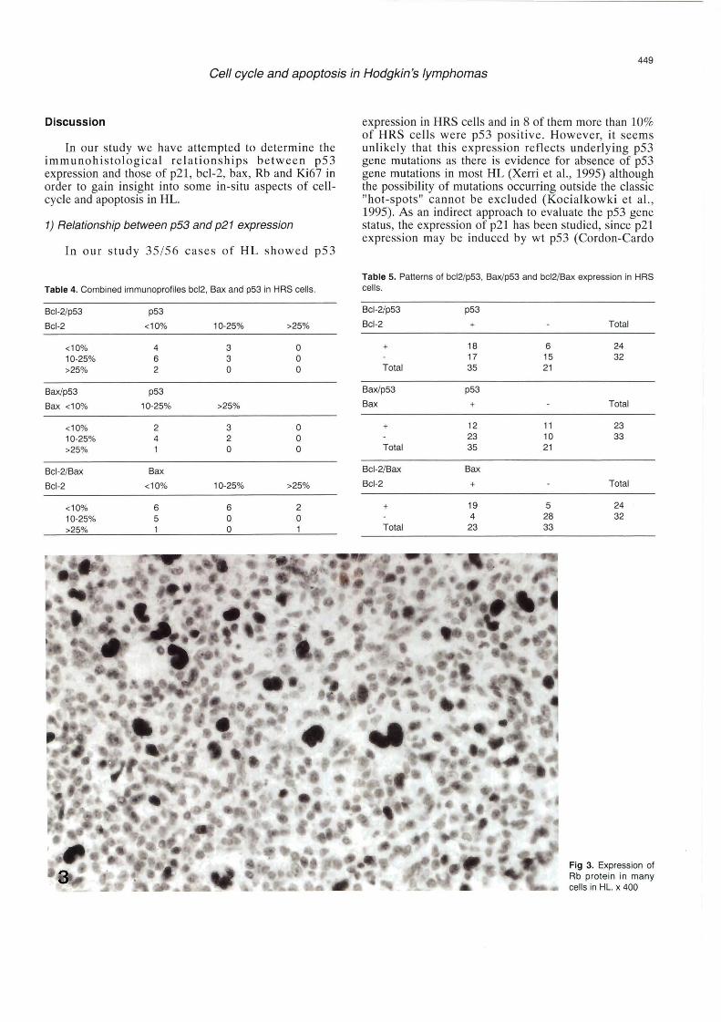
p53 , bcJ-2 and bax were completely undetect able in HRS cells in 21/56, 32/56, and 33/56 cases, respectively. Ki67 (Fig. 2), p21 and Rb expression (Fig. 3) was found in HRS cells in alJ cases (ranging from very few HRS cells to more than 25 % of HRS cells).
No correlation was found between the expression of p53 , p21 , Rb, Ki67 , bcl-2 and bax proteins with the histotypes of HL (Table 1) or the EBV status.
In ali ten "reactive" Jymph nodes numerous Rbpositive cells (10-25 %) parall eled Ki67-positive cells in germinal centers and interfollicular areas (Cannell et al., 1996). p53- and p21-positive cells were absent or very
Table 2. EBV status in HRS cells in relationship with the histotypes of HL.
NS MC Total
EBER
2/18 15/38 17/38
LMP-1
2/18 15/38 17/38
Fig. 1. Expression of p53 prote in in HRS cells of HL. x 400

448
Gel! cycle and apoptosis in Hodgkin 's lymphomas
Table 3. Combined immunoprofiles of Ki67 , Rb, p53 and p21 in HAS rare ( <1 % ) in 7 cases and localised in the same areas cells .
Ki67/Rb Rb
Ki67 <10%
<10% 3 10-25% 2 >25% 2
Ki67/p21 p21
Ki67 <10%
<10% 5 10-25% 2 >25% 2
Ki67/p53 p53
Ki67 <10%
<10% 17 10-25% 7 >25% 3
P53/p21 p21
p53 <10%
<10% 11 10-25% 1 >25% o
P53/Rb Rb
p53 <10%
<10% 2 10-25% 5 >25% o
10-25% >25%
17 9 1 6
13 3
10-25% >25%
18 6 3 4 11 5
10-25% >25%
1 o 3 o 4 o
10-25% >25%
13 3 6 1 o o
10-25% >25%
17 8 2 1 o o
(Mateo et al., 1997). Numerous bax-positive cells were found in germinal centres and interfollicul ar areas in ali cases of "reactive" lymph node s . Bcl-2 stain ed mantle roue and interfollicular areas while germin al centres were mainly negative.
Relationship between p53/p21, p53/bcl-2 , p53/bax and p53/Rb expression
Since p53 can influence the expression of p21, bcl-2 , bax and Rb the correspondin g immunoprofiles in HRS cells were determined.
No statistically significant coexpression or inverse expression was found between p53/Rb or p53/bcl-2 or p53/bax or p53/p21 proteins.
Relationship between bc/-2 and bax expression
A statistic ally significant (p<0.001) coexpression or absence of expression of both proteins was found (Table s 4 and 5). In contrast , there was no statistically significant relationship between each of the pattern s of the Table 5 and the histotype or the EBV statu s.
Relationship between Ki67/Rb, Ki67/p21 and Ki67/p53 expression (Table 3)
No statistically significant coexpression or inverse expression was found between Ki67 /Rb , Ki67 /p21 or Ki67/p53 .
Fig 2. Expression of MIB1 (Ki67) in many cells in HL. x 400
449
Gel/ cycle and apoptosis in Hodgkin 's lymphomas
Discussion
ln our study we have attempted to determine the immunohistological relationships between p53 expression and those of p21 , bcl-2, bax, Rb and }(j67 in order to gain insight into sorne in-situ aspects of ce llcycle and apoptosis in HL.
1) Relationship between p53 and p21 expression
In our st ud y 35 / 56 cases of HL showed p53
Table 4. Combined immunoprofiles bcl2 , Bax and p53 in HRS cells.
Bcl-2/p53 p53
Bcl-2 <10%
<10% 4 10-25% 6 >25% 2
Bax/p53 p53
Bax <10% 10-25%
<10% 2 10-25% 4 >25% 1
Bcl-2/Bax Bax
Bcl-2 <10%
<10% 6 10-25% 5 >25%
10-25%
3 3 o
>25%
3 2 o
10-25%
6 o o
>25%
o o o
o o o
>25%
2 o
expression in HRS cells and in 8 of them more than 10% of HRS ce lls were p53 positive. However, it seems unlikely that th is expression reflects underJying p53 gene mutations as there is ev idence for abse nce of p53 gene mutations in most HL (Xerri et al., 1995) although the possibility of mutations occurri ng outside the classic "hot-spots" can not be exc lud ed (Kocialkowki et al., 1995). As an indirect approach to eva luate the p53 gene status, the expres sion of p21 has been studied, since p21 express ion may be indu ced by wt p53 (Cordon -Cardo
Table 5. Patterns of bcl2/p53 , Bax/p53 and bcl2/Bax expression in HRS cells.
Bcl-2/p53 p53
Bcl-2 +
+ 18 17
Total 35
Bax/p53 p53
Bax +
+ 12 23
Total 35
Bcl-2/Bax Bax
Bcl-2 +
+ 19 4
Total 23
6 15 21
11 10 21
5 28 33
Total
24 32
Total
23 33
Total
24 32
Fig 3. Expression of Rb protein in many cells in HL. x 400

450
Gel/ cycle and apoptosis in Hodgkin 's lymphomas
and Richon, 1994; Sanchez-Beato et al., 1996a; Villuendas et al., 1997; Prives and Hall, 1999). Thus, we have determined the p53/p21 patterns: a) The pattern p53- /p21+ (21 cases). This pattern suggests p53-independent pathway of p21 induction of expression (Steinmann et al., 1994; Cox 1997). However, negative p53 immunostaining may also result from biallelic deletion of p53 gene (Kocialkowski et al., 1995; Cox, 1997), but this seems very unlikely in most HL (Trumper et al. , 1993; Xerri et al., 1995). b) The pattern p53+ /p21 + (35 case). This pattern indicates wild type p53 protein able to induce p21 expression (Cox, 1997). A previous study on lymphomas (Villuendas et al., 1997) showed that the p53+/p21- phenotype was associated to p53 mutations. Interestingly, no p53+ /p21- HL cases were found in our study supporting the previous evidence that p53 mutations are rare in HL (Xerri et al., 1995). However, another study (Trumper et al., 1993) demonstrated mutation of p53 gene codon 246 in 5 of 7 HRS cells examined by single cell PCR in 3/7 patients with HL. lt has been suggested that p53 gene mutation may represent a late event in the pathogenesis of sorne HL associated with disease progression, since only a proportion of HRS cells harboured this mutation (Trumper et al., 1993). Therefore, the search for p53 mutations in HL seems to require single-cell PCR. Besides p53 gene mutations it is possible that accumulation of p53 protein could occur as a consequence of absence of mdm-2 expression, since the p53 / mdm-2 interaction can result in enhanced p53 degradation (Save et al., 1998). However, the detection of mdm-2 protein and mRNA in ali HL studied does not favour this interpretation (Sanchez-Beato et al., 1996b). p53 protein immunodetection could also be the result of molecular complex with the EBNA-5 protein of EBV (Szekely et al., 1993), but no correlation was found in our study between EBV status and p53 expression. In addition, there is no evidence that EBNA-5 is expressed in HRS cells of HL and it is noteworthy that EBVinduced immortalisation does not seem to inactivate the function of p53 (Cannell et al., 1996). An alternative hypothesis to explain p53 protein accumulation without mutation could be the stabilisation of wild-type p53 protein through direct association to hypoxia-inducible factor la (An et al., 1998)
1t should be mentioned, however, that an important caveat of the immunohistochemical analysis of p53/p21 pattern for prediction of p53 activity are the data that each of p53 and p21 proteins can probably regulate more than one pathway which may be mutually exclusive (e.g apoptosis and cell-cycle arrest) (Cox , 1997). Thus, although the p53 /p21 pattern can be informative in certain circumstances (Lorenzen et al., 1997; Villuendas et al., 1997), reliable assessment of p53 protein activity should be based on DNA and RNA studies (Cox, 1997). Taken together, the present and previous data on p53 expression in HRS cells (Xerri et al., 1995; Lorenzen et al., 1997), favour the interpretation of a wt protein able to upregulate p21 expression and involved in an attempt
for cell-cycle arrest. A role of p53 expression in HRS cells for apoptosis could be considered. However, these cells did not exhibit classical apoptotic features when studied by electron microscopy and the TUNNEL method although the possibility of apoptosis without typical features cannot be excluded (Lorenzen et al., 1997).
In line with previous data (Lorenzen et al. , 1997; Naresh et al., 1997), many HL expressed the p21 protein in more than 10 % of HRS cells in this study. Furthermore, we found no statistically significant tendency for parallel or inverse expression of Ki67 and p21 proteins. lt should be noticed, however, that 5 cases showed expression of both proteins in more than 25% of HRS cells, and ali were p53 immunopositive. This suggests that the presumptive p53-induced p21-mediated growth arrest is somehow overriden in this subset of HL. The inability of p21 to induce growth arrest could be related to p21 gene structural alterations but this is unlikely in most human tumours (Shiohara et al., 1994). Other possibilities could be insufficient amounts of p21 protein to inhibit cell-cycle or alterations of the p21 targets (Elledge et al., 1996).
2) Relationship between bax, bcl-2 and p53 expression
In our study bax expression was undetectable in HRS cells in 33 cases, whereas most small lymphocytes in HD as well as most normal lymphoid cells in reactive lymph nades were bax positive. This suggests deregulated expression and could be interesting in view of recent data (Yin et al., 1997) that bax acts as tumour suppressor. The putative role of absence of bax in HL could be related to recent findings in acute lymphoblastic leukaemia cell lines that bax gene mutations resulted in absence of detectable amounts of bax protein (Meijerink et al. , 1998). Thus it is possible that reduced bax expression in HRS cells may be related, at least in sorne cases, to underlying mutations which could confer survival advantage during the development and progression of HL. Therefore, it would be interesting to analyse possible genetic alterations of bax in HL although the one HL-cell line studied showed wild-type bax gene (Meijerink et al., 1998). Another possibility is that reduced bax expression can be due to posttranscriptional regulation of bax protein levels as described in lymphoma cell lines (Leff et al. , 1996). Mention should be made, however, to sorne discrepancies in previous studies (Brousset et al., 1996; Messineo et al., 1998) with respect to bax immunoexpression in HL. Whereas in one study (Brousset et al., 1996) bax was found in HRS cells in most HL, in our study as well as in another study (Messineo et al., 1998) bax protein and mRNA were rarely expressed in HRS cells. This discrepancy is likely to be due to the use of polyclonal (Brousset et al., 1996) or monoclonal antibodies (Messineo et al., 1998, our study). Since nonspecific staining was suggested with the use of polyclonal anti-bax antibody (Messineo et al., 1998) it

451
Ce/1 cycle and apoptosis in Hodgkin 's lymphomas
seems likely that bax expression is not frequent in HRS cells of HL. On the other hand the comparison between bax and bcl-2 expression in HRS cells of HL revealed a s tatistically significant tendency for absence of expression of both proteins or for coexpression. These findings suggest that a delicate balance between proapoptotic (b ax) and anti-apoptotic stimuli (bcl-2) is retained in many HL (19 cases) . Interestingly , 28 baxnegative tumours of our study were also bcl-2 negativ e. lt could be suggested that bcl-2-negati ve tumours may have evolved to a point where they are less dependen! on bcl-2 for their survival and that one of the reasons for this reduced dependence on bcl-2 may be the low bax expression. However, since bcl-2 is not the only antiapoptotic member of the bcl-2 protein family (Adams and Cory, 1998) , it is possible that low bax expre ssion may be importan! because the ratio of bax with other anti-apoptotíc proteins such as bel-xi and mcl-1 presumably has been modified in tumours with low bax expression . In this regard previous studie s (Brou sset et al., 1996; Messineo et al. , 1998) showed frequent ímmonoexpre ssíon of mcl-1 and bcl-x and frequent mRNA expre ssíon of bel-xi (Messineo et al. , 1998) in HRS cells sugg estíng a role of these apoptosi s-related proteins in HL. The mechanísms of reduced bax expression in a subset of HL may be related to p53 since this gene can transactivate the bax gene promoter whích contaín s four consen sus bindíng site sequences for p53 (Miyashita and Reed , 1995). However , no statistically sígníficant correlation was found between increased p53 and bax ímmunoexpression in our study. This finding could be related to previous evidence for p53-independent mechanisms for regulating bax gene expression (Miy ashita et al., 1994).
Although , bcl-2 expression can be down-regulated in vitro by wild-type and mutant p53 (Adams and Cory , 1998), in agreement with previous data (Doussis et al., 1993), we found no statistically significant coexpre ssion or inverse expression between bcl-2 and p53 in HL. This finding can also be compared with our previous data that there was an inverse bcl2/p53 relationship in thymom as (Stefanaki et al., 1997). These data suggest that bcl-2/p53 interactions may be tumour-type dependent and do not seem to be influ enced by EBV infection in EBVassociated tumours such as HL.
3) Relationship between Rb, and Ki67 expression
In sorne case s of HL the relationship between RB and Ki67 expression has been disturbed and two main deviations from the normal pattern of the reactive lymph nodes have been observed: a) A subset of cases (2 cases) showed low ( <10 %) Rb pro te in expression in relation ship with high (>25 %) Ki67 expression . This could reflect loss of Rb expression due to loss of chromosome 13 since Rb gene is located in 13q14 (Wiman, 1993; Cordon-Cardo and Richon, 1994). In this respect, a previous study (Tilly et al., 1991) in untreated cases of HL showed frequent loss of chromosome 13
suggesting the possibility of loss of heterozygo sity of Rb in the pathogenesis of sorne HL. Another possibility is Rb gene mutation or defects in the transcription and translation of Rb gene; b) Another subset of cas es (9 cases) showed high Rb (>25 %) with low Ki67 (<10%) expression . This could reflect an attempt of the Rb protein in exce ss to induce cell cycle arres!. Another hypothesis is increased Rb expression due to infection from EBV. Indeed , Rb protein can form complexes with EBNA-5 protein (Sz ekely et al., 1993) and EBVtransformed B-cell s show increased leve! of hyperphosphorylated Rb protein in compari son to primary non-infected B-cells (Cannell et al. , 1996) . Evidence was provided that EBV uses a s trategy involving hyp erpho sphorylation of pRb and altered expression of other important cell-cycle proteins (i.e . p21 , p53 , bcl-2) in order to drive cell proliferation (Cannell et al. , 1996) . However , in our se ries , no positive correlation was found between increased Rb expression and EBV status in HRS cells .
In summary , our findings provide combined immunohistological evidence for a deregulation of the expression of apoptotic and cell cycle proteins, such as bax , bcl-2, p53, p21, Rb and Ki67, that may play a role in the pathogene sis of HL.
Acknowledgments . The authors are grateful to Mrs Maria Papadaki for excellent secretaria! assistance and to Mrs Elli Karidi for technical work.
References
Adams J.M. and Cory S. (1998). The BCL-2 protein family: Arbiters of cell survival. Science 281, 1322-1326.
An W.G., Kanekal M., Simon M.C., Maltepe E., Blagosklonny M.V. and Neckers L.M. (1998). Stabilisation of wild-type p53 by hypoxia inducible factor 1 a. Natura 392, 405-408.
Brousset P., Benharroch D., Krajewki S., Laurent G., Meggeto F., RigalHuguet F., Gopas J., Prinsloo l., Pris J., Delsol G., Reed J.C. and Schlaifer D. (1996). Frequent expression of the cell death-inducing gene Bax in Reed-Sternberg cells of Hodgkin's disease. Blood 87, 2470-2475.
Cannell E., Farrell P.J . and Sinclair A.J . (1996). Epstein-Barr virus exploits the normal cell pathway to regulate Rb activity during !he immortalisation of primary B-cells. Oncogene 13, 1413-1421.
Cordon-Cardo C . and Richon V .M. (1994) . Expression of !he retinoblastoma protein is regulated in normal human tissues. Am. J. Pathol. 3, 144-153.
Cox L.S (1997) . Multiple pathways control cell growth and transformation: Overlapping and independent activities of p53 and p21. J. Pathol.183, 134-140.
Doussis I.A. , Pezzella F., Lane P.L., Gatter K.C. and Mason D.Y. (1993). An immunohistochemical study of p53 and bcl-2 protein expression in Hodgkin's disease. Am. J. Clin. Pathol. 99, 663.
El Deiry W.S., Tokino T., Velculescu V.E., Levy DB., Parsons R., Tren! J.M., Lin D., Mercar W.E., Kinzler K.W. and Vogelstein B. (1993). Waf-1 a potential mediator of p53 tumour suppression. Cell 75, 817-825.
Evan G. and Littlewood T. (1998). A matter of lile and cell death .
452
Gel/ cyc/e and apoptosis in Hodgkin 's lymphomas
Science 281, 1317-1322.
Elledge S.J. (1996). Cell-cycle checkpoints: preventing an identity cris is.
Science 274, 1664-1672.
Greenblat M., Bennerr W ., Abllstein M. and Harris e.e. (1994) .
Mutations in the p53 tumor suppressor gene : clues to cancer etiology and molecular pathogenesis. Cancer Res. 54, 4855-4878 .
Harris N.L., Jaffe E.S., Stein H., Banks P.M., Chan J.K.C., Cleary M.L.,
Delsol G., De Wolf-Peeters C., Falini B., Gatter K.C., Grogan T.M.,
lsaacson P.G., Knowels D.M., Masan O.Y., Muller-Hermelink H.K.,
Pileri S.A. , Piris M.A ., Ralfkiaer E. and Warnke A.A. (1994) . A
revisad European-American classificat ion of lymphoid neoplasms : a
proposal from the lnternational Lymphoma Study Group . Blood 84, 1361-1392.
Jiwa N., Kanavaros P., Van der Valk P., Walboomers J., Hostman A.,
Vos W., Mullink H. and Meijer C. (1993). Reed-Sternberg cells in
Hodgkin 's disease express frequently c-myc and bcl-2 oncogene
products independently of the presence of EBV. J. Clin. Pathol. 46,
211-217.
Kanavaros P .. Sakalidou A. , Tzardi M., Darivianaki K. and Delidis G.
(1994). Frequent detection of EBV-EBER transcripts and LMP -1
protein in tumor cells in Hodgkin's disease arising in childhood.
Pathol. Res. Pract. 190, 1026-1030.
Kanavaros P., Lescs M.C., Briere J., Divine M., Galateau F., Joab l.,
Bosq J., Farcet J.P., Reyes F. and Gaulard P. (1993). Nasal T-cell
lymphomas : a clínico pathologic ent ity associated with peculiar phenotype and with Epstein-Barr virus . Blood 81, 2688-2691.
Kocialkowski S., Pezzela F., Morrison H., Janes M., Laha S. and Harris
AL. (1995). Mutations in the p53 gene are not limited to classic "hot
spots" and are not predictiva of p53 protein expression in high-grade non-Hodgkin's lymphoma . Br. J. Haematol. 89, 55-60.
Leff M.A. , Buckley D.J. and Krumenacker J.S. (1996). Rapid modulat ion
of the apoptosis regulatory genes, bcl-2 and bax by prolactin in rat Nb2 lymphoma cells . Endocrinology 137, 5456-5462.
Leoncini L., Donatela S., Glose P., Minacci C., Megla T., De Luka F.,
Tasi P., Pileri S., Kraft R. , Laissue J.A. and Cottier H. (1997). Mitotic
activity and nuclear DNA damage of Large cells in Hodgkin 's
disease: comparison with expression of p53 and bcl-2 proteins and
the presence of Epstein-Barr virus. Leukemia Lymphoma 25, 153-161.
Lorenzen J., Thile J. and Fischer R. (1997) . The mummified Hodgkin
cell: cell death in Hodgkin's disease . J. Pathol. 182, 288-298 .
Mateo M.S., Saer A.I., Sanchez-Beato M., García P., Sancher-Verde L.,
Martinez J.C., Orradre J.L. and Piris M.A. (1997). Expression of p21
in fetal and adults tissues : Simultaneous analysis with Ki67 and p53. J. Clin. Pathol. 50, 645-653 .
Meijerink J., Mensink E., Wang K., Sedlak T. , Sloetjes A. , deWitte T. ,
Waksman G. and Korsmeyer J. (1998). Hematopoietic malignancies
demonstrate loss of function mutations of bax. Blood 91, 2991-2997 .
Messineo C., Jameson H., Huntec E., Brariel R., Bagg A., lrving S.G.
and Cossman J. (1998). Gene expression by single reed-sternberg
cells. Pathways of apoptosis and activation . Blood 91, 2243-2245.
Miyash ita T. and Reed J. (1995) . Tumour suppressor p53 is a direct transriptional activator ot human bax gene. Cell 80, 293-299.
Miyashita T ., Krajewsk i S ., Krajewska M., Wang H.G. , Lin H.K.,
Lieberman O.A ., Hoftman B . and Rees J .C . (1994) . Tumour
suppressor p53 is a regulator ot bcl-2 and bax in gene expression in
vitre and in vivo . Oncogene 9, 1799-1805.
Morente M.M., Piris M.A. , Abraira V., Acevedo A., Aguilera B., Bellas C.,
Fraga M., García-Del-Moral R., Gomez-Marcos F., Menarguez J.,
Oliva H., Sanchez-Beato M. and Monta lban C. (1997). Adv erse
clinical outcome in Hodgkin 's disease is assoc iated with loss ot Retinoblastoma protein expression , high Ki67 proliferation index,
and absence of Epstein-Barr virus Latent Membran a Prote in 1
expression. Blood 90, 2429-2436. Naresh K.N., O'Conor G., Sornan C., Johnson J., Advani S., Magrath l.
and Bhatia K.G. (1997) . A study of p53 protein , prol iferation cell
nuclear antigen and p21 in Hodgkin's disease at presentation and at
relapse. Hum. Pathol. 28, 549-555.
Polyak K., Xia Y., Zweler J.L., Kinzler K.W. and Vogelstein B. (1997). A
modal far p53-induced apoptosis . Natura 389, 300-305.
Prives C. and Hall P. (1999). The p53 pathway. J. Pathol. 187, 112-1
26.
Sanchez -Beato M., Martinez -Montero L.C., Doussis-Anagnostopoulou
T.A. , Gatter K.C., García J., García J.F., Loret E.L. and Piris M.A.
(1996a). Anomalous retinoblastoma protein expression in Sternberg
Reed cells in Hodgkin's disease : a comparative study with p53 and
Ki67 expression. Br. J. Cancer 74, 1056-1062.
Sanchez-Beato M., Pir is M., Martinez-Mo ntero J .C ., García J .F., Villuendas R., Garcia F.J. , Orradre J.L. and Martinez P. (1996b).
Mdm2 and p21 wild-type p53- induced proteins, are regularly
expressed by Sternberg-Reed cells in Hodgkins disease. J Pathol.
180, 58-64.
Save V ., Nylande r K. and Hall P. (1998) . Why is p53 stab ilized in
neoplasia? Sorne answers but many more questions . J. Pathol. 184,
348-353.
Shiio Y., Yamamoto T. and Yamaguchi N. (1992). Negativa regulation ot Rb express ion by the p53 gene product. Proc. Natl. Acad . Sci. USA
89, 5206-5210. Shiohara M., El Deiry W.S ., Wada M., Nakamaki T ., Takeuchi S., Yang
R., Chen D., Vogelstein B. and Koeffler P.H. (1994). Absence ot waf1 mutations in a variety of human malignancies . Blood 84, 3781-
3784.
Sinclair A .J., Fenton M. and Dellikat S (1998). lnteractions between
Epst ein-Barr virus and the cell cyc le control machin ery. Histol.
Histopathol. 13, 461-467.
Stefanaki K. , Rontogianni D. , Kouv idou C., Boliot i S. , Delides G.,
Pantelidaki A., Sotsiou F. and Kanavaros P. (1997). lmmunohisto
chemical expression of bcl-2 , p53, mdm-2 and p21/waf1 proteins in
thymomas . Histopathology 30, 549-555 .
Steinmann A.A. , Haffman B., Ira A., Guillouf C., Liebermann O.A. and
Hou seini M.E. (1994) . lnduction of p2 1 (WAF1 / CIP 1) du ring
differentiatio n. Oncogene 9, 3389-3396 .
Szekely L., Selivanova G., Magnusson K.P., Klein G. and Wiman K.G.
(1993) . EBNA-5 , an Epstein -Barr virus-encoded nuclear antigen
binds to retinoblastoma and p53 protein. Proc. Natl. Acad . Sci. USA 90, 5455-5459 .
Tilly H., Bastard C., Delastre T., Duval C., Biret M., Lenomard B., Dance
J.P., Moncondrut M. and Pignet H. (1991). Cytogenet ic studies in
untreated Hodgkin's disease. Blood 77, 1298-1304.
Trumper L.H., Brady G.B., Bagg A. , Gray D., Loke S.L., Griesser H.,
Wagman R. , Braziel R., Gascoyne R.O., Vicini S., lscove N.N., Cossman J. and Mak T.W. (1993) . Single-cell analysis of Hodgkin
and Reed -Sternberg cells: molecular heterogeneity of ge ne
expression and p53 mutations . Blood 81, 3097-3115 .
Villuendas R., Pezzella F., Gatter K., Algara P., Sanchez -Beato M.,
Mart inez P., Martinez J .C., Minoz K., Gar cía P., San chez L.,
Kocialkowsky S., Campo E., Orra nde J.L. and Piris M.A. (1997).
p21WAF1/CIP1 and MDM2 expression in non-hodgkin 's lymphoma

453
Gel/ cycle and apoptosis in Hodgkin's lymphomas
and t he ir relationship to p53 status : A p53+ , mdm2+ , p21-immunophenotype associated with missense p53 mutat ions . J .
Pathol. 181, 51-61. Waga S., Hannon G.J. , Beach D. and Stilman B. (1994) . The p21
inhib itor of cyc lin-dependent kinase controls DNA replicat ion by interactions with PCNA. Nature 369, 574-578.
Wiman K.G. (1993). The retinoblastoma gene: Role in cell cycle control
and cell differentiation FASEB J. 7, 841-845 . Xerri L., Pare P., Bouabdallash R., Camerlo J. and Hassoun J. (1995).
PCR-mismatch analysis of p53 gene mutations in Hodgk in's disease. J. Pathol. 175, 189-194.
Xiong Y., Hannon G.J., Zhang H., Casso D., Kobayashi R. and Beach D. (1993). P21 is universal inhibitor of cyclin kinase. Nature 366 , 701-704.
Yin C ., Knudson M., Korsmeye r S.J . and Dyke T . (1997 ) . Bax suppresses tumorigenesis and stimulates apoptosis in vivo. Nature 385, 637-640.
Zhu V .M., Haynes A .P. , Keith F.J . and Russell N.H. (1995).
Abnormalities of retinoblastoma gene expression in hemato logical malignancies. Leukemia Lymphoma 18, 61-67.
Accepted November 16, 1999