Expression of NF-L in both Neuronal and Nonneuronal Cells ... · conservation of the intron...
15
Expression of NF-L in both Neuronal and Nonneuronal Cells of Transgenic Mice: Increased Neurofilament Density in Axons without Affecting Caliber Mervyn J. Monteiro,* Paul N. Hoffman,§ John D. Gearhart,* and Don W. Cleveland* Departments of *Biological Chemistry, ~ Physiology, and § Ophthalmology and Neurology, The Johns Hopkins University School of Medicine, Baltimore, Maryland 21205 Abstract. We have generated transgenic mice contain- ing additional copies of the murine NF-L gene in or- der to examine the consequences of neurofilament-L overexpression on axonal morphology. Founder mice were constructed to carry a transgene in which the presumptive 5' promoter sequences of NF-L were replaced with the strong murine sarcoma virus long terminal repeat promoter. The transgenes were ex- pressed prominently in several tissues, including skele- tal muscle and kidney where NF-L accumulated to ,,02% of cell protein. This was not accompanied by an overt phenotype, except that expression in lens led to cataract formation. In the brains of these animals, transgene RNA levels exceeded the endogenous NF-L RNAs by up to 20-fold, although no additional protein accumulated, indicating posttranscriptional regulation of NF-L expression. However, in peripheral neurons transgene RNA was approximately fourfold higher than endogenous NF-L mRNAs, and a corresponding increase in NF-L subunits was found in axons arising from these neurons. Myelinated nerve fibers of trans- genic animals contained increased numbers of NFs, assembled predominantly of NF-L. This was reflected in an increase in the density of axonal NFs; axonal caliber was not affected. I NTERMEDIATE filaments (IFs) 1 are 10-nm filaments which, together with microtubules and microfilaments, form the cytoskeleton of eukaryotic cells. However, un- like microtubules and microfilaments whose subunits are well conserved in sequence, IFs from different tissues are as- sembled from a more divergent subunit composition. This heterogeneity has been shown by biochemical, immunologi- cal, and molecular characterization to be due principally to differences in the "head" and "tail" domains of the subunit polypeptides which otherwise all contain a central, structur- ally conserved, a-helical coiled-coil domain of ~ 310 resi- dues (Geisler and Weber, 1986; Franke, 1987; Steinert and Roop, 1988). On the basis of sequence differences and on the conservation of the intron positions, IFs have been divided into five generally accepted classes. Class I and class II are composed of the type I (acidic) and type II (basic) keratins, respectively, both of which are expressed in epithelial cells. Class III consists of vimentin expressed in mesenchymal cells, desmin expressed in muscle cells, glial fibrillary acid Mervyn J. Monteiro's present address is Department of Neurology and the Medical Biotechnology Center, The University of Maryland School of Medicine, Baltimore, Maryland 21201. 1. Abbreviations used in this paper: DRG, dorsal root ganglia; IF, inter- mediate filament; LTR, long terminal repeat; MSV, murine sarcoma virus; NF, neurofilament. protein expressed in glial cells, and peripherin expressed in a subset of neurons of the peripheral nervous system. Class IV comprises the major neurofilament (NF) proteins that are probably expressed in all neuronal cells. Class V comprises the nuclear lamins. The newly discovered IF subunit nestin, found in neuroepithelial stem cells, has been proposed to represent a separate sixth IF class (Lendahl et al., 1990). In mammals, NFs are composed primarily of three subunits, NF-L (68 kD), NF-M (160 kD), and NF-H (200 kD) (Hoffman and Lasek, 1975). Each carries a 310-amino acid c~-helical rod domain following an •100-residue head domain, and differences in size are primarily due to the length of the carboxy-terminal tail sequences (Lewis and Cowan, 1986; Levy et al., 1987; Myers et al., 1987; Julien et al., 1988; Lees et al., 1988). The disposition of the three NF proteins in the native polymer is not known; however, since all three subunits contain the rod domain, almost cer- tainly each is an integral filament subunit. The role of head and tail domains is less clear but immunocytochemical and in vitro reassembly experiments suggest that the tail domains of NF-M and NF-H are mostly involved in the formation of NF cross-bridges (Geisler and Weber, 1981; Sharp et al., 1982; Geisler et al., 1983; Julien and Mushynski, 1983; Minami and Sakai, 1985; Hirokawa et al., 1984; Hisanaga and Hirokawa, 1988). A convincing functional role for any IF has yet to be © The Rockefeller University Press, 0021-9525/90/10/1543/15 $2.00 The Journal of Cell Biology, Volume 11 l, October 1990 1543-1557 1543
Transcript of Expression of NF-L in both Neuronal and Nonneuronal Cells ... · conservation of the intron...
Expression of NF-L in both Neuronal and Nonneuronal Cells of Transgenic Mice: Increased Neurofilament Density in Axons without Affecting Caliber M e r v y n J. Monteiro ,* Pau l N. Hof fman ,§ J o h n D. Gea rha r t , * a n d D o n W. Cleveland*
Departments of * Biological Chemistry, ~ Physiology, and § Ophthalmology and Neurology, The Johns Hopkins University School of Medicine, Baltimore, Maryland 21205
Abstract. We have generated transgenic mice contain- ing additional copies of the murine NF-L gene in or- der to examine the consequences of neurofilament-L overexpression on axonal morphology. Founder mice were constructed to carry a transgene in which the presumptive 5' promoter sequences of NF-L were replaced with the strong murine sarcoma virus long terminal repeat promoter. The transgenes were ex- pressed prominently in several tissues, including skele- tal muscle and kidney where NF-L accumulated to ,,02% of cell protein. This was not accompanied by an overt phenotype, except that expression in lens led to cataract formation. In the brains of these animals,
transgene RNA levels exceeded the endogenous NF-L RNAs by up to 20-fold, although no additional protein accumulated, indicating posttranscriptional regulation of NF-L expression. However, in peripheral neurons transgene RNA was approximately fourfold higher than endogenous NF-L mRNAs, and a corresponding increase in NF-L subunits was found in axons arising from these neurons. Myelinated nerve fibers of trans- genic animals contained increased numbers of NFs, assembled predominantly of NF-L. This was reflected in an increase in the density of axonal NFs; axonal caliber was not affected.
I NTERMEDIATE filaments ( IFs) 1 a r e 10-nm filaments which, together with microtubules and microfilaments, form the cytoskeleton of eukaryotic cells. However, un-
like microtubules and microfilaments whose subunits are well conserved in sequence, IFs from different tissues are as- sembled from a more divergent subunit composition. This heterogeneity has been shown by biochemical, immunologi- cal, and molecular characterization to be due principally to differences in the "head" and "tail" domains of the subunit polypeptides which otherwise all contain a central, structur- ally conserved, a-helical coiled-coil domain of ~ 310 resi- dues (Geisler and Weber, 1986; Franke, 1987; Steinert and Roop, 1988). On the basis of sequence differences and on the conservation of the intron positions, IFs have been divided into five generally accepted classes. Class I and class II are composed of the type I (acidic) and type II (basic) keratins, respectively, both of which are expressed in epithelial cells. Class III consists of vimentin expressed in mesenchymal cells, desmin expressed in muscle cells, glial fibrillary acid
Mervyn J. Monteiro's present address is Department of Neurology and the Medical Biotechnology Center, The University of Maryland School of Medicine, Baltimore, Maryland 21201.
1. Abbreviations used in this paper: DRG, dorsal root ganglia; IF, inter- mediate filament; LTR, long terminal repeat; MSV, murine sarcoma virus; NF, neurofilament.
protein expressed in glial cells, and peripherin expressed in a subset of neurons of the peripheral nervous system. Class IV comprises the major neurofilament (NF) proteins that are probably expressed in all neuronal cells. Class V comprises the nuclear lamins. The newly discovered IF subunit nestin, found in neuroepithelial stem cells, has been proposed to represent a separate sixth IF class (Lendahl et al., 1990).
In mammals, NFs are composed primarily of three subunits, NF-L (68 kD), NF-M (160 kD), and NF-H (200 kD) (Hoffman and Lasek, 1975). Each carries a 310-amino acid c~-helical rod domain following an •100-residue head domain, and differences in size are primarily due to the length of the carboxy-terminal tail sequences (Lewis and Cowan, 1986; Levy et al., 1987; Myers et al., 1987; Julien et al., 1988; Lees et al., 1988). The disposition of the three NF proteins in the native polymer is not known; however, since all three subunits contain the rod domain, almost cer- tainly each is an integral filament subunit. The role of head and tail domains is less clear but immunocytochemical and in vitro reassembly experiments suggest that the tail domains of NF-M and NF-H are mostly involved in the formation of NF cross-bridges (Geisler and Weber, 1981; Sharp et al., 1982; Geisler et al., 1983; Julien and Mushynski, 1983; Minami and Sakai, 1985; Hirokawa et al., 1984; Hisanaga and Hirokawa, 1988).
A convincing functional role for any IF has yet to be
© The Rockefeller University Press, 0021-9525/90/10/1543/15 $2.00 The Journal of Cell Biology, Volume 11 l, October 1990 1543-1557 1543
l kb A N F - L Mstll Aval
Av.aal I / BssHII Xbal Xbal _, / Hindlll ~-COHI/Sacl BssH'I ~ATG[ r I I Sacl Hindlll
I I f l I , , , , , , , , , , , , , , , , , , , ,
NF-L(Bam) B BamHI Xbal
I / CCCCCC GACGGATCCTCTAGAGTC TGAGGG Xbal Xbal
Hindlll H i ~ I ~ A ~ ~ i sac, TGAEClOR'/ S~C, I
BssHII Aval BssHII
MSV-NF-L C Hindlll Aval
• Aval I BssHII Xia/ac I Xbal I Sacl [ ATIGr "" . Hindlll TGAEC°RI~ ' Sacl
1 NF-L promoter ~ NF-Lcoding region [ I NF-L introns
NF-L5'or 3' ut ,~mu NF-L3' flanking region ~ MSV LTR
Figure 1. Schematic drawings of the mouse NF-L gene and the two NF-L transgene constructs. (A) Restriction map and principal features of the 7-kb Hind III fragment containing the mouse NF-L gene. (B) Schematic drawing of pNF-L(Bam). This gene was constructed by inserting a 18-bp sequence, contain- ing Bam HI and Xba I restriction sites, between the Ava I and Mst II sites of pNF-L (5 bases were deleted in this pro- cess leaving a net insertion of 13 bp). (C) Schematic drawing of pMSV-NF-L. This gene was constructed by replacing the 5' 1.7-kb Hind III-Bam HI fragment of pNF-L(Bam) with the 0.6-kb Hind III-Bgl I fragment that contains the MSV promoter segment.
proven, although the keratin and lamin networks are almost certainly required for structural integrity of the epidermis and nuclear lamina, respectively. For NFs, a functional role was first suggested on the basis of the close correlation be- tween the number of axonal NFs and the cross-sectional areas of large myelinated nerve fibers (Friede and Samoraj- ski, 1970; Weiss and Mayr, 1971; Berthold, 1978). More di- rect evidence for a role of NFs in the control of axonal caliber was provided by the observation that reductions in NF gene expression after axotomy correlate with somatofugal axonal atrophy (Hoffman et al., 1987). This axonal atrophy, which is associated with reductions in both the amount of pulse- labeled NF protein undergoing axonal transport in the prox- imal stumps of transected nerve fibers and axonal NF con- tent, begins proximally near the cell body (soma) and spreads somatofugally along the nerve fiber at a rate equal to the ve- locity of NF transport along the axon (Hoffrnan et al., 1984; 1985). Additional evidence for the role of NFs in the control of caliber comes from recent studies which show that the radial growth of myelinated nerve fibers, which occurs dur- ing postnatal development, correlates temporally with the induction of NF gene expression in developing sensory neu- rons (Muma, N. A., and P. N. Hoffman, unpublished obser- vations).
NF-L is the most abundant NF subunit and in vitro experi- ments have shown it to be capable of assembly without the other subunits (Geisler and Weber, 1981; Liem and Hutchin-
son, 1982). We previously demonstrated that forced expres- sion of NF-L in fibroblasts resulted in the assembly of the protein into filaments and was not detrimental to the growth of fibroblasts (Monteiro and Cleveland, 1989). However, it was not known if similar high levels of inappropriate expres- sion of an IF would affect embryogenesis or development. To begin to address such questions, including the role of each NF subunit in neuronal function, we have generated trans- genic mice expressing high levels of NF-L in both neuronal and nonneuronal cell types. The resultant increased expres- sion and assembly of NF-L in neurons allowed us a first test of the hypothesis that NF expression is an intrinsic deter- minant of axonal caliber. We show that increased NF-L ex- pression alone is in fact not sufficient to affect axonal caliber; rather, it generates disorganization of the NF array in axons.
Materials and Methods
Molecular Cloning of the Mouse pNF-L Gene: Construction of pNF-L(Bam) and pMSV-NF-L Transgenes As described previously, the complete NF-L gene of mouse was isolated as a 7-kb Hind III fragment, and subcloned into pUC9 to yield pNF-L (Mon- teiro and Cleveland, 1989). For transgenic animals, pNF-L was manipu- lated to produce two additional clones, pNF-L(Bam) and pMSV-NF-L (Fig. 1). pNF-L(Bam) contains the entire NF-L coding sequences including 1,7 kb of sequences 5' to the ATG start codon and 1.7 kb of 3' terminal noncod-
The Journal of Cell Biology, Volume 111, 1990 1544
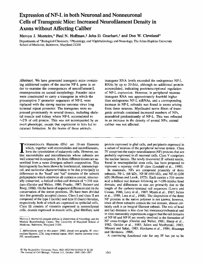
lqgure 2. Identification of pNF-L(Bam) transgenic mice and analysis of its ex- pression in various mouse tissues. (A) A blot of mouse tail DNA (10 #g) was (digested with both Barn HI and Eco RI) probed with a radiolabeled probe corresponding to the 4.2-kb Bam HI- Eco RI fragment of pNF-L(Bam). ( lane 1) Transgenic mouse ceNF-9 DNA; (lane 2) transgenic mouse qNF-16 DNA; (lane C) nontransgenic litter- mate. The endogenous NF-L gene is contained on a 9-kb fragment whereas the transgene is contained on a 4.2-kb fragment. Both transgenic mice con- tained approximately one to two in- tegrated transgene copies. (B) RNA expression of pNF-L(Bam) in trans- genic mouse tissues. RNA was iso- lated from various tissues of a trans- genic F1 progeny of line cYNF-9. 10-#g aliquots were analyzed for NF-L RNAs using S1 nuclease protection of an end-labeled probe corresponding to the Hind III-Ava I fragment of pNF- L(Bam). RNAs from the transgene are expected to yield a 183-base protected fragment, whereas a 93-base fragment is expected from endogenous NF-L mRNAs. Two protected fragments seen in all lanes ('~114 and 125 bases) are contaminants in the probe and were seen even when no RNA was added (left lane). The position of migration of pBR322-digested Hpa II DNA frag- ments of known size are marked at the right.
ing sequences. This clone differs from the authentic gene by insertion of an 18-base linker sequence at the abutting Ava I-Mst II sites in the 5' untrans- lated region, resulting in the loss of 5 NF-L nucleotides but a net gain of 13 nucleotides. Using S1 nuclease protection methods, transcripts derived from pNF-L(Bam) can be distinguished from authentic NF-L RNAs by the presence of the linker sequence (Monteiro and Cleveland, 1989). pMSV- NF-L was constructed by replacing the Hind III-Bam HI fragment of pNF- L(Bam) with a 0.6-kb Hind III-Bgl II fragment containing the murine sar- coma virus (MSV) long terminal repeat (LTR) (Fig. 1 C; see also Monteiro and Cleveland, 1989).
Production of Transgenic Mice
Embryos were recovered from the oviducts of superovulated B6AFIJ fe- males ",12 h after fertilization. The female pronucleus of one-cell embryos was injected with '~300 copies of DNA fragments containing either the 7-kb Hind HI fragment of the pNF-L(Bam) gene construct (Fig. 1 B) or with 5.9- kb Hind III fragment of pMSV-NF-L (Fig. 1 C). The DNA fragments were gel purified from vector sequences using DEAB paper (Dretzen et al., 1981). After incubation at 37°C for 18 h in Whitten's medium, the surviving two-cell embryos were transferred to the oviducts of pseudopregnant CD1 females. Mice born from these mothers were analyzed for the presence of the transgenes as described below.
Southern Blotting of Mouse Tail DNA
Transgenic mice were identified by screening DNA isolated from mouse tail for the presence of the injected DNA fragments. 3 wk after birth a portion of the mouse tail was excised with a razor blade, and the tissue dispersed with a polytron homogenizer into 10 ml of 10 mM Tris-HC1, pH 7.5, 100 mM NaC1, 1 mM sodium EDTA. The samples were incubated at 37"C for
1 h after addition of Sarkosyl to 1% (wt/vol), pronase to 200 ttg/ml, and RNase A to 100 #g/ml. The DNA was extracted once with phenol/chloro- form (1:1), once with ether, then adjusted to 0.3 M with potassium acetate and precipitated at -20oc with 2 vol ethanol. The DNA precipitate was collected by centrifugation, resuspended in 10 mM Tris-HCl, 1 mM sodium EDTA, and a 10-#g aliquot was cleaved with restriction enzymes (Eco RI and Barn HI for analysis of the pNF-L[Bam] transgene and with Sac I for the analysis of the pMSV-NF-L transgene), separated by agarose gel elec- trophoresis, and transferred onto nitrocellulose paper (Southern, 1975). The nitrocellulose was baked at 80°C for 1 h; prehybridized at 42°C in 50% formamide, 5x SSC (lx SSC is 0.15 M NaC1, 0.015 M sodium citrate, pH 7.0), 50 mM Tris-HCl, pH 7.5, 0.5% (wt/vol) SDS, 1 mM sodium EDTA, 5× Denhardt's solution (Denhardt, 1966), and 100 #g/ml of sonicated, denatured salmon sperm DNA; and then hybridized for 48 h with 1 x 10⁶ cpm/ml of labeled probe prepared by random primer extension using radio- active t~-32p-dATP and AMV reverse transcriptase. For detection of the pNF-L(Bam) transgene, the probe was the 4.2-kb Barn HI-Eco RI fragment of pNF-L(Bam) (Fig. 1 B).
SDS-PAGE and Immunoblot Detection of NF-L in Tissue Samples
Mouse tissues were lysed in 0.5 % (wt/vol) SDS, 50 mM Tris-HCl, pH 6.8, 8 M urea, then boiled in Laemmli loading buffer (Laemmli, 1970) contain- ing 2 % fl-mereaptoethanol, and the proteins were separated by electropho- resis on 6 or 8.5% polyacrylamide gels (Laemmli, 1970). Proteins were transferred onto nitrocellulose filters (type BA83; Schleicher and Schuell, Inc., Keene, NH) and immunoblotted as described by Lopata and Cleveland (1987). NF-L protein was detected by autoradiography by incubating the filters first with the mouse monoclonal anti-NF-68 antibody (Boehringer
Monteiro et al. Expression of NF-L in Transgenic Mice 1545
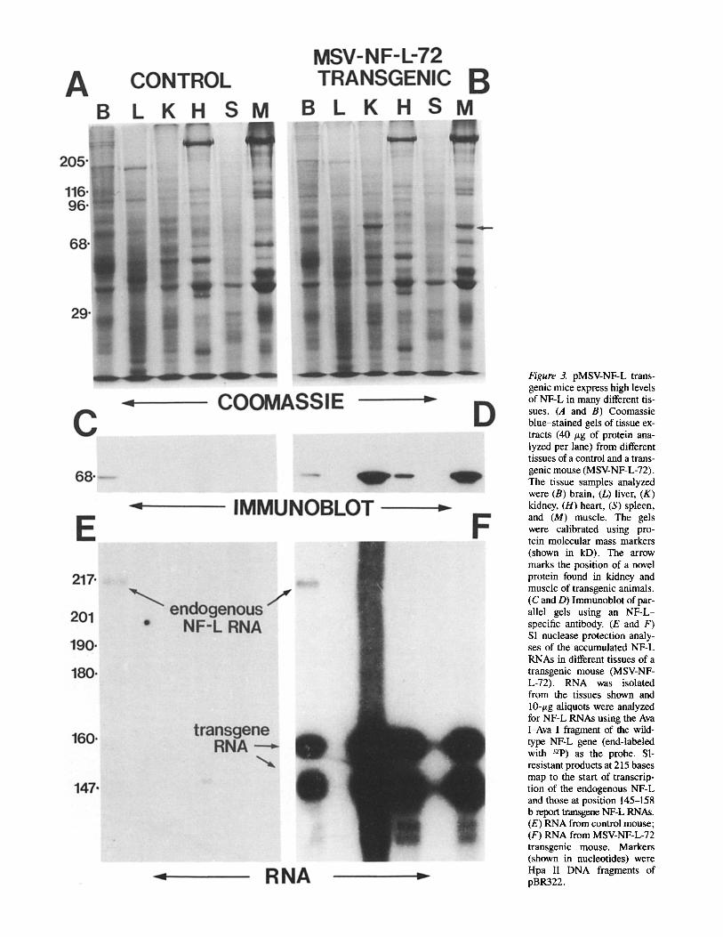
Figure 3. pMSV-NF-L trans- genic mice express high levels of NF-L in many different tis- sues. (A and B) Coomassie blue-stained gels of tissue ex- tracts (40 /~g of protein ana- lyzed per lane) from different tissues of a control and a trans- genic mouse (MSV-NF-L-72). The tissue samples analyzed were (B) brain, (L) liver, (K) kidney, (H) heart, (S) spleen, and (M) muscle. The gels were calibrated using pro- tein molecular mass markers (shown in kD). The arrow marks the position of a novel protein found in kidney and muscle of transgenic animals. (C and D) Immunoblot of par- allel gels using an NF-L- specific antibody. (E and F) S1 nuclease protection analy- ses of the accumulated NF-L RNAs in different tissues of a transgenic mouse (MSV-NF- L-72). RNA was isolated from the tissues shown and 10-/~g aliquots were analyzed for NF-L RNAs using the Ava I-Ava I fragment of the wild- type NF-L gene (end-labeled with 32p) as the probe. SI- resistant products at 215 bases map to the start of transcrip- tion of the endogenous NF-L and those at position 145-158 b report transgene NF-L RNAs. (E) RNA from control mouse; (F) RNA from MSV-NF-L-72 transgenic mouse. Markers (shown in nucleotides) were Hpa II DNA fragments of pBR322.

Figure 4. Assembly of NF-L in primary cul- tures of transgenic fibroblasts. Primary fi- broblasts from mouse embryos of line MSV- NF-L-58 were stained in A for NF-L using a commercially available monoclonal anti- body to rnurine NF-L and a secondary anti- body coupled to fluorescein. (B) Vimentin filaments were localized simultaneously using a rabbit polyclonal antibody to vi- mentin and a rhodamine-linked anti-rabbit antibody. Bar, 20 t~m.
Marmheim Diagnostics, Inc., Houston, TX) followed by 125I-labeled sheep anti-mouse antibody (Amersham Corp., Arlington Heights, IL).
RNA Isolation and S1 Nuclease Protection of mRNA NF-L expression in mouse tissues was examined by S1 nuclease protection of mRNA. Total RNA was isolated from mouse tissue by the guanidinium isothiocyanate/cesium chloride method (Chirgwin et al., 1979). To analyze for accumulation of the pNF-LfBam) transgene RNAs, the 1.9-kb Hind [lI-Ava I fragment of pNF-L(Bam) (Fig. 1 B) was gel purified, 5' end- labeled with -r-32P-dATP using T4 polynucleotide kinase and hybridized to 5 or 10 #g of total RNA for 18 h at 52°C. Analysis for expression of the pMSV-NF-L transgene was performed with 5' end-labeled probes from the 340-bp Ava I-Ava I of pMSV-NF-L or the 1-kb Bss HII fragment of pNF-L. RNA/DNA hybrids were precipitated with ethanol and treated for 1 h at room temperature with 100 U of S1 nuclease in 100 t~l of 0.25 M NaCI, 30 mM sodium acetate, 5 mM ZnCI2, and 7.5 /~g sonicated, denatured salmon sperm DNA. The Sl-resistant radioactive products were separated by electrophoresis through a 6% polyacrylamide, 8 M urea gel and identified by autoradiography. The size of the protected fragments was de- termined by comparison with the mobilities of 32P-labeled fragments of Hpa II--digested pBR322.
Immunofluorescence Microscopy of Transgenic Fibroblasts Cultures of fibroblastic cells were prepared from adult (3-mo-old) trans- genie or control mice by mincing sections of thigh muscle with a razor blade, followed by brief trypsinization. The resultant cell suspension was plated onto plastic culture dishes and grown for 2 wk in DME plus 10% FCS. Media were changed every 3 d. This yielded a population of remaining cells that was enriched in fibroblasts.
For irnmunoftuorescence, cells were trypsinized and plated onto glass coverslips. For visualization of NF and vimentin arrays, cells were washed for 30 s at room temperature in 4 M glycerol, 100 mM Pipes, pH 6.9, and 1 mM EGTA, and then extracted for 30 s in the same solution containing 0.5% Triton X-100. The first wash was repeated and the samples were fixed in -20°C methanol for 5 min. The samples were rehydrated in PBS and incubated with an antibody solution (containing PBS supplemented with 1% nonfat dry milk) for 1 h at room temperature. After a similar incubation with a solution containing secondary antibodies, the coverslips were washed in PBS and mounted on glass slides using Aqua-mount (Lerner Laborato- ries, New Haven, CT). Cells were examined on an Olympus BH-2 micro- scope using epifluorescence optics and photographed on TMAX film (East- man Kodak Co., Rochester, NY). Primary antibodies used were a mouse mAb to NF-L (Boehringer Mannheim Biochemicals, Indianapolis, IN) and
Monteiro et al. Expression of NF-L in Transgenic Mice 1547
The Journal of Cell Biology, Volume 111, 1990 1548

a goat polyclonal antibody to vimentin (ICN Immunobiologicals, Lisle, IL). Affinity-purified fluorescein- and rhodamine-conjngated antibodies specific for each of the primary antibodies were obtained from ICN Immunobiolog- icals.
Pulse Labeling of Proteins in Isolated Dorsal Root Ganglia
The right and left L4 and L5 dorsal root ganglia (DRG) were isolated from anesthetized mice (chloral hydrate 400 mg/kg, i.p.) and immediately placed in warm (37"C) DME. Newly synthesized proteins were labeled by incubat- ing the DR(] for 15 rain at 37°C in 150 ~1 of methionine-free DME sup- plemented with 250 t~Ci of L-[35Slmethionine. The DRG were then washed in medium lacking the radioactive label and frozen at -700C. To analyze the proteins labeled, the DRG were lysed by homogenization in 0.5 % SDS (wt/vol), 50 mM Tris, pH 6.8. Protein concentration was determined by the bicinchonic acid method (Smith et al., 1985) and the amount of radiolabel incorporated determined by TCA precipitation. Either equal TCA-insoluble counts or equal protein fractions were analyzed by SDS-PAGE from trans- genic mice and n0ntransgenic linermates to determine whether there was increased NF-L synthesis in the transgenic samples.
In Situ Hybridization
In situ hybridization was performed following the method of Lewis and Cowan (1985a). Lumbar DRG were removed from anesthetized mice, fixed by im- mersion in 4% paraformaldehyde fixative, embedded in paraffin, and sec- tioned (8 #m). Sections were acetylated with acetic anhydride before hybrid- ization with 3~S-labeled NF-L cDNAs (Lewis and Cowan, 1985b). Slides were washed under low stringency conditions (150 mM NaC1, 15 mM Na- citrate, 50°C) dipped in Kodak NTB3 emulsion, and exposed for 5-10 d.
Morphological Analyses of Axonal Cross-sectional Area and Axonal NF Content
The sciatic nerves of anesthetized mice were exposed and fixed in situ with 5 % glutaraldehyde. Nerve segments were excised, fixed by immersion in fixative for an additional 24 h, postfixed in osmium, dehydrated, and em- bedded in Epon. For analyses of axonal caliber, 1-/~m thick light microscope sections were stained with toluidine blue. Axonal cross-sectional areas were measured in corresponding regions of sciatic nerves from transgenic and nontransgenic littermate animals using a Bioquant computer-based system (R. M. Biometrics, Inc., Nashville, TN). For electron microscopic analyses of axonal NF content, representative areas of axoplasm from age-matched transgenic and control nerve fibers were photographed at a magnification of 25,000. Photographs were printed at a final magnification of 62,500, and NF cross sections were counted by two of the authors (Hoffman and Cleveland).
Immunohistochemistry
DRG and kidney, fixed by intracardiac perfusion of anesthetized mice with 4% paraformaldehyde, were embedded in parraffin, and sectioned (8 #m). Tissue sections were deparaffinized, incubated for 1 h in 5% nonfat de- hydrated milk, and then incubated for 24 h in a rabbit polyclonal anti-NF-L antibody (kindly provided by Dr. Gerry Shaw, University of Florida) used at a 1:200 dilution in 5% nonfat dehydrated milk. (The commercial mono- clonal antibody to NF-L could not be used because it did not recognize NF-L in formalin-fixed tissue.) Sections were then washed, incubated for 1 h in goat anti-rabbit antibody (diluted 1:20), washed again, and finally in- cubated for 1 h in rabbit peroxidase-antiperoxidase. Development was per- formed with 0.05% diaminobenzidine tetrahydrochloride/0.01% hydrogen peroxidase (5 rain). All washes and solutions were in 0.05 M Tris-buffered 0.9% saline, pH Z6. All incubations were carried out at room temperature. Sections were coverslipped, examined by light microscopy, and pho- tographed.
Results
To produce mice bearing NF-L transgenes that might express increased NF-L in axons, we initially chose to inject the 7-kb Hind III DNA fragment containing the mouse NF-L gene into one-cell mouse embryos. We marked the injected DNA with a 13-base insertion within the 5' untranslated region of the NF-L gene so that the RNA from this transgene (pNF- L[Bam]; Fig. 1 B; see also Monteiro and Cleveland, 1989) could be clearly distinguished from that transcribed from the maternal and paternal NF-L genes. The encoded transgene polypeptide was the authentic, wild-type NF-L subunit. Two transgenic founder mice were obtained (crNF-9 and 9NF- 16) and DNA blotting showed that each contained one to two copies of the complete NF-L gene (Fig. 2 A). Using S1 pro- tection of RNA isolated from various tissues, transgenic F1 progeny from each founder were assayed for expression of the transgene. By choosing an end-labeled S1 probe that con- tained the linker sequence that we had placed in the 5' un- translated region of the transgene (the Hind III-Ava I frag- ment of pNF-L[Bam]; Fig. 1 B), this probe simultaneously reported the RNA levels of the transgene (a 183-base pro- tected fragment extending from the site of labeling to the site of transcription initiation) and the endogenous NF-L genes ('093-base fragment extending from the site of labeling to the site of linker insertion). In both founders, transgene RNA accumulated only in brain, but not in liver, kidney, heart, spleen, or muscle (see Fig. 2 B for cy NF-9). However, nei- ther transgene was as highly expressed as the endogenous NF-L RNAs, with cyNF-9 RNA accumulation ,020% of the endogenous and 9NF-16 RNA ,°3% of the endogenous. Since the transgene encodes a wild-type NF-L protein, we could not assay directly for accumulation of protein encoded by it; however, immunoblot analysis of total NF-L protein in brain tissue was not detectably different in transgenic animals compared with control littermates (data not shown).
Production of Transgenic Mice Containing the NF-L Gene under the Control of the MSV LTR Promoter
Previously, we demonstrated in transfection experiments that in cultured mouse L cells the NF-L gene was expressed "010-20-fold higher when the NF-L promoter was replaced with the strong LTR promoter from MSV (Monteiro and Cleveland, 1989). Thus, we reasoned that the MSV LTR might also be suitable to increase expression of the NF-L transgene in neurons of transgenic mice. While we were also concerned that this viral promoter would probably yield ex- pression in a wide variety of cell types and that this might be detrimental to the survival of the transgenic animals, we nevertheless injected mouse embryos with the MSV-NF-L gene construct (see Fig. 1 C). We obtained 13 transgenic founder mice (from 48 born) which blot analysis revealed to contain from 1 to 300 copies of the MSV-NF-L transgene (data not shown). Indeed, a simple assay for transgenic
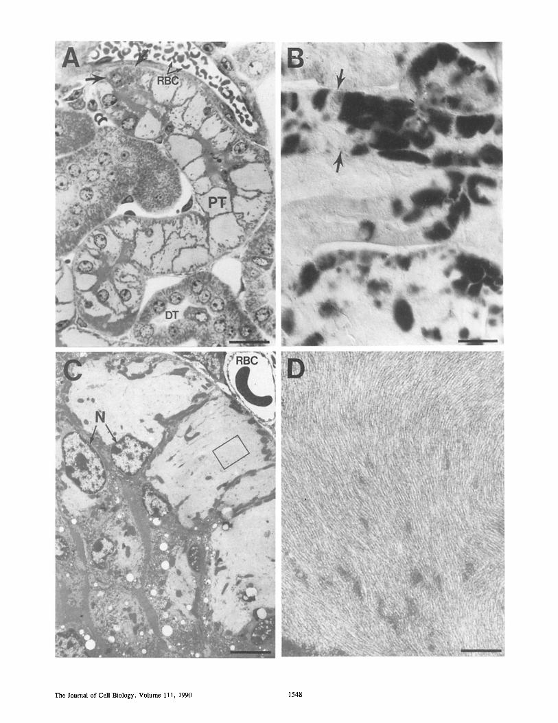
Figure 5. Accumulation of NF-L filaments in transgenic kidney. (A) Light micrograph of a l-#m-thick plastic section of a kidney from mouse line MSV-NF-L-58 (stained with toluidine blue). Bar, 20 #m. (B) Nomarski image of a similar, but unstained section after incubation with a polyclonal antibody to NF-L followed by a secondary antibody conjugated to peroxidase. Bar, 20 #m. (C) Higher magnification view using electron microscopy. Bar, 5 #m. (D) The area delineated by the rectangle in C was further magnified to reveal a densely packed array of filaments. Bar, 0.5 #m. Arrows in A and B denote nearby proximal tubule cells adjacent to others that accumulate high amounts of NF-L. P/;, proximal tubule; DE, distal tubule; RBC, red blood cell; N, proximal tubule cell nuclei.
Monteiro et al. Expression of NF-L in Transgenic Mice 1549
animals emerged when an immunoblot survey of proteins from mouse tails revealed the presence of NF-L in all trans- genic founders, but not in control mice. The level of NF-L expression in tail correlated to a certain degree with copy number but was always similar for littermates acquiring the same transmitted transgene(s) (data not shown). For exam- ple, transgene expression showed some dependence on copy number up to •30 copies, but this relationship was not ap- parent at higher copy numbers and in some cases other fac- tors clearly influenced expression.
To determine the pattern of transgene expression in differ- ent tissues, we used immunoblotting to examine whole cell proteins from a series of tissues from control or transgenic animals. Fig. 3 displays such an analysis for line MSV-NF-L- 72. Of the tissues examined in control animals, NF-L was restricted to brain as expected; however, in transgenic tis- sues, NF-L accumulation was easily detectable by Coomassie staining of whole cell extracts of skeletal muscle or kidney. Quantitation of these findings by parallel immunoblotting of known amounts of partially purified neurofilaments revealed NF-L accumulation in these tissues to be ,x,2.5 % of total cell protein. In heart, NF-L contributed 0.5 % of cell protein, while little transgene expression was observable in liver or spleen. However, in brain NF-L did not accumulate to a level in excess of that in nontransgenic littermates.
With one notable exception, a parallel comparison of transgene RNA levels in the same tissues revealed a pattern similar to that found for accumulated NF-L protein (Fig. 3 E and F). The striking exception was brain where transgene RNAs were more abundant than endogenous NF-L mRNAs (Fig. 3 E and F, lanes B). For example, using S1 analysis and an end-labeled probe derived from the authentic NF-L gene, the endogenous NF-L RNA protected the probe to the start site of transcription while transgene RNAs (that diverge from the probe at the site of linker insertion) yielded two ma- jor protected fragments of 145 and 158 bases (Fig. 3 F). (These major transgene protected fragments are ~11 and ,x,24 bases larger than the 134 bases predicted, probably be- cause of the failure of $1 to cleave efficiently within either of two GC-rich domains that lie just 5' to the point of trans- gene RNA/probe DNA sequence divergence.) Inspection of the data revealed that transgene RNAs were ~20-fold more abundant than NF-L RNAs. Despite this, no increase in ac- cumulated NF-L protein was detectable in brain (Fig. 3, C and D, lanes B). These results were not unique to the MSV- NF-L-72 line; similar relative levels of transgene RNA and protein levels were found in analyses of tissues from two other founder lines that express the transgene highly (MSV- NF-L-58 and MSV-NF-L-103) (not shown).
The failure of NF-L protein to accumulate in brain may have resulted from the transgene encoding an assembly- incompetent NF-L subunit which might have been specifical- ly susceptible to degradation. To test this, we prepared pri- mary cultures of cells enriched in fibroblasts derived from transgenic mice and determined the localization of NF-L by immunofluorescence light microscopy. An antibody to NF-L stained a filamentous array in almost all of the cells from transgenic cultures (Fig. 4 A), but none of those derived from nontransgenic littermates (not shown). In fact, double im- munofluorescence revealed that NF-L coassembles with vimentin (Fig. 4 B), the endogenous IF that the fibroblasts constitutively express.
To confirm further that the transgene encoded assembly- competent molecules, we used light and electron microscopy to analyze the kidneys of MSV-NF-L-58, in which NF-L ac- cumulated to 2.5 % of cell protein. Sections obtained from transgenic kidneys showed that proximal tubule cells con- tained substantial levels of NF-L, which was easily observ- able by immunoperoxidase staining with the NF-L antibody (Fig. 5 B). No NF-L was observed in distal tubules or the surrounding cel ls-for example, red blood cells (Fig. 5 A), or in kidneys from control animals. In fact, in many affected tubule cells NF-L accumulation was to such a level that it was easily observable as lightly staining domains encompassing most of the cell cytoplasm (Fig. 5 A). Higher magnification views using electron microscopy more clearly revealed these large accumulations of NF-L within tubule cells (Fig. 5 C), while even higher magnifications revealed them to be com- prised of massive arrays of densely packed filaments 8-10 nm in diameter (Fig. 5 D). The presence of these accumula- tions of filaments had no discernible effect on the longevity of the mice (the founder mice are now 2 y old). That such massive filament accumulation had no overt phenotypic con- sequence is probably because not all proximal tubules were affected. For example, morphologically normal tubule cells were found to lie adjacent to those most strongly accumulat- ing NF-L (see arrows in Fig. 5 A, or the uneven immuno- staining within an affected tubule in Fig. 5 B). Why only some of the tubule cells are affected is unknown.
The only obvious phenotypic consequence of expression of NF-L outside neurons was the formation of cataracts (Fig. 6 A). These were especially noticeable in the three lines of mice that most highly express the pMSV-NF-L transgene. Although we have not followed the levels of NF-L accumu- lated during development nor have we carefully monitored the onset of cataract formation, cataracts were correlated with the presence of NF-L polypeptides in the lens. For ex- ample, Coomassie blue staining of proteins separated by electrophoresis revealed a major 68-kD protein unique to transgenic animals (Fig. 6 B) and immunoblotting con- firmed its identity as NF-L (Fig. 6 C). The immunoblot fur- ther revealed that the NF-L antibody reacted not only with the expected 68-kD NF-L polypeptide but also with a num- ber of smaller polypeptides which we presume to be proteol- ysis products of the 68-kD polypeptide. Comparison of the Coomassie staining at the NF-L position with that of the 50-57-kD region containing vimentin revealed that NF-L must be at least fivefold more abundant than vimentin, the IF normally expressed in lens.
In view of NF-L accumulation in nonneuronal tissues, the failure of the transgene RNA to elevate NF-L in brain is sur- prising, but obviously cannot be the result of an inadvertent mutation in the transgene. Rather, it may indicate the pres- ence of a posttranscriptional regulatory mechanism that acts to limit NF-L accumulation in some (and perhaps most) brain cells.
Expression of NF-L in Peripheral Sensory Neurons of Transgenic Mice Although we did not detect any increase in accumulated NF-L protein in the brains of transgenic mice despite substantially increased NF-L mRNAs, we also examined NF-L accumula- tion in peripheral neurons. As a tissue rich in neurons, DRG
The Journal of Cell Biology, Volume 111, 1990 1550
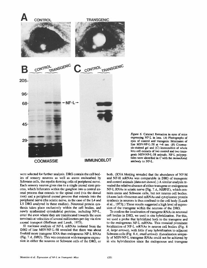
Figure 6. Cataract formation in eyes of mice expressing NF-L in lens. (A) Photographs of eyes of control and transgenic littermates of line MSV-NF-L-58 at '~6 mo. (B) Coomas- sie-stained gel and (C) immunoblot of whole lens cell extracts of two control and two trans- genic MSV-NF-L-58 animals. NF-L polypep- tides were identified in C with the monoclonal antibody to NF-L.
were selected for further analysis. DRG contain the cell bod- ies of sensory neurons as well as axons ensheathed by Schwann cells, the myelin-forming cells of peripheral nerve. Each sensory neuron gives rise to a single axonal stem pro- cess, which bifurcates within the ganglion into a central ax- onal process that extends to the spinal cord (via the dorsal root) and a peripheral axonal process that extends into the peripheral nerve (the sciatic nerve, in the case of the L4 and L5 DRG analyzed in these studies). Neuronal protein syn- thesis takes place exclusively within the cell bodies, and newly synthesized cytoskeletal proteins, including NF-L, enter the axon where they are translocated towards the axon terminals at velocities of several millimeters per day via slow axonal transport (Hoffman and Lasek, 1975).
S1 nuclease analysis of NF-L mRNAs isolated from the DRG of line MSV-NF-L-58 revealed that there was about fivefold more transgene RNA than endogenous NF-L RNAs (Fig. 7 A, DRG). This was consistent with transgene expres- sion in either the neurons or Schwann cells of the DRG, or
both. (RNA blotting revealed that the abundance of NF-M and NF-H mRNAs was comparable in DRG of transgenic and control animals [data not shown].) A similar analysis re- vealed the relative absence of either transgene or endogenous NF-L RNAs in sciatic nerve (Fig. 7 A, NERVE), which con- tains axons and Schwann cells, but not neuron cell bodies. (Axons lack ribosomes and mRNAs and cytoplasmic protein synthesis in neurons is thus confined to the cell body [Lasek et al., 1973].) These results suggested a high level of expres- sion of the transgene within the neurons of the DRG.
To confirm the localization of transgene RNAs in neuronal cell bodies in DRG, we used in situ hybridization. For this, we used a probe that hybridized both to the transgene and to the endogenous NF-L mRNAs. This revealed prominent localization of NF-L mRNAs in neuron cell bodies (Fig. 8 A, large arrows), with little if any hybridization to adjacent Schwann cells (Fig. 8 A, small arrows). (Localization unique- ly of MSV-NF-L transgene RNAs could not be achieved by in situ hybridization since the endogenous and transgene
Monteiro et al. Expression of NF-L in Transgenic Mice 1551

Figure 7. Increased expression of NF-L in DRG and sciatic nerves of MSV-NF-L transgenic mice. (A) RNA purified from DRG and sciatic nerves of MSV transgenic mice (MSV-NF-L- 58) was analyzed for abundance of NFoL RNAs by SI nuclease protection using the Ava I-Ava I fragment of pMSV-NF-L (end-labeled with 32p) as the probe. Transgene RNAs are re- ported as a 140-base protected fragment; the endogenous NF-L genes yield 92-96-base pro- tected fragments. (B) Isolated DRG from MSV-transgenic mice synthesize increased lev- els of NF-L. L4 and L5 DRG were isolated from two MSV-NF-L-58 transgenic mice and from two control nontransgenic littermates. The DRG were pulse labeled for 15 rain with [35S]methionine and the radioactive proteins separated by SDS-PAGE and analyzed by fluorography. (C and D) ~ t e j n , !ysates of sciatic nerves (10 /~g in each 1/m~. obtained from two control and two transgenic mice were separated by electrophoresis. C displays a gel stained with Coomassie blue; D displays an im- munoblot analysis of a parallel gel probed with a monoclonal antibody to NF-L.
RNAs differ only in the 5'-most 35 bases and we were unable to detect a signal using this 35-base region of the transgene. Nevertheless, since the probe reported both endogenous and transgene NF-L RNAs localized only to neuronal cells and the transgene RNAs are fivefold more abundant in the DRG, we infer that the MSV-promoted NF-L transcripts must con- tribute at least five-sixths of the hybridization signal seen over the cell bodies of the neurons.) Further, the lack of in situ hybridization in regions of DRG enriched in Schwann cells and axons but lacking neuron cell bodies (lower region of Fig. 8 A) is consistent with the relative absence of NF-L RNAs in peripheral nerve (Fig. 7 A, NERVE), whose RNA content is contributed primarily by Schwann cells. Previous in situ hybridization studies have shown that these Schwann cells contain high levels of tubulin mRNAs (Hoffman, P. N., and D. W. Cleveland, unpublished observations) and thus the
lack of hybridization with the NF-L cDNA does not reflect limited access of probes to the regions of DRG tissue sec- tions enriched in Schwann cells.
That transgene NF-L expression was absent in the Schwann cells and largely confined to the large neurons was fur- ther confirmed by localization of NF-L protein using a poly- clonal antibody that recognizes NF-L in formalin-fixed tissue sections. (The mAb used for earlier immunoblot and im- munofluorescence analyses did not recognize NF-L in for- malin-fixed tissue.) As shown in Fig. 8 B, large neuronal cell bodies were intensely stained, with little or no detectable NF-L in Schwann cells or smaller neurons. While the anti- body cannot distinguish between the endogenous and trans- gene NF-L, these results clearly demonstrate that if the transgene product is present at substantial levels, its accumu- lation must be restricted to these large neurons.
The Journal of Cell Biology, Volume 111, 1990 1552

Figure 8. Localization of NF- L mRNAs and protein in transgenic sensory neurons. (A) In situ hybridization of 35S-labeled cDNA that specif- ically hybridizes with NF-L mRNAs was performed on tis- sue sections containing lum- bar sensory neurons from an animal expressing the pMSV- NF-L transgene. The autora- diogram was exposed for 5 d. Large arrows point to neuron cell bodies covered with sil- ver grains. Small arrows point to areas enriched in Schwann cells. (B) Immunohistochem- ical localization of NF-L in transgenic sensory neurons. This formalin-fixed tissue was stained with a polyclonal anti- body to NF-L. Bars, 25 /~m.
We next determined whether comparable increases in NF- L synthesis were detectable in these DRG neurons that con- tain elevated levels of NF-L mRNAs. To do this, we isolated L4 and L5 DRG of transgenic animals and nontransgenic lit- termate controls. These were incubated with psS]methio- nine for 15 rain to label newly synthesized proteins, which
were then separated by SDS-PAGE and analyzed by fluorog- raphy. The pattern of newly synthesized proteins was identi- cal in control and transgenic mice with the exception that in transgenic animals there was a clear elevation in a 68-kD polypeptide, which is the expected size of NF-L (see Fig. 7 B). This increase in the rate of synthesis of NF-L is compara-
Monteiro et al. Expression of NF-L in Transgenic Mice 1553

Figure 9. Increased NF density in transgenic axons. Electron micrographs compare representative areas of axoplasm in large myelinated nerve fibers of (A) a control and (B) a transgenic animal expressing pMSV NF-L. NF density is greater and the orientation of NF is more variable in transgenic as compared to control axons. Bars, 0.5/~m. (C) Quantitation of NF density in large myelinated nerve fibers of transgenic (light bars) and littermate control animals (dark bars). NF density is ranked from greatest to least for each group in order to facilitate comparison.
The Journal of Cell Biology, Volume 111, 1990 1554
8
6
~6
~ 2
[ , ~ ~ Contro l .
0 2 4 6
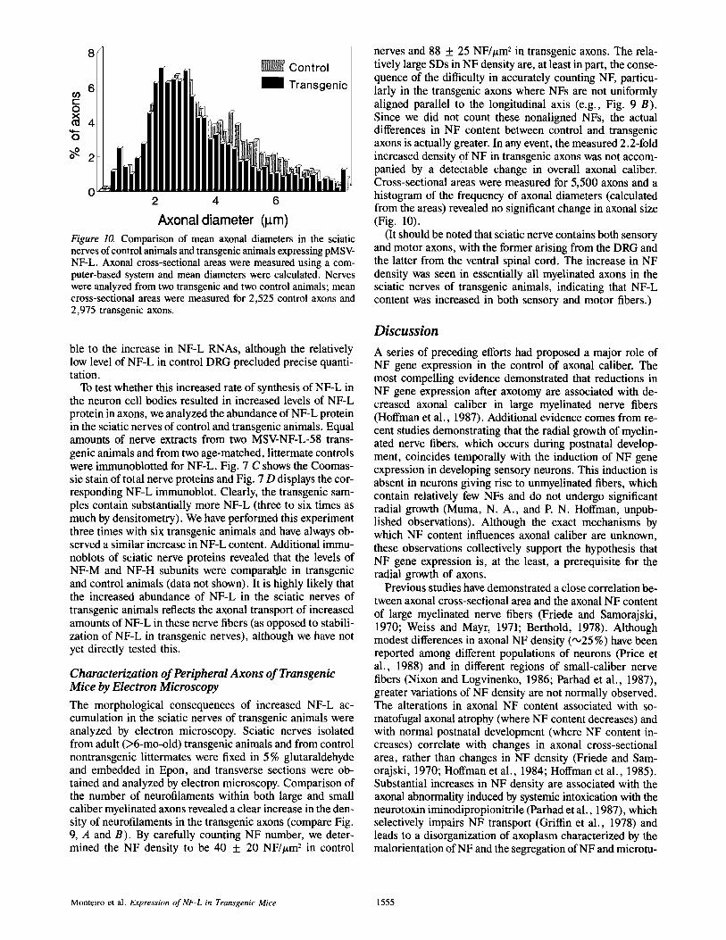
Axonal diameter (l~m) Figure 10. Comparison of mean axonal diameters in the sciatic nerves of control animals and transgenic animals expressing pMSV- NF-L. Axonal cross-sectional areas were measured using a com- puter-based system and mean diameters were calculated. Nerves were analyzed from two transgenic and two control animals; mean cross-sectional areas were measured for 2,525 control axons and 2,975 transgenic axons.
ble to the increase in NF-L RNAs, although the relatively low level of NF-L in control DRG precluded precise quanti- tation.
To test whether this increased rate of synthesis of NF-L in the neuron cell bodies resulted in increased levels of NF-L protein in axons, we analyzed the abundance of NF-L protein in the sciatic nerves of control and transgenic animals. Equal amounts of nerve extracts from two MSV-NF-L-58 trans- genic animals and from two age-matched, littermate controls were immunoblotted for NF-L. Fig. 7 C shows the Coomas- sie stain of total nerve proteins and Fig. 7 D displays the cor- responding NF-L immunoblot. Clearly, the transgenic sam- ples contain substantially more NF-L (three to six times as much by densitometry). We have performed this experiment three times with six transgenic animals and have always ob- served a similar increase in NF-L content. Additional immu- noblots of sciatic nerve proteins revealed that the levels of NF-M and NF-H subunits were comparable in transgenic and control animals (data not shown). It is highly likely that the increased abundance of NF-L in the sciatic nerves of transgenic animals reflects the axonal transport of increased amounts of NF-L in these nerve fibers (as opposed to stabili- zation of NF-L in transgenic nerves), although we have not yet directly tested this.
Characterization of Peripheral Axons of Transgenic Mice by Electron Microscopy The morphological consequences of increased NF-L ac- cumulation in the sciatic nerves of transgenic animals were analyzed by electron microscopy. Sciatic nerves isolated from adult (>6-mo-old) transgenic animals and from control nontransgenic littermates were fixed in 5 % glutaraldehyde and embedded in Epon, and transverse sections were ob- tained and analyzed by electron microscopy. Comparison of the number of neurofilaments within both large and small caliber myelinated axons revealed a clear increase in the den- sity of neurofilaments in the transgenic axons (compare Fig. 9, A and B). By carefully counting NF number, we deter- mined the NF density to be 40 ___ 20 NF//~m 2 in control
nerves and 88 + 25 NF//~m 2 in transgenic axons. The rela- tively large SDs in NF density are, at least in part, the conse- quence of the difficulty in accurately counting NF, particu- larly in the transgenic axons where NFs are not uniformly aligned parallel to the longitudinal axis (e.g., Fig. 9 B). Since we did not count these nonaligned NFs, the actual differences in NF content between control and transgenic axons is actually greater. In any event, the measured 2.2-fold increased density of NF in transgenic axons was not accom- panied by a detectable change in overall axonal caliber. Cross-sectional areas were measured for 5,500 axons and a histogram of the frequency of axonal diameters (calculated from the areas) revealed no significant change in axonal size (Fig. 10).
(It should be noted that sciatic nerve contains both sensory and motor axons, with the former arising from the DRG and the latter from the ventral spinal cord. The increase in NF density was seen in essentially all myelinated axons in the sciatic nerves of transgenic animals, indicating that NF-L content was increased in both sensory and motor fibers.)
Discussion
A series of preceding efforts had proposed a major role of NF gene expression in the control of axonal caliber. The most compelling evidence demonstrated that reductions in NF gene expression after axotomy are associated with de- creased axonal caliber in large myelinated nerve fibers (Hoffman et al., 1987). Additional evidence comes from re- cent studies demonstrating that the radial growth of myelin- ated nerve fibers, which occurs during postnatal develop- ment, coincides temporally with the induction of NF gene expression in developing sensory neurons. This induction is absent in neurons giving rise to unmyelinated fibers, which contain relatively few NFs and do not undergo significant radial growth (Muma, N. A., and P. N. Hoffman, unpub- lished observations). Although the exact mechanisms by which NF content influences axonal caliber are unknown, these observations collectively support the hypothesis that NF gene expression is, at the least, a prerequisite for the radial growth of axons.
Previous studies have demonstrated a close correlation be- tween axonal cross-sectional area and the axonal NF content of large myelinated nerve fibers (Friede and Samorajski, 1970; Weiss and Mayr, 1971; Berthold, 1978). Although modest differences in axonal NF density (,'025 %) have been reported among different populations of neurons (Price et al., 1988) and in different regions of small-caliber nerve fibers (Nixon and Logvinenko, 1986; Parhad et al., 1987), greater variations of NF density are not normally observed. The alterations in axonal NF content associated with so- matofugal axonal atrophy (where NF content decreases) and with normal postnatal development (where NF content in- creases) correlate with changes in axonal cross-sectional area, rather than changes in NF density (Friede and Sam- orajski, 1970; Hoffman et al., 1984; Hoffman et al., 1985). Substantial increases in NF density are associated with the axonal abnormality induced by systemic intoxication with the neurotoxin iminodipropionitrile (Parhad et al., 1987), which selectively impairs NF transport (Griffin et al., 1978) and leads to a disorganization of axoplasm characterized by the malorientation of NF and the segregation of NF and microtu-
Monteiro et al. Expression of NF-L in Transgenic Mice 1555
bules (Clark et al., 1980; Papasozomenos et al., 1981; Griffin et al., 1983). Like the iminodipropionitrile example, overex- pression of wild-type NF-L yields not only an abnormal in- crease in NF density, but also disorganization of the axonal cytoskeleton.
Since the additional NF in the axons of transgenic animals are constructed primarily from NF-L, our findings demon- strate that an increase in filaments assembled primarily from NF-L has little effect on axonal caliber. Of course, we cannot exclude the possibility that a greater increase in NF-L ac- cumulation would ultimately have resulted in altered axonal caliber, as happens in animals intoxicated with iminodipro- pionitrile. However, since NFs are normally composed of three different subunits, a simpler view is that an increase in NF-L alone may not result in the formation of filaments with the same properties as those composed of normal ratios of the three subunits.
Indeed, although NF-L can form filaments in the absence of the other subunits (Geisler and Weber, 1981; Liem and Hutchinson, 1982), it does not carry the extended carboxy- terminal tail shared by NF-M and NF-H (Sharp et al., 1982; Hirokawa et al., 1984; Hisanaga and Hirokawa, 1988), which in the case of NF-H appears to be associated NF cross-bridges (Hirokawa et al., 1984; Hisanaga and Hiro- kawa, 1988). It is possible that these cross-bridges play a critical role in the regulation of axonal caliber. In fact, the carboxy-terminal tail of NF-H is highly enriched in both charged amino acids and potential phosphorylation sites (Lees et al., 1988), suggesting that these cross-bridges may repel adjacent NF, thereby reducing NF density. The possi- ble role of NF cross-bridges in the control of axonal caliber can now be explored by producing additional transgenic mice expressing higher levels of NF-M and NF-H subunits.
Two other transgenic founder mice obtained with the authentic NF-L gene promoter (pNF-L[Bam]) showed ac- cumulation of NF-L RNAs restricted to tissue enriched in neurons (e.g., brain), similar to the endogenous NF-L gene. This indicates that most, if not all, of the elements that specify neuronal-specific expression must be contained with- in the 7-kb transgene fragment. Although we have not shown it directly, we assume that expression of the pNF-L(Bam) transgene is restricted to neurons based on the work of Julien et al. (1987) who demonstrated by in situ hybridization that expression of a human NF-L transgene contained on a 21.5- kb genomic DNA fragment was localized to neurons. Fur- ther, Byrne and Ruddle (1989) had previously demonstrated that the 1.5-kb segment of the mouse NF-L gene just 5' to transcription initiation was sufficient to yield brain-specific expression of a marker gene, but they could not determine the level of expression. Using the same presumptive NF-L promoter domain, we find transgene RNAs at a level 20% of the endogenous NF-L mRNAs in brain. Since NF-L RNA is moderately abundant, this indicates that use of the NF-L controlling sequences should produce a relatively high level of transgene expression targeted to most (and perhaps all) neurons.
MSV LTR-promoted transgenes produced very high ex- pression of NF-L in various tissues, including many where NFs are not normally expressed. Somewhat surprisingly, NF-L accumulated to as much as 2-3 % of total cell protein in tissues like kidney and skeletal muscle, and 0.5% in heart, and yet it had no catastrophic effect on the function of these tissues nor did it affect mouse development or survival. (The
founder mice are now 2 yr old.) This may at first seem sur- prising since NF-L is normally expressed only in postmitotic neurons. The combination of its unique expression in neu- rons and its extreme insolubility initially suggested that inap- propriate expression of NF-L might be detrimental to contin- uing mitosis. However, using DNA transfection of cultured cells we showed that NF-L could accumulate to be the most abundant cellular protein without affecting cell growth (Monteiro and Cleveland, 1989). Our current results extend this finding to a true in vivo situation: inappropriate expres- sion of NF-L did not affect the viability of the many different mitotic cell types in these transgenic mice. Furthermore, we have observed massive accumulation of NF-L filaments in kidney of transgenic mice without any obvious effect on lon- gevity. The only phenotypic effect we have observed upon nontissue-specific expression of NF-L is the formation of cataracts. Capetanaki et al. (1989) also observed cataracts in transgenic mice that expressed the chicken vimentin gene in lens and concluded that vimentin expression specifically prevented normal lens cell differentiation, ultimately result- ing in cataract formation. An alternative explanation that may in fact explain both that observation and ours is that the lens has a very restricted tolerance for expression of "for- eign" proteins without the phenotypic consequence of cata- ract formation.
One set of previous transgenic animals had demonstrated that MSV directed transgene expression at high levels in lens and testis, with lower levels in brain and kidney (Khillan et al., 1987). Our findings confirm that the MSV promoter can be used to yield extraordinarily high levels of transgene ex- pression albeit in a different set of tissues than seen earlier. Specifically, we have now documented MSV-directed expres- sion in tissues rich in neurons. Although lower than that seen in the highest expressing tissues, transgene expression in DRG and in brain was nevertheless substantial enough that MSV-NF-L RNAs were 5-20-fold higher than those from the endogenous NF-L genes. Since a similar pattern was ob- served in three different transgenic lines, this suggests that the MSV LTR may also be of general use to force high level expression of selected gene products within neurons.
An unexpected finding was that the increase of MSV- promoted NF-L RNAs in the brains of transgenic mice did not result in a similar increase in NF-L protein. This clearly indicates the presence of posttranscriptional mechanisms that maintain the level of NF-L protein in brain. With the current mice we can not easily pursue the actual mechanism involved since the transgene translation product is identical to the endogenous NF-L. But in any event, the present data suggest interesting and novel posttranscriptional regulation of NF-L in the central nervous system.
We wish to thank Ms. Brenda Klaunberg for the production of transgenic mice and Ms. Suzanne Schrock and Mr. Paul Kang for maintaining the mouse colony. We also thank Margaret Lopata for the in situ hybridization analysis of DRG. Dr. Gerry Shaw kindly provided the polyclonal antibody to NF-L.
This work has been supported by grants from the National Institutes of Health to D. W. Cleveland and P. N. Hoffman (NS-27036 and NS-22849). Initial phases of the work were supported by a grant from the March of Dimes to D. W. Cleveland (I-1013). M. J. Monteiro was supported in part by a postdoctoral fellowship from the Maryland Affiliate of the American Heart Association.
Received for publication 18 May 1990 and in revised form 5 June 1990.
The Journal of Cell Biology, Volume 111, 1990 1556

References
Berthold, C. H. 1978. Morphology of normal peripheral axons. In Physiology and Pathobiology of Axons. S. G. Waxman, editor. Raven Press, New York. 3-63.
Byrne, G. W., and F. H. Ruddle. 1989. Multiplex gene regulation: a two tiered approach to transgene regulation in transgenic mice. Proc. Natl. Acad. Sci. USA. 86:5473-5477.
Capetanaki, Y., S. Smith, and J. P. Heath. 1989. Overexpression of the vimen- tin gene in transgenic mice inhibits normal lens cell differentiation. J. Cell Biol. 109:1653-1664.
Chirgwin, J. M., A. E. Przbyla, R. J. MacDonald, and W. J. Rutter. 1979. Isolation of biologically active ribonucleic acid from sources enriched in ribonuclease. Biochemistry. 18:5294-5299.
Clark, A. W., J. W. Griffin, and D. L. Price. 1980. The axonal pathology in chronic IDPN intoxication. J. Neuropathol. Exp. & Neurol. 39:42-55.
Denhardt, D. T. 1966. A membrane-filter technique for the detection of com- plementary DNA. Biochem. Biophys. Res. Commun. 23:641-646.
Dretzen, G., M. Bellard, O. Sassone-Corsi, and P. Chambon. 1981. A reliable method for the recovery of DNA fragments from agarose and acrylamide gels. Anal. Biochem. 112:295-298.
Franke, W. W. 1987. Nuclear lamins and cytoskeletal intermediate filament proteins: a growing multigene family. Cell. 48:3--4.
Friede, R. L., and T. Samorajski. 1970. Axon caliber related to neurofilaments and microtubules in sciatic nerve fibers of rats and mice. Anat. Rec. 167: 379-388.
Geisler, N., and K. Weber. 1981. Self-assembly in vitro of the 68,000 molecu- lar weight component of the mammalian neurofilament triplet proteins into intermediate-size filaments. J. Mol. Biol. 151:565-571.
G-eisler, N., and K. Weber. 1986. Structural aspects of intermediate filaments. In Cell and Molecular Biology of the Cytoskeleton. J. W. Shay, editor. Ple- num Press, New York. 41-68.
Geisler, N., E. Kanfmann, S. Fischer, U. Plessmann, and K. Weber. 1983. Neurofilament architecture combines structural principles of .:atermediate filaments with carboxy-terminal extensions increasing in size between triplet proteins. EMBO (Eur. Mol. Biol. Organ.)J. 2:1295-1302.
Griffin, J. W., K. E. Fahnestock, D. L. Price, and P. N. Hoffman. 1983. Microtubule-neurofilament segregation produced by/~,ff-iminodipropioni- trile: evidence for the association of fast axonal transport with microtubules. Z Neurosci. 3:557--566.
Griffin, J. W., P. N. Hoffman, A. W. Clark, P. 1". Carroll, and D. L. Price. 1978. Slow axonal transport of neurofilament proteins: impairment by ~,ff- iminodipropionitrile administration. Science (Wash. DC). 202:633-635.
Hirokawa, N., M. A. Glicksman, and M. B. Willard. 1984. Organization of mammalian neurofilament polypeptides within the neuronal cytoskeleton. J. Cell Biol. 98:1523-1536.
Hisanaga, S.-I., and N. Hirokawa. 1988. Structure of the peripheral domains of neurofilaments revealed by low angle rotary shadowing. J. Mol. Biol. 202:297-305.
Hoffman, P. N., and R. J. Lasek. 1975. The slow component of axonal trans- port: identification of the major structural polypeptides of the axon and their generality among mammalian neurons. J. Cell Biol. 66:351-366.
Hoffman, P. N., J. W. Griffin, and D. L. Price. 1984. Control of axonal caliber by neuroflament transport. J. Cell Biol. 99:705-714.
Hoffman, P. N., G. W. Thompson, J. W. Griffin, and D. L. Price. 1985. Changes in neurofilament transport coincide temporally with alterations in the caliber of axons in regenerating motor fibers. J. Cell Biol. 101:1332- 1340.
Hoffman, P. N., D. W. Cleveland, J. W. Griffin, P. W. Landes, N. J. Cowan, and D. L. Price. 1987. Neurofilament gene expression: a major determinant of axonal caliber. Proc. Natl. Acad. Sci. USA. 84:3472-3476.
Julien, J.-P., and W. E. Mushynski. 1983. The distribution of phosphorylation sites among identified proteolytic fragments of mammalian neurofilaments. J. Biol. Chem. 258:4019-4025.
Julien, J.-P., I. Tretjakoff, L. Beaudet, and A. Peterson. 1987. Expression and assembly of a human neurofilament protein in transgenic mice provide a novel neuronal marking system. Genes & Dev. 1:1085-1095.
Julien, J.-P., F. Cote, L. Beaudet, M. Sidky, D. Flavell, F. Grosveld, and W. Mushynski. 1988. Sequence and structure of the mouse gene coding for the
largest neurofilament subunit. Gene. 68:307-314. Khillan, J. S., M. K. Oskarsson, F. Propst, T. Kuwabara, G. F. Vande Woude
and H. Westphal. 1987. Defects in lens fiber differentiation are linked to c-mos overexpression in transgenic mice. Genes & Dev. 1:1327-1335.
Laemmli, U. K. 1970. Cleavage of structural proteins during the assembly of the head of bacteriophage T4. Nature (Lond.). 227:680-685.
Lasek, R. J., C. Dabrowski, and R. Nordlander. 1973. Analysis of axoplasmic RNA from invertebrate giant axons. Nat. New Biol. 244:162-165.
Lees, J. F., P. S. Shneidman, S. F. Skuntz, M. J. Carden, and R. A. Lazarini. 1988. The structure and organization of the human heavy neurofilament subunit (NF-H) and the gene encoding it. EMBO (Eur. Mol. Biol. Organ.) J. 7:1947-1955.
Lendahl, U., L. B. Zimmerman, and R. D. G. McKay. 1990. CNS stem cells express a new class of intermediate filament protein. Cell. 60:585-595.
Levy, E., R. H. K. Liem, P. D'Eustachio, and N. J. Cowan. 1987. Structure and evolutionary origin of the gene encoding mouse NF-M, the middle- molecular-mass neuroflament protein. Eur. J. Biochem. 166:71-77.
Lewis, S. A., and N. J. Cowan. 1985a. Temporal expression of mouse glial fibrillary acid protein mRNA studied by a rapid in situ hybridization proce- dure. J. Neurochem. 45:913-919.
Lewis, S. A., and N. J. Cowan. 1985b. Genetics, evolution, and expression of the 68,000 mol. wt. neuroflament protein: isolation of a cloned cDNA probe. J. Cell Biol. 100:843-850.
Lewis, S. A., and N. J. Cowan. 1986. Anomalous placement of introns in a member of the intermediate filament multigene family: an evolutionary conundrum. Mol. Cell. Biol. 6:1529-1534.
Liem, R. K. H., and S. B. Hutchinson. 1982. Purification of individual compo- nents of the neurofilament triplet: flament assembly from the 70,000 dalton subunit. Biochemistry. 21:3221-3226.
Lopata, M. A., and D. W. Cleveland. 1987. In vivo microtubules are copolymers of available t3-tubulin isotypes: localization of each of six ver- tebrate/3-tubulin isotypes using polyclonal antibodies elicited by synthetic peptide antigens. J. Cell Biol. 105:1707-1720.
Minami, Y., and H. Sakai. 1985. Dephosphorylation suppresses the activity of neurofilament to promote tubulin polymerization. FEBS (Fed. Eur. Biochem. Soc.) Lett. 185:239-242.
Monteiro, M. J., and D. W. Cleveland. 1989. Expression of NF-L and NF-M in fibroblasts reveals coassembly of neurofilament and vimentin subunits. J. Cell Biol. 108:579-593.
Myers, M. W., R. A. Lazzarini, V. M. Y. Lee, W. W. Schlaepfer, and D. L. Nelson. 1987. The human mid-size neurofilament subunit: a repeated protein sequence and the relationship of its gene to the intermediate filament gene family. EMBO (Eur. Mol. BioL Organ.) J. 6:1617-1626.
Nixon, R. A., and K. B. Logvinenko. 1986. Multiple fates of newly synthesized neurofilament proteins: evidence for a stationary neurofilament network dis- tributed nonuniformly along axons of retinal ganglion cell neurons. J. Cell BioL 102:647-659.
Papasozomenos, S. C. H., L. Autilio-Gambetti, and P. Gambetti. 1981. Reor- ganization of axoplasmic organelles following/3,ff-iminodipropionitrile ad- ministration. J. Cell Biol. 91:866-871.
Parhad, I. M., A. W. Clark, and J. W. Griffin. 1987. Effect of changes in neurofilament content on caliber of small axons: the iminodipropionitrile model. J. Neurosci. 7:2256-2263.
Price, R. L., P. Paggi, R. J. Lasek and M. J. Katz. 1988. Neurofilaments are spaced randomly in the radial dimensions of axons. J. Neurocytol. 17:55-62.
Sharp, G. A., G. Shaw, and K. Weber. 1982. Immunoelectron microscopic local- ization of the three neurofilament triplet proteins along neurofilaments of cul- tured dorsal root ganglion neurones. Exp. Cell Res. 137:403-413.
Smith, P. K., P. I. Krohn, G. T. Hermanson, A. K. Mallia, E H. Gartner, M. D. Provenzaus, E. K. Fujimoto, N. M. Goeke, B. J. Olson, and D. C. Klenk. 1985. Measurement of protein using bicinchonic acid. Anal. Biochem. 150: 76-85.
Southern, E. M. 1975. Detection of specific sequences among DNA fragments separated by gel electrophoresis. J. Mol. Biol. 98:503-517.
Steinert, P. M., and D. R. Roop. 1988. Molecular and cellular biology of inter- mediate filaments. Anna. Re~. Biochem. 57:593-625.
Weiss, P. A., and R. Mayr. 1971. Organelles of neuroplasmic ("axonal') flow: neuroffiaments. Proc. Natl. Acad. Sci. USA. 68:846-850.
Monteiro et al. Expression of NF-L in Transgenic Mice 1557