Expression of c-MET in Invasive Meningiomajpatholtm.org/upload/pdf/jptm-49-1-44.pdf ·...
8
44 pISSN 2383-7837 eISSN 2383-7845 © 2015 The Korean Society of Pathologists/The Korean Society for Cytopathology This is an Open Access article distributed under the terms of the Creative Commons Attribution Non-Commercial License (http://creativecommons.org/licenses/ by-nc/3.0) which permits unrestricted non-commercial use, distribution, and reproduction in any medium, provided the original work is properly cited. Meningioma is a common intracranial tumor arising from the meningothelial (arachnoid) cells. Meningiomas are divided into 15 histologic subtypes and three grades, including benign (grade I), atypical (grade II), and anaplastic (grade III). 1 Most meningi- omas are benign, corresponding to World Health Organization (WHO) grade I, and they have a favorable outcome. Alterna- tively, grade II atypical meningiomas and grade III anaplastic meningiomas have less favorable outcomes. 2,3 Even in benign cases, meningiomas have high recurrence rates after curative sur- gical treatment. They have been estimated to recur in 7%–25%, 29%–52%, and 50%–94% of cases in grades I, II, and III, re- spectively. 4 Several markers, such as the proliferation index, the vascular density marker, and the expression of sex hormone receptors, are suggested for predicting the recurrence rates of tumors. Howev- er, it is generally accepted that both histopathological features, including histologic subtypes and clinical data, have limitations in their use as reliable markers for predicting tumor recurrence due to low accuracy. 5-8 Previous studies report an association among meningiomas, brain or bone invasion, and higher tumor recurrence rates. However, the mechanism of invasion has not been well established. 9-12 Also called a hepatocyte growth factor (HGF), c-MET is a re- ceptor tyrosine kinase that, upon binding of its ligand, is phos- phorylated. Subsequently, c-MET activates the signaling path- way of cell proliferation and migration. The c-MET/HGF sig- naling pathway was first described as an oncogene in the 1980s. Accordingly, it has been known to induce tumor cell prolifera- tion, motility, and invasion, as well as to promote angiogenesis in several human cancers, such as breast, lung and hepatocellular Expression of c-MET in Invasive Meningioma Sumi Yun · Jae Moon Koh Kyu Sang Lee 1 · An Na Seo 2 Kyung Han Nam 3 · Gheeyoung Choe 1 Department of Pathology, Seoul National University Hospital, Seoul National University College of Medicine, Seoul; 1 Department of Pathology, Seoul National University Bundang Hospital, Seoul National University College of Medicine, Seongnam; 2 Department of Pathology, Kyungpook National University Hospital, Kyungpook National University School of Medicine, Daegu; 3 Department of Pathology, Haeundae Paik Hospital, Inje University, Busan, Korea Background: Meningiomas show high recurrence rates even after curative tumor removal. The invasiveness of meningiomas may contribute to their high recurrence rates. Recently, c-MET and hepatocyte growth factor (HGF) have been reported to be involved in cancer invasion. Methods: We examined the immunohistochemical expression of c-MET and HGF in 100 cases of patients with meningiomas who have undergone complete tumor removal. Results: c-MET -High and HGF - High were found in 17% and 13% of meningiomas, respectively. Brain invasion was observed in 17.6% of c-MET -High meningiomas, but in only 2.4% of c-MET -Low meningiomas (p = .033). Bone/ soft tissue invasion was observed in 23.5% of c-MET -High meningiomas and in 9.6% of c-MET -Low meningiomas (p = .119). HGF -High did not show statistical association with brain invasion or bone/ soft tissue invasion. c-MET -High demonstrated shorter recurrence-free survival (RFS, 93.5 ± 8.2 months vs 96.1 ± 1.9 months); however, this difference was not statistically significant (p = .139). There was no association of HGF -High with RFS. Conclusions: This study demonstrates that c- MET -High is associated with brain invasion of meningiomas, and that c-MET expression may be a useful predictive marker for meningioma recurrence. Patients with invasive meningiomas with high expressions of c-MET may be good candidates for targeted therapy using c-MET inhibitors. Key Words: Meningioma; Proto-oncogene proteins c-MET; Hepatocyte growth factor; Neoplasm invasiveness; Immunohistochemistry Received: August 29, 2014 Revised: October 7, 2014 Accepted: October 13, 2014 Corresponding Author Gheeyoung Choe, M.D. Department of Pathology, Seoul National University Bundang Hospital, Seoul National University College of Medicine, 82 Gumi-ro 173 Beon-gil, Bundang- gu, Seongnam 463-707, Korea Tel: +82-31-787-7711 Fax: +82-31-787-4012 E-mail: [email protected] Journal of Pathology and Translational Medicine 2015; 49: 44-51 http://dx.doi.org/10.4132/jptm.2014.10.13 ▒ ORIGINAL ARTICLE ▒
Transcript of Expression of c-MET in Invasive Meningiomajpatholtm.org/upload/pdf/jptm-49-1-44.pdf ·...

44
pISSN 2383-7837eISSN 2383-7845
© 2015 The Korean Society of Pathologists/The Korean Society for CytopathologyThis is an Open Access article distributed under the terms of the Creative Commons Attribution Non-Commercial License (http://creativecommons.org/licenses/
by-nc/3.0) which permits unrestricted non-commercial use, distribution, and reproduction in any medium, provided the original work is properly cited.
Meningioma is a common intracranial tumor arising from the meningothelial (arachnoid) cells. Meningiomas are divided into 15 histologic subtypes and three grades, including benign (grade I), atypical (grade II), and anaplastic (grade III).1 Most meningi-omas are benign, corresponding to World Health Organization (WHO) grade I, and they have a favorable outcome. Alterna-tively, grade II atypical meningiomas and grade III anaplastic meningiomas have less favorable outcomes.2,3 Even in benign cases, meningiomas have high recurrence rates after curative sur-gical treatment. They have been estimated to recur in 7%–25%, 29%–52%, and 50%–94% of cases in grades I, II, and III, re-spectively.4
Several markers, such as the proliferation index, the vascular density marker, and the expression of sex hormone receptors, are suggested for predicting the recurrence rates of tumors. Howev-
er, it is generally accepted that both histopathological features, including histologic subtypes and clinical data, have limitations in their use as reliable markers for predicting tumor recurrence due to low accuracy.5-8 Previous studies report an association among meningiomas, brain or bone invasion, and higher tumor recurrence rates. However, the mechanism of invasion has not been well established.9-12
Also called a hepatocyte growth factor (HGF), c-MET is a re-ceptor tyrosine kinase that, upon binding of its ligand, is phos-phorylated. Subsequently, c-MET activates the signaling path-way of cell proliferation and migration. The c-MET/HGF sig-naling pathway was first described as an oncogene in the 1980s. Accordingly, it has been known to induce tumor cell prolifera-tion, motility, and invasion, as well as to promote angiogenesis in several human cancers, such as breast, lung and hepatocellular
Expression of c-MET in Invasive Meningioma
Sumi Yun · Jae Moon Koh Kyu Sang Lee1 · An Na Seo2 Kyung Han Nam3 · Gheeyoung Choe1
Department of Pathology, Seoul National University Hospital, Seoul National University College of Medicine, Seoul; 1Department of Pathology, Seoul National University Bundang Hospital, Seoul National University College of Medicine, Seongnam; 2Department of Pathology, Kyungpook National University Hospital, Kyungpook National University School of Medicine, Daegu; 3Department of Pathology, Haeundae Paik Hospital, Inje University, Busan, Korea
Background: Meningiomas show high recurrence rates even after curative tumor removal. The invasiveness of meningiomas may contribute to their high recurrence rates. Recently, c-MET and hepatocyte growth factor (HGF) have been reported to be involved in cancer invasion. Methods: We examined the immunohistochemical expression of c-MET and HGF in 100 cases of patients with meningiomas who have undergone complete tumor removal. Results: c-MET-High and HGF-
High were found in 17% and 13% of meningiomas, respectively. Brain invasion was observed in 17.6% of c-MET-High meningiomas, but in only 2.4% of c-MET-Low meningiomas (p= .033). Bone/soft tissue invasion was observed in 23.5% of c-MET-High meningiomas and in 9.6% of c-MET-Low meningiomas (p= .119). HGF-High did not show statistical association with brain invasion or bone/soft tissue invasion. c-MET-High demonstrated shorter recurrence-free survival (RFS, 93.5±8.2 months vs 96.1±1.9 months); however, this difference was not statistically significant (p= .139). There was no association of HGF-High with RFS. Conclusions: This study demonstrates that c-MET-High is associated with brain invasion of meningiomas, and that c-MET expression may be a useful predictive marker for meningioma recurrence. Patients with invasive meningiomas with high expressions of c-MET may be good candidates for targeted therapy using c-MET inhibitors.
Key Words: Meningioma; Proto-oncogene proteins c-MET; Hepatocyte growth factor; Neoplasm invasiveness; Immunohistochemistry
Received: August 29, 2014Revised: October 7, 2014Accepted: October 13, 2014
Corresponding AuthorGheeyoung Choe, M.D.Department of Pathology, Seoul National University Bundang Hospital, Seoul National University College of Medicine, 82 Gumi-ro 173 Beon-gil, Bundang-gu, Seongnam 463-707, KoreaTel: +82-31-787-7711Fax: +82-31-787-4012 E-mail: [email protected]
Journal of Pathology and Translational Medicine 2015; 49: 44-51http://dx.doi.org/10.4132/jptm.2014.10.13
▒ ORIGINAL ARTICLE ▒

http://jpatholtm.org/http://dx.doi.org/10.4132/jptm.2014.10.13
Expression of c-MET in Meningioma • 45
carcinomas.13-16 In meningiomas, the expression of c-MET/HGF has been reported to have a diverse relationship with tumor re-currence, angiogenesis, histologic subtypes, and the invasiveness of the meningioma. Existing reports are based on a limited number of samples and different methods, such as enzyme-linked immunosorbent assay, immunohistochemistry or reverse transcription polymerase chain reaction (RT-PCR).17-19 To date, little is known about their expression in invasive meningiomas. The aim of this study, therefore, is to elucidate whether the pro-tein expressions of c-MET and HGF are associated with clinico-pathologic variables, as well as brain and bone/soft tissue inva-sion of meningiomas, in large scale studies of meningioma.
MATERIALS AND METHODS
Meningioma cases
Formalin-fixed, paraffin-embedded archival tissue samples from 100 patients who underwent complete surgical resection (Simpson grade I) of meningiomas between August 2003 and December 2012 were collected from the databases of the Depart-ment of Pathology, Seoul National University Bundang Hospi-tal in Korea. Clinical and pathological data were obtained by re-viewing medical records and pathology reports. Two patholo-gists (G.C. and S.Y.) independently reviewed the hematoxylin and eosin–stained slides, confirmed the diagnosis according to the 2007 WHO classification system, and classified the histo-logical subtypes and grading of the meningiomas. All the pa-tients received regular follow-up after surgery, without postoper-ative chemoradiation therapy. The recurrence-free survival (RFS) was calculated from the time of surgery to the first suspected re-currence of meningioma. Evidence of tumor recurrence was pro-vided by a computed tomography scan or a magnetic resonance image showing a meningioma in a location contiguous with the previous operation site.
Demographic data
The clinicopathologic features of patients are summarized in Table 1. The 100 patients consisted of 23 males (23%) and 77 females (77%), with median age of 60 years (range, 36 to 85 years). Of these, bone/soft tissue invasion was observed in 12 cas-es, brain invasion was observed in five cases, and both bone/soft tissue invasion and brain invasion were observed in one case. We defined “soft tissue invasion” as invasion of the meningioma to the scalp or paranasal sinus. According to the 2007 WHO clas-sification, all were cases of benign meningiomas (grade I). Histo-logically, the 100 cases consisted of meningothelial types
(n=32), transitional types (n=32), fibrous types (n=16), angio-matous types (n=13), psammomatous types (n=2), microcystic types (n=4), and one metaplastic type (n=1) (Table 1).
Construction of tissue microarray
We chose one representative tumor block in each case, and harvested cores with diameters of 3 mm from the most represen-tative tumor areas of the donor blocks. The cores were precisely arranged into new recipient tissue microarray (TMA) blocks us-ing a trephine apparatus according to previously described pro-tocols.20
Immunohistochemistry staining and interpretation
Immunohistochemical staining was carried out using TMA according to a previously described method.20 Briefly, sections of 4 µm were transferred to poly-L-lysine coated adhesive slides and dried, deparaffinized, and rehydrated. The slides were subse-quently subjected to heat-induced antigen retrieval. The follow-ing antibodies were used according to manufacturer instruction: c-MET (pre-dilution, rabbit monoclonal antibody, Ventana Medical Systems, Inc., Tucson, AZ, USA) and HGFα (1:100, rabbit polyclonal antibody, Santa Cruz Biotechnology, Santa Cruz, CA, USA). The sections were incubated with appropriate reagents from the Dako REAL EnVision Detection System (DAKO, Glostrup, Denmark), and were counterstained with Mayer’s hematoxylin. Evaluation of c-MET and HGF expression was assessed according to previous reports.21 As a positive con-trol for c-MET and HGF, tubular cells of a normal kidney and normal colonic mucosa were used, respectively. Immunostaining without the primary antibody was used as the negative control. Each case was categorized as positive when it showed moderate-to-strong cytoplasmic and/or membranous positivity in tumor cells. Accordingly, we scored expression as c-MET-High and HGF-
High (>25% positive cells) and c-MET-Low and HGF-Low (0%–25%
Table 1. Clinicopathologic characteristics of meningioma cases
Characteristic No.
Meadian age (range, yr) 60 (36–85)Sex Male Female
2377
Histologic subtype Meningothelial Transitional Fibrous Angiomatous Psammomatous Microcystic Metaplastic
32321613 2 4 1

http://jpatholtm.org/ http://dx.doi.org/10.4132/jptm.2014.10.13
46 • Yun S, et al.
tumor cells) (Fig. 1).22
Ethics statements
The study was conducted according to the ethics standards of the World Medical Association’s Declaration of Helsinki.
Statistical analysis
All statistical analyses were conducted using the SPSS ver. 21.0 (IBM Co., Armonk, NY, USA). Associations between the protein expressions of each antibody and the categorical variables were assessed using chi-square tests or Fisher exact tests, if ap-
C
A
D
B
E F
Fig. 1. Immunohistochemical staining in meningiomas. (A) c-MET staining in tubular cells in normal kidney. (B) c-MET weak staining in me-ningioma. (C) c-MET moderate staining in meningioma. (D) c-MET strong staining in meningioma. (E) Hepatocyte growth factor (HGF) stain-ing in colonic mucosa. (F) HGF weak staining in meningioma. (Continued to the next page)

http://jpatholtm.org/http://dx.doi.org/10.4132/jptm.2014.10.13
Expression of c-MET in Meningioma • 47
propriate. Kaplan-Meier survival curves for RFS were plotted for each antibody, and the survival comparison was determined us-ing log rank tests. All tests were two-tailed, and statistical sig-nificance was set as p<.05.
RESULTS
Clinical characteristics according to brain invasion and bone/soft tissue invasion
Among the 100 cases of meningiomas, brain invasion was ob-served in five cases (5%). The median age of cases with brain in-vasion was 73 years (range, 47 to 77 years). Additionally, 13% (3/23) of male patients presented with brain invasion, and 2.6% (2/77) of female patients presented with brain invasion. There-fore, brain invasion was found to be more common in male pa-tients. Even in male patients, however, statistical significance was not reached (p=.078). Bone and/or soft tissue invasion was observed in 12 cases of meningiomas, consisting of two males (8.7%, 2/23) and 10 females (13%, 10/77). Therefore, there was no significant association between bone/soft tissue invasion and sex (p=.728). The median age of patients with bone/soft tissue invasion was 58 years (range, 42 to 75 years) (Table 2).
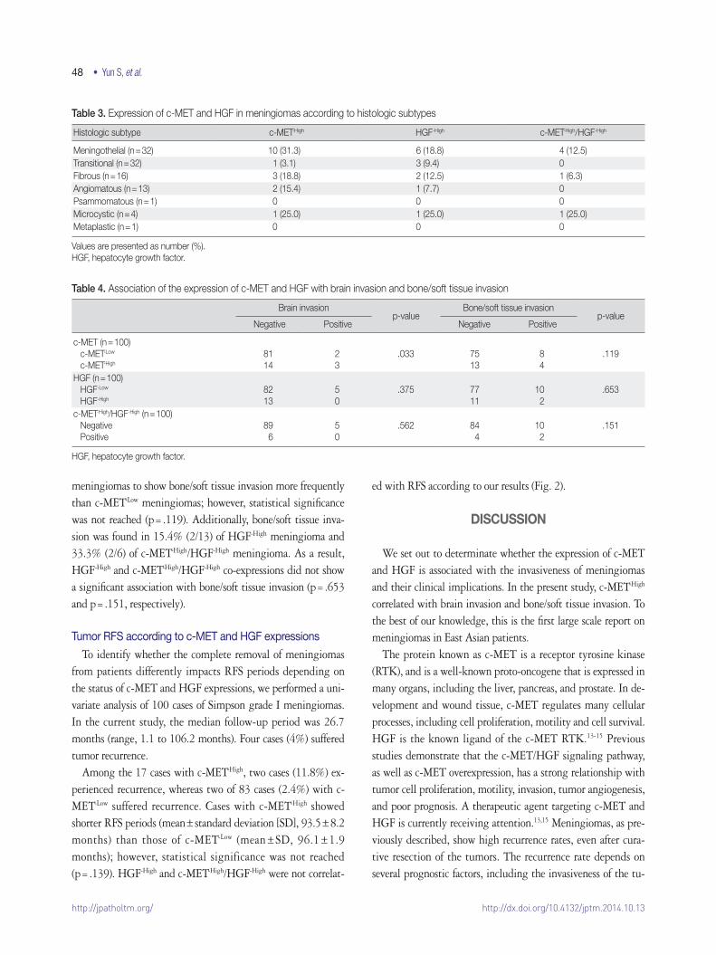
Expression of c-MET and HGF in meningiomas according to histologic subtypes
Of the cases, c-MET-High and HGF-High were found in 17% (17/100) and 13% (13/100) of meningiomas, respectively, and c-MET-High/HGF-High co-expressions were observed in 1% (1/100) of meningiomas. c-MET-High and HGF-High showed no significant correlation with the histologic subtypes of meningi-omas (Table 3).
Association of the expression of c-MET and HGF with brain invasion
Brain invasion was observed in 3/17 (17.6%) of c-MET-High meningiomas and in 2/83 (2.4%) of c-MET-Low meningiomas. Therefore, there was a statistically significant correlation between c-MET-High and brain invasion (p=.033). Neither HGF-High nor c-MET-High/HGF-High co-expression showed statistical associations with brain invasion (p=.375 and p=.562, respectively) (Table 4).
Association of the expression of c-MET and HGF with bone/soft tissue invasion
Of the cases, bone/soft tissue invasion was observed in 4/17 (23.5%) of c-MET-High meningiomas and in 8/83 (9.6%) of c-MET-Low meningiomas. There was a tendency for c-MET-High
G H
Fig. 1. (Continued) (G) HGF moderate staining in meningioma. (H) HGF strong staining in meningioma.
Table 2. Summary of brain and bone/soft tissue invasion in meningiomas
CharacteristicBrain invasion Bone/soft tissue invasion
Yes (n=5) No (n=95) p-value Yes (n=12) No (n=88) p-value
Age (yr) 73 (47–77) 60 (36–85) - 58 (42–75) 60 (36–85) -Sex Male 3 (13.0) 20 (87.0) .078 2 (8.7) 21 (91.3) .728 Female 2 (2.6) 75 (97.4) 10 (13.0) 67 (87.0)
Values are presented as median (range) or number (%).
http://jpatholtm.org/ http://dx.doi.org/10.4132/jptm.2014.10.13
48 • Yun S, et al.
meningiomas to show bone/soft tissue invasion more frequently than c-MET-Low meningiomas; however, statistical significance was not reached (p=.119). Additionally, bone/soft tissue inva-sion was found in 15.4% (2/13) of HGF-High meningioma and 33.3% (2/6) of c-MET-High/HGF-High meningioma. As a result, HGF-High and c-MET-High/HGF-High co-expressions did not show a significant association with bone/soft tissue invasion (p=.653 and p=.151, respectively).
Tumor RFS according to c-MET and HGF expressions
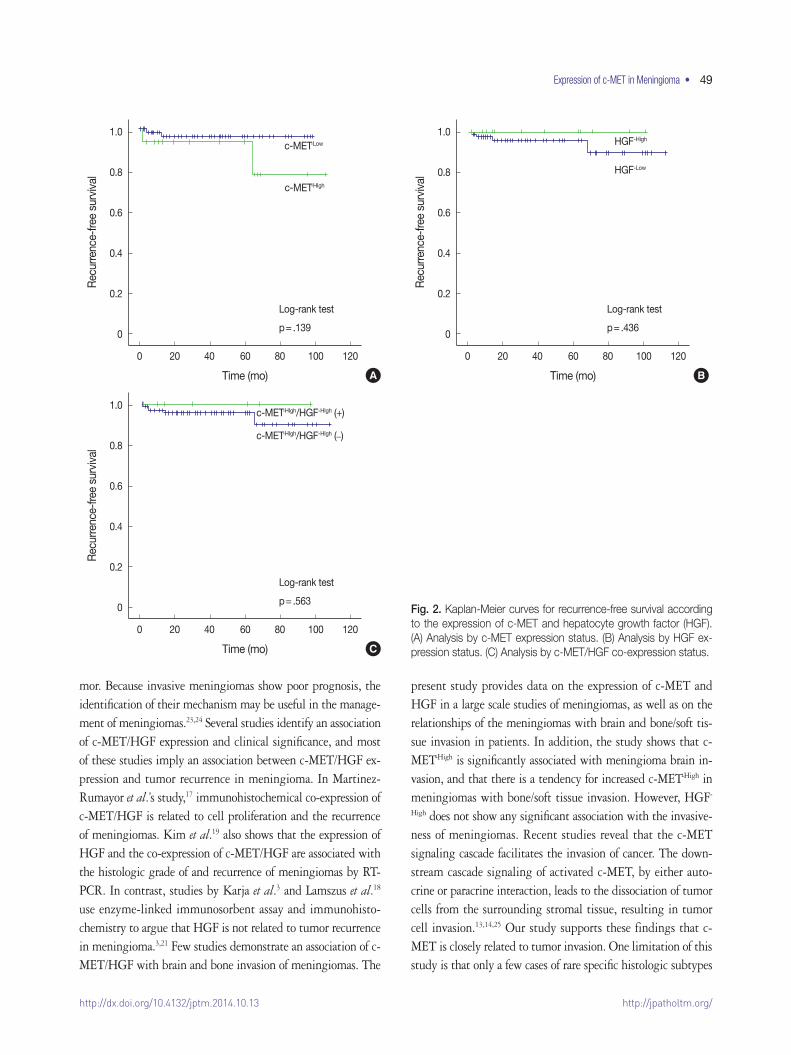
To identify whether the complete removal of meningiomas from patients differently impacts RFS periods depending on the status of c-MET and HGF expressions, we performed a uni-variate analysis of 100 cases of Simpson grade I meningiomas. In the current study, the median follow-up period was 26.7 months (range, 1.1 to 106.2 months). Four cases (4%) suffered tumor recurrence.
Among the 17 cases with c-MET-High, two cases (11.8%) ex-perienced recurrence, whereas two of 83 cases (2.4%) with c-MET-Low suffered recurrence. Cases with c-MET-High showed shorter RFS periods (mean±standard deviation [SD], 93.5±8.2 months) than those of c-MET-Low (mean±SD, 96.1±1.9 months); however, statistical significance was not reached (p=.139). HGF-High and c-MET-High/HGF-High were not correlat-
ed with RFS according to our results (Fig. 2).
DISCUSSION
We set out to determinate whether the expression of c-MET and HGF is associated with the invasiveness of meningiomas and their clinical implications. In the present study, c-MET-High correlated with brain invasion and bone/soft tissue invasion. To the best of our knowledge, this is the first large scale report on meningiomas in East Asian patients.
The protein known as c-MET is a receptor tyrosine kinase (RTK), and is a well-known proto-oncogene that is expressed in many organs, including the liver, pancreas, and prostate. In de-velopment and wound tissue, c-MET regulates many cellular processes, including cell proliferation, motility and cell survival. HGF is the known ligand of the c-MET RTK.13-15 Previous studies demonstrate that the c-MET/HGF signaling pathway, as well as c-MET overexpression, has a strong relationship with tumor cell proliferation, motility, invasion, tumor angiogenesis, and poor prognosis. A therapeutic agent targeting c-MET and HGF is currently receiving attention.13,15 Meningiomas, as pre-viously described, show high recurrence rates, even after cura-tive resection of the tumors. The recurrence rate depends on several prognostic factors, including the invasiveness of the tu-
Table 3. Expression of c-MET and HGF in meningiomas according to histologic subtypes
Histologic subtype c-MET-High HGF-High c-MET-High/HGF-High
Meningothelial (n=32) 10 (31.3) 6 (18.8) 4 (12.5)Transitional (n=32) 1 (3.1) 3 (9.4) 0Fibrous (n=16) 3 (18.8) 2 (12.5) 1 (6.3)Angiomatous (n=13) 2 (15.4) 1 (7.7) 0Psammomatous (n=1) 0 0 0Microcystic (n=4) 1 (25.0) 1 (25.0) 1 (25.0)Metaplastic (n=1) 0 0 0
Values are presented as number (%).HGF, hepatocyte growth factor.
Table 4. Association of the expression of c-MET and HGF with brain invasion and bone/soft tissue invasion
Brain invasionp-value
Bone/soft tissue invasionp-value
Negative Positive Negative Positive
c-MET (n=100) c-MET-Low
c-MET-High
8114
23
.033 7513
84
.119
HGF (n=100) HGF-Low
HGF-High
8213
50
.375 7711
102
.653
c-MET-High/HGF-High (n=100) Negative Positive
896
50
.562 844
102
.151
HGF, hepatocyte growth factor.
http://jpatholtm.org/http://dx.doi.org/10.4132/jptm.2014.10.13
Expression of c-MET in Meningioma • 49
mor. Because invasive meningiomas show poor prognosis, the identification of their mechanism may be useful in the manage-ment of meningiomas.23,24 Several studies identify an association of c-MET/HGF expression and clinical significance, and most of these studies imply an association between c-MET/HGF ex-pression and tumor recurrence in meningioma. In Martinez-Rumayor et al.’s study,17 immunohistochemical co-expression of c-MET/HGF is related to cell proliferation and the recurrence of meningiomas. Kim et al.19 also shows that the expression of HGF and the co-expression of c-MET/HGF are associated with the histologic grade of and recurrence of meningiomas by RT-PCR. In contrast, studies by Karja et al.3 and Lamszus et al.18 use enzyme-linked immunosorbent assay and immunohisto-chemistry to argue that HGF is not related to tumor recurrence in meningioma.3,21 Few studies demonstrate an association of c-MET/HGF with brain and bone invasion of meningiomas. The
present study provides data on the expression of c-MET and HGF in a large scale studies of meningiomas, as well as on the relationships of the meningiomas with brain and bone/soft tis-sue invasion in patients. In addition, the study shows that c-MET-High is significantly associated with meningioma brain in-vasion, and that there is a tendency for increased c-MET-High in meningiomas with bone/soft tissue invasion. However, HGF-
High does not show any significant association with the invasive-ness of meningiomas. Recent studies reveal that the c-MET signaling cascade facilitates the invasion of cancer. The down-stream cascade signaling of activated c-MET, by either auto-crine or paracrine interaction, leads to the dissociation of tumor cells from the surrounding stromal tissue, resulting in tumor cell invasion.13,14,25 Our study supports these findings that c-MET is closely related to tumor invasion. One limitation of this study is that only a few cases of rare specific histologic subtypes
Fig. 2. Kaplan-Meier curves for recurrence-free survival according to the expression of c-MET and hepatocyte growth factor (HGF). (A) Analysis by c-MET expression status. (B) Analysis by HGF ex-pression status. (C) Analysis by c-MET/HGF co-expression status.
1.0
0.8
0.6
0.4
0.2
0
1.0
0.8
0.6
0.4
0.2
0
Rec
urre
nce-
free
surv
ival
Rec
urre
nce-
free
surv
ival
Time (mo) Time (mo)
0 20 40 60 80 100 120 0 20 40 60 80 100 120
p=.139 p=.436
Log-rank test Log-rank test
c-MET-Low HGF-High
HGF-Low
A B
c-MET-HIgh
1.0
0.8
0.6
0.4
0.2
0
Rec
urre
nce-
free
surv
ival
Time (mo)
0 20 40 60 80 100 120
p=.563
Log-rank test
C
c-MET-HIgh/HGF-HIgh (+)
c-MET-HIgh/HGF-HIgh (–)
http://jpatholtm.org/ http://dx.doi.org/10.4132/jptm.2014.10.13
50 • Yun S, et al.
are included in the data. Nevertheless, the results suggest that c-MET may participate in tumor invasion.
We also evaluate a possible association between the c-MET and HGF expression and disease recurrence. In this study, the recurrence rate of meningiomas with complete tumor resection is 5%, a finding which is slightly lower than findings in previ-ous reports.26 Also, we demonstrate that c-MET-High only shows a tendency for association with shorter RFS periods. In general, the recurrence of meningiomas occurred within two years of sur-gical treatment, and up to 94% of patients with meningiomas experienced recurrence within five years.27 However, the vast majority of meningiomas are slow-growing tumors, and benign meningiomas that have been completely removed from patients recur at a rate of 19% after 20 years of follow-up.28 Thus our findings about recurrence rates are limited due to an insufficient follow-up period (median follow-up time in this study, 26.7 months). Several studies report an intratumoral heterogeneity of c-MET and HGF expression, revealing an increase in these fac-tors at cancer-invading fronts in breast carcinoma and cholangio-carcinoma.29,30 Accordingly, further studies are needed to eluci-date intratumoral heterogeneity in meningiomas, and the asso-ciation between c-MET overexpression and RFS.
In summary, our results demonstrate that c-MET is associated with the brain invasion of meningiomas, and that c-MET ex-pression may be useful predictive markers for meningioma re-currence.
Many previous studies reveal that c-MET signaling is in-volved in the progression and spread of several cancers.16-19,25,28 The collective understanding of c-MET’s role in cancers has evoked considerable interest in c-MET and HGF as major tar-gets in the development of cancer drugs. This has led to the de-velopment of a variety of c-MET pathway antagonists with po-tential clinical applications. Several c-MET antagonists are now under clinical investigation.13,14,25 We conclude that c-MET ex-pression may be a useful predictive marker for meningioma re-currence, and that invasive meningiomas with high expression of c-MET may be good candidates for targeted therapy using selective c-MET inhibitors.
Conflicts of InterestNo potential conflict of interest relevant to this article was
reported.REFERENCES
1.LouisDN,OhgakiH,WiestlerOD,CaveneeWK.WHOclassifica-tion of tumours of the central nervous system. Lyon: IARC Press,
2007.2. Choy W, Kim W, Nagasawa D, et al. The molecular genetics and tu-
mor pathogenesis of meningiomas and the future directions of me-ningioma treatments. Neurosurg Focus 2011; 30: E6.
3.KarjaV,SandellPJ,KauppinenT,AlafuzoffI.Doesproteinexpres-sion predict recurrence of benign World Health Organization grade I meningioma? Hum Pathol 2010; 41: 199-207.
4.PerryA,StaffordSL,ScheithauerBW,SumanVJ,LohseCM.Me-ningioma grading: an analysis of histologic parameters. Am J Surg Pathol 1997; 21: 1455-65.
5. Ildan F, Erman T, Göçer AI, et al. Predicting the probability of me-ningioma recurrence in the preoperative and early postoperative period: a multivariate analysis in the midterm follow-up. Skull Base2007;17:157-71.
6. Durand A, Labrousse F, Jouvet A, et al. WHO grade II and III me-ningiomas: a study of prognostic factors. J Neurooncol 2009; 95: 367-75.
7.CaltabianoR,BarbagalloGM,CastaingM,et al. Prognostic value of EGFRexpressioninde novo and progressed atypical and anaplastic meningiomas: an immunohistochemical and fluorescence in situ hybridization pilot study. J Neurosurg Sci 2013; 57: 139-51.
8.BarresiV,VitarelliE,TuccariG,BarresiG.MMP-9expressioninmeningiomas: a prognostic marker for recurrence risk? J Neuroon-col 2011; 102: 189-96.
9.FathiAR,RoelckeU.Meningioma.CurrNeurolNeurosciRep2013; 13: 337.
10. Alahmadi H, Croul SE. Pathology and genetics of meningiomas. Semin Diagn Pathol 2011; 28: 314-24.
11.TerziA,SaglamEA,BarakA,SoylemezogluF.ThesignificanceofimmunohistochemicalexpressionofKi-67,p53,p21,andp16inmeningiomas tissue arrays. Pathol Res Pract 2008; 204: 305-14.
12. Kliese N, Gobrecht P, Pachow D, et al. miRNA-145 is downregulat-ed in atypical and anaplastic meningiomas and negatively regu-lates motility and proliferation of meningioma cells. Oncogene 2013; 32: 4712-20.
13.LiuX,NewtonRC,ScherlePA.Developingc-METpathwayinhibi-torsforcancertherapy:progressandchallenges.TrendsMolMed2010; 16: 37-45.
14. Guessous F, Zhang Y, diPierro C, et al.Anorallybioavailablec-Metkinase inhibitor potently inhibits brain tumor malignancy and growth.AnticancerAgentsMedChem2010;10:28-35.
15.OrganSL,TsaoMS.Anoverviewofthec-METsignalingpathway.TherAdvMedOncol2011;3(1Suppl):S7-19.
16.FengY,ThiagarajanPS,MaPC.METsignaling:noveltargetedin-hibition and its clinical development in lung cancer. J Thorac Oncol 2012; 7: 459-67.

http://jpatholtm.org/http://dx.doi.org/10.4132/jptm.2014.10.13
Expression of c-MET in Meningioma • 51
17.Martínez-RumayorA,ArrietaO,GuevaraP,et al.Coexpressionofhepatocytegrowthfactor/scatterfactor(HGF/SF)anditsreceptorcMETpredictrecurrenceofmeningiomas.CancerLett2004;213:117-24.
18. Lamszus K, Lengler U, Schmidt NO, Stavrou D, Ergün S, Westphal M.Vascularendothelialgrowthfactor,hepatocytegrowthfactor/scatterfactor,basicfibroblastgrowthfactor,andplacentagrowthfactor in human meningiomas and their relation to angiogenesis and malignancy. Neurosurgery 2000; 46: 938-47.
19.KimNR,ChaeYS,LimWJ,ChoSJ.Expressionofhepatocytegrowth factor/c-met by RT-PCR in meningiomas. Korean J Pathol 2011; 45: 463-8.
20.SuhJH,ParkJW,LeeC,MoonKC.ERGimmunohistochemistryand clinicopathologic characteristics in Korean prostate adenocarci-noma patients. Korean J Pathol 2012; 46: 423-8.
21.BozkayaG,KorhanP,CokakliM,et al. Cooperative interaction of MUC1withtheHGF/c-Metpathwayduringhepatocarcinogenesis.MolCancer2012;11:64.
22. Kong DS, Song SY, Kim DH, et al.Prognosticsignificanceofc-Metexpressioninglioblastomas.Cancer2009;115:140-8.
23. Palma L, Celli P, Franco C, Cervoni L, Cantore G. Long-term prog-nosis for atypical and malignant meningiomas: a study of 71 surgi-
cal cases. J Neurosurg 1997; 86: 793-800.24.Gabeau-LacetD,AghiM,BetenskyRA,BarkerFG,LoefflerJS,LouisDN.Boneinvolvementpredictspooroutcomeinatypicalmeningioma. J Neurosurg 2009; 111: 464-71.
25.EderJP,VandeWoudeGF,BoernerSA,LoRussoPM.Novelthera-peuticinhibitorsofthec-Metsignalingpathwayincancer.ClinCancer Res 2009; 15: 2207-14.
26. Yamasaki F, Yoshioka H, Hama S, Sugiyama K, Arita K, Kurisu K. Recurrence of meningiomas. Cancer 2000; 89: 1102-10.
27. Violaris K, Katsarides V, Sakellariou P. The recurrence rate in me-ningiomas:analysisoftumorlocation,histologicalgrading,andex-tentofresection.OpenJModNeurosurg2011;2:6-10.
28. Jääskeläinen J. Seemingly complete removal of histologically be-nign intracranial meningioma: late recurrence rate and factors pre-dicting recurrence in 657 patients: a multivariate analysis. Surg Neurol 1986; 26: 461-9.
29.EdakuniG,SasatomiE,SatohT,TokunagaO,MiyazakiK.Expres-sionofthehepatocytegrowthfactor/c-Metpathwayisincreasedatthe cancer front in breast carcinoma. Pathol Int 2001; 51: 172-8.
30. Terada T, Nakanuma Y, Sirica AE. Immunohistochemical demon-strationofMEToverexpressioninhumanintrahepaticcholangio-carcinoma and in hepatolithiasis. Hum Pathol 1998; 29: 175-80.