Expression and Regulation of Human Neutrophil-derived ...
10
Expression and Regulation of Human Neutrophil-derived Macrophage Inflammatory Protein lee By Tsuyoshi Kasama,* Robert M. Strieter,~ Theodore J. Standiford,~ Marie D. Burdick,~ and Steven L. Kunkel* From the Departments of *Pathology and ~Internal Medicine, Division of Pulmonary and Critical Care Medicine, The University of Michigan Medical School, Ann Arbor, Michigan 481O9 Swmmar~ Neutrophil (polymorphonuclearleukocyte [PMN]) sequestration is one of the histologic hallmarks of an acute inflammatory response. During the natural evolution of an inflammatory response, PMNs are often replaced by mononuclear cells. This shift in the elicitation of specific leukocyte populations usually occurs as the inflammatory lesion enters either the repair/resolution stage or progresses to a chronic inflammation. To elucidate a potential mechanism for the temporal change from predominantly PMN recruitment to the presence of monocytes, we postulated that PMNs could be a rich source of monocyte chemotactic factors. In our studies, we have identified a dose-dependent induction of monocyte chemotactic activity by PMNs treated with lipopoly- saccharide (LPS; 1-100 ng/ml). Interestingly, this monocyte chemotactic activity was significantly attenuated in the presence of neutralizing anti-human macrophage inflammatory protein lc~ (MIP-I~x) antibodies. Moreover, immunolocalization studies demonstrated the expression of MIP-lot by stimulated PMNs. These findings showed that a significant amount of PMN-derived monocyte chemotactic activity was attributable to MIP-loe. Subsequent characterization of MIP-lo~ steady- state mRNA and antigen expression demonstrated both a dose- and time-dependent production by LPS-treated PMNs. Granulocyte/macrophage colony-stimulating factor (GM-CSF), a potent PMN activator, failed to induce the expression of MIP-lc~ over a wide range of concentrations. However, PMNs stimulated in the presence of both LPS and GM-CSF resulted in a synergistic expression pattern for MIP-lc~. PMNs stimulated in the presence of both GM-CSF and LPS demonstrated an enhanced and prolonged expression for both MIP-loe mRNA and antigen, as compared with LPS alone. Messenger RNA stabilization analyses demonstrated that MIP-lc~ mRNA isolated from PMNs stimulated in the presence of GM-CSF and LPS had a prolonged mRNA tl/2, as compared with LPS alone. These findings support the notion that PMNs are capable of producing MIP-lol in the presence of LPS, and that GM-CSF can influence this production through prolongation of MIP-lc~ mRNA tl/2. The production of PMN-derived MIP-lcx, in association with the expression of appropriate adhesion molecules at a site of inflammation, may be one of the central events that contributes to the temporal shift from predominantly PMNs to monocytes during the evolution of inflammation. H uman peripheral PMNs are the predominant leukocyte population at sites of acute inflammatory reactions (1). These leukocytes produce a number of inflammatory medi- ators, including reactive oxygen intermediates, arachidonic acid derivatives, and enzymes. Recently, a number of investi- gators have reported that PMNs can produce several poly- peptide mediators of inflammation. Activated PMNs have been shown to produce specific cytokines, such as IL-1 (2-5), IL-6 (6), IL-8 (7-10), and TNF (11, 12). Since PMNs may com- prise up to 80% of the circulating leukocyte pool in humans, this biosynthetically active leukocyte population must be con- sidered an important source of cytokines during the early phases of inflammation. Previous reports have identified PMNs as a source of IL-8, a chemotactic cytokine for neutrophils and lymphocytes, at nanomolar and picomolar concentrations, respectively. Thus, PMN-derived cytokines may play an im- portant role in the early initiation phase of an inflammatory response by the induction of the early response cytokines, 63 J. Exp. Med. The Rockefeller University Press * 0022-1007/93/07/0063/10 $2.00 Volume 178 July 1993 63-72
Transcript of Expression and Regulation of Human Neutrophil-derived ...

Expression and Regulation of Human Neutrophil-derived Macrophage Inflammatory Protein lee By Tsuyoshi Kasama,* Robert M. Strieter,~ Theodore J. Standiford,~ Marie D. Burdick,~ and Steven L. Kunkel*
From the Departments of *Pathology and ~Internal Medicine, Division of Pulmonary and Critical Care Medicine, The University of Michigan Medical School, Ann Arbor, Michigan 481O9
S w m m a r ~
Neutrophil (polymorphonuclear leukocyte [PMN]) sequestration is one of the histologic hallmarks of an acute inflammatory response. During the natural evolution of an inflammatory response, PMNs are often replaced by mononuclear cells. This shift in the elicitation of specific leukocyte populations usually occurs as the inflammatory lesion enters either the repair/resolution stage or progresses to a chronic inflammation. To elucidate a potential mechanism for the temporal change from predominantly PMN recruitment to the presence of monocytes, we postulated that PMNs could be a rich source of monocyte chemotactic factors. In our studies, we have identified a dose-dependent induction of monocyte chemotactic activity by PMNs treated with lipopoly- saccharide (LPS; 1-100 ng/ml). Interestingly, this monocyte chemotactic activity was significantly attenuated in the presence of neutralizing anti-human macrophage inflammatory protein lc~ (MIP-I~x) antibodies. Moreover, immunolocalization studies demonstrated the expression of MIP-lot by stimulated PMNs. These findings showed that a significant amount of PMN-derived monocyte chemotactic activity was attributable to MIP-loe. Subsequent characterization of MIP-lo~ steady- state mRNA and antigen expression demonstrated both a dose- and time-dependent production by LPS-treated PMNs. Granulocyte/macrophage colony-stimulating factor (GM-CSF), a potent PMN activator, failed to induce the expression of MIP-lc~ over a wide range of concentrations. However, PMNs stimulated in the presence of both LPS and GM-CSF resulted in a synergistic expression pattern for MIP-lc~. PMNs stimulated in the presence of both GM-CSF and LPS demonstrated an enhanced and prolonged expression for both MIP-loe mRNA and antigen, as compared with LPS alone. Messenger RNA stabilization analyses demonstrated that MIP-lc~ mRNA isolated from PMNs stimulated in the presence of GM-CSF and LPS had a prolonged mRNA tl/2, as compared with LPS alone. These findings support the notion that PMNs are capable of producing MIP-lol in the presence of LPS, and that GM-CSF can influence this production through prolongation of MIP-lc~ mRNA tl/2. The production of PMN-derived MIP-lcx, in association with the expression of appropriate adhesion molecules at a site of inflammation, may be one of the central events that contributes to the temporal shift from predominantly PMNs to monocytes during the evolution of inflammation.
H uman peripheral PMNs are the predominant leukocyte population at sites of acute inflammatory reactions (1).
These leukocytes produce a number of inflammatory medi- ators, including reactive oxygen intermediates, arachidonic acid derivatives, and enzymes. Recently, a number of investi- gators have reported that PMNs can produce several poly- peptide mediators of inflammation. Activated PMNs have been shown to produce specific cytokines, such as IL-1 (2-5), IL-6 (6), IL-8 (7-10), and TNF (11, 12). Since PMNs may com-
prise up to 80% of the circulating leukocyte pool in humans, this biosynthetically active leukocyte population must be con- sidered an important source of cytokines during the early phases of inflammation. Previous reports have identified PMNs as a source of IL-8, a chemotactic cytokine for neutrophils and lymphocytes, at nanomolar and picomolar concentrations, respectively. Thus, PMN-derived cytokines may play an im- portant role in the early initiation phase of an inflammatory response by the induction of the early response cytokines,
63 J. Exp. Med. �9 The Rockefeller University Press * 0022-1007/93/07/0063/10 $2.00 Volume 178 July 1993 63-72

IL-1 and TNF, and the expression of the chemotactic cytokine, IL-8. Whereas this latter cytokine will likely promote the continued recruitment of neutrophils to the inflammatory lesion, other recruitment factors must be expressed in order for the responses to progress to one characterized by the ac- cumulation of monocytes/macrophages.
Recent investigations have demonstrated that the chemotac- tic cytokine, macrophage inflammatory protein 1 (MIP-1) 1, possesses biological activity for activating both neutrophils and monocytes/macrophages. Native MIP-1 is comprised of two 8-kD polypeptides termed MIP-lo~ and MIP-I~. This chemotactic cytokine is a member of the C-C chemokine su- pergene family and is an LPS-inducible, heparin-binding poly- peptide first isolated from LPS-treated murine RAW 264.7 cells (13-16). MIP-lot induces neutrophil chemokinesis, and activates neutrophils to generate hydrogen peroxide (14). In vivo, MIP-lot can induce a pyrogenic response by acting as a prostaglandin-independent endogenous pyrogen (17). Re- cently, it has been reported that MIP-lot-treated murine peri- toneal exudate macrophages exhibited enhanced antibody- independent macrophage cytotoxicity for tumor targets, in addition to stimulating the secretion of TNF, IL-1, and IL-6 from this macrophage population (18). These data indicate that MIP-lot may play a crucial role in the biology of inflam- mation and the pathogenesis of inflammatory diseases.
In the present studies, we present data demonstrating that PMNs may play an important role in altering the composi- tion of leukocytes recruited to an area of inflammation via their ability to generate monocyte chemotactic factors. Fur- thermore, one of the major neutrophil-derived monocyte chemotactic factors is MIP-lot. PMNs challenged with LPS demonstrated a time- and dose-dependent increase in both steady-state MIP-lot mRNA expression, and antigen and chemotactic activity. The latter activity was suppressed by the addition of neutralizing MIP-lot antibody. Furthermore, the expression of MIP-lot mRNA by PMNs stimulated with LPS was augmented by GM-CSF, which resulted in the prolon- gation of the tt/2 for MIP-lcx mRNA. These investigations suggest that PMNs possess multifunctional activities during the initiation and maintenance of an inflammatory reaction and are an important source of this monocyte chemotactic cytokine.
Materials and Methods Reagent Preparation. Human recombinant GM-CSF (sp act, 4
x 107 U/mg protein); G-CSF (sp act, 2 x 10 s U/rag protein); Ib3 (sp act, 0.1-0.4 ng/ml); and MIP-lc~ (sp act, EDs0, 25 ng/ml) were purchased from R & D Systems, Inc. (Minneapolis, MN). Cycloheximide (CHX) was purchased from Sigma Chemical Co. (St. Louis, MO). Stock LPS (Escherichia coli 0111:B4; Sigma Chem- ical Co.) and actinomycin D (Ac-D) (Sigma Chemical Co.) were prepared at concentrations of 200/~g/ml in complete medium and
1 Abbreviations used in this paper: Ac-D, actinomycin D; CHX, cyclohexi- mide; hpf, high power field; MCP-1, monocyte chemoattractant protein 1; MIP-lct, macrophage inflammatory protein lc~.
5 mg/ml in DMSO (Sigma Chemical Co.), respectively. Complete medium consisted of ILPMI 1640 (Gibco Laboratories, Grand Is- land, NY) supplemented with 2 mM t-glutamine, 25 mM Hepes, 100 U/ml penicillin, 100 ng/ml streptomycin (Hazelton Research Products, Lenexa, KS) and 5% heat-inactivated FCS (Gibco Labora- tories). Antisera to MIP-lc~ were raised in rabbits immunized with recombinant human MIP-lcx using procedures established in our laboratory. The antisera to human MIP-lc~ did not cross-react with other known members of this chemoattractant cytokine family, including RANTES, monocyte chemoattractant protein 1 (MCP- 1), or MIP-1B. All reagents, except LPS, were checked for endo- toxin contamination using a limulus amebocyte lysate test kit (QCb 1000; Whittaker Bioproducts, Inc., Walkersville, MD). The con- centration of endotoxin was consistently below 0.01 endotoxin units (EU)/ml in all reagents.
Isolation and Culture Conditions of PMNs and Monocytes. Human peripheral blood PMNs were obtained from heparinized venous blood of healthy volunteers by Ficoll-Hypaque (Pharmacia LKB Biotechnology Inc., Piscataway, NJ) centrifugation and sedimen- tation in 5% dextran/0.9% saline. PMNs were subsequently sepa- rated from erythrocytes by lysis of erythrocytes in a solution of 0.15 M NH4C1, 0.01 M NaHCO3, and 0.01 M tetra EDTA. The recovered PMNs were washed three times and resuspended at a density of 5 x 10⁶ cells/ml in complete medium. The final cell preparation contained more than 99% PMN by morphology and nonspecific esterase staining, and viability was more than 98% by trypan blue dye exclusion. After Ficoll-Hypaque centrifugation, monocytes were purified by counterflow centrifugal elutriation from PBMC (19), and resuspended in complete medium at 104-106 cells/ml. PMNs and monocytes were incubated with various con- centrations of LPS, and/or GM-CSF. After various periods of time, cell-free supernatants of PMN cultures and total PMN RNA were collected and stored at -20~ In some experiments, cell lysates of PMN were resuspended and stored in the same volume of com- plete medium.
Assay of Monocyte Chemotactic Activity. Monocyte chemotaxis was performed as previously described (20). For monocyte chemo- taxis, either 150 #1 of supernatant specimen, 10 -s M FMLP (Sigma Chemical Co.), MIP-lot (1-100 ng/ml), or HBSS (Gibco Laboratories) was placed in duplicate bottomed wells of a modified Boyden chemotaxis chamber (Neuro Probe Inc., Cabin John, MD). A 5-#m pore size polycarbonate filter (polyvinylpyrrolidone-free, Nucleopore Corp., Pleasanton, CA) was placed in the assembly and 250/xl of mononuclear cell suspension (4 x 10⁶/ml in HBSS) placed in each of the top wells. Chemotaxis chamber assemblies were incubated at 37~ in humidified 95% air/5% COz for 75 min and the filters removed, fixed in absolute methanol, and stained with 2% toluidine blue (Sigma Chemical Co.). Monocytes that had migrated through to the bottom of the filter were counted in 10 high power fields using the following system: a Javelin chro- machip camera (Javelin Electronics, Torrance, CA) attached to a Olympus BH-2 microscope, which was interfaced to a computer (Macintosh* II; Apple Computer, Inc., Cupertino, CA) with a frame grabber (Image Capture 1000; Scion Corp., Walkersvilh, MD) and Image 1.40 software .(National Institutes of Health [Nil-I] Public Software, Bethesda, MD). Chemotactic Bioactivity was represented by the count of migrated monocytes per high power field. In neu- tralization experiments, sample supematants were preincubated with 1:1,000 dilution of rabbit preimmune serum or neutralizing rabbit anti-human MIP-lc~ antiserum for 30 min at 37~ and then as- sayed for monocyte chemotactic bioactivity.
Antigen-specific ELISA. PMN-derived antigenic MIP-lcx quan- tified using a modification of a double ligand method, as previ-
64 Macrophage Inflammatory Protein l~x Expression in Neutrophils

ously described (21). Briefly, fiat-bottomed 96-weU microtiter plates were coated with 50 /xl/weU of rabbit anti-MIP-lc~ antibody (1/xg/ml in 0.6 mol/liter NaC1, 0.26 tool/liter H3BO4, and 0.08 N NaOH, pH 9.6) for 16 h at 4~ and then washed with PBS, pH 7.5, 0.05% Tween 20 (wash buffer). Nonspecific binding sites on microtiter plates were blocked with 2% BSA in PBS and in- cubated for 90 min at 37~ Plates were rinsed four times with wash buffer, and diluted neutrophil-derived conditioned media (50 /~I) were added, followed by incubation for 1 h at 37~ Plates were washed four times with wash buffer, then 50/~l/well of bi- otinylated rabbit anti-MIP-lot was added and incubated for 30 min at 37~ After washing of plates, chromogen substrate was added. The plates were incubated at room temperature to the desired ex- tinction, and the reaction terminated with 50/~l/well of 3 M H3SO4 solution, and were read at 490 nm in an ELISA reader. This ELISA method consistently had a sensitivity limit of ,,~50 pg/ml.
lmmunohistochemistry. PMNs (5.0 x 10⁶/ml) were incubated with LPS (100 ng/ml) and/or GM-CSF (10 U/ml) for 24 h, and deposited on a glass slide by using a Cytospin II (Shandon Southern Instruments, Inc., Sewickley, PA). After air-drying, slides were fixed by 4% paraformaldehyde in PBS for 10 min. Before staining, the slides were again fixed for 30 min in ice-cold acetone. Endogenous peroxidase activity was quenched by incubating the slides for 30 min in absolute methanol and 3% hydrogen peroxide. After rinsing in PBS, the slides were blocked with a 1:50 dilution of normal goat serum for 30 min at 37~ then treated for 2 h at 37~ with a 1:800 dilution of rabbit anti-human MIP-lcz serum or the same dilution of preimmune rabbit serum. After incubation, prepara- tions were rinsed three times with PBS, overlaid with biotinylated goat anti-rabbit IgG (1:200; Vector Laboratories Inc., Burlingame, CA), incubated 20 min, and rinsed three times with PBS. The slides were then treated with streptavidin conjugated to peroxidase for 15 min at 37~ rinsed three time with PBS, overlaid with 100 txl of equal vol of 3-amino-9-ethylcarbazole 40 mg/ml in N,N- dimethylformamide, (Sigma Chemical Co.), and treated with 3% hydrogen peroxide in 0.1 M sodium acetate for 15 min at 37~ to allow color development. After rinsing with distilled water, the slides were stained with Mayer's hematoxylin. In competitive inhi- bition studies, to demonstrate antibody specifidty, immunostaining for human MIP-lce showed 100% inhibition by the addition of exogenous recombinant MIP-lcr
Isolation of Total PMN RNA and Northern Blot Analysis. Total cellular RNA from PMN was isolated as previously described (8). Briefly, guanidine isothiocyanate solution containing 25 mM Tris, pH 8.0, 4.2 M guanidine isothiocyanate, 0.5% Sarkosyl, and 0.1 M 2-ME was added to PMN pellets. After homogenization, the above suspension was added to a solution containing an equal volume of 100 mM Tris, pH 8.0, 10 mM EDTA, and 1.0% SDS. The mix- ture was then extracted with chloroform-phenol and chloroform- isoamyl alcohol. The RNA was alcohol-precipitated and the pellet dissolved in diethyl pyrocarbonate-treated H20. Total RNA was separated by Northern blot analysis using formaldehyde, 1% agarose gels, transblotted to nitrocellulose, baked, prehybridized, and hy- bridized with a 32p-5' end-labeled oligonucleotide probe specific for human MIP-lc~ or/~-actin. The 30-mer oligonucleotide probe for MIP-lcr was complementary to nucleotides 105-134 (5'-GAG- AGC-CAT-GGT-GCA~GAG-GAG-GAC-AGC-AAG-3') (22). The 42-mer oligonucleotide probe for ~/-actin was complementary to nucleotides 432-473 (5'-GGC-TGG-GGT-GTT-GAA-GGT-CTC- AAA-CAT-GAT-CTG-GGT-CAT-CTT-3') (23). Blots were washed and exposed to X-ray film. Specific cytokine mRNA was quantified
using imaging analysis with the Image Capture 1000 flame grabber and Image 1.40 software.
StatisticalAnalysis. Data were analyzed by Macintosh | II com- puter using a statistical software package (Statview II; Abacus Con- cepts, Inc., Berkeley, CA) and expressed as mean _+ SEM. Groups of data were evaluated by analysis of variance. Data that appeared statistically significant were compared by Student's t test for com- paring the means of multiple groups, and considered significant if p values were <0.05.
Results
Activity of Monocyte Chemotaxis in PMN-conditioned Medium. In initial studies, we examined whether LPS-stirnulated PMNs were capable of producing chemotactic factors for monocytes. Conditioned media from PMNs (PMN-CM) stimulated with graded doses of LPS (1-100 ng/ml) for 24 h were shown to possess significant monocyte chemotactic activity (Table 1). PMN-CM challenged with 10 ng/ml LPS demonstrated a significant increase in monocyte chemotactic activity, as com- pared with control (CTR)-CM (11.9 vs 5.5 ceUs/high power field [hpq, respectively). PMNs challenged with the max- imum concentration of LPS used in this study, 100 ng/ml, produced a monocyte chemotactic activity which was ,,050% of the FMLP positive control, 14.0 vs 30.5 cells/hpf, respec- tivdy. To address the potential role of residual LPS in the PMN supernatant to alter monocyte chemotaxis, we assessed the monocyte chemotactic activity of recombinant human MIP-lot in the presence and absence of graded doses (1-100 ng/ml) of LPS. In these studies LPS, at various doses, did not alter in vitro monocyte chemotaxis to 10 ng/ml of MIP- lc~ (data not shown). We examined the specificity of the mono- cyte chemotactic activity produced by the PMNs by adding neutralizing antibody directed against MIP-lc~ to the PMN- CM before assessing chemotactic activity. The neutralizing antibody to MIP-lot reduced the PMN-derived chemotactic activity by "~60% (Table 1). hrMIP-lot also had the ability to dose dependently induce monocyte chemotaxis, which was suppressed by our antibody to MIP-lc~. Antibodies from preimmune sera had no significant effect on the PMN-derived monocyte chemotactic activity. These initial studies demon- strated that part of the activity produced by LPS-stimulated PMNs was attributable to MIP-lcr
Production of Antigenic MIP-lcr from PMNs. Based on the above findings, we began a series of investigations to assess the expression of PMN-derived antigenic MIP-lc~. As shown in Fig. 1, PMNs produced MIP-lol over a wide concentration range of LPS (0.1-1,000 ng/ml). Significant levels of PMN- derived MIP-lot were produced in response to LPS concentra- tions in the nanogram per milliliter range. Since GM-CSF was previously shown to induce PMN cytokine expression (3, 6, 10, 24), we examined the effect of this polypeptide on PMN-derived MIP-lcr (Fig. t). In this study, GM-CSF was not an effective stimulus for the production of PMN- derived MIP-lc~ when assessed over a 5-log concentration range of GM-CSF. In addition, both IL-3 and G-CSF were also found to be ineffective in inducing MIP-lot expression
65 Kasama et al.
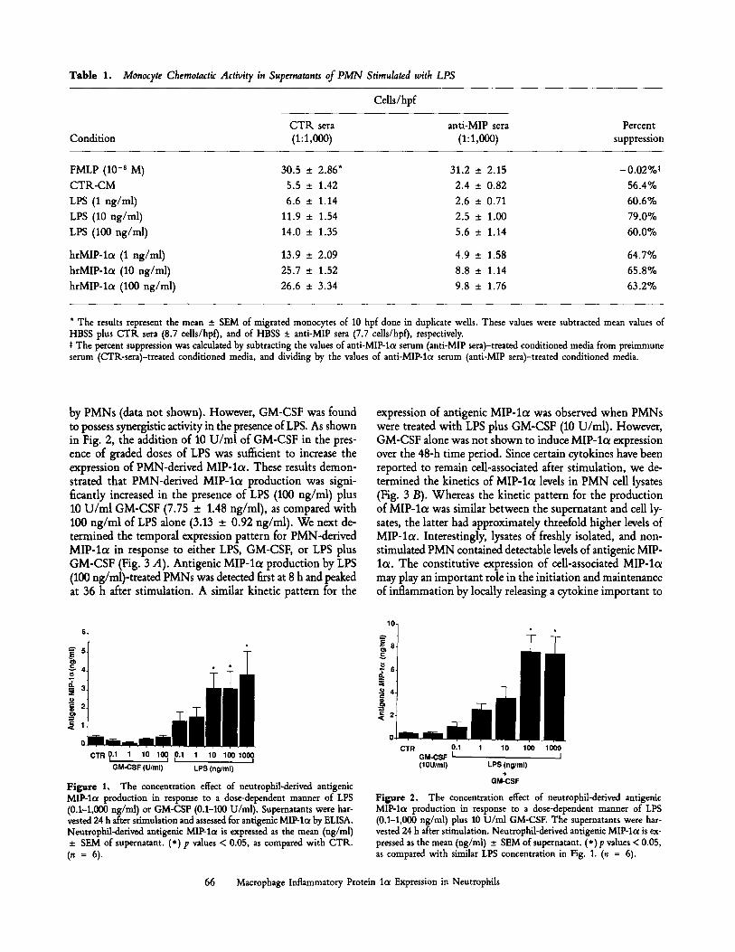
Tab le 1. Monocyte Chemotactic Activity in Supernatants of PMN Stimulated with LPS
Cells/hpf
C T K sera anti-MIP sera Percent Condition (1:1,000) (1:1,000) suppression
FMLP (10 -s M) 30.5 _+ 2.86* 31,2 _+ 2.15 -0.02%~
C T R - C M 5.5 -+ 1.42 2.4 + 0.82 56.4%
LPS (1 ng/ml) 6.6 + 1.14 2.6 _+ 0.71 60.6%
LPS (10 ng/ml) 11.9 _+ 1.54 2.5 + 1.00 79.0%
LPS (100 ng/ml) 14.0 _+ 1.35 5.6 +_ 1.14 60.0%
hrMIP-lc~ (1 ng/ml) 13.9 _+ 2.09 4.9 + 1.58 64.7%
hrMIP-lo~ (10 ng/ml) 25.7 +_ 1.52 8.8 _+ 1.14 65.8%
hrMIP-lc~ (100 ng/ml) 26.6 + 3.34 9.8 + 1.76 63.2%
* The results represent the mean _ SEM of migrated monocytes of 10 hpf done in duplicate wells. These values were subtracted mean values of HBSS plus CTR sera (8.7 cells/hpf), and of HBSS • anti-MlP sera (7.7 cells/hpf), respectively. t The percent suppression was calculated by subtracting the values of anti-MIP-l~x serum (anti-MIP sera)-treated conditioned media from preimmune serum (CTR-sera)-treated conditioned media, and dividing by the values of anti-MIP-lcx serum (anti-MIP sera)-treated conditioned media.
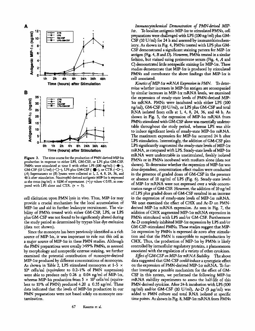
by PMNs (data not shown). However, GM-CSF was found to possess synergistic activity in the presence of LPS. As shown in Fig. 2, the addition of 10 U/ml of GM-CSF in the pres- ence of graded doses of LPS was sufficient to increase the expression of PMN-derived MIP-lo~. These results demon- strated that PMN-derived MIP-la production was signi- ficantly increased in the presence of LPS (100 ng/ml) plus 10 U/ml GM-CSF (7.75 _+ 1.48 ng/ml), as compared with 100 ng/ml of LPS alone (3.13 +_ 0.92 ng/ml). We next de- termined the temporal expression pattern for PMN-derived MIP-lol in response to either LPS, GM-CSF, or LPS plus GM-CSF (Fig. 3 A). Antigenic MIP-lo~ production by LPS (100 ng/ml)-treated PMNs was detected first at 8 h and peaked at 36 h after stimulation. A similar kinetic pattern for the
c T . o.1 , lo 1~ ,o.1 1 lo loo,ooo GM-CSF (U/ml) LPS (ng/ml)
Figure 1, The concentration effect of neutrophil-derived antigenic MIP-la production in response to a dose-dependent manner of LPS (0.1-1,000 ng/ml) or GM-CSF (0.1-100 U/ml). Supernatants were har- vested 24 h after stimulation and assessed for antigenic MIP-I~ by ELISA. Neutrophil-derived antigenic MIP-la is expressed as the mean (ng/ml) _+ SEM of supernatant. (*) p values < 0.05, as compared with CTR. (. = 6 ) .
expression of antigenic MIP-let was observed when PMNs were treated with LPS plus GM-CSF (10 U/ml). However, GM-CSF alone was not shown to induce MIP-lc~ expression over the 48-h time period. Since certain cytokines have been reported to remain ceU-associated after stimulation, we de- termined the kinetics of MIP-lot levels in PMN cell lysates (Fig. 3 B). Whereas the kinetic pattern for the production of MIP-lot was similar between the supernatant and cell ly- sates, the latter had approximately threefold higher levels of MIP-loe. Interestingly, lysates of freshly isolated, and non- stimulated PMN contained detectable levels of antigenic MIP- lo~. The constitutive expression of cell-associated MIP-lc~ may play an important role in the initiation and maintenance of inflammation by locally releasing a cytokine important to
a
CTR 0.1 1 10 100 1000 GM-CSF I I (lOU/ml) LPS (ng/ml)
+ GM-CSF
Figure 2. The concentration effect of neutrophil-derived antigenic MIP-lo~ production in response to a dose-dependent manner of LPS (0.1-1,000 ng/ml) plus 10 U/ml GM-CSF. The superuatants were har- vested 24 h after stimulation. Neutrophil-derived antigenic MIP-lo~ is ex- pressed as the mean (ng/ml) _+ SEM of supernatant. (*)p values < 0.05, as compared with similar LPS concentration in Fig. 1. (n = 6).
66 Macrophage Inflammatory Protein lot Expression in Neutrophils
A . - , 1 2
E ~ 1 0
8 "T, ----- 6
�9 ~ 4 r
�9 ~ 2"
'~ 0
! I " - '~ ~ I �9 . ~ .
l h 2h 4h 8h 24h 36h 4 8 h
T i m e ( h o u r s ) a f te r S t i m u l a t i o n
B 35"
i �9 L~ I T * I= 30" i - - o - - GM-CSF , ~ .
~ 2s -
.~ 1s- o '
g 10-
~ 5 - e ,
'~ 0 ' , . . . . . . . . . . . . . . . Oh l h 2h 4h 8h 24h 36h 48h
Time (hours) after Stimulation
Figure 3. The time course for the production of PMN-derived MIP-I(x production in response to either LPS, GM-CSF, or LPS plus GM-CSF. PMNs were stimulated at time 0 with either LPS (100 ng/ml) (--4~), GM-CSF (10 U/ml) ( ~ ) , LPS plus GM-CSF ( - l - ) , or CTR (-O-) . (.4) Supernatants or (B) lysates were collected at 1, 2, 4, 8, 24, 36, and 48 h after stimulation. Neutrophil-derived antigenic MIP-lc~ is expressed as the mean (ng/ml) -+ SEM of supernatant. (*)p values < 0.05, as com- pared with LPS alone and CTR. (n = 5).
cell elicitation upon PMN lysis in vivo. Thus, MIP-la may provide a crucial mechanism for the local accumulation of MIP-loc and aid in further leukocyte recruitment. The via- bility of PMNs treated with either GM-CSF, LPS, or LPS plus GM-CSF was not found to be significantly altered during the study period as determined by trypan blue dye exclusion (data not shown).
Since the monocyte has been previously identified as a rich source of MIP-la, it was important to rule out this cell as a major source of MIP-lo~ in these PMN studies. Although the PMN preparations were usually >99% PMNs, as assessed by morphology and nonspecific esterase staining, we further examined the potential contribution of monocyte-derived MIP-lc~ produced by different concentrations of monocytes. As shown in Table 2, LPS stimulated monocytes at 1-5 x 10⁴ cells/ml (equivalent to 0.2-1% of PMN suspension) were able to produce only 0.26 _+ 0.04 ng/ml of MIP-lol, whereas MIP-lo~ production from 5 x 10 s cells/ml (equiva- lent to 10% of PMN) produced 4.20 +_ 0.35 ng/ml. These data indicated that the levels of MIP-lc~ production in our PMN preparations were not based solely on monocyte con- tamination.
67 Kasama et al.
Imraunocytocheraical Demonstration of PMN-derived MIP- 1or. To localize antigenic MIP-lol to stimulated PMNs, cell preparations were challenged with LPS (100 ng/rrd) plus GM- CSF (10 U/m1) for 24 h and assessed by immunohistochem- istry. As shown in Fig. 4, PMNs treated with LPS plus GM- CSF demonstrated a significant staining pattern for MIP-lol antigen (Fig. 4, B and/9). However, PMNs treated in a similar fashion, but stained using preimmune serum (Fig. 4, A and C) demonstrated little nonspecific staining for MIP-lo~. These studies demonstrate that MIP-loL is produced by stimulated PMNs and corroborate the above findings that MIP-lo~ is cell associated.
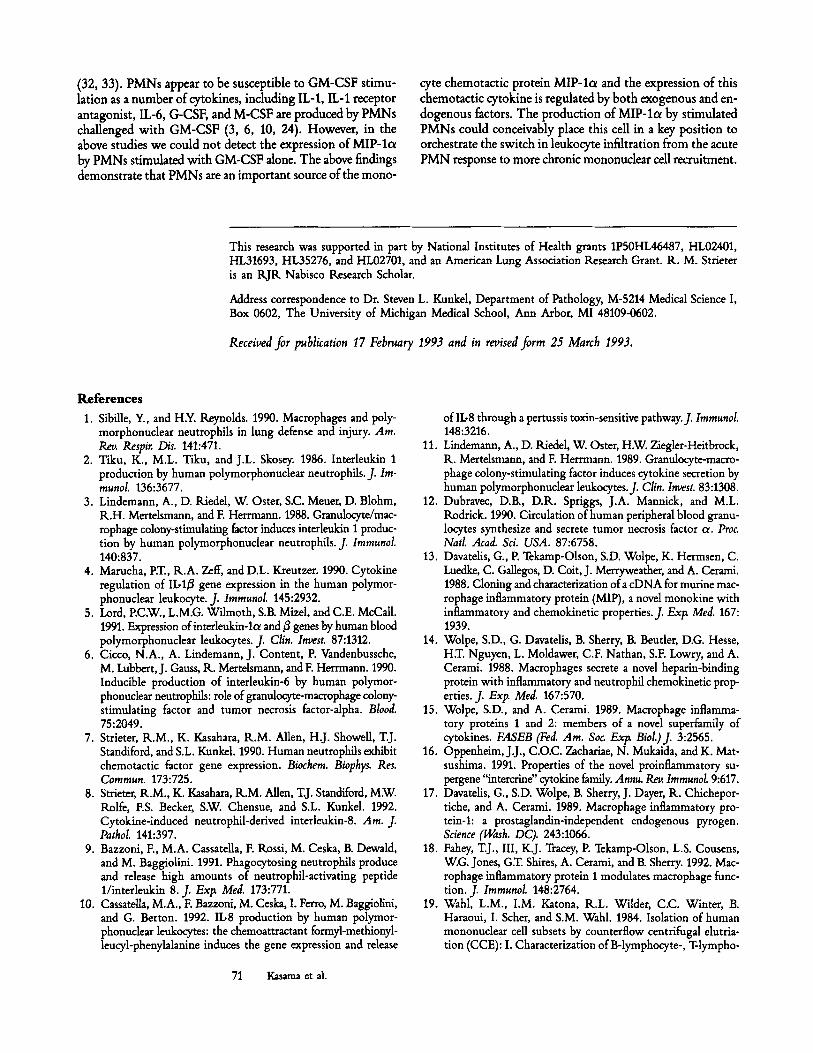
Kinetics of MIP-Icr mRNA Expression in PMN. To deter- mine whether increases in MIP-lcx antigen are accompanied by similar increases in MIP-lot mRNA levels, we examined the expression of steady-state levels of PMN-derived MIP- lo~ mRNA. PMNs were incubated with either LPS (100 ng/ml), GM-CSF (10 U/m1), or LPS plus GM-CSF and total RNA isolated from cells at 1, 4, 8, 24, 36, and 48 h. As shown in Fig. 5, the expression of MIP-la mRNA from PMNs stimulated with GM-CSF alone was essentially undetec- table throughout the study period, whereas LPS was able to induce significant levels of steady-state MIP-lol mRNA. The maximum expression for MIP-lc~ occurred 24 h after LPS stimulation. Interestingly, the addition of GM-CSF plus LPS significantly augmented the steady-state levels of MIP-lc~ mRNA, as compared with LPS. Steady-state levels of MIP-lc~ mRNA were undetectable in unstimulated, freshly isolated PMNs or in PMNs incubated with medium alone (data not shown). To determine whether the expression of MIP-lc~ was dose dependent, concentration effect studies were conducted in the presence of graded doses of GM-CSF in the presence or absence of 10 ng/ml of LPS (Fig. 6). Steady-state levels of MIP-loe mtLNA were not expressed over a wide concen- tration range of GM-CSF. However, the addition of 10 ng/ml of LPS plus graded doses of GM-CSF resulted in an increase in the expression of steady-state levels of MIP-lc~ mRNA. We next examined the effect of CHX and Ac-D on PMN- derived MIP-lot mRNA expression. As seen in Fig. 7, the addition of CHX augmented MIP-lo~ mRNA expression in PMNs stimulated with LPS and/or GM-CSF. Furthermore Ac-D completely inhibited MIP-lo~ expression by LPS and/or GM-CSF-stimulated PMNs. These studies suggest that MIP- lot expression by PMNs is expressed de novo after stimula- tion and that the PMN is susceptible to superinduction by CHX. Thus, the production of MIP-lcr by PMNs is likely controlled by intracellular regulatory proteins, a phenomenon associated with the regulation of a variety of other cytokines.
Effect of GM-CSF on MIP-Icr mRNA Stability. The above data suggested that GM-CSF could induce a synergistic effect on the expression of PMN-derived MIP-lo~ mtLNA. To fur- ther investigate a possible mechanism for the effect of GM- CSF in this system, we performed the following MIP-lo~ mRNA stability experiments to assess the half-life of this PMN-derived cytokine. After 24-h incubation with LPS (100 ng/ml) and/or GM-CSF (10 U/ml), Ac-D (5/zg/ml) was added to PMN culture and total RNA isolated at specific time points. As shown in Fig. 8, MIP-lc~ mRNA from PMNs
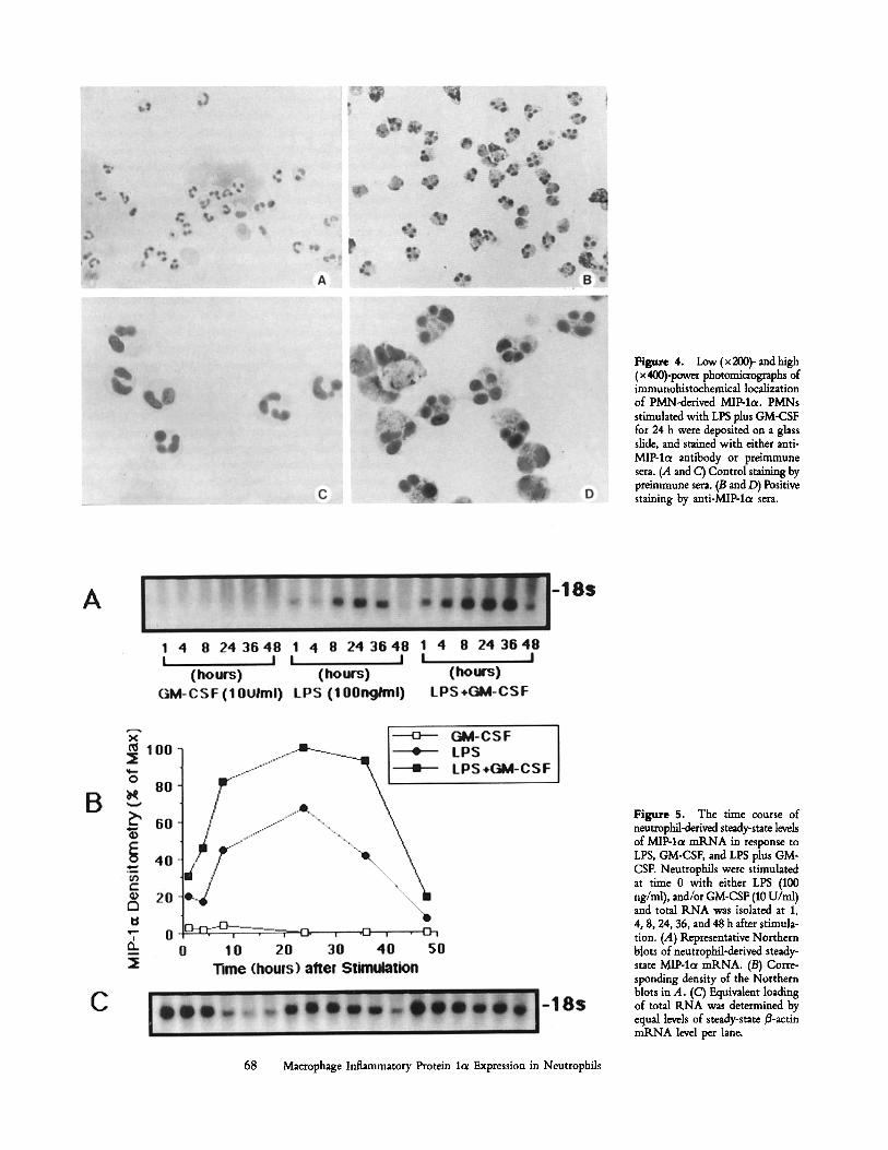
Figure 4. Low (x 200)- and high (x 400)-power photomicrographs of immunohistochemical localization of PMN-derived MIP-lo~. PMNs stimulated with LPS plus GM-CSF for 24 h were deposited on a glass slide, and stained with either anti- MIP-lc~ antibody or preimmune sera. (A and C) Control staining by preimmune sera. (B and D) Positive staining by anti-MIP-l~ sera.
Figure 5. The time course of neutmphil-derived steady-state levels of MIP-lol mRNA in response to LPS, GM-CSF, and LPS plus GM- CSF. Neutrophils were stimulated at time 0 with either LPS (100 ng/ml), and/or GM-CSF (10 U/ml) and total RNA was isolated at 1, 4, 8, 24, 36, and 48 h after stimula- tion. (A) Representative Northern b~ots of neutrophil-derived steady- state MIP-1oe mRNA. (B) Corre- sponding density of the Northern blots in A. (C) Equivalent loading of total RNA was determined by equal levels of steady-state/~-actin mRNA level per lane.
68 Macrophage Inflammatory Protein lot Expression in Neutrophils

Tab le 2. MIP-lot Production from Monocytes
MIP-loe
1 x 104* 5 x 104 1 x 10⁵ 5 x 10⁵ 1 x 10⁶ Condition (0.2%o)* (1~ (2%0) (10%) (20%)
C T R NDS 0.07 _+ 0.02 0.16 _+ 0.03 0.36 _+ 0.06 3.32 _+ 0.56
GM-CSF (10 U /ml ) N D 0.05 + 0.02 0.19 _+ 0.04 0.51 + 0.08 3.19 _+ 0.37
LPS (100 ng/ml) N D 0.26 _+ 0.04 0.97 _+ 0.13 4.20 _+ 0.35 10.24 _+ 1.20
LPS + GM-CSF N D 0.26 -+ 0.02 0.76 _+ 0.07 3.92 _+ 0.42 12.50 _+ 1.73
* Monocytes (cells/m1) were resuspended in complete media at various concentrations, and incubated for 24 h. * Percentage would he equivalent to that of monocytes potentially contaminating the PMN cultures. S ND, Not detected.
treated with GM-CSF plus LPS possessed a markedly pro- longed tl/2, as compared with LPS alone (LPS, 2 h vs LPS plus GM-CSF, 5 h) (Fig. 8 B). In these studies, the stability of B-actin m R N A isolated from PMNs treated with LPS or LPS plus GM-CSF did not appear to be altered. In total, the above studies demonstrate that GM-CSF may be an impor- tant polypeptide mediator for the continued maintenance of an inflammatory response via its ability to augment the ex- pression of PMN-derived chemotactic cytokines.
Discuss ion
PMNs are usually recognized as an important leukocyte population involved in acute inflammatory responses. Histor- ically, this cell population was not considered an important source of de novo polypeptide mediators, and its role in host defense was felt to occur only via its phagocytic activity and
Figure 6. The concentration effect of neutrophil-derived steady-state levels of MIP-lo~ mRNA in response to a dose-dependent manner of GM- CSF in the absence or presence of 10 ng/ml of LPS. Neutrophils were stimulated with graded concentrations of GM-CSF (0.1-100 U/ml) with or without LPS (10 ng/ml), and total RNA was isolated at 8 h after stim- ulation. (A) Representative Northern blots of neutrophil-derived steady- state MIP-lol mRNA. (B) Corresponding density of the Northern blots as assessed by image analysis. (C) Equivalent loading of total RNA as de- termined by equal levels of steady-state 3-actin mRNA level per lane.
Figure 7. The effect of CHX, or Ac-D on neutrophil-derived steady- state levels of MIP-lce mRNA in response to LPS, GM-CSF, or LPS plus GM-CSF. Neutrophils were preincubated with CHX (5/~g/ml), or Ac-D (5/ig/ml) for 1 h and stimulated with LPS (100 ng/ml), GM-CSF (10 U/ml), or LPS plus GM-CSF. Total RNA was isolated at 4 h after stimu- lation. (A) Representative Northern blots of neutrophil-derived steady- state MIP-lc~ mRNA. (B) Corresponding density of imaging analysis. (C) Equivalent loading of total RNA was determined by equal levels of steady- state 13-actin mRNA level per lane.
69 Kasama et al.

B
~1 2 5 2
~ 1 0 0 '
@
0
ffl
@
Q 25
"7 el
I ~ LP5 ]
0 2 4 6 8 1' Time (hours) af ter 24 h-st imulat ion
Ac-D
Figure 8. The half-life of neutrophil-defived steady- state levels of MIP-la mRNA in response to LPS, or LPS plus GM-CSF. Neutrophils were stimulated with LPS (100 ng/ml) or LPS plus GM-CSF (10 U/ml). After 24-h incu- bation, Ac-D (5 #g/ml) was added and total RNA was isolated at various time points. (A) (I and I/) Representa- tive Northern blots of nentrophil-derived steady-state MIP-lot mRNA. (III and IV) Equivalent loading of total RNA was determined by equal levels of steady-state /3-actin mRNA level per lane. (B) Corresponding density of the image analysis.
the production and release of enzymes and reactive oxygen species. However, recent data have demonstrated that PMNs are biosynthetically active and can produce a variety of cytokines known to play an important role in inflammation. These cytokines include: IL-1 c~ and 3 (2-5), IL-6 (6), IL-8 (7-10), TNF-ot (11, 12), and IL-1 receptor antagonist (24). Since PMNs are one of the first cells to arrive at a site of inflammation, the cytokine secreting activity of these cells may indicate a role for PMNs during the maintenance of an inflammatory site.
The progression and maintenance of many inflammatory responses are often histologically defined by a significant PMN recruitment phase followed by the elicitation of mononuclear cells. This temporal appearance of specific leukocytes would suggest that the PMN may play a role in mononuclear cell recruitment. However, few studies have actually demonstrated that PMNs can generate a chemotactic factor(s) for mono- cytes. In the present study, we demonstrated that peripheral blood PMNs were a significant source of MIP-lot, which is a potent chemoattractant for monocyte. MIP-lc~ belongs to a supergene family of chemotactic cytokines which are identified by the location of four cysteine amino acids; two of the cysteines are found in juxtaposition to each other. This family contains other chernotactic cytokines, including MCP-1, R.ANTES, and MIP-I~. In addition, this group of chemotactic cytokines is distantly related to another supergene family of neutrophil chemotactic factors, which includes IL-8. These cytokines appear to play a key role in inflammation and im- mune responses by their chemotactic activities and their ability to activate PMNs, monocytes, T ceils, eosinophils, and baso- phils (16, 25-27). MIP-lot has been shown to possess a va- riety of biological activities, including the induction of neu- trophil chemokinesis and activation of neutrophils to generate
hy&ogen peroxide (14), serving as a prostaglandin-independent endogenous pyrogen (17), stimulating the secretion of TNF, IL-lot, and IL-6 from murine peritoneal macrophages (18), and inducing the chemotaxis of basophils and histamine re- lease from mast cells (28).
In this study, LPS challenge (8-36 h) was identified as an important stimulus for the expression of PMN-derived MIP- lot. Interestingly, GM-CSF, an inflammatory mediator pre- viously identified as a stimulus for PMN-derived cytokines, did not induce the expression of PMN-derived MIP-lc~ alone, but did cause a significant synergistic rise in the production of PMN-derived MIP-lol. The mechanism for the increase in MIP-lot production by GM-CSF plus LPS appeared to be due to the stabilization of MIP-lc~ mRNA, resulting in a prolonged mRNA tv2. Whereas mRNAs which code for various structural proteins are quite stable, recent studies have identified mRNAs that code certain inducible proteins, in- cluding cytokines, oncogenes, and some enzymes, are rela- tively unstable and possess half-lives on the order of 10-60 rain (29). Furthermore, the "AUUUA" nucleotide motif in the 3' untranslated region has been identified as playing an important role in regulating the longevity of many cytokine mRNA species. This motif appears to have relevance for the rapid decay of specific cytokine mRNA (29, 30). Interest- ingly, MIP-lcr does possess this motif in the 3' untranslated region of its mRNA (14, 15, 23, 31).
GM-CSF is likely to play an important role during the maintenance of an inflammatory lesion not only by increasing the production of a PMN-derived monocyte chemotactic cytokine, but also by increasing the longevity of PMNs them- selves. Recent studies have demonstrated that GM-CSF can increase the survival of PMNs as they participate in inflam- mation and prolong the circulating halfqife of PMNs in vivo
70 Macrophage Inflammatory Protein lcx Expression in Neutrophils
(32, 33). PMNs appear to be susceptible to GM-CSF stimu- lation as a number of cytokines, including IL-1, IL-1 receptor antagonist, IL-6, G-CSF, and M-CSF are produced by PMNs challenged with GM-CSF (3, 6, 10, 24). However, in the above studies we could not detect the expression of MIP-lol by PMNs stimulated with GM-CSF alone. The above findings demonstrate that PMNs are an important source of the mono-
cyte chemotactic protein MIP-lc~ and the expression of this chemotactic cytokine is regulated by both exogenous and en- dogenous factors. The production of MIP-lol by stimulated PMNs could conceivably place this cell in a key position to orchestrate the switch in leukocyte infiltration from the acute PMN response to more chronic mononuclear cell recruitment.
This research was supported in part by National Institutes of Health grants 1P50HL46487, HL02401, HL31693, HL35276, and HL02701, and an American Lung Association Research Grant. R. M. Strieter is an RJR Nabisco Research Scholar.
Address correspondence to Dr. Steven L. Kunkel, Department of Pathology, M-5214 Medical Science I, Box 0602, The University of Michigan Medical School, Ann Arbor, MI 48109-0602.
Received.for publication 17 February 1993 and in revised.form 25 March 1993.
References 1. SibiUe, Y., and H.Y. Reynolds. 1990. Macrophages and poly-
morphonuclear neutrophils in lung defense and injury. Am. Rev. Respir. Dis. 141:471.
2. Tiku, K., M.L. Tiku, and J.L. Skosey. 1986. Interleukin 1 production by human polymorphonuclear neutrophils. J. Im- munol. 136:3677.
3. Lindemann, A., D. Riedel, W. Oster, S.C. Meuer, D. Blohm, R.H. Mertelsmann, and F. Herrmann. 1988. Granulocyte/mac- rophage colony-stimulating factor induces interleukin I produc- tion by human polymorphonuclear neutrophils. J. Immunol. 140:837.
4. Marucha, P.T., R.A. Zeff, and D.L. Kreutzer. 1990. Cytokine regulation of IL-13 gene expression in the human polymor- phonuclear leukocyte. J. Immunol. 145:2932.
5. Lord, P.C.W., L.M.G. Wilmoth, S.B. Mizel, and C.E. McCall. 1991. Expression of interleukin-lc~ and ~ genes by human blood polymorphonuclear leukocytes. J. Clin. Invest. 87:1312.
6. Cicco, N.A., A. Lindemann, J. Content, P. Vandenbussche, M. Lubbert, J. Gauss, Ik. Mertelsmann, and F. Herrmann. 1990. Inducible production of interleukin-6 by human polymor- phonuclear neutrophils: role of granulocyte-macrophage colony- stimulating factor and tumor necrosis factor-alpha. Blood. 75:2049.
7. Strieter, R.M., K. Kasahara, R.M. Allen, H.J. Showell, T.J. Standiford, and S.L. Kunkel. 1990. Human neutrophils exhibit chemotactic factor gene expression. Biochem. Biophys. Res. Comraun. 173:725.
8. Strieter, R.M., K. Kasahara, R.M. Allen, T.J. Standiford, M.W. Rolfe, ES. Becket, S.W. Chensue, and S.L. Kunkel. 1992. Cytokine-induced neutrophil-derived interleukin-8. Am. J. Pathol. 141:397.
9. Bazzoni, E, M.A. Cassatella, F. Rossi, M. Ceska, B. Dewald, and M. Baggiolini. 1991. Phagocytosing neutrophils produce and release high amounts of neutrophil-activating peptide 1/interleukin 8. J. Exla filed. 173:771.
10. CassateUa, M.A., F. Bazzoni, M. Ceska, I. Ferro, M. Baggiolini, and G. Berton. 1992. IIr production by human polymor- phonudear leukocytes: the chemoattractant formyl-methionyl- leucyl-phenylalanine induces the gene expression and release
of II.-8 through a pertussis toxin-sensitive pathway.J. Immunol. 148:3216.
11. Lindemann, A., D. Riedel, W. Oster, H.W. Ziegler-Heitbrock, R. Mertelsmann, and F. Herrmann. 1989. Grantdocyte-macro- phage colony-stimulating factor induces cytokine secretion by human polymorphonuclear leukocytes.f Clin. Invest. 83:1308.
12. Dubravec, D.B., D.R. Spriggs, J.A. Mannick, and M.L. Rodrick. 1990. Circulation of human peripheral blood granu- locytes synthesize and secrete tumor necrosis factor ~. Proa Natl. Acad. Sci. USA. 87:6758.
13. Davatdis, G., P. Tekamp-Olson, S.D. Wolpe, K. Hermsen, C. Luedke, C. GaUegos, D. Coit, J. Merryweather, and A. Cerami. 1988. Cloning and characterization of a cDNA for routine mac- rophage inflammatory protein (MIP), a novel monokine with inflammatory and chemokinetic properties.f Exl~ Med. 167: 1939.
14. Wolpe, S.D., G. Davatelis, B. Sherry, B. Beutler, D.G. Hesse, H.T. Nguyen, L. Moldawer, C.F. Nathan, S.F. Lowry, and A. Cerami. 1988. Macrophages secrete a novel heparin-binding protein with inflammatory and neutrophil chemokinetic prop- erties. J. Extz Med. 167:570.
15. Wolpe, S.D., and A. Cerami. 1989. Macrophage inflamma- tory proteins 1 and 2: members of a novel superfamily of cytokines. FASEB (Fed. Am. Soa Extx Biol.) f 3:2565.
16. Oppenheim, J.J., C.O.C. Zachariae, N. Mukaida, and K. Mat- sushima. 1991. Properties of the novel proinflammatory su- pergene "intercrine" cytokine family. Annu. Rev. Immunol. 9:617.
17. Davatelis, G., S.D. Wolpe, 13. Sherry, J. Dayer, R. Chichepor- tiche, and A. Cerami. 1989. Macrophage inflammatory pro- tein-l: a prostaglandin-independent endogenous pyrogen. Science (Wash. DC). 243:1066.
18. Fahey, T.J., III, K.J. Tracey, P. Tekamp-Olson, L.S. Cousens, W.G. Jones, G.T. Shires, A. Cerami, and B. Sherry. 1992. Mac- rophage inflammatory protein I modulates macrophage func- tion. J. Immunol. 148:2764.
19. Wahl, L.M., I.M. Katona, R.L. Wilder, C.C. Winter, B. Haraoui, I. Scher, and S.M. Wahl. 1984. Isolation of human mononuclear cell subsets by counterflow centrifugal elutria- tion (CCE): I. Characterization of B-lymphocyte-, T-lympho-
71 Kasama eta|.
cyte-, and monocyte-enriched fractions by flow cytometric anal- ysis. Cell. Immunol. 85:373.
20. Standiford, T.J., S.L. Kunkel, M.A. Basha, S.W. Chensue, J.P. Lynch III, G.B. Toews, J. Westwick, and R.M. Strieter. 1990. Interleukin-8 gene expression by a pulmonary epithelial cell line. A model for cytokine networks in the lung. J. Clin. In- vest. 86:1945.
21. Evanoff, H.L., M.D. Burdick, S.A. Moore, S.L. Kunkel, and R.M. Strieter. 1992. A sensitive ELISA for the detection of human monocyte chemoattractant protein (MCP-1). Immunol. Invest. 21:39.
22. Zipfel, P.F., J. Balke, S.G. Irving, K. Kelly, and U. Siebenlist. 1989. Mitogenic activation of human T cells induces two closely related genes which share structural similarities with a new family of secreted factors. J. Immunol. 142:1582.
23. Tokunaga, K., H. Taniguchi, K. Yoda, M. Shimizu, and S. Sakayama. 1986. Nucleotide sequence of a full-length cDNA for mouse cytoskeletal beta-actin mRNA. Nucleic Acids Res. 14:229.
24. McColl, S.R.., R. Paquin, C. Menard, and A.D. Beaulieu. 1992. Human neutrophils produce high levels of the interleukin 1 receptor antagonist in response to granulocyte/macrophage colony-stimulating factor and tumor necrosis factor c~.J. Exp. Med. 176:593.
25. Bischoff, S.C., M. Krieger, T. Brunner, and C.A. Dahinden. 1992. Monocyte chemotactic protein 1 is a potent activator of human basophils. J. Ex F Med. 175:1271.
26. Kameyoshi, Y., A. Dorschner, A.I. Mallet, E. Christophers, and J.-M. Schroder. 1992. Cytokine RANTES released by thrombin-stimulated platelets is a potent attractant for human eosinophils. J. Extx Med. 176:587.
27. Kuna, P., S.R. Reddigari, D. Rucinski, J.J. Oppenheim, and A.P. Kaplan. 1992. Monocyte chemotactic and activating factor is a potent histamine-releasing factor for human basophils. J. Extx Med. 175:489.
28. Alam, R., P.A. Forsythe, S. Stafford, M.A. Lett-Brown, and J.A. Grant. 1992. Macrophage inflammatory protein-l~ acti- vates basophils and mast cells. J. Exp. Med. 176:781.
29. Carter, B.Z., andJ.S. Maker. 1991. Regulation ofmRNA sta- bility and its relevance to disease. Lab Invest. 65:610.
30. Caput, D., B. Beutler, K. Hartog, R. Thayer, S. Brown-Shimer, and A. Cerami. 1986. Identification of a common nucleotide sequence in the Y-unstimulated region of mRNA molecules specifying inflammatory mediators. Proa Natl. Acad. Sci. USA. 83:1670.
31. Irving, S.G., P.F. Zipfel, J. Balke, O.W. McBride, C.C. Morton, P.R. Burd, U. Siebenlist, and K. Kelly. 1990. Two inflamma- tory mediator cytokine genes are closely linked and variably amplified on chromosome 17q. Nucleic Acids Res. 18:3261.
32. Lieschke, G.J., and A.W. Burgess. 1992. Granulocyte colony- stimulating factor and granulocyte-macrophage colony-stimu- lating factor. N. Engl. J. Med. 327:28.
33. Gasson, J.C. 1991. Molecular physiology of granulocyte-macro- phage colony-stimulating factor. Blood. 77:1131.
72 Macrophage Inflammatory Protein lc~ Expression in Neutrophils