EXPLORING THE PEDIATRIC ABDOMEN: A RADIOLOGICAL … · of neck, chest, abdomen and pelvis may be...
19
2018 January Edition |www.jbino.com | Innovative Association J.Bio.Innov7(1), pp: 47-65, 2018 |ISSN 2277-8330 (Electronic) Meena et al., EXPLORING THE PEDIATRIC ABDOMEN: A RADIOLOGICAL EVALUATION. Dr.SURBHI GUPTA 1 ., Dr. MD. Khizer Razak 2 & Dr.G.L.MEENA 3* 1 2,3* DEPARTMENT OF RADIODIAGNOSIS, S.P.MEDICAL COLLEGE, BIKANER, RAJASTHAN. (Received on Date: 1 January 2018 Date of Acceptance:29 January 2018) ABSTRACT To study and characterize various paediatric abdominal masses by Multidetector CT.To assess the role of Multidetector CT in imaging of various abdominal masses in paediatric age group. This hospital based study was conducted in Department of Radio-diagnosis and Modern imaging of PBM Hospital, Bikaner, Rajasthan. Data for the study was collected from patients of paediatric age group attending/ referred to the department of Radio- Diagnosis.A preliminary ultrasound scanning was done in all cases using GE LOGIQ P5 sonography machine with transducers of appropriate frequency. Color Doppler imaging was done as and when required based on gray scale characteristics.Non-contrast and contrast enhanced CT examination of the patients was carried out, using PHILLIPS BRILLIANCE MDCT 64 SLICE CT SCAN. Scanning protocol were tailored according to the age, weight of the child and the clinical situation. Imaging findings were correlated with the clinical course of disease and/or surgical/cytological findings as far as possible. The results were subjected to statistical analysis wherever applicable and expressed as percentages.The recent advances have expanded the usefulness of CT in the evaluation of pediatric abdominal masses. The advantage of single breath hold acquisition in cooperative children, improved vascular contrast enhancement, increased detection of parenchymal lesions, and multiplanar and three dimentional reconstruction may make it one of the modalities of choice in evaluation of pediatric abdominal masses. KEYWORDS: Pediatric; imaging; CT; Abdomen; tumor. No: of figures : 8 No: of Tables:6 No: of Charts: 6 No: of References: 25
Transcript of EXPLORING THE PEDIATRIC ABDOMEN: A RADIOLOGICAL … · of neck, chest, abdomen and pelvis may be...

2018 January Edition |www.jbino.com | Innovative Association
J.Bio.Innov7(1), pp: 47-65, 2018 |ISSN 2277-8330 (Electronic)
Meena et al.,
EXPLORING THE PEDIATRIC ABDOMEN: A RADIOLOGICAL EVALUATION.
Dr.SURBHI GUPTA1 ., Dr. MD. Khizer Razak2 & Dr.G.L.MEENA3*
1 2,3* DEPARTMENT OF RADIODIAGNOSIS, S.P.MEDICAL COLLEGE, BIKANER, RAJASTHAN.
(Received on Date: 1 January 2018 Date of Acceptance:29 January 2018)
ABSTRACT
To study and characterize various paediatric abdominal masses by Multidetector CT.To
assess the role of Multidetector CT in imaging of various abdominal masses in paediatric age
group. This hospital based study was conducted in Department of Radio-diagnosis and
Modern imaging of PBM Hospital, Bikaner, Rajasthan. Data for the study was collected from
patients of paediatric age group attending/ referred to the department of Radio-
Diagnosis.A preliminary ultrasound scanning was done in all cases using GE LOGIQ P5
sonography machine with transducers of appropriate frequency. Color Doppler imaging was
done as and when required based on gray scale characteristics.Non-contrast and contrast
enhanced CT examination of the patients was carried out, using PHILLIPS BRILLIANCE MDCT
64 SLICE CT SCAN. Scanning protocol were tailored according to the age, weight of the
child and the clinical situation. Imaging findings were correlated with the clinical course of
disease and/or surgical/cytological findings as far as possible. The results were subjected to
statistical analysis wherever applicable and expressed as percentages.The recent advances
have expanded the usefulness of CT in the evaluation of pediatric abdominal masses. The
advantage of single breath hold acquisition in cooperative children, improved vascular
contrast enhancement, increased detection of parenchymal lesions, and multiplanar and
three dimentional reconstruction may make it one of the modalities of choice in evaluation
of pediatric abdominal masses.
KEYWORDS: Pediatric; imaging; CT; Abdomen; tumor.
No: of figures : 8 No: of Tables:6 No: of Charts: 6 No: of References: 25

2018 January Edition |www.jbino.com | Innovative Association
J.Bio.Innov7(1), pp: 47-65, 2018 |ISSN 2277-8330 (Electronic)
Meena et al.,
INTRODUCTION
Abdominal masses in the paediatric age
group include a spectrum of lesions of
diverse origin and significance. They may
occur at any age- from the new born
period through adolescence l. In most
cases, their common aspect is the lack of
peculiar clinical features that may help in
early differential diagnosis. In many cases,
the mass is detected late after a long
period of vague, non- specific symptoms.
The role of diagnostic imaging is to identify
the precise anatomic location and extent
of the pathologic process with a minimal
number of imaging procedures.
Most abdominal masses in children
are initially imaged by abdominal
radiography. Abdominal radiographs
provide information as to the location of
the mass and presence or absence of
calcification1. Their role ranges from a
screening process, providing non-specific
information in some cases, to providing
specific information in some cases, to
providing a specific diagnosis in
others.However, patients are exposed to
radiation and radiographs have the
intrinsic limitation in that only four basic
densities (bone or mineral, soft tissue, fat, or
air) are identified3. Ultrasonography is
particularly useful imaging modality for the
paediatric patients since it does not utilize
radiation2. It allows imaging in multiple
planes, permits repetitive examinations
and requires no physiologic function for
anatomic visualization2. It is also a portable
means for
examination of complications and does
not generally require sedation. It can be
used in directing a location for biopsies
and drainage of fluid collections. It aids in
localization of the tumor, identification of
associated adenopathy and examination
of adjacent vascular structures by Colour
and Duplex Doppler. Thus, USG is
diagnostic in some cases while limits the
differential diagnoses in others and hence,
is useful as a general screening
procedure. However, USG is highly
operator dependent and is adversely
affected by bone or gas artefacts4. In
addition, ultrasonography provides less
precise anatomic details and smaller
section areas of interest.
In recent times, computed tomography
has found increasing application in the
evaluation of paediatric abdominal
masses2. It is currently one of the most
powerful and versatile imaging procedure
for the evaluation of abdominal masses.
The anatomic detail provided by CT is
superior to any other imaging modality
currently available. It obtains an entire
anatomic section of tissue, which aids in
determining the precise extent of disease.
The technique is not operator dependent
and permits the accurate measurement of
tissue attenuation coefficient.
Enhancement with contrast medium
facilitates measurement of blood flow to
an organ or pathologic abnormality. Bolus
injection permits visualization of vascular
structures. Anatomic and physiologic
information may be obtained in severely
compromised organs, and structures may
bevisualized despite overlying gas and

2018 January Edition |www.jbino.com | Innovative Association
J.Bio.Innov7(1), pp: 47-65, 2018 |ISSN 2277-8330 (Electronic)
Meena et al.,
bone. However, the paucity of tilt in
children makes delineation of anatomic
margins in the retroperitoneum difficult. In
addition, conventional CT requires
sedation or anaesthesia in infants and
small children, intravenous and enteric
contrast medium, immobilization and
alteration of environment, and is time
consuming.
The technical improvements, in the
form of Multislice helical CT recently, have
resulted in improved resolution and
considerable reductions in scan acquisition
and display time.
One of the most notable effects of
faster scanning with present CT technology
in children is the reduced need for
sedation. In cases of neoplasms, Dual
phase imaging of the organ concerned is
important to obtain information about the
vascular status. Multislice CT has improved
temporal resolution into arterial and
venous phases.
In certain cases, like lymphomas, CT
of neck, chest, abdomen and pelvis may
be necessary for staging or follow-up. With
the advent of Multislice CT, the imaging
time is reduced considerably. This has
facilitated optimal contrast enhancement
during CT of neck, chest, abdomen and
pelvis using a single i.v contrast material
bolus of the standard paediatric dose of
contrast material. Another advantage of
recent technological advancements is
volume acquisition of data. This furnishes
several important benefits for children.
Reconstruction can be performed
conveniently once the patient has left the
scanner. With Multislice CT, now isotropic
viewing has become a reality. Unlimited
reformations are possible without any
difficulty, leading to increased conspicuity.
Thus, Multislice helical technology
has expanded the usefulness of CT in
evaluation of paediatric abdominal
masses. The advantages of single breath-
hold acquisition in cooperative children,
improved vascular contrast enhancement,
increased detection of parenchymal
lesions and multiplaner and three-
dimensional reconstructions may make it
one of the modalities of choice in
evaluation of paediatric abdominal
masses.
MATERIAL & METHODS:
This hospital based study will be conducted
in Department of Radio-diagnosis and
Modern imaging of PBM Hospital, Bikaner,
Rajasthan.
Source of data:
Data for the study will be collected from
patients of paediatric age
groupattending/ referred to the
department of Radio-Diagnosis, PBM
Hospital, Bikaner for evaluation of
abdominal mass.
Inclusion criteria:
All patients 0 to 14 yrs who have clinical
suspicion as well as sonographic evidence
of mass in abdomen.
Exclusion criteria:
1. Patients above the age of 14 yrs.

2018 January Edition |www.jbino.com | Innovative Association
J.Bio.Innov7(1), pp: 47-65, 2018 |ISSN 2277-8330 (Electronic)
Meena et al.,
2. Patients with bleeding diatheses.
3. Patients with previous history of contrast
sensitivity.
Methods:
The study will be carried out in the
Department of Radiodiagnosis, SPMC and
PBM Hospital, Bikaner on the following lines:
a. A detailed clinical history will be
recorded.
b. Relevant clinical examination will be
done.
c. Required lab investigations will be done.
d. Radiological Examination :-
i) A preliminary ultrasound scanning
will be done in all cases using GE
LOGIQ P5 sonography machine with
transducers of appropriate
frequency.
ii) Color Doppler imaging will be
done as and when required based
on gray scale characteristics.
iii) Non-contrast and contrast
enhanced CT examination of the
patients will be carried out, using
PHILLIPS BRILLIANCE MDCT 64 SLICE
CT SCAN. Scanning protocol shall be
tailored according to the age,
weight of the child and the clinical
situation. Dual-phase imaging and
angiography sequences shall be
used as and when required.
iv) Other radiological investigations
will also be done wherever required.
v) Ultrasound/CT guided
FNAC/biopsy shall be done
wherever indicated.
e. Imaging findings will be correlated with
the clinical course of disease and/or
surgical/cytological findings as far as
possible.
Statistical Analysis
The results will be subjected to
statistical analysis wherever applicable
and expressed as percentages.
RESULTS:
Abdominal masses in the neonatal period
are predominantly benign lesions, usually
representing defects in the embryonic
development2. The majority of neonatal
masses are retroperitoneal in location
(52%) out of which 54% are of renal
origin6.Paediatric abdominal masses
occurring after the neonatal period are still
predominantly retroperitoneal; however,
there is a significant increase in malignant
tumors and some differences in the
incidence of specific masses2. We studied
50 cases of pediatric abdominal masses in
various age groups. Quite a large
spectrum of lesions was found.
Biona et al24 in 1983 and Rastogi et al25 in
1988 reviewed the pattern of pediatric
abdominal masses.
The age wise incidence in our series in 3
defined groups was 16% (8/50) in 0-1 years,
44% (22/50) in 1-5years and 40% (20/50) in
>5years. This is quite similar to the age
incidence given by Biona et al24 of 16.4%,

2018 January Edition |www.jbino.com | Innovative Association
J.Bio.Innov7(1), pp: 47-65, 2018 |ISSN 2277-8330 (Electronic)
Meena et al.,
51% and 32.6% respectively for the three
age groups. Age wise incidence of
masses(exclusive of hydronephrosis) given
by Rastogi et al25 (27% in 0-1year, 39% in 1-
5years and 34% in >5years). However, the
difference can be attributed to exclusiom
of patients with hydronephrosis in 1-6 year
age group in their study.
Biona et al24 reported that males were
affected more than females (1.5:1).
Rastogi et al also reported that males were
affected more often than females (2.4:1).
In our study, the male:female ratio was
29:21 i.e. 1.4:1.
Majority of the patient presented with
progressively increasing abdominal lump
and USG was the initial investigation
requested.
In our study, out of 50 cases 22 (44%) were
malignant and 28 (56%) were benign. This is
in accordance with incidence reported by
Rastogi et al25 where 58% passes were
benign. However, in Biona24
series, excluding hydronephrosis 18 of the
26 cases (70%) were malignant.
In our study, 52% (26/50) of the masses
were retroperitoneal out of which 14 (54%
of retroperitoneal and 28% of total) were
renal, an incidence quite similar to that
reported by Biona et al (58%
retroperitoneal and 31% renal) and Rastogi
et al (53% retroperitoneal and 32% renal).
Egeibor and Jabral23 also state that the
majority of the abdominal masses
occurring in childhood are retroperitoneal
in location, and greater than 50% of these
masses arise from the kidney.
According to Egeibor and Jabral23,
approximately 87% of solid renal
neoplasms in children are Wilms’ tumors;
other renal tumors include clear cell
sarcomas (6%), mesoblastic nephroma
(2%), rhabdoid tumors (2%), lymphoma
(<O.5%) and renal cell carcinoma (<0.5%).
In our study, out of the 14 renal masses, 8
were neoplastic (57%). Out of these 5
(62.5%) had Wilms’ tumor, 1 had rhabdoid
tumor of kidney and 1 had renal cell
carcinoma. In cases reported by Rastogi et
al, 63% of renal lesions were neoplastic and
all had Wilms’ tumor while in Biona series,
all the renal masses (exclusive
of hydronephrosis) had Wilms’ tumor.
Approximately 50% of children with Wilms’
tumor present before 3 years, 80% before
5 years.In Biona series24, 50% of the cases of
Wilms’ tumor were in 1-6years age group
while 55% were in the group in Rastogi
series25. In our study, 60% of patients
presented before 5 years of age.Other
renal lesions included renal cell carcinoma,
rhabdoid tumor of kidney,
perinephric/renal abscess and one infant
with multicystic dysplastic kidney.
Non-renal retroperitoneal masses
constituted 24% (12/50) of the cases.This is
similar to the incidence reported by
Griscom (29%)3, Rastogi et al25 (20%) and
Biona et al24 (30%). Out of these, 6 cases
were of neuroblastoma (50%). In series by
Griscom3, neuroblastoma constituted 60%
of non-renal retroperitoneal cases while in
series by Rastogi et al25 and Biona et al24,
39% and 11.8% were neuroblastomas.

2018 January Edition |www.jbino.com | Innovative Association
J.Bio.Innov7(1), pp: 47-65, 2018 |ISSN 2277-8330 (Electronic)
Meena et al.,
Peak age of incidence of neuroblastoma is
in first 5 years of life (85 percent), with 50%
in less than 2 years of age. In our study, out
of 6 cases, all 6 cases were <5 years of
age. The mean age of presentation was
43.4 months as compared to 31.7 months
in Biona series.
Other non-renal retroperitoneal masses
included one case each of psoas abscess;
retroperitoneal yolk sac tumor, sacro-
coccygeal and pelvic teratoma.
Next category of masses included those of
gastro intestinal /mesenteric origin. These
constituted 12% (6/50) of total cases as
compared to 32% in Rastogi series25.
However, in cases illustrated by Griscom6,
16% cases were of
gastrointestinal/mesenteric origin while
Biona series24 had only 2 such cases. Out of
the cases in our study, all 6 children were
>5years of age. The diagnosis
included mesenteric lymphangioma,
chronic midgut volvulus,
omphalomeseneric cyst,etc.
Hepatobiliary masses contributed 8(16%)
cases. Out of these, 2 cases were of
hepatoblastoma, one of Hepatocellular
carcinoma, and one of choledochal cyst.
Cases of liver abscess and hydatid cyst
were also observed.
Cases involving genital system were also
found 6/50 (12%). Of these 1 was
malignant (dysgerminoma) while others
were benign and included teratoma,
ovarian cysts and ovarian tortion.
Eight of the case could not be ascertained
to a particular category. Out of these, one
case had abscess in the abdominal wall.
Another interesting case was of a neonate
with heterotaxy syndrome.
Plain radiographs and contrast studies
were done in 10 patients. Plain radiographs
were taken in five patients, out of whom 2
had Wilms’ tumor, 1 case was of renal cell
carcinoma, 2 had neuroblastoma, 1 had
chronic midgut volvulus. Loss of renal
outline with soft tissue mass was seen in all
the three cases with renal tumors, thus
providing aclue to the origin of the mass.
USG and CT were done in every patient.
While USG was found quite useful in
majority of the cases, its accuracy was
found to be consistently less as compared
to CT in all aspects. While the accuracy of
USG in predicting nature of the mass, its
localisation, extent and exact diagnosis
was 81%, 64.5%, 59% and 54.5%
respectively, the accuracy of CT for same
was found to be 100%, 97%, 100% and 81%
respectively.
2018 January Edition |www.jbino.com | Innovative Association
J.Bio.Innov7(1), pp: 47-65, 2018 |ISSN 2277-8330 (Electronic)
Meena et al.,
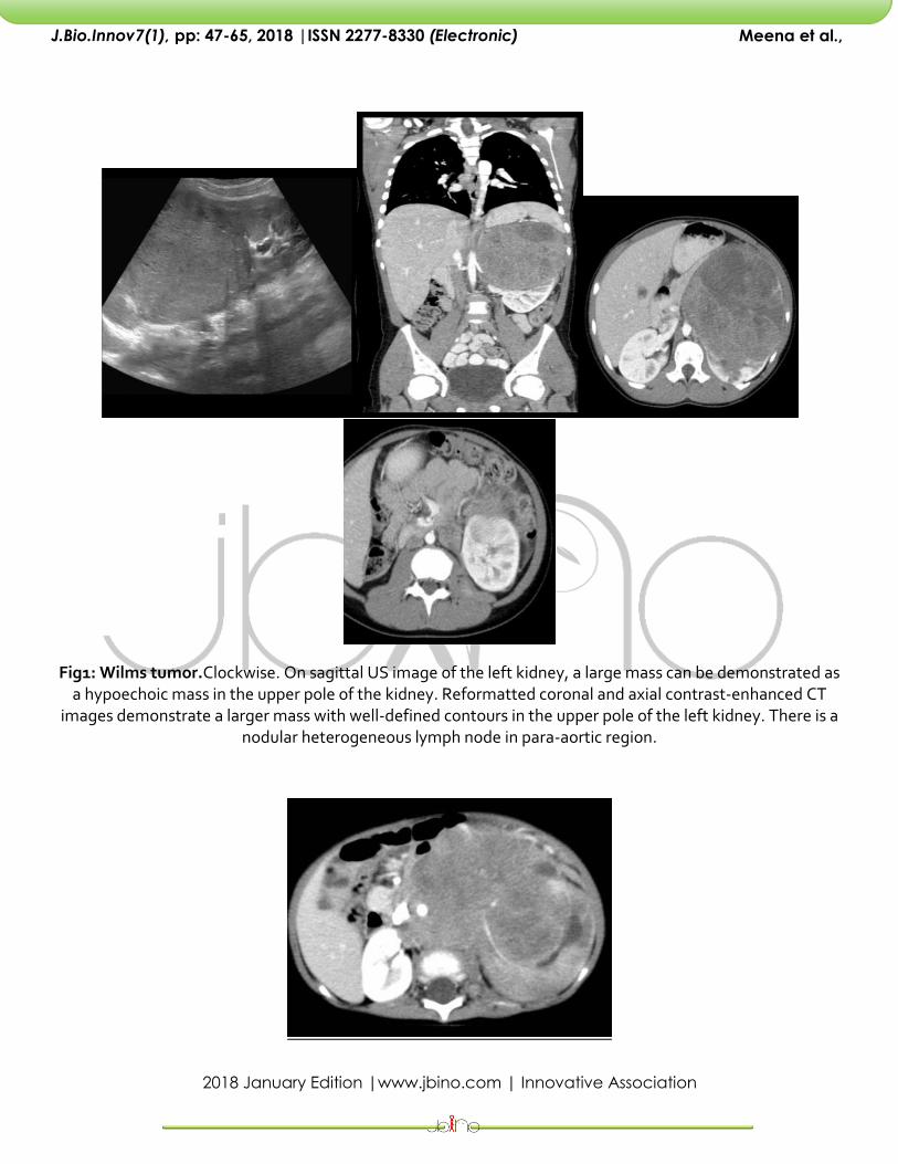
Fig1: Wilms tumor.Clockwise. On sagittal US image of the left kidney, a large mass can be demonstrated as a hypoechoic mass in the upper pole of the kidney. Reformatted coronal and axial contrast-enhanced CT
images demonstrate a larger mass with well-defined contours in the upper pole of the left kidney. There is a nodular heterogeneous lymph node in para-aortic region.
2018 January Edition |www.jbino.com | Innovative Association
J.Bio.Innov7(1), pp: 47-65, 2018 |ISSN 2277-8330 (Electronic)
Meena et al.,
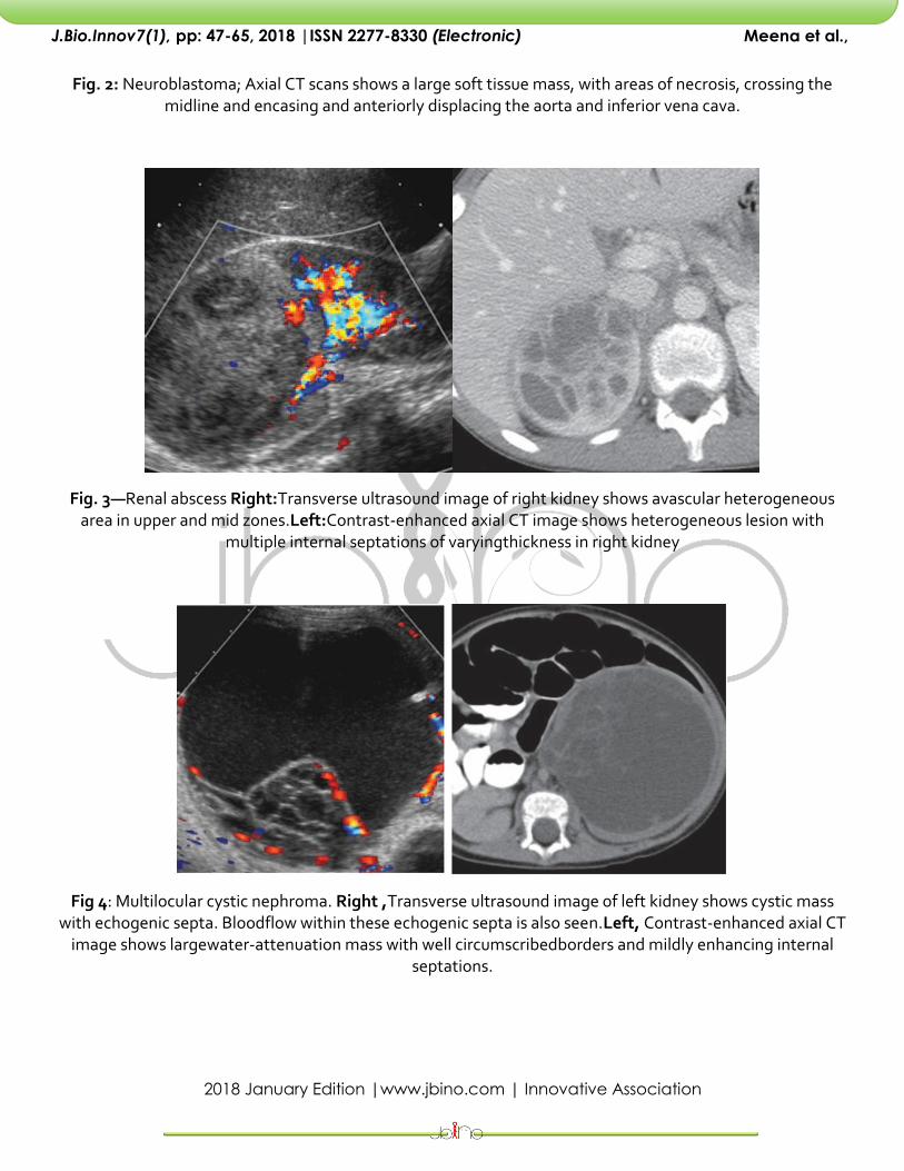
Fig. 2: Neuroblastoma; Axial CT scans shows a large soft tissue mass, with areas of necrosis, crossing the midline and encasing and anteriorly displacing the aorta and inferior vena cava.
Fig. 3—Renal abscess Right:Transverse ultrasound image of right kidney shows avascular heterogeneous area in upper and mid zones.Left:Contrast-enhanced axial CT image shows heterogeneous lesion with
multiple internal septations of varyingthickness in right kidney
Fig 4: Multilocular cystic nephroma. Right ,Transverse ultrasound image of left kidney shows cystic mass with echogenic septa. Bloodflow within these echogenic septa is also seen.Left, Contrast-enhanced axial CT
image shows largewater-attenuation mass with well circumscribedborders and mildly enhancing internal septations.

2018 January Edition |www.jbino.com | Innovative Association
J.Bio.Innov7(1), pp: 47-65, 2018 |ISSN 2277-8330 (Electronic)
Meena et al.,
Fig 5:Mesenteric cyst Right, Transverse ultrasound image of midabdomen shows large anechoic cystic mass with imperceptible wall.Left, Contrast-enhanced axial CT image shows well-circumscribed cystic mass
without enhancement in mid abdomen.
Fig 6:Hydatid infection Contrast-enhanced axial CT image showswell-defined cystic mass with several internalendocyst membranes.
2018 January Edition |www.jbino.com | Innovative Association
J.Bio.Innov7(1), pp: 47-65, 2018 |ISSN 2277-8330 (Electronic)
Meena et al.,
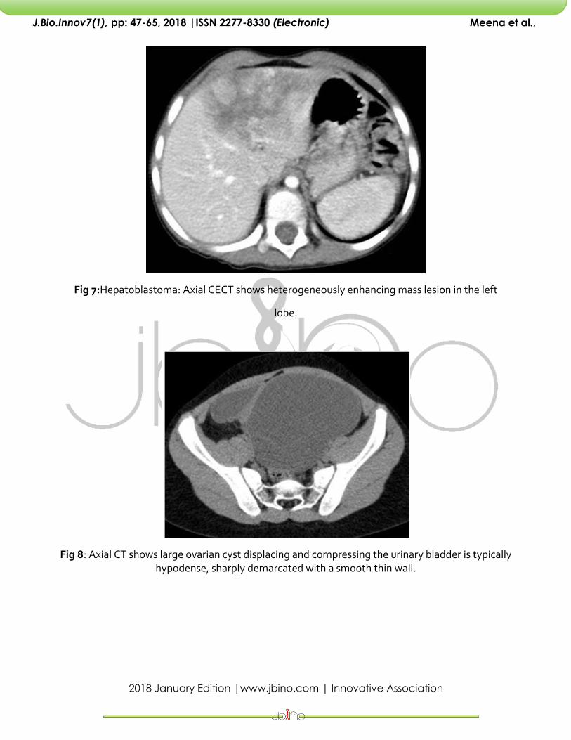
Fig 7:Hepatoblastoma: Axial CECT shows heterogeneously enhancing mass lesion in the left
lobe.
Fig 8: Axial CT shows large ovarian cyst displacing and compressing the urinary bladder is typically hypodense, sharply demarcated with a smooth thin wall.

2018 January Edition |www.jbino.com | Innovative Association
J.Bio.Innov7(1), pp: 47-65, 2018 |ISSN 2277-8330 (Electronic)
Meena et al.,
Table 1: Age Distribution
AGE GROUP NO. OF CASES PERCENTAGE
≤1 year 8 16
1-5 years 21 42
>5 years 21 42
Total 50 100
Maximum: More than 1yr
Chart 1: Age Distribution
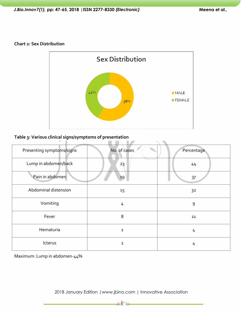
Table 2: Sex distribution
CATEGORY NUMBER OF CASES PERCENTAGE (%)
MALE 29 58
FEMALE 21 42
TOTAL 50 100
Maximum: Males 58%
2018 January Edition |www.jbino.com | Innovative Association
J.Bio.Innov7(1), pp: 47-65, 2018 |ISSN 2277-8330 (Electronic)
Meena et al.,
Chart 2: Sex Distribution
Table 3: Various clinical signs/symptoms of presentation
Presenting symptoms/signs No. of cases Percentage
Lump in abdomen/back 23 44
Pain in abdomen 19 37
Abdominal distension 15 32
Vomiting 4 9
Fever 8 11
Hematuria 2 4
Icterus 2 4
Maximum: Lump in abdomen-44%

2018 January Edition |www.jbino.com | Innovative Association
J.Bio.Innov7(1), pp: 47-65, 2018 |ISSN 2277-8330 (Electronic)
Meena et al.,
Chart 3: Various clinical signs/symptoms of presentation
Table 4: Initial investigation for which patient was referred
Investigation No. of cases Percentage
Plain radiograph 2 4%
Contrast study 0 0%
USG 44 88%
CT 4 8%
Maximum: USG- 88%

2018 January Edition |www.jbino.com | Innovative Association
J.Bio.Innov7(1), pp: 47-65, 2018 |ISSN 2277-8330 (Electronic)
Meena et al.,
Chart 4: Initial investigation for which patient was referred
Table 5: Distribution of masses according to nature
Nature of Mass No. of cases Percentage
Malignant 22 44%
Benign 28 56%
Congenital 3 6%
Infective/Inflammatory 13 26%
Neoplastic 4 8%
Miscellaneous 7 14%

2018 January Edition |www.jbino.com | Innovative Association
J.Bio.Innov7(1), pp: 47-65, 2018 |ISSN 2277-8330 (Electronic)
Meena et al.,
Chart 5: Distribution of masses according to nature
Table 6: Accuracy of CT and USG vis-à-vis surgical/cytological findings
USG CT
Nature of Mass 81% 100%
Localization 64.5% 97%
Extent 59% 100%
Diagnosis 54.5% 86%
2018 January Edition |www.jbino.com | Innovative Association
J.Bio.Innov7(1), pp: 47-65, 2018 |ISSN 2277-8330 (Electronic)
Meena et al.,
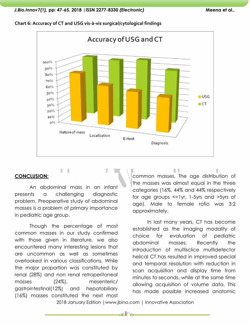
Chart 6: Accuracy of CT and USG vis-à-vis surgical/cytological findings
CONCLUSION:
An abdominal mass in an infant
presents a challenging diagnostic
problem. Preoperative study of abdominal
masses is a problem of primary importance
in pediatric age group.
Though the percentage of most
common masses in our study confirmed
with those given in literature, we also
encountered many interesting lesions that
are uncommon as well as sometimes
overlooked in various classifications. While
the major proportion was constituted by
renal (28%) and non renal retroperitoneal
masses (24%), mesenteric/
gastrointestinal(12%) and hepatobiliary
(16%) masses constituted the next most
common masses. The age distribution of
the masses was almost equal in the three
categories (16%, 44% and 44% respectively
for age groups <=1yr, 1-5yrs and >5yrs of
age). Male to female ratio was 3:2
approximately.
In last many years, CT has become
established as the imaging modality of
choice for evaluation of pediatric
abdominal masses. Recently the
introduction of multisclice multidetector
helical CT has resulted in improved special
and temporal resolution with reduction in
scan acquisition and display time from
minutes to seconds, while at the same time
allowing acquisition of volume data. This
has made possible increased anatomic
2018 January Edition |www.jbino.com | Innovative Association
J.Bio.Innov7(1), pp: 47-65, 2018 |ISSN 2277-8330 (Electronic)
Meena et al.,
coverage lesional dual phase imaging, Ct
angiography for vascular status and
retrospective multiplanar reconstruction of
area of interest with images of high quality
to predict the exact extent of the lesion. In
our study, while the accuracy of USG for
predicting nature of the mass, its
localisation, extent and exact diagnosis
was 81%, 64.5%, 59% and 54.5%
respectively, the accuracy of CT for same
was found to be 100%, 97%, 100% and 81%
respectively.
While CT was found to be 100%
accurate in determining the exact location
as well as extent of the mass lesions, the
accuracy for diagnosis was found to be
81% reflecting the inherent limitation of the
imaging modality in terms of non specific
findings in certain lesions. An important
aspect was optimal vascular
enhancement and excellent multiplanar
reconstructions with our CT that enabled
an appropriate evaluation of the extent of
the lesion as well as relation of the mass to
various vessels at the same time also
conclusively telling about vascular
invasion/encasement, an important finding
in context of various malignancies and
their staging. In context of children, the
motion artifacts encountered in our study
were very infrequent and in no case were
they of such significance as to impede the
diagnostic value of the examination.
Thus we would conclude that the recent
advances have expanded the usefulness
of CT in the evaluation of pediatric
abdominal masses. The advantage of
single breath hold acquisition in
cooperative children, improved vascular
contrast enhancement, increased
detection of parenchymal lesions, and
multiplanar and three dimentional
reconstruction may make it one of the
modalities of choice in evaluation of
pediatric abdominal masses.
BIBLIOGRAPHY:
Merten DF and Stuart GH: Radiological
staging of thoracoabdominal tumors in
childhood. Radiologic clinics of North
America, 1994; 32(1): 133-149
Goldberg BB, Pollack HM, Capitanio MA,
Kirkpatrick JA.: Ultrasonography: an aid in
the diagnosis of masses in paediatric
patients. Paediatrics, 1975; 56(3): 421-8.
Griscom NT: The roentgenology of
neonatal abdominal masses. AJR, 1965; 93:
447-463.
Yamaguchi M, Takeuchi S, Akiyama H,
Sawaguchi S: Ultrasonic evaluation of
abdominal masses in the paediatric
patient. Tohoku J Exp Med, 1980; 130(1):
25-39.
Scott DJ, Wallace WH, Hendry GM. With
advances in radiological imaging can the
radiologist reliably diagnose Wilm’s tumor?
Clin. Radiol. 1999; 54(5): 321-327.
Ohtsuka Y, Takahashi H et al.: Detection of
tumor thrombus in children using colour
Doppler ultrasonography. J. Paed. Surgery,
1997; 32(10): 1507-1510.
Riccabona M, Uggowitzer M et al.: Echo-
enhanced color Doppler ultrasonography

2018 January Edition |www.jbino.com | Innovative Association
J.Bio.Innov7(1), pp: 47-65, 2018 |ISSN 2277-8330 (Electronic)
Meena et al.,
in children and adolescents. J Ultrasound
Med., 2000; 19(11): 789-796.
Bates SM, Keller MS, Ramos IM,Carter D,
Taylor KJ: Hepatoblastoma: Detection of
tumor vascularity with duplex Doppler US.
Radiology 1990; 176(2): 505-7.
Brasch RC: Computed tomography in the
evaluation of pediatric genitourinary
disease. Urol Clin North America,
1980;7(2):223-30.
Babcock DS, Kaufman RA.:
Ultrasonography and computed
tomography in the evaluation of acutely ill
pediatric patients. Radiol. Clin. Of North
Am. 1983; 21(3):527-50
Plumley DA, Grosfeld JL, Kopecky KK,
Buckwalter KA, Vaughan WG: The role of
spiral CT with 3D reconstruction in pediatric
solid tumors. J Pediatr Surg, 1995; 30(2):
317-21.
Miele V, Galluzo M, Bellusi A, Valenti M:
Spiral CT in the study of renal neoplasms in
children. Radiol. Med (Torino).1998;
95(5):486-92.
Gualdi GF, Ferriano MG, Casciani E,
Pollettini E: Volumetric Spiral CT in the
diagnosis, staging and programmed
therapy of kidney tumors: Comparison with
conventional CT. Clin Ter, 1998;149(5): 335-
41.
Frush DP, Siegel MJ, Bisset GS 3rd:
Challenges of pediatric spiral CT.
Radiographics, 1997;17(4):939-59.
Sanders RC and Hartman Ds: The
sonographic distinction between neonatal
multicystic kidney and hydronephrosis.
Radiology,1984; 151:621-625.
Gilbert R, Garra B, Gibbons MD: Renal
Duplex Doppler Ultrasound: An adjunct in
the evaluation of hydronephrosis in the
child. J Urol 1993 Oct; 150(4):1192-4.
Lim GY, Jang HS, Lee EJ, Lim YS, Jung SE,
Lee JM, Parkn SH: Utility of the resistance
index ratio in differentiating obstructive
from nonobstructive hydronephrosis in
children. J Clin Ultraound 1999 May;
27(4):187-93.
Report of the international Reflux Study
Committee. Medical versus Surgical
treatment of primary vesicoureteral reflux:
a prospective international reflux study in
children. J Urol 1992; 148:1688-1692.
Berrocal T, Gaya F, Arjonilla A, Lonergan
GJ: Vesicoureteral Reflux: Diagnosis and
grading with Echo-enhanced
cystosonography versus voiding
cystourethrography.Radiology 2001; 221:
359-365.
Agarwal R: Sonographic assessment of
fetal abdominal cystic lesions. A pictorial
essay. Ind. Journal Radiol Img 1999;9:4:169-
182.
Ganeshan S, Indrajit IK:Images: Prune Belly
Syndrome: Antenatal Ultrasound. Ind.
Journal Radiol Img 2001;11:1:25-28.
2018 January Edition |www.jbino.com | Innovative Association
J.Bio.Innov7(1), pp: 47-65, 2018 |ISSN 2277-8330 (Electronic)
Meena et al.,
Walker D, Fennell R, Garin E, Richard G:
Spectrum of multicystic renal dysplasia:
diagnosis and management. Urology 1978:
11(5):433-6.
Egeibor OO and Jebral AA: Pediatric renal
masses: CT findings. Applied Radiology,
1999; 28(2):20-26.
Biona K, Bazaz R, and BhargavaS:
Roentgen evaluation of abdominal
masses in children. IJRI 1983; 37(4): 337-342
Rastogi V, Singhal PK, Aseri A, and Taneja
SB: Ind J Ped 1988; 55: 295-300.