Exploring the Autoimmune Diseases: A Systemic Approach
86
Exploring the Autoimmune Diseases: A Systemic Approach Blair B Lonsberry, MS, OD, MEd. , FAAO Professor of Optometry Pacific University College of Optometry [email protected]
Transcript of Exploring the Autoimmune Diseases: A Systemic Approach

Exploring the Autoimmune Diseases: A Systemic Approach
Blair B Lonsberry, MS, OD, MEd., FAAOProfessor of Optometry
Pacific University College of [email protected]

Disclosures
Paid consultant for:Maculogix: Honoraria-Advisory BoardSun Pharmaceuticals: Advisory Board

Autoimmune Diseases• Group of acquired diseases in which genetic factors
appear to play a role• They have in common widespread immunologic and
inflammatory alterations of connective tissue• The illnesses share certain clinical features and
differentiation between them is often difficult because of this.
• Although thought to be acquired diseases, often their causes cannot be determined .

Case• 55 yr white female complains of fluctuating vision
– Worse at near– Spends 8-10 hours/day on the computer
• Medical Hx:– Hypertension for 10 years – Joint pain
• Medications:– HCTZ for HTN– Celebrex for her joint pain
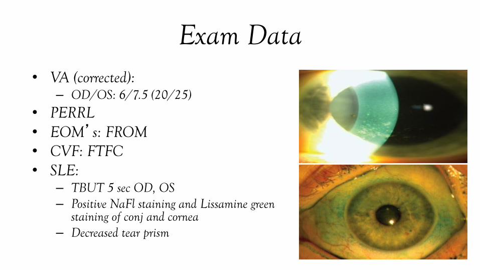
Exam Data• VA (corrected):
– OD/OS: 6/7.5 (20/25) • PERRL• EOM’s: FROM• CVF: FTFC• SLE:
– TBUT 5 sec OD, OS– Positive NaFl staining and Lissamine green
staining of conj and cornea– Decreased tear prism
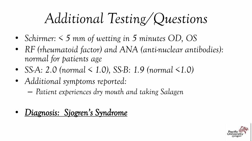
Additional Testing/Questions• Schirmer: < 5 mm of wetting in 5 minutes OD, OS• RF (rheumatoid factor) and ANA (anti-nuclear antibodies):
normal for patients age• SS-A: 2.0 (normal < 1.0), SS-B: 1.9 (normal <1.0)• Additional symptoms reported:
– Patient experiences dry mouth and taking Salagen
• Diagnosis: Sjogren’s Syndrome
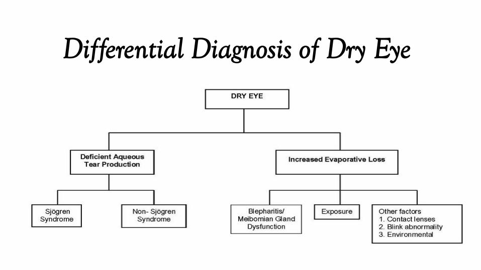
Differential Diagnosis of Dry Eye

DEWS 2: DED Definition
“Dry eye is a multifactorial disease of the ocular surface characterized by a loss of homeostasis of the tear film, and accompanied by ocular symptoms, in which tear film instability and hyperosmolarity, ocular surface inflammationand damage, and neurosensory abnormalities play etiological roles.”

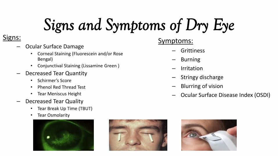
Signs and Symptoms of Dry EyeSigns:
– Ocular Surface Damage• Corneal Staining (Fluorescein and/or Rose
Bengal)
• Conjunctival Staining (Lissamine Green )
– Decreased Tear Quantity• Schirmer’s Score
• Phenol Red Thread Test
• Tear Meniscus Height
– Decreased Tear Quality• Tear Break Up Time (TBUT)
• Tear Osmolarity
Symptoms:– Grittiness
– Burning
– Irritation
– Stringy discharge
– Blurring of vision
– Ocular Surface Disease Index (OSDI)

Treatment• We initiated:
– Omega-3 supplements (2 grams per day)– Recommended warm compresses and lid washes qhs– Testosterone cream 3% applied to upper lid bid
• Patient had significant improvement in symptoms with the use of the topical testosterone cream.– However, she was still symptomatic at the end of the day and she still had significant
staining on her cornea and conjunctiva– Initiated FML tid for 1 month, Restasis bid after 2 weeks
• 2 months later patient reported further improvement in her symptoms• No conjunctival staining was noted and only slight SPK• Schirmer’s values improved to OD: 9 mm, OS: 10 mm

Role of Androgens?• Recent studies have suggested that androgen deficiency may be the
main cause of the meibomian gland dysfunction, tear-film instability and evaporative dry eye seen in Sjogren patients
• Transdermal testosterone 3% promotes increased tear production and meibomian gland secretion, thereby reducing dry eye symptoms (Dr. Charles Connor).
• Progesterone 0.05%/Testosterone 0.05% Ophthalmic Solution BID (local compounding pharmacy?)
• Topical Testosterone 0.5% drops BID (compounding pharmacy)
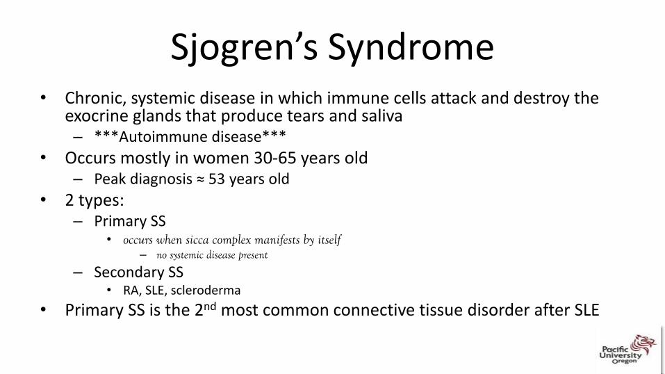
Sjogren’s Syndrome• Chronic, systemic disease in which immune cells attack and destroy the
exocrine glands that produce tears and saliva– ***Autoimmune disease***
• Occurs mostly in women 30-65 years old– Peak diagnosis ≈ 53 years old
• 2 types:– Primary SS
• occurs when sicca complex manifests by itself – no systemic disease present
– Secondary SS• RA, SLE, scleroderma
• Primary SS is the 2nd most common connective tissue disorder after SLE
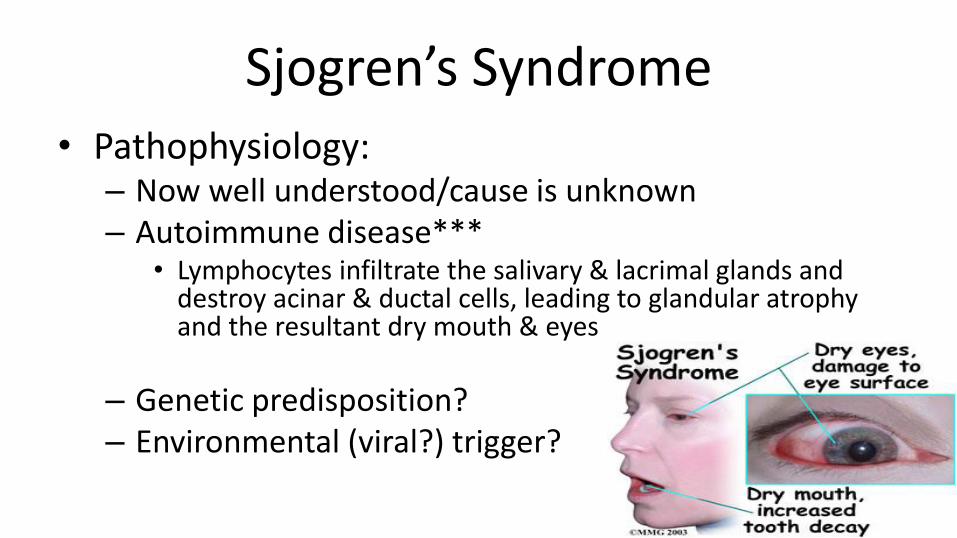
Sjogren’s Syndrome• Pathophysiology:
– Now well understood/cause is unknown– Autoimmune disease***
• Lymphocytes infiltrate the salivary & lacrimal glands and destroy acinar & ductal cells, leading to glandular atrophy and the resultant dry mouth & eyes
– Genetic predisposition?– Environmental (viral?) trigger?

Antibodies to SS-A and SS-B• Sjogren’s Syndrome Antibodies A and B• Typically tested by ELISA and immunoblot• Associated Conditions:
– Uncommon in the normal population and in patients with rheumatic diseases other than Sjogren’s syndrome and SLE
– Present in 75% of patients with “primary” Sjogren’s but only 10-15% of patients with RA and secondary Sjogren’s Syndrome
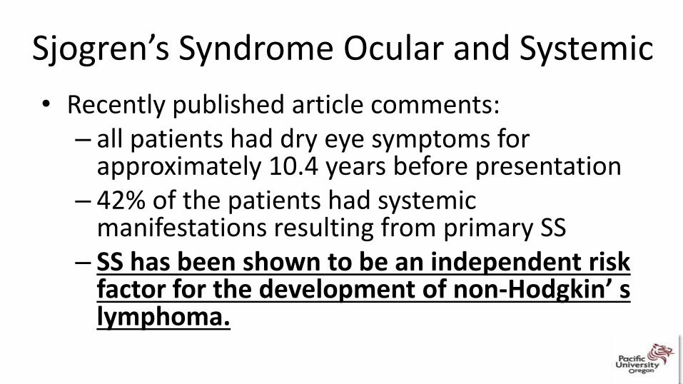
Sjogren’s Syndrome Ocular and Systemic
• Recently published article comments:– all patients had dry eye symptoms for
approximately 10.4 years before presentation – 42% of the patients had systemic
manifestations resulting from primary SS– SS has been shown to be an independent risk
factor for the development of non-Hodgkin’ s lymphoma.

Sjogren’s Syndrome Ocular and Systemic
• Authors recommendation:
– primary SS is associated with vision- and life-threatening complications
– presence of SS needs to be explored in patients with clinically significant dry eye because dry eye precedes the occurrence of the systemic manifestations

Sjogren’s Syndrome Management• Management:
– Generally supportive eye & mouth therapy• Artificial tears, gels, ointments, punctal plugs, anti-inflammatories, cyclosporine, lifitegrast• Scleral contact lenses?• fish oil• Sipping water more frequently, good dental care***
– Cevimeline (Evoxac)– Pilocarpine (Salagen)– NSAIDs– Steroids– DMARDs (Plaquenil, Methotrexate, Cyclosporine)
• Prognosis:– Overall pretty good– Higher rate of lymphoma*
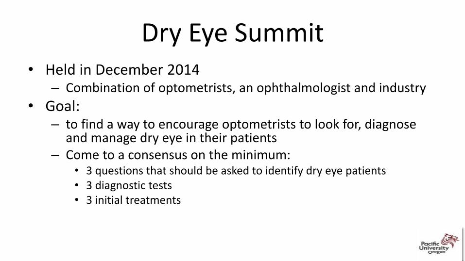
Dry Eye Summit• Held in December 2014
– Combination of optometrists, an ophthalmologist and industry
• Goal:– to find a way to encourage optometrists to look for, diagnose
and manage dry eye in their patients– Come to a consensus on the minimum:
• 3 questions that should be asked to identify dry eye patients• 3 diagnostic tests• 3 initial treatments

Recommendations from the Dry Eye Summit 2014
1. Do your eyes ever feel dry or uncomfortable?
2. Are you bothered by changes in your vision throughout the day?
3. Are you ever bothered by red eyes?
4. Do you ever use or feel the need to use drops?
Consensus on Screening QuestionsREV. as of March 13, 2015

Recommendations from the Dry Eye Summit 2014
1. The lid
2. Staining
3. Tear stability
Consensus on Baseline Diagnostic Options for Entry Level Dry Eye
Disease
REV. as of March 13, 2015

Recommendations from the Dry Eye Summit 2014
1. For all patients:
A. Ocular lubrication
B. Lid hygiene
C. Nutrition
2. Topical anti-inflammatories
Consensus on Baseline ManagementREV. as of March 13, 2015
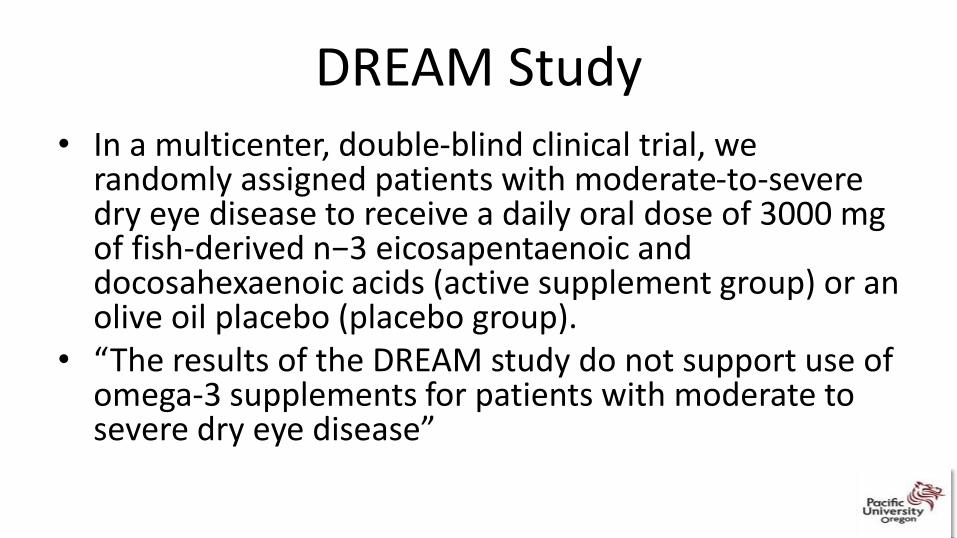
DREAM Study• In a multicenter, double-blind clinical trial, we
randomly assigned patients with moderate-to-severe dry eye disease to receive a daily oral dose of 3000 mg of fish-derived n−3 eicosapentaenoic and docosahexaenoic acids (active supplement group) or an olive oil placebo (placebo group).
• “The results of the DREAM study do not support use of omega-3 supplements for patients with moderate to severe dry eye disease”

DREAM Study
• In DREAM, most dry eye symptoms and signs appear to improve in both arms.
• In each trial group, there was a meaningful statistical change between baseline and 12 months (with time as a continuous variable) in the conjunctival staining score, the corneal staining score and the tear break-up time

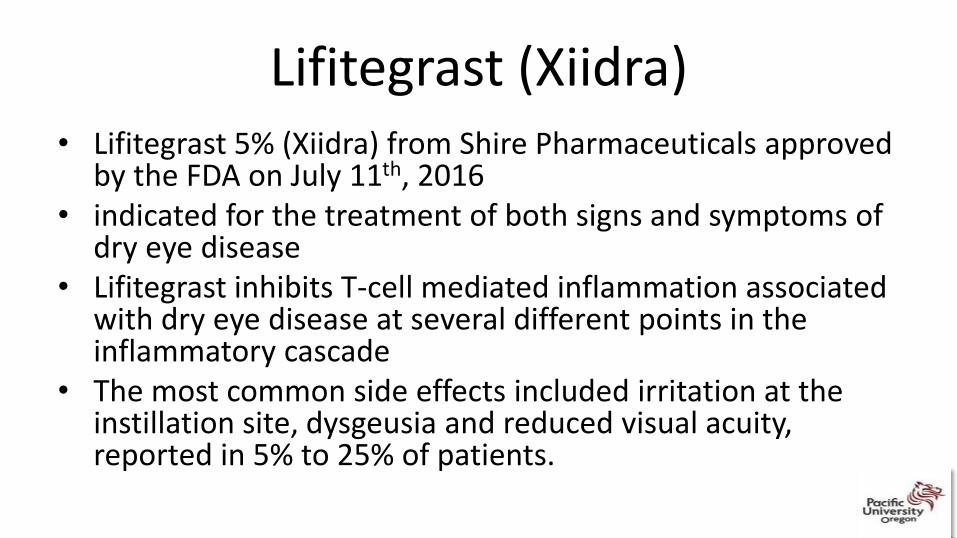
Lifitegrast (Xiidra)• Lifitegrast 5% (Xiidra) from Shire Pharmaceuticals approved
by the FDA on July 11th, 2016• indicated for the treatment of both signs and symptoms of
dry eye disease• Lifitegrast inhibits T-cell mediated inflammation associated
with dry eye disease at several different points in the inflammatory cascade
• The most common side effects included irritation at the instillation site, dysgeusia and reduced visual acuity, reported in 5% to 25% of patients.

Cequa (cyclosporine 0.09%)
• From Sun Pharmaceuticals
• Offers a novel nanomicelle formulation that helps improve the delivery of cyclosporine
• Enhanced solubility and increased ocular penetration of cyclosporine
Case: Gonzalez• 33 HF presents with a painful, red right eye
– Started a couple of days ago, deep boring pain– Has tried Visine but hasn’t helped the redness
• PMHx: patient reports she has been diagnosed with rheumatoid arthritis 3 years ago– Takes Celebrex for the joint pain– Patient reports she occasionally gets a skin rash when she is outdoors in the
sun• POHx: unremarkable• PMHx: mother has rheumatoid arthritis
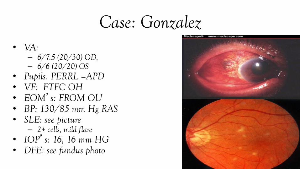
Case: Gonzalez• VA:
– 6/7.5 (20/30) OD, – 6/6 (20/20) OS
• Pupils: PERRL –APD• VF: FTFC OH• EOM’s: FROM OU• BP: 130/85 mm Hg RAS• SLE: see picture
– 2+ cells, mild flare• IOP’s: 16, 16 mm HG• DFE: see fundus photo

Etiologies of Cotton Wool SpotsVascular Occlusive Disease Hypertension Ocular Ischemic Syndrome
Autoimmune Disease e.g. SLE Hyperviscosity syndromes Trauma
Pre-eclampsia Radiation Retinopathy Toxic e.g. interferon
Neoplastic e.g. leukemia Anterior Ischemic Syndrome Infectious e.g. HIV

Patient Update• Patient was worked up for lupus and diagnosed with
lupus.• Patient was already taking Celebrex which was not
effective in treating the scleritis she presented with– upon referral to rheumatology it was discovered that she
had several organs already being affected by the lupus– she was put on immunosuppressive agents to treat the
systemic and ocular manifestations
• Patient was taken off of Celebrex and put on plaquenil(hydroxychloroquine) 400 mg po qd

RHEUMATOID ARTHRITIS
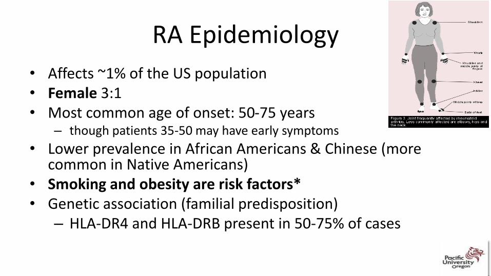
RA Epidemiology
• Affects ~1% of the US population• Female 3:1• Most common age of onset: 50-75 years
– though patients 35-50 may have early symptoms
• Lower prevalence in African Americans & Chinese (more common in Native Americans)
• Smoking and obesity are risk factors*• Genetic association (familial predisposition)
– HLA-DR4 and HLA-DRB present in 50-75% of cases
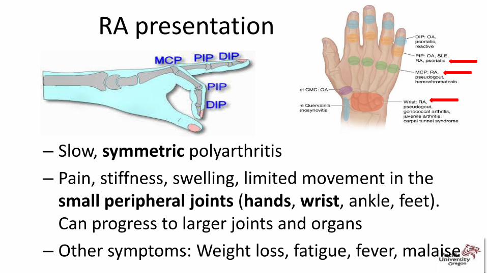
RA presentation
– Slow, symmetric polyarthritis
– Pain, stiffness, swelling, limited movement in the small peripheral joints (hands, wrist, ankle, feet). Can progress to larger joints and organs
– Other symptoms: Weight loss, fatigue, fever, malaise

Rheumatoid Arthritis• Rheumatoid Arthritis (RA) is not a benign disease. • RA is associated with decreased life expectancy.
– The risk of cardiovascular mortality is twice that of the general population.
• Affecting approximately 1% of the adult population, RA is associated with considerable disability.
• It is now well recognized that there is a "window of opportunity" early in the disease process to initiate treatment which will fundamentally change the course of the disease.

Rheumatoid Arthritis

Other Diagnostic Criteria for RACutaneous Ocular Pulmonary Cardiac Neurological Hematological
Nodules Sicca Pleuritis Pericarditis Peripheral neuropathy
Leukopenia
Vasculitis Episcleritis Nodules Atherosclerosis Cervical myelopathy Anemia of chronic disease
Scleritis Interstitial lung disease
Myocardial infarction Lymphadenopathy
Fibrosis

Osteoarthritis (OA) vs. RA• Etiology of RA is inflammatory
which improves with activity while OA is mechanical and worsens with activity
• Inflammation secondary to mechanical insults in OA while no previous insult required in RA
• Joint cartilage is primary site of articular involvement in OA while its the bony surfaces of the joints in RA
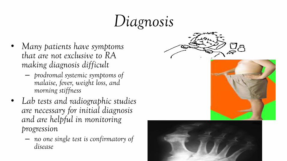
Diagnosis• Many patients have symptoms
that are not exclusive to RA making diagnosis difficult– prodromal systemic symptoms of
malaise, fever, weight loss, and morning stiffness
• Lab tests and radiographic studies are necessary for initial diagnosis and are helpful in monitoring progression– no one single test is confirmatory of
disease

Criteria for Diagnosis of RA
RA likely if:– Morning stiffness > 30 minutes– Painful swelling of 3 or more joints– Involvement of hands and feet (especially MCP and MTP joints)– Duration of 4 or more weeks– Differential diagnoses include: crystal arthropathy, psoriatic
arthritis, lupus, reactive arthritis, spondyloarthropathies.

Lab Testing for RATests Diagnostic Value Disease Activity Monitoring
ESR or CRP Indicate only inflammatory process- Very low specificity
ESR elevated in many but not all active inflammation.Maybe useful in monitoring disease activity and response to treatment
RF RF has a low sensitivity and specificity for RA.Seropositive RA has worse prognosis.
No value
ANA Positive in severe RA, SLE, or other connective tissue disorders (CTD)
No value-do not repeat
X-rays Diagnostic erosions rarely seen in disease of <3 mo’s duration
Serial x-rays over many years may show disease progression and indicate med change
Joint aspiration Indicated if infection suspected

Giant Cell Arteritis• vessels most often involved are the
arteries over the temples, – GCA = "temporal arteritis.”
• symptoms, such as fatigue, loss of appetite, weight loss or a flu-like feeling– pain in the jaw with chewing (jaw
claudication).
– Sometimes the only sign of GCA is unexplained fever.
– Less common symptoms include pains in the face, tongue or throat.
Giant Cell Arteritis
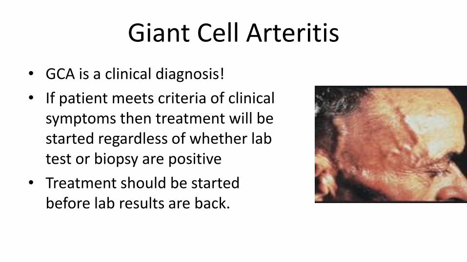
• GCA is a clinical diagnosis!
• If patient meets criteria of clinical symptoms then treatment will be started regardless of whether lab test or biopsy are positive
• Treatment should be started before lab results are back.

GCA Treatment Update!!• May 22, 2017:
– FDA expanded and approved the use of subcutaneous Acetemra (tocilizumab) to treat adults with giant cell arteritis.• Approved in Canada
– First FDA approved therapy, specific to this type of vasculitis
– Compared to placebo and standardized prednisone treatment
Rheumatoid Factor (RF)• RF is an autoantibody directed against IgG• Most common lab testing are latex fixation and nephelometry• RF present in 70-90% of patients with RA
– However RF is not specific for RA– Occurs in a wide range of autoimmune disorders– Prevalence of positive RF increases with age
• As many as 25% of persons over age of 65 may test positive– High titer for RF almost always reflects an underlying disease

Antibodies to Cyclic Citrullinated Peptides (ACPA)
• Proteins that contain citrulline are the target of an AB response that is highly specific for RA
• Associated conditions:– Appears to be quite specific for RA
• Specificity as high as 97%– Sensitivity in the range of 70-80% for established RA and 50% for
early-onset– Has superior specificity and comparable sensitivity for diagnosis of
RA as compared to RF
• 80-97% of patients have RA if they are RF+ and ACPA+
Diagnosis
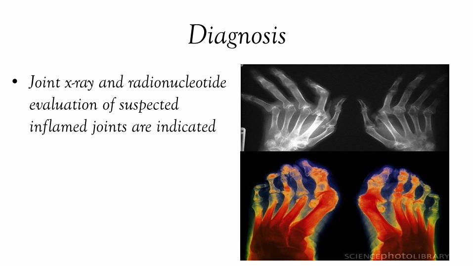
• Joint x-ray and radionucleotide evaluation of suspected inflamed joints are indicated

Rheumatoid Arthritis: Treatment• Treatment must be started early to maximize the benefits of
medications and prevent joint damage. • The use of traditional medications in combination and the new
biologic therapies has revolutionized the paradigm of RA treatment in recent years.
• There is no curative treatment for RA– treatment is to minimize inflammation– minimize damage and – maximize patient functioning
Treatment and Management-Systemic• Current Tx regimens utilize a step-down approach with initiation of
one or more DMARD’s at time of diagnosis. • RA most destructive early in disease• “Easier” and more effective if Tx initiated early.• DMARD-disease modifying antirheumatic drug
– these drugs not only reduce inflammation but also change the immune response in a long-term and more dramatically than NSAID’s
– give chance of permanent remission
Treatment and Management: Antimalarials
• Hydroxychloroquine (HCQ) more common and less toxic than more effective chorloquine
• usual dose is 200-400 mg/d @night with onset of action after a period of 2-4 months
• has mild DMARD effect, does not slow radiographic progression and has relatively slow onset of action, useful with other DMARD’s
28
Treatment and Management: Antimalarial Ocular Complications
• Have affinity for pigmented structures such as iris, choroid and RPE
• Toxic affect on the RPE and photoreceptors leading to rod and cone loss.
• Have slow excretion rate out of body with toxicity and functional loss continuing to occur despite drug discontinuation.

Treatment and Management: Steroids
• usually used in short-term pulse dosages (e.g. 7.5 mg/day in combination with DMARD to reduce joint damage in early disease Tx). 26


Treatment and Management: Methotrexate
• now considered as part of mainstay treatment
• antimetabolite used in cancer therapy that inhibits DNA synthesis (thought to cause suppression of lymphocyte proliferation)
• low dose in RA (7.5-25mg) once weekly orally or injection with onset of action 6-8 weeks
30
Treatment and Management: Methotrexate
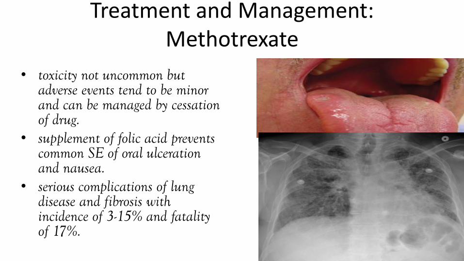
• toxicity not uncommon but adverse events tend to be minor and can be managed by cessation of drug.
• supplement of folic acid prevents common SE of oral ulceration and nausea.
• serious complications of lung disease and fibrosis with incidence of 3-15% and fatality of 17%.
31

Treatment and Management: Biological Therapies-TNF Inhibitor
• inhibitors bind TNF before it can be bound to the receptor (infliximab [Remicade], etanercept [Enbrel], adalimumab [Humira]) and newest golimumab (Simponi)
• quicker onset of action (several weeks)
• new studies indicate use as first line therapy, potentially combined with methotrexate
36

Treatment and Management: Biological
Therapies-TNF Inhibitor• Remicade: 3 mg/k as IV infusion followed
by similar doses at 2 and 6 weeks and then every 8 weeks after
• Enbrel and Humira are SC injections every 2weeks
• Newer is Simponi which is once a month injection
• Adverse affects include increased risk of opportunistic infections (TB most common), malignancies (lymphoma) and neurological disease.
• common SE’s include nausea and vomiting37

Episcleritis
• self-limiting, recurring, idiopathic inflammation of the episcleraltissue that does not threaten vision
• Symptoms are a localized area of hyperemia of the globe, irritation, and lacrimation. Diagnosis is clinical. Treatment is symptomatic
• Unilateral (bilateral possible but rarely simultaneously)

Episcleritis
• occurs in young adults, more commonly among women. It is usually idiopathic; it can be associated with connective tissue diseases and rarely with serious systemic diseases.
• Recurrent episodes of episcleritisusually manifest prior to active periods of arthritis and a better indicator than dry eye
• Episcleritis will recur despite systemic treatment

Treatment and Management: Episcleritis
• Treatment of episcleritis is dependent upon severity and chronicity.
• Palliative care maybe considered for mild cases (ocular lubrication).
• Utilization of vasoconstrictors, NSAIDs and steroid (Pred mild, Lotemax) use for more severe or chronic cases.
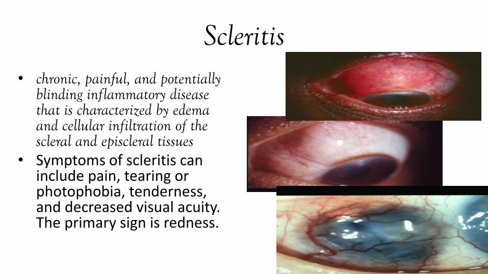
Scleritis• chronic, painful, and potentially
blinding inflammatory disease that is characterized by edema and cellular infiltration of the scleral and episcleral tissues
• Symptoms of scleritis can include pain, tearing or photophobia, tenderness, and decreased visual acuity. The primary sign is redness.

Ocular Manifestations-Scleritis• classified into anterior and posterior.• Anterior:
– Diffuse and nodular forms– Necrotizing (with/without inflammation) less
frequent• Have the most serious systemic implications• Scleromalacia perforans
• Posterior:– characterized by flattening of the
posterior aspect of the globe, thickening of the posterior coats of the eye (choroid and sclera), and retrobulbar edema.
Treatment and Management: Scleritis
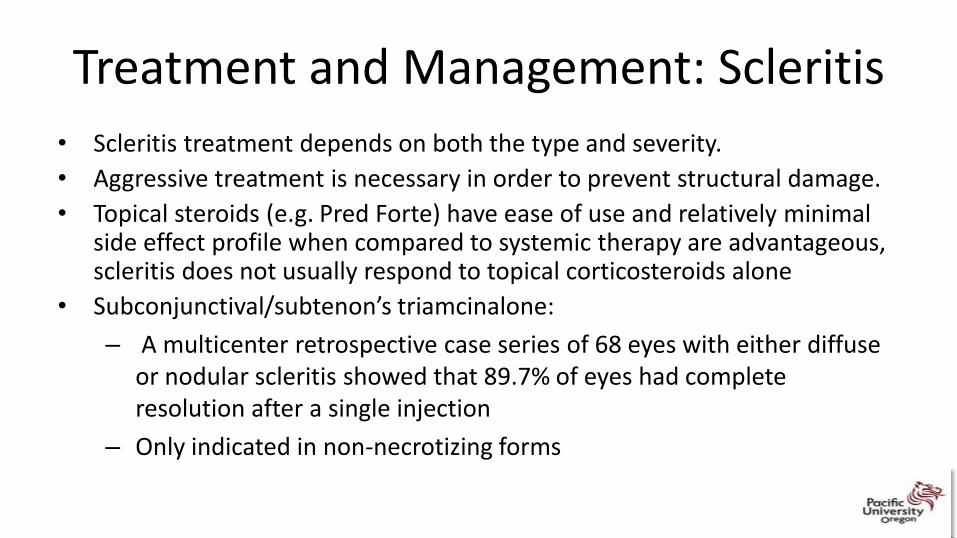
• Scleritis treatment depends on both the type and severity.
• Aggressive treatment is necessary in order to prevent structural damage.
• Topical steroids (e.g. Pred Forte) have ease of use and relatively minimal side effect profile when compared to systemic therapy are advantageous, scleritis does not usually respond to topical corticosteroids alone
• Subconjunctival/subtenon’s triamcinalone:
– A multicenter retrospective case series of 68 eyes with either diffuse or nodular scleritis showed that 89.7% of eyes had complete resolution after a single injection
– Only indicated in non-necrotizing forms
Treatment and Management: Scleritis
• Oral NSAIDs:– considered first-line therapy for scleritis for their ease of
use, cost, and relatively mild side effect profile for both anterior and posterior scleritis
– E.g. Ibuprofen 400-600 mg QID, Naproxen 250-500 mg BID, or Indomethacin 25-50 mg TID
– short term use of an NSAID is often well tolerated, NSAIDs can cause adverse effects which include peptic ulcer disease, hypertension, increased heart disease, bleeding, fluid retention, renal disease, and mood change
Treatment and Management: Scleritis
• Oral Prednisone:
– considered to be the first line therapy for the treatment of non necrotizing scleritis in the setting of poor control on oral NSAIDs, or as a first line agent for necrotizing scleritis.
– Typically start at between 40-60 mg until resolution with a slow taper

Treatment and Management: Scleritis
• If necrotizing present patient needs to receive aggressive medical therapy by rheumatologist– patients have better prognosis when immunosuppressive
therapy is instituted

SYSTEMIC LUPUS ERYTHEMATOSUS (SLE)

Systemic Lupus Erythematosus (SLE)
• Idiopathic, multisystemicinflammation disorder characterized by hyperactivity of immune system and prominent auto-antibody production – against components of cell membranes
and nuclear material• Acute periods followed by periods of
remission are common – gives disease an unpredictable course
Systemic Lupus Erythematosus (SLE)
• Definite genetic predisposition has been demonstrated– environmental factors also play a role especially as triggers
• Clinical course varies from mild episodic disorder to rapidly developing fatal disease

Epidemiology• SLE is not uncommon with prevalence exceeding
1:2000 persons with 85% being female• Disease may occur at any age though most patients
are b/w ages 20-40– AA being affected 3x more than any other race (and more
severely)
Epidemiology• Have to ensure that condition is not secondary to a
drug response (several drugs produce lupus-like syndrome)– Agents strongly associated include:
• Procainamide (cardiac arrhythmias), hydralazine (high blood pressure) and isoniazid (anti-tuberculosis)
• Others include: phenytoin, quinidine, tetracyclines and TNF inhibitors.
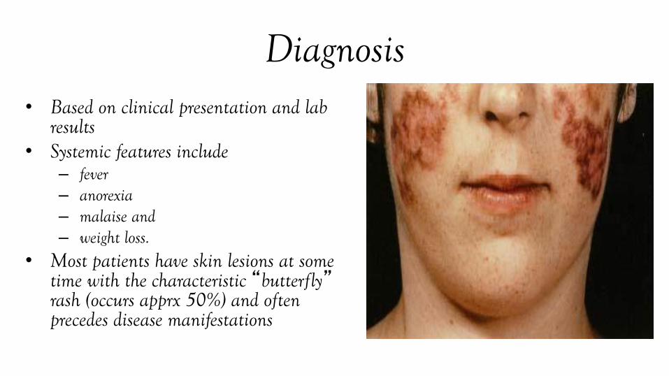
Diagnosis• Based on clinical presentation and lab
results• Systemic features include
– fever– anorexia– malaise and – weight loss.
• Most patients have skin lesions at some time with the characteristic “butterfly”rash (occurs apprx 50%) and often precedes disease manifestations

Diagnosis
• Joint symptoms (with/without active synovitis) occur in >90% of patients and are often the earliest manifestation.
• Other organs affected include heart, kidney, lungs, CNS.
• American Rheumatology Association established 11 criteria for diagnosis (8 clinical manifestations and 3 lab). – Minimum of 4 needed serially or
simultaneously.

Lab Tests: Antinuclear Antibodies (ANA)
• AB’s directed against nuclear material:• Detection is via indirect immunofluorescence
• ANA with titers > 1:40 considered positive
• Associated conditions:– Positive tests occur in a wide variety of conditions
• Low-titer ANA are relatively common among healthy adults
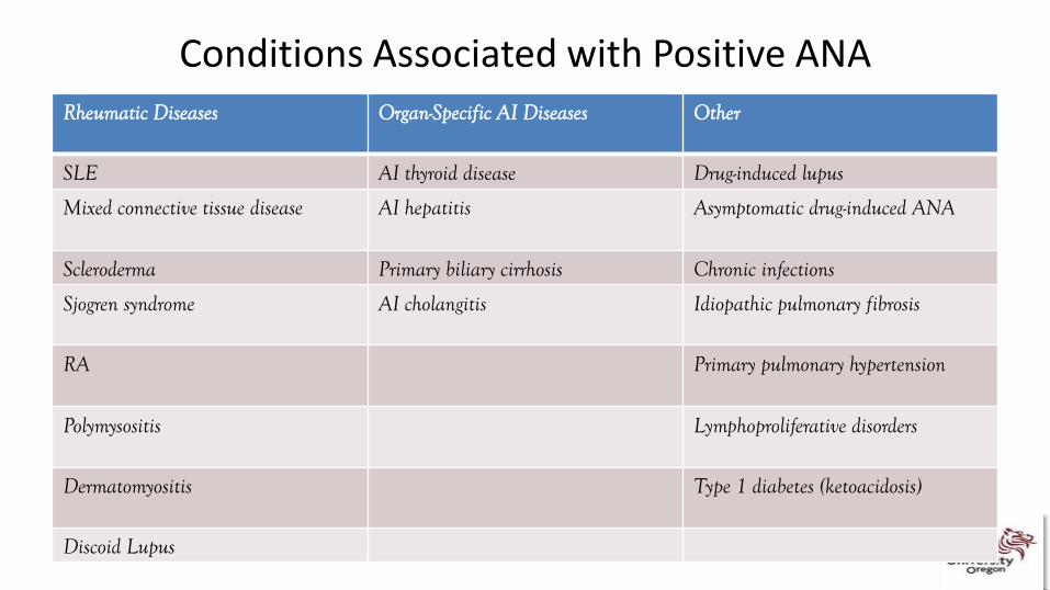
Conditions Associated with Positive ANA Rheumatic Diseases Organ-Specific AI Diseases Other
SLE AI thyroid disease Drug-induced lupus
Mixed connective tissue disease AI hepatitis Asymptomatic drug-induced ANA
Scleroderma Primary biliary cirrhosis Chronic infections
Sjogren syndrome AI cholangitis Idiopathic pulmonary fibrosis
RA Primary pulmonary hypertension
Polymysositis Lymphoproliferative disorders
Dermatomyositis Type 1 diabetes (ketoacidosis)
Discoid Lupus
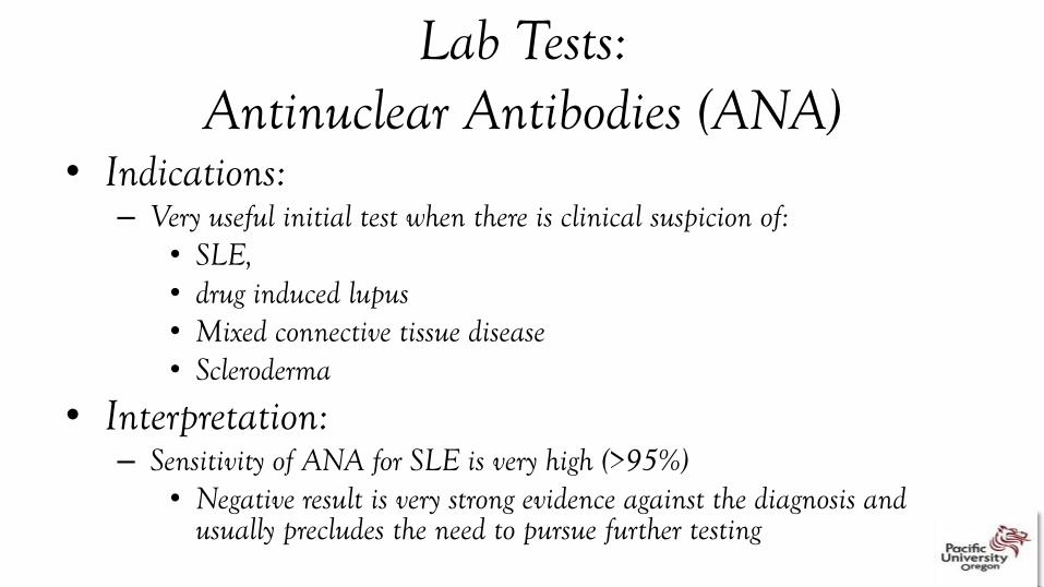
Lab Tests: Antinuclear Antibodies (ANA)
• Indications:– Very useful initial test when there is clinical suspicion of:
• SLE, • drug induced lupus• Mixed connective tissue disease• Scleroderma
• Interpretation:– Sensitivity of ANA for SLE is very high (>95%)
• Negative result is very strong evidence against the diagnosis and usually precludes the need to pursue further testing
Lab Tests: Antinuclear Antibodies (ANA)
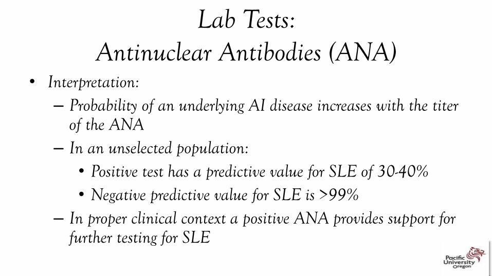
• Interpretation:– Probability of an underlying AI disease increases with the titer
of the ANA– In an unselected population:
• Positive test has a predictive value for SLE of 30-40%• Negative predictive value for SLE is >99%
– In proper clinical context a positive ANA provides support for further testing for SLE

Lab Tests: Antibodies to Double-Stranded DNA
• ELISA is most commonly used• Associated conditions:
– Occurs in SLE and is rare in other diseases and in healthy persons
• Indications:– Should be measured when there is clinical suspicion of SLE and the ANA
is positive
• Interpretation:– Specificity for SLE is 97% and approaches 100% when titer is high– AB’s occur in 60-80% of patients with SLE
Lab Tests
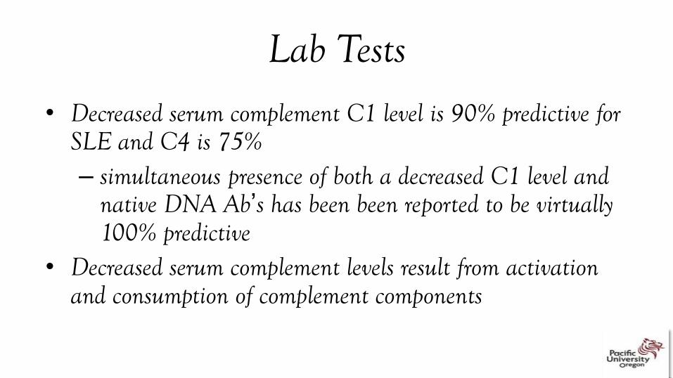
• Decreased serum complement C1 level is 90% predictive for SLE and C4 is 75% – simultaneous presence of both a decreased C1 level and
native DNA Ab’s has been been reported to be virtually 100% predictive
• Decreased serum complement levels result from activation and consumption of complement components
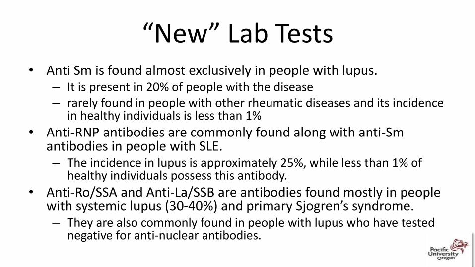
“New” Lab Tests• Anti Sm is found almost exclusively in people with lupus.
– It is present in 20% of people with the disease– rarely found in people with other rheumatic diseases and its incidence
in healthy individuals is less than 1%
• Anti-RNP antibodies are commonly found along with anti-Smantibodies in people with SLE. – The incidence in lupus is approximately 25%, while less than 1% of
healthy individuals possess this antibody.
• Anti-Ro/SSA and Anti-La/SSB are antibodies found mostly in people with systemic lupus (30-40%) and primary Sjogren’s syndrome. – They are also commonly found in people with lupus who have tested
negative for anti-nuclear antibodies.

Treatment and Management• No cure for SLE (rest, reduce stress and avoid UV exposure)• Medical management includes:
– Salicylates and NSAIDs employed to treat arthralgias, arthritis, myalgias and fever in 20-30% of Px with mild disease
– Antimalarials (Plaquenil) used to treat discoid lesions and joint disease– High dose, short-acting steroids are used in life-threatening and severely disabling
cases. Prolonged maintenance at low dosages needed after.– Cytotoxic controversial-used when steroids ineffective– Exp therapy: high dose immunoglobulin injections
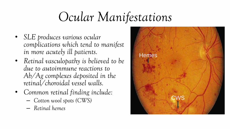
Ocular Manifestations• SLE produces various ocular
complications which tend to manifest in more acutely ill patients.
• Retinal vasculopathy is believed to be due to autoimmune reactions to Ab/Ag complexes deposited in the retinal/choroidal vessel walls.
• Common retinal finding include:– Cotton wool spots (CWS)– Retinal hemes
Hemes
CWS

Ocular Manifestations• Occlusions are uncommon but
occur more frequently in arteries and can result in nonperfusion and hypoxia.
• Optic nerve and retinal neo may arise.
• Vitreous heme and RD may also occur.
• Optic atrophy and blindness may result in severe occlusions.
ONH
Neo
Vitreous
Heme
BRAO
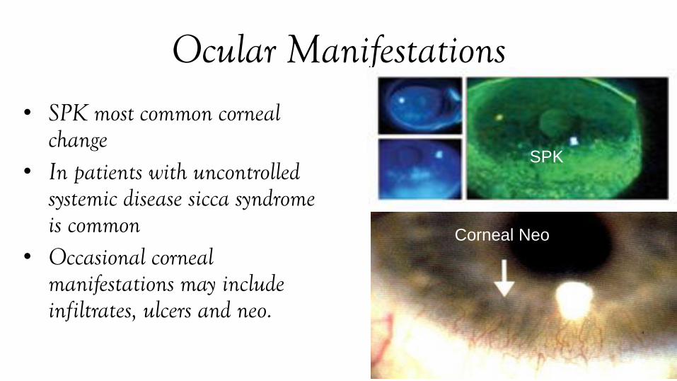
Ocular Manifestations• SPK most common corneal
change • In patients with uncontrolled
systemic disease sicca syndrome is common
• Occasional corneal manifestations may include infiltrates, ulcers and neo.
SPK
Corneal Neo

Ocular Manifestations
• Scleritis is usually diffuse and nodular and is fairly common. It may be the presenting feature of SLE.
• Non-granulomatous uveitis is sometimes found
• Diplopia and pupillary abnormalities secondary to cranial nerve palsies also arise

THANK [email protected]