
Exploring different models of stroke unit care and outcome ...
305
School of Physiotherapy Exploring different models of stroke unit care and outcome: The Stroke Rehabilitation Outcome (SRO) study Diane Dennis This thesis is presented for the Degree of Doctor of Philosophy of Curtin University of Technology January 2013
Transcript of Exploring different models of stroke unit care and outcome ...

School of Physiotherapy
Exploring different models of stroke unit care and outcome:
The Stroke Rehabilitation Outcome (SRO) study
Diane Dennis
This thesis is presented for the Degree of
Doctor of Philosophy
of
Curtin University of Technology
January 2013


i
DECLARATION
To the best of my knowledge and belief this thesis contains no material previously
published by any other person except where due acknowledgment has been made.
This thesis contains no material that has been accepted for the award of any other
degree or diploma in any university.
Diane Dennis
January 2013

ii

iii
ABSTRACT
Introduction:
Stroke is a significant cardiovascular event requiring sub-acute rehabilitation, best
provided in a stroke unit (SU). These units include dedicated neurological SUs
usually catering only for patients with stroke and more generic SUs existing within
geriatric rehabilitation units (GRUs). There exists a “grey” group of survivors of
stroke whose allocation to one type of rehabilitation facility over another is arbitrary,
in that the referring physician had no evidence to suggest advantages of SU versus
GRU rehabilitation.
Objectives:
The aim of this inception cohort study was to provide a direct comparison of quality
of life and functional outcome between two commonly applied models of organised
multidisciplinary SU rehabilitation for the “grey” group of stroke survivors. Further,
it evaluated differences in the intensity of treatment and the environment in which
rehabilitation was implemented.
Method:
All patients presenting to Royal Perth Hospital-Wellington Street Campus acute
stroke unit (RPH-WSC ASU) with a diagnosis of recent stroke requiring
hospitalisation and subacute rehabilitation were considered for inclusion into the
study. Patients were selected based on their age, absence of dementia and their
acceptance by incumbent medical staff for rehabilitation transfer at either Royal
Perth Hospital-Shenton Park Campus stroke unit (SPC SU) or at a GRU
geographically closest to their home (located at either Mercy hospital, Bentley
hospital or Swan health campus).
Baseline data was collected in order to establish the underlying level of disability and
compare groups for comparability, and also to be used as covariates in data analysis.

iv
All treatments received were those considered standard for the individual facility,
administered as usual by registered health professionals. During the study, periodic
behaviour mapping at each of the study facilities was undertaken by a research
assistant in order to quantify differences in the rehabilitation environment. In
addition, attending therapists at each facility recorded the frequency and duration of
their intervention with individual patients involved in the study in a patient diary
designed for that purpose. Six and twelve months following their transfer from
RPH-WSC ASU, patients attended follow-up outpatient appointments at neutral
rooms where objective and subjective assessments were undertaken by an
independent assessor (a physiotherapist) who was blinded as to which rehabilitation
facility the patient had attended. The primary outcome measure was the MOS 36-
Item Short Form Health Survey (SF-36) and secondary outcome measures included
the Functional Independence Measure (FIM) and other functional measures.
Results:
Between July 2004 and June 2007, 354 patients with stroke were age appropriate (60
years of age or older) for recruitment into the study and of these, 94 consented to
participate (SPC SU n=22; GRUs n=72). Patients referred to SPC SU were younger,
more likely to be male, and have speech abnormality, peripheral vascular disease and
diabetes than those referred to GRUs. Otherwise there were no significant
differences between groups in any of the characteristics measured at baseline.
Rehabilitation data demonstrated a significant difference in both the total allied
health professional (AHP) therapy time (p<0.001) and the indirect support time such
as telephone calls and meetings with family (p=0.022), with SPC SU therapists
utilising more time compared with GRU therapists. There was no significant
difference in time spent undertaking administration including writing notes and
reports (p=0.957). Data showed significant difference in length of stay (LOS),
whereby patients spent a longer time at SPC SU (p=0.036), however there was no
significant difference in discharge destination between facilities (p=0.312). Of the
10 unadjusted patient measures in this study, there were significant differences

v
between groups in only two, the Berg balance score and the Chedoke McMaster
posture inventory. The differences in both of these secondary outcomes favoured the
SPC SU group. In addition there were differences in the SF36 Mental component
summary (MCS) and Physical component summary (PCS) scores that approached
significance. The difference in the PCS scores also favoured the SPC SU group but
for the MCS score it was the GRU group that had more favourable scores.
As the study was not randomized, age and gender, which differed between groups at
baseline, and Barthel Index score, known to be associated with length of stay in
stroke patients, were added to the models as covariates. As data from 6 and 12 month
follow-ups was included in the dependent variable, “visit” was added to the models.
After these adjustments there were no significant differences between facilities in
any quality of life or functional outcomes.
Discussion:
Overall there was relatively high quality of life, and low anxiety and depression
reported and results were not influenced by where rehabilitation took place. Selection
criteria excluding dementia and young age may in part explain this, as both have
been found to predict worse quality of life outcome in stroke. Significant differences
in both where patients were, and what they were doing throughout the day reflected
different ethos between facilities in the way rehabilitation was delivered. However,
there was no difference in functional outcome despite these environmental
differences and the fact that patients experienced more intensive treatment over a
more prolonged hospital stay at SPC SU.
Conclusion:
In most cases, rehabilitation of this “grey” subgroup of the wider population of
stroke may be more cost-effective if carried out at GRUs (with higher patient/ staff
ratios, less intensive treatment and shorter LOS) rather than the neurological SUs.

vi
ACKNOWLEDGEMENTS
Heartfelt thanks to the patients with Stroke who willingly agreed to participate in the
project in spite of the sudden onset of their illness and the functional impairment
experienced. Obviously there would have been no study without your involvement,
and hopefully your participation will have helped others in the future. Also a big
thank you to the patient carers who agreed to facilitate outpatient assessments by
providing transport to appointments despite the often significant impact of the
patient’s illness on their own lives.
To all of the medical and allied health professionals who cooperated with the
implementation of the study protocol at all sites, and especially those involved in the
collection of data, and those who agreed to be observed during behaviour mapping, a
huge thanks. Particular thanks to physiotherapists Jacquie Ancliffe and the team at
RPH-WSC as well as Karen Smith at SPC SU for their incredible patience and
wonderful feedback before, during and after data was collected. Your dedication to
stroke rehabilitation is something we all aspire to.
Thank you for the monumental efforts of my supervisor and mentor, Dr Kathy
Briffa, who has been extraordinarily intelligent, energetic, encouraging, patient, and
inspirational over such a prolonged period. In addition to her overall supervision of
my work, her input was fundamentally important in the successful National Heart
Foundation grant application. Her continued faith in my ability to actually complete
the study and her resourcefulness in helping me to do so has continued to the very
end, and I will be forever grateful. Kathy was a great friend long before she was a
supervisor, and I wouldn’t and couldn’t have done this without her.
To Professor’s Graeme Hankey and Leon Flicker I extend a special
acknowledgment. Graeme suggested the project in the first place so many years ago,
and agreed to help, design and instigate the study on his stroke unit at RPH-WSC.
Since then, both he and Leon have been a great source of information and advice in

vii
helping to get the project through the various Ethics committees, acquire a grant-in-
aid from the National Heart Foundation and provide informed positive feedback on
everything from the study design, to the implementation and interpretation of results.
Their co-operation has essentially ensured and enabled the project to be successful
and I am hugely indebted to them both.
I would also like to acknowledge the generous assistance of Jeff Ewen to design and
adapt the study database, and extract data when needed. Also for all those late night
phone calls demanding immediate IT help when various computers unexplainably
swallowed data or text. Thanks Jeff, for always being ridiculously accessible and
willing to help – I’m sure you are equally as excited as I am to have this completed!
To physiotherapists Jo Bouckley and Louise Wise, a huge thankyou. Cheerfully
reliable Jo managed to coordinate patient recruitment and outpatient appointments
whilst juggling her own final years of university study; Louise, with young children
of her own, managed to complete follow-up assessments at 6 and 12 months over 2
years. Her warm, thorough and professional approach to everything she does made
my life very easy. Also thanks to Gill Vinton for diligently collecting the behaviour
mapping data during that time.
To my physiotherapist-now-medical-doctor friend, Rose Wyllie, I am extremely
grateful for help in setting up the behaviour mapping component of the study. It’s
nice to surround yourself with friends when you undertake a huge project such as
this, and Rose is one of those people who have remained encouraging and excited at
the little things over a long period of time.
I would also like to acknowledge Dr Anne Smith for her expert and timely statistical
advice and analyses, and her guidance in the interpretation of all things statistical. I
would also like to thank Dr Richard Parsons for his early work on statistical data.

viii
Thanks to Dr Sophie Coleman for her editing, formatting and overall organization of
my thesis; and to Professor Joan Cole for her help in proofreading early versions and
providing insightful comments and feedback.
I would also like to acknowledge my work colleagues at the Sir Charles Gairdner
Hospital in Perth, Western Australia whom I regard as some of my closest friends.
Particularly Tracy Hedben-Todd, Ian Cooper and Wendy Jacob, who have afforded
me the time to finish the writing up of my thesis and who have always been a huge
source of encouragement and support. It is a privilege to work with such caring,
friendly and professional individuals.
I also gratefully acknowledge the financial assistance provided by the grant-in-aid
from the National Heart Foundation. There is no doubt in my mind that the study
would have not been completed were it not for this support.
Finally, a very special mention of my closest support and inspiration, my lovely
husband Jeff and our beautiful children Madeleine, Dimity and Grayson. This study
and all it involves has been a part of this family for a very long time, as we carried it
with us to Sydney and then to Canada and back to Perth again. It is exciting to have
it finally completed and to move forward toward the next chapter of our lives. I wish
to also thank my wonderful parents Maria and Fred Acott for their love and
encouragement and not least of all, their babysitting. And so now, what next?

ix
ABBREVIATIONS
ADL activities of daily living
AHP(s) Allied health professional(s)
ARAT Action Research Arm Test
ASU acute stroke unit
Bentley Bentley health service
BBS Berg Balance Scale
BI Barthel Index
CT Computer Tomography
EQ5D VAS European Quality of Life-5 Dimension Visual Analogue Scale
FIM Functional Independence Measure
FH Fremantle Hospital
FTE full time equivalency
GRU(s) geriatric rehabilitation unit(s)
GW general ward
HADS Hospital Anxiety and Depression Scale
LAC lacunar infarction
LOS length of stay
Mercy Mercy hospital
MCS Mental Component Summary score of the SF36 measure
MMSE Mini-mental State Examination
MRI Magnetic Resonance Imaging
PAC partial anterior circulation infarction
PCS Physical Component Summary score of the SF36 measure
POC posterior circulation infarction
PVD peripheral vascular disease
Rankin Rankin score
RCT Randomized controlled trial
RPH-WSC Royal Perth Hospital-Wellington Street Campus

x
SCGH Sir Charles Gairdner Hospital
SF36 MOS 36-Item Short Form Health Survey
SPC SU Royal Perth Hospital-Shenton Park Campus
SRO Stroke Rehabilitation Outcome
SSS Scandinavian Stroke score
Swan Swan health service
SU stroke unit
TAC total anterior circulation infarction
tPA tissue plasminogen activator
VAS visual analogue scale
WHO World Health Organization

xi
TABLE OF CONTENTS
DECLARATION..................................................................................................................... I
ABSTRACT .......................................................................................................................... III
ACKNOWLEDGEMENTS ................................................................................................ VI
ABBREVIATIONS .............................................................................................................. IX
TABLE OF CONTENTS .................................................................................................... XI
CHAPTER 1 ........................................................................................................................... 1
INTRODUCTION .................................................................................................................. 1
1.1 OVERVIEW OF STROKE .................................................................................................... 1
1.2 MANAGEMENT OF STROKE ............................................................................................. 1
1.3 CURRENT PRACTICE IN STROKE REHABILITATION, PERTH, WESTERN AUSTRALIA ........ 2
1.4 AIM AND HYPOTHESIS ..................................................................................................... 3
1.5 STUDY TIMEFRAME ......................................................................................................... 4
FIGURE 1.5.1 CHAPTER FLOW CHART...................................................................................... 6
CHAPTER 2 ........................................................................................................................... 7
STROKE ................................................................................................................................. 7
2.1 DEFINITION OF STROKE ................................................................................................... 7
2.2 EXTENT OF STROKE WORLD-WIDE, AND IN AUSTRALIA ................................................. 8
2.2.1 INCIDENCE ................................................................................................................... 8
2.2.2 AGE AND GENDER DISTRIBUTION ................................................................................. 9
2.2.3 SOCIOECONOMIC AND RACIAL DISTRIBUTION ............................................................. 9
2.3 ACUTE CARE MANAGEMENT ........................................................................................ 10
2.3.1 DIAGNOSIS ................................................................................................................. 10
2.3.2 MANAGEMENT ........................................................................................................... 11

xii
2.3.3 ACUTE STROKE UNITS ................................................................................................ 11
2.4 REHABILITATION ........................................................................................................... 13
2.4.1 OVERVIEW ................................................................................................................. 13
2.4.2 REHABILITATION MODELS .......................................................................................... 14
2.4.3 COMPONENTS AND INTENSITY OF THERAPY ................................................................ 15
2.4.4 AGE-SPECIFIC ADMISSION CRITERIA ........................................................................... 15
2.4.6 SOCIAL SUPPORT ......................................................................................................... 17
2.5 CONTEXT OF CURRENT RESEARCH ............................................................................... 17
CHAPTER 3 ......................................................................................................................... 19
PILOT STUDIES .................................................................................................................. 19
3.1 INTRODUCTION .............................................................................................................. 19
3.1.1 FACILITIES .................................................................................................................. 19
3.1.2 NUMBER OF STROKE PATIENTS .................................................................................. 19
FIGURE 3.1.2A: INPATIENT DESTINATION OF PATIENTS WITH STROKE FOLLOWING
ADMISSION TO RPH-WSC BETWEEN JULY 1999 AND JUNE 2000 .......................................... 21
FIGURE 3.1.2B: DISCHARGE DESTINATION OF PATIENTS WITH STROKE FOLLOWING
ADMISSION TO RPH-WSC BETWEEN JULY 1999 AND JUNE 2000 .......................................... 22
FIGURE 3.1.2C: DISCHARGE DESTINATION OF PATIENTS WITH STROKE CARED FOR BY THE
RPH-WSC ASU FOLLOWING ADMISSION BETWEEN JULY 1999 AND JUNE 2000 .................. 23
3.1.3 REHABILITATION ........................................................................................................ 23
3.1.4 TOOLS ......................................................................................................................... 24
3.2 PILOT STUDIES UNDERTAKEN ....................................................................................... 25
3.2.1 ALLIED HEALTH PROFESSIONAL (AHP) QUESTIONNAIRE ......................................... 25
3.2.1.1 INTRODUCTION ........................................................................................................... 25
3.2.1.2 DEVELOPMENT OF TOOL ............................................................................................. 25
Method ................................................................................................................................... 25
Trial ........................................................................................................................................ 26
Results .................................................................................................................................... 26
Discussion .............................................................................................................................. 26
3.2.1.3 IMPLEMENTATION OF THE TOOL ................................................................................. 27
3.2.1.4 RESULTS OF THE PILOT STUDY .................................................................................... 27

xiii
AHP Experience, Education and Research ............................................................................ 27
TABLE 3.2.1.4A: CHARACTERISTICS OF ALLIED HEALTH PROFESSIONAL (AHP)
RESPONDENTS, N (%). ............................................................................................................ 28
FIGURE 3.2.1.4A: YEARS SINCE QUALIFICATION FOR STAFF AT EACH FACILITY ................... 29
FIGURE 3.2.1.4B: INDIVIDUAL AHP’S EXPERIENCE TREATING PATIENTS WITH STROKE,
MONTHS .................................................................................................................................. 30
AHP Treatment Philosophies ................................................................................................. 31
TABLE 3.2.1.4B: PROPORTIONS OF DIFFERENT TREATMENT PHILOSOPHIES UTILISED AT EACH
FACILITY ................................................................................................................................. 32
Workload and casemix ........................................................................................................... 32
FIGURE 3.2.1.4C: NUMBER PATIENTS SEEN PER DAY AT EACH FACILITY............................... 33
FIGURE 3.2.1.4D: NUMBER OF STROKE PATIENTS SEEN PER DAY AT EACH FACILITY ............ 34
FIGURE 3.2.1.4E: TIME AHP TREATS ALL PATIENTS WITH STROKE PER DAY, MINUTES ....... 35
FIGURE 3.2.1.4F: AVERAGE TREATMENT TIME PER PATIENT WITH STROKE, MINUTES .......... 36
Care Provided......................................................................................................................... 36
FIGURE 3.2.1.4G: PERCEIVED RATING OF AHPS OPTIMAL CARE, RATED ON 100MM VAS .... 37
FIGURE 3.2.1.4H: AHP’S PERCEPTION OF FACILITY'S OPTIMAL CARE, RATED ON 100MM VAS
............................................................................................................................................... 38
Length of Stay (LOS) ............................................................................................................. 39
TABLE 3.2.1.4C: AHP LEVEL OF INFLUENCE ON PATIENT LOS, N (%).................................. 39
3.2.1.5 DISCUSSION AND SUMMARY ...................................................................................... 39
3.2.1.6 IMPLICATIONS FOR SRO STUDY ................................................................................. 40
3.2.2 PATIENT DIARIES ...................................................................................................... 41
3.2.2.1 INTRODUCTION ........................................................................................................... 41
3.2.2.2 DEVELOPMENT OF TOOL ............................................................................................. 41
Diary Design .......................................................................................................................... 41
FIGURE 3.2.2.2A: EXTRACT FROM PILOT PATIENT DIARY ...................................................... 42
3.2.2.3 IMPLEMENTATION OF TOOL ........................................................................................ 43
Data collection ....................................................................................................................... 43
3.2.2.4 RESULTS OF PILOT STUDY .......................................................................................... 43
FIGURE 3.2.2.4A: BREAKDOWN OF TREATMENT AND ADMINISTRATIVE TIME FROM 12
PATIENT DIARIES, MINUTES .................................................................................................... 45
FIGURE 3.2.2.4B: TOTAL OCCASIONS OF SERVICE FROM 12 PATIENT DIARIES BY AHP ........ 46

xiv
FIGURE 3.2.2.4C: TOTAL OCCASIONS OF SERVICE FROM 12 PATIENT DIARIES BY LEVEL OF
AHP APPOINTMENT ................................................................................................................ 47
FIGURE 3.2.2.4D: BREAKDOWN OF INDIVIDUAL AND GROUP TREATMENTS FROM 12 PATIENT
DIARIES ................................................................................................................................... 48
3.2.2.5 IMPLICATIONS FOR MAIN STUDY ................................................................................ 49
3.2 PILOT STUDIES UNDERTAKEN ....................................................................................... 50
3.2.3 BEHAVIOUR MAPPING ................................................................................................ 50
3.2.3.1 INTRODUCTION ........................................................................................................ 50
3.2.3.2 DEVELOPMENT OF TOOL ............................................................................................. 51
Behaviour Mapping Design .................................................................................................... 51
Assessment of Daily Routine ................................................................................................. 51
TABLE 3.2.3.2A: CATEGORIES OF BEHAVIOUR (KENNEDY, FISHER ET AL. 1988) .................. 53
3.2.3.3 IMPLEMENTATION OF TOOL ........................................................................................ 54
3.2.3.4 RESULTS OF PILOT STUDY ........................................................................................... 54
FIGURE 3.2.3.4A: WARD MAPPING: FREQUENCY OF EACH BEHAVIOUR (ALL PATIENTS) ...... 56
FIGURE 3.2.3.4B: WARD MAPPING: FREQUENCY OF EACH BEHAVIOUR (STROKE PATIENTS) 57
PATIENT-BASED MAPPING: FREQUENCY OF EACH BEHAVIOUR: ............................................. 58
FIGURE 3.2.3.4C: INDIVIDUAL MAPPING: TOTAL FREQUENCY EACH BEHAVIOUR ............... 59
PATIENT-BASED MAPPING: TIME OBSERVED EACH BEHAVIOUR (FIGURE 3.2.3.4D): ............. 60
FIGURE 3.2.3.4D: INDIVIDUAL MAPPING: TOTAL TIME EACH BEHAVIOUR ........................... 61
TOPOGRAPHICAL MAPPING: LOCATION OF PATIENTS (FIGURE 3.2.3.4E &3.2.3.4F): ............ 62
FIGURE 3.2.3.4E: WARD MAPPING: LOCATION (ALL PATIENTS) ............................................ 62
FIGURE 3.2.3.4F: WARD MAPPING: LOCATION (STROKE PATIENTS) .................................... 63
PATIENT-BASED MAPPING: LOCATION WHERE BEHAVIOUR WAS OBSERVED: ........................ 63
FIGURE 3.2.3.4G: PATIENT ROOM OBSERVATIONS................................................................. 64
3.2.3.5 IMPLICATIONS FOR SRO STUDY ................................................................................. 64
TABLE 3.2.3.5A: CLASSIFICATION OF PATIENT BEHAVIOUR TO BE USED IN SRO TRIAL ....... 66
3.3 CONCLUSIONS FROM PILOT STUDIES ............................................................................. 67
CHAPTER 4 ......................................................................................................................... 69
LITERATURE REVIEW OF OUTCOME METHODS ................................................................. 69
4.1 INTRODUCTION .............................................................................................................. 69

xv
4.2 BASELINE MEASURES .................................................................................................... 69
4.2.1 AGE ........................................................................................................................... 69
4.2.2 BARTHEL INDEX (BI) ................................................................................................. 70
Development, indication and description ............................................................................... 70
Reliability and validity ........................................................................................................... 70
Clinically relevant cut-off scores ........................................................................................... 71
Justification ............................................................................................................................ 71
4.2.3 MINI-MENTAL STATE EXAMINATION (MMSE) ......................................................... 71
Development, indication, description, reliability and validity ............................................... 71
Clinically relevant cut-off scores ........................................................................................... 72
Justification ............................................................................................................................ 72
4.2.4 RANKIN SCALE ........................................................................................................... 72
Development, indication, description, reliability and validity ............................................... 72
Justification ............................................................................................................................ 73
4.2.5 SCANDINAVIAN STROKE SCORE (SSS) ...................................................................... 73
Justification ............................................................................................................................ 74
4.3 REHABILITATION MEASURES ........................................................................................ 74
4.3.1 PATIENT DIARY .......................................................................................................... 74
Justification ............................................................................................................................ 74
4.3.2 HOSPITAL LENGTH OF STAY (LOS) ............................................................................ 74
Development, indication, description, reliability and validity ............................................... 74
Justification ............................................................................................................................ 75
4.4 PATIENT MEASURES ...................................................................................................... 75
4.4.1 ACTION RESEARCH ARM TEST (ARAT) .................................................................... 75
Development, indication and description ............................................................................... 75
Reliability and validity ........................................................................................................... 76
Clinically significant difference ............................................................................................. 77
Justification ............................................................................................................................ 77
4.4.2 BEHAVIOUR MAPPING ................................................................................................ 78
Development, indication and description; Reliability and validity ........................................ 78
Justification ............................................................................................................................ 78
4.4.3 BERG BALANCE SCALE (BBS) ................................................................................... 78
Development, indication and description ............................................................................... 78

xvi
Reliability and validity ........................................................................................................... 79
Minimal detectable and clinically significant difference ....................................................... 79
Justification ............................................................................................................................ 79
4.4.4 CHEDOKE-MCMASTER POSTURAL CONTROL & SHOULDER IMPAIRMENT SCORES .. 80
Development, indication and description ............................................................................... 80
Reliability and validity ........................................................................................................... 80
Justification ............................................................................................................................ 81
4.4.5 EUROPEAN QUALITY OF LIFE-5 DIMENSION VISUAL ANALOGUE SCALE (EQ5D VAS)
81
Development, indication and description ............................................................................... 81
Interpreting data ..................................................................................................................... 82
Justification ............................................................................................................................ 82
4.4.6 FUNCTIONAL INDEPENDENCE MEASURE (FIM) ............................................................ 82
Development, indication and description ............................................................................... 82
Reliability and validity ........................................................................................................... 83
Indications for using tool ........................................................................................................ 83
Clinically significant differences ............................................................................................ 84
Justification ............................................................................................................................ 84
4.4.7 HOSPITAL ANXIETY AND DEPRESSION SCALE (HADS) ............................................. 85
Description and development of tool ..................................................................................... 85
Reliability and Sensitivity of tool ........................................................................................... 85
Indications for using tool ........................................................................................................ 85
Clinical thresholds .................................................................................................................. 86
Justification of choice of outcome measure ........................................................................... 86
4.4.8 MOS 36-ITEM SHORT FORM HEALTH SURVEY (SF-36) ............................................. 87
Description and development of tool ..................................................................................... 87
Reliability and Sensitivity of tool ........................................................................................... 87
Indications for using tool ........................................................................................................ 88
Normative values and clinically significant difference for patients with stroke .................... 88
Justification of choice of outcome measure ........................................................................... 88
4.4.9 TEN-METRE WALK TEST.............................................................................................. 89
Description, development, reliability and sensitivity of tool.................................................. 89
Indications for using tool ........................................................................................................ 89

xvii
Clinically significant difference ............................................................................................. 90
Justification of choice of outcome measure ........................................................................... 90
CHAPTER 5 ......................................................................................................................... 91
METHODS .............................................................................................................................. 91
5.1 INTRODUCTION .............................................................................................................. 91
5.2 RANDOMIZED CONTROLLED TRIAL (RCT) PROTOCOL .................................................. 92
5.2.1 STUDY DESIGN ........................................................................................................... 92
FIGURE 5.2.1A: STUDY DESIGN OF SRO RCT ........................................................................ 93
5.2.2 STUDY POPULATION .................................................................................................. 94
5.2.2.1 INCLUSIONS AND EXCLUSIONS ................................................................................... 94
5.2.2.2 RECRUITMENT AND PATIENT CONSENT ................................................................... 94
5.2.2.3 POWER AND SAMPLE SIZE ........................................................................................... 94
5.2.2.4 FEASIBILITY................................................................................................................ 95
5.2.3 STUDY PROCEDURE ................................................................................................... 95
5.2.3.1 BASELINE DATA COLLECTION ................................................................................. 95
5.2.3.2 RANDOMIZATION .................................................................................................... 96
5.2.3.3 TREATMENT PROCEDURE AND DOCUMENTATION ................................................... 96
5.2.3.4 FOLLOW-UP PROCEDURE AND OUTCOME DATA COLLECTION ................................. 96
5.2.4 IMPLEMENTATION OF PROTOCOL ............................................................................... 97
FIGURE 5.2.4.1: INITIAL RCT RECRUITMENT PERIOD ........................................................... 98
FIGURE5.2.4.2: SECOND RCT RECRUITMENT PERIOD ......................................................... 100
5.2.6 CONCLUSION ............................................................................................................ 101
5.3 OBSERVATIONAL TRIAL PROTOCOL ............................................................................ 102
5.3.1 STUDY DESIGN ......................................................................................................... 102
FIGURE 5.3.1A: STUDY DESIGN OF SRO OBSERVATIONAL TRIAL ....................................... 103
5.3.2 STUDY POPULATION ................................................................................................. 104
5.3.2.1 INCLUSIONS AND EXCLUSIONS .............................................................................. 104
5.3.2.2 RECRUITMENT AND PATIENT CONSENT .................................................................... 104
5.3.3 STUDY PROCEDURE .................................................................................................. 104
5.3.3.1 BASELINE DATA COLLECTION ............................................................................... 104
5.3.3.2 TRANSFER PROCEDURE ......................................................................................... 105

xviii
5.3.3.3 TREATMENT PROCEDURE AND DOCUMENTATION ................................................. 105
5.3.3.4 FOLLOW-UP PROCEDURE ....................................................................................... 105
5.3.4 OUTCOME MEASURES ............................................................................................... 106
5.3.4.1 QUALITY OF LIFE ..................................................................................................... 106
5.3.4.2 FUNCTION ............................................................................................................... 106
5.3.4.3 TREATMENT ENVIRONMENT: BEHAVIOUR MAPPING ............................................... 106
5.3.4.4 COST: LENGTH OF STAY AND AHP DIARY INFORMATION ....................................... 107
5.3.4.5 DISCHARGE OUTCOME .............................................................................................. 107
5.4 STATISTICAL METHODS ............................................................................................... 107
CHAPTER 6 ....................................................................................................................... 109
RESULTS ............................................................................................................................ 109
6.1 INTRODUCTION ........................................................................................................... 109
6.2 PATIENT FLOW ............................................................................................................. 109
TABLE 6.2A: ANNUAL NUMBER OF PATIENTS DISCHARGED FROM RPH-WSC WITH THE
DIAGNOSIS OF STROKE ......................................................................................................... 110
FIGURE 6.2A: INPATIENT DESTINATION OF ALL PATIENTS WITH STROKE FOLLOWING
ADMISSION TO RPH-WSC BETWEEN JULY 2004 AND JUNE 2007 ........................................ 111
FIGURE 6.2B: DISCHARGE DESTINATION OF ALL PATIENTS WITH STROKE FOLLOWING
ADMISSION TO RPH-WSC BETWEEN JULY 2004 AND JUNE 2007 ........................................ 112
6.3 BASELINE DATA .......................................................................................................... 113
FIGURE 6.2C: DESTINATION OF PATIENTS WITH STROKE TRANSFERRED TO OTHER FACILITIES
FOLLOWING ADMISSION TO RPH-WSC BETWEEN JULY 2004 AND JUNE 2007, AND
RECRUITMENT OF AGE APPROPRIATE PATIENTS INTO THE SRO TRIAL. ............................... 114
TABLE 6.3.1: DEMOGRAPHIC CHARACTERISTICS OF PATIENTS (N=94) ............................... 115
TABLE 6.3.2: ADMISSION STATUS OF PATIENTS (N=94) ...................................................... 116
TABLE 6.3.3: PAST MEDICAL HISTORY OF PATIENTS (N=94) ............................................... 117
TABLE 6.4.1: UNADJUSTED REHABILITATION DATA ............................................................ 119
TABLE 6.4.2: DISCHARGE DESTINATION OF PATIENTS (N=94) ............................................ 120
TABLE 6.4.3: ADJUSTED LENGTH OF REHABILITATION STAY DATA .................................... 121
TABLE 6.4.4: ADJUSTED AHP TIME* ................................................................................... 124
TABLE 6.4.5: ADJUSTED AHP INPUT BY PROFESSION* ........................................................ 126

xix
TABLE 6.4.6: UNADJUSTED DIFFERENCES BETWEEN GROUPS IN THE OUTCOME VARIABLES
MEAN SCORES (SE) OVERALL AT BOTH TIME POINTS ........................................................... 128
TABLE 6.4.7: ADJUSTED OUTCOME VARIABLES AT 6 AND 12 MONTHS ............................... 129
TABLE 6.4.8: UNADJUSTED OUTCOME VARIABLE MEASURES, MEAN SCORES (SE) ............ 132
TABLE 6.4.9 OUTCOME VARIABLE MEASURES, MEAN SCORES (SE) ADJUSTED FOR AGE,
GENDER, BARTHEL INDEX SCORE AND THE REPEATED FACTOR “VISIT” (6 MONTH AND 12
MONTH FOLLOW UP) ............................................................................................................. 133
CHAPTER 7 ....................................................................................................................... 135
PUBLICATION ...................................................................................................................... 135
CHAPTER 8 ....................................................................................................................... 163
DISCUSSION ......................................................................................................................... 163
8.1 INTRODUCTION ............................................................................................................ 163
8.2 MAJOR FINDINGS ......................................................................................................... 164
8.2.1 OVERVIEW ................................................................................................................ 164
8.2.2 PATIENT RECRUITMENT AND CHARACTERISTICS ..................................................... 164
8.2.3 OUTCOME MEASURES AND COVARIATES ................................................................. 167
8.2.3.1 QUALITY OF LIFE, ANXIETY AND DEPRESSION ...................................................... 168
8.2.3.2 FUNCTION .............................................................................................................. 171
8.2.3.3 TREATMENT ENVIRONMENT: BEHAVIOUR MAPPING ............................................. 175
8.2.3.4 COST: LENGTH OF STAY AND AHP DIARY INFORMATION ..................................... 176
8.2.3.5 DISCHARGE OUTCOMES......................................................................................... 179
8.2.4 BED BLOCKAGE ........................................................................................................ 180
8.2.5 STUDY ENDPOINT AT 12 MONTHS............................................................................. 180
8.3 LIMITATIONS ............................................................................................................... 181
8.3.1 OUTCOME MEASURES FOR SPEECH THERAPY ........................................................... 181
8.3.2 ONGOING COST ASSOCIATED WITH DISCHARGE HOME ............................................ 181
8.3.3 THERAPIST BIAS AND COMPLIANCE ......................................................................... 181
8.3.4 PATIENT RECRUITMENT ........................................................................................... 182
8.3.5 STUDY TIMELINE ..................................................................................................... 183

xx
CHAPTER 9 ....................................................................................................................... 185
CONCLUSION ....................................................................................................................... 185
REFERENCES ................................................................................................................... 189
APPENDIX 1 ...................................................................................................................... 201
PILOT ALLIED HEALTH PROFESSIONAL QUESTIONNAIRE ................................................ 201
APPENDIX 2 ...................................................................................................................... 205
FINAL ALLIED HEALTH PROFESSIONAL QUESTIONNAIRE ................................................ 205
APPENDIX 3 ...................................................................................................................... 209
PATIENT CONSENT FORM FOR DIARY PILOT STUDY ......................................................... 209
APPENDIX 4 ...................................................................................................................... 211
FINAL PATIENT DIARY TEMPLATE ..................................................................................... 211
Definitions ............................................................................................................................ 212
APPENDIX 5 ...................................................................................................................... 215
EXAMPLE OF COMPLETED WAR BEHAVIOUR MAP ............................................................ 215
APPENDIX 6 ...................................................................................................................... 217
MAPS .................................................................................................................................... 217
Topographical map – Bentley GRU ..................................................................................... 217
Topographical map - Mercy GRU ........................................................................................ 218
Topographical map SPC SU ................................................................................................. 219
Topographical map – Swan Health Service GRU ................................................................ 220

xxi
APPENDIX 7 ...................................................................................................................... 221
PILOT PATIENT-BASED MAPPING TEMPLATE .................................................................... 221
APPENDIX 8 ...................................................................................................................... 223
PILOT PATIENT-BASED BEHAVIOUR MAPPING PATIENT INFORMATION SHEET .............. 223
APPENDIX 9 ...................................................................................................................... 225
PILOT PATIENT-BASED BEHAVIOUR MAPPING PATIENT CONSENT FORM ....................... 225
APPENDIX 10 .................................................................................................................... 227
PILOT PATIENT-BASED BEHAVIOUR MAPPING PATIENT THERAPIST INFORMATION SHEET
............................................................................................................................................. 227
APPENDIX 11 .................................................................................................................... 229
GENERIC DAILY ROUTINE TEMPLATE ............................................................................... 229
GENERIC TOPOGRAPHICAL DEMOGRAPHIC TEMPLATE .................................................. 230
APPENDIX 12 .................................................................................................................... 233
FINAL PATIENT-BASED MAPPING TEMPLATE .................................................................... 233
APPENDIX 13 .................................................................................................................... 235
ACTION RESEARCH ARM TEST (ARAT) ........................................................................... 235
APPENDIX 14 .................................................................................................................... 237
BARTHEL INDEX (BI) .......................................................................................................... 237

xxii
APPENDIX 15 .................................................................................................................... 241
BERG BALANCE SCALE (BBS) ............................................................................................ 241
APPENDIX 16 .................................................................................................................... 243
CHEDOKE MCMASTER POSTURAL CONTROL SCORE ...................................................... 243
APPENDIX 17 .................................................................................................................... 245
CHEDOKE MCMASTER SHOULDER IMPAIRMENT SCORE ................................................ 245
APPENDIX 18 .................................................................................................................... 247
EQ5VAS .............................................................................................................................. 247
APPENDIX 19 .................................................................................................................... 249
FUNCTIONAL INDEPENDENCE MEASURE (FIM) ............................................................... 249
APPENDIX 20 .................................................................................................................... 251
HOSPITAL ANXIETY AND DEPRESSION SCALE (HADS) ................................................... 251
APPENDIX 21 .................................................................................................................... 253
MMSE ................................................................................................................................. 253
APPENDIX 22 .................................................................................................................... 255
MOS 36-ITEM SHORT FORM HEALTH SURVEY (SF-36) .................................................. 255
APPENDIX 23 .................................................................................................................... 259

xxiii
RANKIN SCALE .................................................................................................................... 259
APPENDIX 24 .................................................................................................................... 261
SCANDINAVIAN STROKE SCORE ........................................................................................ 261
APPENDIX 25 .................................................................................................................... 263
RESEARCH PROTOCOL ....................................................................................................... 263
APPENDIX 26 .................................................................................................................... 265
PATIENT INFORMATION SHEET AND CONSENT ................................................................. 265
APPENDIX 27 .................................................................................................................... 269
BASELINE DATA COLLECTION FORM ................................................................................. 269
APPENDIX 28 .................................................................................................................... 273
ETHICS APPROVAL LETTERS .............................................................................................. 273

xxiv

1
CHAPTER 1
1.1 Overview of Stroke
A diagnosis of “stroke” may be made in the presence of rapidly developing
symptoms or signs of focal or global loss of cerebral function lasting more than 24
hours or leading to death (Bamford, Sandercock et al. 1991). Stroke is a major
public health problem in Australia, as it is throughout the world (Hankey 1999) with
more than 40,000 Australians being affected by stroke every year (Hankey, Jamrozik
et al. 2000; Thrift, Dewey et al. 2000). In 2006 stroke became Australia’s second
biggest killer after coronary heart disease (Australian Institute of Health and Welfare
1996-1997). Among the 28,000 survivors of stroke each year, at least 12,000 remain
permanently disabled, and dependent on carers to help them with regular activities of
daily living (Hankey, Jamrozik et al. 2002).
The consequences of stroke to the individual, their carer(s) and the community are
significant in terms of disability, burden on carers, use of medical and community
resources, productivity losses and personal financial costs. The estimated total
lifetime costs for stroke in 1997 was A$1.3 billion (Dewey, Thrift et al. 2003). Two
complementary strategies to reduce the burden of stroke on the individual, their
carers, and the community are prevention of a first-ever and recurrent stroke, and
effective treatment of survivors of stroke.
1.2 Management of Stroke
Medical and nursing staff are responsible for the initial stability of the primary
medical condition of patients with stroke. This includes the prevention and/or
minimization of secondary effects and this care may be overseen by a number of
medical clinical specialities in the acute setting.
INTRODUCTION

2
In sub-acute recovery, a complex package of rehabilitation is the most frequently
applied treatment modality to improve the quality of life (Kwakkel, Wagenaar et al.
1999, Pomeroy, 2000). The members of the team depend on the patient’s physical,
cognitive and emotional disorders, as well as the availability of rehabilitation
resources. Physiotherapists, occupational therapists and speech pathologists provide
therapy programs according to the resources available and the individual preference
to different treatment concepts and philosophies, and optimal functional recovery is
the ultimate goal (Kwakkel, Wagenaar et al. 1999).
Rehabilitation outcome for patients with stroke has been shown to be superior in
dedicated stroke units (SU) (Aborderin 1996) and at least two different types of SUs
have been identified (Langhorne and Pollock 2002). There are dedicated
neurological stroke units providing care for only patients with neurological
disabilities, and more generic stroke units within geriatric facilities (GRUs). Both
types of SUs have been associated with better outcomes for patients with stroke than
care in a general medical ward (Stroke Unit Trialist's Collaboration 1997).
1.3 Current practice in stroke rehabilitation, Perth, Western Australia
Following acute care at the Royal Perth Hospital, Wellington Street Campus (RPH-
WSC), patients may be transferred to either the SU at the Royal Perth Hospital,
Shenton Park Campus (SPC SU) or to a GRU in another hospital.
In general, the SPC SU aims to facilitate recovery of normal function, and initially
withhold physical aids. This approach may require longer length of inpatient stay in
order for patients to achieve independence. The GRUs may be more likely to accept
adaptation to functional impairment and disability enabling earlier discharge home,
albeit with less than normal movement. This may potentate social benefits such as
less isolation and faster return to their familiar environment.

3
Consequently, younger patients (some with an excellent potential for full recovery)
have been routinely transferred to SPC SU for high intensity rehabilitation. Older
patients who have more co-morbidities and perhaps less capacity to cope with an
intensive rehabilitation program have been transferred to a local GRUs where they
may be closer to their own social support network.
There remains a mid-aged group of stroke patients with both the potential to return to
some form of independent living as well as the ability to cope with intensive
rehabilitation. Historically in this group, allocation to one type of rehabilitative
facility over another has been arbitrary, in that the attending physician had no
objective evidence to suggest advantages of one model or other.
1.4 Aim and hypothesis
The aim of this inception cohort study was to provide a direct comparison of
functional outcome between two commonly applied models of organised
multidisciplinary SU rehabilitation for survivors of stroke whose allocation to one
type of rehabilitative facility over another was arbitrary. Further, it evaluated
differences in the intensity of treatment and the environment in which rehabilitation
was implemented.
The independent variable for this study was the type of facility where the
rehabilitation was undertaken (SPC SU versus GRU), and the null hypothesis was
that both types of facilities were equivalent. The primary outcome variable was the
MOS 36-Item Short Form Health Survey (SF36) score. Equivalence was also
assessed by secondary variables that addressed function, anxiety and depression,
resource utilization, treatment environment and discharge destination.

4
1.5 Study timeframe
This study was first conceived in 2001. Review of the literature involving stroke and
stroke rehabilitation was undertaken in order to design and develop this research
protocol (Chapter 2). In the period between conception and conclusion of the study,
literature was periodically updated in order to identify changes in stroke
management. This has resulted in the final thesis reporting the most up to date trends
of the incidence and distribution of stroke, as well as the progressive changes leading
to the current medical management of acute stroke, including thrombolysis. The
need for rehabilitation and the preference for stroke unit management has remained
unchanged during this time. Pilot data were collected regarding sample size and
feasibility of the trial during 2001 and 2002. Further pilot work was undertaken in
2001 and 2004 in order to both test some perceptions held by various health
professionals about the facilities, and develop tools for use in the final trial (Chapter
3). The selection of outcome measures was undertaken in collaboration with expert
clinicians within the specialties of neurology and geriatrics, and was in keeping with
published data related to the reliability and specificity of the measures for use in
research relating to stroke (Chapter 4). Both historical papers (where measures were
first published) and those published more recently (where tools were validated for
use in the cohort of patients with stroke) have been cited, the latter of which were
identified during annual literature updates during the implementation and write up of
the study. Initial methodology was trialled in 2003 and 2004 and the final study
commenced in June 2004 with the recruitment of the first patient (Chapter 5). First
six month follow-ups were completed in December 2004. Patient recruitment was
completed at the end of November 2006. Final twelve-month follow-ups were
completed in November 2007 (Chapters 6, and 7). Final conclusions are detailed in
Chapter 8, followed by Appendices.

5
Addendum:
There has been some time between follow up final data collection and the write up of
this thesis. Anecdotally, on discussion with key staff at each study site, during this
time there has been little change in
the referral pattern of patients with stroke from RPH-WSC
the criteria for acceptance of patients with stroke for rehabilitation at RPH-
SPC SU
the geographical layout of each study site (RPH-SPC SU or the GRUs)
the overall implementation of rehabilitation at each study site (RPH-SPC SU
or the GRUs)
the staffing levels at each study site (RPH-SPC SU or the GRUs)
Consequently, there is no reason to believe that the conclusions drawn are not as
reasonable or as relevant today as they were when final data collection was
completed in 2007.

6
Figure 1.5.1 Chapter Flow Chart
Stroke Rehabilitation Outcome (SRO)
Study
Introduction
(Chapter 1)
Literature review of “Stroke” (Chapter 2)
Pilot Studies (Chapter 3)
Literature review of outcome measures
(Chapter 4) Methods
(Chapter 5)
Results
(Chapter 6)
Behaviour Mapping
(Chapter 7)
Discussion and Conclusion
(Chapter 8)

7
CHAPTER 2
2.1 Definition of Stroke
In normal healthy humans, blood is carried through the brain by arteries and veins,
thereby supplying both oxygen and nutrients to sustain brain tissue and function.
When this blood supply is interrupted either by a blockage to the blood vessel
(causing lack of blood) or by a burst blood vessel (causing a bleed within the brain)
brain cells may be damaged or may die. The medical term for this condition is
known as a “cerebrovascular accident” or “stroke”. The word stroke was first used
as a synonym for the Greek word, apoplexy, which means to be struck down, as in a
seizure (Nilsen 2010).
Strokes may be ischaemic or haemorrhagic. Ischaemic strokes are most commonly
caused by a blood clot, whereas bleeds are commonly caused by long standing high
blood pressure and disorders where there are weaknesses in the blood vessel wall,
like aneurysms. It is important to distinguish between the type of stroke because the
treatment of each is quite different. The Perth Community Stroke Study (Islam,
Anderson et al. 2008) of 183 first-ever stroke patients in 2001 found 75.4% of
strokes were ischaemic, and this is consistent with world-wide data (Hisham and
Bayraktutan 2012).
Stroke may cause brain cell damage resulting in permanent impairment in function,
or untreated, may result in death. The anatomical location of the stroke is important
because different areas of the brain are responsible for different functions, including
breathing and heartbeat, movement, sensation, language, swallowing and cognition.
Although two people sustaining a stroke in the same location may be affected
differently, there are similarities between strokes occurring in the same location. A
STROKE

8
stroke located deep within the brain may affect the vital functions of breathing and
heartbeat. A stroke on the right side of the brain generally causes problems on the
left side of the body, and a stroke on the left side of the brain causes problems on the
right side of the body. The Oxfordshire Community Stroke Project (Lindley,
Warlow et al. 1993) defined a clinical classification of stroke subtypes that has been
adopted by many centres in everyday clinical and research practice and in order to
predict early mortality and functional outcome. This classification comprises total
anterior circulation infarction (TAC), partial anterior circulation infarction (PAC),
lacunar infarction (LAC), and posterior circulation infarction (POC). The most
common stroke subtype is the PAC (Hallström, Jönsson et al. 2008), and it is
generally accepted that the TAC subtype has the worst outcome in terms of mortality
and morbidity (Stone, Allder et al. 2000).
The timing of the event is also important. Brain cells may die quickly, but with
collateral supply, sometimes they may last for a few hours. How much brain tissue
is permanently damaged may depend upon early medical intervention. Death may
occur early as a result of pressure on vital breathing centres or it may occur later as a
result of secondary complications such as pneumonia brought about by poor airway
protection from cranial nerve damage (http://strokefoundation.com.au).
2.2 Extent of Stroke world-wide, and in Australia
2.2.1 Incidence
According to the World Health Organization, cardiovascular diseases are the leading
cause of death in the world (WHO 2004). Acute cardiovascular events include both
myocardial infarction and stroke, and the incidence of stroke currently exceeds the
incidence of coronary heart disease (Rothwell, Coull et al. 2005). Although there has
been a decline in incidence of stroke on the western side of Australia between 1989
and 2001 (Islam, Anderson et al. 2008) and this trend has been reported elsewhere in
high-income countries (Feigin, Lawes et al. 2009), it remains a major public health
problem in Australia, as it is throughout the world. Risk factors include hypertension

9
and current smoking (O'Donnell, Xavier et al. 2010; Phillips, Leyden et al. 2011; von
Sarnowski, Putaala et al. 2012).
2.2.2 Age and gender distribution
In terms of sex differences, a worldwide review of 98 articles reporting stroke
epidemiology concluded that the mean age of first-ever stroke in North America and
Australasia was 68.6/73.3 years (male/female) and stroke was more common in men
(33% higher incidence rate than women) (Appelros, Stegmayr et al. 2009). Other
studies have suggested that women are more globally older than men at stroke onset
(Xiao-ying Yao 2012). The Perth Community Stroke Study reported a slightly
higher overall median age of stroke onset of 77 years (range 32-98) and almost equal
gender distribution (% males/females: 48.1/51.9) (Islam, Anderson et al. 2008).
2.2.3 Socioeconomic and racial distribution
Recent data from the United States of America has suggested that community
poverty is associated with worse stroke severity and that socioeconomic status may
impact stroke severity via medication compliance, access to care, and cultural factors
(Kleindorfer, Lindsell et al. 2012). There is also evidence for an influence of
socioeconomic status and racial disparity in the United States in the treatment of
stroke, whereby African Americans, Hispanics, and low median income patients are
less likely to receive thrombolysis for ischemic stroke, and low median income
patients are less likely to be treated at high-volume hospitals (Kimball, Neal et al.
2012). Like Australian data is unavailable but Western Australian data relating to the
disparities in health status between Aboriginal and Torres Strait Islander peoples and
the total Australian population has suggested that stroke contributes to 3% of the
indigenous health gap in Australia (Vos, Barker et al. 2009).

10
2.3 Acute care management
Although the management of acute stroke advanced considerably with the advent of
Computer Tomography (CT) head scanning in the late 1970s, when this tool became
the first choice and primary diagnostic test in the diagnosis of stroke (Azar Kia 1977;
Davis 1977; Walshe 1977), basic resuscitation has remained the same for some
years. Medical personnel of various clinical specialties are responsible for patients’
initial stability in terms of resuscitation (providing patent airway, breathing support
and blood circulation pressure and volume) and this may include the prevention
and/or minimization of secondary effects, such as pneumonia.
2.3.1 Diagnosis
As suggested previously, establishing the type of stroke is very important in the early
management of the patient, as the treatment of each is quite different. A CT scan
will be undertaken at most facilities to determine the location, extent and type of
stroke. In addition, since the early 1990’s, a Magnetic Resonance Imaging (MRI)
scan may be also be available in some major centres. It is generally accepted that CT
should occur for all stroke patients as soon as possible after a stroke (Wardlaw,
Lewis et al. 1999). Blood tests are also undertaken to check the clotting ability of
blood, and other blood chemistry levels (for example, iron which can influence
oxygenation of brain cells and potassium which can alter heart rhythms). If the
stroke is found to be ischaemic, there may also be tests done relating to cardiac
function, as abnormal rhythms and heart valve dysfunction can cause abnormal blood
clotting. A neck ultrasound or carotid angiogram may also be undertaken to look for
clots or narrowing of blood vessels that may have contributed to the initial stroke, or
that may contribute to further deterioration.

11
2.3.2 Management
A landmark study in 2006 quantified the rate of neuronal loss in an untreated stroke
patient at 1.9 million neurons per minute (Saver 2006), and this has demonstrated
that definitive timely treatment of stroke is of the utmost importance.
If a clot causes the stroke there are two definitive therapies, one to remove the clot
(thrombectomy) and one to break the clot down (thrombolysis). Thrombectomy
involves the insertion of a catheter into the femoral artery, directing it into the
cerebral circulation, ensnaring the clot, and withdrawing it. This technique has
become increasingly popular with advances in interventional medical imaging
(Berlis, Lutsep et al. 2004; Smith, Sung et al. 2005; Smith 2006). Thrombolysis
involves the administration of a drug (usually tissue plasminogen activator (tPA) that
breaks the clot down, but this therapy must begin within 4.5 hours of the stroke to be
effective (Davis and Donnan 2009). Other drugs aimed at preventing new clots
forming or clot enlargement may also be administered. These include aspirin,
clopidogrel and dipyridamole.
If the stroke has been caused by a bleed, the cause of the bleed needs to be detected
(utilising investigations such as cerebral angiogram), and may require medical
management (in the case of high blood pressure) or neurosurgical management (in
the case of burst blood vessel wall). Removal of the bleed itself may also be
indicated if it is causing pressure on the brain that may cause secondary damage.
2.3.3 Acute Stroke Units
Care of a patient with acute stroke may be undertaken in a number of different
settings within departments of general medicine, neurology or geriatric medicine,
where patients may be managed alongside a range of other patient groups. The term
‘Stroke Unit’ (SU) has been used to describe organised inpatient care occurring at a
geographically distinct location or in a specialised facility housing a specialist
coordinated team dealing exclusively with patients diagnosed with stroke (Stroke

12
Unit Trialist's Collaboration 1997).. The arguments for and against the acute stroke
unit (ASU) concept have been debated in the literature since the late 1980s, but
repeated systematic reviews (Stroke Unit Trialist's Collaboration 1997; Stroke Unit
Trialists’ Collaboration 2007) and independent studies (Hankey and Warlow 1999)
have demonstrated that compared with conventional generic care, treatment of stroke
in a designated ASU decreases the odds of death, institutionalization and dependency
levels compared to treatment in a general ward (GW).
A Norwegian study first identified the differences between ASU and GW treatment
programs, evaluating the care of 206 acute stroke patients (Indredavik, Bakke et al.
1999). Characteristic ASU features included a standardized acute medical treatment
program (leading to increased use of oxygen, heparin, intravenous saline solutions,
and antipyretics, and shorter time to stabilization of diastolic blood pressure
compared to the GW), early and intensive mobilisation/rehabilitation (leading to
shorter time to start of the systematic mobilization/training), an organised team
approach that emphasized functional training with both patient and family
involvement, and the education of a multidisciplinary team that included integrated
physiotherapy and nursing. In contrast, the GW treatment program was
characterized by a non-standardized and non-systematic approach, and physiotherapy
and occupational therapy were only provided when the ward physician specifically
prescribed them. No close co-operation between the physiotherapist and the other
staff existed in the GW, where staff were trained to give a generally good quality of
care, non-specific to stroke. The shorter time to start of mobilization/training in the
ASU was the most important factor associated with discharge to home.
In addition, treatment in combined acute and rehabilitation SU seems to have
important long-term effects on outcome for stroke patients. Stroke unit care has
been shown to improve survival and functional state and increases the proportion of
patients able to live at home at both five (Indredavik, Slørdahl et al. 1997) and 10
years (Indredavik, Bakke et al. 1999) post stroke.

13
2.4 Rehabilitation
2.4.1 Overview
Whether in an ASU or a GW, most patients with stroke do not fully recover during
acute rehabilitation and there may be diversity in recovery of people with seemingly
similar conditions. In addition, while the stroke itself may impose limitations, other
factors including medical therapies and the patient’s environment may be influential.
Once a patient is deemed medically stable, other health professionals become
involved in patient management as, because there is no cure, rehabilitation is the
most frequent approach to treatment after stroke (Kwakkel, Wagenaar et al. 1999).
Rehabilitation is a proactive, person-centred and goal-oriented process that should
begin the first day after stroke (Australian National Stroke Foundation 2007) as
studies have demonstrated better functional outcome when it is initiated very early
(Aborderin 1996). In support of this, a large Canadian retrospective review of 553
stroke patient charts found that patients admitted to stroke rehabilitation within 30
days of first-ever, unilateral stroke experienced greater functional gains (as measured
by the Functional Independence Measure) and shorter lengths of stay than those
whose admission to rehabilitation was delayed beyond 30 days (Salter, Jutai et al.
2006), with both groups similar in terms of age, gender, side of lesion and the
presence of risk factors for stroke. Differences in gender distribution may be
important as women have demonstrated poorer outcomes both in terms of disability
and rate of institutionalization compared to men (Petrea, Beiser et al. 2009). Long
term rehabilitation, taking place in outpatient clinics and in the home occurs later,
and has been studied in less detail. The outcomes and cost-effectiveness of
treatments are inconclusive (Aziz NA, Leonardi-Bee J et al. 2008).
Rehabilitation aims to improve function and/or prevent deterioration of function, and
to bring about the highest possible level of independence – physically,
psychologically, socially and financially (Australian National Stroke Foundation
2007). Although a number of organised care models exist (Kalra, Evans et al. 2000),

14
the central aspect of best-practice rehabilitation is the provision of a coordinated
program by specialized, interdisciplinary team of health professionals within a SU
(Stroke Unit Trialist's Collaboration 1997; Stroke Unit Trialists’ Collaboration
2007). This rehabilitation team involves combined and coordinated use of medical,
nursing and allied health skills, along with social, educational and vocational
services, to provide individual assessment, treatment, regular review, discharge
planning and follow-up (Australian National Stroke Foundation 2007). The
objective is to ensure that patients are provided with every opportunity to achieve
their maximal potential in terms of physical, cognitive and emotional domains.
2.4.2 Rehabilitation models
Although there is little doubt that an organised multidisciplinary, goal-directed,
rehabilitation program is associated with reduced dependency and mortality after
stroke (Australian National Stroke Foundation 2007), there is limited evidence about
which components of rehabilitation are effective or how intensive and early the
intervention should be. Ongoing sub-acute rehabilitation can be provided in a variety
of institutional and community based settings. As with acute stroke, rehabilitation
outcomes have also been shown to be superior when undertaken in dedicated SUs by
trained teams of professionals who use systematic care plans (Aborderin 1996).
Stroke units have also been found to be more effective than a specialist stroke team
(who consult throughout the hospital facility when bed restraints preclude the
patients admission to the specialist SU) or specialist domiciliary care in reducing
mortality, institutionalization, and dependence after stroke (Kalra, Evans et al. 2000).
As discussed in Chapter 1 retrospective analysis of components of care in effective
stoke rehabilitation units identified at least two different types of SUs (Langhorne
and Pollock 2002), dedicated neurological SUs and more generic SUs existing within
geriatric rehabilitation units (GRUs). Both are associated with better outcomes for
patients with stroke than care in a GW (Foley, Teasell et al. 2003).

15
2.4.3 Components and intensity of therapy
Rehabilitation is a generic term and specific components including ‘therapy’ have
not been well defined (Wade 1999). Physiotherapists are amongst those who make
substantial contributions to rehabilitation, specifically in the re-education of all
motor functions. These include transfers, walking and the strength and mobility of
the trunk and all limbs. Occupational therapists are also involved in the assessment
and treatment of motor dysfunction, particularly in relation to its impact on activities
of daily living (ADL). They also assess and manage perceptual and cognitive
impairment. Speech pathologists assess and treat orofacial dysfunction in relation to
the functions of speech and swallowing.
Programs vary greatly from one facility to another as a result of the variable
resources available and the individual preference to different treatment concepts and
philosophies. Irrespective of the therapeutic approach applied, it is generally
accepted that intensive therapy is most beneficial (Kwakkel, Wagenaar et al. 1997)
and therapy is more effective if it is task specific (Wagenaar and Meijer 1991).
2.4.4 Age-specific admission criteria
Although data on the influence of age on outcome are inconclusive and there may be
no justification to deny patients access to rehabilitation solely because of advanced
age (Bagg, Pombo et al. 2002; Black-Schaffer, Winston et al. 2004; Calmels, Defay
et al. 2005), some neurological SU facilities have an age-specific ceiling as part of
their admission criteria. Justification for this is often the potential incapacity of an
elderly cohort to engage in the intensive therapy required for beneficial outcome
(Kwakkel, Wagenaar et al. 1997). Geriatric rehabilitation units, by their very nature,
tend to undertake the rehabilitation of an older cohort. Age on admission data were
collected in the present study in order to correct for differences between the groups at
baseline.

16
There are relatively few studies concerned with the economic implications of stroke
care and even fewer for socioeconomic implications (Australian National Stroke
Foundation 2007). The comparative cost and effect of two alternative treatments is
an important consideration in health care decision-making. The economic burden of
stroke is substantial and relates more to the resulting physical disability that dictates
length of stay in hospital than the need for prolonged investigation or medical
treatment (Wade and Langton 1987). In the longer term, it is the costs related to
ongoing care of disabled survivors that dominate including domiciliary support
(Bergman, van der Meulen et al. 1995), and this is likely to increase with the
increasing number of elderly individuals in the population. It follows that quality
outcomes that result in lower readmission and reduce follow-up care required are
likely to curtail discounted lifetime costs. For these reasons, it is important that
therapy both optimises rehabilitation potential, and does so in the most cost efficient
way.
It may be that neurological SUs may pursue higher functional recovery levels (for
example, independent ambulation, without walking aid) compared with the GRUs in
view of their comparatively younger cohort, who may be expected to live relatively
longer, and potentially require return to employment. In order to achieve this,
patients may remain inpatients for longer periods of time, and may undertake more
extensive therapy sessions (both in terms of frequency and duration of services).
Conversely, it may be advantageous for an older person to return home to their
familiar environment as soon as possible, and the addition of a walking aid may be
an effective way of achieving an early discharge. To date, there have been no studies
comparing the rehabilitation outcomes or the direct hospital costs in each of the two
settings discussed.
Indicators of cost recorded in the present study were length of stay in the
rehabilitation unit and the length of treatment time provided by allied health staff.
Although these measures provide only a gross indication of cost difference, this may

17
be important if there are no significant differences in functional outcome between
groups.
2.4.6 Social support
High levels of social support have been associated with faster and more extensive
recovery of functional status after stroke and socially isolated patients may be at
particular risk of poor outcome (Glass, Matchar et al. 1993). This was relevant to the
present study as the SPC SU in Perth is in a central location that may have been
further away from the individual patient’s home, friends and family. In contrast
there are more GRUs – some in outer urban areas - and patients were more likely to
be transferred to a unit closer to their home address. It follows that patients
transferred to GRUs may be more likely to have more social contact with their
friends and family during their rehabilitation stay.
2.5 Context of current research
Stroke is a significant cardiovascular event requiring sub-acute rehabilitation, best
provided in a SU. Patient factors such as age and social network may influence the
choice of facility best suited for an individual to undergo rehabilitation. This study
takes its cohort from that group of RPH-WSC patients with stroke in whom the
decision as to the type of rehabilitation SU facility preferred was arbitrary, and has
provided a direct comparison between the two rehabilitation models for this group of
stroke survivors. It has evaluated the differences in the intensity of rehabilitation at
each type of facility and the respective benefits in terms of physical and emotional
outcomes of patients at 6 and 12 months post-stroke.

18

19
CHAPTER 3
3.1 Introduction
3.1.1 Facilities
The majority of metropolitan adult patients with stroke in Perth, Western Australia
are admitted and treated in one of three tertiary teaching hospitals. These are the
Royal Perth Hospital – Wellington St Campus (RPH-WSC), Sir Charles Gairdner
Hospital (SCGH) and Fremantle Hospital (FH). In addition, there are emergency
departments at 5 other hospitals (Armadale-Kelmscot, Joondalup, Bentley, Murdoch
and Swan) responsible for the acute management of some stroke patients.
This study involves those patients with acute stroke admitted and treated at the RPH-
WSC. Within this hospital, there are a number of medical specialties able to manage
this group. Management is triaged in the emergency department, based on the age of
the patient, the number of co-morbidities, the degree of impairment, and the
prognosis for recovery. The majority of patients with stroke are managed in the
acute stroke unit (ASU), the neurology and neurosurgery wards, the general medical
wards, or the geriatric wards where medical and nursing staff are responsible for the
initial stability of the patient.
3.1.2 Number of Stroke Patients
Between July 1999 and June 2000, 531 patients with stroke presented to RPH-WSC.
The medical specialties undertaking case management are summarized in Figure
3.1.2a. Almost half these patients (n=221) were assessed and treated in the ASU.
Discharge destination of the whole cohort is summarized in Figure 3.1.2b, with 204
patients requiring transfer to other facilities for ongoing rehabilitation. For the 221
inpatients managed by the ASU, discharge destination varied (Figure 3.1.2c) with 95
PILOT STUDIES

20
patients discharged to another hospital or institution for rehabilitation. Of these, 64
were aged ≥ 60 years. Forty-eight were transferred to SPC stroke unit (SU), with 24,
aged ≥ 60 years. Another 40 patients were transferred to geriatric rehabilitation units
(GRUs) (Bentley, n=18; Mercy, n=16 and Swan, n=6), all aged ≥ 60 years.

21
Figure 3.1.2a: Inpatient destination of patients with Stroke following admission to
RPH-WSC between July 1999 and June 2000
RPH-WSC= Royal Perth Hospital-Wellington Street Campus
Patients admitted to RPH-WSC with acute
stroke n=531
July1999 to June 2000, n=531
Managed by acute stroke unit
n=221
Managed by general medicine
n=121
Managed by geriatric medicine
n=79
Managed by neurology
n=40
Managed by neurosurgery
n=45
Managed by other specialties
n=25

22
Figure 3.1.2b: Discharge destination of patients with Stroke following admission to
RPH-WSC between July 1999 and June 2000
RPH-WSC= Royal Perth Hospital-Wellington Street Campus
Patients admitted RPH-WSC with acute stroke
July1999 to June 2000, n=531
Deceased
n=88
Discharged home, with or without domiciliary care
n=239
Transferred to other hospitals/institutions
n=204

23
Figure 3.1.2c: Discharge destination of patients with Stroke cared for by the RPH-
WSC ASU following admission between July 1999 and June 2000
RPH-WSC= Royal Perth Hospital –Wellington Street Campus; ASU=Acute Stroke Unit; SPC=
Royal Perth Hospital – Shenton Park Campus; GRU=Geriatric rehabilitation unit
3.1.3 Rehabilitation
As discussed in Chapter 2, the rehabilitation of stroke is complex (Pomeroy and
Tallis 2000) and different types of facilities provide different emphasis and care
(Langhorne and Pollock 2002). Anecdotally in our city, more young patients with
fewer co-morbidities are managed by neurologists at the SPC SU, and undergo high
intensity rehabilitation. Older patients who have more co-morbidities and are less
capacity to cope with an intensive rehabilitation program are rehabilitated under the
care of geriatricians on GRUs. These units include ward 4 at Bentley health service
(Bentley), and the restorative units at Mercy hospital (Mercy) and Swan health
service (Swan).
Patients with acute stroke managed by
RPH-WSC ASU, n=221
Deceased
n=11
Discharged home
n=115
Transferred to other facilities
n=95
SPC SU
n=48
Bentley
GRU
n=16
Mercy
GRU
n=16
Swan
GRU
n=6
Other
hospital
n=9

24
The allocation to one type of facility over the other based solely on age may be quite
clearly defined, but there remains a group of mid-aged stroke patients with both the
potential to return to some form of independent living as well as the ability to cope
with intensive rehabilitation where the age factor may be less definitive. For
example, an older individual with few co-morbidities and small stroke, or a younger
individual with significant co-morbidities and large stroke could justifiably be sent to
either type of facility. As a consequence, in this group, allocation to one type of
rehabilitative facility over another may be more random, with no objective evidence
to suggest advantages of stroke rehabilitation under one model or other. This
uncertainty may also be influenced by a perception that the organisation and delivery
of rehabilitation services is fundamentally different between these facilities. These
patients in the “grey” area are the ones of particular interest in this study.
Progressive pilot studies were undertaken between 2001 and 2004 in order to explore
whether this perception of difference between facility type was true, and to identify
trends in a pilot sample that could be tested in a larger population of patients with
stroke. Pilot studies were also used in order to develop tools to measure potential
differences in the organisation and delivery of rehabilitation services between the
two different types of facility.
Although the hypothesis was that there would be no significant differences between
facilities, the sample size of the pilot data was too small to generate meaningful
results. These studies were also not designed in order to gather data to power the
final study (which was to be powered using other previously validated robust
rehabilitation outcome measures).
3.1.4 Tools
Allied health professional (AHP) questionnaires (3.2.1) were developed to establish
whether or not the perceptions held by incumbent AHPs at the different facilities
demonstrated differences in terms of the environment and approach to treatment that

25
rehabilitation offered at their facility. The questionnaires were pilot tested at the
facilities receiving RPH-WSC stroke patients (seen in Figure 3.1.2c). Differences in
frequency and intensity of treatment highlighted here led to the development and trial
of patient diaries (3.2.2) to record the frequency and duration of patients’ daily
rehabilitation, including details of which professionals provided the interventions. To
further capture differences in the quantity of rehabilitation and the environment in
which it was undertaken, behaviour mapping (of both individual stroke patients, and
the ward as a whole) was then developed and trialed (3.2.3) as a tool for
implementation during the period of the Stroke Rehabilitation Outcome (SRO) Trial.
3.2 Pilot Studies Undertaken
3.2.1 Allied Health Professional (AHP) Questionnaire
3.2.1.1 Introduction
During 2001, an AHP questionnaire was developed in order to determine the
experience, education and training, treatment philosophy and workload of AHP staff
at four facilities involved in the rehabilitation of RPH-WSC acute stroke patients.
The questionnaire also examined the individual’s perception of the care provided by
both them and their facility, and the amount of influence they felt they had on patient
length of stay. The hypothesis was that there would be significant differences in
these factors between facilities.
3.2.1.2 Development of tool
Method
The 18-question survey (Appendix 1) developed comprised 4 tick-box questions, 8
close-ended questions and 3 open-ended questions. There were also 3 100millimetre
visual analogue scales (VAS) where AHPs indicated their philosophy of therapy, in
terms of it being compensatory (0) or facilitatory (100), and the level of care they
provided as individual health professionals and their facility as a whole to patients
with stroke, where 100 was optimal.

26
Trial
The questionnaire was trialed by AHPs employed at SCGH. Questionnaires were
distributed to 8 AHPs at this facility, at various levels of appointment, all working in
neurology (3 physiotherapists, 3 occupational therapists, and 2 speech pathologists).
Questionnaires were returned by mail to the investigator. Responses were validated
by individual telephone interviews with the respondents on receipt of their completed
questionnaires.
Results
All questionnaires were returned, and all respondents completed the questionnaire in
less than 10 minutes. None found any questions ambiguous, and discussion with
respondents suggested that their interpretation was consistent with the intention of
the questions. Although respondents understood the visual analogue scale question
regarding philosophy of treatment, they suggested that an additional question
regarding the breakdown of treatment philosophy might be useful. It became evident
that there was no question that quantified whether or not workload reflected
employment status (full or part time), and no question regarding involvement in
research.
Discussion
The revised questionnaire used for pilot testing included an additional 3 questions
(Appendix 2). These ascertained employment full time equivalency (FTE), research
experience, and a percentage breakdown of treatment philosophy according to well
established neurological rehabilitation approaches (in addition to the VAS question).
In addition to the VAS, room was provided for comments regarding the perception of
care.

27
3.2.1.3 Implementation of the tool
Questionnaires were distributed to all incumbent AHPs on the rehabilitation wards at
each of the 4 facilities involved with the SRO Trial. Participants were told about the
study by telephone, and return of the completed questionnaire inferred consent.
3.2.1.4 Results of the pilot study
Questionnaires were completed by 29 out of 30 AHPs across the four facilities
surveyed. The cohort included physiotherapists, occupational therapists and speech
pathologists. In view of the small sample size, only descriptive statistics have been
used to summarize the data.
AHP Experience, Education and Research
Responses that characterize the AHPs are summarized in Table 3.2.1.4a. The
majority worked fulltime (69%) on inpatient rehabilitation wards (72%). Staff at
SPC SU attended ongoing education of any kind more frequently (at least weekly)
than any other facility. Across all sites, only 9 AHPs had enrolled in some form of
postgraduate study with only 4 completing a postgraduate qualification. Only SPC
SU AHPs had been involved in research.
At least one AHP at every facility had more than 20 years post-graduate general
experience, with Mercy staff having the highest median experience of 18 years
(range 10-25) (Figure 3.2.1.4a). The experience of staff in the specific treatment of
stroke patients varied considerably between facilities (Figure 3.2.1.4b), with Mercy
again having the highest median experience of 156 months (range 36 – 204). SPC
SU had the lowest median (24 months; range 1-324).

28
Table 3.2.1.4a: Characteristics of Allied Health Professional (AHP) respondents, n
(%).
SPC
SU
n=9
Bentley
hospital
GRU
n=11
Mercy
hospital
GRU
n=3
Swan
hospital
GRU
n=6
Total
n=29
Allied Health Profession
Physiotherapist
Occupational Therapist
Speech Pathologist
5 (56)
4 (44)
0
6 (55)
2 (18)
3 (27)
1 (33)
1 (33)
1 (33)
2 (33)
3 (50)
1 (17)
14 (48)
10 (34)
5 (18)
Location
Rehabilitation ward
Day Hospital
Both
8 (89)
1 (11)
0
4 (36)
5 (46)
2 (18)
3 (100)
0
0
6 (100)
0
0
21 (72)
6 (21)
2 (7)
Fulltime equivalency
Fulltime
Part time
7 (78)
2 (22)
10 (91)
1 (9)
1 (33)
2 (67)
2 (33)
4 (67)
20 (69)
9 (31)
Continuing Education
Twice weekly
Weekly
Fortnightly
Monthly
Six monthly
Annually
3 (33)
6 (67)
0
0
0
0
0
0
0
2 (18)
7 (64)
2 (18)
0
1 (33)
0
0
2 (67)
0
0
0
2 (33)
1 (17)
2 (33)
1 (17)
3 (10)
7 (24)
2 (7)
3 (10)
11 (39)
3 (10)
Postgraduate study 2 (22) 3 (27) 1 (33) 3 (50) 9 (31)
Research Involvement 4 (44) 0 0 0 4 (14)
SPC SU= Royal Perth Hospital – Shenton Park Campus Stroke Unit; GRU=Geriatric
rehabilitation unit

29
Figure 3.2.1.4a: Years since qualification for staff at each facility
SPC SU= Royal Perth Hospital – Shenton Park Campus Stroke Unit; GRU=Geriatric
rehabilitation unit; AHP=Allied health professional
Swan GRU,
n=6
Mercy GRU,
n=3
Bentley GRU,
n=11
SPC SU,
n=9
Tota
l ti
me
qual
ifie
d, yea
rs
40
30
20
10
0
Facility of AHP

30
Figure 3.2.1.4b: Individual AHP’s experience treating patients with stroke, months
SPC SU= Royal Perth Hospital – Shenton Park Campus Stroke Unit; GRU=Geriatric
rehabilitation unit; AHP=Allied health professional
In summary, whilst all facilities had some longstanding senior staff, SPC SU,
Bentley and Swan also had a number of rotating junior staff. The number of junior
staff was reflected in the median amount of specific stroke rehabilitation experience
across facilities, with SPC SU recording the lowest level of experience. They also
had the highest rate of continuing education and research involvement compared
with all of the other facilities. Post-graduate enrolments were equally spread over
facilities.
Facility of AHP
Swan GRU,
n=6
Mercy GRU,
n=3
Bentley GRU,
n=11
SPC SU,
n=9
Exper
ience
tre
atin
g p
atie
nts
wit
h s
troke,
month
s 400
300
200
100
22

31
AHP Treatment Philosophies
Shenton Park Campus SU staff perceived that they were more facilitatory in their
treatment approach (mean 80.65 on 100millimetre VAS) than staff at the other
facilities (mean 50.44 at Bentley, mean 59.98 at Swan, data not provided by Mercy).
In terms of the philosophy of treatment approaches, the Bobath Concept was more
widely embraced (75% of overall approach) at SPC SU than at the other facilities
(range 8.3 - 48.3%). At other facilities, there was more emphasis on combined
approaches for the management of comparable patient groups (Table 3.2.1.4b).

32
Table 3.2.1.4b: Proportions of different treatment philosophies utilised at each
facility
SPC SU= Royal Perth Hospital – Shenton Park Campus Stroke Unit; GRU=Geriatric
rehabilitation unit
Workload and casemix
AHPs at Swan reported a consistently higher (median 12 IQR 4) number of
treatments per day than AHPs at SPC SU, Bentley and Mercy who treated a similar
number of patients each day (median (IQR) 8 (5), 9(10) and 8 (10.5) respectively)
(Figure 3.2.1.4c). AHPs at SPC SU treated more patients with stroke per day
(median 6, IQR 2.5) (Figure 3.2.1.4d) and therefore also spent the most time treating
patients with stroke each day (median 360 minutes, IQR 180) (Figure 3.2.1.4e). The
duration of a treatment session for stroke patients however was similar across
facilities (median 60 minutes) (Figure 3.2.1.4f).
0
10
20
30
40
50
60
70
80
Bentley GRU
Mercy GRU
Swan GRU
SPC SU
BOBATH 8.3 48.3 9 75
PNF 5.2 6.7 23 8.75
ROOD 0 0 14 2.67
MOTOR LEARNING 43.3 41.7 19 9.3
BRUNNSTROM 17.3 1.7 9 3.3
OTHER 27.5 3.3 26 0.92
Ph
ilo
so
ph
y p
erc
en
tag
e
Treatment approaches utilized

33
Figure 3.2.1.4c: Number patients seen per day at each facility
SPC SU= Royal Perth Hospital – Shenton Park Campus Stroke Unit; GRU=Geriatric
rehabilitation unit; AHP=Allied health professional
Facility of AHP
Swan GRU,
n=6
Mercy GRU,
n=3
Bentley GRU,
n=11
SPC SU,
n=9
Num
ber
of
pat
ients
see
n p
er d
ay
25
20
15
10
5
0
12

34
Figure 3.2.1.4d: Number of stroke patients seen per day at each facility
SPC SU= Royal Perth Hospital – Shenton Park Campus Stroke Unit; GRU=Geriatric
rehabilitation unit; AHP=Allied health professional
Swan GRU,
n=6
Mercy GRU,
n=3
Bentley GRU,
n=11
SPC SU,
n=9
10
8
6
4
2
0
19
20
21 Num
ber
of
pat
ients
wit
h s
troke
seen
per
day
Facility of AHP

35
Figure 3.2.1.4e: Time AHP treats all patients with stroke per day, minutes
SPC SU= Royal Perth Hospital – Shenton Park Campus Stroke Unit; GRU=Geriatric
rehabilitation unit; AHP=Allied health professional
Facility of AHP
Swan GRU,
n=6
Mercy GRU,
n=3
Bentley GRU,
n=11
SPC SU,
n=9
Tota
l ti
me
all
pat
ients
wit
h s
troke,
min
ute
s 500
400
300
200
100
20 22
9
10

36
Figure 3.2.1.4f: Average treatment time per patient with stroke, minutes
SPC SU= Royal Perth Hospital – Shenton Park Campus Stroke Unit; GRU=Geriatric
rehabilitation unit; AHP=Allied health professional
Care Provided
At SPC SU, Bentley and Swan respondents perceived the level of care provided by
AHPs was higher (median rated 81, 76 and 80 respectively on 100millimetre VAS)
compared with Mercy (median rated 64) and the reasons provided for “less than
optimal care” included lack of resources, lack of experience, and workload and time
restraints. Respondents from Mercy did not specify any factors impinging on the
level of care provided (Figure 3.2.1.4g). SPC SU and Swan perceived a higher level
of overall care by their facility (median rated 86 by both on 100millimetre VAS)
compared with Bentley and Mercy (median rated 75 and 74 respectively) (Figure
3.2.1.4h).
Facility of AHP
Swan GRU,
n=6
Mercy GRU,
n=3
Bentley GRU,
n=11
SPC SU,
n=9
Aver
age
trea
tmen
t ti
me
each
pat
ient
wit
h s
troke,
min
ute
s
120
100
80
60
40
20
29
22
21
4
5 10

37
Figure 3.2.1.4g: Perceived rating of AHPs optimal care, rated on 100mm VAS
SPC SU= Royal Perth Hospital – Shenton Park Campus Stroke Unit; GRU=Geriatric
rehabilitation unit; AHP=Allied health professional
Facility of AHP
Swan GRU,
n=6
Mercy GRU,
n=3
Bentley GRU,
n=11
SPC SU,
n=9
100
90
80
70
60
50 12
Opti
mal
car
e th
erap
ist
pro
vid
es,
rat
ed o
n 1
00m
m V
AS

38
Figure 3.2.1.4h: AHP’s perception of facility's optimal care, rated on 100mm VAS
SPC SU= Royal Perth Hospital – Shenton Park Campus Stroke Unit; GRU=Geriatric
rehabilitation unit; AHP=Allied health professional; VAS=visual analogue scale
Swan GRU,
n=6
Mercy GRU,
n=3
Bentley GRU,
n=11
SPC SU,
n=9
100
80
60
40
4
5
6
12
Facility of AHP
Opti
mal
car
e fa
cili
ty p
rovid
es,
rate
d o
n 1
00m
m V
AS

39
Length of Stay (LOS)
AHPs reported that LOS in days (mean, range) for patients with stroke were similar
at SPC SU (75, 60-90) and Bentley (70, 52-100), while LOS at Mercy (41, 40-84)
and Swan (50, 21-90) were somewhat shorter. AHPs at all facilities had comparable
influence over stroke patient LOS at their facility, with 78% of all respondents
having more than moderate or maximal input (Table 3.2.1.4c).
Table 3.2.1.4c: AHP level of influence on patient LOS, n (%)
RPH-SPC
n=8
Bentley
n=6
Mercy
n=3
Swan
n=6
Total
n=23
Level of input
Minimal
Moderate
More than moderate
Maximal
1 (12)
2 (25)
5 (63)
0
1 (17)
1 (17)
2 (33)
2 (33)
0
0
2 (67)
1 (33)
0
0
6 (100)
0
2 (9)
3 (13)
15 (65)
3 (13)
SPC SU= Royal Perth Hospital – Shenton Park Campus Stroke Unit; GRU=Geriatric
rehabilitation unit; AHP=Allied health professional; LOS=length of stay
3.2.1.5 Discussion and Summary
Using this self-reporting approach, there were clear differences in the 4 rehabilitation
wards themselves and the AHPs who staff them. One of the major areas of
difference between facilities appeared to be the treatment philosophy, however it
became obvious from the responses that every AHP’s interpretation of what was
“facilitatory” and what was “compensatory” was not always the same. One
respondent indicated, for example, that they were very facilitatory in their
philosophy, utilizing a high level of Brunstrom and Motor Learning in their
approach. Another respondent said that they were also largely facilitatory, utilizing
the Bobath approach almost always. It is clear to most AHPs that these approaches
are somewhat polarized on the facilitatory continuum, there remains debate as to
which of the two treatment philosophies is facilitatory.

40
More than 60% of AHPs from all facilities felt that they had more than moderate
influence over stroke patient length of stay at their facility. This may reflect the
general high regard in which they are held for their role in the rehabilitation of
patients with stroke. Moreover, they felt that within some constraints (including lack
of resources, lack of experience, and workload and time limits), as individuals and as
a facility they provided better than average care for the rehabilitation of patients with
stroke.
3.2.1.6 Implications for SRO study
These preliminary findings suggested that there were differences in the self report of
experience, training, treatment philosophy and workload of AHP staff at four
facilities involved in the rehabilitation of stroke patients referred from RPH-WSC.
Whilst a self-reported questionnaire was adequate to validate the Investigator’s
perception, a more objective tool was needed for the final study, and this led to the
development and pilot testing of patient diaries.

41
3.2 Pilot Studies Undertaken
3.2.2 Patient Diaries
3.2.2.1 Introduction
A patient diary was developed in order to examine variations in rehabilitation
practices and to establish if there was a difference in the way stroke patients were
managed at four facilities involved in the rehabilitation of RPH-WSC ASU patients.
The aim was to measure the frequency and duration of treatment sessions, record the
profession and level of appointment of the AHP performing the treatment and
differentiate between individual or group sessions. The hypothesis was that there
would be significant differences across all of these domains between facilities.
3.2.2.2 Development of tool
Diary Design
In consultation with various AHP team members experienced in stroke rehabilitation,
items in the diary were determined by the objectives of the pilot study. It was
decided that the most practical size and format would be an A5 booklet. The cover
was to be a bright colour so that it could not easily be lost on the ward. This booklet
would include 4 separate pages for each major AHP group involved in the
rehabilitation of stroke patients. These were deemed to be physiotherapists,
occupational therapists, speech pathologists, social workers, and “others” (which was
to include any other groups such as dieticians and podiatrists). These pages would
be colour coded to differentiate between professional groups.
In each of these professional sections, tables were devised so that AHPs could
identify themselves (and thereby their level of appointment), the date, the frequency
and length of contact with the patient, whether treatment was undertaken in a group
or individually, and whether or not the interaction was directly related to treatment or
the administrative tasks relevant to patient care. An extract of the diary is shown in
Figure 3.2.2.2a.

42
Figure 3.2.2.2a: Extract from pilot patient diary
Date
Initials
of
therapist
THERAPY Time
(minutes)
Spent with patient
over the number of patients
concurrently in therapy area
ADMINISTRATION Time (minutes)
Spent away from patient on
patient notes, general paper work, or
ward rounds
04/06/04 DD
45/3
(45 minutes spent with this pt
whilst 2 others pts in gym)
04/06/04 DD
5

43
3.2.2.3 Implementation of tool
All AHPs treating stroke patients at their facility agreed to participate in the pilot
study. They were educated in recording their interventions in the pilot diary at in-
service presentations at each facility.
Twelve patients (3 at each facility) volunteered for the pilot study and provided
written informed consent (Appendix 3). The only selection criteria were that they
were newly diagnosed with stroke, and were receiving some level of input from the
AHPs. The patients were provided with a pilot diary that was kept at a location
mutually convenient to all incumbent AHPs. This location was individualized for
each facility, and decided upon following discussion and trial with the subjects and
AHPs concerned.
Data collection
Incumbent AHPs recorded data for 5 weekdays. Diaries were then collected for
review by the investigator. Informal discussions were then held between the
investigator and incumbent AHPs regarding the ease of completion, and usefulness
of the information generated.
3.2.2.4 Results of pilot study
During 2004, pilot data were collected from 12 stroke patients, 3 from each of the
four facilities receiving RPH-WSC ASU patients, using purpose designed patient
diaries.
All diaries were fully completed by the AHP team. None were misplaced or lost on
the ward during the trial. The manner in which the diaries were managed on
individual wards was individualized for each facility. At SPC SU, it was found most
effective if the diary remained attached to the patient’s wheelchair, or if no
wheelchair, in their bed space. The patient was then responsible for the whereabouts

44
of the diary. At Bentley and Swan, the diary remained with the patient’s observation
charts at the bedside. At Mercy, the diary remained with the AHPs.
The diaries were found to be effective in measuring the frequency and duration of
treatment and the characteristics of the AHPs attending. These data indicated
differences between facilities in terms of individual AHP interventions. Specifically:
Although total administrative times were similar, there were differences in the total
time spent with the three patients across facilities, with SPC SU spending
considerably more time than the other facilities (total minutes SPC SU=2320)
(Figure 3.2.2.4a).
Mercy had a higher total number of occasions of service (74) compared with the
other facilities that were similar to each other (Figure 3.2.2.4b). There were variable
amounts of interventions of all AHP groups other than physiotherapy at each facility
(Figure 3.2.2.4b).
There were clear differences across facilities in terms of the levels of appointment of
the AHPs providing the occasions of service (Figure 3.2.2.4c). There was no senior
staff at Bentley, no junior staff at Swan and no students at either Mercy or Swan. All
facilities utilized therapy aides.
There were also differences between the setting in which contact took place (Figure
3.2.2.4d). The majority of Bentley (95%) and Mercy (81%) occasions of service
took place in isolation, with a much smaller proportion occurring in a group setting.
SPC SU and Swan had almost equal occasions of service in isolation and in group
settings.

45
Figure 3.2.2.4a: Breakdown of treatment and administrative time from 12 patient
diaries, minutes
SPC SU= Royal Perth Hospital – Shenton Park Campus Stroke Unit; GRU=Geriatric
rehabilitation unit; AHP=Allied health professional
0
500
1000
1500
2000
2500
3000
Tota
l tim
e,
min
ute
s
Total administrative time 255 260 265 305
Total treatment time 2320 1060 1779 1027
SPC SU Bentley GRU Mercy GRU Swan GRU

46
Figure 3.2.2.4b: Total occasions of service from 12 patient diaries by AHP
SPC SU= Royal Perth Hospital – Shenton Park Campus Stroke Unit; GRU=Geriatric
rehabilitation unit; AHP=Allied health professional
0
10
20
30
40
50
60
70
80
Occa
sio
ns o
f se
rvic
e,
nu
mb
er
Other Allied Health Groups 0 0 9 9
Social Work 0 4 3 0
Speech Pathology 9 6 3 1
Occupational Therapy 21 7 34 14
Physiotherapy 20 24 25 15
SPC SU Bentley GRU Mercy GRU Swan GRU

47
Figure 3.2.2.4c: Total occasions of service from 12 patient diaries by level of AHP
appointment
SPC SU= Royal Perth Hospital – Shenton Park Campus Stroke Unit; GRU=Geriatric
rehabilitation unit; AHP=Allied health professional
0
20
40
60
80
Therapy Student 3 5 0 0
Therapy Aide 6 8 5 9
Junior therapist 21 28 55 0
Senior therapist 20 0 14 30
SPC SU Bentley GRU Mercy GRU Swan GRU Occa
sio
ns o
f se
rvic
e,
nu
mb
er

48
Figure 3.2.2.4d: Breakdown of individual and group treatments from 12 patient
diaries
SPC SU= Royal Perth Hospital – Shenton Park Campus Stroke Unit; GRU=Geriatric
rehabilitation unit; AHP=Allied health professional
0
20
40
60
80
Occa
sio
ns o
f se
rvic
e,
nu
mb
er
Number of treatments with other patients present
30 2 14 19
Number of treatments in Isolation
20 39 60 20
SPC SU
Bentley GRU Mercy GRU Swan GRU

49
3.2.2.5 Implications for main study
All AHPs found the diaries straightforward to complete. Following analysis of the
data entered by AHPs, and their direct feedback, some changes were made for the
final SRO Trial patient diary (Appendix 4). An additional column in the daily
treatment area was added where AHPs could indicate the location of the intervention,
and space was also provided to differentiate between administration time (involving
paperwork and recording of statistics) versus ward rounds and meetings.
Utilization of patient diaries on the rehabilitation wards was feasible, and the data
collected was useful and informative. Based on the information already collected
from AHP questionnaires, it was not surprising that the diary data suggested
differences between the facilities in terms of AHP attendance, and the treatment
frequency and duration in stroke rehabilitation. Patient diaries were a valuable tool
to measure these differences and were therefore considered to be useful for the main
study following the minor changes outlined.
Although the individual patient intervention would now be measured, the
environment in which the rehabilitation took place had not been objectively defined.
This led to the development of ward behaviour mapping that will be discussed next.

50
3.2 Pilot Studies Undertaken
3.2.3 Behaviour Mapping
3.2.3.1 Introduction
Behavioural mapping has been used since the 1970’s (Fairbanks, McGuire et al.
1977; Keith 1980; Keith and Cowell 1987) to measure activity patterns, time usage,
and thereby the culture and the functional organization of a facility. It involves
observational study whereby subjects are monitored for specific time periods during
daily activities.
In health, studies of time have historically focused on observation of health care
providers, and their efficiency in the delivery of services to patients under their care.
The number of health professionals involved in the rehabilitation of patients with
stroke may be considerable, and another approach is to observe the patients
themselves.
Previous studies in the observation of patients with stroke have focused on the static
time patients were seen in solitary behaviour, treatment behaviour or social
behaviour (Keith 1980; Lincoln, Gamlen et al. 1989; Tinson 1989), and the specific
activities undertaken at the time of observation (Newall, Wood et al. 1997; Ada
1999). Results have shown that formal therapy occupies a small portion of patients'
average day and many patients remain solitary and inactive for long periods.
What was needed for this trial was a tool to measure the ward environment in terms
of the location, activity and interactions of inpatients at each of the four study
facilities.

51
3.2.3.2 Development of tool
Behaviour Mapping Design
Four customized topographical ward maps were designed by walking around the
ward and treatment areas at each facility. These were drawn freehand,
approximately to scale. They included the physical layout of the ward (room
positions, the location of toilets, showers, lounge and dining areas, nursing and
medical stations), as well as the number of patients normally allocated to each room.
Each form also had generic close-ended questions regarding the number of empty
beds, the total number of patients, and the total number of stroke patients on that
unit, on that day. These maps were to be utilized for “topographical” mapping
(Appendices 5 and 6).
In addition, patient-based behaviour maps were designed so that the individual
patient’s category of behaviour and, if receiving therapy, the nature of the
intervention (including the discipline, the location, the philosophy applied and the
presence of others) could be recorded (Appendix 7).
Assessment of Daily Routine
A ward familiarisation process involving consultation with incumbent ward AHPs
and clerks at each facility provided details of the day to day ward routines including
the room allocation system, patient treatment programming, and areas where
treatment interventions were actually carried out was undertaken. This enabled the
observer to locate all ward patients efficiently on a 15-minute walk-through the ward
and rehabilitation areas.
Patient information, consent forms and Therapist information (Appendices 8-10)
forms were developed. A generic daily routine template (Appendix 11) was also
devised in order to utilize time most efficiently, and provide as many maps as
possible between the hours of 0830 and 1630. Five topographical maps and 4
patient-based maps were scheduled over the period. Utilizing this form, the observer

52
could plan each day and rotate the observation order of the subjects such that they
were not observed at exactly the same time each day.
Each topographical map had an additional demographic template in the top left hand
corner (Appendix 5). This template enabled details of the number of beds, number
of empty beds, number of patients and number of patients with stroke to be recorded
at the beginning of each episode of mapping.
The categories of behaviour used in a 1988 study of patients with spinal injury
(Kennedy, Fisher et al. 1988) (Table 3.2.3.2a) were used as a basis for development
of behaviour categories for this study.

53
Table 3.2.3.2a: Categories of behaviour (Kennedy, Fisher et al. 1988)
SOLITARY BEHAVIOUR
1. ISOLATED DISENGAGEMENT: Little external activity, physical isolation
(eg sitting and gazing)
2. INACTIVE INDIVIDUAL TASK: Activity not related to self care involving
engagement with the environment (eg focused attention like watching tv)
3. ACTIVE INDIVIDUAL TASK: Solitary tasks involving gross motor skills (eg
walking, exercises)
4. INDEPENDENT SELF MAINTENANCE: Activity related to self care (eg
eating, brushing teeth)
5. DEVIANT BEHAVIOUR: Obstruction to usual care (eg abusive or refusing
treatment)
INTERACTIVE BEHAVIOUR
6. INDIVIDUAL INTERACTION TASK: One-to-one communication associated
with task activities
7. INDIVIDUAL INTERACTION VERBAL: One-to-one communication
associated with non-task activity (eg talking with roommate)
8. GROUP INTERACTION TASK: As 6 but involving more than 2 people
9. GROUP INTERACTION VERBAL: As 7 but involving more than 2 people
10. FORMAL MEETINGS: With family or carers

54
3.2.3.3 Implementation of tool
Ward staff and AHPs at each of the trial facilities agreed to be observed with their
patients during a 3-day pilot trial of behaviour mapping. The observer undertaking
the mapping was a newly graduated physiotherapist.
Topographical and patient-based mapping were undertaken between 8.30am until
4.30pm over 3 consecutive days at each of the four ward facilities. The daily routine
(Appendix 3) was consistent over the three days. Topographical mapping: For 15
minutes each hour, using a floor plan, the observer moved on a predetermined route
through the ward and rehabilitation areas applying and recording the categories of
behaviour to all ward patients. This provided a total of 60 observation sheets (15 at
each facility) over 3 days. Within the general population, patients with stroke were
identified and recorded.
Patient-based mapping:
The activity and location of four subjects with stroke was observed for 15 minutes in
every hour. This provided a total of 192 observation sheets (48 at each facility) over
3 days. The information required to define the ward environment included what the
activity was, whether it was solitary or interactive, and if task related, whether it was
verbal or physical. In addition, during the 15 minutes of individual observation, the
observer documented a description of the therapy undertaken, as described on the
generic mapping form.
3.2.3.4 Results of pilot study
Sixteen patients (4 at each facility) volunteered for the pilot study of the patient-
based mapping, and provided informed written consent. The only selection criteria
were that they were newly diagnosed with stroke, and were receiving some level of
input from the AHPs. The daily routine was found to be appropriate and generated a
useful number of maps at each facility. Familiarity with the individual ward’s

55
protocols and routine made locating all patients trouble-free. All inpatients were
located and accounted for over the 12 days.
The categories of behaviour were clearly defined and their application in
topographical mapping was straightforward. Patients with stroke were easy to
identify using the “Ps” notation. An example of a completed topographical map of
the Swan Health Service GRU can be found in Appendix 3.
The patient-based mapping forms however were not easy to complete. If there was
no therapy taking place, the location of the behaviour that was mapped was not
documented. There was also a high level of detail required to adequately describe
individual treatments, and the level of detail was inconsistent. In addition, the
distinction between approaches was often unclear.
There was only one episode of “deviant behaviour” (during topographical mapping
at Bentley) and only one formal meeting (during topographical mapping at SPC SU)
over the 12 days of mapping. Neither episode involved patients with stroke.
Results suggest that the tool was able to demonstrate behaviour and location at the
two types of facilities. Topographical mapping of both “All patients” (Figure
3.2.3.4a) and “Stroke patients-specific” (Figure 3.2.3.4b) demonstrated similar trends
of frequency of each behaviour across all categories.
Mapping of all patients indicated that patients were more likely to be isolated at the
GRUs (range 72.29 – 74.19%) compared with the SPC SU (59.37%). Stroke patient-
specific results demonstrated a similar trend although the difference was less
(restorative unit range 61.29 – 68.83%, SPC SU 57.02%). The higher rate of
“Isolated Disengagement” at the GRUs (range 38.79 – 46.19% for topographical
mapping) compared to SPC SU (20.11% for topographical mapping) accounted for
the majority of this difference.

56
Figure 3.2.3.4a: Ward Mapping: Frequency of each behaviour (all patients)
0%
10%
20%
30%
40%
50%
60%
70%
80%
90%
100%
Formal Meetings 0.14 0.00 0.00 0.00
Group Interaction - verbal 3.05 1.98 2.43 2.55
Group Interaction - task 2.64 2.64 3.73 2.72
Individ Interaction - verbal 9.02 7.25 8.43 6.45
Individ Interaction - task 25.80 14.62 13.13 14.09
Deviant Behaviour 0.00 0.11 0.00 0.00
Indep Self Maintenance 10.82 17.91 15.24 11.71 Active Individual Task 15.40 5.16 2.92 2.04
Inactive Individual Task 13.04 11.54 7.94 14.94
Isolated Disengagement 20.11 38.79 46.19 45.50
SPC SU
Bentley GRU Mercy GRU Swan GRU

57
Figure 3.2.3.4b: Ward Mapping: Frequency of each behaviour (stroke patients)
0%
10%
20%
30%
40%
50%
60%
70%
80%
90%
100%
Formal Meetings 0.00 0.00 0.00 0.00
Group Interaction - verbal 3.55 3.46 4.03 4.55 Group Interaction - task 3.05 4.33 5.65 2.84 Individ Interaction - verbal 9.14 9.52 12.10 7.95 Individ Interaction - task 27.24 13.85 16.94 16.48 Deviant Behaviour 0.00 0.00 0.00 0.00 Indep Self Maintenance 10.66 16.45 8.06 11.93 Active Individual Task 14.38 2.16 0.00 1.14 Indep Self Maintenance 12.18 8.66 7.26 16.48 Isolated Disengagement 19.80 41.56 45.97 38.64
SPC SU
Bentley GRU Mercy GRU Swan GRU

58
There were a higher proportion of SPC SU patient observations involving individual
interaction with a task compared with the other facilities (Whole ward mapping:
GRU range 13.13 – 14.62%, SPC SU 25.8%; Stroke patient-specific results: GRU
range 13.85 – 16.94%, SPC SU 27.24%).
Patient-based mapping: Frequency of each behaviour:
The patient-based mapping of stroke patients in terms of frequency of each
behaviour demonstrated similar trends to those of the topographical mapping across
all categories (Figure 3.2.3.4c).
Patients were more likely to be isolated at the GRUs (range 60.47-63.29%) compared
with SPC SU (51.5%). The higher rate of “Isolated Disengagement” at the GRUs
(range 32.56-40.20%) compared to SPC SU (8.33%) again accounted for the
majority of this difference.
There was a difference in the proportion of patient observations that involved
individual interaction with a task but this difference was less than during Whole
ward mapping (GRU range 18.87-25.49 %, SPC SU 28.03%).

59
Figure 3.2.3.4c: Individual Mapping: Total frequency each behaviour
0%
10%
20%
30%
40%
50%
60%
70%
80%
90%
100%
Formal Meetings 0.00 0.00 0.00 0.00 Group Interaction - verbal 1.52 0.94 1.96 2.33 Group Interaction - task 5.30 9.43 4.90 3.88 Individ Interaction - verbal 13.64 7.55 6.86 8.53 Individ Interaction - task 28.03 18.87 25.49 24.81 Deviant Behaviour 0.00 0.00 0.00 0.00 Indep Self Maintenance 17.42 16.98 9.80 11.63 Active Individual Task 11.36 1.89 0.00 4.65 Inactive Individual Task 14.39 6.60 10.78 11.63 Isolated Disengagement 8.33 37.74 40.20 32.56
SPC SU
Bentley GRU Mercy GRU Swan GRU

60
Patient-based mapping: Time observed each behaviour (Figure 3.2.3.4d):
The patient-based mapping of stroke patients in terms of the total observation time of
each behaviour demonstrated similar trends as the frequency of each behaviour.
Patients were more likely to spend time isolated at the GRUs (range 61.57-64.09%)
compared with the SPC SU (49.09%). The higher rate of “Isolated Disengagement”
at the GRUs (range 36.56-42.31%) compared with SPC SU (6.67%) again accounted
for the majority of this difference.
There was a difference in the proportion of time patients were observed in individual
interaction with a task and this difference was comparable to the frequency of the
patient-based observations cited previously (GRU range 14.35-23.43 %, SPC SU
27.73%).
Across both topographical and patient-based mapping, some categories of behaviour
in Kennedy’s scale were not utilized, and as such provided no useful information
regarding rehabilitation practices and the rehabilitation environment. Other
categories were highly utilized.

61
Figure 3.2.3.4d: Individual Mapping: Total time each behaviour
0%
10%
20%
30%
40%
50%
60%
70%
80%
90%
100%
Formal Meetings 0.00 0.00 0.00 0.00 Group Interaction - verbal 1.45 1.39 2.78 2.33 Group Interaction - task 7.83 12.96 5.74 1.86 Individ Interaction - verbal 13.91 7.69 6.48 10.70 Individ Interaction - task 27.73 14.35 23.43 21.02 Deviant Behaviour 0.00 0.00 0.00 0.00 Indep Self Maintenance 18.55 16.67 7.96 11.53 Active Individual Task 10.34 1.39 0.00 3.07 Inactive Individual Task 13.53 6.02 11.30 12.93 Isolated Disengagement 6.67 39.54 42.31 36.56
SPC SU
Bentley GRU MercyGRU SwanGRU

62
Topographical mapping: Location of patients (Figure 3.2.3.4e &3.2.3.4f):
Patients at SPC SU were more likely to be in a therapy area during the day than in
their rooms (284 out of 721 of all patient observations and 236 out of 591 of the total
Stroke patient observations) than subjects at the GRUs (7/231 at Bentley, 13/124 at
Mercy and 13/176 at Swan).
Patients at SPC SU were more likely to venture outside the hospital building (47 out
of 721 of the total observations) than subjects at the GRUs (15 out of 910 of the total
observations at Bentley, 20/617 at Mercy, and 0/589 at Swan). Observation of
patients with stroke demonstrated the same trends (31 out of 591 of the total
observations) than subjects at the GRUs (only 5 out of 124 of the total observations
at Mercy, and 0 at the other units).
Figure 3.2.3.4e: Ward Mapping: Location (all patients)
SPC SU= Royal Perth Hospital – Shenton Park Campus Stroke Unit; GRU=Geriatric
rehabilitation unit; AHP=Allied health professional
0
200
400
600
800
1000
Total number of ward pt observations 721 910 617 589 Total number of observations where pt was in therapy area
284 20 29 22
Total number of observations where pt was outside
47 15 20 0
SPC SU
Bentley GRU Mercy GRU Swan GRU

63
Figure 3.2.3.4f: Ward Mapping: Location (stroke patients)
SPC SU= Royal Perth Hospital – Shenton Park Campus Stroke Unit; GRU=Geriatric
rehabilitation unit; AHP=Allied health professional
Patient-based mapping: Location where behaviour was observed:
Other than if the patient was involved in therapy (a small number of observations
overall and variable across sites), there were no data that qualified where behaviour
was observed.
Topographical mapping: Empty patient room observations (Figure 3.2.3.4g): There
were less occasions of empty rooms at Swan (27 out of 378 observations) compared
with any of the other facilities.
0
100
200
300
400
500
600
700
Total number of Stroke pt observations 591 231 124 176 Total number of observations where Stroke patient was in therapy area
236 7 13 13
Total number of observations where Stroke patient was outside
31 0 5 0
SPC SU
Bentley GRU Mercy GRU Swan GRU

64
Figure 3.2.3.4g: Patient room observations
SPC SU= Royal Perth Hospital – Shenton Park Campus Stroke Unit; GRU=Geriatric
rehabilitation unit; AHP=Allied health professional
3.2.3.5 Implications for SRO study
The topographical maps were found to be useful in measuring the activity and
location of the inpatients at each facility. The patient-based maps were useful in
measuring the activity of the patients in terms of the category of the behaviour, but
the descriptors of the behaviour had variable detail and covered too broad a range of
activities to be useful. In addition, these descriptions had potential for observer bias
based on the profession or qualification of the observer.
Following descriptive analysis of the data entered, some changes were made for the
final SRO Trial behaviour mapping forms. The patient-based mapping sheets were
simplified so the observer need only categorize the behaviour and account for the
patient’s location during the observation period (Appendix 12).
The categories of behaviour were redefined from a 10 to 15 point descriptive
dichotomy that was more relevant for use in the observation of rehabilitating Stroke
0
100
200
300
400
500
600
Total number of patient room observations
297 567 486 378
Total number of empty room observations
95 156 138 27
SPC SU Bentley GRU Mercy GRU Swan GRU

65
patients (Table 3.2.3.5a). This redefinition involved the breakdown of categories
having high frequency in pilot mapping and utilized descriptions of interaction
provided from some of the completed individual maps to enable more meaningful
conclusions from the results.
In the new dichotomy, “Solitary Behaviour” still contained 5 categories, however
Kennedy’s category 1 (“Isolated Disengagement”) was broken down into 2
categories (1 and 2) according to whether the patient was in or out of bed whilst
disengaged. Kennedy’s categories 2 – 4 were thus renumbered categories 3 – 5.
Kennedy’s category 5 (“Deviant Behaviour”) was omitted.
It was difficult to classify some interactions as solitary or interactive as they
appeared to be unique, and somewhere in between. These were therefore classed as
“Mid-Interactive” and included “Independent activity in a Group environment” (new
category 6) and “Being transferred between activities” (new category 7).
“Interactive Behaviour” contained a further 5 categories. Kennedy’s category 6
(“Individual Task”) was divided into 3 categories whereby category 10 related to
visitors (“Individual or Group Task”), category 11 related to medical or nursing care
(“Individual Task-medical or nursing”), and category 12 related to therapy
(“Individual Task–therapy”). Kennedy’s category 7 (“Individual Verbal”) was
divided into 2 categories whereby category 8 related to medical, nursing or therapy
staff, and category 9 related to visitors. Kennedy’s categories 8 and 9 remained the
same but were renumbered 13 and 14. Kennedy’s category 10 (“Formal Meetings”)
was replaced with category 15 that described patient task orientated interaction in
group setting.

66
Table 3.2.3.5a: Classification of patient behaviour to be used in SRO Trial
SOLITARY
1 ISOLATED DISENGAGEMENT- PATIENT IN/ ON BED (e.g staring into space, asleep)
2 ISOLATED DISENGAGEMENT- PATIENT OUT OF BED (e.g staring into space, asleep)
3 INACTIVE INDIVIDUAL TASK (e.g watching television, reading)
4 ACTIVE INDIVIDUAL TASK (e.g. practicing exercises by themselves)
5 INDEPENDENT SELF MAINTENANCE (e.g. indep toileting, showering, self-care)
MID-INTERACTIVE
6 INDEPENDENT ACTIVITY IN GROUP ENVIRONMENT
7 BEING TRANSFERRED BETWEEN ACTIVITIES BY STAFF
INTERACTIVE
8 VERBAL INDIVIDUAL INTERACTION RELATING TO NURSING/MEDICAL/THERAPY
9 INDIVIDUAL OR GROUP INTERACTION THAT IS VERBAL ONLY AND RELATES TO
SOCIAL VISITORS / FRIENDS (Also includes visitors who take patients out of the ward for a drink,
etc if they remain non-ambulant)
10 INDIVIDUAL OR GROUP INTERACTION ASSOCIATED WITH TASK AND RELATES TO
SOCIAL VISITORS / FRIENDS (Includes visitors who take patients out of the ward for a drink, etc if
the patient ambulates)
11 INDIVIDUAL INTERACTION WITH TASK – RELATED TO MEDICAL/NURSING SELF
CARE (e.g one nurse assisting with showering, toileting,)
12 INDIVIDUAL INTERACTION WITH TASK – RELATED TO THERAPY STAFF (e.g during
treatment session with one AHP – includes shower assessments with OT)
13 AHP GROUP INTERACTION WITH TASK – RELATED TO THERAPY STAFF (e.g during
treatment session with more than one AHP – where 2 or more therapy staff – including aides – are
interacting with the patient)
14 GROUP INTERACTION THAT IS VERBAL ONLY AND RELATES TO NURSING,
MEDICAL OR THERAPY STAFF (e.g family meetings)
15 PATIENT GROUP INTERACTION WITH TASK – RELATED TO THERAPY STAFF (e.g
group exercise class)
SRO=Stroke rehabilitation outcome

67
Utilising the new behaviour categories outlined above, behaviour mapping was
considered to be a valuable tool in measuring the differences between the four ward
facilities. The main trial methodology was now ready to be developed, and will be
outlined in the next chapter.
3.3 Conclusions from pilot studies
At the conclusion of these pilot studies, there was some evidence to suggest that
there was a difference between the rehabilitative facilities. Specifically, visual
examination of the descriptive results suggested that the 3 GRUs had more similar
characteristics that were different to that of the SPC SU. Examples include the
number of years qualification and the experience of treating patients with stroke, the
level of continuing education, the treatment philosophies and the number of patients
with stroke seen daily (AHP questionnaire data); the amount of time spent
undertaking treatment versus administration, and individual versus group therapy
(patient diary data); and the patient’s location and activity (behaviour mapping data).
Although the sample sizes used in the pilot data were too small to generate
meaningful results, they supported the premise that the GRUs were more similar than
different, and could therefore be grouped together for analyses in the main study.
These pilot studies also provided 2 tools that had been considerably tested, revised
and developed for use in the main study. All SRO patients would subsequently carry
rehabilitation diaries for the duration of their inpatient rehabilitation stay, and regular
ward and SRO subject behaviour mapping would be carried out at each facility,
using the newly developed maps and classification of behaviours.

68

69
CHAPTER 4
4.1 Introduction
Baseline data were used to determine if patients undergoing rehabilitation at the two
different types of facility were significantly different. Data pertaining to inpatient
sub-acute rehabilitation were recorded in order to determine whether or not there was
a direct relationship between the amount of allied health input and the length of stay,
and the level of functional outcome at different facilities. They also enabled a global
measure of comparative cost analysis between groups. Post-rehabilitation data were
collected at 6 and 12 months to measure functional ability and quality of life at these
time points.
4.2 Baseline measures
4.2.1 Age
Chronological age is routinely recorded on acute hospital admission. In
rehabilitation it is often assumed that younger patients will make faster and more
significant functional gains compared with their older counterparts, however there is
inconclusive evidence on the influence that age has on the potential for recovery and
positive outcome in stroke rehabilitation. Studies have found that increasing age is
associated with higher death rate (Kammersgaard, Jørgensen et al. 2004; Calmels,
Defay et al. 2005) but not poorer outcome (Calmels, Defay et al. 2005), no difference
in outcome (Bagg, Pombo et al. 2002) or poorer outcome (Granger, Hamilton et al.
1992; Black-Schaffer, Winston et al. 2004; Kammersgaard, Jørgensen et al. 2004).
Studies have also purported age as an important clinical determinant of the quality
and quantity of stroke care provided (Palnum KD, Petersen P et al. 2008; Luker,
Wall et al. 2011). In order to rule out age at baseline as a confounding factor in the
Literature Review of Outcome Methods

70
present study, the age data for both groups was collected and compared to ensure that
the SU and GRU cohorts were similar, or if dissimilar used as covariates.
4.2.2 Barthel Index (BI)
Development, indication and description
This tool was developed for use in rehabilitation patients with stroke and other
neuromuscular or musculoskeletal disorders (Mahoney 1965). It measures the extent
to which somebody can function independently in their activities of daily living
(ADL) and thereby the need for assistance in care. It is now frequently used in
stroke research to determine the extent of post-stroke disability, self-care activities
and ability to live independently. It has also been found to predict length of stay in
hospital (Granger, Albrecht et al. 1979).
The 10-item form of the BI addresses 10 common ADL activities: feeding, bathing,
grooming, dressing, bowel control, bladder control, toileting, chair transfer,
ambulation and stair climbing. Items are rated in terms of whether individuals can
perform activities independently, with some assistance, or are dependent (scored as
10, 5, or 0 respectively). The score of the BI is a summed aggregate and yields a total
score out of 100 - the higher the score, the greater the degree of functional
independence (Appendix 14).
Reliability and validity
Since first published (Mahoney 1965), the tool has shown to be a reliable and valid
measure of basic ADL (Hsueh, Lee et al. 2001; Leung, Chan et al. 2007). A
common criticism is the limited range of disability within which it is able to detect
change. In their study of 90 Spanish stroke survivors, Carod-Artal et al found the
measure demonstrated a ceiling effect, as it was insensitive to subjective dysfunction
in patients with high level of ADL performance (Carod-Artal, Egido et al. 2000).
This suggests that the BI is not sensitive to change among the least impaired stroke
survivors. However, an earlier study concluded that while the BI may not be able to

71
detect change within an individual who is independent, it is able to detect when a
patient requires assistance (Wade and Collin 1988).
Clinically relevant cut-off scores
A recent study of patients with stroke defined good or bad outcome, as BI ≥ 95 or <
95 respectively (Berge, Fjaertoft et al. 2001).
Justification
It was decided to use the measure to identify potential differences between groups at
baseline in terms of the level of dependency. Although the ceiling effect in the BI is
well documented (Carod-Artal, Egido et al. 2000), it was thought that it would be
irrelevant at that time point, because the level of function would most likely be low
in view of the need for ongoing rehabilitation (and thereby inclusion in the trial). At
6 and 12-month follow-up, when the patient had the potential to be at a higher
functional level, a more sensitive measure of ADL was required and used (see 4.4.6
FIM). Importantly, the literature has demonstrated good construct validity between
the two measures (Gosman-Hedstrom and Svensson 2000).
4.2.3 Mini-Mental State Examination (MMSE)
Development, indication, description, reliability and validity
The Mini-Mental State Examination (MMSE) is a well-validated observer-related
measure of cognitive ability, designed originally to screen for dementia in the elderly
and now widely used across all settings. (Folstein, Folstein et al. 1975). It is used to
detect the presence of cognitive impairment, rather than provide a diagnosis, and has
been used extensively in patients with stroke (Grace, Nadler et al. 1995; Suhr and
Grace 1999; Agrell and Dehlin 2000; Ozdemir, Birtane et al. 2001). The MMSE is
not used to measure changes over time, as a reduction in its validity has been shown
with repeated measures over a short time period (Folstein, Robins et al. 1983).

72
It consists of 11 simple questions or tasks that look at various functions including:
arithmetic, memory and orientation (Appendix 21). The score is the number of
correct items with a total possible score of 30, and a lower score indicating greater
impairment in cognition (Folstein, Folstein et al. 1975).
Clinically relevant cut-off scores
Since 1993, the MMSE has been available with an attached table that enables
patient-specific norms to be identified on the basis of age and educational level
(Crum, Anthony et al. 1993). Serious cognitive function was defined as a score
lower than 17 (Folstein, Folstein et al. 1975).
Justification
It has been shown that higher-order cognitive impairments (comprehension,
judgment, short-term verbal memory, and abstract thinking) are important factors in
extending length of stay and increasing referrals for outpatient therapies and home
services after discharge for stroke (Galski, Bruno et al. 1993). In order to rule out
differences in cognitive levels at baseline as a confounding factor in the present
study, the MMSE data from both groups were collected and compared to ensure that
the SU and GRU cohorts were similar on this factor.
4.2.4 Rankin Scale
Development, indication, description, reliability and validity
The Rankin scale is an observer-rated global measure of performance in ADL
assessing handicap and any limitation in the patient’s social role on a scale of 0 – 5
(Rankin 1957) (Appendix 23). It should be viewed as a global functional health
index with a strong accent on physical disability (de Haan, Limburg et al. 1995). A
score of 0 reflects no handicap and 5 reflects severe handicap, whereby the
individual is totally dependent, requiring constant attention, day and night (Bamford,
Sandercock et al. 1989). The index is useful as a simple and time-efficient tool, and
has shown acceptable inter-observer reliability (van Swieten, Koudstaal et al. 1988).

73
Justification
The Rankin score was used to provide a global measure of ADL in order to rule out
differences in functional levels at baseline as a confounding factor in the present
study. Data from both groups were collected and compared to ensure that the SU
and GRU cohorts were similar.
4.2.5 Scandinavian Stroke Score (SSS)
Development, indication, description, reliability and validity
This is a prognostic score that was developed by the Scandinavian Stroke Group
(Scandinavian 1985) in the absence of any available scoring systems for use by non-
neurologists (Appendix 24). The group conducted a multi-centred trial looking into
haemodilution in acute ischaemic stroke, and used the score to further stratify
treatment groups (choosing initial prognostic score ranges of 0-15 points and 16-22
points) across patients already stratified for age. The initial prognostic score (range
0-22 total) includes level of consciousness, eye movements and severity of paresis
whereby the higher the score, the higher function and better prognosis.
The long term functional score (range 0-48 total) focuses on items easy to assess and
of functional significance to the patient. These include orientation and speech and
strength of facial muscles, limbs and gait independence. As with the initial score, the
higher the long term score, the higher the function and better prognosis.
During the haemodilution trial (Scandinavian 1985; Scandinavian 1987;
Scandinavian 1988) the long term functional score was performed on day 1 and 6
and at 3 months. The study group reported high validity of the scoring system, as
initial scoring strongly predicted mortality and function at 3 months in stroke
survivors (Scandinavian 1987; Scandinavian 1988).

74
Justification
In the present study, the SSS was used to provide a global measure of prognosis and
function in order to rule out differences between the two groups at baseline. Data
from both groups were collected and compared to ensure that the SU and GRU
cohorts were similar.
4.3 Rehabilitation measures
4.3.1 Patient diary
Development, indication and description; Reliability and validity; Justification
See Chapter 2: Pilot Studies
Justification
Patient diaries were used in order to observe if there were any differences between
the length and frequency of patient treatment by allied health staff at SPC SU,
compared with three GRUs.
4.3.2 Hospital length of stay (LOS)
Development, indication, description, reliability and validity
Hospital length of stay (LOS) is routinely recorded on discharge. There are a
number of indications for collecting LOS data. It may be used in cost analysis as the
proxy for resource use because of its high correlation with charges and its freedom
from distortion due to the labor market and local cost factors (Heinemann, Linacre et
al. 1994). It may also be used as a time point where level of functional gain is
defined, as significant gains in functional independence prior to acute hospital
discharge are more and more difficult to achieve (Bohannon 2003) and different rates
of functional improvement in stroke have been found to directly relate to length of
hospital stay (Alexander 1994).

75
Justification
Basic allied health professional statistics pertaining to the duration of interventions
as well as hospital LOS data were collected for both groups and compared in order to
detect a significant difference between groups both in terms of therapy input and
acute hospital and rehabilitation hospital LOS.
4.4 Patient measures
4.4.1 Action Research Arm Test (ARAT)
Development, indication and description
The ARAT (Appendix 13) is an upper limb-specific measure of activity limitation
assessing the individual’s ability to manipulate both small and large quantifiable
objects (Lyle 1981). It is based on the upper extremity function test (Carroll 1965)
and was constructed for assessing the recovery of upper extremity function (focal
disability) following cortical injury (Hsueh, Hsieh et al. 2002). It assesses the ability
to handle objects differing in size, weight and shape and therefore is considered to be
an arm-specific measure of activity limitation (Platz, Pinkowski et al. 2005). It is
one of the most common outcome measure used to assess upper limb function in
cortically injured patients with upper limb paresis (Platz, Pinkowski et al. 2005).
The ARAT has been used as the primary outcome measure in several studies
(Kwakkel, Wagenaar et al. 1999; Parry, Lincoln et al. 1999; van der Lee, Wagenaar
et al. 1999).
The tool includes 19 items and contains four subscales: ‘grasp’, ‘grip’, ‘pinch and
‘gross movement’. The items are graded on a four-point scale (whereby 0 = cannot
perform any part of the test; 1 = performs the test partially; 2 = completes the test,
but takes abnormally long, time and 3 = performs the test normally). If a patient
accomplishes the most difficult item (scoring 3), this predicts success with all less
difficult subscale items and the patient is credited with succeeding with all items of
the subtest for that limb (all scoring 3). If the patient is unable to complete the most
difficult item (scoring between 0-2), then the easiest item in this specific subscale
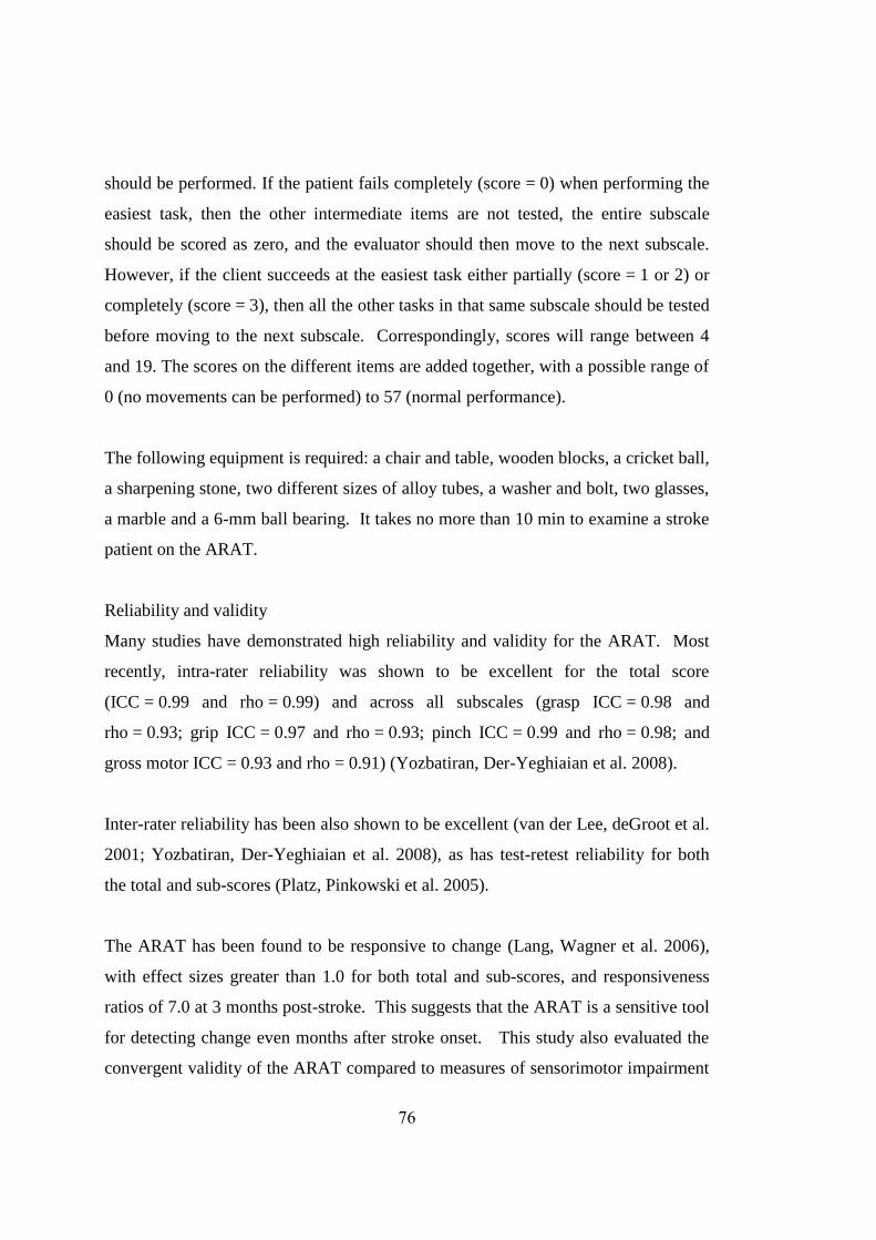
76
should be performed. If the patient fails completely (score = 0) when performing the
easiest task, then the other intermediate items are not tested, the entire subscale
should be scored as zero, and the evaluator should then move to the next subscale.
However, if the client succeeds at the easiest task either partially (score = 1 or 2) or
completely (score = 3), then all the other tasks in that same subscale should be tested
before moving to the next subscale. Correspondingly, scores will range between 4
and 19. The scores on the different items are added together, with a possible range of
0 (no movements can be performed) to 57 (normal performance).
The following equipment is required: a chair and table, wooden blocks, a cricket ball,
a sharpening stone, two different sizes of alloy tubes, a washer and bolt, two glasses,
a marble and a 6-mm ball bearing. It takes no more than 10 min to examine a stroke
patient on the ARAT.
Reliability and validity
Many studies have demonstrated high reliability and validity for the ARAT. Most
recently, intra-rater reliability was shown to be excellent for the total score
(ICC = 0.99 and rho = 0.99) and across all subscales (grasp ICC = 0.98 and
rho = 0.93; grip ICC = 0.97 and rho = 0.93; pinch ICC = 0.99 and rho = 0.98; and
gross motor ICC = 0.93 and rho = 0.91) (Yozbatiran, Der-Yeghiaian et al. 2008).
Inter-rater reliability has been also shown to be excellent (van der Lee, deGroot et al.
2001; Yozbatiran, Der-Yeghiaian et al. 2008), as has test-retest reliability for both
the total and sub-scores (Platz, Pinkowski et al. 2005).
The ARAT has been found to be responsive to change (Lang, Wagner et al. 2006),
with effect sizes greater than 1.0 for both total and sub-scores, and responsiveness
ratios of 7.0 at 3 months post-stroke. This suggests that the ARAT is a sensitive tool
for detecting change even months after stroke onset. This study also evaluated the
convergent validity of the ARAT compared to measures of sensorimotor impairment

77
(e.g. light touch sensation, pain, elbow joint spasticity, upper extremity strength), to
kinematic measures (e.g. reach and grasp), to the Functional Independence Measure
(FIM), and to the National Institutes of Health Stroke Scale (NIHSS). At all 3 time
points, ARAT scores related to comparative measures, and the ARAT was therefore
found to be a valid measure of upper extremity functional limitation post-stroke.
Clinically significant difference
The ARAT scores is a continuous measure, with no categorical cutoff scores.
Therefore the score obtained at the ARAT does not allow classifying scores into
categories such as normal, mild limited, or severely limited. There are published
normative values available for stroke patients (Kwakkel, Wagenaar et al. 1999;
Parry, Lincoln et al. 1999; van der Lee, Wagenaar et al. 1999), and a 10% difference
in total score represents a minimally important clinical difference (van der Lee,
Beckerman et al. 2001; van der Lee, Leo et al. 2002).
Justification
Up to fifty percent of stroke patients who experience initial upper limb impairment
post-stroke will still have impairment up to four years on (Broeks, Lankhorst et al.
1999), and the management of this pain is multidimensional (Zeferino and Aycock
2010). The ARAT is designed for evaluation of both sides of patients with cortical
injuries, helping to obtain a more total description of the upper extremity function
than investigation of only the hemiplegic side. From the point of view of dependency
it is necessary to know whether the patient has unlimited function on the non-
affected side (Hsieh, Hsueh et al. 1998). It is quick and easy to use and assesses both
proximal and distal strength as well as dexterity. This makes it preferable to other
tools such as the nine-hole peg test (finger dexterity) or grip strength (distal strength)
that focus on only one component of arm-hand function.

78
4.4.2 Behaviour Mapping
Development, indication and description; Reliability and validity
See Chapter 2: Pilot Studies
Justification
Behaviour mapping was used in order to observe if there were any differences
between where and how patients with stroke spent their time in a SU, compared with
three GRUs.
4.4.3 Berg Balance Scale (BBS)
Development, indication and description
This scale was originally developed for older adults as a measure of dynamic balance
performance (Smith, Hembree et al. 2004) through a process that used interviews
with rehabilitation professionals and individuals with balance deficits to generate a
pool of 38 balance items. Items were then excluded systematically on the basis of
perceived usefulness and clarity, consideration of the internal consistency and
examination of reliability, until 14 tasks remained (Berg, Wood-Dauphinee et al.
1989).
Since the introduction of the tool in the early 1990’s the BBS has been used for a
variety of functions, including an evaluative manner in projects examining the
effectiveness of different interventions (Stevenson 2001). The multiple-item format
of the assessment provides the clinician with additional insight into the possible
aetiology for the balance disturbance (Smith, Hembree et al. 2004) including the
ability to tolerate the internally produced perturbation to standing balance associated
with forward arm flexion (Stevenson, Garland et al. 1996) and its use for the
assessment of standing balance in subjects with hemiparesis from stroke is therefore
supported (Appendix 15).

79
Used widely by physiotherapists, this series of 14 static and dynamic observable
balance tasks is scored on a 5-point ordinal scale (0-4) according to the quality of
performance, the time taken to complete the skill or maintain a specific posture
(Smith, Hembree et al. 2004). Scores therefore may range from 0-56 with 56 being
the maximal score possible. Equipment requirements include a chair with and
without arms, a 15cm stepstool, a 30cm ruler, a stopwatch, and a slipper.
Reliability and validity
The initial examination of reliability resulted in a calculated inter-rater ICC=0.98 and
intra-rater ICC=0.97 for the total BBS score (Berg, Wood-Dauphinee et al. 1989).
Studies have also shown excellent internal consistency and both inter-rater and intra-
rater reliability (Berg, Wood-Dauphinee et al. 1995) in elderly stroke patients. It is
also sensitive to change (Salbach, Mayo et al. 2001; English, Hillier et al. 2006) and
demonstrates large effect size (English, Hillier et al. 2006). In terms of validity, it
has been found to correlate with the Barthel Index and the balance subscale of the
Fugl-Meyer and the Timed Up and Go Test (Berg, Wood-Dauphinee et al. 1992),
and the Functional Independence measure (Wee, Bagg et al. 1999).
Minimal detectable and clinically significant difference
In a study of 48 subjects receiving inpatient rehabilitation after stroke, the minimal
detectable change score analysis suggested that a change of ±6 BBS points is
necessary to be 90% confident of genuine change (Stevenson 2001). A score greater
than 45 is clinically associated with a lower risk of falling (Berg, Wood-Dauphinee
et al. 1992), and normative values have been published (English, Hillier et al. 2006).
Justification
Impaired balance is a common characteristic of patients with stroke, secondary to
neurological impairment in strength and tone and/or the co-ordination of both fine
and gross movement that may be compounded by the effect of increasing age
(Winter, Patla et al. 1990). Sensory impairment (both central and peripheral) may

80
also result in balance disturbance. Because the BBS is a sensitive and valid measure
of balance that has been widely published, it was chosen in order to identify
differences in outcome between the two types of facilities.
4.4.4 Chedoke-McMaster Postural Control & Shoulder Impairment Scores
Development, indication and description
The Chedoke-McMaster Stroke Assessment is a performance-based measure that
consists of two inventories: the Impairment Inventory and the Activity Inventory. It
consists of specific tasks that classify the stage of motor control (1-7) across all 6
areas of function from limb movement to trunk control and shoulder pain
(Appendices 16 and 17).
This scale is utilised as both a clinical and a research tool that can discriminate
among subjects and evaluate patient outcomes. The measure has three overall
purposes: 1) to stage motor recovery to classify individuals in terms of clinical
characteristics, 2) to predict rehabilitation outcomes, and 3) to measure clinically
important change in physical function (Gowland, Stratford et al. 1993).
The minimum score for the overall Impairment Inventory is therefore 6 and the
maximum score is 42 (Gowland, Stratford et al. 1993). In addition, each area of
assessment has been independently validated. The 7-point scale for shoulder pain is
based on pain severity and 7-point scale for postural control defines recovery over 7
stages (Stage 1 relating to poor function and stage 7 relating to normal function).
The scale is predicated on the knowledge that motor recovery progresses in discrete
phases that reflect the degree of neurological impairment and recovery (Gowland,
Stratford et al. 1993).
Reliability and validity
The Chedoke-McMaster Stroke Assessment yields both reliable and valid results. In
a study of 32 subjects from a stroke rehabilitation unit, Gowland et al (1993)

81
assessed the intra-rater, inter-rater, and test-retest reliabilities of the impairment and
disability inventories of the Chedoke-McMaster Stroke Assessment. Reliability
coefficients for the total scores ranged from 0.97 to 0.99. Construct and concurrent
validities were studied by examining the correlations between this and other
measures. Correlations were found to be greater than 0.60, with the impairment
inventory total score was found to correlate with the Fugl-Meyer Test (r = 0.95, p <
0.001) and the disability inventory with the FIM (r = 0.79, p < 0.05) (Gowland,
Stratford et al. 1993).
Justification
Both the shoulder subscale score and the postural control subscale scores were
chosen in view of the fact that these domains were not covered in any other chosen
outcome measures and are accepted as a common clinical focus in the rehabilitation
of stroke patients.
4.4.5 European Quality of Life-5 Dimension visual analogue scale (EQ5D VAS)
Development, indication and description
This tool measures health-related quality of life (HRQoL) and was developed and
validated in Europe (EuroQol 1990). It describes health status according to five
dimensions: mobility, self-care, usual activities, pain/ discomfort and
anxiety/depression. Each dimension has three levels, namely, "no problems", "some
problems" and "severe problems". This yields 243 potential combinations of health
states across the five dimensions. Besides the five dimensions, the EQ-5D consists
of a visual analogue scale (EQ5D VAS) ranging from 0 (worst imaginable health
state) to 100 (best imaginable health state).
It is applicable to a wide range of health conditions and treatments and provides a
simple descriptive profile and a single index value for health status that can be used
in the clinical and economic evaluation of health care. One limitation is that it could

82
be insensitive to changes that are considered clinically or socially significant,
therefore it is not the focus of the economic analysis.
Interpreting data
Data can be presented in various ways. The digits for 5 dimensions can be combined
in a 5- digit number describing the respondent’s health state (the numerals 1-3 have
no arithmetic properties and should not be used as a cardinal score, and a total of 243
unique health states may be defined in this way); the EQ VAS may be reported as an
overall self-rated health status; or results from the descriptive system may be
presented as a weighted index.
Justification
The EQ5D VAS (Appendix 18) was chosen for the present study in order to identify
differences in outcome between the two types of facilities as it was cognitively
undemanding and took only a few minutes to complete.
4.4.6 Functional Independence Measure (FIM)
Development, indication and description
The FIM (Appendix 19) was originally developed by a national taskforce following a
recommendation in 1984 from the American Congress of Rehabilitation Medicine
and the American Academy of Physical Medicine and Rehabilitation (Keith, Granger
et al. 1987), and is part of the Uniform Data System for Medical Rehabilitation
(UDS) which also contains demographic, diagnostic, impairment groups, length of
hospital inpatient stay and cost information (Fricke 1993); (Heinemann, Linacre et
al. 1993).
The scale consists of 18 items each assessed on 7 levels which, when summed, may
be used to estimate a person’s need for assistance (burden of care) or resource cost of
disability. The scale is anchored by extreme ratings of total assistance (1) and
complete independence (7), and considers extent of assistance, supervision, and use

83
of adaptive equipment (Heinemann, Linacre et al. 1993). Possible scores range from
18 to 126. In addition to a total score, the FIM provides two domain scores (motor
and cognitive), six subscale scores (self-care, sphincter control, transfers,
locomotion, communication and social cognition), and 18 individual item scores
(Guide87). It does not include domestic or community ADL (Fricke 1993).
Though each FIM item contributes its own, unique information, it is impractical
clinically, and burdensome analytically to allow each item to act as a separate
measurement device. The FIM was intended to quantify one unambiguous disability
indicator by combining its 18 items to yield a summary score.
Reliability and validity
The FIM has demonstrated no ceiling effect when compared with other measures
(van der Putten, Hobart et al. 1999) and is sensitive to change (Dromerick, Dorothy
et al. 2003) and has acceptable levels of reliability (Ottenbacher, Hsu et al. 1996,
Fricke, 1993 #433, Granger, 1990 #456); and validity (Dodds 1993; Kelly, Furie et
al. 2003). It has also demonstrated construct validity with the BI (Gosman-Hedstrom
and Svensson 2000).
Indications for using tool
The FIM represents the “burden of care” for the care provider, whether the provider
be spouse, family or institution (Granger and Hamilton 1990). It is designed for use
by any discipline, is suitable for a wide range of diagnoses (Fricke 1993) and gives
rehabilitation providers a common language with which to discuss disability in terms
of functional activities or activities of daily living (Black, Soltis et al. 1999).
Although it does not measure more specific functional skills, such as fine motor
ability, speed, and ease of task completion, or quality of task execution (Dodds
1993), as a generic assessment tool, it may serve several purposes, ranging from
outcomes research to program evaluation. Scores from the FIM are commonly used

84
to predict and demonstrate rehabilitation success (Sandstrom, Mokler et al.
1998);(Oczkowski, Barreca et al. 1993).
Clinically significant differences
While no recommendations exist for what constitutes a clinically significant change
on the FIM, a 10-point improvement in raw score decreases by almost 50 percent the
time required to care for a group of stroke patients in the community (Granger,
Cotter et al. 1993).
Admission FIM scores of less than 40 have been associated with less change in FIM
score over time, high level of impairment and disability, and heavy “burden of care”
necessitating institutionalization on discharge (Ween, Alexander et al. 1996,
Oczkowski, 1993). At the other end of the spectrum, stroke patients with an
admission FIM greater than 80 almost universally are discharged to their own home
(Black, Soltis et al. 1999). An admission FIM of 60 or more was associated with a
higher probability of functional improvement during rehabilitation. (Ween,
Alexander et al. 1996). Admission FIM scores above 96 have also demonstrated
little change over time (perhaps relating to a ceiling effect), and correspondingly low
levels of impairment and disability, and discharge to home (Oczkowski, Barreca et
al. 1993). Overall, those mid-range patients with admission FIM scores between 36
and 96 changed the most in their FIM scores, had moderate impairment and
disability, and destination on discharge was more difficult to predict (Oczkowski,
Barreca et al. 1993). It may be important to consider that age has shown a significant
but small effect on functional outcome when outcome is measured as FIM score
(Bagg, Pombo et al. 2002).
Justification
A measure of ADL was needed, as they have been found to be more useful in
treatment trials (Dromerick, Dorothy et al. 2003) due to their sensitivity to change in
disability compared with other global measures of health. The FIM was chosen as

85
the primary measure to assess differences in functional outcome between the two
different types of facilities in the SRO study, as a sensitive, inclusive, (Granger,
Cotter et al. 1993) and responsive ADL measure.
4.4.7 Hospital Anxiety and Depression Scale (HADS)
Description and development of tool
The Hospital Anxiety and Depression Scale (HADS) was specifically designed to be
a brief self-assessment tool for use in patients with somatic co-morbidity, and limited
to the two most common aspects of neurosis presenting in hospital practice: anxiety
and depression (Zigmond and Snaith 1983). It takes only 2 to 5minutes to complete,
and consists of 7-item subscales for both depression and anxiety (Aben, Frans et al.
2002). Items were selected based solely on the psychic symptoms of neurosis rather
than emotional or physical disorders (Zigmond and Snaith 1983) such that symptoms
such as dizziness, headaches, insomnia and fatigue were excluded as well as those
relating to serious mental disorders (Bjelland, Dahl et al. 2002). Each item has a 4-
point response category (0-3) so that the possible scores range from 0-21 for anxiety
and 0-21 for depression (Appendix 20).
Reliability and Sensitivity of tool
The tool has demonstrated excellent internal consistency and sensitivity (Olsson,
Mykletun et al. 2005, Aben, 2002, Zigmond, 1983) and validity has been well
established across different countries and disorders (Snaith 2003).
Indications for using tool
The HADS was specifically designed to facilitate the large task of detection and
management of psychiatric disorders in patients under investigation and treatment in
medical and surgical departments (Zigmond and Snaith 1983). A review of the
literature in 2002 identified 747 papers that had used HADS to address research
questions. Overall it was found to perform well in assessing the symptom severity
and the presence of anxiety disorders and depression in both somatic, psychiatric and

86
primary care patients and in the general population (Bjelland, Dahl et al. 2002).
Specifically in patients with stroke, the total HADS score is accurate in detecting
cases of post-stroke depression (Aben, Frans et al. 2002).
The internal consistency of the scale was high, and only slightly decreased in a
subset of patients with low MMSE scores (≤ 23). In this group, non-response was
clearly higher. This suggests that, overall, stroke patients are able to fill out these
scales reliably, but problems arise in the more cognitively impaired patients (Aben,
Frans et al. 2002).
Clinical thresholds
For the depression subscale, a score of 7 or less for non-cases, 8-10 for doubtful
cases and 11 or more for definite cases; for the anxiety subscale, the same score
ranges fit (Zigmond and Snaith 1983). Aben et al (2002) found that the optimal
cutoff point for the depression subscale was a score of 8 (sensitivity: 73.1,
specificity: 81.6), and the anxiety subscale was a score of 5 (sensitivity: 91.7,
specificity: 56.1) (Aben, Frans et al. 2002). Screening for both major and minor
depression did not change the overall accuracy of the instrument, however the
optimum cutoff score decreased by one point to 7 on the depression subscale (Aben,
Frans et al. 2002). Olssen (2005) replicated these optimal cut off scores in a general
practice caseload with sensitivity 0.89, specificity .75 for anxiety and sensitivity 0.80
and specificity 0.88 for depression (Olsson, Mykletun et al. 2005).
The scores on the subscales are indicators of the severity of the depression and
anxiety respectively (r=0.70 and r=0.74) with p<0.001. The subscales are thought to
assess different aspects of mood disorder (Zigmond and Snaith 1983),
Justification of choice of outcome measure
The HADS is a widely used screening tool in medically ill patients (Tang, Wong et
al. 2007) and seems to have at least as good screening properties as similar more

87
comprehensive instruments used to identify anxiety and depression (Bjelland, Dahl
et al. 2002). Although the SRO study cohort was medically stable, because it is a
simple to use and is well validated in stroke, often demonstrating the presence of
significant anxiety and depression within this cohort (Robinson, Starr et al. 1984;
Robinson, Bolduc et al. 1987; Astrom 1996), it was deemed suitable for study.
4.4.8 MOS 36-Item Short Form Health Survey (SF-36)
Description and development of tool
The SF-36 (Appendix 22) is the most widely used generic instrument for measuring
quality of life (de Haan 2002) and for which Australian normative data are available
(McCallum 1995). It provides a comprehensive, psychometrically sound, and
efficient way to measure health from a patient’s point of view by scoring
standardized responses to standardized questions.
The SF-36 comprises eight health scales: physical functioning (10 items), role
limitations – physical (4 items), bodily pain (2 items), general health (5 items),
vitality (4 items), social functioning (2 items), role limitations – emotional (3 items),
and mental health (5 items). Two core dimensions of health, physical and mental can
be derived from these eight scales. There is also a single separate item that is used to
assess any change in health from 1 year before (Anderson, Laubscher et al. 1996).
Reliability and Sensitivity of tool
The tool has been translated into numerous languages, and the validity of the 8
subscales is confirmed in general populations and in a wide variety of patient groups
in more than 2000 articles including stroke (McCallum 1995, Brazier, 1992; de Haan
2002). Moreover, when compared to the ceiling effect of the BI, the physical
functioning scale of the SF-36 showed a uniform distribution of scores that reflected
a broader range of physical disability (Anderson, Laubscher et al. 1996).

88
Indications for using tool
The focus of the SF-36 is on the subjective perception of health (Carod-Artal, Egido
et al. 2000). This is in the context that isolated measures of physical health domains
may not adequately reflect the full impact of the long-term disability in stroke that a
quality of life measurement may provide. These additional subjective components
show the effect of survivor attitudes, health beliefs, and social interaction.
Normative values and clinically significant difference for patients with stroke
Compared to the general population, overall quality of life outcomes reported for
patients with stroke vary. A Scottish study reporting on a large sample of mildly
disabled patients with little cognitive impairment and using the SF-36 measure
described outcomes significantly worse than healthy individuals (Bugge, Hagen et al.
2001) and these findings are consistent with other studies using different outcome
measures (Haacke, Althaus et al. 2006). Conversely, a more recent Australian study
reported outcomes close to healthy patients (Anderson 2000; Anderson 2000).
Normative data for the SF36 is available across versions and across countries
(Jenkinson, Coulter et al. 1993), including Australia, and these data were utilised in
the analyses of this study.
Justification of choice of outcome measure
In the current study, no other measure combined quality of life with physical
functioning, and given the well established validity and reliability of this measure, as
well as the availability of normative values, it was chosen as an outcome measure for
the SRO study.
Pre-testing of the authorized Australian version of the SF-36 among patients with
stroke showed that a high proportion were unable to self-complete the questionnaire
because of visual problems, confusion, and physical disability (McCallum 1995).
Further, in a study of geriatric day unit stroke and musculoskeletal patients found
that administration of SF36 by an interviewer is essential to obtain meaningful

89
results in older people with poor physical health (Fowler, Congdon et al. 2000).
Consequently, although patients were asked to self-complete questionnaires that
were posted to them prior to follow-up if it was possible for them to do so, these data
were also then checked for completeness and ambiguity during face-to face interview
by the assessor.
4.4.9 Ten-metre walk test
Description, development, reliability and sensitivity of tool
Gait speed is a continuous measure with a natural zero, and published target gait
speeds provide meaningful goals for treatment planning and clinical decision making
(Salbach, Mayo et al. 2001). It has been found to be reliable (Flansbjer, Holmback et
al. 2005) and sensitive to change (English, Hillier et al. 2006).
It has been shown that the 6 and 12minute walk tests (6MWT, 12MWT) and self-
paced gait speed over 10metres are all highly correlated with one another all also
related to the severity of impairments. Stroke-specific impairments including
weakness of the affected hip flexors and knee extensors are the major limitations to
the distance walked in individuals with stroke (Eng, Chu et al. 2002). Gait velocity
is less likely to show a ceiling effect than other measures given that it is only limited
by the physical capacity of the individual and not the structure of the test (Salbach,
Mayo et al. 2001).
Although various instructions have been published (Hsu, Tang et al. 2003), those
used in the current study were: “I am going to measure your comfortable walking
speed. When I say ‘go’, walk in a straight line at a pace which is safe and
comfortable for you, until you reach the second pylon” (Salbach, Mayo et al. 2001).
Indications for using tool
The retraining of gait is a major focus in the rehabilitation of patients with stroke
(Kim, Eng et al. 2004), with literature suggesting that the majority of patients will

90
walk again (Friedman 1990). Contrary to the gait of healthy individuals, which is
fairly consistent in pattern across subjects, marked variation in gait patterns has been
noted in patients with stroke (Kim, Eng et al. 2004). Gait performance in patients
with stroke is characterized by slower gait velocity and residual spatial and temporal
left-right asymmetry, compared with that of healthy adults (Hsu, Tang et al. 2003).
Achieving normal gait patterns and speed are usually the ultimate goals of gait
training (Lin, Yang et al. 2006), with gait speed predicting functional mobility and
socialization (Jette, Keysor et al. 2005).
Clinically significant difference
Normative values have been published for both patients with stroke (Perry, Garrett et
al. 1995) and non-stroke (Bohannon 1997) with minimal clinically important
difference for acute stroke equal to 0.16m/s (Oberg, Karsznia et al. 1993; Tilson,
Sullivan et al. 2010).
Justification of choice of outcome measure
As the retraining of walking is a major focus in the rehabilitation of persons with
stroke (Kim, Eng et al. 2004), it was important to include a measure of gait
performance to assess differences between facilities. The measurement of gait speed
was chosen as it could be performed quickly and without equipment or space.

91
CHAPTER 5
5.1 Introduction
The primary aim of the Stroke Rehabilitation Outcome (SRO) trial was to determine
whether patients identified as having some potential to return to independent living
as well as the ability to cope with intensive rehabilitation have better quality of life
and functional outcome following rehabilitation at the stroke unit at Royal Perth
Hospital–Shenton Park Campus (SPC SU) compared with a stroke unit located
within a geriatric rehabilitation unit (GRU). The secondary aims were to determine
whether there was a difference in the average amount of rehabilitation received by
sub-acute stroke patients in the two different types of units (SPC SU versus GRU),
and to make a preliminary assessment of the indirect costs associated with each type
of facility based on length of hospital stay and amount of therapy input.
The SRO trial was initially designed as a randomized controlled trial (RCT). It was
planned to recruite patients at the weekly acute stroke unit (ASU) team meetings at
Royal Perth Hospital – Wellington Street Campus (RPH-WSC). The protocol for the
proposed RCT is described in part 5.2 of this chapter. Despite comprehensive pilot
work suggesting the feasibility of recruiting the required number of subjects in an
appropriate time frame, recruitment was much more difficult than anticipated.
Difficulties predominantly related to therapeutic equipoise made it impossible to
proceed according to the intended randomized protocol and the study was revised
into an observational trial, described in part 5.3 of this chapter. The aims of the
observational study remained unchanged from those of the original RCT.
Methods

92
The primary Null Hypothesis was that there was no difference in functional outcome
and quality of life of patients with stroke who met the inclusion criteria and received
rehabilitation at SPC SU compared with a GRU.
5.2 Randomized controlled trial (RCT) protocol
5.2.1 Study design
The study was designed as a RCT whereby acute patients with stroke would be
randomized into either rehabilitation group. Baseline data (Appendix 27) were to be
collected from medical records, and inpatient length of stay (at both RPH-WSC and
the subsequent rehabilitation facility) and amount of therapy, and functional status
and quality of life would be measured at 6 and 12 months post-randomization
(Figure 5.2.1a).
The study was approved by the ethics committees of Royal Perth Hospital (reference
2003/022), Bentley Health Service (reference 6/02), Mercy Hospital (reference
MC/ad) and Swan Health Service (reference 7.23) and Curtin University (reference
HR 192/2002). Copies of approval letters can be found in Appendix 28. It was not
registered with the clinical trials registry, as this was not a requirement at the
inception of the study.

93
Figure 5.2.1a: Study design of SRO RCT
SRO= Stroke Rehabilitation Outcome; RPH - WSC= Royal Perth Hospital – Wellington Street
Campus; SPC SU= Royal Perth Hospital – Shenton Park Campus stroke unit; GRU=Geriatric
rehabilitation unit
RPH-WSC patients with stroke
deemed suitable for randomization
Informed consent obtained
Baseline data collected
Randomized stratified assignment into
study: age, gender, stroke type and site
SPC SU rehabilitation
GRU rehabilitation
6 and 12 months post-recruitment into study,
follow-up subjective and objective assessments
by independent assessor
6 and 12 months post-recruitment into study,
follow-up subjective and objective
assessments by independent assessor

94
5.2.2 Study population
5.2.2.1 Inclusions and exclusions
All patients presenting to RPH-WSC ASU with a diagnosis of recent stroke requiring
hospitalisation and sub-acute rehabilitation were considered for inclusion into the
study. Patients were to be invited into the study when the attending physician had no
evidence to suggest advantages of SPC SU versus GRU rehabilitation. Patients
younger than 60years of age would be excluded as they would not normally be
admitted to geriatric facilities. Older patients with no potential to return to some form
of independent living or an inability to cope with intensive rehabilitation would also
be excluded, as would those with pre-existing dementia. Those patients with any
severe co-morbid condition that could cause death within one year; an inability to
understand English well enough to complete assessments or sign consent; or those
with geographic or social factors that were expected to impede study participation
were also to be excluded.
5.2.2.2 Recruitment and patient consent
Patients were considered for inclusion in the study at weekly ward meetings
involving all frontline staff at RPH-WSC ASU. Having been identified by the team,
the senior medical registrar would explain the study to the patient and invite them to
participate. Those potentially interested would be asked to read the Patient
Information Sheet (Appendix 26), and given the opportunity to ask questions.
Written consent would then be obtained from those wishing to volunteer (Appendix
26).
5.2.2.3 Power and sample size
Published data on the SF36 is widely available so this variable was used for a priori
power calculations. Based on a within-group standard deviation of up to 18 points
for 6 of the 8 SF36 domains, 50 patients per group would provide approximately

95
80% power to detect a 10 point difference in most of the SF36 domains if statistical
significance was inferred at a 2-tailed p< 0.05. A difference of this magnitude is
considered clinically meaningful by the SF36 developers. In addition, an allowance
was made for a 16% dropout or loss to follow-up.
5.2.2.4 Feasibility
Pilot data related to the number of patients with stroke presenting to RPH-WSC
(Chapter 3, section 3.1.2) suggested that it would be feasible to recruit this number of
stroke patients within 18 months.
5.2.3 Study procedure
5.2.3.1 Baseline data collection
Baseline data (Appendix 27) would be collected from the medical notes of all
patients after patient consent and prior to randomization, hence assessor blinding was
not an issue. Where possible, these data would be derived from routine clinical
assessment by RPH-WSC medical and AHP staff and collated for the purposes of
this study. Any data not available from this source was collected separately for the
study. The purpose of these data was to establish the underlying level of disability
and compare groups for comparability. They would also be valuable as covariates in
data analysis. Data included demographic information, social circumstance, past
medical history, pre-morbid function, stroke classification and functional status at the
time of consent into the study. Functional status was measured using measures
previously outlined in Chapter 4. They included the Barthel Index (BI) score,
Rankin score, Scandinavian Stroke score (SSS), Mini mental state examination
(MMSE), 10m-walk time, Berg balance scale score (BBS) and Chedoke-McMaster
tests. The number of days between admission and consent was also recorded.

96
5.2.3.2 Randomization
Patient volunteers were to be randomized to continue their rehabilitation at either the
SPC SU or the GRU geographically located closest to their home (Mercy, Swan or
Bentley Hospitals). In order to be considered for randomization, patients would need
to be reviewed and accepted for admission by medical staff from both relevant
rehabilitation facilities. Those patients accepted for both facilities would then be
randomized to determine their rehabilitation destination. To ensure groups were
balanced for prognostic factors, randomization was to be stratified using the
minimization method balancing groups for patient age (> or ≤ 75 years), gender (M
or F), and stroke severity as defined by dependency at the time of randomization
(mild stroke - BI 10-20/20 or moderate to severe stroke – BI <10/20). This dynamic
process would be administered by the clinical coordinator who did not have clinical
contact with the participants and was not to be involved with recruitment.
Recruitment was to take place prior to group assignment. Group assignment would
not be simple to predict when stratifying across 3 factors.
5.2.3.3 Treatment procedure and documentation
In this pragmatic trial, all treatments received would be those considered standard for
the individual facility, administered as usual by registered rehabilitation/therapy
health professionals.
5.2.3.4 Follow-up procedure and outcome data collection
Six and twelve months following their transfer from RPH-WSC ASU, patients would
attend follow-up outpatient appointments at neutral rooms where objective and
subjective assessments would be undertaken by an independent assessor who would
not be informed about which rehabilitation facility the patient had attended. The
blind independent assessor was to be an experienced physiotherapist who had never
worked at RPH-WSC previously to ensure previous knowledge of the patients would

97
not confound assessments. The duration of each assessment visit was planned to be
approximately 1 hour.
5.2.4 Implementation of protocol
Recruitment was commenced in late January 2003 by a research collaborator who
planned to recruit the caregivers of the study patients for a concurrent study
exploring the role of the caregiver in stroke recovery. This collaborator attended the
medical and allied health team weekly ward meetings where ongoing rehabilitation
and discharge plans of patients were discussed. Over 14 weeks, a total of 45 patients
were age appropriate for the trial (Figure 5.2.4.1). Of these, 24 patients were
excluded (13 discharged home; 2 transferred to country hospitals; 4 transferred to
other medical specialties within RPH-WSC; 3 discharged directly to permanent care;
and 2 with poor understanding of the English language) and 10 had no discussion of
discharge planning at the team meeting. This left 11 potential patients suitable for
randomization, however none were of these were able to be recruited. The reasons
for this included clinical staff deciding they should be directed to one facility in
preference to the other (for example, one patient who made “better than expected
early improvements” at RPH-WSC being directed to SPC SU where they considered
rehabilitation would be more appropriate) and patient or family preference (for
example, where the geographical location of a GRU enabled an elderly spouse to
visit his wife more frequently).

98
Figure 5.2.4.1: Initial RCT recruitment period
Age appropriate patients with stroke n=45
Excluded
n=24
Home
n=13
Country hospital
n=2
Managed by
non-ASU
n=4
Discharged to nursing home directly n=3
Poor English
n=2
Potential patients
n=21
No discharge plan discussed
n=10
Considered for randomisation
n=11
Transferred to SPC SU
n=7
Transferred to GRU
n=4

99
Recruitment was suspended in May 2003 in order that the primary investigator could
meet and discuss the randomization process with key RPH-WSC medical and allied
health staff. Following meetings with key staff, it was decided that it may be more
feasible to amend the study design and recommence the study using an observational
rather than a randomized design. Using an amended design, all patients with stroke
who met the inclusion criteria would be invited to participate and those who
volunteered would be followed up at 6 and 12 months post-stroke no matter which
type of rehabilitation facility they had attended. However, as this approach was less
rigorous than the original randomized design, it was decided that one more trial of
RCT recruitment should be undertaken before commencing an observational study.
At about the same time the collaborator who had been attending the ward meetings
moved and could no longer continue her involvement with the project. Therefore,
during the second attempt at recruitment, the principal investigator attended meetings
and promoted the RCT.
After the Christmas 2003 break (where staffing was transient and discontinuous), the
second period of recruitment began in February 2004. Over 12 weeks, the principal
investigator attended all weekly ASU ward team meetings. During this time 26 new
patients were age appropriate for the trial (Figure 5.2.4.2). Of these, 12 patients were
excluded (8 discharged home; 1 transferred to country hospitals; 1 transferred to
other medical specialties within RPH-WSC; 2 discharged directly to permanent care)
and 3 had no discussion of discharge planning. This left 11 potential patients suitable
for randomization, however none were able to be recruited. Once again, no patients
could be recruited based on clinical decision making or family preference for one
facility over another.

100
Figure5.2.4.2: Second RCT recruitment period
Age appropriate patients with stroke
n=26
Excluded
n=12
Home
n=8
Country hospital
n=1
Managed by non-ASU
n=1
Discharged to nursing home directly
n=2
Potential patients
n=14
No discharge plan discussed
n=3
Considered for randomisation
n=11
Transferred to SPC SU
n=4
Transferred to GRU
n=7

101
5.2.5 Discussion
After a total of 6 months of recruitment no patients had been recruited for the study.
Attendance at the weekly team meetings and discussions with the rehabilitation staff
revealed that there were strong preconceived ideas about the rehabilitation facilities
that prompted staff to form opinions about the “best fit” rehabilitation facility for any
prospective patient. This led to reluctance by these staff to sanction randomization of
patients despite the lack of empirical evidence to inform decisions about which
patients should go to which facility. These preconceptions may or may not have been
held by incumbent staff at the SPC SU or GRUs (see Chapter 3, section 3.2.1), and
may or may not have been accurate. It should be recognized however that staff
attending the RPH-WSC ASU team meetings were very experienced in dealing with
patients with stroke.
Reasons offered for why patients should be referred to one or other facility included
that:
therapy was more intensive at SPC SU compared with the GRUs.
patients suitable for referral to SPC SU needed to have the capacity to cope
with more intensive therapy.
the patient’s capacity to cope with rehabilitation may be indirectly
proportional to the number of co-morbidities present.
patients needed to be motivated for best outcome at SPC SU.
some GRUs did not have appropriate AHP staffing (in particular speech
pathologists) to provide optimal treatment for some patients.
5.2.6 Conclusion
Despite support for the trial by the RPH-WSC ASU medical team, and full ethics
approval from all hospitals involved, no patients were successfully recruited into the
trial over two periods, lasting a total of 26 weeks. Ultimately, based on their clinical
experience and perceptions about the different rehabilitation facilities, AHPs at the
RPH-WSC ASU felt ethically obliged to recommend one or other rehabilitation

102
course for their patients despite the lack of objective evidence for these perceptions.
It was similarly ethically difficult for the author to influence staff in any way, given
their views. To a lesser extent, patients’ preference to be transferred to facilities
closer to home also had a negative effect on recruitment.
The inability to recruit subjects for randomization into the study required there to be
significant changes to the methodology. Although less rigorous, it was decided to
amend the study, changing from an experimental to an observational design. The
aims of the observational study remained unchanged from those of the original RCT.
5.3 Observational trial protocol
5.3.1 Study design
The study was an inception cohort study whereby patients were selected based on
their age, absence of dementia and their acceptance by incumbent medical staff for
rehabilitation transfer at either SPC SU or a GRU (Figure 5.3.1a).
The amended study design was submitted to and approved by the ethics committees
of Royal Perth Hospital (reference 2003/022), Bentley Health Service (reference
6/02), Mercy Hospital (reference MC/ad) and Swan Health Service (reference 7.23)
and Curtin University (reference HR192/2002).

103
Figure 5.3.1a: Study design of SRO observational trial
SRO= Stroke Rehabilitation Outcome; RPH - WSC= Royal Perth Hospital – Wellington Street
Campus; SPC SU= Royal Perth Hospital – Shenton Park Campus stroke unit; GRU=Geriatric
rehabilitation unit
All RPH—WS patients with acute stroke referred
with sub-acute rehabilitation
Patients
> 60 years without dementia
Patients
< 60 years or with dementia
Informed consent provided Excluded
Base line data collected
(demographic information, past medical history, stroke data, functional
ability)
Transferred to SPU
SU
Transferred to Bentley, Mercy or Swan
GRU
Data Collection
6 and 12 months post stroke
Data Collection
6 and 12 months post
stroke

104
5.3.2 Study population
5.3.2.1 Inclusions and exclusions
No adjustments were made to the inclusion and exclusion criteria (see 5.2.2.1) or
sample size (see 5.2.2.3).
5.3.2.2 Recruitment and patient consent
All patients from RPH-WSC with acute stroke requiring further rehabilitation were
identified and screened according to the standard referral procedures for the ASU
(Appendix 25). This system involved a written referral to the SPC SU or one of the
GRUs. Written referrals were followed up by the medical staff from the unit
specified and if the patient was deemed appropriate, transfer was arranged. For the
purposes of the study, all referrals were copied by the medical units receiving them,
and these referrals were collected and reviewed on a biweekly basis by a research
assistant employed for the study to identify patients suitable for the SRO trial.
A research assistant obtained informed written consent from all participants prior to
commencement of data collection (Appendix 26). All procedures were clearly
explained to each participant, and it was made clear that they had the right to
withdraw from the study at any time without prejudice to their ongoing medical care.
5.3.3 Study procedure
5.3.3.1 Baseline data collection
The same baseline data (Appendix 27) were collected as proposed for the RCT
design (see 5.2.3.1).

105
5.3.3.2 Transfer procedure
Participants were transferred to continue their rehabilitation at either the SPC SU or
the GRU geographically located closest to their home or the home of their primary
caregiver (within Bentley, Mercy or Swan hospital). If bed availability delayed
transfer to the rehabilitation unit, the number of days delayed was recorded as part of
baseline data in order to assess differences between facilities at this timepoint. If
transfer was delayed patients continued to receive usual care from staff in the ASU at
RPH-WSC while awaiting transfer as per normal practice. If patients awaiting
transfer recovered sufficiently at RPH-WSC ASU for direct discharge home, then
this was recorded. If patients were discharged home directly from RPH-WSC they
were excluded from the data set, as their outcome was no longer influenced by their
sub-acute rehabilitation stay.
5.3.3.3 Treatment procedure and documentation
All allied health professionals (AHPs) involved in the management of patients in the
trial had the opportunity to attend information sessions where they were made aware
of the study design and data collection. Ongoing education of new staff was
attended to as the need arose at each facility.
In this pragmatic trial, all treatments provided were standard for the individual
facility and administered as usual by incumbent AHPs. Participating patients were
provided with an AHP treatment diary (see Chapter 3, section 3.2.2), where AHPs
recorded the date, duration and location of all treatments received by that patient. In
addition, behaviour mapping was conducted for 2 consecutive days once every 2
months at each facility for the duration of the trial (see Chapter 7).
5.3.3.4 Follow-up procedure
The protocol for follow-up procedure remained as planned for the RCT design.
Patients were instructed not to disclose the facility where they received rehabilitation

106
to the assessor. Self-completed questionnaires were mailed to patients prior to these
appointments to allow completion ahead of time. Questionnaires were checked for
missing data at the time of the appointment.
5.3.4 Outcome measures
Health outcomes were selected in consultation with clinicians with recognized
expertise in the management of stroke, taking into account validity, reliability and
responsiveness in the stroke population. All chosen variables have been previously
discussed in Chapter 4 and are widely used in published studies. The independent
variable was the group assignment to either rehabilitation at the SPC SU or one of 3
GRUs. The dependant variables covered 6 domains:
5.3.4.1 Quality of life
MOS 36-Item Short Form Health Survey (SF-36)
European Quality of life 5 dimension visual analogue scale (EQ5D VAS)
Hospital Anxiety and Depression (HAD) scale
5.3.4.2 Function
Functional independence measure (FIM)
Berg balance scale score (BBS)
Ten metre walk test
Chedoke McMaster shoulder pain inventory
Chedoke McMaster postural control impairment inventory
Action Research Arm (ARAT) test
5.3.4.3 Treatment environment: behaviour mapping
Behaviour mapping was used to monitor and characterize the rehabilitation setting
and aspects of treatment. Pilot data (see Chapter 3, section 3.2.3) indicated that the

107
mapping data were sensitive to variations in the rehabilitation environment within
and between facilities.
5.3.4.4 Cost: Length of stay and AHP diary information
The length of inpatient stay within the rehabilitation facility
The frequency and duration of therapy: Information was generated from the
patient diaries. Pilot data (see Chapter 3, section 3.2.2) indicated that the
diaries were sensitive to variations in the way rehabilitation was administered
within and between facilities. These data were used to compare the treatment
frequency and duration at each type of facility.
5.3.4.5 Discharge outcome
Discharge destination
5.4 Statistical methods
Data were analysed using Statistical Package for the Social Sciences (SPSS®
version, IBM USA and other countries) and statistical significance was set at α=0.05.
Descriptive statistics were reported as counts (percentage) for categorical data and
for continuous data mean (SD) for normally distributed data or median (IQR) where
data were not normally distributed. Characteristics of patients at baseline were
compared between facilities using Pearson Chi–Square for categorical variables and
unpaired t-tests or Mann-Whitney tests as appropriate for continuous variables.
Multilevel linear models were used to compare rehabilitation outcomes between SPC
SU and GRU. Additional factors included as covariates in the models were age,
gender, and baseline Barthel Index score. Separate models were constructed for each
of the dependent variables.

108
Generalized estimating equations were used to compare rehabilitation outcomes
between SPC SU and GRU. Additional factors included as covariates in the models
were age, gender, baseline Barthel Index score and visit as a repeated factor.
Separate models were constructed for each of the 10 dependent variables but
adjustments for multiple comparisons were not implemented to reduce the risk of
Type II error (Perneger 1998; Feise 2002).

109
CHAPTER 6
6.1 Introduction
This chapter reports the results of the stroke rehabilitation outcome (SRO)
observational study that compared functional outcome of a group of stroke patients
receiving rehabilitation at a neurological stroke unit located at Royal Perth Hospital –
Shenton Park campus (SPC SU) with a group of patients receiving rehabilitation at a
geriatric rehabilitation unit (GRU) (at either Bentley, Mercy or Swan hospital) with 6
and 12 month follow-up. Ninety-four patients with stroke were recruited from the
Royal Perth Hospital – Wellington Street campus (RPH-WSC) acute stroke unit
between June 2004 and November 2006.
6.2 Patient flow
Since 1999/2000 when pilot data were first collected for the study (see Chapter 3,
section 3.1.2), data from RPH-WSC the annual number of patients discharged with
the diagnosis of stroke increased by almost 50 percent (Table 6.2a).
RESULTS

110
Table 6.2a: Annual number of patients discharged from RPH-WSC with the
diagnosis of Stroke
Year Number of patients
1999/2000 531
2000/2001 695
2001/2002 659
2002/2003 767
2003/2004 853
2004/2005 798
2005/2006 922
2006/2007 802
Although the SRO study recruited subjects for 30 months between July 2004 and
December 2006, only annual hospital data was available to the author. Thus, during
the 36 months between July 2004 and June 2007, 2522 patients with stroke presented
to RPH-WSC. Of these, 550 patients were assessed and treated at the ASU, the
remaining patients were treated in other areas of the hospital (Figure 6.2a).

111
.
Figure 6.2a: Inpatient destination of all patients with Stroke following admission to
RPH-WSC between July 2004 and June 2007
.
2522 patients admitted to RPH-WSC
with acute stroke
between June 2004 and July 2007
550 patients
cared for by
acute stroke unit
498 patients
cared for by
general medicine
259 patients
cared for by
geriatric medicine
456 patients
cared for by
neurology
507 patients
cared for
by neurosurgery
66 patients
cared for by
cardiology
186 patients
cared for by
other specialities

112
Figure 6.2b: Discharge Destination of all patients with Stroke following admission
to RPH-WSC between July 2004 and June 2007
Of the total cohort of 2522 patients, 370 died as inpatients. A further 1354 were
discharged directly home from the hospital, with or without domiciliary support and
under the care of their general practitioner. The remaining 798 were transferred to
other facilities (Figure 6.2b)
Of the 798 patients that were transferred to another hospital or institution between
July 2004 and June 2007, 524 patients with stroke transferred to the facilities
involved in this trial (Figure 6.2c: SPC SU n=250; GRUs n=274). The remaining
274 patients transferred to one of 14 other metropolitan hospitals (106 patients),
2522 patients admitted to RPH-WSC
with acute stroke
between June 2004 and July 2007
370 patients
deceased
1354 patients
discharged home,
with or without domiciliary care
798 patients
transferred
to other hospitals/ institutions

113
various nursing home/ hostels (93 patients), country hospitals (56 patients),
interstate/ overseas hospitals (8 patients) or hospice care (11 patients).
Of the 524 patients that transferred to the facilities included in the SRO trial, there
were 354 who were age appropriate (60 years of age or older) for recruitment into
the study (Figure 6.2c: SPC SU n=84; GRUs n=270). Of these, 94 consented to
participate (SPC SU n=22; GRUs n=72). This reflected an overall recruitment rate
of 26.5% (SPC SU 26%; GRU 27%).
6.3 Baseline Data
Patients referred to SPC SU were younger, more likely to be male, and have speech
abnormality, peripheral vascular disease and diabetes than those referred to GRUs.
Otherwise there were no significant differences between groups in demographic
(Table 6.3.1) admission characteristics (Table 6.3.2) or past medical history (Table
6.3.3) measured at baseline.

114
Figure 6.2c: Destination of patients with Stroke transferred to other facilities
following admission to RPH-WSC between July 2004 and June 2007, and
recruitment of age appropriate patients into the SRO trial.
798 patients admitted to RPH
with diagnosis of Acute Stroke
between June 2004 and July 2007
and transferred to other facilities
250 patients SPC SU
84 patients age approriate for SRO trial
(60 years of age or older)
22 patients consented for
SRO trial
(26%)
274 patients GRUs involved with SRO study
270 patients age appropriate for SRO trial
(60 years of age or older)
72 patients consented for
SRO trial
(27%)
274 patients to other facilities including other GRUs and
hospitals

115
Table 6.3.1: Demographic characteristics of patients (n=94)
Data presented as number (%) unless indicated; p value for Pearson Chi–Square; ^p value for
t-test; LAC= lacunar infarction; TAC= total anterior circulation infarction; PAC=partial
anterior circulation infarction; POC=posterior circulation infarction
Variable SPC SU
n=22
GRU
n=72
P value
Gender 0.009
Male 19 (86.4) 40 (55.6)
Female 3 (13.6) 32 (44.4)
Age, mean (SD) 63.95 (4.32) 77.19 (8.25) <0.001^
Living Arrangements Pre-Stroke 0.073
Alone 5 (22.7) 29 (40.3)
With Relative other than
partner
0 6 (8.3)
With Partner 17 (77.3) 37 (51.4)
Level of function Pre-Stroke 0.429
Independent 22 (100) 70 (97.2)
Dependent 0 2 (2.8)
Stroke Site 0.599
Right 9 (40.9) 34 (47.2)
Left 13 (59.1) 36 (50)
Brainstem 0 2 (2.8)
Stroke Pathology 0.685
Ischaemic 18 (81.8) 56 (77.8)
Haemorrhagic 4 (18.2) 16 (22.2)
Stroke Type 0.689
LAC 5 (22.7) 22 (30.6)
TAC 8 (36.4) 18 (25)
POC 1 (4.5) 6 (8.3)
PAC 8 (36.4) 26 (36.1)

116
Table 6.3.2: Admission status of patients (n=94)
Data presented as number (%) unless indicated; p value for Pearson Chi–Square; ^p value for
t-test; BI= Barthel Index; BBB= Berg balance score; CMM= Chedoke McMaster; MMSE= Mini
mental state examination; SSS=Scandinavian stroke score
Variable SPC SU
n=22
GRU
n=72
P value
BI total score, mean (SD) 38.86 (28.91) 30.70 (25.78) 0.211^
BBB mean (SD) 18.05 (18.17) 16.01 (13.92) 0.583^
CMM shoulder inventory, mean (SD) 3.68 (1.43) 3.42 (1.34) 0.513^
CMM postural inventory, mean (SD) 3.68 (1.43) 3.42 (1.34) 0.436^
MMSE score, mean (SD) 23.43 (6.69) 19.95 (7.20) 0.104*^
Rankin score, mean (SD) 3.91 (1.06) 4.18 (0.70) 0.165^
SSS – prognostic, mean (SD) 15.82 (4.93) 16.79 (3.71) 0.325^
SSS – long term, mean (SD) 23.59 (12.52) 26.20 (11.35) 0.361^
Abnormal language present 10 (45.5) 15 (20.8) 0.022
Abnormal speech present 17 (77.3) 48 (66.7) 0.346
Abnormal swallow present 5 (22.7) 22 (30.6) 0.478
Visual field loss/ diplopia present 3 (13.6) 15 (20.8) 0.453
Vertigo present 0 2 (2.8) 0.429
Sensory deficit arm or leg present 6 (27.3) 21 (29.2) 0.864
Weakness arm or leg present 20 (90.9) 63 (87.5) 0.663
Ataxia present 3 (13.6) 5 (6.9) 0.325
Incontinence present 0 7 (9.7) 0.128
Facial muscle weakness present 8 (36.4) 31 (43.1) 0.577

117
Table 6.3.3: Past medical history of patients (n=94)
Data presented as number (%) unless indicated; p value for Pearson Chi–Square
Variable SPC SU
n=22
GRU
n=72
P value
Smoking History 0.098
Non-Smoker 9 (40.9) 44 (61.1)
Current Smoker 4 (18.2) 4 (5.6)
Past Smoker 9 (40.9) 24 (33.3)
Stroke 2 (9.1) 14 (19.4) 0.258
Transient Ischaemic Attack 2 (9.1) 14 (19.4) 0.258
Myocardial Infarction 3 (13.6) 7 (9.7) 0.602
Hypertension 13 (59.1) 55 (76.4) 0.112
Peripheral Vascular Disease 4 (18.2) 2 (2.8) 0.010
Diabetes 12 (54.5) 12 (16.7) <0.001
Ischaemic Heart Disease 3 (13.6) 15 (20.8) 0.453
Atrial Fibrillation 2 (9.1) 14 (19.4) 0.258
Hypercholesterolaemia 8 (36.4) 24 (33.3) 0.793
Coronary Artery Bypass Grafting 2 (9.1) 7 (9.7) 0.930
Depression 2 (9.1) 1 (1.4) 0.072
Alcohol Abuse 0 4 (5.6) 0.259
Psychiatric history 0 0

118
6.4 Outcome data
Rehabilitation data (Tables 6.4.1 to 6.4.3) demonstrated no significant differences in
either the number of days between stroke and transfer (p=0.201) or the number of
days between referral and transfer (p=0.403). Length of stay was longer at SPC SU
(p=0.036; Table 6.4.1) and absolute duration of time spent by AHP on
therapy/treatment, administration and indirect support, was significantly greater at
SPC SU compared with GRUs. Similarly overall occasions of service split by
profession (Table 6.4.1) also reflected significant differences in occupational
therapy, speech therapy and social work with SPC SU staff delivering more
occasions of service. There was no difference between facilities in the occasions of
service provided by physiotherapists. There was also no significant difference in the
number of different health professions providing treatment between facilities
(p=0.191; Table 6.4.1). Professions such as podiatrists, dietitians and clinical
psychologists were among those providing treatment, however their numbers were
not reported as they represent only a small component of overall allied health
attendance. When both total AHP and individual profession occasions of service
were expressed as occasions of service per week, consequently adjusting for LOS,
the overall and physiotherapy only occasions of service did not differ between
facilities but the number of services provided at SPC SU by occupational and speech
therapy were still significantly greater than the GRUs. There was also no significant
difference in discharge destination between facilities (p=0.312; Table 6.4.2).

119
Table 6.4.1: Unadjusted rehabilitation data
Data presented as median (IQR) unless stated otherwise; p value for Mann-Whitney U Test
except for ^p value for Pearson Chi-Square; *data only available for 77 (82%) of patients;
†data relates to only those patients receiving speech therapy, SPC SU n=13 and GRU n=25;
AHP=Allied health professional
SPC SU,
Median (IQR)
GRU,
Median (IQR)
P value
Time between stroke and transfer to rehabilitation,
days
23 (42) 20 (18) 0.201
Time between referral and transfer to
rehabilitation, days
8 (17) 8 (17) 0.403
Length of rehabilitation stay, days 53 (124) 39 (39) 0.036
AHP Time*
Total AHP time, hours
Therapy 65.7 (135.4) 18.1 (21.8) <0.001
Administration 8.0 (7.2) 4.8 (4.8) 0.034
Indirect support 4.2 (6.2) 2.1 (2.8) <0.001
Total AHP time, minutes per weekday
Therapy 99.2 (53.3) 39.6 (32.9) <0.001
Administration 12.7 (9.7) 11.8 (9.4) 0.957
Indirect support 6.8 (5.2) 3.9 (3.6) 0.022
Total number of professions delivering AHP,
number per patient per weekday
3 (2-3) 3 (2-5) 0.191
AHP Occasions of Service*
Total number AHP occasions of service 75.5 (220) 56.0 (59.0) 0.020
Physiotherapy 31.0 (72.0) 25.0 (26.0) 0.084
Occupational Therapy 33.0 (118.0) 19.0 (27.0) 0.012
Speech Therapy, n(%) 13 (72.0) 25 (42.4) 0.033^
Social Work, n (%) 1 (5.9) 29 (49.2) <0.001^
Total number AHP occasions of service per week 11.9 (3.9) 9.0 (5.3) 0.055
Physiotherapy 5.1 (1.4) 4.7 (2.6) 0.592
Occupational Therapy 5.0 (1.5) 3.9 (3.3) 0.042
Speech Therapy† 2.3 (2.0) 0.6 (0.8) <0.001

120
Table 6.4.2: Discharge destination of patients (n=94)
Data presented as number (%); p value for Pearson Chi-Square
As the study was not randomized, age and gender, which differed between groups at
baseline, and Barthel Index score, known to be associated with length of stay in
stroke patients, were included as covariates in the models presented in the tables
following.
Differences between facilities in LOS remained significant (p<0.001; Table 6.4.3), as
did overall AHP time in terms of therapy, administration and indirect support (Table
6.4.4). Moreover, even after AHP time was expressed as minutes per weekday the
difference between therapy time remained significant with overall differences
between the facilities of the order of 40 minutes per day. It is of interest that age was
also a significant predictor of therapy time indicating that older patients received less
therapy minutes regardless of which facility they were admitted to.
Discharge destination, number (%) 0.312
Usual home 19 (86.4) 49 (68.1)
Hostel 0 6 (8.3)
Another acute hospital 0 1 (1.4)
Another non-acute hospital 1 (4.5) 1 (1.4)
Nursing home 1 (4.5) 13 (18.1)
Deceased 1 (4.5) 2 (2.8)

121
Table 6.4.3: Adjusted Length of rehabilitation stay data
Estimated marginal mean
(SE)
Adjusted β value
(95% CI)
p-value
Length of rehabilitation stay, days
SPC SU 99.6 (10.2)
GRU 50.6 (5.0) -49.1 (-72.8 to -25.3) <0.001
Age (yrs) 0.3 (-0.8 to 1.4) 0.631
Female sex 4.8 (-12.8 to 22.5) 0.593
Barthel Index -1.0 (-1.3 to -0.7) <0.001
Data are presented as estimated marginal means from generalized estimating equations (SE);
SPC SU= Royal Perth Hospital – Shenton Park Campus Stroke Unit; GRU=Geriatric
rehabilitation unit

122
After adjustment for covariates, total AHP occasions of service were more than
double at SPC SU than GRUs (Table 6.4.4), however when expressed as a number
per week the patients received the same average number of occasions of service
regardless of facility. Age was not associated with the number of occasions of
service.
When the absolute number of occasions of service were considered by profession the
patients admitted to SPC SU were seen more often than those at the GRUs by each
individual profession (p<0.003; Table 6.4.5) even after adjustment for covariates.
However, when expressed as the number of visits per week, physiotherapists and
occupational therapists provided similar numbers of services per week at both types
of facility (p>0.59).
Prior to adjustment for the covariates age, gender, Barthel Index and the repeated
factor “visit” (6 month and 12 month follow up) patients from SPC SU had
significantly better outcomes for Berg balance score and Chedoke McMaster posture
inventory (p<0.04; Table 6.4.6). In addition, there were differences favoring the
SPC SU group in the two general functional variables SF36PCS and FIM total that
approached significance (p=0.064 and p=0.102 respectively). Differences between
groups in the SF36 MCS score in favor of the GRU group also approached
significance (p=0.088) however there were no notable differences in either of the
other two outcomes indicative of mental health (EQ5D or HAD). In adjusted models
age and baseline Barthel Index were significantly associated with Berg balance,
Chedoke McMaster posture inventory, SF36 PCS and FIM total. After adjustment
for these and the other covariates there were no differences (significant or
approaching significance) in outcome (Table 6.4.7).
Unadjusted and adjusted outcome scores at 6 and 12 months for the SPC SU and
GRUs are shown in Tables 6.4.8 and 6.4.9 respectively. In general, changes in the 6
months between follow-ups were small, however it is interesting to note that “visit”

123
was significantly associated with SF36 PCS score and EQ5D in the adjusted models
with improvements occurring with time in both of these outcomes.

124
Table 6.4.4: Adjusted AHP time*
Estimated marginal mean
(SE)
Adjusted β value
(95% CI)
p-value
Total AHP Therapy time, minutes
SPC SU 5705.5 (716.9)
GRU 1457.0 (354.2) -4248.5 (-5926.6 to -2570.4) <0.001
Age (yrs) -41.4 (-119.5 to 36.8) 0.300
Female sex -250.4 (-1492.3 to 991.4) 0.693
BI
-45.17 (-66.3 to -24.1) <0.001
Total AHP Therapy time, minutes per weekday
SPC SU 86.3 (7.4)
GRU 45.9 (3.6) -40.4 (-57.6 to -23.2) <0.001
Age (yrs) -1.0 (-1.8 to -0.2) 0.010
Female sex -3.2 (-15.9 to 9.6) 0.625
BI
0.03 (-0.2 to 0.2) 0.744
Total AHP Administration time, minutes
SPC SU 563.2 (76.6)
GRU 372.2 (37.9) -191.0 (-370.4 to -11.6) 0.037
Age (yrs) -1.7 (-10.1 to 6.6) 0.686
Female sex -62.6 (-195.4 to 70.1) 0.355
BI
-3.8 (-6.1 to -1.5) 0.001
Total AHP Administration time, minutes per weekday
SPC SU 9.6 (1.9)
GRU 13.9 (0.9) 4.3 (-0.2 to 8.8) 0.060
Age (yrs) -0.2 (-0.4 to -0.01) 0.033
Female sex -1.5 (-4.8 to 1.8) 0.384
BI
0.1 (0.02 to 0.1) 0.008
Total AHP Support time, minutes
SPC SU 352.8 (51.5)
GRU 158.9 (25.4) -193 (-314.4 to -73.3) 0.002
Age (yrs) -0.4 (-6.1 to 5.2) 0.873
Female sex 38.0 (-51.2 to 127.2) 0.404
BI
-2.4 (-3.9 to -0.9) 0.002

125
Total AHP Support time, minutes per weekday
SPC SU 5.8 (1.2)
GRU 5.4 (0.6) -0.4 (-3.1 to 2.4) 0.798
Age (yrs) -0.1 (-0.2 to 0.04) 0.197
Female sex 1.1 (-0.9 to 3.1) 0.278
BI 0.03 (0 to 0.1) 0.068
Total AHP occasions of service, number
SPC SU 148.2 (17.0)
GRU 62.7 (8.4) -85.4 (-125.4 to -45.5) <0.001
Age (yrs) -0.1 (-2.0 to 1.7) 0.882
Female sex -12.8 (-42.4 to 16.7) 0.396
BI -1.2 (-1.7 to -0.7) <0.001
Total AHP occasions of service, number per week
SPC SU 11.0 (1.2)
GRU 10.7 (0.6) -0.3 (-3.1 to 2.5) 0.824
Age (yrs) -0.1 (-0.2 to 0.01) 0.084
Female sex -1.7 (-3.7 to 0.4) 0.113
BI 0.02(-0.01 to 0.06) 0.201
Data are presented as estimated marginal means from generalized estimating equations (SE);
*data only available for 77 (82%) of patients; SPC SU= Royal Perth Hospital – Shenton Park
Campus Stroke Unit; GRU=Geriatric rehabilitation unit; AHP=Allied health professional;
BI=Barthel Index

126
Table 6.4.5: Adjusted AHP input by profession*
Estimated marginal
mean (SE)
Adjusted β value
(95% CI)
p-value
Total number physiotherapy occasions of service
SPC SU 57.5 (7.7)
GRU 30.4 (3.8) -27.1 (-45.1 to -9.0) 0.003
Age (yrs) 0.1 (-0.7 to 1.0) 0.772
Female sex -1.5 (-14.8 to 11.9) 0.830
BI -0.5 (-0.8 to -0.3) <0.001
Total number physiotherapy occasions of service per week
SPC SU 4.7 (0.6)
GRU 5.0 (0.3) 0.3 (-1.1to 1.7) 0.675
Age (yrs) -0.02 (-0.1 to 0.04) 0.523
Female sex -0.6 (-1.6 to 0.4) 0.277
BI 0.004 (-0.01 to 0.02) 0.673
Total number occupational therapy occasions of service
SPC SU 66.8 (8.4)
GRU 26.2 (4.2) -40.6 (-60.4 to -20.8) <0.001
Age (yrs) -0.1 (-1.1 to 0.8) 0.785
Female sex -7.6 (-22.4 to 7.2) 0.314
BI -0.6 (-0.8 to -0.3) <0.001
Total number occupational therapy occasions of service per week
SPC SU 4.8 (0.6)
GRU 4.4 (0.3) -0.4 (-1.8 to 1.0) 0.589
Age (yrs) -0.04 (-0.1 to 0.02) 0.194
Female sex -1.0 (-2.1 to 0.1) 0.066
BI 0.01 (-0.01 to 0.03) 0.288
Total number speech therapy occasions of service^
SPC SU 31.6 (4.7)
GRU 4.1 (3.1) -27.5 (-39.6 to -15.3) <0.001
Age (yrs) -0.1 (-0.8 to 0.5) 0.664
Female sex -3.9 (-13.7 to 5.8) 0.431
BI -0.2 (-0.4 to -0.1) 0.005

127
Total number speech therapy occasions of service per week^
SPC SU 2.7 (0.3)
GRU 0.8 (0.2) -1.8 (-2.7 to -1.0) <0.001
Age (yrs) 0.01 (-0.04 to 0.06) 0.674
Female sex 0.3 (-0.4 to 1.0) 0.434
BI 0.01 (-0.001 to 0.02) 0.071
Total number social work occasions of service†
SPC SU 34.3 (5.4)
GRU 8.0 (0.9) -26.2 (-36.9 to -15.6) <0.001
Age (yrs) -0.2 (-0.4 to 0.1) 0.242
Female sex -2.7 (-7.2 to 1.7) 0.230
BI -0.1 (-0.2 to 0.1) 0.051
Data are presented as estimated marginal means from generalized estimating equations (SE);
*data only available for 77 (82%) of patients; ^data relates to only those patients receiving
speech therapy, SPC SU n=13 and GRU n=25; † data relates to only those patients receiving
social work, SPC SU n=1 and GRU n=29; SPC SU= Royal Perth Hospital – Shenton Park
Campus Stroke Unit; GRU=Geriatric rehabilitation unit; AHP=Allied health professional;
BI=Barthel Index score

128
Table 6.4.6: Unadjusted differences between groups in the outcome variables mean
scores (SE) overall at both time points
Total
population,
mean (SD)
SPC SU
n=22
GRU
n=72
P value
Primary variables:
SF36 MCS score 52.3 (11.5) 48.9 (2.2) 53.3 (1.3) 0.088
SF36 PCS score 40.3 (10.3) 43.2 (2.0) 39.0 (1.1) 0.064
FIM total score 99.9 (24.7) 107.2 (5.4) 97.1 (3.0) 0.104
Secondary variables:
ARAT total score 101.3 (20.2) 100.5 (4.3) 100.4 (2.5) 0.993
BBS 34.0 (19.0) 41.9 (4.2) 31.9 (2.3) 0.039
CMM shoulder inventory 4.6 (2.0) 4.4 (0.4) 4.7 (0.2) 0.576
CMM posture inventory 4.4 (1.4) 4.9 (0.3) 4.2 (0.2) 0.032
EQ5D VAS (centimetres) 6.4 (2.1) 6.8 (0.4) 6.3 (0.2) 0.293
HAD score 8.9 (7.0) 8.8 (1.3) 9.0 (0.8) 0.880
Ten metre walk time (seconds) 39.9 (122.1) 33.7 (22.8) 41.8 (12.7) 0.757
Data presented as median (SE); ^p value for t-test; SF36=; MCS=Mental component summary
score; PCS=Physical component summary score; FIM=Functional independence measure;
ARAT=Action Research Arm Test; BBS= Berg balance score; CMM= Chedoke McMaster;
EQ5D VAS=European Quality of Life-5 Dimension Visual Analogue Scale; HAD=Hospital
anxiety and depression scale

129
Table 6.4.7: Adjusted outcome variables at 6 and 12 months
Estimated marginal
mean (SE)
Adjusted β value
(95% CI)
p-value
FIM Total (score)
SPC SU 93.58 (5.6)
GRU 99.99 (2.8) 6.41(-6.48 to 19.30) 0.330
Age (yrs) -0.65 (-1.25 to -0.04) 0.036
Female sex 6.66 (-3.36 to 16.68) 0.193
BI
Visit
0.51 (0.34 to 0.68)
-0.30 (-1.58 to 0.98)
<0.001
0.648
SF36 MCS (score)
SPC SU 49.92 (2.7)
GRU 53.64 (1.4) 3.71 (-2.70 to 10.12) 0.256
Age (yrs) 0.03 (-0.28 to 0.33) 0.851
Female sex -2.26 (-7.41 to 2.88) 0.389
BI
Visit
0.02 (-0.07 to 0.11)
-0.70 (-4.12 to 2.72)
0.710
0.687
SF36 PCS (score)
SPC SU 38.22 (2.2)
GRU 40.12 (1.2) 1.91 (-3.32 to 7.13) 0.475
Age (yrs) -0.30 (-0.55 to -0.05) 0.018
Female sex 3.71 (-0.50 to 7.90) 0.083
BI
0.08 (0.01 to 0.15) 0.033
Visit -6.05 (-8.70 to -3.40) <0.001
ARAT (score)
SPC SU 96.14 (5.3)
GRU 102.23 (2.7) 6.09 (-6.14 to 18.33) 0.329
Age (yrs) -0.11 (-0.65 to 0.42) 0.675
Female sex -1.78 (-11.61 to 8.06) 0.723
BI
0.34 (0.15 to 0.52) <0.001
Visit -1.02 (-6.92 to 4.87) 0.733

130
BBS (score)
SPC SU 28.5 (4.1)
GRU 35.0 (2.0) 6.53 (-3.0 to 16.0) 0.177
Age (yrs) -0.80 (-1.2 to -0.4) <0.001
Female sex 6.0 (-1.3 to 13.2) 0.108
BI
0.35 (0.2 to 0.5) <0.001
Visit -0.13 (-1.4 to 1.2) 0.844
CMM shoulder inventory (score)
SPC SU 3.95 (0.5)
GRU 4.89 (0.2) 0.94 (-0.16 to 2.04) 0.094
Age (yrs) -0.02 (-0.07 to 0.03) 0.406
Female sex -0.23 (-1.09 to 0.63) 0.602
BI
0.03 (0.02 to 0.05) <0.001
Visit -0.29 (-0.68 to 0.09) 0.138
CMM posture inventory (score)
SPC SU 3.99 (0.3)
GRU 4.28 (0.2) 0.30 (-0.48 to 1.07) 0.454
Age (yrs) -0.04 (-0.07 to 0) 0.043
Female sex 0.84 (0.23 to 1.45) 0.007
BI 0.02 (0.02 to 0.04) <0.001
Visit -0.02 (-0.14 to 0.10) 0.780
EQ5D VAS (centimetres)
SPC SU 6.95 (0.5)
GRU 6.31 (0.2) -0.64 (-1.80 to 0.51) 0.276
Age (yrs) 0.01(-0.04 to 0.07) 0.641
Female sex -0.14 (-1.05 to 0.78) 0.765
BI 0 (-0.13 to 0.02) 0.699
Visit -0.64 (-1.14 to -0.15) 0.011
HAD (score)
SPC SU 10.63 (1.7)
GRU 8.64 (0.9) -1.99 (-5.87 to 1.89) 0.315
Age (yrs) 0.08 (-1.00 to 0.26) 0.379
Female sex -1.99 (-5.02 to 1.05) 0.200
BI -0.02 (-0.08 to 0.03) 0.383
Visit 1.46 (-0.45 to 3.38) 0.134

131
Ten metre walk time (seconds)
SPC SU 53.35 (27.8)
GRU 39.61 (14.6) -13.74 (-77.60 to 50.12) 0.673
Age (yrs) 1.49 (-1.45 to 4.44) 0.320
Female sex -16.79 (-69.60 to 36.02) 0.533
BI 0.25 (-0.64 to 1.14) 0.583
Visit 24.01 (-20.50 to 68.52) 0.290
Data presented as median (SE); ^p value for t-test; SF36=; MCS=Mental component summary
score; PCS=Physical component summary score; FIM=Functional independence measure;
ARAT=Action Research Arm Test; BBS= Berg balance score; CMM= Chedoke McMaster;
EQ5D VAS=European Quality of Life-5 Dimension Visual Analogue Scale; HAD=Hospital
anxiety and depression score ; BI=Barthel Index score

132
Table 6.4.8: Unadjusted outcome variable measures, mean scores (SE)
SPC SU GRU
6 months 12 months 6 months 12 months
Primary variables:
SF36 MCS score 47.8 (2.8) 50.1 (2.8) 52.9 (1.6) 53.6 (1.6)
SF36 PCS score 39.4 (2.3) 47.1 (2.4) 36.4 (1.4) 41.6 (1.4)
FIM total score 106.2 (5.5) 108.4 (5.5) 97.2 (3.1) 97.0 (3.1)
Secondary variables:
ARAT total score 96.0 (5.4) 104.7 (5.2) 101.6 (3.0) 99.0 (3.1)
Berg balance score 41.2 (4.3) 42.9 (4.3) 32.1 (2.4) 31.7 (2.4)
CMM shoulder inventory 4.1 (0.5) 4.7 (0.5) 4.6 (0.3) 4.8 (0.3)
CMM posture inventory 4.8 (0.3) 5.0 (0.3) 4.1 (0.2) 4.2 (0.2)
EQ5D VAS (centimetres) 6.8 (0.4) 6.8 (0.5) 5.9 (0.3) 6.8 (0.3)
HAD score 9.5 (1.6) 8.1 (1.6) 9.7 (0.9) 8.2 (1.0)
Ten metre walk time (seconds) 48.8 (32.9) 19.6 (31.8) 52.0 (17.6) 30.4 (18.5)
Data presented as median (SE); ^p value for t-test; SF36=; MCS=Mental component summary
score; PCS=Physical component summary score; FIM=Functional independence measure;
ARAT=Action Research Arm Test; BBS= Berg balance score; CMM= Chedoke McMaster;
EQ5D VAS=European Quality of Life-5 Dimension Visual Analogue Scale; HAD=Hospital
anxiety and depression scale

133
Table 6.4.9 Outcome variable measures, mean scores (SE) adjusted for age, gender,
Barthel Index score and the repeated factor “visit” (6 month and 12 month follow up)
SPC SU GRU
6 months 12 months 6months 12 months
Primary variables:
SF36 MCS score 48.8 (3.2) 51.1 (3.3) 53.6 (1.8) 53.7 (1.8)
SF36 PCS score 34.5 (2.5) 42.0 (2.6) 37.3 (1.4) 42.9 (1.4)
FIM total score 92.6 (5.6) 94.7 (5.6) 101.0 (2.8) 99.8 (2.8)
Secondary variables:
ARAT total score 91.3 (6.1) 100.4 (6.0) 103.0 (3.2) 101.1 (3.3)
Berg balance score 27.7 (4.2) 29.4 (4.2) 35.2 (2.1) 34.8 (2.1)
CMM shoulder inventory 3.7 (0.5) 4.2 (0.5) 4.8 (0.3) 5.0 (0.3)
CMM posture inventory 3.9 (0.3) 4.0 (0.3) 4.3 (0.2) 4.3 (0.2)
EQ5D VAS (centimetres) 6.9 (0.5) 6.9 (0.6) 5.9 (0.3) 6.7 (0.3)
HAD score 11.3 (1.9) 10.0 (1.9) 9.4 (1.0) 7.9 (1.1)
Ten metre walk time (seconds) 67.9 (36.6) 38.9 (36.0) 50.8 (19.1) 28.4 (20.1)
Data presented as median (SE); ^p value for t-test; SF36=; MCS=Mental component summary
score; PCS=Physical component summary score; FIM=Functional independence measure;
ARAT=Action Research Arm Test; BBS= Berg balance score; CMM= Chedoke McMaster;
EQ5D VAS=European Quality of Life-5 Dimension Visual Analogue Scale; HAD=Hospital
anxiety and depression scale

134

135
CHAPTER 7
This chapter consists of a paper that has been submitted for publication. Because of
the requirements of publication, the introduction and background section contain
some repetition of information presented earlier in the thesis.
Publication

136
Title:
Where and how do stroke patients spend their time on a stroke unit compared to a
geriatric rehabilitation unit?
ABSTRACT
Objective: To observe where and how hospitalized stroke patients spend their time in
a stroke rehabilitation unit (SU), compared with geriatric rehabilitation units (GRUs).
Design: Prospective observational study.
Setting and Participants: All inpatients at one SU and 3 GRUs within Perth
metropolitan area, between 2002 and 2004. Patients with diagnosis of “stroke” were
identified within the cohort.
Intervention: The location and behaviour of all inpatients was observed and
recorded for 30 minutes on ten separate occasions over two consecutive days every
eight weeks. In addition, the specific location and activities of up to four inpatients
with stroke were monitored for 15 minutes on eight occasions over two days every
eight weeks.
Main Outcome measures: Location was classified under 7 categories, relating to the
physical layout of the unit. Behaviour was classified using 15 categories that were
defined according to whether isolated or engaged, and active or inactive.
Results: Stroke inpatients on SU were more likely to be located in a therapy area
(OR=9.41; 95%CI 7.58 to 11.68) and more likely to be actively engaged in
rehabilitation (OR=3.77; 95%CI 3.24 to 4.38) than patients in a GRU who, in turn,
were more likely to be located in bed (OR=4.01; 95%CI 3.47 to 4.64) and to be
isolated rather than engaged (OR 2.90; 95%CI: 2.52-3.35). Analyses of individual
mapping data of stroke patients adjusted for potential confounding of age and/or
Barthel Index showed patients in GRUs spent significantly more time in the
bedspace (Incidence Rate Ratio (IRR): 1.41; 95% CI: 1.01-1.98) and in isolated
behaviour (IRR: 1.34; 95%CI: 1.04-1.72), and less time in active behaviour (IRR:
0.66; 95%CI: 0.45-0.98) than did patients in the SU.

137
Conclusions: There was a difference in the location and behaviour of patients
undergoing rehabilitation in a SU compared to the GRUs. More research is needed
to explore whether these differences have effects on functional outcome.
Introduction
Stroke units improve outcome and reduce hospital stay without increasing therapy
time (Kalra, Dale et al. 1993; Kalra, Evans et al. 2005). Retrospective analysis of the
components of care in effective stoke rehabilitation units have identified at least two
different types of stroke units (Langhorne, 2002). The dedicated neurological stroke
unit (SU) provides care specifically for those patients with neurological disabilities
including stroke, and the mixed assessment general rehabilitation unit exists within
geriatric and general rehabilitation services. Both provide different forms of
organized multidisciplinary coordinated care consistent with the basic definition of
“stroke unit”, and both are associated with better outcomes for stroke patients
compared to the care provided in a general medical ward (Stroke Unit Trialist's
Collaboration 1997) or by a roving stroke rehabilitation team (Kalra, 2000).
However, it is unknown whether the culture and ethos towards stroke rehabilitation
is different between these two types of units, and whether these two types of units
operate quite differently on a day-to-day basis. This study has sought to examine the
culture and ethos of the units by quantifying the amount of time patients spend in
different locations and the activity or behaviour they undertake during this time.
Behavioural mapping involves observational study whereby subjects are monitored
for specific time periods during daily activities, and details of those activities or
behaviours are recorded. It has been used since the 1970’s to measure activity
patterns, time usage, and thereby the culture and the functional organization of a
facility [Keith, 1980; Keith, 1987; Fairbanks, 1977).
Early studies involving the observation of patients with stroke using behaviour
mapping focused on the static time patients were seen in solitary behaviour,

138
treatment behaviour or social behaviour (Keith 1980; Lincoln, Gamlen et al. 1989;
Tinson 1989). Other studies have looked at the specific activities undertaken at the
time of observation (Ada, 1999; Bernhardt, 2007; Bernhardt, 2008; Bernhardt, 2004 ;
Mackey, 1996 ; Newall, 1997). Overall, results have shown that formal therapy
occupies only a small portion of patients' average day.
The purpose of this study was to observe if there were any differences between
where and how patients with stroke spent their time in a SU, compared with three
geriatric rehabilitation units (GRUs).
Methods
Ethics
Ethics approval was granted by relevant committees at all participating hospitals, and
Curtin University. Ward staff and allied health professionals at each facility attended
group information sessions conducted by the principal investigator and subsequently
provided written consent to be observed with their patients on multiple occasions
over 2 consecutive days between November 2004 and December 2006.
Design and setting
A prospective observational cohort study was undertaken in one stroke rehabilitation
unit (SU) and three geriatric stroke units (GRUs) within Perth metropolitan area
between 2002 and 2004.
Participants
Participants were inpatients of either the SU or one of three GRUs at the time of the
surveys. Included in the cohort were disabled survivors of a recent acute stroke who
were undergoing active inpatient rehabilitation, and all of these were identified as
“stroke” patients during data collection. Some of this group were participating in
another study documenting functional outcome of patients with stroke.

139
Baseline data
Baseline data were collected from each unit regarding the characteristics of the ward,
including numbers of therapy staff, daily routines, case-mix, floor-plan, bed numbers
and infrastructure. Other descriptive data was also collected at baseline from the
functional outcome stroke patient cohort. These data included age, gender and
Barthel score on admission.
Intervention
The location, behaviour and level of interaction of all patients on each of the
rehabilitation units were observed on a rotation basis for 2 weekdays (Wednesday
and Thursday) every 8 weeks, over 25 months between November 2002 and
December 2004. This resulted in complete data from 51 visits (13 each at three
facilities, and 12 at the other facility), and included a total of 13,360 patient
observations, of which 4119 were stroke patient observations.
Training
A single observer (physiotherapy assistant, JV) undertook the behaviour mapping
who was trained (DD) and practiced in the use of the mapping forms for half a day at
each facility prior to the study. A ward familiarization process involving
consultation with incumbent ward staff at each facility was also undertaken, and this
provided details of the day to day ward routines including room allocation system,
patient treatment programming, and areas where treatment interventions were
actually carried out. This enabled the observer to locate all ward patients efficiently
on a 30-minute walk-through the ward and rehabilitation areas. This observer was
not blinded to type of unit.
Procedure
Two forms of mapping were conducted. The first was topographical. For this form
of mapping a floor plan of the ward and treatment areas was utilised (Appendix 5). A
routine was devised so that the observer was able to walk through the ward and

140
therapy areas 5 times each day at 9.00, 11.00, 13.00, 14.30 and 16.00 following a
pre-planned path and visiting each room and treatment area. Thirty minutes was
allowed for each sweep of the ward. This was sufficient time for all patients on the
ward, regardless of their diagnosis, to be identified and their location and behaviour
to be recorded on the map. When patients were away from the ward but their
location was known (e.g. at the podiatrist or X-ray), this was noted. Their behaviour
was not recorded, except if the interaction was known (e.g. one on one interaction
with therapist). Therefore, for each patient there were potentially ten observation
maps of their whereabouts and behaviour over 2 days.
The second form of mapping was patient-based (Appendix 2). For these maps, up to
four patients with stroke who were participating in another study documenting the
functional outcome of patients with stroke were identified for more detailed
observation during the one-hour interval between the location-based sweeps. These
patients were individually located and their behaviour observed for 15 minutes each.
During this 15-minute period, behaviour was observed at 1-minute intervals and
recorded. This provided 8 maps for each patient from a total of 2 hours of individual
observations over the two days with a potential total of 32 individual maps from each
facility over this time.
Prior to the commencement of data collection each day, the observer ascertained the
number of inpatients and which of those had stroke and planned the order of
individual patient observations so patients were not observed at exactly the same
time each day (Appendix 3).
Categories of behaviour
Patient behaviour was defined as isolated or engaged with others, independent or
dependent, and active or inactive. From previous research (Lincoln, 1989) and in
other pilot work for this study relating to the performance of rehabilitation, a 15-
point descriptive classification was devised (Table 1).

141
In this classification, there were 5 categories of “Isolated behaviour” that
differentiated between disengaged behaviour in (1) and out of bed (2), whilst
physically inactive (3), or active (4) and active independent self-maintenance (5).
“Mid-Engaged behaviour” included independent activity in a group environment (6)
and being transferred between activities (7). “Engaged behaviour” was defined by
individual verbal interactive behaviour relating to medical, nursing or therapy staff
(8) and individual or group interactions relating to visitors (9). There was also
differentiation between behaviour involving a physical task relating to visitors (10),
nursing or medical care (11) or therapy (12). Finally, in terms of groups and therapy
behaviours, there were those behaviours related to a task group intervention by
therapists (13), a verbal group intervention by multiple staff (14) or a patient group
intervention related to a task (15). Specific examples of each behaviour are provided
in Table 1.
During both forms of mapping, the location of the patient was documented on the
floor plan, and this was classified into one of 7 categories (Table 2).
Statistical methods
Data were extracted from the topographical maps as counts. The number of patients
in each location was summed across all sweeps of the ward. Similarly the numbers
of patients participating in each behaviour were summed across all sweeps of the
ward. For the patient-based mapping, each 1 minute interval was considered a count
and the counts for each location and behaviour were summed across all 15 minute
periods of observation.
Comparisons between SU and GRU of counts from topographical mapping were
analysed using log linear analysis with robust standard errors due to repeated
observations within patients. For the location mapping, two contrasts of interest
were tested: “at the bedside” versus “all other areas” and “in a therapy area” versus

142
“all other areas”. For the behaviour mapping two contrasts of interest were tested:
“isolated” versus “mid-engaged and engaged” and “active” versus “inactive”.
For the patient-based mapping, counts were summed for the locations “at the
bedside” and “in a therapy area”, and for the behaviours “isolated” and “active”.
Comparisons between SU and GRU of counts from patient-based mapping were
made using four separate negative binomial regression models, accounting for
differences between individuals in total observation minutes. Comparisons between
SU and GRU were examined adjusting for age, sex and Barthel Index measured on
hospital admission, and final multivariable models are presented where these
variables displayed associations with the outcome variable at p<0.200.
Data were analysed using Stata/IC 10.1 for Windows (StataCorp LP: College
Station, TX). Statistical significance was set at α=0.05.
Results
Environment
All units were situated on the ground floor of the hospital and the characteristics of
the ward environments are summarized in Appendix 4. The SU had no dining room
and communal shower/ toilet facilities, and 74% of beds were within 4bed rooms.
All GRUs had dining rooms, individual room shower/toilet facilities and 83-100% of
beds with within single and double rooms.
Full time equivalent (FTE) staff numbers and ward routines are summarized in
Appendices 4 and 5 respectively. The SU had a total of 8.2 FTE while the GRUs
had 3.2 (mean total, range 2.2 to 3.9) FTE. All units had equivalent meal breaks and
comparable length of ward rounds, and rest periods. The SU had no formal ward
meetings or group sessions on survey days, and visiting hours were more restricted
and enforced compared with the GRUs.

143
Case mix
A higher proportion of SU case mix was stroke, such that 2689 (81.7%) of the total
3291 patient observations were of stroke patients compared with 1430 (14.2%) of the
total 10069 patient observations at the GRUs (Appendix 4).
Mapping
Topographical data
Overall patients in GRU and SU were found in different locations (p<0.001). The
number (percentage) of observations in each location at both units is shown in Table
2. Of the 10.069 observations from GRU, 7,024 (69.8%) were at the bed space,
whereas of the 3.291 observations from the SU, only 1,258 (38.2%) were at the bed
space. Therefore, compared with patients in the SU patients in the GRU were more
likely to be located in the bed space than anywhere else on the ward (OR 3.72,
95%CI 3.42 to 4.06, p<0.001) (Table 2). Of the 10,069 observations from GRU, 694
(6.9%) were in a therapy location, compared to 1,366 of 3,291 (41.5%) observations
from the SU. Therefore, patients in the SU were more likely than patients in the
GRU to be located in a therapy area than anywhere else on the ward (OR=9.59;
95%CI 8.55 to 10.74, p<0.001)(Table 2). A similar pattern was observed in patients
with stroke being more likely to be found in the bed space in the GRU (OR=4.01;
95%CI 3.47 to 4.64, p<0.001) and in therapy areas in the SU (OR=9.41; 95%CI 7.58
to 11.68, p<0.001)(Table 2).
Overall patient behaviours differed between GRU and SU (p<0.001). The number
and percentage of observations for each behaviour in both units is shown in Table 3.
Of the 9875 observations of behavior from the GRU, 7,114 (72.0%) were of isolated
behavior, compared to 1,338 of the 3,245(41.2%) observations in the SU.
Compared with patients in the SU patients in the GRU were more likely to be
isolated than engaged (OR3.67; 95% CI 2.37 to 4.00, p<0.001). Furthermore, 1,855
of 3,245 (57.2%) observations from the SU were of active behaviour, compared to
2,547 of 9,875 (25.8%) observations from the GRU. Patients in the SU were more

144
likely to be active than patients in the GRU unit (OR=3.84; 95%CI 3.52 to 4.19,
p<0.001). A similar pattern was observed in patients with stroke being more likely
to be isolated in the GRU (OR=2.90; 95%CI 2.52to 3.35, p<0.001) and patients are
more likely to be active in the SU (OR=3.77; 95%CI 3.24 to 4.38, p<0.001).
Patient based data
Characteristics of the 51 patients with stroke that were individually located and
observed for 15 minute periods are displayed in Table 4. Patients in the SU were
significantly younger than those in the GRUs but were comparable with regard to
Barthel Index and sex (Table 4). The results of the univariable and multivariable
negative binomial regression analyses are displayed in Table 5. Patients in GRUs
spent significantly more time in the bed space (adjusted Incidence Rate Ratio (IRR):
1.41; 95% CI: 1.01-1.98, p=0.045) and in isolated behaviour (adjusted IRR: 1.34;
95%CI:1.04-1.72, p=0.021), and less time in active behaviour (adjusted IRR: 0.66;
95%CI:0.45-0.98, p=0.039) than did patients in the SU. There were no significant
differences observed in time spent in therapy areas between SU and GRUs (Table 4).
Discussion
This is the first published study that reports whether stroke patients in a dedicated SU
staffed by neurologists spend time in different locations and exhibit different
behaviours to stroke patients in mixed GRUs staffed by geriatricians. The study
examined the differences between units by both topographical and patient-based
mapping.
Topographical mapping of location and behaviour
Overall, SU patients were more likely to be in a rehabilitation-specific area
(gymnasium, occupational or speech therapy areas) and less likely to be in the bed
space compared to GRU patients. There was also a difference in frequencies of
patient behaviours between GRU and SU (p<0.001) whereby SU patients were less

145
isolated (behaviours 1 to 5 in table 1) and more active (behaviours 4, 5, 6, 11, 12, 13
and 15in table 3) than GRU patients.
The fact that SU were more likely to be found in the gymnasium or other therapy
areas may have been a direct result of a SU culture whereby patients were
encouraged to be out of their rooms and attending rehabilitation, or at least be
located in the rehabilitation area, observing other patients undergoing treatment. A
comparatively larger SU gymnasium, able to contain more people may have enabled
this difference.
A recent Australian study found that acute stroke patients in acute SUs spend a lot of
time resting in bed (Bernhardt, Dewey et al. 2004), but in our sub-acute study, this
was more evident in the GRUs than in the SU. The relatively smaller therapy areas
in the GRUs may in part explain the fact that GRU patients were more likely to be
found in the bed space. It is possible that the single and double bed spaces of the
GRUs afforded relatively more space than the communal gymnasiums, and also
supported a GRU culture that enabled private socialization with visitors.
The fact that SU patients were less isolated may also have been a direct result of the
different room configuration of each type of facility, whereby there was a higher
proportion of single bedrooms in the GRUs compared with the SU. The SU
gymnasium was also significantly larger than any of the GRU gymnasiums, and was
therefore conducive to fitting multiple people into the space. This may have
potentiated relatively more patient/therapist and patient/patient interaction in the SU.
In addition to the comparisons made between units, this study has also shown that
overall, sub-acute stroke patients spend a large proportion of their time alone and this
is consistent with other observational studies of stroke patients (Keith 1980; Lincoln,
Gamlen et al. 1989; Tinson 1989).

146
The fact that patients on the SU were more likely to be engaged in activity either by
themselves or in a group setting is important in the context of effective rehabilitation.
It has been stated previously that in order to maximize physical recovery from stroke,
rehabilitation units should be structured so that patients are challenged to repeatedly
practice the motor skills they are unable to perform (Mackey, 1996). It may be that
the SU model potentiates a culture of exercise and participation as patients are more
active, even when they are alone.
Patient-based mapping
Association of patient-level factors with study outcome
Patient-based mapping of locations and behaviours allowed differences between
units to be adjusted for potential patient-level factors. Age was significantly
associated with bed space location, isolated and active behaviour. Older patients
with stroke were more likely to be in the bed space and isolated, and less likely to be
involved in active behaviour. This finding could be interpreted to be consistent with
other studies that older age is associated with inferior care for patients with stroke
(Palnum KD, Petersen P et al. 2008; Luker, Wall et al. 2011). However, not all
studies demonstrate associations between age and quality of stroke care (Luker, Wall
et al. 2011) and others have reported that age proxies such as stroke severity and
higher levels of comorbidity may be stronger predictors of whether recommended
care is provided (Luker, Wall et al. 2011).
Gender was not significantly associated with bed space location, therapy area
location, isolated or active behaviour. This lack of sex-related difference is also
consistent with the literature (Palnum, Andersen et al. 2009).
Patients with a higher Barthel score, indicating greater functional independence,
were more likely to be isolated. This suggests that more able patients are less
dependent upon and therefore less interactive with staff, whilst less able patients
require more assistance and thereby are more engaged with staff.

147
Patient-based location
The results of the patient-based analysis was consistent with the topographical
mapping with regard to the finding that the GRU patients were more likely to be in
the bed space, and this association remained statistically significant after adjusting
for age. This suggests that the differences in cultural values of each type of unit
(with emphasis placed on rehabilitation versus socialization with visitors) remain
regardless of the indication for admission (treatment of stroke or other pathology)
within the cohort.
The results of the patient-based analysis were inconsistent with the topographical
mapping with regard to the finding that the patient-based mapping did not confirm
the finding from the topographical mapping that patients in the SU were more likely
to be found in a therapy area than patients in GRUs. It may be that topographical
mapping captured a broader range of functionally dependent stroke patients, in
whom treatment priorities differed, (whether socialization over rehabilitation, or vise
versa) compared with the much smaller population enrolled in the functional
outcome study (where rehabilitation may have been prioritized, see limitations
section).
Patient-based behaviour
The results of the patient-based analysis was consistent with the topographical
mapping with regard to the finding that GRU patients were more likely than SU
patients to be engaged in isolated behaviour, with this association remaining
statistically significant after adjusting for age and Barthel Index. Similiarly, the
results of the patient-based analysis was consistent with the topographical mapping
with regard to the finding that SU patients were more likely than GRU patients to be
engaged in active behaviour, with this association remaining statistically significant
after adjusting for age. These findings support the premise that no matter what the
admission age and diagnosis (and for some factors, the level of dependency), patients

148
will exhibit different behaviours according to whether they are admitted to a GRU or
a SU.
Unit-based factors
The fact that only one SU was included prevented any statistical evaluation of the
contribution of unit-based factors to the explanation of the differences in location and
behaviour observed. Some unit-based factors that are potentially explanatory are
staffing and the opportunity for social support within the facility.
Staffing
The SU had between 2 and almost 4 times the staffing levels of the GRUs across
physiotherapy, occupational therapy and speech therapy. Higher staffing levels may
be conducive to a more rehabilitation-focused culture whereby patients are less likely
to be bed or bedroom-bound and more likely to be in therapy areas.
It has also been found that in well staffed and resourced units, patients still received
little therapy and had low levels of physical activity (Mackey, Ada et al. 1996;
Bernhardt, Chan et al. 2007). A relative lack of spontaneous activity is a pervasive
feature of the behaviour of institutionalized people, and is important in view of the
likely relationships between inactivity and both depression and physical deterioration
(Burton 1980). Whether the level of interaction demonstrated here translates to better
or worse functional recovery is also beyond the scope of this study and remains
questionable considering that another study has estimated more than half of the time
stroke subjects spent in therapy was undertaking activity unrelated to physical
outcome (58%), while only one third was task practice (34%) and minimal time was
spent exercising (8%) (Ada 1999).
Social support
Studies have shown that the support of family and friends is important to improved
functional outcome and that high levels of social support is associated with faster and

149
more extensive recovery (Kwakkel, Wagenaar et al. 1996) and that socially isolated
patients may be at particular risk for poor outcome (Glass, Matchar et al. 1993). It
was interesting to note that GRU patients spent more time interacting with visitors
than those in the SU. This most likely related directly to the culture of the unit,
whereby visitors were able to access patients for longer periods throughout the day,
but may also be a reflection on the central location of the SU, and the satellite nature
of the GRUs that were generally located closer to the homes of resident inpatients.
Whether these environmental differences impacted on functional recovery from
stroke is beyond the scope of this study. A randomized controlled trial of a cohort
equally suited for rehabilitation in either facility is needed to explore the effect of the
environmental differences found here on functional outcome after rehabilitation.
Study Strengths and Limitations
A limitation of the study was that although there was an established difference in the
proportion of stroke patients between the SU and GRUs, only a small proportion of
the cohort had data that defined their age, gender and dependency (baseline Barthel
score). It is likely that while some of the stroke patients were comparable between
the two different settings, other stroke and non-stroke patients had large differences
in dependency levels and the potential for aggressive rehabilitation. Higher
dependency levels and other comorbid conditions in GRUs may explain differences
in the location and behaviour of patients at these facilities.
Another limitation of the study was that of observer bias, as the mapping assessor
could not be blind as to the rehabilitation unit. This potential bias was minimized by
uniform data collection, such that there were well defined criteria for classifying
locations and behaviours, careful preparation of topographical maps and rigorous
training of the assessor at each research site prior to data collection.
A strength of the study was that there was one assessor across all units.

150
Conclusion
This study has demonstrated a difference in the location and behaviour of patients
undergoing rehabilitation for stroke in a neurological stroke unit compared to three
geriatric rehabilitation units, after adjustment for patient age and dependency. More
research is required to measure whether or not these differences impact on long term
functional outcome, whether cost in terms of resource use and length of stay are
different between type of facility, and whether improved outcome might justify cost
differences.
ACKNOWLEDGEMENTS
The authors gratefully acknowledge the following:
Jill Vinton (JV) for her assistance in data collection
Rose Wyllie (RW) for her technical help to design and pilot ward maps and routines
Jo Bouckley (JB) for her general collaboration
The staff and patients observed at participating hospitals

151
REFERENCES
Kalra L, Dale P, Crome P. Improving stroke rehabilitation. A controlled study.
Stroke 1993;24(10):1462-7.
Kalra L, Evans A, Perez I, Knapp M, Swift C, Donaldson N. A randomised
controlled comparison of alternative strategies in stroke care. Health technology
assessment 2005;9(18):iii-iv, 1.
Langhorne P, Pollock A. What are the components of effective stroke unit care? Age
and ageing 2002;31(5):365-71.
Collaborative systematic review of the randomised trials of organised inpatient
(stroke unit) care after stroke. BMJ 1997;314(7088):1151.
Kalra L, Evans A, Perez I, Knapp M, Donaldson N, Swift CG. Alternative strategies
for stroke care: a prospective randomised controlled trial. Lancet
2000;356(9233):894-9.
Keith R. Activity patterns of a stroke rehabilitation unit. Social Science Medicine
1980;14A:575-80.
Keith R, Cowell K. Time use of stroke patients in three rehabilitation hospitals.
Social Science Medicine 1987;24:529-33.
Fairbanks LA, McGuire MT, Cole SR, Sbordone R, Silvers FM, Richards M et al.
The ethological study of four psychiatric wards: patient, staff and system behaviors.
Journal of psychiatric research 1977;13(4):193-209.
Lincoln N, Gamlen R, Thomason H. Behaviour mapping of patients on a stroke unit.
International Journal of Disability Studies 1989;11:149-54.
Tinson D. How stroke patients spend their days: an observational study of the
treatment regime offered to patients in hospital with movement disorders following
stroke. International Journal of Disability Studies 1989;11:45-9.
Ada L, Mackey, F, Heard, R, and Adams, R. Stroke rehabilitation: Does the therapy
area provide a physical challenge? Australian Journal of Physiotherapy 1999;45:33-
45.

152
Bernhardt J, Chan J, Nicola I, Collier J. Little therapy, little physical activity:
rehabilitation within the first 14 days of organized stroke unit care. Journal of
rehabilitation medicine 2007;39(1):43-8.
Bernhardt J, Chitravas N, Meslo I, Thrift A, Indredavik B. Not all stroke units are the
same: a comparison of physical activity patterns in Melbourne, Australia, and
Trondheim, Norway. Stroke 2008;39(7):2059-65.
Bernhardt J, Dewey H, Thrift A, Donnan G. Inactive and alone: physical activity
within the first 14 days of acute stroke unit care. Stroke 2004;35(4):1005-9.
Mackey F, Ada L, Heard R, Adams R. Stroke Rehabilitation: Are highly structured
units more conducive to physical activity than less structured units? Archives of
Physical Medicine and Rehabilitation 1996;77:1066-70.
Newall J, Wood V, Langton H. Development of a neurological rehabilitation
environment: an observational study. Clinical Rehabilitation 1997;11:146-55.
Palnum KD, Petersen P, Sorensen HT, Ingeman A, Mainz J, Bartels P et al. Older
patients with acute stroke in Denmark: quality care and short-term mortality. A
nationwife follow-up study. Age and Aging 2008;37:90-5.
Luker JA, Wall K, Bernhardt J, Edwards I, Grimmer-Somers KA. Patients' age as a
determinant of care received following acute stroke: A systematic review. BMC
Health Services Research 2011;11:161.
Palnum KD, Andersen G, Ingeman A, Krog B, Bartels P, Johnsen SP. Sex-Related
differences in quality of care and short-term mortality among patients with acute
stroke in Denmark: A Nationwide follow-up study. Stroke 2009;40(1134-1139).
Burton M. Evaluation and Change in a Psychogeriatric Ward Through Direct
Observation and Feedback. British Journal of Psychiatry 1980;137:566-71.
Kwakkel G, Wagenaar RC, Kollen BJ, Lankhorst GJ. Predicting disability in stroke--
a critical review of the literature. Age and ageing 1996;25(6):479-89.
Glass T, Matchar D, Belyea M, Feussner J. Impact of social support on outcome in
first stroke. Stroke 1993;24:64-70.

153
Tables and Figures
Table 1: Categories of Behaviour
ISOLATED BEHAVIOUR
1 Isolated disengagement, with patient in or on the bed staring into space, asleep
2 Isolated disengagement, with patient out of bed staring into space, asleep
3 Inactive individual task watching television, reading
4 Active individual task practicing exercises by them self
5 Independent self maintenance independent toileting, showering, or self-care
MID-ENGAGED BEHAVIOUR
6 Independent activity in a group environment practicing exercises indep in a gym with others
7 Transferring between activities by staff

154
ENGAGED BEHAVIOUR
8 Individual interaction that is verbal only talking to individual patient, one on one
9 Individual or group interaction that is verbal and social non-ambulant interaction with visitors
10 Individual or group interaction that is associated with a task visitors who take patients for a walk
11 Individual interaction with task, related to self care Eg one nurse assisting with showering or toileting
12 Individual interaction with task, related to therapy staff shower assessment with occupational therapist
13 Therapist group interaction involving task 2 therapists undertaking gait retraining
14 Group interaction that is verbal only relates to care family meeting
15 Patient group interaction with a task, related to therapy staff group exercise class

155
Table 2: Location of patients on ward, utilizing Topographical-based mapping
* Based on 13360 weekday observations (52 observation periods x 2 days per period x number of patients on ward during that observation day); ** Based on 4119
weekday observations (52 observation periods x 2 days per period x number of Stroke patients on ward during that observation day)
All patients mapped
during 30 minute whole ward sweep*
N, (% observations)
P-value Stroke patients mapped
during 30 minute whole ward sweep**
N, (% observations)
P-value
GRU SU GRU SU
Bed space 7024 (69.8) 1258 (38.2) 977 (68.3) 940 (35.0)
Gymnasium 209 (2.1) 1036 (31.5) 44 (3.1) 972 (36.2)
Other therapy area 485 (4.8) 330 (10.0) 80 (5.9) 297 (11.0)
Day room 606 (6.0) 40 (1.2) 99 (6.9) 34 (1.3)
Outside 290 (2.9) 170 (5.2) 43 (3.0) 117 (4.4)
Shower/ toilet 610 (6.1) 247 (7.5) 103 (7.2) 164 (6.1)
Dining Room 347 (3.4) 2 (0.1) 35 (2.4) 2 (0.1)
Hallway 498 (5.0) 208 (6.3) <0.001 49 (3.4) 163 (6.1) <0.001

156
Table 3: Behaviour of patients on ward, topographical mapping method
All patients mapped
during 30 minute whole ward
sweep*
N (% observations)
Stroke patients mapped
during 30 minute whole ward
sweep**
N (% observations)
GRU SU GRU SU
Isolated behaviour^
1 (inactive)
2 (inactive)
3 (inactive)
4 (active)
5 (active)
2393 (24.4)
2106 (21.3)
1446 (14.6)
610 (6.2)
559 (5.7)
255 (7.9)
299 (9.2)
366 (11.3)
201 (6.2)
217 (6.7)
317 (22.9)
294 (21.2)
197 (14.2)
48 (3.5)
62 (4.5)
184 (7.5)
234 (9.6)
264 (10.8)
156 (6.4)
148 (6.1)
Mid- engaged behaviour^^
6 (active)
7 (inactive)
67 (0.7)
50 (0.5)
702 (21.6)
64 (2.0)
15 (1.1)
4 (0.3)
492 (20.2)
51 (2.1)

157
Engaged behaviour^^^
8 (inactive)
9 (inactive)
10 (inactive)
11 (active)
12 (active)
13 (active)
14 (inactive)
15 (active)
279 (2.8)
945 (9.6)
69 (0.7)
633 (6.4)
341 (3.4)
29 (0.3)
40 (0.4)
308 (3.1)
79 (2.4)
282 (8.7)
25 (0.8)
191 (5.9)
516 (15.9)
23 (0.7)
20 (0.6)
5 (0.2)
36 (2.6)
149 (10.8)
12 (0.9)
123 (8.9)
76 (5.5)
5 (0.4)
4 (0.3)
42 (3.0)
56 (2.3)
200 (8.2)
21 (0.9)
136 (5.6)
458 (18.8)
19 (0.8)
15 (0.6)
5 (0.2)
Total isolated behaviour 7114 (72.0) 1338 (41.2) 918 (66.3) 986 (40.3)
Total mid-engaged behaviour 117 (1.2) 766 (23.6) 19 (1.4) 543 (22.3)
Total engaged behaviour 2644 (26.8) 1141 (35.2) 447 (32.3) 910 (37.3)
Total active behaviour 2547 (25.8) 1855 (57.2) 371 (26.8) 1414 (58.0)
Total inactive behaviour 7328 (74.2) 1390 (42.8) 1013 (73.2) 1025 (42.0)

158
Table 3: Behaviour of patients on ward, topographical mapping method
* Based on 13120 weekday observations (52 observation periods x 2 days per period x number
of patients on ward during that observation day)
** Based on 3823 weekday observations (52 observation periods x 2 days per period x number
of Stroke patients on ward during that observation day)
^1= Isolated disengagement in/on bed; 2=Isolated disengagement out of bed; 3= Inactive
individual task; 4=Active individual task; 5=Independent self maintenance
^^6= Independent activity in a group environment; 7=Being transferred between activities
^^^8= Individual verbal interaction with nursing, medical or therapy staff; 9= Individual or
group verbal interaction with visitors/friends; 10= Individual or group physical task interaction
with visitors/friends; 11=Individual interaction with task related to medical or nursing self care;
12= Individual interaction with staff related to therapy staff; 13= Therapist group interaction
involving task; 14= Group interaction which is verbal and relates to nursing, medical or therapy
staff; 15= Patient group interaction with a task related to therapy staff

159
Table 4: Characteristics of individually mapped stroke patients***
SU
N=15
GRU1
N=8
GRU2
N=10
GRU3
N=18
P-value
Age (yrs)
Mean(SD)
Min-max
62.9
(3.5)
60.3-73.0
78.8 (7.2)
66.0-88.7
79.5 (8.8)
67.3-91.0
75.4 (7.9)
64.0-92.3
<0.001
Female sex, N(%) 3 (20.0) 3 (37.5) 5 (50.0) 6 (33.3 0.541
Barthel Index
Median (IQR)
Min-max
25 (55)
0-95
10 (20)
0-95
12 (50)
0-75
20 (42)
0-75
0.903
*** Based on a total of 8460 minutes hours of observation (52 observation periods x 2 days per
period x up to 4 stroke patients on
ward during that observation day)

160
Table 5: Unadjusted and Adjusted Incidence Rate Ratios from negative Binomial
Regression Analysis unadjusted and adjusted for potential confounding factors
Unadjusted IRR
(95% CI)
p-value Adjusted IRR
(95% CI)
p-value
Bed space location
SU 1.00 1.00
GRU 1.52 (1.20-1.93) 0.001 1.41 (1.01-1.98) 0.045
Age (yrs) 1.02 (1.00-1.03) 0.008 1.01 (0.99-1.02) 0.515
Female sex 1.07 (0.82-1.39) 0.635
Barthel Index1 1.02 (0.97-1.06) 0.438
Therapy area location
SU 1.00 1.00
GRU 0.55 (0.22-1.40) 0.210 0.552 (0.22-1.40) 0.210
Age (yrs) 0.98 (0.94-1.02) 0.396
Female sex 1.20 (0.46-3.16) 0.706
Barthel Index1 1.07 (0.92-1.25) 0.383
Isolated behaviour
SU 1.00 1.00
GRU 1.58 (1.30-1.91) <0.001 1.34 (1.04-1.72) 0.021
Age (yrs) 1.02 (1.01,1.03) <0.001 1.01 (1.00-1.02) 0.062
Female sex 1.11 (0.89-1.39) 0.363
Barthel Index1 1.04 (1.00-1.08) 0.029 1.04 (1.01-1.07) 0.009
Active behaviour
SU 1.00 1.00
GRU 0.51 (0.39-0.69) <0.001 0.66 (0.45-0.98) 0.039
Age (yrs) 0.97 (0.95-0.98) <0.001 0.98 (0.96-1.00) 0.043
Female sex 0.86 (0.61-1.22) 0.398
Barthel Index1 1.00 (0.95-1.06) 0.917

161
Table 5: Unadjusted and Adjusted Incidence Rate Ratios from negative Binomial
Regression Analysis unadjusted and adjusted for potential confounding factors
1Incidence Rate Ratio for an increase in 10 points in Barthel Index
2Univariable estimates only as neither age, sex or Barthel were identified as potential
confounders
*** Based on a total of 8460 minutes hours of observation (52 observation periods x 2 days per
period x up to 4 stroke patients
on ward during that observation day)

162

163
CHAPTER 8
8.1 Introduction
Stroke is a significant cardiovascular event requiring sub-acute rehabilitation, best
provided in a SU (Stroke Unit Trialist's Collaboration 1997; Stroke Unit Trialists’
Collaboration 2007; Hankey and Warlow 1999). Patient factors such as age and
social network may influence the choice of facility best suited for an individual to
undergo rehabilitation. Stroke units include dedicated neurological SUs usually
catering only for patients with stroke and more generic SUs existing within geriatric
rehabilitation units (GRUs). Both are associated with better outcomes for patients
with stroke than care in general medical wards (Foley, Teasell et al. 2003). In Perth
both types of units are available for patients requiring sub-acute rehabilitation.
The studies comprising this thesis have recruited from patients with acute stroke
admitted to RPH-WSC. In addition, the operation of four facilities where this group
of patients are routinely referred for sub-acute rehabilitation have been considered in
detail. The four facilities include one dedicated neurological SU and three GRUs.
The patients of particular interest were those for whom the decision regarding the
optimal type of rehabilitation SU facility was arbitrary, enabling a direct comparison
between the two rehabilitation models for this group of stroke survivors. In the
thesis the differences in the intensity of rehabilitation at each type of facility, the
staffing resources and approaches to rehabilitation have been considered in relation
to the benefits in terms of physical and emotional outcomes of patients at 6 and 12
months post-stroke. To our knowledge, no previous studies have directly compared
the outcomes of patients attending the two types of sub-acute stroke units that have
been the focus of the studies presented in this thesis.
Discussion

164
8.2 Major findings
8.2.1 Overview
In preliminary studies a number of differences were identified between facilities in
terms of the intensity of rehabilitation, staffing resources and ward ethos in terms of
with whom and where patients spend most of their time. In brief, there was a
perception that the intensity of rehabilitation and staffing resources were greater at
SPC SU than the GRUs. There were also differences in patient behaviour and
location of patients in the different types of facilities. In the GRUs patients were
more likely to be in isolated locations and unengaged than those in SPC SU. Despite
this, there were few differences in outcomes for patients attending the different types
of facilities. After adjustment for four potential confounding variables there were no
detectable differences in outcome between the two groups of patients. While
considering these findings it is important to recall that the patient group of interest
were those patients of intermediate age who had the potential for sufficient recovery
to return to independent living and the physical capacity to participate in intensive
rehabilitation. These patients have been described as a “grey” group as there is no
objective evidence indicating whether they should attend a more intensive SU or
whether they would do as well with rehabilitation attending a GRU.
8.2.2 Patient recruitment and characteristics
Baseline data collected from the cohort were comprehensive as they were considered
important both to establish the underlying level of disability and to be used as
covariates in data analysis. The differences between groups at baseline were that
patients referred to SPC SU were younger, more likely to be male, more likely to
have abnormal language, more likely to have peripheral vascular disease and more
likely to have diabetes.
Considering that age-specific ceilings are applied to all admissions to SPC SU, it was
not surprising that there were a higher number of younger patients in this group. It

165
was an important difference to identify and adjust for however, because the evidence
for an association between increasing age and poorer outcome is controversial in the
literature and older age has been associated with inferior care for patients with stroke
(Palnum KD, Petersen P et al. 2008; Luker, Wall et al. 2011).
In terms of gender, although the Perth Community stroke study reported equal
gender distribution for stroke overall (Islam, Anderson et al. 2008), other studies
have suggested that women are more globally older than men at stroke onset (Xiao-
ying Yao 2012). It fits therefore that the younger SPC SU cohort were male
dominant. Differences in gender distribution may be important as women have
demonstrated poorer outcomes both in terms of disability and rate of
institutionalization compared to men (Petrea, Beiser et al. 2009).
During the weekly ward meetings where rehabilitation options for current inpatients
were discussed for the RCT SRO study design, one of the reasons provided by ASU
staff as to why particular patients should be transferred to SPC SU rather than a GRU
was the extent of speech therapy services available (see Chapter 5 section 5.2.5).
There was a perception amongst staff in the acute SU at RPH-WSC that more
extensive speech therapy rehabilitation was available at SPC SU compared to the
GRUs as a whole. Data collected during behaviour mapping (see Chapter 7, Results
and Appendix 4) supported this in terms of FTE staffing across sites. It follows that
significantly more SPC SU patients had abnormal language at baseline, having been
transferred specifically for more intensive speech therapy intervention.
The explanation for the significantly different incidence of past history of diabetes
and peripheral vascular disease at SPC SU is more difficult, as one might expect
these co-morbidities to be associated with the significantly older GRU cohort -
although this assumption may or may not be true in a population of patients with
stroke. Diabetes, peripheral vascular disease and hypertension share many
predisposing vascular risk factors and often occur together. The fact that there was

166
no significant difference in the rate of past history of hypertension between groups,
yet significant differences in the other 2 variables may reflect a Type I error in
analyses of multiple baseline measures. In addition, although there are statistically
significant differences, for peripheral vascular disease at least, the overall incidence
is very small (6% of the whole cohort), and results should be interpreted with this in
mind.
In terms of describing the whole “grey” cohort, most patients had previously lived
independently (with or without partner) and this was not unexpected, given the
common clinical history of stroke whereby there is a sudden deterioration in a
previously high functioning individual (Bamford, Sandercock et al. 1991). Of
interest is the fact that 44% of the grey group were present or past-smokers. This
proportion is below that reported in a recent Australian study of young (less than50
years of age) stroke survivors (Phillips, Leyden et al. 2011) and like-European data
(von Sarnowski, Putaala et al. 2012) which found smoking to be a strong predictor of
stroke.
In the majority of patients the stroke pathology was ischaemic (see Chapter 2, section
2.1) and in terms of stroke type, there was a higher number of PAC compared to
LAC and TAC infarcts, and only a small number of POC infarcts. The fact that there
were no differences in stroke type was important, given the literature that describes
better outcome in some subtypes, particularly TAC stroke (Stone, Allder et al. 2000).
Along with a predominant history of hypertension (72% of the whole cohort) these
findings were consistent with other published data on stroke (Hallström, Jönsson et
al. 2008; O'Donnell, Xavier et al. 2010).
On admission, overall mean Rankin (4), Barthel (34.8), Berg balance (17.03),
Chedoke McMaster (shoulder 3; posture 3) and Scandinavian stroke scores
(prognostic 16.3; long term 24.9) reflected a dependent population with considerable
handicap, in keeping with the requirement for rehabilitation. Mean MMSE score

167
(21.7) suggested that cognitive impairment was not a characteristic of the cohort, and
this is consistent with study exclusion criteria for dementia and confusion.
8.2.3 Outcome measures and covariates
Of the 10 unadjusted patient outcomes measured in this study, there were significant
differences between groups in only two, the Berg balance score and the Chedoke
McMaster posture inventory. The differences in both of these secondary outcomes
favoured the SPC SU group. In addition there were differences in the SF36 MCS
and PCS scores that approached significance. The difference in the PCS scores also
favoured the SPC SU group but for the MCS score the GRU group had more
favourable scores.
Median LOS data suggests that most follow-up appointments were undertaken after
the patients had been discharged from rehabilitation, with the majority of patients
returning to their own home. Within facility comparison indicated that SPC SU
patients showed some improvements in most measures between 6 and 12 months,
compared to GRU patients who improved in some measures and deteriorated in
others. Although longitudinal data was not collected past 12 months, it may be that
these differences would have increased over time, resulting in better long term
outcomes for the SPC SU group.
Overall, scores reflected a relatively disabled population, requiring assistance with
the more demanding aspects of activities of daily living. Raw scores were then
adjusted using the covariates of age, gender, BI score and visit (whether 6 or 12
months). The choice of covariates was made on a pragmatic basis, and the same
covariates were applied to all dependent variables.
Both age (Granger, Hamilton et al. 1992; Bagg, Pombo et al. 2002; Black-Schaffer,
Winston et al. 2004; Kammersgaard, Jørgensen et al. 2004; Calmels, Defay et al.
2005) and gender (Petrea, Beiser et al. 2009) may independently predict outcome,

168
and they were chosen as there were significant differences between facility type at
baseline. In addition, age was a selection criterion for SPC SU patients. Although
both were added as covariates, only age was significant for some variables.
Stroke severity has been found to predict both functional outcomes and the level of
care provided (Luker, Wall et al. 2011), so a functional scale was also needed in case
there were differences at baseline that carried through to 6 and 12 months. Although
it would have been ideal to measure and collect the FIM score at baseline (with its
sensitivity and lack of ceiling effect), RPH-WSC staff routinely used the BI to
measure dependency in the acute setting, so these data were collected and used for
baseline adjustment between facility type.
In reviewing results, it is important to remember that this “grey” stroke-survivor
cohort represented only a small proportion of the total Perth stroke population (see
Chapter 6, section 6.2), and that comparing data to other more inclusive populations
of stroke patients should be done with caution. The sample recruited represented
26.5% of the population of patients meeting the selection criteria for the study during
the period of recruitment. While it would have been ideal to recruit a larger sample
this proportion is higher than that reported in other clinical studies of stroke cohorts
(Anderson 2000).
8.2.3.1 Quality of life, anxiety and depression
The SF36 scores presented were normalised to a healthy population so that a score of
50 represents the average score for a healthy population. Results for both the SF36
and the EQ5D VAS scores suggested no significant differences between patients
according to facility type either before or after adjustment for covariates. In
addition, changes to the SF36 MCS and the EQ5D VAS scores after adjustment were
small. Level of patient anxiety and depression was also not influenced by where
rehabilitation took place as HAD data showed no significant difference between
facility before adjustment for covariates, and even less difference after adjustment.

169
Visit was the only covariate that predicted the EQ5D VAS score with higher scores
at 12 months.
Overall SF-36 MCS (mean 52.3) scores reflect a population who perceived the
mental health component of their quality of life as being just above average for a
healthy population. These data are consistent with other published SF-36 data from
stroke cohorts similar in terms of age, function (as defined by BI) and cognitive
ability as defined by MMSE score (Anderson 2000). Other studies of more inclusive
stroke populations have demonstrated that a substantial proportion of stroke-
survivors have very poor quality of life outcomes (Haacke, Althaus et al. 2006). It
would have been interesting to know the patients’ perceived quality of life at
baseline as it may be associated with their quality of life before and after
rehabilitation. However, it was considered that measurement so soon after admission
to hospital with an acute neurological event would confound the validity of the data.
Despite significantly different environments encompassing different rehabilitation
ethos in terms of treatment goals and expectations, determined by behaviour
mapping (see Chapter 7), the individual’s sense of well-being and quality of life
post-stroke did not differ between groups. This may reflect the fact that though
different, the goals and expectations of both groups had been met during
rehabilitation. For example, as the GRU cohort were geographically closer to their
own homes, they received more socialisation (as demonstrated in the behaviour
mapping data, Chapter 7) and this has been shown to be an important factor in
successful rehabilitation without depression (Carod-Artal, Egido et al. 2000). The
SPC SU cohort was exposed to a culture of intense treatment (see Chapter 6, section
and Chapter 7), and their higher rate of interaction during therapy time may have
been an important factor in their positive sense of well being. A worse quality of life
outcome may have resulted in circumstances where the patient’s expectations of the
rehabilitation environment did not match their experience, whereby they perceived
that they had been transferred to a poor quality facility. Exploring this notion was

170
irrelevant in the present study, as quality of life and level of anxiety and depression
were close to that of healthy Australians.
Quality of life assessment is multidimensional and should consider physical,
functional, psychological and social health. Both depression and disability have
been found to be the strongest predictors of overall quality of life (Carod-Artal,
Egido et al. 2000), with disability a stronger predictor of low quality of life than
depression at 12 months. The finding of SF36 PCS scores lower than those of a
healthy normal population is consistent with the other functional outcome measures
used in the study that define the disability of the groups, and discussion of these
follows. In multivariate models, BI, visit and age all predicted the SF36 PCS score
and this is consistent with published data relating to increasing disability and health
problems in older age (Haacke, Althaus et al. 2006) and therefore over time.
The finding of little anxiety and depression in the cohort is also in contrast to
previous more inclusive population studies of stroke-survivors that have
demonstrated that depression is a common characteristic post-stroke occurring in up
to 60% of stroke survivors at 6 months post-stroke (Robinson, Starr et al. 1984;
Robinson, Bolduc et al. 1987). The same studies demonstrated a correlation between
the severity of the functional impairment, increasing age, the quality of social
supports available, and the severity of depression. Similarly, generalised anxiety
disorder after stroke has been reported as being common, long lasting and with the
potential to impact substantially on functional recovery and outcome (Astrom 1996).
Mean HAD total score (8.9) overall reflected a group that were neither anxious nor
depressed according to published cut-off scores (Bjelland, Dahl et al. 2002). The
explanation as to why our study group was neither anxious nor depressed may lie in
our patient selection criteria. Although functional outcome influences level of
anxiety and depression, the extent of coexisting cognitive deficit and history of
psychiatric illness also appear to play important roles in the presence and severity of

171
the depression observed in stroke (Perneger 1998). Our selection criteria (excluding
presence or history of dementia) and baseline data supporting high cognitive function
(mean MMSE 21.7) without psychiatric history (no cases) may have made the “grey”
cohort even smaller in terms of capturing a depressed or anxious group within the
larger population of patients with stroke. Excluding patients with dementia creating
a biased sample has been suggested in other studies of quality of life outcome in
stroke (Carod-Artal, Egido et al. 2000) although it could be argued that the
alternative of including a more diverse group with less valid data isn’t a better
option.
Failure to recover the ability to work has been identified as a major source of low
quality of life for the stroke population (Carod-Artal, Egido et al. 2000). Although
employment data was not collected for the SRO study, it might be presumed that
return to work may be less of a cause for concern in this cohort where all patients
were over 60 years of age. This may also have contributed to the low levels of
depression and anxiety among the patients in the SRO study.
8.2.3.2 Function
Results from unadjusted functional outcome measures suggested significant
differences in both the BBS and the Chedoke McMaster posture inventory scores,
but no other differences between facilities. After adjusting for covariates, there were
no significant differences in any variable between facilities. Not surprisingly, BI, the
baseline functional measure predicted most of the functional outcomes. In addition,
age predicted FIM, BBS and Chedoke McMaster posture inventory scores. There are
important factors to consider for each measure in interpreting these results.
Firstly, despite the fact that the FIM was the major functional outcome measure, it is
important to consider that the SRO study was powered around the SF36 quality of
life measure, and patient numbers limit the extent to which FIM data should be
interpreted, as it turned out to be underpowered to detect significant differences

172
between facilities that may have been present and detectable in a larger sample.
Unadjusted data suggested a magnitude of difference that although not statistically
significant, approached the minimally important clinical difference before
adjustment (Granger, Cotter et al. 1993). As the literature has demonstrated good
construct validity between BI and FIM scores (Gosman-Hedstrom and Svensson
2000) and it is reasonable to assume that the baseline functional status would be
associated with rehabilitation outcome, it is not surprising that the BI predicted FIM
score in the adjusted model. The fact that FIM score was also predicted by age has
been demonstrated previously in the literature (Bagg, Pombo et al. 2002).
Considering the overall mean score, data suggests that patients remained somewhat
dependant, and this reflects the sensitivity of the measure to detect disability.
The BBS evaluates both static and dynamic aspects of balance (see Chapter 4,
section 4.4.3) and as discussed previously, unadjusted mean values at both time
points demonstrated a statistically significant difference between facilities that was
also clinically significant (more than 6) (Stevenson 2001). The SPC SU patients had
better scores, although still below the threshold of 44, consistent with less risk of
falling (Berg, Wood-Dauphinee et al. 1992). A Chedoke McMaster postural control
inventory mean score of four fits with this picture, as it reflects lack of static righting
and trunk segmentation required for higher balance function. After adjusting for
covariates both variables showed no statistically significant differences, but scores
remained below the safe threshold for falls. Both baseline BI and age predicted these
scores, and as with the FIM, this was not surprising as the BI is another functional
score that has been shown to correlate with the BBS (Berg, Wood-Dauphinee et al.
1992), and increasing age is associated with biomechanical changes that may affect
balance and walking (Winter, Patla et al. 1990).
The literature suggests that the majority of stroke-survivors will walk again
(Friedman 1990) and although gait speed has been shown to predict functional
mobility and level of social activity (Jette, Keysor et al. 2005) there were no

173
statistically significant differences between groups in this study either before or after
adjustment for covariates. It is interesting however that there was a change in the
direction of the difference between facilities after adjustment, with SPC SU time
becoming slower than those at GRUs.
It would have been useful to know the extent and timing of the provision of walking
aids at both facility types because anecdotally, there is a perception that these aids
are provided earlier in the GRUs compared to SPC SU in order to facilitate early
discharge. The significantly shorter GRU length of stay supports this, although if
walking aids were provided to this cohort, it did not follow that those patients were
able to walk any faster than patients at SPC SU.
Several studies have suggested that gait speed is only one component of a complex
package of measures defining “community ambulation”, such as the ability to walk
on different terrains, and undertake other tasks concurrently (Jette, Keysor et al.
2005). The addition of a walking aid may have provided different scores in these
domains highlighting differences between facility type that were not apparent with
simple gait speed.
Overall gait speeds were slow compared to published healthy patient normative data
(Bohannon 1997) and equivalent to speeds reported for household walkers post-
stroke (Perry, Garrett et al. 1995) although the stroke cohort reported by Perry was
slightly younger (minimum age 57 years), and there was no measure of their overall
functional level in terms of ADL that could be compared with the present study
sample. Perhaps it may simply be that a slow and steady walking pace was
encouraged across all facilities rather than the faster gait speed of healthy
individuals.
It has been suggested that 50% of stroke patients with initial upper limb impairment
continue to have significant problems four years later (Broeks, Lankhorst et al.

174
1999). For the purposes of this study we used the total ARAT score to evaluate both
sides of patients with cortical injuries thereby providing a holistic description of
overall upper extremity function and therefore overall dependency, rather than only
the hemiplegic side (Hsieh, Hsueh et al. 1998). Although no data is available that
defines cut off scores (see Chapter 4, section 4.4.1), some papers propose that a 10%
difference in total score represents a minimally important clinical difference (van der
Lee, Beckerman et al. 2001; van der Lee, Leo et al. 2002). Differences between
facilities (unadjusted difference of 1 and adjusted difference of 6) were therefore
neither statistically nor clinically significant, and represented less than normal
function in both groups.
Considering the relationship between poor upper limb function and shoulder pain
(Feise 2002), it was not surprising that shoulder pain was evident in both groups at 6
and 12 months post-stroke. It follows that after adjusting for the facility type, overall
function at baseline (as measured using BI score) predicted both ARAT and shoulder
pain scores.
The incidence and severity of shoulder pain following stroke requires a management
programme comprised of multiple components, including correct handling,
positioning and education (Zeferino and Aycock 2010). Two specific components of
SU care compared with care in the general ward (GW) are an organised team
approach, and the education of a multidisciplinary team (Indredavik, Bakke et al.
1999). The demonstration of equivalent outcomes in this study suggests that staff in
both the SPC SU and the GRUs provided good care for the upper limb. Another
contributor to the similarity in outcome may be that all patients came from the same
referring institution (RPH-WSC) and that practise relating to the care and protection
of the upper limb at this facility is good and consistent there also.
With the overall non-significant differences across all of the adjusted functional
measures, it is finally of interest to use the measures to define the functional level of

175
the “grey” cohort as a whole at follow-up. Overall the group remained somewhat
dependent, with unadjusted mean FIM score 99.9, and at high risk of falling (Berg,
Wood-Dauphinee et al. 1992), with Chedoke McMaster postural inventory score of 4
and BBS score less than 45. The Chedoke McMaster shoulder inventory score of 4
suggested that mild intermittent shoulder pain was experienced by the majority of
patients.
8.2.3.3 Treatment environment: behaviour mapping
Behaviour mapping was used to monitor and characterize the rehabilitation setting
(see Chapter 7) as pilot data (see Chapter 3, section 3.2.3) had indicated that these
data would provide useful information pertaining to the location and activity of
patients within each facility, and detect differences if they existed.
Results revealed significant differences between facilities both in terms of where
patients were located and how they spent their time. Whereas across all diagnoses,
patients at the SPC SU were more likely than those in the GRUs to be in
rehabilitation-specific areas such as the gymnasium, data pertaining specifically to
the stroke patients suggested no differences in the amount of time spent in therapy
areas between facilities. Patients at SPC SU were also less likely to be in the bed
space and more likely to be active compared to GRU patients. The fact that
differences were detected in the whole ward cohort suggests the rehabilitation ethos
differs between facilities, whereby SPC SU patients were encouraged to be out of
their rooms and attending rehabilitation, or at least be located in the rehabilitation
area, observing other patients undergoing treatment. It also may be that the SPC SU
model potentiates a culture of exercise and participation as patients were more
active, even when they were alone.
Data also suggested that GRU patients spent more time interacting with visitors than
those in the SU. This highlighted other differences in rehabilitation ethos, whereby
visitors were able to access patients for longer periods throughout the day, but may

176
also be a reflection on the satellite nature of the GRUs that were generally located
closer to the homes of resident inpatients. Overall, data reflected differences in
rehabilitation attitudes between units, with emphasis placed on treatment (SPC SU)
versus socialization with visitors (GRUs) regardless of the indication for admission
(treatment of stroke or other pathology) or age within the cohort.
Importantly, age was significantly associated with bed space location, isolated and
active behaviour whereby older patients with stroke were more likely to be in the bed
space and isolated, and less likely to be involved in active behaviour.
8.2.3.4 Cost: Length of stay and AHP diary information
Although the small numbers preclude rigorous statistical analysis, gross staffing
levels as measured by FTE (see Chapter 7, Results and Appendix 4) reflect variation
across facility type, with higher staffing at the SPC SU and this may in part explain
the significant differences in the intensity of therapy applied there. This difference
impacts on the total cost of rehabilitation services however, and although it may be
expected that these costs would be offset by a shorter LOS at SPC SU, data suggests
the opposite, with a significantly longer SPC SU LOS compared to the GRUs.
An explanation for this may be that the generally younger population at SPC SU
might aim for higher functional recovery, whereby components of care such as the
provision of walking aids (which may promote an earlier but more dependent
discharge) are withheld (see Chapter 2, section 2.4.5) and LOS is prolonged. In the
end, if functional outcome was no better in this group, our results demonstrated that
shorter LOS with less intensive therapy is more cost efficient. It is important to
remember that the conclusions may only be drawn for the “grey” group, and that in
the overall scheme of the annual number of patients with stroke, this group is small
(see Chapter 6, section 6.2).

177
Interestingly, the study comparing stroke rehabilitation models (Kalra, Evans et al.
2000) reported a median LOS of 22.5 days which was shorter than either SPC SU or
the GRUs in the present study. This may reflect inherent differences in the level of
national health funding for healthcare between Britain and Australia, or it may reflect
a difference in practise in the ten years since the data was collected.
Results indicate that there were no differences in the number of different professions
available to patients at each facility type, and that these included physiotherapy,
occupational therapy, speech therapy and social work. This finding was in keeping
with the overall SU ethos (whether at SPC SU or at any of the GRUs) of the
availability of well trained multidisciplinary staff. Other professions (such as
podiatry and clinical psychology) were also represented across both facility type, but
not reported in view of the low frequency of their attendance during the study.
Data from the AHP diaries reflected that the intensity of therapy input in terms of
total length of therapy time and indirect support (ward rounds and patient related
meetings) were greater at the SPC SU. After adjustment for covariates,
administration time (paper work and the recording of treatment statistics) was also
found to be significantly higher at SPC SU. All of these factors were predicted by
the baseline disability (BI score) of the patients, reflecting the fact that more disabled
patients received most input.
Although these results fit with the anecdotal perceptions of the wider rehabilitation
professional community and it is one of the factors believed to have inhibited
recruitment into the RCT study design (see Chapter 5, section 5.2.5), they are
different from those perceptions reported in pilot data whereby incumbent AHPs at
each facility reported the same average length of stroke patient treatment sessions
(see Chapter 3, section3.2.1.4, Figure 3.2.1.4f ).

178
These treatment intensity data were recorded in patient diaries that were pilot tested
in the development of the study protocol (see Chapter 3, section 3.2.2). The manner
in which data was recorded is important, as it took into account the environment in
which the interaction took place. As stated previously (see 8.2.3.3), behaviour
mapping of the units (see Chapter7) demonstrated that overall, patients on the SPC
SU were more likely to be in therapy areas like the gymnasium interacting in a group
setting, and more likely to be active, whereas patients in the GRUs were more likely
to be isolated in their bed space and inactive. At SPC SU mapping indicated that the
therapist frequently had a number of patients simultaneously exercising, while he/she
moved between patients providing individualised treatment. The therapist then
divided their total time between as many patients as he/she was treating, only
recording a fraction of time that the patient was actually undergoing supervised
(though perhaps not “hands on”) treatment. In this way, although the cost of
treatment in terms of AHP time was accurate and enabled efficient treatment, the
extent of treatment intensity itself may have been underestimated at SPC SU. In
contrast, diary data at the GRUs probably more accurately reflected both cost and
treatment intensity, as treatments were more likely to be one-on-one.
There is little information in the literature about the amount of therapy received in
different rehabilitation facilities. A landmark British randomised controlled trial
published over 10 years ago was one of the first studies to attempt to quantify the
amount of therapy received in different rehabilitation models (SU versus GW versus
domiciliary care) for stroke patients (Kalra, Evans et al. 2000). The Kalra study
cohort was similar in age and disability to that of the present study and their SU staff
delivered a considerable amount of therapy. Almost all patients received
physiotherapy (99%) and occupational therapy (99%) and the majority received
speech therapy (70%). In terms of total treatment hours per patient, physiotherapists
delivered the most (21.5), followed by occupational (6) and speech therapists (4).
The present study has taken the evaluation of the SU model a step further by
examining whether different models of SU provided different numbers of

179
professionals, variable duration of treatment and variable frequency of contact
including hands on treatment, administrative care or indirect support. These data can
also now be tied to the functional outcomes of each SU model that have been
discussed previously in order that cost-effectiveness is examined.
Data relating to overall occasions of service split by profession reflect significant
differences in occupational therapy, speech therapy and social work before adjusting
for covariates, and in addition, significant differences in physiotherapy after
adjustment. Data suggested that a higher number of SPC SU patients undergo
speech therapy compared with GRU patients, and in addition, those that do have
more intensive treatment.
Once again, all differences were predicted by disability (BI score), but it is of interest
that when the longer SPC SU LOS was accounted for, the significance disappeared
across all professions, except for speech therapy. This implies that patients from the
“grey” group receive the same amount of input at each facility type, except for
speech. This is not consistent with the general perception of nurological
rehabilitation professionals within Western Australia that rehabilitation treatment is
more intensive at SPC SU. That does not necessarily mean that the general
perception is incorrect across the whole population of patients referred to SPC SU;
but may just be that the lack of a difference for the subgroup of patients from the
“grey” group is not perceptible when the population is considered anecdotally and as
a whole.
8.2.3.5 Discharge outcomes
Discharge destination data demonstrated no significant difference between facility
type, with most patients discharged home. It may have been anticipated that the SPC
SU patients would more likely be discharged home in the context of more intensive
treatment over a longer hospital stay and a younger population. The large number of
GRU patients who, perhaps given sufficient support, can also function at home was

180
not anticipated. This may correspond with the higher frequency of social work
contact in the GRU group, enabling more home support services to be put in place
for this cohort compared with the SPC SU. Overall, data is therefore suggestive of a
“ceiling” effect for this “grey” cohort, whereby most patients are eventually
discharged home.
8.2.4 Bed blockage
Although RPH-WSC staff FTEs were not measured during the study, it is reasonable
to assume that the resources available for rehabilitation in the acute setting were
different from both the SPC SU and the GRUs. Bed blockage to either the SPC SU
or the GRUs may have resulted in an acute rehabilitation intensity that did not reflect
the rehabilitation facility patients were ultimately transferred to, and this could have
influenced eventual functional outcome in either group. It is reassuring therefore
that there were no significant differences between groups in either the time between
stroke and referral, or the time between stroke and transfer to sub-acute rehabilitation
despite there being relatively more GRU beds available, with three facilities
compared to one SPC SU. The potential for bed blockage to influence outcome was
equal in both groups and therefore not adjusted for in data analysis.
8.2.5 Study endpoint at 12 months
Literature supports early rehabilitation in view of the rate of early functional
improvement in stroke recovery (Aborderin 1996; Salter, Jutai et al. 2006). At the
other end of the timeline, there are few studies of long term rehabilitation, such that
both the outcomes and cost-effectiveness of such treatments are inconclusive (Aziz
NA, Leonardi-Bee J et al. 2008). In the current study, no follow-up data were
collected after 12 months post-stroke. We are therefore limited in the extent to
which conclusions can be drawn relating to differences in the long term functional
outcome of patients at each facility type. These data may detect differences and in
any case would provide more information about the “grey” cohort if collected in the

181
future, however in the current study, the changes between 6 and 12 months were very
small for most variables, and not significant.
8.3 Limitations
8.3.1 Outcome measures for speech therapy
Although a wide range of appropriate outcome measures were chosen to detect
differences between facility type, there was no speech therapy-specific outcome.
Baseline data reflected a significantly different incidence of abnormal language of
stroke patients in the SPC SU cohort compared to the GRUs, and correspondingly,
rehabilitation measures suggested differences in the amount of speech therapy
intervention undertaken at SPC SU. These data support the premise that patients
requiring speech therapy may be directed to SPC SU based on better speech therapy
outcome, but we did not measure this to support or refute the assumption.
8.3.2 Ongoing cost associated with discharge home
An interesting outcome of the study was that no significant differences were found in
the discharge destination between groups, with most patients being discharged home.
A limitation of the study is the fact that the extent of this domiciliary assistance (such
as the showering and dressing assistance and meal preparation) was not measured to
enable more detailed cost-comparison between facility type.
8.3.3 Therapist bias and compliance
All incumbent therapy staff agreed to participate in the collection of data for the SRO
trial, and all therapists were aware of which of their patients were enrolled in the
study. This was required for ethical approval, but also in order to have staff
complete patient diary information. The fact that staff were not blind to the SRO
cohort may have meant that they treated study patients differently than usual

182
(perhaps more often or for longer). There was no way to control for this, but the
potential for the bias was the same in both groups.
In addition, although patients were asked to remind attending staff, their compliance
in completing diary data, although monitored periodically with informal telephone
calls, was not measured. It may be for example that the social workers at SPC SU,
who are underrepresented in occasions of service, did not comply with the recording
of diary data related to their care.
8.3.4 Patient recruitment
Pilot data collected during 2001 (see Chapter 3, section 3.1.2; Chapter 5, sections
5.2.2.4 and 5.3.2.4) suggested that there would be enough appropriately aged
patients with stroke presenting to RPH-WSC to allow recruitment of sufficient
numbers for the SRO study within 18 months. Results reflect a slower rate of
recruitment, within a period of 30 months. This may be explained by both patient
characteristics and the practical considerations.
Patient characteristics that may in part explain slow recruitment included the number
of patients having to be excluded from the cohort due to the presence or history of
dementia or confusion, which although not an uncommon characteristic of stroke,
was not anticipated. There were also patients exhibiting dysphasia (either receptive
or expressive) that precluded informed written consent and thereby recruitment. In
addition, a number of patients refused to be involved in the study. Reasons provided
included the perception that the research would add burden to the stress of the acute
illness; that they did not wish to be followed up for assessments not considered a
specific component of their treatment; and that they did not want to add to their
partner or carer’s burden by having them have to transport them to additional
outpatient appointments following hospital discharge.

183
From a practical standpoint, although the incumbent referral system (described in
Chapter 5, section 5.3.2.2) accommodated our study, there were patients who were
“missed” for screening and consent. These “missed” patients were usually
transferred earlier than anticipated when a bed became available at one type of
facility or other. In addition, although the number of patients presenting during the
period had increased since pilot data was first collected (see Chapter 6, section 6.2)
there were still a large number of patients that were discharged to facilities other than
those involved in the SRO study (see Chapter 6, Figure 6.2c) and these patients were
excluded from the study.
After all of these considerations it may also simply have been that there were just not
as many patients in the “grey” area as had been anticipated, and capturing those that
were for the purposes of a research study took longer based on these numbers.
Importantly, after 30 months and considering the number of patients discharged
home rather than attending inpatient rehabilitation, actual numbers recruited were
sufficient for powering the primary outcome measure.
8.3.5 Study timeline
The timeline for this study was extended over four periods, with initial delay due to
difficulties with recruitment into the RCT study design, subsequent delay during the
changeover to a new observational study design, and delay with slower than
expected rate of patient recruitment into this model,. These extensions have
culminated in the drawing of conclusions from data collected almost 8 years ago.
During this period, there has been no change in patient admission criteria to the
rehabilitation units concerned (SPC SU and GRUs), the number of rehabilitation
beds available or gross staffing levels, such that conclusions drawn from past data
should remain as relevant and applicable to the 2012 cohort of RPH-WSC patients
with stroke as they were 8 years ago.

184

185
CHAPTER 9
While there is no doubt that the literature supports the treatment of stroke patients in
dedicated stroke units rather than general wards, questions remain regarding whether
the overall organisation and operation of the stroke units situated in neurological
wards versus those existing within geriatric facilities are different. The impact of
these differences on the different subgroups of the stroke patient population are also
therefore unknown, but may in themselves potentiate different functional outcomes
in certain groups.
Whatever the organisation and operation, it is generally accepted that young patients
with the capacity for intensive treatment and the requirement for extensive
rehabilitation in order to return to work should receive rehabilitation at neurological
stroke units. At the other end of the spectrum, older patients with less potential for
intensive treatment but a more urgent requirement for early return to a familiar home
environment should be transferred for treatment in geriatric stroke units. The
intermediate “grey” group of stroke-survivors comprising those patients falling
somewhere in between the other more discrete groups have been the focus of this
study. These patients were over 60 years of age and had the capacity to undertake
reasonably intensive rehabilitation in view of a limited number of comorbidities and
good social support, such that the decision to recommend one type of unit over
another was arbitrary in that the referring physician had no evidence to support
superior outcome at either facility.
Pilot data suggested that the GRU population as a whole comprised a more varied
group in terms of diagnosis, age, dependency, and cognitive function compared to
the SPC SU pool. Patients at either end of the rehabilitation spectrum (young and
able or older and with cognitive deficit) who were not the subject of this study none
Conclusion

186
the less helped define the environment in which the “grey” cohort undertook their
rehabilitation. Behaviour mapping data quantified the differences that had been
perceived anecdotally in pilot surveys of incumbent staff, by describing where
patients were and what they were doing during the day at the different facilities.
Patients undergoing rehabilitation at the GRUs were less likely to be located in
therapy and less likely to be actively engaged in rehabilitation than SU patients. They
were also more likely to be located in the bed space and to be isolated rather than
engaged.
Diary data quantified whether these environmental differences impacted on the
likelihood of equivalent care in terms of the professionals delivering care and the
frequency and length of treatment at each facility. Although unadjusted diary data
demonstrated differences in terms of treatment intensity, when adjustments were
made for covariates, results reflect that both groups received similar treatment, albeit
in significantly different surroundings as defined by the overall rehabilitation
population and the physical environment.
Moreover, actual (unadjusted) measures demonstrate that despite a longer LOS with
more intensive, costly therapy in locations specific to therapy at SPC SU, quality of
life and functional outcomes were not significantly different from the outcomes of
patients on GRUs. Consideration should also be taken of the fact that the study was
underpowered to detect differences with the primary functional outcome (the FIM).
Although the statement cannot be generalised to all patients with stroke, it would
appear that this “grey” cohort do equally as well at either type of stroke unit, as there
is no evidence to support the superiority of one over another in this specific cohort.
It follows that the recommendations for future referral of the “grey” patient cohort
may be that

187
from a cost/outcome perspective, in most cases, they should preferably be
transferred to GRUs rather than the SPC SU
higher patient/ staff ratios, and less intensive treatments with shorter LOS
may not be associated with poorer outcome at GRUs
there may be exceptions in particular patients requiring speech therapy, who
may be more suited to SPC SU than the GRUs despite increased cost and the
same functional outcomes in other measures
Future functional outcome studies powered around the FIM are needed to
investigate possible differences that were not detected in the present study.
Further, studies evaluating differences in speech therapy outcomes in this
population are also needed in this cohort.

188

189
REFERENCES
Aben, I., V. Frans, et al. (2002). "Validity of the beck depression inventory, hospital
anxiety and depression scale, SCL-90, and hamilton depression rating scale
as screening instruments for depression in stroke patients." Psychosomatics
43(5): 386.
Aborderin, I., Venables, G (1996). "Stroke management in Europe." Journal of
Internal Medicine 240: 173-180.
Ada, L., Mackey, F, Heard, R, and Adams, R (1999). "Stroke rehabilitation: Does
the therapy area provide a physical challenge?" Australian Journal of
Physiotherapy 45: 33-45.
Agrell, B. and O. Dehlin (2000). "Mini mental state examination in geriatric stroke
patients. Validity, differences between subgroups of patients, and
relationships to somatic and mental variables." Aging 12(6): 439-444.
Alexander, M. (1994). "Stroke Rehabilitation Outcome. A Potential Use of
Predictive Variables to Establish Levels of Care." Stroke 25: 128-134.
Anderson, C., S. Laubscher, et al. (1996). "Validation of the Short Form 36 (SF-36)
health survey questionnaire among stroke patients.[see comment]." Stroke
27(10): 1812-1816.
Anderson, C., Rubenach, S, Mhurchu, C, Clark, M, Spencer, C, and Winsor, A
(2000). "Home or Hospital for Stroke Rehabilitation? Results of a
Randomized Controlled Trial I: Health Outcomes at 6 months." Stroke 31:
1024-1031.
Anderson, C., Rubenach, S, Mhurchu, C, Clark, M, Spencer, C, and Winsor, A
(2000). "Home or Hospital for Stroke Rehabilitation? Results of a
Randomized Controlled Trial II: Cost Minimization Analysis at 6 months."
Stroke 31: 1032-1037.
Appelros, P., B. Stegmayr, et al. (2009). "Sex differences in stroke epidemiology: a
systematic review." Stroke 40(4): 1082-1090.
Astrom, M. (1996). "Generalized anxiety disorder in stroke patients." Stroke 27: 270-
275.
Australian Institute of Health and Welfare. (1996-1997).
Australian National Stroke Foundation (2007). "Clinical Guidelines for acute stroke
management."
Azar Kia, B. (1977). "Evaluation of intracerebral hematoma by computed
tomography. ." Computerized tomography 1(4): 339 -348.
Aziz NA, Leonardi-Bee J, et al. (2008). "Therapy-based rehabilitation services for
patients living at home more than one year after stroke (Review)." Cochrane
Database of Systematic Reviews (2.Art. No.: CD005952.
DOI:10.1002/14651858.CD005952.pub2).
Bagg, S., A. P. Pombo, et al. (2002). "Effect of age on functional outcomes after
stroke rehabilitation." Stroke 33(1): 179-185.
Bamford, J., P. Sandercock, et al. (1991). "Classification and natural history of
clinically identifiable subtypes of cerebral infarction." Lancet 337(8756):
1521-1526.

190
Bamford, J., P. Sandercock, et al. (1989). "Interobserver agreement for the
assessment of handicap in stroke patients." Stroke 20(6): 828.
Berg, K., S. Wood-Dauphinee, et al. (1995). "The Balance Scale: reliability
assessment with elderly residents and patients with an acute stroke."
Scandinavian Journal of Rehabilitation Medicine 27: 27-36.
Berg, K., S. Wood-Dauphinee, et al. (1989). "Measuring balance in the elderly:
preliminary development of an instrument." Physiotherpay Canada 41: 304-
311.
Berg, K., S. Wood-Dauphinee, et al. (1992). "Measuring balance in the elderly:
validation of an instrument." Canadian Journal of Public Health 83(Suppl 2):
S7-11.
Berge, E., H. Fjaertoft, et al. (2001). "Validity and reliability of simple questions in
assessing short- and long-term outcome in Norwegian stroke patients."
Cerebrovascular Diseases 11(4): 305-310.
Bergman, L., J. H. van der Meulen, et al. (1995). "Costs of medical care after first-
ever stroke in The Netherlands." Stroke 26(10): 1830-1836.
Berlis, A., H. Lutsep, et al. (2004). "Mechanical thrombolysis in acute ischemic
stroke with endovascular photoacoustic recanalization. ." Stroke 35: 1112–
1116.
Bernhardt, J., J. Chan, et al. (2007). "Little therapy, little physical activity:
rehabilitation within the first 14 days of organized stroke unit care." Journal
of rehabilitation medicine 39(1): 43-48.
Bernhardt, J., H. Dewey, et al. (2004). "Inactive and alone: physical activity within
the first 14 days of acute stroke unit care." Stroke 35(4): 1005-1009.
Bjelland, I., A. A. Dahl, et al. (2002). "The validity of the Hospital Anxiety and
Depression Scale: An updated literature review." Journal of Psychosomatic
Research 52(2): 69-77.
Black-Schaffer, R. M., C. Winston, et al. (2004). "Age and functional outcome after
stroke.[erratum appears in Top Stroke Rehabil. 2004 Summer;11(3):viii]."
Topics in Stroke Rehabilitation 11(2): 23-32.
Black, T., T. Soltis, et al. (1999). "Using the functional independence measure
instrument to predict stroke rehabilitation outcomes." Rehabilitation Nursing
24(3): 109-121.
Bohannon, R. W. (1997). "Comfortable and maximal walking speed of adults aged
20-79 years: reference values and determinants." Age and ageing 26: 15-19.
Bohannon, R. W., Ahlquist, M., Lee, N., Maljanian, R. (2003). "Functional Gains
during Acute Hospitalization for Stroke." The American Society of
Neurorehabilitation 17(3): 192-195.
Brazier, J., et al. (1992). "Validating the SF-36 health survey questionnaire: new
oucome measure for primary care." British Medical Journal 305: 160-164.
Broeks, J., G. Lankhorst, et al. (1999). "The long term outcome of arm function after
stroke: the results of a followup study." Disability and rehabilitation 21: 357-
364.

191
Bugge, C., S. Hagen, et al. (2001). "Measuring stroke patients' health status in the
early post-stroke phase using the SF36." International Journal of Nursing
Studies 38(3): 319-327.
Burton, M. (1980). "Evaluation and Change in a Psychogeriatric Ward Through
Direct Observation and Feedback." British Journal of Psychiatry 137: 566-
571.
Calmels, P., C. Defay, et al. (2005). "[Is very old age a prognostic factor for outcome
after a first stroke?]." Annales de Readaptation et de Medecine Physique
48(9): 675-681.
Carod-Artal, J., J. A. Egido, et al. (2000). "Quality of life among stroke survivors
evaluated 1 year after stroke: Experience of a stroke unit." Stroke 31: 2995-
3000.
Carroll, D. (1965). "A quantitative test of upper extremity function." Journal of
Chronic Disability 18: 479-491.
Crum, R. M., J. C. Anthony, et al. (1993). "Population-based norms for the Mini-
Mental State Examination by age and educational level." JAMA (Chicago,
Ill.) 269(18): 2386-2391.
Davis, K. R. (1977). "Computed tomography of cerebral infarction: hemorrhagic,
contrast enhancement, and time of appearance. ." Computerized tomography
1(1): 71-86.
Davis, S. M. and G. A. Donnan (2009). "4.5 Hours : The New Time Window for
Tissue Plasminogen Activator in Stroke." Stroke 40: 2266-2267.
de Haan, R., M. Limburg, et al. (1995). "The Clinical Meaning of Rankin `Handicap'
Grades After Stroke." Stroke 26(11): 2027-2030.
de Haan, R. J. (2002). "Measuring quality of life after stroke using the SF-36."
Stroke 33(5): 1176-1177.
Dewey, H., A. Thrift, et al. (2003). "Lifetime cost of stroke subtypes in Australia:
findings from the North East Melbourne Stroke Incidence Study
(NEMESIS)." Stroke 34(10): 2502-2507.
Dodds, T. A., Martin, D.P., Stolov, W.C., Deyo, R.A. (1993). "A Validation of the
Functional Independence Measurement and its Performance Among
Rehabilitation Inpatients." Archives of Physical Medicine and Rehabilitation
74: 531-536.
Dromerick, A., W, F. E. Dorothy, et al. (2003). "Sensitivity to changes in disability
after stroke: A comparison of four scales useful in clinical trials." Journal of
Rehabilitation Research and Development 40(1): 1.
Eng, J. J., K. S. Chu, et al. (2002). "Functional walk tests in individuals with stroke:
relation to perceived exertion and myocardial exertion." Stroke 33(3): 756-
761.
English, C. K., S. L. Hillier, et al. (2006). "The sensitivity of three commonly used
outcome measures to detect change amongst patients receiving inpatient
rehabilitation following stroke." Clinical Rehabilitation 20(1): 52-55.
EuroQol, G. (1990). "EuroQol - a new facility for the measurement of health-related
quality of life." Health Policy 16(3): 199-208.

192
Fairbanks, L. A., M. T. McGuire, et al. (1977). "The ethological study of four
psychiatric wards: patient, staff and system behaviors." Journal of psychiatric
research 13(4): 193-209.
Feigin, V. L., C. M. M. Lawes, et al. (2009). "Worldwide stroke incidence and early
case fatality reported in 56 population-based studies: a systematic review."
Lancet Neurology 8(4): 355-369.
Feise, R. (2002). "Do multiple outcome measures require p-value adjustment?"
BioMed Central Medical Research 2(8).
Flansbjer, U. B., A. M. Holmback, et al. (2005). "Reliability of gait performance
tests in men and women with hemiparesis after stroke." Journal of
Rehabilitation Medicine 37(2): 75-82.
Foley, N. C., R. W. Teasell, et al. (2003). "The efficacy of stroke rehabilitation: a
qualitative review." Topics in Stroke Rehabilitation 10(2): 1-18.
Folstein, M., S. Folstein, et al. (1975). "Mini-Mental state: a practical method for
grading the cognitive state of patients for the clinician." Journal of Psychiatry
Research 12: 189-198.
Folstein, M. F., L. N. Robins, et al. (1983). "The Mini-Mental State Examination."
Archives of general psychiatry 40(7): 812-812.
Fowler, R. W., P. Congdon, et al. (2000). "Assessing health status and outcomes in a
geriatric day hospital." Public Health 114(6): 440-445.
Fricke, J., Unsworth, C, Worrell, D (1993). "Reliability of the Functional Outcome
Measure with Occupational Therapists." The Australian Occupational
Therapy Journal 40(1): 7 - 15.
Friedman, P. (1990). "Gait recovery after hemiplegic stroke." International Journal of
Disability Studies 12: 119-122.
Galski, T., R. L. Bruno, et al. (1993). "Predicting length of stay, functional outcome,
and aftercare in the rehabilitation of stroke patients. The dominant role of
higher-order cognition.[see comment]." Stroke 24(12): 1794-1800.
Glass, T., D. Matchar, et al. (1993). "Impact of social support on outcome in first
stroke." Stroke 24: 64-70.
Gosman-Hedstrom, G. and E. Svensson (2000). "Parallel reliability of the functional
independence measure and the Barthel ADL index." Disability and
rehabilitation 22(16): 702-715.
Gowland, C., P. Stratford, et al. (1993). "Measuring physical impairment and
disability with the Chedoke-McMaster Stroke Assessment." Stroke 24(1): 58-
63.
Grace, J., J. D. Nadler, et al. (1995). "Folstein vs modified Mini-Mental State
Examination in geriatric stroke. Stability, validity, and screening utility."
Archives of neurology 52(5): 477-484.
Granger, C., A. Cotter, et al. (1993). "Functional Assessment Scales: A Study of
Persons After Stroke." Archives of Physical Medicine and Rehabilitation 74:
133-138.
Granger, C. and R. Hamilton (1990). "Measurement of stroke rehabilitation
outcomnes in the 1980s." Stroke 21((Suppl 2)): 46-47.

193
Granger, C. V., G. L. Albrecht, et al. (1979). "Outcome of comprehensive medical
rehabilitation: measurement by PULSES profile and the Barthel Index."
Archives of Physical Medicine and Rehabilitation 60(4): 145-154.
Granger, C. V., B. B. Hamilton, et al. (1992). "Discharge outcome after stroke
rehabilitation." Stroke 23(7): 978-982.
Haacke, C., A. Althaus, et al. (2006). "Long term outcome after stroke: Evaluating
health-related quality of life using utility measures." Stroke 37: 193-198.
Hallström, B., A.-C. Jönsson, et al. (2008). "Stroke Incidence and Survival in the
Beginning of the 21st Century in Southern Sweden. Comparisons With the
Late 20th Century and Projections Into the Future " Stroke 39: 10-15.
Hankey, G. J. (1999). "Stroke: how large a public health problem, and how can the
neurologist help?" Archives of neurology 56(6): 748-754.
Hankey, G. J., K. Jamrozik, et al. (2002). "Long-Term disability after first-ever
stroke and related prognostic factors in the Perth community stroke study,
1989-1990." Stroke 33(4): 1034-1040.
Hankey, G. J., K. Jamrozik, et al. (2000). "Five-year survival after first-ever stroke
and related prognostic factors in the Perth Community Stroke Study." Stroke
31(9): 2080-2086.
Hankey, G. J. and C. P. Warlow (1999). "Treatment and secondary prevention of
stroke: evidence, costs, and effects on individuals and populations." Lancet
354(9188): 1457-1463.
Heinemann, A., J. Linacre, et al. (1993). "Relationships between Impairment and
Physical Disability as measured by the Functional Independence Measure."
Archives of Physical Medicine and Rehabilitation 74: 566 - 573.
Heinemann, A., J. Linacre, et al. (1994). "Prediction of Rehabilitation Outcomes
with Disability Measures." Archives of Physical Medicine and Rehabilitation
75: 133-143.
Hisham, N. and U. Bayraktutan (2012). "Epidemiology, Pathophysiology, and
Treatment of Hypertension in Ischaemic Stroke Patients. ." Journal of stroke
and cerebrovascular diseases 1052-3057.
Hsieh, C. L., I. P. Hsueh, et al. (1998). "Inter-rater reliability and validity of the
action research arm test in stroke patients." Age & Ageing 27(2): 107-113.
Hsu, A. L., P. F. Tang, et al. (2003). "Analysis of impairments influencing gait
velocity and asymmetry of hemiplegic patients after mild to moderate
stroke." Archives of Physical Medicine & Rehabilitation 84(8): 1185-1193.
Hsueh, I. P., C. L. Hsieh, et al. (2002). "Responsiveness of two upper extremity
function instruments for stroke inpatients receiving rehabilitation." Clinical
Rehabilitation 16(6): 617-624.
Hsueh, I. P., M. M. Lee, et al. (2001). "Psychometric characteristics of the Barthel
activities of daily living index in stroke patients." Journal of the Formosan
Medical Association 100(8): 526-532.
Indredavik, B., F. Bakke, et al. (1999). "Stroke unit treatment. 10-year follow-up."
Stroke 30(8): 1524-1527.

194
Indredavik, B., F. Bakke, et al. (1999). "Treatment in a combined acute and
rehabilitation stroke unit: which aspects are most important?" Stroke 30(5):
917-923.
Indredavik, B., S. Slørdahl, et al. (1997). "Stroke unit treatment. Long-term effects."
Stroke 28(10): 1861-1866.
Islam, M. S., C. S. Anderson, et al. (2008). "Trends in Incidence and Outcome of
Stroke in Perth, Western Australia During 1989 to 2001." Stroke 39(3): 776-
782.
Jenkinson, C., A. Coulter, et al. (1993). "Short Form 36 (SF-36) health survey
questionnaire: normative data for adults of working age." British Medical
Journal 306: 1437-1440.
Jette, A., J. Keysor, et al. (2005). "Beyond function: predicting participation in a
rehabilitation cohort." Archives of physical medicine and rehabilitation
86(11): 2087-2094.
Kalra, L., P. Dale, et al. (1993). "Improving stroke rehabilitation. A controlled
study." Stroke 24(10): 1462-1467.
Kalra, L., A. Evans, et al. (2000). "Alternative strategies for stroke care: a
prospective randomised controlled trial." Lancet 356(9233): 894-899.
Kalra, L., A. Evans, et al. (2005). "A randomised controlled comparison of
alternative strategies in stroke care." Health technology assessment 9(18): iii-
iv, 1.
Kammersgaard, L. P., H. S. Jørgensen, et al. (2004). "Short- and long-term prognosis
for very old stroke patients. The Copenhagen Stroke Study." Age and Ageing
33(2): 149.
Keith, R. (1980). "Activity patterns of a stroke rehabilitation unit." Social Science
Medicine 14A: 575-580.
Keith, R. and K. Cowell (1987). "Time use of stroke patients in three rehabilitation
hospitals." Social Science Medicine 24: 529-533.
Keith, R., C. V. Granger, et al. (1987). "The functional independence measure: a new
tool for rehabilitation." Advances in clinical rehabilitation 1: 6-18.
Kelly, P. J., K. L. Furie, et al. (2003). "Functional recovery following rehabilitation
after hemorrhagic and ischemic stroke." Archives of Physical Medicine &
Rehabilitation 84(7): 968-972.
Kennedy, P., K. Fisher, et al. (1988). "Ecological evaluation of a rehabilitative
environment for spinal cord injured people: behavioural mapping and
feedback." British journal of clinical psychology 27(3): 239-246.
Kim, C. M., J. J. Eng, et al. (2004). "Magnitude and pattern of 3D kinematic and
kinetic gait profiles in persons with stroke: relationship to walking speed."
Gait & Posture 20(2): 140-146.
Kimball, M. M., D. Neal, et al. (2012). "Race and Income Disparity in Ischemic
Stroke Care: Nationwide Inpatient Sample Database, 2002 to 2008 " J Stroke
Cerebrovasc Disease Epub.
Kleindorfer, D., C. Lindsell, et al. (2012). "Patients living in impoverished areas
have more severe ischemic strokes." Stroke 43(8): 2055-2059.
195
Kwakkel, G., R. Wagenaar, et al. (1999). "Intensity of leg and arm training after
primary middle-cerebral-artery stroke: a randomised trial." The Lancet 354:
191-196.
Kwakkel, G., R. C. Wagenaar, et al. (1997). "Effects of intensity of rehabilitation
after stroke. A research synthesis." Stroke 28(8): 1550-1556.
Kwakkel, G., R. C. Wagenaar, et al. (1996). "Predicting disability in stroke--a critical
review of the literature." Age and ageing 25(6): 479-489.
Lang, C., J. Wagner, et al. (2006). "Measurement of Upper-Extremity Function Early
After Stroke: Properties of the Action Research Arm Test." Archives of
physical medicine and rehabilitation 87(12): 1605-1610.
Langhorne, P. and A. Pollock (2002). "What are the components of effective stroke
unit care?" Age and ageing 31(5): 365-371.
Leung, S. O. C., C. C. H. Chan, et al. (2007). "Development of a Chinese version of
the Modified Barthel Index-- validity and reliability." Clinical Rehabilitation
21(10): 912-922.
Lin, P. Y., Y. R. Yang, et al. (2006). "The relation between ankle impairments and
gait velocity and symmetry in people with stroke." Archives of Physical
Medicine & Rehabilitation 87(4): 562-568.
Lincoln, N., R. Gamlen, et al. (1989). "Behaviour mapping of patients on a stroke
unit." International Journal of Disability Studies 11: 149-154.
Lindley, R. I., C. P. Warlow, CP, Wardlaw, JM, Dennis, MS, Slattery, J, Sandercock,
PA (1993). "Interobserver Reliability of a Clinical Classification of Acute
Cerebral Infarction." Stroke 24: 1801-1804.
Luker, J. A., K. Wall, et al. (2011). "Patients' age as a determinant of care received
following acute stroke: A systematic review." BMC Health Services
Research 11: 161.
Lyle, R. (1981). "A performance test for assessment of upper limb function in
physical rehabilitation treatment and research." International Journal of
Rehabilitation Research 4: 483-492.
Mackey, F., L. Ada, et al. (1996). "Stroke Rehabilitation: Are highly structured units
more conducive to physical activity than less structured units?" Archives of
Physical Medicine and Rehabilitation 77: 1066-1070.
Mahoney, F. a. B., DW (1965). "Functional Evaluation:The Barthel Index."
Maryland State medical journal 14: 61-65.
McCallum, J. (1995). "The SF-36 in an Australian sample: validating a new generic
health status measure." Australian Journal of Public Health Medicine 19:
160-166.
Newall, J., V. Wood, et al. (1997). "Development of a neurological rehabilitation
environment: an observational study." Clinical Rehabilitation 11: 146-155.
Nilsen, M. L. (2010). "A Historical Account of Stroke and the Evolution of Nursing
Care for Stroke Patients." Journal of Neuroscience Nursing 42(1): 19-27
10.1097/JNN.1090b1013e3181c1091fdad.
O'Donnell, M., D. Xavier, et al. (2010). "Risk factors for ischaemic and intracerebral
haemorrhagic stroke in 22 countries (the INTERSTROKE study): a case
control study." Lancet 376(9735): 112-123.

196
Oberg, T., A. Karsznia, et al. (1993). "Basic gait parameters: reference data for
normal subjects. 10-79 years of age." Journal of Rehabilitation Research 30:
210-223.
Oczkowski, W. J., S. Barreca, et al. (1993). "The functional independence measure:
its use to identify rehabilitation needs in stroke survivors." Archives of
Physical Medicine & Rehabilitation 74(12): 1291-1294.
Olsson, I., A. Mykletun, et al. (2005). "The hospital anxiety and depression rating
scale: A cross-sectional study of psychometrics and case finding abilities in
general practice." BMC Psychiatry 5(1): 46.
Ottenbacher, K. J., Y. Hsu, et al. (1996). "The Reliability of the Functional
Independent Measure: A Quantitative Review." Archives of Physical
Medicine and Rehabilitation 77(12): 1226 - 1232.
Ozdemir, F., M. Birtane, et al. (2001). "Cognitive evaluation and functional outcome
after stroke." American Journal of Physical Medicine & Rehabilitation 80(6):
410-415.
Palnum KD, Petersen P, et al. (2008). "Older patients with acute stroke in Denmark:
quality care and short-term mortality. A nationwife follow-up study. ." Age
and Aging 37: 90-95.
Palnum, K. D., G. Andersen, et al. (2009). "Sex-Related differences in quality of care
and short-term mortality among patients with acute stroke in Denmark: A
Nationwide follow-up study." Stroke 40(1134-1139).
Parry, R. H., N. B. Lincoln, et al. (1999). "Effect of severity of arm impairment on
response to additional physiotherapy early after stroke." Clinical
Rehabilitation 13(3): 187-198.
Perneger, T. (1998). "What's wrong with Bonferroni adjustments." British Medical
Journal 316(7139): 1236-1239.
Perry, J., M. Garrett, et al. (1995). "Classification of walking handicap in the stroke
population." Stroke 26: 982-989.
Petrea, R., A. Beiser, et al. (2009). "Stroke in women - Gender differences in stroke
incidence and post-stroke disability in the Framingham Heart study." Stroke
40(4): 1032-1037.
Phillips, M. C. L., J. Leyden, et al. (2011). "Ischaemic stroke among young people
aged 15 to 50 years in Adelaide, South Australia." Medical journal of
Australia 195(10): 610-614.
Platz, T., C. Pinkowski, et al. (2005). "Reliability and validity of arm function
assessment with standardized guidelines for the Fugl-Meyer Test, Action
Research Arm Test and Box and Block Test: a multicentre study " Clinical
Rehabilitation 19(4): 404-411.
Pomeroy, V. M. and R. C. Tallis (2000). "Need to focus research in stroke
rehabilitation." Lancet 355(9206): 836-837.
Rankin, J. (1957). "Cerebral vascular accidents in patients over the age of 60."
Scottish Medical Journal 2: 200-215.
Robinson, R., P. Bolduc, et al. (1987). "A two-year longitudinalstudy of post-stroke
mood disorders:diagnosis and outcome at one and two years." Stroke 18:
837-843.

197
Robinson, R., L. Starr, et al. (1984). "A two-year longitudinalstudy of post-stroke
mood disorders: dynamic changes in associated variables over the first six
months of followup." Stroke 15: 510-517.
Rothwell, P. M., A. J. Coull, et al. (2005). "Population-based study of event-rate,
incidence, case fatality, and mortality for all acute vascular events in all
arterial territories (Oxford Vascular Study)." The Lancet 366(9499): 1773-
1783.
Salbach, N. M., N. E. Mayo, et al. (2001). "Responsiveness and predictability of gait
speed and other disability measures in acute stroke." Archives of Physical
Medicine & Rehabilitation 82(9): 1204-1212.
Salter, K., J. Jutai, et al. (2006). "Impact of early vs delayed admission to
rehabilitation on functional outcomes in persons with stroke." Journal of
Rehabilitation Medicine 38(2): 113-117.
Sandstrom, R., P. Mokler, et al. (1998). "Discharge destination and motor function
outcome in severe stroke as measured by the functional independence
measure/ function-related group classification system." Archives of Physical
Medicine and Rehabilitation 79: 762-765.
Saver, J. L. (2006). "Time is Brain - Quantified." Stroke 37: 263-266.
Scandinavian, S. S. G. (1985). "Multicenter trial of hemodilution in ischemic stroke--
background and study protocol. ." Stroke 16(5): 885-890.
Scandinavian, S. S. G. (1987). "Multicenter trial of hemodilution in acute ischemic
stroke. I. Results in the total patient population. ." Stroke 18(4): 691-699.
Scandinavian, S. S. G. (1988). "Multicenter trial of hemodilution in acute ischemic
stroke. Results of subgroup analyses. ." Stroke 19(4): 464-471.
Smith, P. S., J. A. Hembree, et al. (2004). "Berg Balance Scale and Functional
Reach: determining the best clinical tool for individuals post acute stroke."
Clinical Rehabilitation 18(7): 811-818.
Smith, W. (2006). "Safety of mechanical thrombectomy and intravenous tissue
plasminogen activator in acute ischemic stroke. Results of the Multi
Mechanical Embolus Removal in Cerebral Ischemia (MERCI) trial, part I. ."
Am J Neuroradiol 27: 1177–1182.
Smith, W., G. Sung, et al. (2005). "Safety and efficacy of mechanical embolectomy
in acute ischemic stroke: results of the MERCI trial. ." Stroke 36: 1432–1438.
Snaith, R. P. (2003). "The Hospital Anxiety And Depression Scale." Health and
Quality of Life Outcomes 1(1): 29.
Stevenson, T. J. (2001). "Detecting change in patients with stroke using the Berg
Balance Scale." Australian Journal of Physiotherapy 47(1): 29-38.
Stevenson, T. J., S. J. Garland, et al. (1996). "Standing balance during internally
produced perturbations in subjects with hemiplegia: validation of the balance
scale." Archives of Physical Medicine & Rehabilitation 77(7): 656-662.
Stone, S., S. Allder, et al. (2000). "Predicting outcome in acute stroke." British
Medical Bulletin 56(2): 486-494.
Stroke Unit Trialist's Collaboration (1997). "Collaborative systematic review of the
randomised trials of organised inpatient (stroke unit) care after stroke." BMJ
314(7088): 1151.

198
Stroke Unit Trialists’ Collaboration (2007). "Organised inpatient (stroke unit) care
for stroke. ." Cochrane Database of Systematic Reviews(4).
Suhr, J. A. and J. Grace (1999). "Brief cognitive screening of right hemisphere
stroke: relation to functional outcome." Archives of Physical Medicine and
Rehabilitation 80(7): 773-776.
Tang, W., E. Wong, et al. (2007). "Rasch analysis of the scoring scheme of the
HADS Depression subscale in Chinese stroke patients." Psychiatry research
150(1): 97-103.
Thrift, A. G., H. M. Dewey, et al. (2000). "Stroke incidence on the east coast of
Australia: the North East Melbourne Stroke Incidence Study (NEMESIS)."
Stroke 31(9): 2087-2092.
Tilson, J., K. Sullivan, et al. (2010). "Meaningful gait speed improvement during the
first 60 days poststroke: minimal clinically important difference." Physical
Therapy 90(2): 196-208.
Tinson, D. (1989). "How stroke patients spend their days: an observational study of
the treatment regime offered to patients in hospital with movement disorders
following stroke." International Journal of Disability Studies 11: 45-49.
van der Lee, J., H, R. Wagenaar, et al. (1999). "Forced use of the upper extremity in
chronic stroke patients: results from a single-blind randomised clinical trial."
Stroke 30: 2369-2375.
van der Lee, J., H., V. deGroot, et al. (2001). "The intra- and interrater reliability of
the action research arm test: A practical test of upper extremity function in
patients with stroke." Archives of physical medicine and rehabilitation 82(1):
14-19.
van der Lee, J., H., D. R. Leo, et al. (2002). "Improving the Action Research Arm
test: a unidimensional hierarchical scale." Clinical Rehabilitation 16(6): 646.
van der Lee, J. H., H. Beckerman, et al. (2001). "The responsiveness of the Action
Research Arm test and the Fugl-Meyer Assessment scale in chronic stroke
patients." Journal of Rehabilitation Medicine 33(3): 110-113.
van der Putten, J. J., J. C. Hobart, et al. (1999). "Measuring change in disability after
inpatient rehabilitation: comparison of the responsiveness of the Barthel
index and the Functional Independence Measure." Journal of Neurology,
Neurosurgery & Psychiatry 66(4): 480-484.
van Swieten, J., P. Koudstaal, et al. (1988). "Interobserver agreement for the
assessment of handicap in stroke patients." Stroke 19(5): 604-607.
von Sarnowski, B., J. Putaala, et al. (2012). "Lifestyle Risk Factors for Ischemic
Stroke and Transient Ischemic Attack in Young Adults in the Stroke in
Young Fabry Patients Study." Stroke Epub ahead of print.
Vos, T., B. Barker, et al. (2009). "Burden of disease and injury in Aboriginal and
Torres Strait Islander Peoples: the Indigenous health gap." Int. J. Epidemiol.
38(2): 470-477.
Wade, D. (1999). "Rehabilitation therapy after stroke." Lancet 354(9174): 176-177.
Wade, D. and C. Collin (1988). "The BarthelADL Index: A standard measure of
physical disability?" Disability and Rehabilitation 10(2): 64-67.

199
Wade, D. and H. Langton (1987). "Functional abilities after stroke. Measurement,
natural history and prognosis." Journal of Neurology, Neurosurgery, and
Psychiatry 50: 177-182.
Wagenaar, R. and O. Meijer (1991). "Effects of stroke rehabilitation. A critical
review of the literature." Journal of Rehabilitation Science 4: 61-73.
Walshe, T. M. (1977). "The diagnosis of hypertensive intracerebral hemorrhage: the
contribution of computed tomography. ." Computerized tomography 1(1): 63-
69.
Wardlaw, J. M., S. C. Lewis, et al. (1999). "Why do Italian stroke patients receive
CT scans
earlier than UK patients?" Postgradruate Medicine Journal 75: 18-21.
Wee, J. Y., S. D. Bagg, et al. (1999). "The Berg balance scale as a predictor of length
of stay and discharge destination in an acute stroke rehabilitation setting."
Archives of Physical Medicine & Rehabilitation 80(4): 448-452.
Ween, J. E., M. P. Alexander, et al. (1996). "Factors predictive of stroke outcome in
a rehabilitation setting.[see comment]." Neurology 47(2): 388-392.
WHO (2004). "Gloabal burden disease. ."
Winter, D. A., A. E. Patla, et al. (1990). "Biomechanical walking pattern changes in
the fit and healthy elderly." Physical Therapy 70: 340-347.
Xiao-ying Yao, Y. L., Jie-li Geng, Ya-meng Sun, Ying Chen, Guo-wen Shi, Qun Xu,
and Yan-sheng Li (2012). "Age- and Gender-Specific Prevalence of Risk
Factors in Patients with First-Ever Ischemic Stroke in China." Stroke
Research and Treatment 2012.
Yozbatiran, N., L. Der-Yeghiaian, et al. (2008). "A Standardized Approach to
Performing the Action Research Arm Test." Neurorehabil Neural Repair
22(1): 78-90.
Zeferino, S. I. and D. M. Aycock (2010). "Poststroke shoulder pain: Inevitable or
preventable." Rehabilitation Nursing 35(4): 147-151.
Zigmond, A. and R. Snaith (1983). "The Hospital Anxiety and Depression Scale."
Acta Psychiatr Scandinavia 67: 361-370.
Every reasonable effort has been made to acknowledge the owners of copyright
material. I would be pleased to hear from any copyright owner who has been
omitted or incorrectly acknowledged

200

201
APPENDIX 1
QUESTIONNAIRE TO CLINICIANS IN FACILITIES DEALING IN STROKE
PATIENTS TRANSFERRED FROM ROYAL PERTH HOSPITAL
Please identify the facility in which you currently work:
Royal Perth Hospital, Shenton Park Campus
Swan Districts Hospital
Mercy Hospital
Bentley Hospital
In what year did you graduate and with what qualification
19_____ 200___
Have you enrolled in post-graduate studies?
Yes No
Please give details of post-graduate qualifications (if any)
Please give details of your achievements if a qualification was not completed)
During your career, how many years/months have you been involved in the treatment
of stroke patients?
How often do you attend continuing education dealing with the treatment of
neurological conditions?
weekly monthly six monthly annually other_______
Pilot allied health professional questionnaire

202
Give examples of the length and focus of all continuing education you have attended
in the last 2 years
Indicate which area of care you are primarily involved with:
Rehabilitation ward (inpatient) Day Hospital (outpatient)
On average how many patients are you responsible for the provision of therapy daily
within your facility
Of these, how many of these patients would have stroke as their primary diagnosis?
Estimate the total time (minutes) you allocate to the assessment, treatment and
documentation of all patients with stroke on an average day
Estimate your average treatment time (minutes) with a stroke patient with stroke on
an average day
Mark on the continuum where you consider the underlying philosophy of your
approach to treating patients with acute or subacute stroke to be
----------------------------------------------------------------------------------------------
Compensatory Facilitatory
Mark on the continuum where you consider your perception of the care you are able
to provide to patients with stroke under your care
----------------------------------------------------------------------------------------------
Less than optimal average optimal

203
Mark on the continuum where you consider your perception of the care your facility
provides to patients with stroke under your care
----------------------------------------------------------------------------------------------
Less than optimal average optimal
Can you estimate the average length of stay (in days) for inpatients with stroke at
your facility
If you treat inpatients, how much input do you have in the overall patient length of
stay at your facility?
Minimal Maximal
1 2 3 4 5

204

205
APPENDIX 2
QUESTIONNAIRE TO CLINICIANS IN FACILITIES DEALING IN STROKE
PATIENTS TRANSFERRED FROM ROYAL PERTH HOSPITAL
Please give your FTE working eqivalency:
Fulltime Partime FTE
Please identify the facility in which you currently work:
Royal Perth Hospital, Shenton Park Campus
Swan Districts Hospital
Mercy Hospital
Bentley Hospital
In what year did you graduate and with what qualification
19_____ 200___
Have you enrolled in post-graduate studies at a recognised tertiary institution?
Yes No If yes, which institution?
Please give details of post-graduate qualifications completed at this tertiary
institution
Please give details of your achievements if a qualification was not completed at this
tertiary institution (for example, the number of units completed)
Final allied health professional questionnaire

206
Please give details/ study title of any research in the area of neurological
rehabilitation that you have been directly involved with
During your career, how many years/months have you been involved in the treatment
of patients with stroke
How often do you attend continuing education dealing with the treatment of
neurological conditions weekly
monthly six monthly annually other_______
Please give examples of the length and focus of all continuing education dealing with
the assessment and treatment of neurological conditions you have attended in the last
2 years
Indicate which area of care you are primarily involved with:
Rehabilitation ward (inpatient) Day Hospital (outpatient)
On average how many patients are you responsible for the provision of therapy daily
within your facility
Of these, how many of these patients would have stroke as their primary diagnosis
Estimate the total time (minutes) you allocate to the assessment, treatment and
documentation of all patients with stroke on an average day

207
Estimate your average assessment, treatment and documentation time (minutes)
spent with a single patient with stroke on an average day
Please estimate in percentage the distribution of your assessment and treatment that
is based on each of the following approaches
Bobath _______%
PNF _______%
Rood _______%
Motor Relearning _______%
Brunstrom _______%
Other _______%
100 %
Mark on the continuum where you consider the underlying philosophy of your
approach to treating patients with acute or subacute stroke to be
----------------------------------------------------------------------------------------------
Compensatory Facilitatory
Mark on the continuum where you consider your perception of the care you are able
to provide to patients with stroke under your care
----------------------------------------------------------------------------------------------
Less than optimal average optimal
If less than optimal, please describe why

208
Mark on the continuum where you consider your perception of the care your facility
provides to patients with stroke under your care
----------------------------------------------------------------------------------------------
Less than optimal average optimal
If less than optimal, please describe why
Please estimate the average length of stay (in days) for inpatients with stroke at your
facility
If you treat inpatients, how much input do you have in the overall patient length of
stay at your facility?
Minimal Maximal
1 2 3

209
APPENDIX 3
MAIN STUDY TITLE: Outcomes of different rehabilitation approaches in patients
post-stroke: a randomised controlled trial
PILOT STUDY: Completion of Treatment Diaries
PATIENT CONSENT FORM
Having read the Stroke Rehabilitation Diary, I,
_____________________________________ hereby consent to participate in the
pilot study described, enabling investigators to obtain a better idea about the amount
and type of rehabilitation services offered to patients with Stroke at this hospital
facility. I therefore also agree to carry the diary with me for 5 days, and, where
possible, have all appropriate staff complete treatment details.
I have been given the opportunity to ask questions about the study, and I understand
that I may withdraw from the study at any time without affecting any future medical
treatment, or the treatment of the condition which is the subject of the trial.
Signed ……………………………………. Date …………………
Signature of Investigator ………………………… Date …………………
INVESTIGATORS: DIANE DENNIS, KATHY BRIFFA, GRAEME HANKEY
LEON FLICKER
Patient consent form for diary pilot study

210

211
APPENDIX 4
STROKE REHABILITATION DIARY
The purpose of this Diary
The data required in this diary is an important part of information needed to complete
a Stroke Rehabilitation study being performed at this hospital and other rehabilitation
facilities in Perth. The purpose of the study is to obtain a clear picture of the
rehabilitation costs and patient outcomes at the different facilities.
What information is required
It is important that we receive accurate information regarding the amount and type of
rehabilitation services offered to each stroke patient enrolled in this study. This
includes the name of the professionals who work with this patient, their profession
(eg. Physiotherapist, Occupational Therapist, Speech Pathologist, Social Worker,
etc), their level of employment and how long they spend with each patient.
How to complete each page
Please complete details of your intervention on the following pages that have been
colour coded according to profession:
Physiotherapists (green)
Occupational Therapists (blue)
Speech Pathologists (pink)
Social Workers (purple)
Other groups (yellow)
Fill in the date, your initials and the time you spend each day with this patient.
Definitions of Therapy Time, General Administration Time and Round
Administrative Time can be found on the last page of this diary. Also specify the
location of treatment in the location column. Please indicate in the white pages any
Final patient diary template

212
significant events (if there should be any) relevant to the patient’s daily routine care.
These events could be positive or negative and could include things such as falls,
new illnesses other than those already documented (eg colds, shingles, chest pain),
outpatient appointments, home visits, day leave or birthdays.
Please Don’t Forget to Complete the Last Page
On the last page, please complete your profession, your printed name, your initials
and your level of appointment (for example Student, Aide, Level 3/5, Level 6).
Diary for: (PATIENT STICKER)
DA
TE
INIT
IAL
S
THERAPY
Time
(in minutes) spent
with patient over
the number of
patients
concurrently in
therapy area
GENERAL
ADMIN Time (in
minutes)
spent on
patient notes,
general paper
work, or statistics
ROUND ADMIN
Time (in minutes)
spent discussing
patient on ward
rounds/ meetings as
fraction of total
time of meeting
LOCATION
Bedside (B)
Therapy Gym/Rm
(T)
Office (O)
Other (X)
Eg.
04/0
6/04
DD 45/3 (45 minutes
spent with this pt
whilst 2 others pts
in gym too)
G
Definitions
Therapy Time refers to time spent with the patient and /or family in rehabilitation. It
should include family meetings and telephone conversations related to the patient.
If there are other patients in the treatment area during your therapy session with this
patient, please record only the individual time you spend with this patient (not the
entire time of the therapy session). Put that number over the number of people in the
therapy area. For example, if 3 patients were being treated concurrently in a gym
setting for a total of 50 minutes, and the stroke study patient received 25 minutes of

213
the therapist’s individual attention, it would be recorded as 25/3. If there were no
other patients present in the gym, and this patient received 25 minutes of therapy, it
would be recorded as 25/1. If more than 1 therapist treats the same patient at the
same time, they should each individually record their times.
General Administration Time refers to time spent writing notes, letters, or
completing statistics pertaining to the patient.
Round Administrative Time refers to time spent attending general ward rounds or
meetings pertaining to the patient. On a ward round, if the total meeting time was 60
minutes and this patient was discussed for 10 of those minutes, each therapist should
individually record a time of 10/60.
Thank you for taking the time to provide us with this valuable information. If you
have any questions regarding this trial or the completion of this diary, please feel free
to contact the study coordinator:
Record of Professionals Working With This Patient
Profession Printed Name Initials Level of Appt. (eg.
Level 6)

214

215
APPENDIX 5
Example of completed war behaviour map

216

217
APPENDIX 6
Maps
Topographical map – Bentley GRU

218
Topographical map - Mercy GRU

219
Topographical map SPC SU

220
Topographical map – Swan Health Service GRU

221
APPENDIX 7
PATIENT A B C DAY 1 2 3
CATEGORY (1-!0) _____
THERAPY
DISCIPLINE:
LOCATION:
PATIENT POSITION:
AHP POSITION:
OTHERS PRESENT: Y N
Detail (number pts, staff ; active/ inactive)
Receiving treatment concurrently?
THERAPY DESCRIPTION
TASK ORIENTATED: Y N
Detail
AHP’S
HANDS ON OFF
VERBAL COMMANDS Proprioceptive Biomechanical
Task Related Other
Frequent Infrequent
Pilot patient-based mapping template

222
OVERALL TREATMENT APPROACH
BODY PART TREATED EXPOSED Y N
DAILY TREATMENT REPEATED Y N (days 2 & 3)
INDEP PRACTISE ENCOURAGED Y N
OTHER (Self propelling, walking with n/staff, etc)

223
APPENDIX 8
MAIN STUDY TITLE: Outcomes of different rehabilitation approaches in patients
post-stroke: a randomised controlled trial
PILOT STUDY: Completion of Behaviour Mapping
PATIENT INFORMATION SHEET
Introduction
Stroke is a common illness in most western countries requiring rehabilitation. This
rehabilitation is complicated and may involve many professionals. The best recovery
of daily function is the ultimate goal, and it is important that patients are provided
with every opportunity to achieve their greatest potential.
There are many different approaches to rehabilitation and little scientific evidence to
favor one approach over another. Many studies have noted that although patients
having suffered similar types of strokes originally, their functional outcome is quite
variable. In Perth, there are a number of different types of facilities that offer stroke
rehabilitation. We are presently unsure of the suitability of one facility type over
another for some patients. The purpose of the main study will be to see whether the
level of recovery differs between rehabilitation facilities.
You have been asked to participate in this PILOT study because you have had a
stroke requiring further rehabilitation.
What does the PILOT STUDY involve?
If you agree to participate in the pilot study, your activity will be observed for
approximately 15 minutes every hour between 8.30am and 5.00 pm over 3
consecutive days. During this time the observer will record details of your activity.
Pilot patient-based behaviour mapping patient information sheet

224
Benefits and Risks
There are no benefits or risks for you in the trial, as you will receive rehabilitation
whether or not you participate in the study. You will however be providing valuable
information for doctors and therapists of future stroke patients by participating.
Withdrawal from the study
Your participation in the study is entirely voluntary. You may withdraw from the
study at any time without explanation and without it affecting your treatment.
If you have any questions at any time during the Behaviour Mapping, you are
welcome to telephone the study coordinator Diane Dennis on 93716319

225
APPENDIX 9
MAIN STUDY TITLE: Outcomes of different rehabilitation approaches in patients
post-stroke: a randomised controlled trial
PILOT STUDY: Completion of Behaviour Mapping PATIENT CONSENT
Having read the Patient Information Sheet, I, __________________
hereby consent to participate in the pilot study described, enabling investigators to
obtain a better idea about the amount and type of rehabilitation services offered to
patients with Stroke at this hospital facility.
I have been given the opportunity to ask questions about the study, and I understand
that I may withdraw from the study at any time without affecting any future medical
treatment, or the treatment of the condition which is the subject of the trial.
Signed ……………………………………. Date…………………
Signature of Investigator ………………… Date…………………
INVESTIGATORS:
DIANE DENNIS
KATHY BRIFFA
GRAEME HANKEY
LEON FLICKER
Pilot patient-based behaviour mapping patient consent form

226

227
APPENDIX 10
MAIN STUDY TITLE: Outcomes of different rehabilitation approaches in patients
post-stroke: a randomised controlled trial
PILOT STUDY: Completion of Behaviour Mapping.
THERAPIST INFORMATION SHEET
Introduction
Stroke is a common illness in most western countries requiring rehabilitation. This
rehabilitation is complicated and may involve many professionals. The best recovery
of daily function is the ultimate goal, and it is important that patients are provided
with every opportunity to achieve their greatest potential.
There are many different approaches to rehabilitation and little scientific evidence to
favor one approach over another. Many studies have noted that although patients
having suffered similar types of strokes originally, their functional outcome is quite
variable. In Perth, there are a number of different types of facilities that offer stroke
rehabilitation. We are presently unsure of the suitability of one facility type over
another for some patients. The purpose of the main study will be to see whether the
level of recovery differs between rehabilitation facilities.
You have been asked to participate in this PILOT study because you are involved in
the treatment of patients with stroke requiring further rehabilitation.
What does the PILOT STUDY involve?
Activity on the restorative ward will be observed between the hours of 8.30am and
5.00pm over 3 consecutive days (Tuesday 27th April to Thursday 29th April,
inclusive). Two methods of observation will be used: Firstly, three patients will each
Pilot patient-based behaviour mapping patient therapist information sheet

228
be followed for 15 minutes of every hour, and their activity will be recorded.
Secondly, an observer will perform a walk-through the ward (during the remaining
15 minutes of each hour) to observe all patient’s activity. Nursing, Allied Health and
Axillary staff’s presence will be recorded in the second method, but not their
activity.
Benefits and Risks
There are no benefits or risks for you in the trial, and patients will receive
rehabilitation whether or not you participate in the study.
Withdrawal from the study
Your participation in the study is entirely voluntary. You may withdraw from the
study at any time without explanation.
Ethics and Consent
Mercy Ethics committee and all of the Medical Consultants involved on the
Restorative Ward have consented to the involvement of their patients in the study. In
addition, the three patients who will be followed during the period will be
individually consented.
If you have any questions at any time during the Behaviour Mapping, you are
welcome to telephone the study coordinator Diane Dennis on 93716319

229
APPENDIX 11
Facility (CIRCLE): MERCY BENTLEY SHENTON PARK SWAN
Date:______________
Preparation:
8.30 – 9.00
Check total number patients on the ward
Identify patients with stroke versus other diagnosis
Document know appointments during the day
Document therapy programs where appropriate
Collate individual patient maps and sheets according to how many SRO
patients are at the facility (max 4 – patients A,B,C,D)
Will need 5 ward and rehab area maps per day and up to 16 individual maps
per day
Mapping Programme:
9.00 – 9.30 Ward and Rehab Area Behaviour Map 1
9.30 – 10.30 Individual (15 minute) Behaviour Maps (up to 4 patients) 1
9.30 – 9.45 Patient A ____________________
9.45 – 10.00 Patient B ____________________
10.00 – 10.15 Patient C ____________________
10.15 – 10.30 Patient D ____________________
10.30 – 11.00 Begin to collate Data
11.00 – 11.30 Ward and Rehab Area Behaviour Map 2
11.30 – 12.30 Individual (15 minute) Behaviour Maps (up to 4 patients) 2
11.30 – 11.45 Patient D ____________________
11.45 – 12.00 Patient A ____________________
12.00 – 12.15 Patient B ____________________
Generic daily routine template

230
12.15 – 12.30 Patient C
____________________
12.30 – 13.00 LUNCHBREAK
13.00 – 13.30 Ward and Rehab Area Behaviour Map 3
13.30 – 14.30 Individual (15 minute) Behaviour Maps (up to 4 patients) 3
14.30 – 14.45 Patient C ____________________
14.45 – 15.00 Patient D ____________________
15.00 – 15.15 Patient A ____________________
15.15 – 15.30 Patient B ____________________
14.30 – 15.00 Ward and Rehab Area Behaviour Map 4
15.00 – 16.00 Individual (15 minute) Behaviour Maps (up to 4 patients) 4
15.00 – 15.15 Patient B ____________________
15.15 – 15.30 Patient C ____________________
15.30 – 15.45 Patient D ____________________
15.45 – 16.00 Patient A ____________________
16.00 – 16.30 Ward and Rehab Area Behaviour Map 5
Generic topographical demographic template

231
WARD AND REHAB AREA BEHAVIOUR MAPPING SHEET
FACILITY: MERCY BENTLEY SHENTON PARK SWAN
TIME (circle):
9.00 – 9.30
DAY (circle): WEDNESDAY THURSDAY 11.00 – 11.30
DATE: ____________________ 13.00 – 13.30
14.30 – 15.00
16.00 – 16.30
TOTAL NUMBER BEDS: ________
TOTAL NUMBER EMPTY BEDS: ________
TOTAL NUMBER PATIENTS OFF WARD: ________
TOTAL NUMBER PATIENTS: ________
TOTAL NUMBER STROKE PATIENTS: ________
KEY:
P (1-?) = pt (number)
Ps = stroke pt
Ps = SRO pt
T = AHP
Ta = therapy aide
Ts = therapy student
D = doctor
O = orderly
X = other staff

232

233
APPENDIX 12
FACILITY (circle): MERCY BENTLEY SHENTON PARK SWAN
DAY (circle): WEDNESDAY THURSDAY
DATE: _________________________
PATIENT (circle): 1 2 3 4
TIME (circle): LOCATION (circle):
1 9.30 – 9.45
9.45 – 10.00
10.00 – 10.15
10.15 – 10.30
B
S
G
T
Bedspace
Shower /Toilet
Gymnasium
Other therapy area
2 11.30 – 11.45
11.45 – 12.00
12.00 – 12.15
12.15 – 12.30
H
D
S
O
Hallway
Dining Room
Sitting/ Day Room
Off ward or outside
3 13.30 – 13.45
13.45 – 14.00
14.00 – 14.15
14.15 – 14.30
4 15.00 – 15.15
15.15 –3015.
15.30 – 15.45
15.45 – 16.00
Final patient-based mapping template

234
CATEGORY
(1 –15)
DESCRIPTION
(when indicated)
LOCATION
(code)
TIME in minutes
(should total 15)

235
APPENDIX 13
Action Research Arm Test (ARAT)

236

237
APPENDIX 14
Feeding
10 = Independent Patient can feed self when someone puts the food within reach.
Must be able to put on mechanical device if needed, cut up the food, use salt and
pepper, spread butter, etc. Must accomplish this in a reasonable time. Must eat
normal foods (not just soft foods).
5 = Some help in necessary Help cutting up food, etc. but can feed self. Soft/pureed
ok
0 = Dependent Cannot do at least 50% of activity by self.
Transfer (from bed to chair or wheel-chair)
15 = Independent in all phases of this activity. Patient can safely approach the bed in
wheelchair, lock brakes, lift footrests, move to bd, lie down, come to a sitting
position on the side of the bed, change wheelchair position if necessary, and return to
the wheelchair. Transfer may be from bed to reular chair and back if wheelchair is
not needed.
10 = Minor help Either minimal help is needed in some step of this activity or
patient needs to be reminded or supervised for safety.
5 = Major help Can come to a sitting position without the help of another person but
needs to be lifted out of bed, or transfers, but with a great deal of help.
0 = Dependent Patient has no sitting balance, in addition to needing to be lifted from
bed.
Barthel Index (BI)

238
Grooming
5 = Independent Can wash hands and face, comb hair, clean teeth. Males can shave
using any kind of razor but must put in blade or plug in razor without help. Females
must put on own make-up, but need not braid or style hair. Implements may be laid
out for patient by helper.
0 = Dependent Needs help with grooming.
Getting on and off toilet
10 = Independent Can get on and off toilet, fasten and unfasten clothes, prevent
soiling of clothes and use toilet paper without help. May use bar for support. If bed
pan is used, must be able to position, empty and clean it.
5 = Needs some help Needs help for balance or with clothes or with toilet paper.
0 = Dependent Cannot manage without major help in all steps of toiletting.
Bathing self
5 = Independent Can use a bath tub or shower or a sponge bath. Must do all steps
alone.
0= Dependent Needs supervision or assistance with bathing.
Walking on a level surface (or self-propels wheelchair)
15 = Independent Can walk at least 50 yards without help or supervision. May wear
prostheses, use crutches, canes or a walker, but not one with wheels. Must be able to
lock and unlock braces, and dispose of aids when standing or sitting.
10 = Walks with help. Needs help or supervision in any of the above but can walk at
least 50 yards with a little help.

239
5 = Able to propel a wheelchair Patient cannot ambulate but can propel a wheelchair
50 yards. Can go around corners, turn around, manoeuvre the chair to a table, bed,
toilet, etc. (Do not assign this score if patient fits either criteria above for walking).
0 = Dependent Cannot meet any of the above criteria.
Ascending and descending stairs
10 = Independent Can go up and down a flight of stairs safely without help or
supervision. May use handrail, and must be able to carry canes or crutches when
needed.
5 = Needs some help Needs help with or supervision, but can ascend and descend
stairs.
0 = Dependent Cannot meet either of the above criteria.
Dressing and undressing
10 = Independent Can put on, remove and fasten all clothing and tie shoe laces.
Includes putting on and removing and fastening prescribed garments (braces,
girdles). Women need not be scored on use of bra. Clothes such as front fastening
dresses and loafers can be worn.
5 = Needs some help Needs help in putting on and removing or fastening any
clothing. Must do at least half the work by self. Must accomplish this in a reasonable
time.
0 = Dependent Cannot meet either of the above criteria
Continence of bowels (in preceding week)

240
10 = Independent Able to control bowels and has no accidents. Able to use own
enema.
5 = Has occasional accident or needs help. Has accident once a week. Needs help
w/enema
0 = Incontinent Has accidents more than once a week or must be given enema by
nurse
Continence of bladder (in preceding week)
10 = Independent Able to control bladder day and night. Able to handle own bag or
catheter.
5 = Has occasional accidents or needs help. Has accidents (one per 24 hours) or
needs help
0 = Incontinent Accidents more often than once per 24 hours..
Total Score ______(Maximum = 100 points)

241
APPENDIX 15
Score 0 – 4 (According to Berg Balance Protocol)
Item Description:
Sitting to standing _____
Standing unsupported _____
Sitting unsupported _____
Standing to sitting _____
Transfers _____
Standing with eyes closed _____
Standing with feet together _____
Reaching forward with outstretched arm _____
Retrieving object from the floor _____
Turning to look behind _____
Turning 360 degrees _____
Placing alternate foot on stool _____
Standing with one foot in front _____
Standing on one foot _____
TOTAL _____/56
Berg Balance Scale (BBS)

242

243
APPENDIX 16
CIRCLE APPROPRIATE TASKS /STAGE ACHIEVED (Begin at stage 4)
Stage 1:
Unable to demonstrate at least two of the stage 2 tasks
Stage 2:
Task1: Facilitated log roll to side lying
Task2: Resistance to trunk rotation
Task3: Static righting with facilitation
Stage 3:
Task1: Log rolling to side lying
Task2: Move forward and backward
Task3: Remain upright for 5 seconds
Stage 4:
Task1: Segmental Rolling to side-lying
Task2: Static righting
Task3: Stand
Stage 5:
Task1: Dynamic righting side to side, feet on floor
Task2: Standing with equal weight bearing
Task3: Step forward onto weak leg, transfer weight
Stage 6:
Chedoke McMaster Postural Control Score

244
Task1: Dynamic righting backward and sideways with displacement, feet off floor
Task2: On weak leg 5 seconds
Task3: Sideways braiding for 2 metres
Stage 7:
Task1: Abduction of strong leg
Task2: Tandem walking 2 metres in 5 seconds
Task3: Walk on toes 2 metres

245
APPENDIX 17
With client in sitting, observe the
position of the scapular spine and the inferior angle of the scapular of the weaker
side in relation to the stronger side.
In supine, with weak arm in a neutral position,
With elbow extended, flex the shoulder and note whether there is less than 90
degrees of painfree range
With elbow flexed to 90 degrees, abduct the shoulder and note whether there is less
than 90 degrees of painfree range
With elbow flexed, externally rotate the shoulder and note whether there is less than
90 degrees of painfree range
Prognostic indicators are:
The arm is in a low stage of recovery (1 or 2)
The scapular is malaligned
Loss of range of movement with flexion or abduction less than 9o degrees, or
external rotation less than 60 degrees
CIRCLE APPROPRIATE STAGE:
Stage 1:
Constant severe arm and shoulder pain with pain pathology in more than just the
shoulder
Stage 2:
Intermittent, severe arm and shoulder pain with pathology in more than just the
shoulder
Chedoke McMaster Shoulder Impairment Score

246
Stage 3:
Constant shoulder pain with pathology in just the shoulder
Stage 4:
Intermittent shoulder pain with pathology in just the shoulder
Stage 5:
Shoulder pain is noted during testing, but the functional activities that the client
normally performs are not affected by the pain
Stage 6:
No shoulder pain, but at least one prognostic indicator is present
Stage 7:
Shoulder pain and prognostic indicators are absent

247
APPENDIX 18
EQ5VAS

248

249
APPENDIX 19
Scoring:
1=Maximal Assistance
2=Total Assistance
3=Moderate Assistance
4=Minimal Assistance
5=Supervision
6=Modified Independence
7=Complete Independence
MOTOR COMPONENT
Self Care:
Eating _____
Grooming _____
Bathing _____
Dressing – upper _____
Dressing – lower _____
Toiletting _____
MOTOR COMPONENT
Sphincter Control:
Bladder Management _____
Bowel Management _____
MOTOR COMPONENT
Transfers:
Bed, Chair, Wheelchair _____
Toilet _____
Bath/ Shower _____
Functional Independence Measure (FIM)

250
MOTOR COMPONENT
Locomotion:
Walk or Wheelchair _____
Stairs _____
MOTOR COMPONENT SUBSCORE: _____ /91
COGNITIVE COMPONENT
Comprehension:
Auditory or Visual _____
COGNITIVE COMPONENT
Expression:
Verbal or Non-verbal _____
COGNITIVE COMPONENT
Social Cognition:
Social Interaction _____
Problem Solving _____
Memory _____
COGNITIVE COMPONENT SUBSCORE: _____ /35
TOTAL FIM SCORE: _____ /126

251
APPENDIX 20
Hospital Anxiety and Depression Scale (HADS)

252

253
APPENDIX 21
MMSE

254

255
APPENDIX 22
MOS 36-Item Short Form Health Survey (SF-36)

256

257

258

259
APPENDIX 23
Rankin Scale

260

261
APPENDIX 24
Scandinavian Stroke Score

262

263
APPENDIX 25
SRO TRIAL RESEARCH ASSISTANT’S PROTOCOL
SUBJECT SOURCES:
A
Collect DGM referrals from DGM Secretary 5th floor Ainslee House
(Murray St);Double check in with RA (down the corridor, or page 2209) re: other
DGM inpts (should be left with DGM Sec @ 1); Sort through referrals for any
relevant stroke pts over 60years old and with no history of dementia; Locate pts
within hospital
CONSENT PTS
B
On 8A, check red file at Ward Clerk’s station for RPH Shenton Park Campus Ward 2
referrals (listed on white paper). These may have originated from anywhere at RPH
Wellington ST, not just 8A. Find out if anyone referred has been accepted for rehab
at Ward 2, and whether or not their diagnosis is stroke. This may involve chasing up
the original referral (on it’s ward of origin) on a Thursday if you do not sight it on
Wednesday or ringing the Ward 2 Clerk directly
Locate pts within hospital
CONSENT PTS
C
Get copy of 8A list post- Wednesday morning ward meeting and discuss with
physios to identify new patients and current management – ie rehab or discharge
planning
Research protocol

264
Check new referrals to DGM from mtg (should be seen straight after mtg by DGM
Reg and Shenton Park. Do not consider those pts going straight to GEM with view
to discharge home from there
CONSENT PATIENTS
POST-PATIENT CONSENT
Document consent in headsheet
Collect Baseline Data (liaising with AHS where appropriate)
Leave copy of pt consent and pt info sheet along with patient diary in a plastic sleeve
in the front of the headsheet. Staple it so diary doesn’t get lost and write “For Allied
Health Staff at _________ Hospital” (Mercy, Bentley, Swans or Shenton as
appropriate) on the plastic sleeve
Establish who is pt’s caregiver with pt
Discuss trial with caregiver (over phone or in person)
Arrange outpatient appointment for 6 months time (at neuro outpatient clinic on 7th
floor) on a Wednesday morning and record in Diary
Contact each peripheral hospital re: discharged pts to keep our list current, and to
thereby flag completed diaries for collection

265
APPENDIX 26
Patient information sheet and consent

266

267

268

269
APPENDIX 27
BASELINE DATA
PATIENT (STICKER)
Telephone: (H): _________________________ (W): _______________________
CAREGIVER DETAILS:
Name:
________________________________________________________________
Relationship to patient (circle): Spouse Daughter Son Sister Brother
Other
Address:
______________________________________________________________
______________________________________________________________
Telephone: (H): ________________________ (W): __________________
Birthdate: ________________ (Age:_________)
GP DETAILS:
Name:
_______________________________________________________________
Address:
_______________________________________________________________
_______________________________________________________________
Telephone: ____________________________ Fax: ___________________
CHECKLIST FOR BASELINE DATA COLLECTION :
PATIENT: CONSENT
BARTHEL (RA) EQ5D (RA)
BERG (Physio) HAD (RA)
Baseline data collection form

270
CMM – SHOULDER (Physio)
CMM – POSTURAL (Physio) DIARY
DISTRIBUTED
10 m WALK (Physio)
DIARY AND COPIES OF CONSENT AND PT INFO SHEET IN
HEADSHEET; DESTINATION WARD RUNG/ EMAILED
DOB: __________ Age: _____
Gender (circle): M F
Date of Stroke: __________
Date of Referral: __________
Date of Transfer: __________
Transferred to (circle): RPH-SPC MH SKHS BHS
Living Arrangements Pre-Stroke(circle): Alone With Partner/ relative/ others
Function Pre-Stroke (circle): Independent Dependent
Stroke Site (circle): Left Right Brainstem/ Cerebellar
Stroke Pathology (circle): Ischaemic Haemorrhagic
Stroke Type (circle): TACS PACS LACS POCS
Clinical Symptoms on admission:
Abnormal language
Abnormal speech
bnormal Swallow
Visual field loss or diplopia
Vertigo
Sensory changes of arm/leg
Weakness of arm/leg
Ataxia
Incontinence
Facial muscle weakness
Other (please detail) ____________
Other (please detail) ____________

271
Relevant Scores since admission (please date in brackets on left):
( ) Scandinavian Stroke Scale Score / 48
( ) Glasgow Coma Scale Score E / 3 V / 5 M / 7
( ) MMSE Score / 30
( ) Rankin (0 – 6) ______
Smoking History (tick if applicable): Non-smoker
Current smoker
Past smoker
Medical History (tick if applicable):
Previous Stroke
Previous TIA
Previous SDH
Myocardial Infarction
Diabetes
Hypertension
Peripheral Vascular Disease
Ischaemic Heart Disease
Atrial Fibrillation
Hypercholesterolaemia (>6.5)
CABG or angioplasty
Endartectomy or angioplasty
Depression
Behavioural problems
Dementia
Pysch problems other than dementia
Alcohol Abuse
Other (please detail)

272

273
APPENDIX 28
Ethics approval letters

274

275

276

277

278

279


![Critical Ultrasound Journal · According to the ‘Neurosonology in Acute Ischemic Stroke study,’ TCCS is an independent predictor for stroke patient's outcome [17]. Assessment](https://static.fdocuments.in/doc/165x107/5f0e68317e708231d43f1868/critical-ultrasound-journal-according-to-the-aneurosonology-in-acute-ischemic.jpg)


